efficiency and treatment outcome of corticotomy …
TRANSCRIPT

EFFICIENCY AND TREATMENT OUTCOME OF
CORTICOTOMY ASSISTED ORTHODONTICS
Dissertation submitted to
THE TAMILNADU DR. M.G.R.MEDICAL UNIVERSITY
In partial fulfillment for the degree of
MASTER OF DENTAL SURGERY
BRANCH V
ORTHODONTICS AND DENTOFACIAL ORTHOPEDICS
APRIL 2013


ACKNOWLEDGEMENTS
At the verge of this explicit work it’s time to pay tribute to all the people
who in some or the other way have inspired and motivated me to bring this
endeavor into shape.
I am so thankful for my parents for their unconditional love and endless
patience. I wouldn’t be who I am today if it weren’t for them. My thankfulness
goes to the almighty God who blessed me with the cognizance and position to
conceptualize this endeavour. I just thank him for all the blessings.
I am indebted to my father for living but to my teacher for living well. It has
been a privilege and honor to work under the able guidance and sharp vigil of my
respected teacher and guide, Prof. Dr. N.R. KRISHNASWAMY,
M.D.S., M.Ortho RCS. (Edin), Diplomat of Indian board of Orthodontics,
Professor and Head, Department of Orthodontics, Ragas Dental College and
Hospital, Chennai, who conceived this study and helped me get most of my
thinking. It was under his watchful eye that I gained so much drive and an ability
to tackle challenges head on. His dynamic personality, scientific outlook and
untiring enthusiasm have been a constant source of encouragement. He was a
constant sounding board for ideas and with his profound knowledge and close
supervision rendered quality and satisfaction to my work. His perpetual energy
and enthusiasm in research has motivated all his students, including me. No
amount of thanks and gratitude can equal all that he has done for me.
My sincere thanks to my Professors Dr. S. VENKATESWARAN, M.D.S. &
Dr. ASHWIN GEORGE, M.D.S. for their constant source of encouragement and

invaluable suggestions that they have imparted to me. I want to take this
opportunity to acknowledge and thank them for their support throughout my post
graduation.
I greatly acknowledge Dr. SHAHUL(Associate Professor), Dr. ANAND
(Reader) Dr. JAYAKUMAR (Reader), Dr. SHAKEEL(Reader), Dr. REKHA
BHARADWAJ (Reader), Dr. SHOBANA, Dr.BIJU TOM, Dr.PRABHU and
Dr.KAVITHA for their support, enthusiasm & professional assistance
throughout my post graduate course.
I am indebted for the excellent work and the fullest co-operation given by
Dr.B.VIKRAMAN, Oral and Maxillofacial Surgeon. A good human being
and a gem of a person for which he’s worthy of admiration. His valuable
guidance, timely procedures for my patient, keen interest remain a source of
inspiration for years to come. I thank Dr. M.VEERABAHU, H.O.D.- Department
of Oral and maxillofacial surgery- who paved the way to utilize his department
facilities and other co-staffs for their ever friendly move.
I am highly obligated to Prof. A.KANAKARAJ Chairman &
Dr. S.RAMACHANDRAN, Principal and . Dr.N. S. Azhagarasan, Vice-Principal
Ragas Dental College & Hospital for providing me with an opportunity to utilize the
facilities available in this institution in order to conduct this study.
I owe a special debt of appreciation to my friends and colleagues
Dr.Ashwin, Dr.Deepak, Dr.Sivasubramanian, Dr.Manikandan, Dr.Ravanth and
Dr.Nupur. I also extend my gratitude to my seniors Dr.Saravanan, Dr.Ayush and
my juniors Dr.Femin, Dr.Murali, Dr.Vishal, Dr.Gayathri , Dr.Manali, Dr.Vikram,
Dr.Regina. Dr.Saptharishi, Dr.JaiKrishna, Dr. Karthik, Dr. Ansalem, Dr.Divya,

Dr.Mercy, Dr.pirathiba and Dr.Avdesh for all their support and for cooperating
with me to conduct this study on their patients.
I thank Mr. Ravanan , for helping me with the statistical analysis for the
study. My thanks to MR. ASHOK, and MR. RAJENDRAN for helping me with the
technical work and the photographs for the study.
It would be unjust on my part if I fail to mention timely support offered by
Sisters Lakshmi, Rathi , Kanaka, Azeena, Ameena, Receptionist Ms.Divya. and
attender Mr. Baskar, Mr. Mani.
Last but not the least acknowledgement is accorded to all those people
whose names I have somehow managed to miss for assisting me to facilitate my
efforts.
CONTENTS
S. No.
TITLE
PAGE NO.
1.
INTRODUCTION
1
2.
REVIEW OF LITERATURE
4
3.
MATERIALS & METHODS
29
4.
RESULTS
46
5.
DISCUSSION
53
6.
SUMMARY & CONCLUSION
65
7.
BIBLIOGRAPHY
66

ABSTRACT
Bimaxillary protrusion is a condition warranting retraction of upper and lower
incisor for optimizing their axial inclination to obtain lip competency and for
straightening the profile. These cases generally require maximum conservation of
anchorage since the entire retraction space must be utilized for retracting the incisors.
Further adults who have bimaxillary proclination often express a desire to expedite the
duration of treatment. In recent times corticotomy has been reported to be a viable
proposition especially in adults who are desirous of reduced treatment duration. Most of
the currently used corticotomy procedure has to be done under conscious sedation or
general anesthesia and many of these techniques are invasive. The challenge in treating
bimaxillary case is during retraction stage wherein the anchorage can be severely taxed,
in addition this stage requires protracted treatment time. The current study employed a
modified corticotomy protocol which utilizes the principle of Regional acceleratory
phenomenon, in addition Demineralized freeze dried bone was also used to augment the
periodontal apparatus. The current modification is relatively simple, less invasive and can
be done under local anesthesia as an outpatient procedure. Extraction of premolars just
prior to retraction in our approach also provides for maximum space for retraction of
incisors.
The aim of the study was to compare the space closure in adults with modified
corticotomy assisted orthodontic protocol and compare the treatment outcome in patients
with similar malocclusion where corticotomy was not employed. The results
conclusively prove that modified corticotomy procedure utilizing selective alveolar
decortication is effective for retraction,reduces the retraction time by half in addition to
conserving anchorage.
Key words: Adult orthodontics; Orthodontic retraction; Corticotomy; Regional acceleratory phenomenon.

Introduction
1
INTRODUCTION
Bimaxillary protrusion is a condition characterized by protrusive and
proclined upper and lower anterior teeth and an increased procumbency of the
lips.43
Many patients with bimaxillary protrusion seek orthodontic treatment to
decrease this procumbency.8
Extracting the first four premolars and retracting the
anterior segments with maximum anchorage is the most common way to reduce
lip protrusion and to straighten the patient’s profile.
However, during retraction, the characteristics of the anterior alveolar bone
can resist the efforts to remodel bone. The anatomic limits set by the cortical
plates of the alveolus at the level of the incisor apices act as barriers to incisor
retraction. Also, orthodontic treatment requiring closure of extraction space, has
side effects such as bone loss,7,34,93,94
root resorption,7,34
gingival recession,7
root
dehiscence,92,66
and fenestration.92,66
The mandibular incisors, more frequently
than the maxillary incisors, is the limiting factor in treatment because of the
thinness of their alveolar housing.7
One way to overcome this limitation is to use anterior segmental osteotomy.93
This procedure is believed to provide optimal retraction of the anteriors and reduce
the duration of treatment. Still its post operative complications and underlying
surgical procedure under general anesthesia is considered unacceptable by many
patients.66,14
Adult patients who seek orthodontic treatment often desire that their treatment
be completed in as short a period as possible.53
At present, however, adult patients

Introduction
2
with bimaxillary protrusion requiring maximum anchorage require at least 2 years
of active treatment.100
In 1959, Köle94
introduced a technique called selective alveolar
decortications to enable movement of a bone segment that included a tooth by
sectioning the layer of compact bone. In 2001, Wilcko et al94,67
developed a new
treatment method combining corticotomy, alveolar augmentation, and orthodontic
treatment. They have observed that orthodontic tooth movement is accelerated by
the increase of bone turnover and decrease of bone density because osteoclasts
and osteoblasts are increased due to a regional acceleratory phenomenon
(RAP).26,98
One possible method for completing treatment in a shorter period is through
orthodontic treatment combined with corticotomy.94,24,27,10
Corticotomy has been
used in difficult adult cases as an alternative to conventional orthodontic treatment
or orthognathic surgery. This period of accelerated tooth movement usually being
4 -6 months94,83,27,9
has provided orthodontist an excellent opportunity to reduce
the duration of treatment.
At present, the studies of corticotomy-facilitated orthodontics in bimaxillary
protrusion are limited with regard to the efficient retraction time, period of
accelerated tooth movement, rate of retraction and anchorage control.

Introduction
3
Thus the aim of the present study is to assess the efficiency of treatment
outcome of patients treated with corticotomy assisted en-masse orthodontic
retraction with a modified protocol as compared with the en-masse retraction
without corticotomy.

Review of Literature
4
REVIEW OF LITERATURE
Calvin C.Case (1897)42
coined the term bimaxillary protrusion in his
textbook published in 1921. He devoted an entire chapter to bimaxillary
protrusion and retrusion. ―Probably no other dentofacial malocclussion‖, he states,
―so often mars or deforms the human face as some gradation of these two
characters, bimaxillary protrusion and retrusion‖. He describes the condition in
which the entire dentition of both jaws are protruded in relation to the mandible
and other bones of the skull, and states that this deformity is always aggravated by
a receding chin.
Samuel J Lewis (1943)42
stated that bimaxillary protrusion is a condition in
which the maxillary and the mandibular incisor teeth protrude severely so that the
lips cannot be closed together. The condition is usually considered as an Angle
Class I, and the anterior teeth are well aligned. However, it sometimes shows
either mild crowding or spacing or mild vertical discrepancies ranging from an
openbite to a deep bite.
Lew et al (1989),42
Keating PJ et al (1985)35
observed that the facial
esthetic problems related to bimaxillary protrusion include extreme protrusion of
the anterior teeth, lip incompetence, and strain with hypermentalis action on
closure, thick looking lips with an everted vermilion border, and a toothy
appearance due to an apparent chin deficiency. This profile is found
predominantly in Africans and Asian adults—including the Chinese and the
Japanese—and in Caucasians. The faces of Asians and peoples of African descent
are more prominent than those of Caucasians. The Chinese generally have a

Review of Literature
5
greater tendency toward dentoalveolar protrusion than Caucasians or even the
Japanese.
Ballard (1963)3 discussed the aetiology of bimaxillary protrusion and
considered it to be of multifactorial origin consisting of a genetic component as
well as environmental factors, such as mouth breathing, tongue and lip habits, and
tongue volume.
Charles H. Tweed (1941)86
stated that, the most unstable and the most
difficult condition to retain successfully are those in which both the maxillary and
mandibular teeth are too far forward in relation to their respective bases.
Miyawaki S. 2000, Yamazaki T et al (1998)53,100
proposed that adult
patients who seek orthodontic treatment often desire that their treatment be
completed in as short a period as possible. At present, however, adult patients with
bimaxillary protrusion requiring maximum anchorage usually requiring atleast 2
years of active treatment.
Bills D A et al (2005)3 examined the success of treatment involving four
premolar extractions in the treatment (of 48 ethnically diverse patients) with
bimaxillary protrusion. The study also showed that the extraction of four
premolars can be extremely successful in reducing the dental and soft tissue
procumbency seen in patients with bimaxillary protrusion, thus providing a
stronger evidence-based rationale for this treatment modality.
Lew (1989)42
looked at profile changes after the extraction of four first
premolars and orthodontic treatment of bimaxillary protrusion in 32 Asian adults.

Review of Literature
6
He reported significant improvement in upper and lower incisor protrusion,
nasolabial angle, upper and lower lip length, and upper and lower lip protrusion.
Cristopher JW(2004) reviewed research work on dental and lateral profile
soft tissue effects of the orthodontic treatment involving the premolar extraction
patterns (1) upper and lower premolars, (2) upper first and lower second premolar,
(3) upper and lower second premolars. In all the groups a mean reduction in the
incisor protrusion was reported in all the first premolar extraction groups. Wide
range of variation was found in the amount of forward molar movement and
incisor retraction. The factors other than just the choice of premolar extraction
influence the positional changes of the lips at the vermillion level.
Considering the above factors the review of literature for this study is categorized
into three groups:-
1. En-masse retraction in orthodontics
2. Interventions for accelerating orthodontic tooth movement
3. Corticotomy assisted orthodontics
1. En-Masse Retraction in Orthodontics
Tweed (1943)86
proposed that for minimizing anchorage loss and
maximizing tooth movement efficiency, emphasized anchorage preparation as the
first step in orthodontic treatment.
Proffit and Fields (2000)61
recommended separate canine retraction for
maximum anchorage, stating that this approach would allow the reaction force to
be constantly dissipated over the large periodontal ligament area in the anchor

Review of Literature
7
unit. They acknowledged, however, that closing the space in two steps rather than
in one would take nearly twice as long.
Roth (1994)25
also recommended separate canine retraction for maximum
anchorage extraction cases but did not recommend it for moderate ones.
Kuhlberg (2001)40
described separate canine retraction as less taxing on
anchorage because the two canines are opposed by several posterior teeth in the
anchor unit.
Staggers and Germane(1991)81
On the other hand, described anchorage as
being taxed twice with a two step retraction, as opposed to once with en masse
retraction, pointing out that the posterior segment is unaware of knowing how
many teeth are being retracted and merely responds according to the force system
involved.
Wook Heo (2007)96
study was performed to determine whether two-step
retraction provides better anchorage preservation than en masse retraction, No
significant differences existed in the degree of anchorage loss of the upper
posterior teeth and the amount of retraction of the upper anterior teeth associated
with en masse retraction and two-step retraction of the anterior teeth.
Among the different space closure (anterior retraction, posterior protraction,
or combination) options which are available today in preadjusted mechanotherapy,
sliding mechanics for en masse retraction have gained a substantial popularity
particularly after the evolution of MBT philosophy. Currently there are several
commonly used methods of applying this force: these are elastic modules25
, elastic

Review of Literature
8
chain or active modules is the significant force decay over time.26-28
NiTi springs
have the reported advantage of giving significantly quicker and more consistent
rates of space closure.25,29,30
V.Dixon (2002)89
, Compared the rates of orthodontic space closure for:
Active ligature, polyurethane powerchain and Nickel titanium springs. Mean rates
of space closure was 0.35mm/month for active ligatures, 0.58mm/month for
powerchain and 0.81mm/month for NiTi springs, showing that NiTi springs gave
the most rapid rate of space closure.
Samuels RHA (1998)69
conducted a clinical study of space closure with
nickel titanium closed coil spring and elastic modules. The study used sliding
mechanics of pitting the six anterior teeth against the second bicuspid and first
molars to examine rate of space closure of 100gms and 200gms nickel titanium
closed coil springs. The result for three springs and elastic module were
compared. The nickel-titanium closedcoil spring produced a faster rate of space
closure than the elastic module. The 150 and 200 gms springs produced a faster
rate of space closure than the elastic module or the 100gms spring. No significant
difference was noted between the rates of closure for the 150gms and 200gms
springs.
Brig SM Londhe (2010)6 studied the efficacy of inclusion of second molar
in treatment at the outset to reinforce anchorage. The study successfully quantified
the anchorage loss and brought out the advantages of including second molar in
treatment at the outset. Not only the anchorage loss is minimized but inclusion of
second molar also helps to maximize incisor retraction and helps control angular

Review of Literature
9
movement of molar and incisor. Extra time required for second molar banding is
well spent, as the benefits are overwhelming.
2. Interventions for Accelerating Orthodontic Tooth Movement
Effects of pharmacological agents on tooth velocity:
Verna et al (2000)90
experimented on rats undergoing maxillary molar
mesial movement, by inducing either hypothyroidism or hyperthyroidism. In rats
with high bone turnover, the rate of tooth movement was increased, while it was
reduced in animals with a low turnover group. Examination of histological
sections from the jaws of these rats showed that root resorption had occurred in
both groups, but that it was more pronounced in the low bone turnover group.
Yamasaki et al (1984)99
injected prostaglandin E1 into the gingiva of
moving teeth in rats and in human subjects, resulting in rapid movement.
Sekhavat et al( 2002)70
had done a systemic application of misoprostol,
PGE1 analog, to rats undergoing tooth movement for 2 weeks increased
significantly the velocity of movement without enhancing root resorption.
Madan et al( 2007)51
had done experimental application of the hormone
relaxin to rats undergoing tooth movement. Maxillary molars were moved for 2–9
days, with or without relaxin application. Tooth velocity was found to be similar
in both groups. However, relaxin reduced the level of PDL organization and
mechanical strength, leading to increased tooth mobility.

Review of Literature
10
Acceleration of tooth velocity with physical stimuli:-
Tweedle (1965)88
used local application of heat to paradental tissues
surrounding orthodontically treated teeth in dogs and found a relatively faster
tooth movement.
Miyoshi et al (2001)54
conducted experiments on rats which were exposed
to light for 24 or 12 hrs per day for 21 days while subjected to orthodontic force
during the light periods. The teeth in the 24 hrs light group presented doubling of
the rates of tooth movement and bone remodeling, as compared with animals that
received the force during the 12 hrs of daily darkness.
Limpanichkul W (2006)44
tested the hypothesis that mechanical forces
combined with low-level laser therapy stimulate the rate of orthodontic tooth
movement. 12 young adult patients who required retraction of maxillary canines
into first premolar extraction spaces using tension coil springs with fixed
edgewise appliance was taken into the study. Low level laser was applied on the
mucosa buccally, distally and palatally to the canine on the test side and using a
pseudo-application on the placebo side. Dental impressions and casts were made
at the commencement of the trial and at the end of the first, second and third
months after starting the trial. Measurement of tooth movements was made on
each stage model using a stereo microscope. The results showed that there was no
significant difference of means of the canine distal movement between the low
level laser therapy side and the placebo side for any time periods. The energy
density of low level laser therapy (GaAlAs) at the surface level in this study

Review of Literature
11
(25J/cm(2)) was probably too low to express either stimulatory effect or inhibitory
effect on the rate of orthodontic tooth movement.
Cruz DR (2004)16
studied the effects of low-intensity laser therapy on the
orthodontic movement velocity of human teeth. Eleven patients were recruited for
this 2-month study. One half of the upper arch was considered control group (CG)
and received mechanical activation of the canine teeth every 30 days. The
opposite half received the same mechanical activation and was also irradiated with
a diode laser emitting light at 780 nm, during 10 seconds at 20 mW, 5 J/cm2, on 4
days of each month. All patients showed significant higher acceleration of the
retraction of canines on the side treated with low intensity laser therapy when
compared to the control.
Sousa MV (2011)76
evaluated the effect of low level laser irradiation on the
speed of orthodontic tooth movement of canines submitted to initial retraction. 26
canines were retracted using NiTi springs (force of 150gms/side). Thirteen of
those were irradiated with diode laser (780nm, 20mW, 10sec, 5J/cm(2)) for
3 days, and the other 13 were not irradiated and thus were considered the control
group. Patients were followed up for 4 months, and nine laser applications were
performed (three each month). A statistically significant increase in the movement
speed of irradiated canines was observed in comparison with non-irradiated
canines in all evaluation periods. The study concluded that the diode laser used
within the protocol guidelines increased the speed of tooth movement and that this
might reduce orthodontic treatment time.

Review of Literature
12
Kim DH (2008)88
determined whether an exogenous electric current to the
alveolar bone surrounding a tooth being orthodontically treated can enhance tooth
movement in human and to verify the effect of electric currents on tooth
movement in a clinical aspect. This study was performed on 7 female orthodontic
patients. The electric appliance was set in the maxilla to provide a direct electric
current of 20 micronA. The maxillary canine on one side was assigned as the
experimental side, and the other as control. The experimental canine was provided
with orthodontic force and electric current. The control side was given orthodontic
force only. Electrical current was applied to experimental canines for 5 hours a
day. The result of the amount of orthodontic tooth movement in the experimental
side during 4 weeks was greater by 30% compared to that of the control side.
These results suggested that the exogenous electric current from the miniature
electric device might accelerate orthodontic tooth movement by one third and
have the potential to reduce orthodontic treatment duration.
Showkatbakhsh R (2010)54
designed a study to determine whether a pulsed
electromagnetic field (PEMF) affects orthodontic tooth movement. The canines
of one side of 10 patients (mean age 23.0 ± 3.3 years) who needed canine
retraction were exposed to a PEMF; the canines on the contralateral sides of the
same patients were not similarly exposed. After extraction of the maxillary first
premolars, both canines were retracted with coil springs. A circuit and a watch
battery were used to generate a PEMF (1 Hz). The generator was embedded in a
removable appliance. Foil was used to obstruct the control group from PEMF
exposure. Patients were instructed to use the device from the commencement of
canine retraction, and it was removed when Class I canine relationship was

Review of Literature
13
achieved in either of the canines after 5.0 ± 0.6 months. The results with exposure
to a PEMF, canine retraction was 1.57 ± 0.83 mm more than the control group and
suggested that application of a PEMF can accelerate orthodontic tooth movement.
Acceleration of tooth movement by surgical means:-
Rudolf Hasler (1997)28
studied the rate of movement of the maxillary
canines into the healed or recent extraction alveolus of the first premolar was
measured in 22 patients of 10-27 years. On one side of the dental arch, the first
premolar was extracted. After a median time of 86 days, the contralateral first
premolar was extracted and the distalization of both canines started using Gjessing
canine retraction springs. The canine on the recent extraction side moved faster
than that on the healed side, but with some amount of tipping.
Liou EJ (1998)45
conducted an invivo studies using fifteen orthodontic
patients (26 canines, including 15 uppers and 11 lowers) who needed canine
retraction and first premolar extraction. At the time of first premolar extraction,
the interseptal bone distal to the canine was undermined with a bone bur, grooving
vertically inside the extraction socket along the buccal and lingual sides and
extending obliquely toward the socket base. Then, a tooth-borne, custom-made,
intraoral distraction device was placed to distract the canine distally into the
extraction space. It was activated 0.5 to 1.0 mm/day immediately after the
extraction. Both the upper and lower canines were distracted bodily 6.5 mm into
the extraction space within 3 weeks. New alveolar bone was generated and
remodeled rapidly in the mesial periodontal ligament of the canine during and
after the distraction. It became mature and indistinguishable from the native
alveolar bone 3 months after distraction. During the distraction, 73% of the first

Review of Literature
14
molars did not move mesially and 27% of them moved less than 0.5 mm mesially
within 3 weeks. The study concluded that the periodontal ligament could be
rapidly distracted without complications. The rapid orthodontic tooth movement
through distracting the periodontal ligament cannot be emulated by current
conventional orthodontic concepts and methods.
Yadav Sumit (2005)45
reviewed canine distraction by corticotomy along
with conventional orthodontic therapy with the help of customized distraction
device. The overall treatment time was reduced by almost 5 months without any
complications. The distraction device however proved to be bulky and caused
discomfort to the patient.
Iseri et al (2005)31
through ―distraction osteogenesis.‖ Their study
consisted of 20 maxillary canines in 10 growing or adult subjects. First premolars
were extracted and the canines were subjected to retraction therapy in a surgical
site using a customized, rigid, tooth-borne retraction device. They moved the
cuspids about 0.8 mm per day. The full retraction of the canines was achieved in a
mean time of 10 + 2 days.
Kharkar VR etal (2010)36
compared using two different surgical
techniques: dento-alveolar distraction and periodontal distraction to bring about
rapid canine retraction using an indigenously designed intra-oral distractor, Six
patients, comprising two groups, were compared. The patients were assessed at
regular intervals with intra-oral periapical radiographs and lateral cephalograms
for gauging the time required for retraction, canine tipping, anchorage loss and
external root resorption. The result suggested that Dento-alveolar distraction was
superior to periodontal distraction in all areas of assessment.

Review of Literature
15
Hu Long (2012)46
evaluated the effectiveness of interventions on
accelerating orthodontic tooth Movement (systematic review) for which databases
of PubMed, Embase, Science Citation Index, CENTRAL, and SIGLE from
January 1990 to August 2011 were searched that assessed the effectiveness of
interventions on accelerating orthodontic tooth movement. Assesed interventions
(low-level laser therapy, corticotomy, electrical current,pulsed electromagnetic
fields, and dentoalveolar or periodontal distraction).
The systematic review revealed that:
a. Corticotomy is effective and safe procedure to accelerate orthodontic tooth
movement.
b. Low-level laser therapy was ineffective to accelerate orthodontic tooth
movement.
c. Current evidence does not reveal whether electrical current and pulsed
electromagnetic fields are effective in accelerating orthodontic tooth
movement.
d. Dentoalveolar or periodontal distraction is promising in accelerating
orthodontic tooth movement but lacks convincing evidence.
3. Corticotomy Assisted Orthodontics
Newman WG (1955)59
quoted that adults, compared with young patients,
possess characteristics such as reduced spongeous bone, an increase in cortical
bone density, a decrease in bone volume, and apical displacement of the marginal
bone level, which limit the usefulness of conventional orthodontic treatment. As a

Review of Literature
16
result, such problems as marginal bone loss, root exposure, root resorption, and
prolonged treatment time often occur in cases involving adults.
Handelman CS (1996)7 described the characteristics of the anterior
alveolar bone have an adverse impact on efforts to remodel bone, particularly in
adult bimaxillary protrusion cases that display incompetence in lip repose. The
anatomic limits set by the cortical plates of the alveolus at the level of the incisor
apices act as orthodontic walls. Post treatment results show less remodeling than
desired, and severe resorption has occurred when conventional orthodontic
treatment was performed alone.
Cunningham (1893)12
first proposed the Surgically Facilitated Orthodontic
Therapy (SFOT) which is a 100 year-old idea that has evoked a progression of
surgical refinements designed to (a) accelerate orthodontic tooth movement, (b)
limit the quantity and pathologic potential of the inevitable bacterial load, (c)
enhance stability, and (d) reduce the morbidity of orthognathic alternatives.
Frost HM(1981)22
found a direct correlation between the severity of bone
corticotomy and/or osteotomy and the intensity of the healing response, leading to
accelerated bone turnover at the surgical site. This was designated ―Regional
Acceleratory Phenomenon‖ (RAP). RAP was explained as a temporary stage of
localized soft and hard-tissue remodeling that resulted in rebuilding of the injured
sites to a normal state through recruitment of osteoclasts and osteoblasts via local
intercellular mediator mechanisms involving precursors supporting cells, blood
capillaries and lymph
Cohn-Stock (1921)11
citing ―Angle’s method,‖ removed the palatal bone
near the maxillary teeth to facilitate retrusion of single or multiple teeth.

Review of Literature
17
Skinner (2000)79
stated that just before World War II, Bichlmayr described
a corticotomy for patients older than 16 years, to accelerate tooth movement and
reduce relapse for maxillary protrusion. This was employed with canine retraction
after first bicuspid extraction, by excising the buccal and lingual cortical plates at
the extraction site.
Skinner (2008)78
mentioned in his publication that Skogborg49
in 1926
divided the interdental bone, with a procedure he called ―septotomy,‖ and later
Ascher47
published a similar procedure, claiming that it reduced treatment
duration by 20-25%. These procedures were combined with Bichlmayr’s
procedure by Neuman48
He divided the inter-radicular bone and ablated a wedge
of bone palatal to the incisors meant to be retracted.
Kretz(1947)12
described a procedure similar to Cunningham’s, creating, in
effect, a therapeutic fracture of the anterior alveolus. His aggressive manipulation
of bone contrasts sharply with modern selective alveolar decortication, a more
conservative decortication designed for a proportionate response and a method
which proscribes against any aggressive bone manipulation that might
compromise vasculature.
Heinrich Kole(1959)38
brought about decortication of the dentoalveolar
process to facilitate OTM. With some notable refinements, this is the basic
technique that is employed today by those who promote the integration of
orthodontic therapy and periodontal surgery. The surgery was limited to the cortex
of the dental alveolus, but subapical decortication was embellished by extending
buccal and lingual cortical plate incisions until they communicated through the

Review of Literature
18
subapical spongiosa. Gross movements with heavy orthodontic forces with active
tooth movement was achieved within 6 to 12 weeks and a period of 6 to 8 months
of retention offered remarkable stability.
Bell and Levy in (1972)4
studied ―corticotomy‖ techniques in Macaca
mulatta, with a lack of specific details combined with disparaging, but
undocumented observations. They noted that it ―had a destructive effect on
maxillary incisors ―but failed to elaborate specifically. The operated tooth-bone
segments were also luxated with a chisel, a procedure which even they admit may
have been a more proximate cause of the ischemia.
Merrill and Pedersen (1976)52
claimed that selective alveolar
decortications (SAD) limited to the labial alveolar cortex is a reasonable variant
where the surgeon may wish to facilitate simple labial movement and wants to
maintain a copious blood supply form the lingual aspect and reflection of lingual
mucoperiosteal flaps for labial movement may also contribute to greater stability
by producing a more dissipated therapeutic osteopenia.
Generson and Porter (1978)23
applied the decortication concept to the
treatment of anterior open bites. They departed from aggressive osteotomies and
segment mobilization explicitly, stating that ―…the surgery was done from both
the labial and lingual approaches... the bony cuts are made though the cortex
…marrow was able to maintain viability of the osseous segments. ― They cite
stability and speed as advantages to their technique, and emphasized full thickness
(mucoperiosteal) flaps, resecting the neurovascular bundle of the incisive canal.
They initiated orthodontic force 3 days after surgery.

Review of Literature
19
Mostafa et al(1985)55
diagrammed a surgical-orthodontic technique to
treat over-erupted maxillary molars. It was a procedure similar to decortication
localized to the alveolus of one tooth as advocated by Kole. They reported a
survey of 15 patients, noting that only the cortex was incised with a surgical bur
and osteotome. No indication was made if the surgery was done on the palatal
aspect as well as the diagrammed buccal procedure. Further, no statistical analysis
or even photographs were presented.
Goldson and Reck (1987)25
reported a similar surgical-orthodontic
treatment of malpositioned cuspids just two years later. They reported on the use
of a bur and osteotome, combination to completely separate the dentoalveolar
segment through both the buccal cortex and medullary bone.
Suya (1991)83
revived with ―corticotomy-facilitated orthodontics‖ by
reporting his experiences in over 300 patients. He did not connect the buccal and
labial incisions, like Kole, but relied on linear interproximal decortication. The
style of decortication, divots, lines or other patterns is irrelevant. Only the sum
total of therapeutic trauma is significant. It should be noted that the particular
pattern of decortication, for example, divots, lines pints or other patterns, is
irrelevant. Only the sum total of all therapeutic ―trauma‖ (stimuli) is significant in
its inducement of osteopenia. Suya’s refinement of Kole’s methods has essentially
set the standard for decortication procedures that followed in the Modern era.
Wilcko (2001)94
demonstrated two case reports (24-year-old man with a
Class I severely crowded malocclusion and an overly constricted maxilla with
concomitant posterior crossbites and a 17-year-old female with a Class I

Review of Literature
20
moderately to severely crowded malocclusion). Surgical technique included
buccal and lingual full-thickness flaps, selective partial decortication of the
cortical plates, concomitant bone grafting/augmentation, and primary flap closure.
From bracketing to debracketing, both cases were completed in approximately 6
months and 2 weeks. The canine and premolars in this area were expanded
buccally by more than 3 mm and an increase in the buccolingual thickness of the
overlying buccal bone. Additionally, a preexisting bony fenestration buccal of the
root of the first premolar was covered. Both of these findings lend credence to the
incorporation of the bone augmentation procedure into the corticotomy surgery
because this made it possible to complete the orthodontic treatment with a more
intact periodontium.
Hajji SS (2001)27
investigated the efficacy of a technique combining
orthodontic with alveolar corticotomy + grafting as an effective treatment for
Class I and II malocclusions in comparison with conventional, non-surgical
orthodontic non-extraction and extraction therapies. He found that there were no
differences between the RAP or AOO procedure and traditional nonextraction
treatments, except that treatment was three to four times faster in the
corticotomyassisted group and B point increased significantly due to the alveolar
augmentation.
Hwang (2001)29
used repelling magnets to apply the orthodontic force after
corticotomy to intrude over-erupted molar. He also proposed that heavier force is
needed than in conventional orthodontic tooth movement, and more frequent
reactivation is recommended—movement will be delayed and the alveolar bone
Review of Literature
21
may heal prematurely if force adjustments are done at the same intervals as
conventional orthodontics.
Machado et al (2002)50
compared root resorption of the upper central
incisors following non-extraction orthodontic treatment with and without alveolar
corticotomy surgery. Treatment duration with corticotomy therapy (6.3 months)
and without corticotomy was (25.9 months). In this study, corticotomy facilitated
non-extraction orthodontic therapy resulted in half as much resorption at
debanding and at long term retention than in conventional non-extraction
orthodontics at debanding.
Chung KR (2003)41
reported a decortication-assisted orthodontic method
for posterior intrusion and anterior retraction. The procedure combined with
conventional orthodontic mechanics avoided undesirable side effects without the
need for orthognathic surgery, thus enhancing the stability of results and shortened
the treatment time.
Shoichiro Iino (2006)74
published case report of adult bimaxillary
protrusion treated with Corticotomy-Facilitated orthodontics and titanium
miniplates. The maxillary first premolars and mandibular second premolars were
extracte,.at the same time, a corticotomy was performed on the cortical bone of
the lingual and buccal sides in the maxillary anterior as well as the mandibular
anterior and posterior regions. Leveling was initiated immediately after the
corticotomy. The extraction spaces were closed with conventional orthodontic
force (approx. 1 N per side). The edgewise appliance was adjusted once every 2
weeks. The total treatment time was 1 year.
Review of Literature
22
Raffaele Spena (2006)62
used Segmental Corticotomy to Enhance Molar
Distalization. Decortication was then performed with a round bur on a high-speed
handpiece (20,000rpm) under normal saline irrigation. Vertical incisions were
made between the roots of the first and second molars and connected by
horizontal cuts beyond the apices, ending 1-2mm below the alveolar crests.
Several holes were then drilled, both buccally and palatally, to create a bleeding
bed for the graft. One week after surgery, molar distalization was initiated by
placing 200g nickel titanium coil springs on the maxillary archwire between the
second premolars and first molars. The corticotomies reduced molar resistance to
distal movement and eliminated the need for anterior anchorage.
Fischer TJ (2007)25
evaluated six consecutive patients presenting with
bilaterally impacted canines were compared. One canine was surgically exposed
using a conventional surgical technique while the contra lateral canine was
exposed using a corticotomy-assisted technique. Both the methods revealed a
reduction of treatment time of 28–33% for the corticotomy-assisted canines. No
significant differences were observed in final periodontal condition between the
canines exposed by these two methods.
Thomas Wilcko (2008)49
named the new interpretation of the rapid
movement as ―bone matrix transportation‖ has made it possible to design a
surgical approach, which permits extraction space closure in 3 to 4 weeks. This
protocol allows conventional OTM 300% to 400% faster, increases the envelope
of movement 2- to 3-fold and alveolar augmentation (periodontally accelerated
osteogenic orthodontics or PAOO), and increases alveolar volume providing an
Review of Literature
23
alternative to bicuspid extraction. He emphasized that ―Mobilization‖ of any
outlined single-tooth blocks of bone (luxation) is absolutely contraindicated and
can lead to intrapulpal and intraosseous morbidity and will not increase the
distance that the tooth can be moved.
Sebaoun (2008)33
investigated the alveolar response to corticotomy as a
function of time and proximity to the surgical injury in rats. Maxillary buccal and
lingual cortical plates were injured in 36 healthy adult rats adjacent to the upper
left first molars. Euthanized animals were subjected to histomorphometric
analysis was performed to study alveolar spongiosa and periodontal ligament. At 3
weeks, the surgery group had significantly less calcified spongiosa bone surface,
greater periodontal ligament surface, higher osteoclast number, and greater lamina
dura apposition width. The catabolic activity (osteoclast count) and anabolic
activity (apposition rate) were three-fold greater, calcified spongiosa decreased by
two-fold, and PDL surface increased by two-fold. Surgical injury to the alveolus
that induced a significant increase in tissue turnover by week 3 dissipated to a
steady state by postoperative week 11. The impact of the injury was localized to
the area immediately adjacent to the decortication injury.
Kim (2009)82
developed an interesting technique that is often contrasted
with flap reflection methods. Although it does not allow the surgeon to visualize
periodontal pathosis, and may indeed exacerbate pre-existing lesions, they
successfully used a method of transmucosal incision ―corticision,‖ wherein a
reinforced scalpel is used as a thin chisel to separate the interproximal cortices
trans-mucosally, without a surgical flap reflection.

Review of Literature
24
Thomas Wilcko (2009)48
again proposed a1-stage surgically facilitated
rapid orthodontic technique with alveolar augmentation were the orthodontic
brackets are placed and a light wire engaged sometime during the week before the
surgery with the subsequent orthodontic adjustments being made at 2-week
intervals. A full case in which upper and lower arches are treated surgically can
require 3 to 4 hours to complete usually was performed under intravenous or oral
sedation. The adviced grafting material be 100% demineralized freezedried bone
allograft (DFDBA), a mixture of DFDBA and bovine bone, or a mixture of
DFDBA and mineralized freeze-dried bone allograft. The movement of the teeth
in the AOO treatment was accomplished through tipping and then uprighting.
Payam A. Sanjideh (2010)60
carried out a split-mouth experimental study
to determine tooth movements in foxhounds after one or two alveolar
corticotomies. He found that the rates of maxillary tooth movement slowed over
time, with significantly more overall tooth movement on the side that had two
(2.3 mm) than one (2.0 mm) corticotomy procedure. He concluded that
performing a second corticotomy procedure after 4 weeks maintained higher rates
of tooth movement over a longer duration and produced greater overall tooth
movement than performing just one initial corticotomy, but the difference was
small.
Ali H Hassan (2010)1
introduced a new technique for treating unilateral
posterior crossbite in adults. Corticotomy was performed both on buccal and
palatal to the around the molar in cross biteand the inter-molar distance was
increased by 3 mm to 4 mm. He proposed that the use of simple expanders, such

Review of Literature
25
as heavy labial wires, combined with regular fixed orthodontic appliances instead
of the conventional bulky palatal expanders.
Aboul-Ela et al (2011)73
evaluated miniscrew implant-supported maxillary
canine retraction with corticotomy-facilitated orthodontics.The invivo study used
miniscrews as anchorage, canine retraction was initiated via closed nickel-
titanium coil springs applying 150 g of force per side. He found that the average
daily rate of canine retraction was significantly higher on the corticotomy than the
control side by 2 times during the first 2 months after the corticotomy surgery.
This rate of tooth movement declined to only 1.6 times higher in the third month
and 1.06 times higher by the end of the fourth month. No molar anchorage loss
occurred during canine retraction on either the operated or the nonoperated side.
There was no statistically significant difference between preoperative and
postoperative measurements of plaque index, probing depth, attachment loss, and
gingival recession.
Hwang et al (2011)20
described the case of a 13-year-old boy with anterior
open bite complicated by an ankylosed maxillary central incisor that was treated
by individual corticotomy and subsequent orthodontic traction. Individual
corticotomy of the ankylosed maxillary right central incisor was performed twice,
ankylosed tooth extruded after two weeks. Thus individual corticotomy and
miniscrew application for posterior intrusion enhanced the efficiency of treatment
for open bite and tooth ankylosis
Baloul et al (2011)68
used a total of 114 Sprague-Dawley rats were included
in 3 treatment groups: selective alveolar decortication alone; tooth movement

Review of Literature
26
alone; and combined therapy. Microcomputed tomography. RNA markers of
osteoclastic cells and key osteoclastic regulators (M-CSF [macrophage
colonystimulating factor], RANKL [receptor activator of nuclear factor kappa-B
ligand], OPG [osteoprotegerin], calcitonin receptor [CTR], TRACP-5b [tartrate-
resistant acid phosphatase 5b], cathepsin K [Ctsk]) all showed expression
indicating increased osteoclastogenesis in the combined group. RNA markers of
osteoblastic cells (OPN [osteopontin], BSP [bone sialoprotein], OCN
[osteocalcin]) also showed increased anabolic activity in response to the
combination of alveolar decortication and tooth movement. The study provided
the first scientific evidence for the role of coupled osteoclastic and osteoblastic
activity in response to alveolar decortication through which the orthodontic tooth
movement is enhanced.
Neal C Murphy (2006)58
stated that demineralized bone matrix may be
used to augment ―basal bone‖ when fenestration or dehiscence are noted upon flap
reflection or when orthodontist needs a larger bony base to avoid extraction of
healthy bicuspid teeth.Otherwise it is not necessary where labial bone is surfeit.
He quoted that augmentation of the alveolus is impossible where grafting is done
without the field of orthodontic tensional stress and proposed that it is a genetic
manipulation (gene therapy). He added that actual dose of the graft and
subperiosteal scarification cannot be standardized for every procedure, because
degree of optimal response depends on type of surgery, dosage and natural
biologic diversity of individual patient’s bone physiology. Osteopenia is necessary
only within 2-3 mm of the teeth to be moved. Keeping many other areas of the
dentition un-operated with SAD provides a relative anchorage module.

Review of Literature
27
Jin- Kyung Lee(2007)71
compared the treatment outcomes of orthodontic
treatment, anterior segmental osteotomy and corticotomy assisted orthodontic
treatment for resolution of bimaxillary dentoalveolar protrusion. 65 Korean adult
female were divided as: group 1 (orthodontic treatment), group 2 (corticotomy-
assisted orthodontic treatment with skeletal anchorage in the maxilla and anterior
segmental osteotomy in the mandible), group 3 (anterior segmental osteotomy in
the maxilla and mandible). He derived his findings as: Orthodontic treatment or
corticotomy-assisted orthodontic treatment is indicated for those with severe
incisor proclination with normal basal bone position, (although advantageous for
adult patients concerned with treatment duration.) and Anterior segmental
osteotomy is recommended for bimaxillary dentoalveolar protrusion patients with
a gummy smile, basal bone prognathism, relatively normal incisor inclination, and
relatively underdeveloped chin position.
Lee JK (2007)80
on emphasizing about the contraindication of corticotomy
assisted orthodontics proposed that patients with active periodontal disease or
gingival recession are not good candidates for CAOT. In addition, CAOT should
not be considered as an alternative for surgically assisted palatal expansion in the
treatment of severe posterior cross-bite. CAOT also should not be used in cases
where bimaxillary protrusion is accompanied with a gummy smile, which might
benefit more from segmental osteotomy.
Neal C Murphy (2012)57
conceptualized that intermittent and random
stimuli (adjustments every 1–2 weeks) keep the bone osteopenic. The best tool is a
kind of transmucosal penetration (TMP) into the alveolus as an intentional,

Review of Literature
28
controlled and therapeutic wounding. accomplished by making these punctuate
penetrations/perturbations directly through the alveolus without flap reflection).
The technique employs a high speed surgical length #2 round bur with external
irrigation. TMP is an attempt to reinvigorate the tissue healing dynamics, after the
regional osteopenia (or RAP) has extinguished. The mechanically induced RAP,
usually lasting only 6–9 months therefore this procedure is sometimes needed to
reassert the induced osteopenic state without resorting to a second surgery.
Richard D Roblee (2009)64
based on considerations for timing of
corticotomy suggested that: If the discrepancies are primarily dentoalveolar
(severe crowding with alveolar bone deficiency), corticotomy is performed within
two weeks of orthodontic appliance placement thus minimizing tooth movement
along a deficient alveolus. When the underlying problem is alveoloskeletal
(dentoalveolar retrusion or protrusion) the osteotomy should be performed after
orthodontic leveling and aligning has been completed which allows use of rigid
archwires to better control the segments.

Materials and Methods
26
MATERIALS AND METHODS
Patient selection and preparation
Criteria for sample selection
Patient records
Armamentarium
Method
Radiograph evaluation
Statistical evaluation
Patient selection and preparation
Patients who reported to the Department of Orthodontics at Ragas Dental College and
Hospital, Chennai, India, between January and June 2012 were screened for the study. 20 adult
patients with bimaxillary protrusion for correcting the bidental proclination were selected for the
study. Among those, 7 patients who were willing to undergoe surgery to speeden their
orthodontic treatment were selected as sample for the study and 7 patients who were reluctant to
undergo any surgical interventions but were desired for conventional orthodontic treatment
constituted the control group.

Materials and Methods
27
Criteria for sample selection
Inclusion criteria:
1. Cases with Bimaxillary protrusion:
a. -Angles Class I malocclusion.
b. -Interincisal angle <125°.
c. -Crowding ≤3mm.
2. Requiring first premolar extraction in all the four quadrants to establish normal axial
inclination of the anteriors.
3. No symptoms of temperomandibular disorders.
4. Adult patients between the age of 18-25yrs.
5. Patients with satisfactory periodontal health, good bone support, adequate attached
gingival.
6. Patients with good oral hygiene.
7. Not under any systemic medication.
Exclusion criteria:
1. Patients with severe skeletal dysplasia in transverse, vertical and sagittal direction
2. Patients with poor periodontal health.
3. Non consenting adults.
4. Patients with severe crowding.
5. Patients on medication for systemic disorders, pregnancy or steroid therapy.
The study protocol was approved by the Institutional Review Board of Ragas Dental College
and Hospital institutional research ethics committee. The ethical consideration in this study was

Materials and Methods
28
of intentionally creating corticotomy cuts and placing allogenic graft in these patients.
(Annexure)
Patient records:
After the cases were screened and found suitable, written informed consent was obtained.
Routine orthodontic records including case history, pre treatment study models, extraoral and
intraoral photographs, lateral cephalograms and orthopantamograms were acquired before the
start of the treatment, before the start of retraction and after completion of retraction.
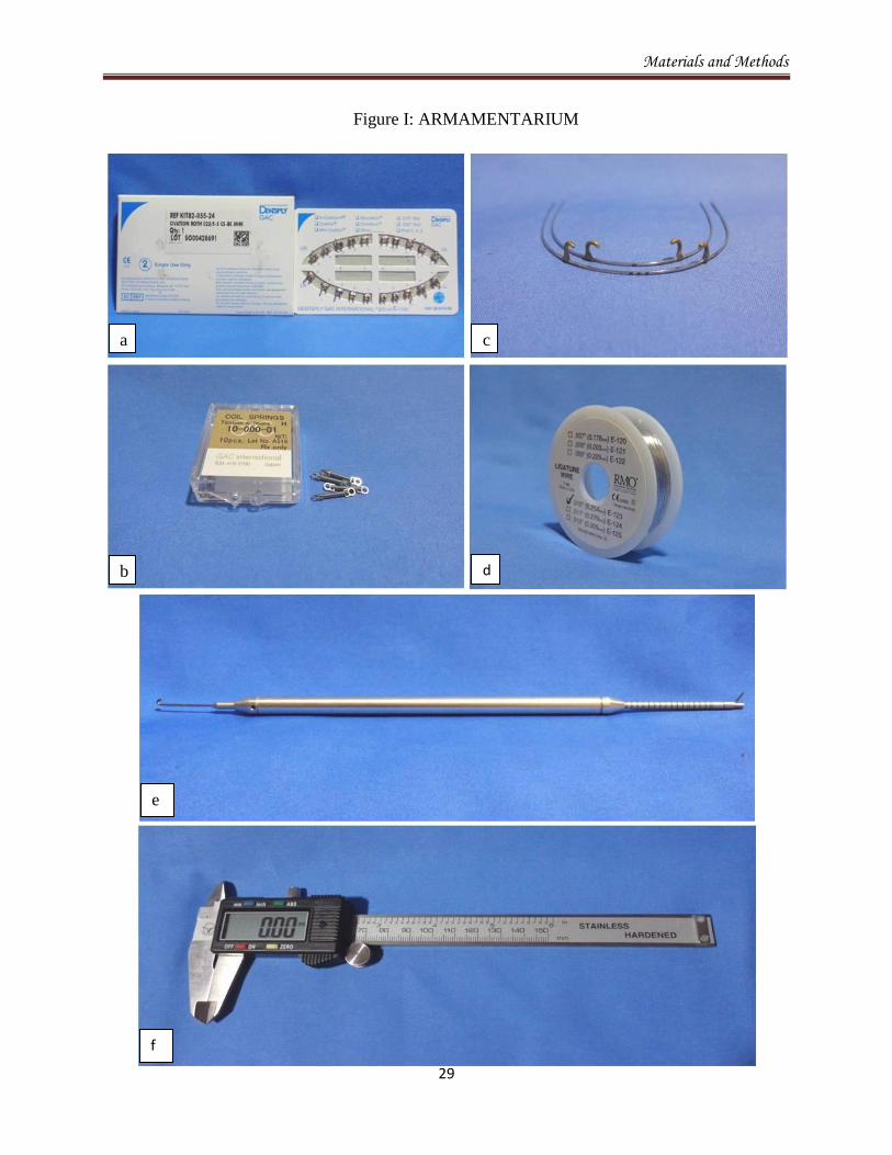
Armamentarium: (Fig.I : Armamentarium)
MiniOvation Roth bracket prescription 0.22’ slot (Dentsply GAC) (fig.I: a)
Closed Niti coil spring (RMO) (fig.I: b)
Stainless steel ligature (fig.I: d)
Dontrix gauge (fig.I: e)
Vernier caliper (fig.I: f)
Surgical armamentarium: (Fig.II : Armamentarium)
#701 fissure bur (fig.II: b)
DFDBA- Rocky mountain graft (fig.II: e)
Vicryl 4-0 suture (fig.II: d)
B.P blade No.15 (fig.II: c)
Materials and Methods
29
Figure I: ARMAMENTARIUM
a c
b d
e
f

Materials and Methods
30
Figure II: ARMAMENTARIUM
a b
c d
e

Materials and Methods
31
Method:
All patients included in the study required extraction of all first premolars and were treated
with 0.022’ slot Roth appliance. In the study group the leveling and aligning was carried out until
0.019”X 0.025” stainless steel archwire could be fully engaged in the bracket slot, the first
premolars were not-extracted till the time the retraction stage in the treatment was commenced
(fig III: a). Whereas in the control group the first premolars were extracted prior to aligning stage
of aligning (fig. IV: a) as it was the conventional method. Brackets were not bonded in the study
group if the first premolars were fairly well aligned. Stainless steel 0.019”X 0.025” working
wires with soldered brass hooks between the lateral incisor and the canine for anterior retraction,
was used in this study (fig.I: c).
The surgery was carried out under local anesthesia(Lignox 2%A) (figII: a). Surgical
procedure was handled by the same Maxillofacial surgeon throught the study. Lower arch
procedure was always executed 2 weeks ahead of the upper arch (fig.III: b,c,h). First premolars
were extracted at the time of the surgery and the stainless steel 0.019”X 0.025” archwire with
soldered brass hooks was palced before flap elevation in the experimental group. Sulcular
incisions using B.P blade no.15 were placed from distal aspect of one canine to the contra lateral
canine and a full thickness flap was elevated involving the anteriors, 3mm above the apical
region of the tooth to expose the underlying cortical bone.
701 fissure bur and no.2 size round bur mounted on a micromotor handpiece under copious
amount of irrigation was used for selective alveolar decortication. If there was a good amount of
inter-root distance between two adjacent teeth vertical interdental scoring was done otherwise
only punch hole perforations were placed in the area available, stopping 2mm short of the
alveolar crest, occlusaly. The vertical decortication was connected by the horizontal

Materials and Methods
32
decortications 2mm beyond the root apex. Selective alveolar decortication from the mesial aspect
of one canine to the mesial surface of the contralateral canine involving the anteriors was
performed. Similarly a careful palatal flap incision was raised for decortications of the palatal
bone (figIII: b,c,d,e). The cuts extend only into the superficial aspect of the medullary bone to
just enhance bleeding for the RAP to occur.
The graft material used in the study was particulate demineralized freeze dried bone
allograft (DFDBA-Rocky mountain tissue bank) (figII: e) wetted with sterile saline solution to
facilitate ease of placement. The quantum of bone grafting was dependent upon the pre-existing
bone, about 0.5-1cc of graft material was required per arch over the decorticated area. Grafting
was done when the quality and quantity of the bone was questionable alone.57
Particulate
grafting material was maintained in the desired position by performing the full thickness flap that
was coronally advanced to cover the grafting material, they were sutured with an interrupted
loop, non resorbable 4-0 black silk suture material. The sutures were left in place for 1-2weeks.
Initiation of orthodontic force was done within 5-7 days after the surgery with the help of
closed Niti coil spring which was engaged from the first molar tube to the soldered hook on the
archwire delivering a force of 250gms for en-masse retraction using sliding mechanics
(fig.III: h,i). In the control group a similar archwire delivering the same amount of force without
the surgical procedure was used for retracting the anteriors (fig.IV: a,b). Retraction was checked
every 2 weeks for distortion of the coil spring and immediately replaced if distorted.

Materials and Methods
33
Figure III: METHODOLOGY- STUDY GROUP
a a
b
c
d
e
f g
h i
Mandible Maxilla
36

Materials and Methods
34
Figure IV: METHODOLOGY- CONTROL
GROUP
a a
b
c d
Figure V: Reference jigs
UR
LR
UL
LL

Materials and Methods
35
Initial measurements were done after extraction using a vernier caliper from the maximum
contour of the the mesial point of second premolar to the distal maximum contour of the canine.
Lateral cephalogram and orthopantamograms were taken with standardization jig placed on the
upper and lower first molars before retraction (fig IV: c,d). Study models were taken at monthly
intervals and the radiographic records were taken every 2 month to assess the treatment time,
period of accelerated tooth movement, anchorage control, soft and hard tissue changes. The
period of study ranged from 4 to 6 months.
Thus the efficacy, efficiency and viability of this new method of retraction with and without
corticotomy were evaluated.
Cephalometric variables used for case selection of the study:
Profile cephalograms were taken in occlusion under standardized conditions with a
cephalostat and tracing were done. Pre and post retraction were carefully traced for each patient
on (8X10 inch) acetate paper. All cepahalograms were manually traced by a single investigator
and checked twice. (figure VI: a,b)

Materials and Methods
36
Cepahalometric landmarks:32,63
used in this study (fig.VI: a)
1.Sella (S) : Geometric center of the pituitary fossa located by visual
inspection.
2.Nasion (N) : The most anterior point on the frontonasal suture in the
midsagittal plane.
3.Porion (Po) : The most superiorly positioned point on external auditory meatus
4.Orbitale (Or) : The lowest point on the inferior rim of the orbit.
5.Basion (Ba) : The lowest point on the anterior rim of foramen magnum.
6.Posterior nasal spine (PNS) : Posterior spine of the palatine bone constituting the hard palate.
7.Anterior nasal spine (ANS) : Anterior tip of the sharp bony process of the maxilla at the lower
margin of anterior nasal opening.
8.Pogonion (Pg) : The most anterior point on the chin.
9.Menton (Me) : The lowest point on the symphyseal shadow of the mandible seen
on the lateral cephalogram.
10.Gonion (Go) : A point on the curvature of the of the angle of the mandible
locating by bisecting the angles formed by lines tangent to the
posterior ramus and the inferior border of the mandible.
11.Gnathion (Gn) : A point located by taking the midpoint between the anterior (Pg)
And inferior (Me) points of bony chin.
12.Pt point (Pt) : The outline of the foramen rotundum can be located by using the
template designed for that purpose or it can be approximated at
the 10.30 (face of a clock) position on the circular outline of the
superior border of the pterygomaxillary fissure.

Materials and Methods
37
13.Point (A) : The most posterior midline point in the concavity between the
anterior nasal spine and the prosthion (the most inferior point on
the alveolar bone overlying the maxillary incisors).
14 U1 E : Incisal tip of maxillary central incisor.
15. U1 A : Upper incisor root apex.
16.U6 D : Most distal point of mesial surface of maxillary first molar crown.
17.Point (B) : The point at the deepest midline concavity on the mandibular
symphysis between infradentale and pogonion.
18.LI E : Incisal tip of mandibular central incisor.
19.LI A : Lower incisor root apex.
20.L6 D : Most distal point of the distal surface of mandibular first molar
crown.
Cephalometric planes: used in this study (fig VI:b)
1.Palatal plane (ANS-PNS plane) : It is a line from the anterior nasal spine to posterior nasal
spine. It will be used as reference plane to find out any
change in vertical plane.
2.Pterygoid vertical plane(Ptv plane): A vertical line drawn through the distal radiographic
outline of the pterygomaxillary fissure and perpendicular
to Frankfort horizontal plane.It will be used as reference
plane to evaluate any change in sagittal plane.
3.S-N plane : It is the cranial line between Sella and Nasion. It represents
the anterior cranial base and will also be used as reference
plane.

Materials and Methods
38
4.Basion-Nasion plane(Ba Nplane) : It is the line connecting the Basion and Nasion point. It
represents the cranial base.
5.Facial axis : A line extanding from the foramen rotundum (Pt) to
cephalometric Gnathion.
6.Facial plane : It is a line from the anterior point of the frontonasal suture
(Nasion) to the most anterior point of the mandible
(Pogonion).
7.Frankfort horizontal plane (FH) : It is a horizontal plane extending from Porion to Orbitale.
8.Mandibular plane (MP) : It is a line extending from Gonion to Menton.
9.E plane : The esthetic line or plane extending from the soft tissue tip
of the nose to the soft tissue chin point.

Materials and Methods
39
Figure VI:a- CEPHALOMETRIC LANDMARKS
Figure VI:b- CEPHALOMETRIC PLANE

Materials and Methods
40
Conventional lateral cephalometric radiographs were taken before the start of retraction and after
the en-masse retraction of the anteriors for both the study and the control group. To differentiate
between the right and left side molar on lateral cephalograms, a 0.017X0.025 inch stainless steel
wire bent in an L shape with 0.5cm horizontal and and 1cm vertical length was inserted from the
mesial side in the buccal tube of the left upper and lower molars inserted from mesial open end.
Similar stainless steel wire bent with an additional horizontal arm of 0.5cm in length was also
inserted from the mesial open end of the buccal tube of the right upper and lower molars. These
markers helped to differentiate the right and left side molars on the orthopantamogram (fig.V:
c,d).
Methodology for evaluation of extraction space closure in the dental cast:
Initial measurements were done after extraction using a digital vernier caliper at the
maximum contour of the the mesial point of second premolar and the distal maximum contour of
the canine on the study models taken at the end of every month interval. Space closure was later
co-related with the molar anchor loss values and the the effective en-masse retraction percentage
was calculated.
Methodology for evaluation of retraction and anchor loss:
Maxilla
In the maxilla the linear measurements was taken from pterygoid vertical along the Frank
fort horizontal plane. The horizontal distance from pterygoid vertical to the jig on the molar was
used to assess anchorage loss on both sides in the study group and the control group. (Fig.V: c,d)
The same reference line was used to assess the retraction of incisors with the horizontal distance
distance measured to the central incisor bracket to cross check the space closure.

Materials and Methods
41
Mandible
In the mandible the linear measurements were taken from sella vertical along the SN
plane. The horizontal distance from the sella vertical to the jig on the molar was used to assess
anchorage loss on both the side in the study and in the control group. (Fig.V: c,d)
The same reference line was used to assess the retraction of incisors with the horizontal distance
measured to the central incisor bracket.
The dental cast measurements were correlated with the amount of anterior retraction. Mean of
these were taken as the amount of space closure.
Measurement of cephalometric error
1.Error due to fatigue : 5-10 cephalograms were analyzed on an average in a day to eliminate
the error due to fatigue of investigator.
2.Inter-investigator error : All the cephalograms were traced and analyzed by a single
investigator.
Comparison with the conventional group for en-masse retraction:
The conventional group was treated with the first premolar extraction and Niti coil spring
retraction, were compared similarly before the start of retraction. The time taken for space
closure in the corticotomy group were compared with the space closure in the conventional
group. Efficiency of corticotomy group was thus compared with the conventional group.

Materials and Methods
42
Statistical analysis:
All statistical analysis was performed by using SPSS software package (SPSS for
Windows XP, version 17.0, Chicago). For each variable measured on the lateral cephalogram,
the mean and the standard deviation were calculated.
Independent T Test was used to determine statistical significance of difference between
the rate of retraction, molar anchor loss before retraction (T1) and after retraction (T2) between
the study group and the control group.
One way Anova followed by Tukey HSD test was done to evaluate and compare the rate
of space closure and anchor loss between the studied monthly time interval.
P < 0.05 was considered statistically significant

Results
46
RESULTS
The study was conducted to evaluate the efficiency and nature of
corticotomy assisted en-masse retraction compared with the conventional method
of en-masse retraction of the anteriors in bimaxillary protrusion patients. The
results are based on 14 patients equally divided into two groups, Study group
(Corticotomy group) and Control group(Control group) irrespective of sex in the
age range of 20 years ±2.5 years. All patients were selected from the patients who
reported to the Department of Orthodontics at Ragas Dental College and Hospital,
Chennai, India, between January and June 2012.
The results are discussed under the following headings:
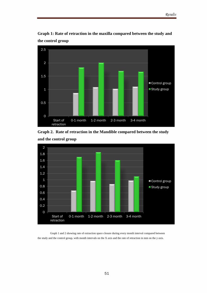
DURATION AND RATE OF RETRACTION:
Maxilla:
There was no significant difference between the extraction spaces in the
maxilla between the study group and the control group.
93.5% of extraction space closure was achieved by the end of 4th
month in
the Study group (Corticotomy group).
54.5% of extraction space closure was achieved during the same time
period, end of 4th
month in the Control group (Conventional group).
Average rate of space closure of 1.8mm/month was achieved in the Study
group.
Average rate of space closure of 1.02mm/month was achieved in the
Control group.

Results
47
Mandible:
There was no significant difference between the extraction spaces in the
mandible between the study group and the control group.
92.6% of extraction space closure was achieved by the end of 4th
month in
the Study group (Corticotomy group).
51.7% of extraction space closure was achieved during the same time
period, end of 4th
month in the Control group (Conventional group).
Average rate of space closure of 1.57mm/month was achieved in the Study
group.
Average rate of space closure of 0.87mm/month was achieved in the
Control group.
Comparison of rate of space closure in the Maxilla and Mandible at monthly
intervals:
Acceleration of rate of space closure was statistically significant during the
first two months of retraction in the study group.
No significant rate of acceleration was found in the maxilla and/or the
mandible during the month intervals in the control group.
No significant difference was found in the rate of space closure at monthly
intervals in the Control group.
.

Results
48
ANCHOR LOSS:
Maxilla:
Molar anchor loss of approximately 0.39mm occurred during the anterior
retraction in the study group within the assessed time period.
Molar anchor loss of approximately 1.47mm occurred during the anterior
retraction in the control group within the assessed time period.
Mandible:
Molar anchor loss of approximately 0.39mm occurred during the anterior
retraction in the study group within the assessed time period.
Molar anchor loss of approximately 1.6mm occurred during the anterior
retraction in the control group within the assessed time period.
Comparison of anchor loss:
Statistically significant difference was present in the anchor loss between
the Study group and the Control group.
The amount of anchor loss increased as time advanced in the Study group.
No significant difference in the amount of anchor loss between the
monthly intervals was found in the Control group.

Results
49
Table 1: Rate of retraction in the Maxilla and Mandible compared during
monthly intervals in Study and Control group.
ARCH MONTH GROUP MEAN S.D P VALUE
MAXILLA Start of
retraction
Control group
Study group
7.51
7.76
.08
.25
0.062
0-1 Control group
Study group
6.64
5.93
.35
.22
0.006**
1-2 Control group
Study group
5.55
3.92
.53
.24
<0.001**
2-3 Control group
Study group
4.52
2.22
.53
.23
0.008**
3-4 Control group
Study group
3.41
.5
.51
.25
0.062*
MANDIBLE Start of
retraction
Control group
Study group
6.75
6.79
.36
.22
0.821
0-1 Control group
Study group
6.08
5.08
.41
.23
0.037**
1-2 Control group
Study group
5.11
3.22
.45
.22
<0.001**
2-3 Control group
Study group
4.24
1.61
.52
.49
0.030*
3-4 Control group
Study group
3.26
.5
.61
.47
0.052*
NS: Not significant;
*p < 0.05 (statistically significant);
**p < 0.001 (statistically highly significant)

Results
50
Table 3: Molar anchor loss in the Maxilla and mandible compared between
the study and the control group (mm)
ARCH MONTH
INTERVALS
GROUP MEAN S.D P VALUE
MAXILLA 0-2 Control group
Study group
.36
.00
.16
.00
0.001**
2-4 Control group
Study group
.42
.06
.10
.09
<0.001**
4-6 Control group
Study group
.41
.33
.12
.17
0.417
MANDIBLE 0-2 Control group
Study group
.64
.00
.33
.00
0.001**
2-4 Control group
Study group
.33
.08
.10
.04
0.002**
4-6 Control group
Study group
.63
.31
.17
.15
0.013*
NS: Not significant;
*p < 0.05 (statistically significant);
**p < 0.001 (statistically highly significant)
Results
51
Graph 1: Rate of retraction in the maxilla compared between the study and
the control group
Graph 2. Rate of retraction in the Mandible compared between the study
and the control group
Graph 1 and 2 showing rate of extraction space closure during every month interval compared between
the study and the control group, with month intervals on the X axis and the rate of retraction in mm on the y axis.
0
0.5
1
1.5
2
2.5
Start of
retraction
0-1 month 1-2 month 2-3 month 3-4 month
Control group
Study group
0
0.2
0.4
0.6
0.8
1
1.2
1.4
1.6
1.8
2
Start of
retraction
0-1 month 1-2 month 2-3 month 3-4 month
Control group
Study group

Results
52
Graph 3: Molar anchor loss in the Maxilla compared between the control and
the study group
Graph 4: Molar anchor loss in Mandible compared between the study and
the control group
Graph 3 and 4 showing the amount of anchor loss compared between the control and the study
group, with month intervals on axis and amount of anchor loss on Y –axis in mm.
0
0.05
0.1
0.15
0.2
0.25
0.3
0.35
0.4
0.45
0-2 month 2-4 month 4-6 month
Control group
Study group
0
0.1
0.2
0.3
0.4
0.5
0.6
0.7
0-2 month 2-4 month 4-6 month
Control group
Study group

Representative Cases
REPRESENTATIVE CASE NO.1: STUDY GROUP
PRE TREATMENT INTRA ORAL PHOTOGRAPH
Leveled and aligned without extraction of first premolars

Representative Cases
CORTICOTOMY- MANDIBULAR ARCH
CORTICOTOMY- MAXILLARY ARCH

Representative Cases
Start of retraction (T1) End of space closure (T2): 3-4 months
0-1 month 1-2 month 2-3 month 2-3 month

Representative Cases
A: Pre treatment B: Pre retraction C: Post retraction
A B
C

Representative Cases
PRE TREATMENT EXTRAORAL PHOTOGRAPH
COMPARISON OF PRE AND POST RETRACTION EXTRAORAL PHOTOGRAPHS
POST RETRACTION EXTRA ORAL PHOTOGRAPH

Representative Cases
POST RETRACTION INTRAORAL PHOTOGRAPH
PROFILE CHANGE

Representative Cases
PRE RETRACTION AND POST RETRACTION CEPHALOMETRIC SUPERIMPOSITION
PRE RETRACTION
POST RETRACTION

Representative Cases
Skeletal parameters for representative case no. 1: Pre and post retraction comparison
Dental parameters for representative case no.1: Pre and post retraction comparison
CEPHALOMETRIC VARIABLES PRE RETRACTION (T1)
POST RETRACTION (T2)
DIFFERENCE
UI to NA(mm) 32˚ 24˚ 8˚
UI to NA(˚) 10mm 4mm 6mm
UI to SN (˚) 112˚ 101˚ 11˚
LI to NB(mm) 36˚ 26˚ 10˚
LI to NB(˚) 12mm 5mm 7mm
LI to Mnd. Plane 102˚ 92˚ 10˚
E line to UL 10mm 2mm 8mm
E line to LL 12mm 4mm 8mm
CEPHALOMETRIC VARIABLES PRE RETRACTION (T1)
POST RETRACTION (T2)
DIFFERENCE
SNA 85˚ 83˚ 2˚
SNB 81˚ 79˚ 1˚
ANB 6˚ 4˚ 2˚
Facial axis angle -1˚ 1˚ 2˚
N perp. to Point A 5mm 2mm 3mm
SN- GoGn 34˚ 33˚ 1˚
Y axis 68˚ 66˚ 2˚
Lower anterior face height 72mm 71mm 1mm

Representative Cases
REPRESENTATIVE CASE NO.1: CONTROL GROUP
PRE TREATMENT INTRAORAL PHOTOGRAPH
LEVELING AND ALIGNING

Representative Cases
2 months Post Retraction 3 months Post Retraction 4 months Post Retraction
START OF RETRACTION
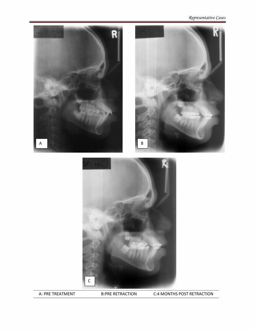
Representative Cases
A: PRE TREATMENT B:PRE RETRACTION C:4 MONTHS POST RETRACTION
A B
C

Discussion
53
DISCUSSION
In the treatment of bimaxillary protrusion, two important factors for consideration
are: esthetic improvement of the dentofacial area and the establishment of the stable
dental relationship that embodies the requirement of line of occlusion. Because of the
negative perception of protrusive dentition and lips many patients with bimaxillary
protrusion seek orthodontic care to decrease this procumbency.8
The treatment approach
usually consists of extracting the four first premolars and retracting the anterior teeth with
maximum anchorage mechanics. Maximum anchorage of posterior teeth allows the
anterior teeth to be retracted to the greatest extent. Time is an important factor during the
course of orthodontic treatment.18
Dimitrios Mavreas18
in a systematic review revealed that extraction treatment
takes longer time than non-extraction treatment where in those involving four
premolar extractions took the longest duration of approximately 18.6 months and
anterior retraction alone, took more than 8 months.
Hu Long46
evaluated the effectiveness of interventions on accelerating
orthodontic tooth movement and the systematic review revealed that: Low-level laser
therapy was ineffective to accelerate orthodontic tooth movement, evidence is still not
conclusive whether electrical current and pulsed electromagnetic fields can
defenetively accelerate tooth movement, Dentoalveolar or periodontal distraction is a
promising method to accelerate orthodontic tooth movement but lacks convincing
evidence. Whereas Corticotomy is documented to be a effective and safe procedure to

Discussion
54
accelerate orthodontic tooth movement. Therefore corticotomy facilitated technique
was used in the current study as documented evidences proved it to be a safe modality
for intervention to accelerate orthodontic tooth movement.
Among the fundamental components of dentofacial problem, Dentoalveolar is
defined as “is relating to or involving the teeth and their sockets”17
which is
interpreted as the relationship of the teeth to the alveolar bone housing the teeth. The
alveoloskeletal component involves relationship of the dentoalveolar complex to its
skeletal base, interpreted as dentoalveolar complex that does not align properly with
its associated skeletal base. Kole suggested that the greatest resistance to tooth
movement is created by cortical bone of the alveolus especially buccal and lingual to
the apices of the teeth referred to as the “Orthodontic Walls”.64
Corticotomy is a surgical technique in which only the cortical bone is cut,
perforated or mechanically altered to the depth of the medullary bone and the
medullary bone remains intact. Corticotomy (selective alveoloar decortictation) can
be effectively used to correct dentoalveolar and moderate alveoloskeletal problems.
As the severity increases from alveoloskeletal to pure skeletal problems osteotomy
and/or distraction osteogenesis would be the choice of treatment.64
Chung et al10
claims that corticotomies when combined with heavy forces
leads to histologic changes called the Compression osteogenesis where the medullary
bone is more plastic and malleable (temporary osteopenia). They produce effective
acceleratory orthodontic rotation and tipping movement. The high medullary bone

Discussion
55
turnover in healthy tissues results in new bone formation with low bone density the
key which provides more rapid tooth movement. Thus the type of tooth movement in
corticotomy assisted retraction is dentoalveolar with tipping followed by uprighting .
There is considerable amount of evidence in the literature as given by William
M. Wilcko94
, Raffaele spena62
, Fischer84
, Dauro Douglas Oliveira15
, Mostafa et
al55
, Generson and Porter,23
Chung et al9, Ali H Hassan
1, Hyo-Won Ahn
30,
Shoichiro Iino67
and many others suggesting that corticotomy is a viable option for
accelerating orthodontic treatment.
However there is very little reference to the treatment efficiency of selective
alveolar decortication for en-masse retraction of anteriors especially in bimaxillary
protrusion patients.
This study differs from other previous studies as the en-masse retraction of
anteriors using sliding mechanics to assess the efficiency of the corticotomy assisted
retraction was assessed with a modified protocol which was compared with the
conventional en-masse retraction in bimaxillary protrusion patients.
Thomas M.Wilcko49
proposed a basic protocol where in orthodontic brackets
are placed and a light wire engaged sometime during the week before the surgery
with subsequent orthodontic adjustments being made at 2-week intervals. This
flexible light wire would bring about faster tipping movements which he used to
accelerate, primarily to decrowd the dentition. Whereas in our study the case required
all first premolar extraction primarily to bring about faster controlled anterior

Discussion
56
retraction and not to level and align the arches, therefore Wilcko’s protocol was
modified where in corticotomy was performed only after leveling and aligning was
completed just before retraction. The teeth was stabilized with a rigid stainless steel
wire (0.019”X 0.025” ) to prevent torque loss and to bring about bodily /controlled
tipping movements during retraction. The posterior segment (second premolar, first
molar and second molar) were not decorticated since osteopenia is required only 2-
3mm around the teeth to be moved and also to act as an anchorage module.
In Wilcko’s48
protocol corticotomy was performed prior to aligning and an
additional procedure namely Ostectomy (surgical removal of cortical plate) in the
extraction space was required which is both cumbersome and expensive to the patient
because of additional grafting required in the area. The outcome is also not much
significantly different from other studies were corticotomy was employed.
Corticotomy-facilitated tooth movement was first described by L.C. Bryan in
1893, Cohn Stock11
surgically removed the palatal bone to retract single/ multiple
teeth. Bichlmayr5 described a corticotomy procedure for adult bimaxillary protrusion
patient requiring all first bicuspid extraction by excising the buccal and lingual
cortical plates at the extraction site. Skogborg and Ascher80
divided the interdental
bone (septotomy). Neuman59
divided the interdental bone and ablated wedge of bone
palatal to the teeth to be retracted. Köle’s38
technique consisted of buccal and lingual
interproximal vertical corticotomy cuts limited to cortical layers, with these vertical
corticotomy cuts being connected by horizontal osteotomy cuts approximately 1 mm

Discussion
57
beyond the apices of the roots, but the bucco lingual communication that was the
stansard procedure in Kole’s technique is considered to be morbid. Suya replaced
supraapical horizontal osteotomy with horizontal corticotomy to facilitate luxation of
the corticotimized bone blocks. Kretz39
described that therapeutic fracture of the
anterior alveolus, again which is aggressive bone manipulation that could
compromise the vascularity. Bell and Levy4
luxated the operated tooth bone
segment with a chisel which had been documented to lead to ischemia in the area.
Germec24
used minimal intervention by limiting corticotomy only on the labial
surface of bone where labial movement of teeth was desired but acceleration duration
started to slow down very easily. Wilcko and Ferguson19
collaborated and reiterated
the fact that no luxation of the bone was required as they considered luxation to be a
mechanical misapprehension. They further modified the corticotomy-assisted
orthodontic technique with the addition of alveolar augmentation. Therefore the
Wilcko- Ferguson technique is dependent upon the cortical bone thickness than on
any preconceived pattern of bone injury such as cuts and dots or a combination of
both was used. Surgical scarring barely penetrated the cortical bone without involving
the medullary bone. The procedure employed in our study has the advantage of being
less invasive, is done under local anesthesia and is a simpler technique.
Derya Germec24
carried out corticotomy for lower incisor retraction done only
under local anesthesia. Thus the surgical procedure in our study was carried under
local anesthesia where full-thickness flaps are reflected labially and lingually using a
sulcular releasing incision. Vertical releasing incisions were used if sufficient

Discussion
58
underlying bone was not accessible. The incision was located distal to the area where
corticotomy cuts were planned.
In our study 701 tapering fissure bur for interdental scoring and no.2 round bur
for punch hole perforation was used which is an acceptable method for decortications.
25,55,8
As proposed by Neal C Murphy,57
the demineralized bone matrix was used to
augment when dehiscence or fenestration was noted as when the quality and quantity
of bone was questionable. Demineralized freeze dried allogenic bone graft (Rocky
mountain) was used as it has more of osteoinductive (stimulation of osteoprogenitor
cells to differentiate into osteoblasts that then begin new bone formation) which
stimulates BMP formation was used. Approximately 1-2cc of particulate graft mixed
with sterile saline was required per arch.
A few case reports: Derya Germec24
, Seong Hun Kim72
, Waranee
Linlawan95
, Aboul Ela et al73
, Schoihiro lino74
, Lee etal 71
are available in the
literature suggesting that corticotomy can be beneficial in bimaxillary protrusion
cases. Most of these studies employ either an implant or involves independent canine
retraction. Hence we chose to employ bimaxillary cases, since they require maximum
anchorage and extended treatment duration.
The molar anchor loss value had not been documented convincingly for such
corticotomy assisted retraction. Therefore our study aimed to evaluate the rate of
retraction and the amount of molar anchor loss that occurred during the retraction

Discussion
59
period. Control group were evaluated for the same period from start to finish of
retraction period as in the corticotomy group. Molar anchor loss studied at certain
monthly intervals served to find the ability of the molars to effectively serve as an
anchorage source as to whether they can be relied as an absolute anchorage source for
such bimaxillary protrusion condition. Temporary anchorage devices was not used to
assess whether the undecorticated posterior teeth can act as an anchorage module.
The duration and rate of retraction in the study group was assessed with the
help of study models. The space closure was assessed between the maximum contour
distal to the canine and mesial surface of the second premolar. Ziegler and
Ingervall77
used the posterior rugae and the midpalatal raphe as reference point and
line whereas Lotzof eta al47
designed an acrylic mold of the anterior palate and two
wires projected to the central fossa of first molar, to assess molar anchor loss during
canine retraction. The palatal rugae or no other special transferable reference acrylic
mold was fabricated to assess anchor loss, since the palatal flap was also reflected and
sutured back and would not serve as a reliable landmark after the corticotomy
procedure. Anchor loss was assessed with the help of lateral cephalogram similar to
the method proposed by Badri Thiruvengadachari2
with modified jigs placed on
the upper and lower, right and left first molar to differentiate the right from the left
molar. Anchor loss was correlated with the space closure and effective retraction in
the Study group was assessed and compared with the control group for the same
period of time as the study group (approximately 4-5 months).

Discussion
60
Fink and Smith21
in a retrospective comparative study observed that non-
extraction cases took, on an average 21.95 months, two premolar extraction cases 25
months, and four premolar extraction cases 26.18 months time period to get
completed and Dimitrios Mavreas18
in a systematic review correlated that the longer
treatment time was required for extraction cases and this was proportional to the
number of teeth extracted.
In our study there was no significant difference in the extraction space in the
study and the control group which accounted to be, on an average of 7.63mm in the
maxilla and 6.77mm in the mandible inspite of early extraction in the control group
when compared with the study group which could be due to the fact that the cases
selected for the study had minimal crowding. Thus the study and the control group
can be effectively compared.
93.5% (7.26mm) of extraction space closure was achieved by the end of 4th
month in the Study group (Corticotomy group). Definitive space closure was
achieved in the maxilla by 4-5 months. Whereas only 54.5% (4.1mm) of extraction
space closure was achieved in the control group for the same period of time i.e by the
end of fourth month. 92.6% (6.29mm) of extraction space closure in the mandible
was achieved by the end of 4th
month in the Study group (Corticotomy group).
Whereas only 51.7% (3.5mm) of extraction space closure was achieved in the control
group for the same period of time i.e by the end of fourth month. Derya Germec24
attained retraction of lower incisors alone into the residual spaces remaining after

Discussion
61
decrowding and segmental canine retraction took almost 1 ½ months for space
closure with corticotomy assisted retraction using loop mechanis, the surgery was an
extensive invasive procedure when compared with the present study. Chung et al9
reported that the complete retraction of anterior teeth combined with corticotomy in a
case with severe bimaxillary protrusion took less than 3½ months and Seo et al71
reported that in case of bimaxillary protrusion using anterior corticotomized segment
required five months for complete retraction with the help of C-lingual retractor,
Schoihiro Lino74
reported that it took about 7 months for completion of space
closure.
The rate of retraction in our study correlates with the outcome reported by
others. However the fact that no Temporary anchorage devices (TAD) were used in
our study in contrast to the above studies along with the time of intervention for
corticotomy highlights the significance of this modification. Wherein only a modified
corticotomy without the aid of TAD supported anchorage not only accelerates the rate
of retraction of tooth movement but also does not tax the anchorage.
The average rate of space closure was 1.8mm/month in the maxilla and
1.6mm/month in the mandible in the Study group. This correlates with the studies of
Aboul Ela et al73
who calculated the mean rates of individual maxillary canine
retraction with buccal corticotomy alone and a retraction force of 150 grams was on
an average about 1.42mm/ month.

Discussion
62
An average rate of space closure was 1.02mm/month in the maxilla and
0.87mm/month in the mandible in the control group. Dixon et al89
in a previous
study used Niti coil spring delivering 200grams force for retraction which showed the
highest rate of space closure of about 0.81mm/ month and attained 32% of extraction
space closure by the 4th
month. The rate of space closure per month in the control
group is highly correlative but approximately 51% of extraction space closure by 4th
month was closed in our study could possibly be because of marginally higher forces
and a more efficient protocol for en-masse retraction.
Though the average rate of retraction in the study group was 1.8mm/month in
the maxilla and 1.6mm in the mandible there was a peak increase during the first two
months of retraction in the maxilla (2.01mm/month) and in the mandible
(1.86mm/month) which started to reduce by the end of fourth month . This finding is
in concurrence with that of Aboul Ela et al73
who reported that the rate of space
closure peaked during the end of 1st and 2
nd month and reduced by the end of 4
th
month. This could be biologically co-related with the transient RAP phenomenon as
mentioned by Frost26
which remains active for 4 months.Thus the time period
assessment of retraction for the first four month period after corticotomy can be
substantiated.There was no significant difference in the rate of retraction in control
group when assessed between monthly intervals.
An average anchor loss of 0.39mm occurred in the maxilla and in the mandible
for a maximum of 4 to 6 months. There is no pertinent literature to substantiate molar
Discussion
63
anchor loss in corticotomy assisted retraction as most of the study used temporary
anchorage devices for retraction purposes and hence there was no anchor loss. Our
study would be the first study to correlate pure anchor loss and the efficiency of the
undecorticated posterior segment to act as an efficient anchorage source.
Whereas molar anchor loss of 1.47mm in the maxilla and 1.6mm in the
mandible occurred in the control group within the same time period assessed.
Similarly in a previous study on conventional en-masse retraction by Wook Heo96
approximately 4mm of retraction of upper incisal edge resulted in 1mm of anchor loss
in the upper molar. Since the complete space closure in the control group was not
assessed in the current study ther could be further anchor loss at the end of retraction.
Vikas Agarwal91
in a previous study reported molar anchorage loss of 2.45mm.
Thiruvenkatachari et al2
reported an anchorage loss of 1.6mm in the maxilla and
1.7mm in the mandible when molars were used as anchorage unit for anterior
retraction.
Thus statistically significant difference in molar anchorage loss was observered
when compared with the study and the control group. The undecorticated posterior
segment can effectively act as an anchorage module. Although not statistically
significant, there was no anchorage loss during the first two months the anchor loss
was found to gradually increase by the end of 4th
month of retraction. Further anchor
loss could not be assessed as most of the cases showed completion of space closure
Discussion
64
by the end of 4th
month in the corticotomy(study) group. The molar anchor loss did
not vary significantly between the monthly intervals assessed in the control group.
Though corticotomy assisted orthodontic treatment (CAOT) has beneficial
outcome in terms of duration of treatment its limitations in cases of active periodontal
disease or gingival recession should be a consideration. In our study in one sample
the mandibular canine roots were completely exposed with dehiscence which was
covered with alveolar grafting. Initially there was gingival recession in that particular
tooth which eventually underwent repair and resulted in improvement.
Although CAOT may be considered a less-invasive procedure than osteotomy-
assisted orthodontics or surgically assisted rapid expansion, there are a few reports
sighting interdental bone loss and loss of attached gingiva, to periodontal defects
observed in some cases with short interdental distance. Subcutaneous hematomas of
the face and the neck have been reported if intensive corticotomies were performed.
Long-term effect of CAOT on root resorption requires further study. In addition,
some post-operative swelling and pain has also been reported for several days.

Summary and Conclusion
65
SUMMARY AND CONCLUSION
The present clinical study was conducted to evaluate the efficiency and treatment
outcome of a modified Corticotomy assisted en-masse retraction with the
conventional method of en-masse retraction in bimaxillary protrusion patients.
The following conclusions were drawn from the present study:
1. Extraction space closure of about 93.5% in the maxilla and 92.6% in the
mandible was achieved by the fourth month of retraction in the
corticotomy group- Study group.
2. Only 54.5% of extraction space closure in the maxilla and 51.7% in the
mandible was achieved by the fourth month of retraction in the
conventional group- Control group.
3. The rate of retraction accelerated during the first two months of retraction.
4. The rate of retraction with corticotomy assisted orthodontics was twice as
quicker when compared to the control group.
5. There was better anchorage control with the undecorticated molar segment
during the retraction period but the amount of anchor loss was found to
increase as time period advanced.
Based on the outcome of this study it is reasonable to conclude that
Corticotomy assisted retraction drastically reduces the overall duration of
Orthodontic treatment. It is particularly useful in maximum anchorage cases since
by Selective decortications the anchorage segment can be made virtually
stationary and the entire extraction space can be utilized for retraction. However
post operatively, the procedure does bring about transient inflammation and
localized swelling.
Bibliography
66
BIBLIOGRAPHY
1. Ali H Hassan, Ali T AlGhamdi, Ahmad A Al-Fraidi, Aziza Al-Hubail and
Manar K Hajrassy- CUnilateral cross bite treated by corticotomy-assisted
expansion: two case reports. Head & Face Medicine 2010, 6:1-9.
2. Badri.Thiruvenkatachari, A. Pavithranand- Comparison of rate of canine
retraction with conventional molar anchorage and titanium implant anchorage.
Am J Orthod Dentofacial Orthop 2008;134:30-5.
3. Ballard CF- Variations of posture and behaviour of the lips and tongue which
determine the position of the labial segments: the implications in orthodontics,
prosthetics and speech. Transactions of the European Orthodontic
Society.1963;67-88.
4. Bell WH and Levy BM- Revascularization and bone healing after maxillary
corticotomies, J Oral Surg 1972; 30:640-648.
5. Bichlmayr A - Ckirurgische Kieferothopaedie und das Verhalten des Knochens
und der Wurzelspitzen nach derselben. Deutsche Zahnaerztl Woschenschrift
1931;34: 835--42.
6. Brig SM Londhe, Lt Col P Kumar, Col R Mitra, Col A Kotwal, SM- Efficacy
of Second Molar to Achieve Anchorage Control in Maximum Anchorage Cases;
2010: 220-224.

Bibliography
67
7. C. S. Handelman-―The Anterior Alveolus: Its Importance in Limiting
Orthodontic Treatment and Its Influence on The Occurrence of Iatrogenic
Sequelae‖. Angle Orthod 1996; 66: 95.
8. Cheol Hyun Moon, Ji-Uk Wee, Hyun Sun Lee- Intrusion of overerupted
molars by corticotomy and orthodontic skeletal anchorage. Angle Orthod.
2007;77:1119-1125.
9. Chung KR, Oh MY, Ko SJ- Corticotomy- assisted orthodontics. J Clin Orthod
2001: 35: 331–339.
10. Chung KR. Text book of Speedy Orthodontics. 1st ed. Seoul, Korea: Jeesung;
2001.
11. Cohn-Stock G- Die chirurgische immediatregulierung der Kiefer Speziell die
Chirurgische Behandlung der Prognathie. Vierteljahrsschrift fur Zahnheilkunde
1921;37:320-354.
12. Cunningham G- Methode sofortiger Regulierung von anomalen Zahnstellungen.
Oesterreichisch-Ungarische Vierteljahrsschrift fur Zahnheilkunde, 1894;10:455-
457.
13. D. A. Bills, C. S. Handelman, and E. A. BeGole- ―Bimaxillary Dentoalveolar
Protrusion: Traits and Orthodontic Correction‖. Angle Orthod. 2005; 75: 333.
14. D. T. Laigan, J. H. Hey, and H. A. West- ―Aseptic Necrosis Following
Maxillary Osteotomies: Report of 36 Cases‖, J Oral Maxillofac Surg 1990; 48:
142.
Bibliography
68
15. Dauro Douglas Oliveira, Bruno Franco de Oliveira, Helio Henrique de
Araújo Brito, Margareth Maria Gomes de Souza, and Paulo José Medeirose-
Selective alveolar corticotomy to intrude overerupted molars. Am J Orthod
Dentofacial Orthop 2008;133:902-8.
16. Delma R. Cruz, Eduardo K. Kohara, Martha S. Ribeiro and Niklaus U.
Wetter- Effects of Low-Intensity Laser Therapy on the Orthodontic Movement
Velocity of Human Teeth: A Preliminary Study. Lasers in Surgery and Medicine
2004;35:117–120.
17. Dentoalveolar. Merrlam- Webster Medical Dictionary. http:// medical.merriam-
Webster.com/ medical/ dentoalveolar. Accessed March 2009.
18. Dimitrios Mavreas, Athanasios E. Athanasiou- Factors affecting the duration
of orthodontic treatment: a systematic review. Eur J Orthod 2008; 30: 386–395.
19. Donald J.Ferguson, William M.Wilcko and M.Thomas Wilcko- Selective
alveolar decortications for Rapid Orthodontic of Skeletal malocclusion
treatment.Efficient orthodontic repositioning of teeth- Chapter:15, Bell and
Proffit.
20. Dong-Hyun Hwanga; Ki-Ho Parkb; Yong-Dae Kwonc; Su-Jung Kim-
Treatment of Class II open bite complicated by an ankylosed maxillary central
incisor. Angle Orthod. 2011;81:726–735.
21. Fink D, Smith R- The duration of orthodontic treatment . Am J of Orthod and
Dentofacial Orthop. 1992;102: 45-51.

Bibliography
69
22. Frost H.M- The regional accelerated phenomenon. Orthop Clin N Am. 1981;12:
725-726.
23. Generson RM. Porter JM. Zell A- Stratigos GT. Combined surgical and
orthodontic management of anterior open bite using corticotomy. J Oral Surg
1978; 36:216-9.
24. Germec D, Giray B, Kocadereli I, Enacar- A lower incisor retraction with a
modified corticotomy, Angle Orthod 2006;76(5):880-888.
25. Goldson L, Reck VJ- Surgical-orthodontic treatment of malpositioned cuspids. J
Clin Orthod 1987;21:847-51.
26. H.M. Frost- ―Biology of fracture healing- An overview for clinicians Part I & II,
Clinical orthopedics and related research 1989; 248: 283.
27. Hajji SS.- The Influence of Accelerated Osteogenic Response on Mandibular
Decrowding [master’s thesis]. St Louis, Mo: St Louis University; 2000.
28. Hasler R, Schmid G, Ingervall B, Gebauer U. A- Clinical comparison of the
rate of maxillary canine retraction into healed and recent extraction sites—a pilot
study. Eur J Orthod. 1997;19:711–719.
29. Hwang HS, Lee KH- Intrusion of overerupted molars by corticotomy and
magnets. Am J Orthod Dentofacial Orthop 2001; 120:209-16.
30. Hyo-Won Ahn, Dong-Yeol Lee, Young-Guk Park, Seong-Hun Kim, Kyu-
Rhim Chung,e and Gerald Nelsonf- Accelerated decompensation of mandibular
incisors in surgical skeletal Class III patients by using augmented corticotomy: A
preliminary study. Am J Orthod Dentofacial Orthop 2012;142:199-206.

Bibliography
70
31. Iseri H, Kisnisci R, Bzizi N, Tuz H- Rapid canine retraction and orthodontic
treatment with dentoalveolar distraction osteogenesis. Am J Orthod Dentofacial
Orthop. 2005;127: 533–541.
32. Jacoboson . A. Caufield.W.Page- Tracing technique and identification of land
mark- Radiographic cephalometry, from basics to videoimaging. 1995;53-62.
33. Jean-David Sebaoun, Alpdogan Kantarci, John W. Turner, Roberto S.
Carvalho, Thomas E. Van Dyke and Donald J. Ferguson- Modeling of
trabecular bone and lamina dura following selective alveolar decortication in rats.
J Periodontol. 2008 ; 79(9): 1679–1688.
34. K. R. Chung, S. H. Kim, and S. L. Baek- ―Speedy Surgical-Orthodontic
Treatment with Temporary Anchorage Devices as An Alternative to Orthognathic
Surgery‖. Am J Orthod Dentofacial Orthop 2009; 135-787.
35. Keating PJ- The treatment of Bimaxillary protrusion. A cephalometric
consideration of changes in the inter-incisal angle and soft tissue profile. Br J
Orthod. 1986;13:209-220.
36. Kharkar VR, Kotrashetti SM, Kulkarni P- Comparative evaluation of dento-
alveolar distraction and periodontal distraction assisted rapid retraction of the
maxillary canine: a pilot study. Int J Oral Maxillofac Surg. 2010;39:1074–1079.
37. Kim DH, Park YG, Kang SG- The effects of electrical current from a micro-
electrical device on tooth movement. Korean J Orthod. 2008;38:337–346.

Bibliography
71
38. Kole H- Surgical operations of the alveolar ridge to correct occlusal
abnormalities. Oral Surg Oral Med Oral Pathol 1959; 12:515-529.
39. Kretz .R- Die chirurgische Immediatregu-lierung der Prognathie, Dtsch.
Zahnheilkd. 1965; 81.
40. Kuhlberg.J.K, Priebe.N.D- space closure and anchorage control. Sem in Orthod
2001;7:42-49.
41. Kyu-rhim chung, Moon-young oh, Su-jin ko- Corticotomy-assisted
orthodontics 2003 - 35: 331-339.
42. Lew K- Profile changes following orthodontic treatment of bimaxillary
protrusion in adults with the Begg appliance. Eur J Orthod. 1989;11:375-381.
43. Lewis SJ- Bimaxillary Protrusion. Angle Orthod. 1943;13:51-59.
44. Limpanichkul W, Godfrey K, Srisuk N, Rattanayatikul C- Effects of low-
level laser therapy on the rate of orthodontic tooth movement. Orthod Craniofac
Res. 2006;9(1):38-43.
45. Liou EJ, Huang CS- Rapid canine retraction through distraction of the
periodontal ligament. Am J Orthod Dentofacial Orthop. 1998;114:372–382.
46. Long H, Pyakurel U, Wang Y, Liao L, Zhou Y, Lai W- Interventions for
accelerating orthodontic tooth movement. Angle Orthod. 2012 Jun 21. [ahead of
print].
47. Lotzof LP, Fine HA, Cisneros GJ- Canine retraction: A comparison of two
preadjusted bracket systems. Am J Orthod Dentofacial Res. 2008; 110: 191-196.
Bibliography
72
48. M. Thomas Wilcko, DMD, William M. Wilcko, Jeffrey J. Pulver, Nabil F.
Bissada, Jerry E. Bouquot- Accelerated Osteogenic Orthodontics Technique: A
1-Stage Surgically Facilitated Rapid Orthodontic Technique With Alveolar
Augmentation. J Oral Maxillofac Surg 2009;67:2149-2159.
49. M. Thomas Wilcko, William M. Wilcko, and Nabil F. Bissada- An Evidence-
Based Analysis of Periodontally Accelerated Orthodontic and Osteogenic
Techniques: A Synthesis of Scientific Perspectives. Semin Orthod 2008;14
:305-316.
50. Machado I., Ferguson DJ., Wilcko WM- Root resorption following
orthodontics with and without alveolar corticotomy. J Dent Res. 2002 : 81,
Abstract #2378.
51. Madan MS, Liu ZJ, Gu GM, King GJ- Effects of human relaxin on orthodontic
tooth movement and periodontal ligaments in rats. American Journal of
Orthodontics and Dentofacial Orthopedics.2007; 131: 8–10.
52. Merrill RG., Pedersen GW- Interdental osteotomy for immediate repositioning
of dental-osseous elements. Journal of Oral Surgery 1976; 34:118-25.
53. Miyawaki S, Koh Y, Kim R, Kobayasi M, Sugimura M- Survey of young
adults women regarding men’s orofacial features. J Clin Orthod. 2000;34:367–
370.
54. Miyoshi K, Igarashi K, Saeki S, Shinoda H, et al- Tooth movement and
changes in periodontal tissue in response to orthodontic force in rats vary
Bibliography
73
depending on the time of day the force is applied. European Journal of
Orthodontics2001; 23: 329–38.
55. Mostafa YA, Tawfik KM, El-Mangoury NH- Surgical-orthodontic treatment
for overerupted maxillary molars. J Clin Orthod 1985;19:350-351.
56. Nakanishi H.- Experimental study on artificial tooth movement with osteotomy
and corticotomy. Shika Gakuho. 1982; 82:219–252.
57. Neal C Murphy, Nabil F Bissada, Ze’ev Davidpvitch, Simone Kucska-
Corticotomy and Stem cell theraphy for Orthodontist and Periodontists: Rationale,
Hypothesis and Protocol. Integrated Clinical Orthodontics, First Edition 2012.
58. Neal C. Murphy, and Michael O. Williams- Beyond the Ligament: A ―Whole
Bone‖ Approach to Dentofacial Orthopedics and Falsification of Universal
Alveolar Immutability. 2007 A special private publication for the American
Association of Orthodontists 107th Annual Meeting Seattle, 19-21.
59. Neuman D- Die Bichlmayrsche keilresektion bei der kieferothopaedischen
Spaetbehandlung. Fortschrift der Kierfer-und Gesichts-Chirurgie,1955; 1:205-
210.
60. Payam A. Sanjideh, P. Emile Rossouw, Phillip M. Campbel , Lynne A.
Opperman and Peter H. Buschang- Tooth movements in foxhounds after one
or two alveolar Corticotomies. European Journal of Orthodontics 2010; 32: 106–
113.
61. Profit WR, and Fields HW - Contemporary orthodontics.3rd ed. St Louis: Mo:
CV Mosby;2000:348.

Bibliography
74
62. Raffaele spena, Alfonso caiazzo, Antonio gracco, Giuseppe siciliani- The Use
of Segmental Corticotomy to Enhance Molar Distalization J Clin Orthod 2007;
VOLUME XLI NUMBER 11: 693- 699.
63. Rakosi Thomas – The introduction of cephalometry to orthodontics. An Atlas
and Manual of cephalometric radiography. 1982;41-42.
64. Richard D. Roblee, Scotty L. Bolding and Jason M.Landers- Surgically
facilitated Orthodontic therapy: A new tool for optimal Interdisciplinary results.
Compendium 2009; 30: 264-276.
65. Roth. H.Ronald, Grabber and vanarssall- Treatment mechanics for straight
wire appliance. Orthodontic current principles and technique. Second edition;
1994:685-711.
66. S. H. Beak, and B. H. Kim- ―Determinants of Successful Treatment of
Bimaxillary Protrusion: Orthodontic Treatment Versus Anterior Segmental
Osteotomy‖, The J of Craniofacial Surgery 2005; 16: 234.
67. S. Lino, S. Sakoda, and S. Miyawaki- ―An Adult Bimaxillary Protrusion
Treated with Corticotomy-Facilitated Orthodontics and Titanium Miniplate‖,
Angle Orthodontist2006; 76: 1074-1082.
68. S. Susan Baloul, Louis C. Gerstenfeld, Elise F. Morgan, Roberto S. Carvalho,
Thomas E. Van Dyke,and Alpdogan Kantarci- Mechanism of action and
morphologic changes in the alveolar bone in response to selective alveolar

Bibliography
75
decortication–facilitated tooth movement. Am J Orthod Dentofacial Orthop
2011;139:S83-101.
69. Samuels RHA, Rudge SJ Mair LH- A comparison of thr rate of space closure
using a nickel titanium spring and an elastic module. Aclinical study. Am J
Orthod 1993; 103: 464-467.
70. Sekhavat AR, Mousavizadeh K, Pakshir HR, Asiani FS- Effect of misoprostol,
a prostaglandin E1 analog, on orthodontic tooth movement in rats. American
Journal of Orthodontics2002; 122: 542-547.
71. Seo YJ, Jung SW, Kang HS, Im JJ, Huh YS, Woo SS, Shim KS, Hwang KG-
The study of efficiency of corticotomy in treatment of bimaxillary protrusion. J
Korean Assoc Maxillofac Plast Reconstr Surg 2005; 27(4):365-371.
72. Seong-Hun Kim, Kye-Bok Lee, Kyu-Rhim Chung, Gerald Nelson, Tae-Woo
Kim- Severe bimaxillary protrusion with adult periodontitis treated by
corticotomy and compression osteogenesis. Korean J Orthod 2009;39(1):54-65.
73. Shadw Mohammed Badr El-Din Aboul-Ela, Amr Ragab El-Beialy, Karim
Mohamed Fawzy El-Sayed, Essam Mohamed Nassef Selim,Nagwa Helmy
EL-Mangoury,and Yehya Ahmed Mostafa- Miniscrew implant-supported
maxillary canine retraction with and without corticotomy-facilitated orthodontics.
Am J Orthod Dentofacial Orthop 2011;139:252-9.
74. Shoichiro Iino, Sumio Sakoda, Shouichi Miyawaki- An Adult Bimaxillary
Protrusion Treated with Corticotomy-Facilitated Orthodontics and Titanium
Miniplates. Angle Orthod 2006; 76 :1074- 1082.

Bibliography
76
75. Showkatbakhsh R, Jamilian A, Showkatbakhsh M- The effect of pulsed
electromagnetic fields on the acceleration of tooth movement. World J Orthod.
2010;11:52–56.
76. Siéssere S, Sousa LG, Issa JP, Iyomasa MM, Pitol DL, Barbosa AP, Semprini
M, Sebald W, Bentley MV, Regalo SC - Application of low-level laser
irradiation (LLLI) and rhBMP-2 in critical bone defect of ovariectomized rats:
histomorphometric evaluation. Photomed Laser Surg 2011;29:453–458.
77. Silvia Geron, Nir Shpack, Samouil Kandos, Moshe Davidovitch, Alexander
D. Vardimon - Anchorage Loss—A Multifactorial Response. Angle Orthod
2003;73:730–737.
78. Skinner HB, Fitzpatrick M, McGraw-Hill- Current Essentials: Orthopedics, ,
New York, 2008.
79. Skinner HB, McGraw Hill- Current Diagnosis & Treatment in Orthopedics, 2nd
Ed., , New York, 2000.
80. Skogsborg C- Die permanente Fixierung der Zähne nach orthodontischer
Behandlung. Vrtlj. f. Zahnh. 1926;45: 278.
81. Staggers. A. j. Germane. N- Clinical consideration in the use of retraction
mechanics. Jou clin orthod. 1991;364-369.
82. Su-Jung Kim, Young-Guk Park, Seung-Goo Kang- Effects of Corticision on
Paradental Remodeling in Orthodontic Tooth Movement. Angle Orthod.
2009;79:284–291.

Bibliography
77
83. Suya H.- Corticotomy in orthodontics. In: Hosl E, Baldauf A, eds. Mechanical
and Biological Basics in Orthodontic Therapy. Heidelberg, Germany: Huthig
Buch Verlag; 1991:207– 226.
84. T. J. Fischer- Orthodontic Treatment Acceleration with Corticotomy-assisted
Exposure of Palatally Impacted Canines A Preliminary Study .Angle Orthodontis
2007; 77: 417-420.
85. Tomaso Vercellotti, Andrea Podesta- Orthodontic microsurgery: A mew
surgically guided technique for Dental movement. Int J Periodontics Restorative
Dent 2007; 27: 325-331.
86. Tweed .C.H- A philosophy of orthodontic treatment. Am J Orthod Oral surgery.
1943;31:74-103.
87. Tweed CH- The Frankfort-Mandibular Incisor Angle in Orthodontic
Diagnosis,Treatment Planning and Prognosis. Angle Orthod. 1954;24:121-169.
88. Tweedle JA- The effect of local heat on tooth movement. Angle Orthodontist
1965; 35: 219–225.
89. V.Dixon, M.J.F. Read, K.D.O’Brien, H.V.Worthington and N.A.Mandall- A
randomized clinical trial to compare three methods of orthodontic space closure.
Journal of Orthodontics; 29: 31-36.
90. Verna C, Michel D, Melsen B- The rate and the type of orthodontic tooth
movement is influenced by bone turnover in a rat model. Europ Orthod
2000;22:343- 352.

Bibliography
78
91. Vikas Agarwal, B.M. Shivalinga, Ragunath- A comparative evaluation of
canine retraction and Anchorage loss during Self- ligating and conventional MBT
Pre adjusted edgewise bracket systems- A Clinical study. JIOS.
92. W. G. Newman- ―Possible Etiologic Factors in External Root Resorption‖. Am J
Orthod Dentofacial Orthop 1975; 67:539–552 .
93. W. Heinrich, B. Waltraud, and D. Peter- ―Mandibular Incisors, Alveolar Bone,
and Symphysis After Orthodontic Treatment. A Retrospective Study‖, Am J
Orthod Dentofacial Orthop 1996; 110: 339.
94. W. M. Wilcko, M. T. Wilcko, J. E. Bouquot, and D. J. Ferguson- ―Rapid
Orthodontics with Alveolar Reshaping: Two Case Reports of Decrowding‖, Int J
of Restorative dentistry 2001; 21: 9-19.
95. Waranee Linlawan, Bancha Sumruajbenjakun1, Chairat Charoemratrote,
Narit Leepong- A clinical study of corticotomy-facilitated anterior teeth
retraction in bimaxillary protrusion patients. Rajamangala University of
Technology Isan, 22-23 December 2011.
96. Wook Heo, Dong Seok Nahm- En-masse retraction and two step retraction of
maxillary anterior teeth in adult class 1 women. Angle Orthod. 2007;77:973-978.
97. Yadav Summit, Keluskar K.M- Canine distraction. Asystematic review Ind
Orthod Soc. 2005.
98. Yaffe A, Fine N, Binderman I- Regional accelerated phenomenon in the
mandible following mucoperiosteal flap surgery. J Periodontal 1994; 65:79-83.

Bibliography
79
99. Yamasaki K. et al- Clinical application of prostaglandin El (PGEl) upon
orthodontic tooth movement. American Journal of Orthodontics 1984;85: 508-18.
100. Yamazaki T, Tamura Y, Nakakuki M, Namura S.- An advantage of using the
SPEED appliance—shortening active treatment time. Orthod Waves.
1998;57:327–339.

80
ANNEXURE I
Participant Informed consent form
Participant O.P number: ……………………..
Title of the study: EFFICIENCY AND TREATMENT OUTCOME OF CORTICOTOMY
ASSISSTED ORTHODONTICS.
The content of the information sheet dated …………… that was provided had been
carefully read by me/ explained in detail to me, in language that I comprehend and fully
understood by me.
I have been explained about the treatment to correct my forwardly placed teeth that
involves a surgical intervention of intentional cuts on the bone over the front teeth region along
with the placement of human bone graft material. The nature and the purpose of the study , it’s
potential risk and benefits like swelling and pain, duration of the treatment, maintenance of the
appliance and keeping of regular prescribed appointments had also been briefed. I agree with the
above treatment plan and I give permission for these individuals who looked at my treatment as
responsible individuals from Ragas Dental College and hospital to have access to my records.
I agree to take part in the above study.
……………………………..
Signature/ Left thumb impression Date & place:
Name of the participant: ________________________________ Age/ Sex: _____________
Complete postal address: ________________________________
________________________________