expression of ig genes - lth · expression of ig genes regulation of transcription and production...
TRANSCRIPT
1
Expression of Ig genes
Regulation of transcription and production ofhuman antibodies
Christina Furebring
Department of Immunotechnology Lund University Lund, Sweden
1996

2
Expression of Ig genes
Regulation of transcription and production ofhuman antibodies
Christina Furebring
Department of ImmunotechnologyLund UniversityLund, Sweden
1996

3
Christina FurebringISBN 91-628-2056-7Printed by Grahns BoktryckeriLund, Sweden
4
Avdelningen för Immunteknologi Lunds Universitet
Expression of Ig genesRegulation of transcription and production
of human antibodies
Christina Furebring
Akademisk avhandling
som för avläggande av teknologie doktorsexamenvid Tekniska fakulteten vid Lunds Universitet
kommer att offentligen försvaras på Kemicentrum, hörsal D,Sölvegatan 39, fredagen den 14 juni 1996 kl 10.00.Fakultetsopponent är Dr. Inger Sandlie, Norge.
Lund 1996
5
CONTENTS PageAbbreviations 2Original papers 3INTRODUCTION 4
Immunoglobulin structure 4The generation of antibody diversity 6B cell development 7B cell activation 9
REGULATION OF TRANSCRIPTION 11VH promoter 13IgH intron enhancer 14IgH 3’ enhancer 16Additional control elements 18Locus control region 18
PRODUCTION OF HUMAN ANTIBODIES 20In vitro generation of antigen-specific Ab 20Immortalization 22Polymerase chain reaction 22Prokaryotic expression 23Eukaryotic expression 24
THE PRESENT INVESTIGATION 28Paper I 28Paper II 29Paper III 31Paper IV 31Paper V 34
Concluding remarks 36Acknowledgements 37REFERENCES 38PAPER I-V

6
Abbreviations
Ab antibodyAPC antigen presenting cellBSAP B cell specific activator proteinC constantCSR class switch recombinationD diversityFR frameworkH heavyHAMA human anti-mouse antibodiesHLH helix loop helixHS hypersensitive siteIg immunoglobulinJ joiningL lightLCR locus control regionLPS lipopolysaccharideMAR matrix attachment regionµE IgH intron enhancerPCR polymerase chain reactionsIg surface immunoglobulin3'E 3' enhancerV variable

7
Original papers
The present thesis is based on the following papers which will be referred to inthe text by their Roman numerals.
I. Arulampalam, V., Furebring, C., Samuelsson, A., Lendahl, U., Borrebaeck,C.A.K., Lundkvist, I. and Pettersson, S. (1996) Elevated expression levels of animmunoglobulin transgene links the IgH 3' enhancer to the regulation of IgHexpression. Int. Immunol. in press.
II. Furebring, C., Borrebaeck, C.A.K. and Pettersson, S. (1996) Evidence that theIgH 3' enhancer can act directly on a natural IgH promoter in vivo. Manuscript.
III. Danielsson, L., Furebring, C., Ohlin, M., Hultman, L., Abrahamson, M.,Carlsson, R. and Borrebaeck, C.A.K. (1991) Human monoclonal antibodies withdifferent fine specificity for digoxin derivates: cloning of heavy and light chainvariable region sequences. Immunology 74, 50-54.
IV. Simonsson Lagerkvist, A.C., Furebring, C. and Borrebaeck, C.A.K. (1995)Single antigen-specific B cells used to generate Fab fragments using CD40-mediated amplification or direct PCR cloning. BioTechniques 5, 862-869.
V. Furebring, C., Ohlin, M., Pettersson, S. and Borrebaeck, C.A.K. (1996)Evaluation of novel control elements by construction of eukaryotic expressionvectors. Submitted.
8
INTRODUCTION
The immune system protects us from being harmed by a variety ofinfectious microbial agents in our environment. One key component in theimmune response against foreign particles is the antibody (Ab) molecule. Twofunctions are characteristic of every Ab molecule; (i) specific binding to anantigenic determinant, and (ii) mediator of effector functions. The latter involvesbinding and activation of complement, stimulation of phagocytosis bymacrophages and triggering of granule release by mast cells. Abs are expressedexclusively by cells of the developmental line of B lymphocytes. Terminallydifferentiated B cells, so-called plasma cells, produce enormous amounts of Abs,which requires the corresponding genes to be very active in these cells. Theexpression of genes encoding the light and heavy-chain immunoglobulin (Ig)polypeptides is stringently regulated by a variety of different regulatory proteins.One of the best studied transcriptional regulatory mechanism is the Ig heavychain gene. In the first part of this thesis, after a short introduction to the Igmolecule and its host the B cell, I will describe how the Ig gene expression isthought to be regulated.
The properties of an Ab makes it a suitable agent for therapeuticapplications. The advent of hybridoma technology in 1975 made it possible toobtain Abs of defined specificity in large quantities (Köhler and Milstein, 1975).The murine Abs, however, suffer from several disadvantages. Therefore mucheffort has been focused on the development of human monoclonal Abs. In thesecond part of this thesis I will discuss different approaches in how to producehuman Abs. The last part of the thesis is a presentation of my own projects,including the original papers whereupon this thesis is based.
Immunoglobulin structureAn antibody molecule consists of two identical light (L) polypeptide chains
and two identical heavy (H) chains held together by a combination ofnoncovalent bonds and covalent disulfide bonds (Figure 1). Each polypeptide

9
chain can be divided into one variable (V) and one to four constant (C) domains.The antigen binding site is combined by the VL and VH domains. The variableregion can be further divided into more conserved regions, the frameworkregions (FR), and hypervariable regions, often called the complementaritydetermining regions (CDR). The effector functions are determined by thestructure of the constant region of the heavy chain. Abs are glycoproteins andthe presence of carbohydrates on the Ab molecule is essential for some of theeffector functions. Cleavage with the enzyme papain splits the Ab molecule into aFab region, the antigen binding site and an Fc region, mediating effectorfunctions.
L-chainH-
Fab
Fc
Variable region
Constant region
Figure 1. The immunoglobulin molecule
In humans, there are five classes of antibodies, IgM, IgD, IgG, IgA and IgE,each with its own isotype of H chain- µ, δ, γ, α and ε, respectively. The twoisotypes of light chain, κ and λ are shared between the different classes.
IgM is the first class of antibody secreted in a primary immune response andit is preferentially produced as a pentamer. The low affinity of the molecule iscompensated for by the high avidity mediated by its pentameric form.
10
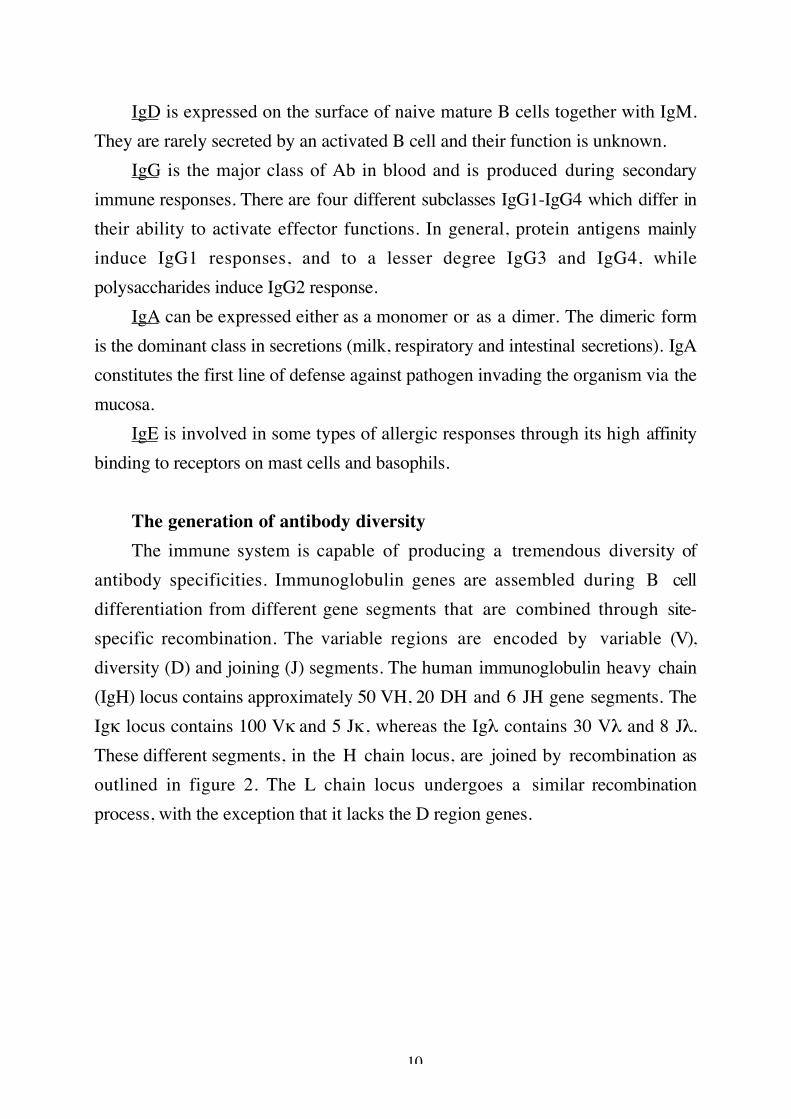
IgD is expressed on the surface of naive mature B cells together with IgM.They are rarely secreted by an activated B cell and their function is unknown.
IgG is the major class of Ab in blood and is produced during secondaryimmune responses. There are four different subclasses IgG1-IgG4 which differ intheir ability to activate effector functions. In general, protein antigens mainlyinduce IgG1 responses, and to a lesser degree IgG3 and IgG4, whilepolysaccharides induce IgG2 response.
IgA can be expressed either as a monomer or as a dimer. The dimeric formis the dominant class in secretions (milk, respiratory and intestinal secretions). IgAconstitutes the first line of defense against pathogen invading the organism via themucosa.
IgE is involved in some types of allergic responses through its high affinitybinding to receptors on mast cells and basophils.
The generation of antibody diversityThe immune system is capable of producing a tremendous diversity of
antibody specificities. Immunoglobulin genes are assembled during B celldifferentiation from different gene segments that are combined through site-specific recombination. The variable regions are encoded by variable (V),diversity (D) and joining (J) segments. The human immunoglobulin heavy chain(IgH) locus contains approximately 50 VH, 20 DH and 6 JH gene segments. TheIgκ locus contains 100 Vκ and 5 Jκ, whereas the Igλ contains 30 Vλ and 8 Jλ.These different segments, in the H chain locus, are joined by recombination asoutlined in figure 2. The L chain locus undergoes a similar recombinationprocess, with the exception that it lacks the D region genes.

11
V D J CµHeavy chain
Recombined DJ genes
Recombined VDJ genes
Figure 2. Recombination of antibody VDJ genes.
Further diversity is generated through junctional diversification, that isnucleotides can either be lost or added (P and N nucleotides) in the joining of V-(D)-J segments. Additional diversity can be derived from pairing of differentVL/VH regions. Furthermore, after the B lymphocyte has been stimulated byantigen a somatic hypermutation mechanism may operate on the assembledvariable regions.
B cell developmentThe cells of the immune system which are specialized to make antibodies
are called B lymphocytes. They originate in the bone marrow and are derivedfrom a multipotent stem cell. The different stages during differentiation of Blymphocytes, from precursor cells to antibody secreting cells, are outlined infigure 3. The different steps are defined by changes in the specific geneexpression pattern.

12
pro-B pre-B Immature B sIgM Mature B
sIgM sIgD
Plasma cell
Memory cell
sCµ/λ5/VpreB
VDJ VL Antigen
Figure 3. B cell differentiation.
At the pro-B cell stage, the Ig genes are in germline configuration but asterile transcript is produced (Lennon and Perry, 1990). In pre-B cells, VDJrecombination of the IgH variable chain generates µ-chains which can associatewith either the surrogate λ5 and VpreB light chains (Kerr et al., 1989; Kitamuraet al., 1992) or the κ protein encoded by a germline JCκ transcript (Francés etal., 1994). This immature B cell receptor is expressed on the surface of the pre-Bcell and can function as a signal-transducing surface receptor (Misener et al.,1991). Following the rearrangement of genes encoding Ig κ or Ig λ light chain,the IgM receptor is displayed on the surface of immature B cells. The mature Bcell is antigen reactive and expresses IgM and IgD receptors. After encounteringthe correct antigen the mature B cell is activated and can differentiate into eithermemory or plasma cells. Differentiation to plasma cells include switch fromsurface Ig (sIg) expression to secretion of Abs and may include somatic mutationof Ig varible regions.
SIg on immature and mature B cells are noncovalently associated with themb-1 gene product Igα and the B29 gene product Igβ (Sakaguchi et al., 1988;Hermanson et al., 1988). These molecules together form the B cell antigenreceptor (BCR) complex. Igα and Igβ facilitates transport of the Ig moleculefrom the endoplasmatic reticulum to the cell surface and also have an importantrole in the signal transduction event (Matsuuchi et al., 1992; Nakamura et al.,

13
1993). Igα and Igβ are expressed at all stages prior to terminal differentiation intoplasma cells.
B cell activationB cell activation initially involves antigen binding to sIg. There are two kinds
of antigen, T cell-dependent and T cell-independent. The T cell-independentpathway is triggered by two kinds of antigen, polysaccharides or potentpolyclonal activators like lipopolysaccharide, (LPS). These antigen crosslink thesIg and thereby activate the B cells. However, the activation still requires T cellderived cytokines to stimulate Ig secretion (Pike et al., 1987). The T cell-dependent pathway is triggered by soluble proteins and relies on the possibility ofthe B cell to act as an antigen-specific presenting cell (APC). The B cell binds theantigen with its sIg. The antigen is internalized, processed into peptides andtherafter presented on the cell surface bound to the MHC class II molecule.Finally the T cell recognizes the processed antigen on the B cell surface and theactivation of the B cell starts. This presentation leads to cognate interactions,involving the release of T cell derived cytokines and increased expression ofaccessory molecules, which results in both T and B cell activation. A key featureof this cognate interaction is the induction on the T cell membrane of a ligand forCD40 (CD40L), which delivers an activational signal to the B cell (Noelle et al.,1992). Other important accessory molecules are the CD80, CD86 on the B celland CD28 and CTLA-4 on the T cell (figure 4). Crosslinking CD40 promotes Bcell proliferation and immunoglobulin class switch (Clark and Ledbetter, 1986;Jabara et al., 1990). If the CD40-CD40L interaction is blocked in vitro withsoluble CD40 or CD40L specific Ab, the B cell can not proliferate indicating thatthis interaction is required for signaling (reviewed by Noelle et al., 1992). Theinteraction of CD28-CD80/CD86 prefentially activates the T cells to producecytokines and to proliferate (Linsley et al., 1991). CTLA-4 deficient mice showmassive expansion of activated T cells and an increase in serum Ig levels(Waterhouse et al., 1995). This data suggest that CTLA-4 may act as a negativeregulator of T cell activation, and indirectly, B cell stimulation.

14
B T
MHCIIAntigen TcR
CD40 CD40L
CD80 CD28
CD86 CTLA-4
Figure 4. Interaction between B and T cell.

15
REGULATION OF TRANSCRIPTION
DNA in the nucleus of eukaryotic cells is wrapped around histone cores,bound by nonhistone proteins and compacted into highly ordered chromatinstructures. Gene activation correlates to an altered chromatin environment. Theopen chromatin has two special characteristics. The first is an increased sensitivityto digestion with DNaseI and the second is hypomethylation. DNA methylation isbelieved to be one way of regulating gene activity, where demethylation reflectsan active gene. Gene transcription can be regulated by cis influences, dependenton the DNA sequences of genetic elements attached to a gene, and transinfluences, dependent on the environment of the gene. Cis-acting controlelements mediate their function by binding of nuclear proteins which assemblethe DNA into a three dimensional configuration that is optimal for thetranscription and expression of genes. Binding of trans-acting nuclear factors tothe control elements may give the signal for opening of the chromatin structure.Promoters and enhancers represent two classes of DNA elements that appear tocooperate in the control of efficient transcription. The promoter region containssequences upstream of the transcription initiation site that are involved in thetissue-specific activation or repression of transcription. Enhancer elements canpotentiate transcription from an appropriate promoter region in a tissue-specificmanner, somewhat independent of their orientation or distance from thepromoter. The proteins important for gene transcription include basaltranscription factors, positive and negative nuclear factors and co-factors whichcan modulate the function of specific nuclear factors. Control of thetranscriptional activity of individual Ig gene loci is central to the regulation of Bcell differentiation. The IgH locus appears to be activated at a very early stage ofB cell development whereas the IgL locus is activated in late pre-B cells. In figure5 the different promoters and enhancers important for IgH gene expression areoutlined. Below the characteristics of these elements will be discussed.
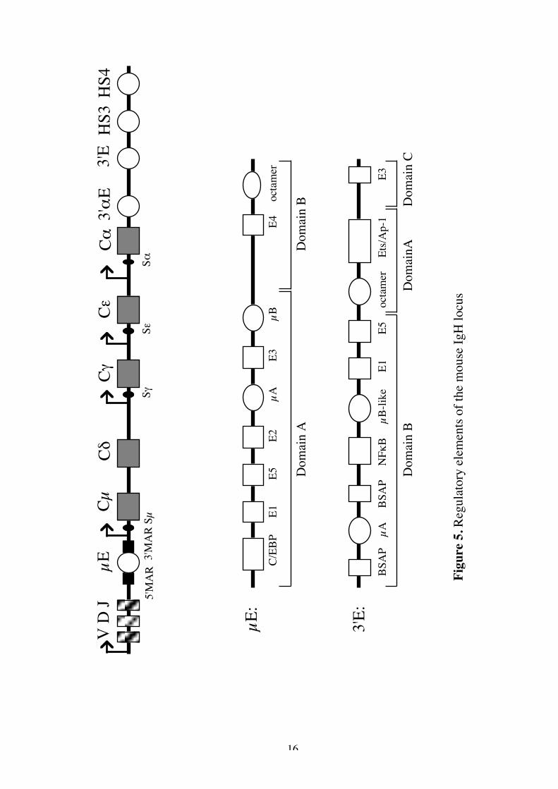
16
V D
J µE
CµCδ
CγCε
Cα3'α
E3'
EH
S3H
S4
5'M
AR
3'M
AR
Sµ
SγSε
Sα
C/EB
P
E1
E5
E2
µA
E3
µ
B
E4
oct
amer
BSA
P
µA
BSA
P
NFκ
B
µB-
like
E
1
E
5
oct
amer
E
ts/A
p-1
E3
Dom
ain
A
Dom
ain
B
Dom
ain
B
Dom
ainA
D
omai
n C
µE:
3'E:
Figu
re 5
. Reg
ulat
ory
elem
ents
of th
e m
ouse
IgH
locu
s

17
VH promoterThe VH promoters are located at the 5’ end of each variable gene segment,
between 150-200 bp upstream of the initiation site (Grosschedl and Baltimore,1985) (figure 6). Two sequence motifs are shared by all Ig promoters, an octamermotif and a TATA box. The TATA box consensus sequence is the core promoterwhich interacts with the basal transcription machinery (Parvin et al., 1992). Thismotif is not cell-restricted. The octamer motif (ATTTGCAT or its inversion) ishighly conserved among the VL and VH promoters (Falkner and Zachau, 1984).Mutations in the octamer motif reduces expression of the IgH gene in bothtransfection assays (Mason et al., 1985) and in transgenic animals (Jenuwein andGrosschedl, 1991). It has been shown that a single octamer motif is sufficient forconferring B cell specificity to a minimal promoter element consisting a TATAbox (Dreyfus et al., 1987). A family of nuclear proteins bind to the octamermotif, two of them are present in B cells, Oct-1 and Oct-2 (Staudt and Lenardo,1991). Oct-1 is expressed ubiquitously whereas the Oct-2 is largely B cellrestricted (Staudt and Lenardo, 1991). Therefore it was proposed that the Oct-2protein has a specialized function in B cells with respect to Ig transcription.However in Oct-2-deficient mice generated by gene targeting, the Ig genes arerearranged and transcribed normally (Corcoran et al., 1993). It was thensuggested that other B cell specific components are responsible for lymphoidrestricted transcription of Ig promoters. Recently an adaptor protein has beenidentified which is tissue specific and it stimulates transcription in conjunctionwith either Oct-1 or Oct-2 (Luo et al., 1992). An adaptor protein does not bind toDNA itself but can interact with DNA-binding proteins. This protein-proteininteraction can then modulate the affinity of the DNA-binding protein. Theadaptor molecule that binds the Oct-proteins have been cloned, by three differentgroups, as Bob-1, OBF-1 and OCA-B (Gstaiger et al., 1995; Strubin et al.,1995;Luo and Roeder, 1995). Several other sequence motifs are conserved in Igpromoters, including the heptamer, a pyrimidine-rich element and two E-boxmotifs. The heptamer is located close to the octamer and the two motifs can bebound cooperatively by Oct-binding proteins (Poellinger and Roeder, 1989). The
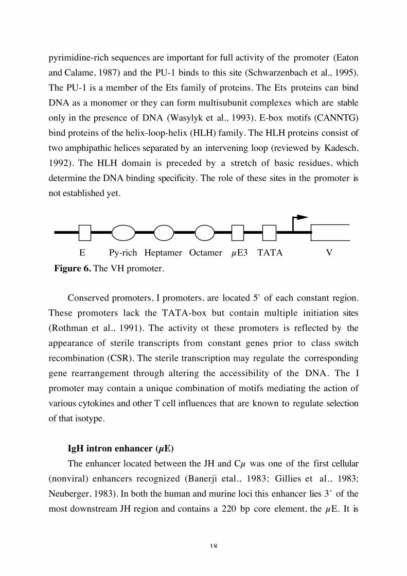
18
pyrimidine-rich sequences are important for full activity of the promoter (Eatonand Calame, 1987) and the PU-1 binds to this site (Schwarzenbach et al., 1995).The PU-1 is a member of the Ets family of proteins. The Ets proteins can bindDNA as a monomer or they can form multisubunit complexes which are stableonly in the presence of DNA (Wasylyk et al., 1993). E-box motifs (CANNTG)bind proteins of the helix-loop-helix (HLH) family. The HLH proteins consist oftwo amphipathic helices separated by an intervening loop (reviewed by Kadesch,1992). The HLH domain is preceded by a stretch of basic residues, whichdetermine the DNA binding specificity. The role of these sites in the promoter isnot established yet.
E Py-rich Heptamer Octamer µE3 TATA V Figure 6. The VH promoter.
Conserved promoters, I promoters, are located 5' of each constant region.These promoters lack the TATA-box but contain multiple initiation sites(Rothman et al., 1991). The activity ot these promoters is reflected by theappearance of sterile transcripts from constant genes prior to class switchrecombination (CSR). The sterile transcription may regulate the correspondinggene rearrangement through altering the accessibility of the DNA. The Ipromoter may contain a unique combination of motifs mediating the action ofvarious cytokines and other T cell influences that are known to regulate selectionof that isotype.
IgH intron enhancer (µE)The enhancer located between the JH and Cµ was one of the first cellular
(nonviral) enhancers recognized (Banerji etal., 1983; Gillies et al., 1983;Neuberger, 1983). In both the human and murine loci this enhancer lies 3’ of themost downstream JH region and contains a 220 bp core element, the µE. It is

19
located 5’ of the switch region and where it is retained after switchrecombination. In transfection assays this enhancer appears to be active at all Bcell stages (Grosschedl and Baltimore, 1985). The generation of mice lacking theµE has demonstrated its importance for efficient DNA demethylation, germlineIg transcription and VDJ recombination during B cell development (Serwe andSablitzky, 1993; Chen et al., 1993). Deletion of µE also strongly suppressesswitch recombination at the Cµ switch region, whereas the switch region of theγ1 gene is efficiently rearranged (Gu et al., 1993).
µE is flanked by two Matrix attachment regions (MARs) which mediate anassociation with the nuclear matrix (Cockerill et al., 1987). These MARs havebeen shown to be essential for transcription of a rearranged µ gene in transgenicB lymphocytes (Forrester et al., 1994). It has been suggested that the µE and itsflanking MARs are largely responsible for inducing chromatin alterations(Cockerill et al., 1987). The MARs contain binding site for two nuclear factors,NFµNR and SATB-1, which may participate in the repression of enhanceractivity in non-B lymphoid cells (Scheuermann and Chen, 1989; Dickinson et al.,1992).
Functional and biochemical analyses have identified a set of nuclear proteinbinding sites in the µE, like the E-boxes, octamer binding and Ets binding motifs(Staudt and Lenardo 1991; Ephrussi et al., 1985) (Figure 5). There are five E-boxes (Eµ1-Eµ5) which bind a variety of homo or heterodimerizing trans-actingfactors belonging to the HLH family. Mutational studies have suggested that theE-box elements contribute to the transcriptional regulation of the IgH gene butdo not determine lymphoid specificity (Lenardo et al., 1987; Perez-Mutal etal.,1988). The octamer element, also present in Ig promoters, bind the Oct-1 andOct-2 proteins. Mutation of these sequences in the context of entire IgH enhancerhas only very little effect on its activity in lymphoid cells (Perez-Mutal et al.,1988; Jenuwein et al., 1991), suggesting the presence of other motifs that conferthe lymphoid specific activity of the µE. The Ets-binding sites, µA and µB, arerequired for optimal µE activity in B cells (Liebermann et al., 1990; Nelson et al.,
20
1993). The µA motif binds at least five different Ets family members, presumablywith varying affinites, whereas the µB motif binds the PU-1 (Nelson et al., 1993).
3’ enhancer (3’E)The observation that certain cell lines, in which the µE was deleted, still
transcribed the IgH gene (Eckhardt and Birshtein, 1985; Wabl and Borrows,1984) led to the notion that the µE might serve an important function in early Bcell stages, while other control elements might be more important in late stages.Transgenic studies have also shown that the IgH transgene was not expressed atthe same high level as the endogenous IgH gene (Sturb, 1987). Subsequently, asecond B cell specific enhancer 3’ of the Cα coding sequence was identified, bothin mice (16kb downstream) and in rat (27kb downstream) (Pettersson et al.,1990; Dariavach et al., 1991; Lieberson et al., 1991). It has not yet been identifiedin the human genome. In transfection assays the 3’E activity was only detected incell lines representing terminally differentiated B cells (Lieberson et al., 1991;Darivach et al., 1991). Transgenic studies confirmed this late B cell activationprofile (Arulampalam et al., 1994). In these animals, enhancer activity wasobserved in both in vivo activated splenocytes and in B cells of the peritonealcavity. The 3’E activity late in B cell development also correlates with thedemethylation and hypersensitive site formation over this region (Giannini et al.,1993). Potential function of the 3’E includes CSR, somatic hypermutation orincreased Ig gene expression. The targeted deletion of the 3’E in mice revealedits role in control of CSR (Cogné et al., 1994). These mice were deficient inserum IgG2a and IgG3, but had normal levels og IgM and IgG1. In transgenicstudies it has been shown that the Ig gene expression is higher if the transgene iscontrolled by the µE+3’E, as compared to the transgene controlled by the µEalone (paper I). This augmentation was seen at both the mRNA and proteinlevels. In cell lines, loss of both the µE and the 3’E completely abolished IgHgene expression, demonstrating the importance of 3’E for gene transcription(Lieberson et al., 1995).

21
The 3’E contains a number of nuclear protein binding sites (figure 5), someof which are identical to those found in the µE. The 3’E can be divided into threefunctional domains (Grant et al., 1992). Domain A and B confer lymphoidspecific reporter gene activity whereas domain C appears to be active in all celllines used.
Domain A, the strongest transcriptional domain, contains an octamer motifand an Ets-1/AP site. Mutations in the octamer site reduces the activity, but thecell specific activity is likely to rest with the Ets/AP-1 site. A DNA bindingcomplex, nuclear factor of activated B cells (NFAB), binds to the Ets/AP-1 site.This complex contains the tissue restricted Ets protein, Elf-1 and the AP-1protein (dimer of Jun-B and c-Fos) (Grant et al., 1995). This binding is onlyobserved in activated B cells.
Domain B contains a range of nuclear factor binding sites. The E-box motifsE1 and E5 have been shown to be important for enhancer activity (Grant et al.,1992; Meyer et al., 1995). The E2A gene products E12 and E47 can bind to theE5 site and the zink finger protein YYI binds to the E1 site. The µA- and µB-likemotifs also bind Ets proteins. The activity of the µB has not been characterised,whereas the µA contribute to the transcriptional actvity of domain B (Grant etal., 1992). There are also two binding sites for B cell specific activator protein(BSAP) in domain B. BSAP, a recently identified member of the Pax-gene familyof transcription factors, is expressed in the B lineage from the pro-B to themature B cell stage, however, it is not found in plasma cells (Singh and Birshtein,1993, Neurath et al., 1994). When the BSAP binds to the motif in the 3’E itexerts a negative effect on the transcriptional activity. This might explain why the3’E is active in plasma cells only.
Domain C has only one protein binding motif, E3, which can bind theDNA-binding protein, USF, in splenic nuclear extracts (Arulampalam et al.,1995). USF is a member of a distinct class, denoted basic-HLH-zip proteins.These proteins are distinguished by the presence of leucine zippers adjacent totheir HLH motifs (Kadesch, 1992).
22
Additional control elementsAn enhancer has been identified close to the Cα gene (Matthias and
Baltimore, 1992). This enhancer has quite weak lymphoid specific activity. Thelocation of this enhancer close to the Cα suggested that it might be involved incontrolling switch recombination to Cα, although this has not yet beenconfirmed.
Analysis of the 3’ end of the IgH locus revealed four DNaseI hypersensitivesites (HS), two of which (HS1 and HS2) mapped to the 3'E (Madisen andGroudine, 1994, Michaelson et al., 1995). The enhancer activity of HS3 and HS4is restricted to B cells, HS3 is only active in plasma cells whereas HS4 is active inpre-B as well as plasma cells. HS3 and HS4 contain multiple binding sites fortranscription activators commonly associated with Ig enhancers, the significanceof these different motifs have not been confirmed.
Locus control regionThe most well-characterised locus control region (LCR) is at the 5' end of
the human β-globin locus. The LCR consists of five different regulatory elementswhich are involved in the regulation of replication, chromatin structure andtranscription of the entire locus (Crossley and Orkin, 1993). Transgene constructscontaining the LCR show copy number dependent expression of linked globingenes independent of the site of integration of the transgene in the host genome(Grosveld et al., 1987; Blom van Assendelft et al., 1989). The LCR regulatestranscription of the different β-globin genes through competion of promoterswith the LCR elements (Wijgerde et al., 1995).
It has been proposed that the IgH locus contains two LCRs. The µE and itsflanking MARs form a potential LCR upstream of the Cµ. This LCR is requiredfor VDJ recombination at early stages of B cell development (Serwe et al., 1993,Gu et al., 1993). The other potential LCR is located at the 3' end of the IgH locusand would include the 3'E, HS3 and HS4. Madisen and Groudine (1994) havedemonstrated that these three elements confer copy number-dependentexpression of a linked myc-gene in vitro. We have shown that the 3'E can confer

23
position independent expression of a linked transgene (paper I) but no copynumber-dependent expression was achieved. The missing elements to achievecopy number-dependent expression might be the HS3 and HS4. The potential 3'end LCR might also regulate transcription of different constant isotypes throughcompetion with the different promoters. At least the 3'E can direct transcriptionalactivity of specific promoters (Cogné et al., 1994). Mice with targeted deletion ofthis enhancer did not produce γ3 and γ2a germline transcript.
24
PRODUCTION OF HUMAN ANTIBODIES
Monoclonal Ab became available when Köhler and Milstein demonstratedthat cultured mouse myeloma cells could be fused to spleen cells from immunizedmice (Köhler and Milstein, 1975). The hybrid cells (hybridoma) grew continouslyin culture (a property aquired from the myeloma cells) and continued to producelarge quantities of Ab (a property obtained from the spleen cells). The mAbs havean enormous potential as therapeutic and diagnostic agents because of theirremarkable specificity and affinity for their target. Rodent mAb unfortunately hasseveral disadvantages, such as a short half-life in serum, only some of thedifferent classes can trigger human effector functions and the Abs elicit anunwanted immune response in patients (human anti-mouse antibodies orHAMA). HAMA can result in enhanced clearance of the Ab from the serum,blocking of its therapeutic effect. Only a fraction of the anti-mouse immuneresponse is directed to the variable region of the rodent Abs. To reduce theHAMA response it is therefore necessary to express potentially therapeutic Abeither as chimeric, humanized (engraftments of mouse CDR into the humanframework region) or human monoclonal Abs. Recent development in bothmonoclonal Ab strategy and the use of recombinant DNA technology have nowmade it feasable to produce human mAb with the desired specificity. Below, thedifferent steps, outlined in figure 7, leading to antigen-specific human Ab will bediscussed.
In vitro generation of antigen-specific AbsDue to ethical problems associated with immunization of humans much
interest has been focused on techniques to generate antigen-specific Ab, using invitro systems. Human B cells may be obtained from spleen, tonsils, lymph nodes,bone marrow or peripheral blood, the latter being the most accessible source. Anapproach to generate antigen-specific Abs is to use in vitro immunizationtechniques. However, in vitro immunization of human lymphocytes was initiallymore difficult to achieve than that of the murine counterpart. The reason for this

25
might be that peripheral blood cells contain cytotoxic T cells, monocytes,granulocytes and NK cells, which abrogate the antigen-specific activation of Bcells (Borrebaeck et al., 1988). These cells must therefore be removed from theperipheral blood cells by L-leucyl-L-leucine methyl ester (LeuLeuOMe) treatmentbefore an in vitro immunization of the B cells can take place (Borrebaeck et al.,1988). Briefly, the cells are cultivated with antigen in medium containingsupernatant from irradiated pokeweed mitogen (PWM) stimulated T cells andrecombinant IL-2. Immunization of naive B cells using this protocol onlygenerates primary immune responses.
Preparation of peripheral blood
PCR of the Ig variable region genes
Generation of antigen-specific Abs
EBV infection and electrofusion
Prokaryotic expression
Eukaryotic expression
Cell line producing specific Ab
Figure 7. Establishment of human monoclonal Ab-producing cell lines.
B cells can be activated antigen-specifically and simultaneously becostimulated in vitro via their CD40 surface molecules. The stimulation can bedelivered using either soluble recombinant CD40L or CD40 specific Ab incombination with cytokines (Nonoyama et al., 1993; Armitage et al., 1993).Using this CD40-system it is possible to maintain a long term B cell culture andpolyclonal isotype switch has also been reported (Splawski et al., 1993).
26
Antigen-specific activation of B cells in vitro could be achieved afterstimulating the B cells with anti-sIg Abs in the presence of T helper cells andStaphylococcal Enterotoxin A (SEA) (Ingvarsson et al., 1995). Increasedcrosslinking of the sIg enhanced Ig production. In this manner specific Ab wasobtained against both primary and secondary antigens.
Chin et al. (1995) have been able to activate B cells antigen-specifically andinduce isotype switch to achieve antigen-specific IgG production, using theirprimary and secondary antigen driven cultures. The secondary immunization issupported by antigen activated T helper cells and stimulation via the CD40pathway.
ImmortalizationOne of the most important technologies in the production of monoclonal Ab
is the establishment of immortal cell lines. The most frequently applied techniqueuses Epstein-Barr virus (EBV) to infect and activate human B cells (Steinitz et al.,1979). To rescue the transformed B cells they can be further fused to myelomacells which stabilizes the Ab production (Kozbor et al., 1982).
Another alternative to immortalizing monoclonal Abs is to use DNArecombinant technology, which will be discussed below.
Polymerase chain reactionSince the first report of specific DNA amplification using the polymerase
chain rection (PCR) in 1985, the number of different applications has grownsteadily, as have modifications of the basic method. An alternative way ofimmortalizing human Ab is to use this powerful PCR technique for amplificationof the VL and VH chains. The concept is simple: a mixture of oligomer primersin the 5' leader sequences or framework 1 region with 3' constant or framework4 region permits the amplification of any Ig variable region (Larrick et al., 1989a;Coloma et al., 1991; Marks et al., 1991a). The method has been used to obtainvariable regions of both heavy and light chains from single human B cells (paper

27
IV; Küppers et al., 1993). These fragments can thereafter be cloned into eitherprokaryotic or eukaryotic expression vectors.
Prokaryotic expressionUsing bacterial expression technology, different types of Ab fragments can
be made in fully functional form in E. coli. This technology can be extendedfurther to cloning and expressing libraries of VL/VH chains from the immunesystem. The construction of libraries of single chain Fv or Fab fragments ofantibody molecules that are expressed on the surface of filamentous phages andthe selection of specific recombinant Ab offers a new and powerful mean forgenerating monoclonal Ab (reviewed by Hoogenboom et al., 1992a; Marks et al.,1992a). A major goal of recombinant Ab technology is to develop Ab libraries oflarge size and diversity to facilitate the isolation of Abs of every conceivablespecificity, among them Abs with high affinity. The rearranged V-genes can bederived from immunized (Barbas et al., 1991; Clacksons et al., 1991) orunimmunized B cells (Marks et al., 1991b). The naive libraries are easier to obtainbut mostly low affinity binders have been isolated (Marks et al., 1991b). Analternative approach to creating diverse libraries is to use a collection of germlineVH segments fused to synthetic CDR3 regions in vitro (varying in lengthbetween 6 and 15 amino acids) and combined with one or multiple light chains(Hoogenboom and Winter, 1992b; de Kruif et al., 1995a). Fully synthetic librarieshave also been described (Barbas et al., 1992; Söderlind et al., 1995).
Several methods for antigen-specific selection of high affinity Abs have beendescribed. These include selection of phage Abs binding to antigen-coated tubes(Marks et al., 1991b) or binding to antigen on a column matrix (Clackson et al.,1991). One alternative is to use flowcytometry based selection for finding Abagainst cell surface antigens (de Kruif et al., 1995b). To mimick in vivo antigenselection, Duenas and Borrebaeck (1994) used a fusion protein consisting ofantigen fused to the pIII protein. Using an engineered helper-phage with deletedfunctional gIII made it possible to select high affinity Abs.
28
Since most libraries are obtained from naive repertoires, methods for affinitymaturation in vitro have been developed. These include in vitro maturation of Abusing codon based mutagenesis (Glaser et al., 1992), error prone PCR (Cassonand Menser, 1995) or spiked oligonucleotide based mutagenesis (Hermes et al.,1989). One alternative is to use the chain-suffling technique (Marks et al., 1992b).
After selecting a high affinity Ab to a desired antigen, it could be eitherexpressed in E.coli and purified, or cloned in an eukaryotic expression vector toproduce the entire Ab molecule.
The use of Ab fragments may be advantageous in some therapeuticalapplications, as they penetrate tissue more readily and are cleared more rapidlyfrom serum. This may help in neutralizing and clearing drugs from the serum.Engineering of recombinant Ab has also resulted in the construction of a newgeneration of molecules. Monovalent sFv Abs have been converted into bivalentmonospecific or bispecific reagents using e.g. dimerization domains (Pack et al.,1993) or direct coupling (Holliger et al., 1993). In addition, Fv can be fused orcoupled to a variety of cytokines, enzymes or radioactive entities.
Eukaryotic expressionGene transfection provides a method for making novel Ig molecules. The
tranfected cells (transfectomas) grow continously in culture and produce the Igspecified by the transfected gene. To create a transfectoma cell line synthesizing anovel Ab, both Ig heavy and light chains must be transfected into the samerecipient cell line. Although both chains can be contained in a single vector, it isusually more convenient to construct separate light and heavy chain vectors,which are simultaneously transferred and expressed. A vector should containregulatory elements, VL/VH region genes, a genomic constant region includingpolyA signal and splice signals and a selectable marker, as outlined in figure 8.
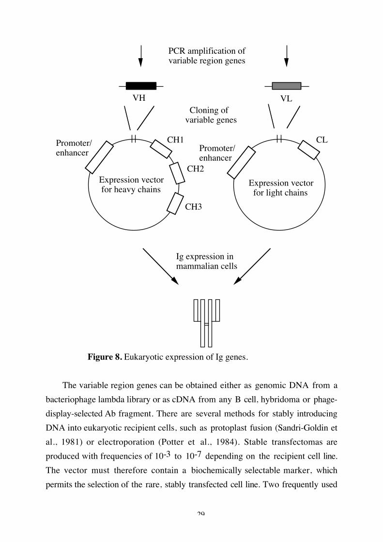
29
PCR amplification of variable region genes
VH VL
Promoter/enhancer Promoter/
enhancer
CH1
CH2
CH3
CL
Ig expression in mammalian cells
Cloning of variable genes
Expression vector for heavy chains
Expression vector for light chains
Figure 8. Eukaryotic expression of Ig genes.
The variable region genes can be obtained either as genomic DNA from abacteriophage lambda library or as cDNA from any B cell, hybridoma or phage-display-selected Ab fragment. There are several methods for stably introducingDNA into eukaryotic recipient cells, such as protoplast fusion (Sandri-Goldin etal., 1981) or electroporation (Potter et al., 1984). Stable transfectomas areproduced with frequencies of 10-3 to 10-7 depending on the recipient cell line.The vector must therefore contain a biochemically selectable marker, whichpermits the selection of the rare, stably transfected cell line. Two frequently used
30
selectable markers are the gpt-gene (bacterial xanthine-guaninephosphoribosyltransferase gene) or the neo-gene (aminoglycosidephosphotransferase gene). Both enzymes, when expressed, endow a dominantbiochemically selectable phenotype to mammalian cells so that they can be usedfor selection in non drug-marked recipient cells.
There are two possible ways of creating an eukaryotic expression vector, touse either viral or endogenous regulatory elements. Viral promoter/enhancer pairsfrom cytomegalovirus (CMV) (Bebbington et al., 1992) and SV40 (Morrison etal., 1995) have been used successfully to achieve high levels of Ab expression.Viral based vectors can be transfected into either myeloma cell lines or Chinesehamster ovary cells (CHO). The Ig gene expression was for a long time thoughtto be regulated by the V promoter and the intron enhancer. Therefore mostvectors based on endogenous control elements use this promoter/enhancercombination. The rate of Ab secretion attainable using these vectors is generallyconsiderably lower than those of comparable parent hybridomas (Nakatani et al.,1989; Walls et al., 1993). Recent observations indicate that other enhancerelements are important for Ig gene expression. Including the 3'E to the IgHvector increased the IgH expression five fold (Mocikat et al., 1993; Mocikat et al.,1995a; Paper V). Vectors based on endogenous control elements are alsotransfected in myeloma cells.
Gene amplification can be used to increase the level of Ig expression. Twoalternative selectable markers have been described in Ig expression vectors, theglutamine synthetase (GS), and the dihydrofolate reductase (DHFR).Cointroduction of heavy and light chain constructs with subsequent amplificationusing the DHFR-marker resulted in as much as 25-fold increase in secretion ofintact Ab relative to unamplified cells (Dorai and Moore, 1987). The GS-markerin combination with a vector, based on viral control elements, containing bothheavy and light chain, also resulted in higher secretion levels of Ab (Bebbingtonet al., 1992).
Using eukaryotic Ig expression vectors it is also possible to introduce novelfunctions not normally found in the Ig molecule. To achieve optimal effectorfunctions either the constant region can be changed into an other region
31
(reviewed by Shin et al., 1992; Brekke et al., 1993) or the IgG1 can be convertedinto a pentamer IgG1 (Smith and Morrison, 1994). It is also possible to producefusion proteins with e. g. cytokines (Morrison et al., 1995).
One alternative way of producing chimeric Abs is to, through homologousrecombination, introduce the desired human constant gene segment into the Igloci of any hybridoma cell line of interest. The advantages of using this system isthat all regulatory elements needed are already in the loci and the variable genedoes not have to be isolated from the hybridoma and finally that onerecombination vector can be used for all specificities. There are two kinds ofrecombination vectors available, replacement or integration vectors (Capecchi etal., 1989). The replacement vector contains a two-sided homology flankneighbouring the heterologous region, whereas the integration vector containsone homology flank within which the construct is linearized, thus giving rise to aduplication of the target sequence. Using these vectors, the Ig expression levelsare comparable with the original hybridoma levels (Mocikat et al., 1995b; Bakeret al., 1994). The disadvantage in using these vectors is a frequent occurance ofclones that coexpress Ig from two species. So far this approach to produce Abshas only been done using murine hybridomas.
32
THE PRESENT INVESTIGATION
The aim of this work has been; (i) to increase the understanding of how theIg gene expression is regulated and (ii) to generate and produce humanmonoclonal Abs.
(i) We have focused our interest on the regulatory elements in the 3' end ofthe IgH locus. We chose these elements because of their potential role inregulating high Ig gene expression. In paper I and II we have investigated, usingtransgenic mice, the 3' enhancers role in controlling Ig gene expression. In paperV we have used different regulatory elements for optimizing an eukaryoticexpression vector.
(ii) Our goal has been to develop techniques for obtaining humanmonoclonal Abs. Different approaches have been used such as PCRimmortalization of variable regions, prokaryotic and eukarotic expression. Theseapproaches will be discussed in paper III-V.
Paper I: Elevation of immunoglobulin gene expression in transgenicanimals.
In paper I we investigated if the 3'E in combination with the µE can inducehigher Ig gene expression in transgenic animals. Mice harbouring rearranged IgHtransgenes, potentiated by the VH promoter/µE pair (Vµ1) or by the VHpromoter in combination with the µE/3'E (Vµ3) were generated. Analysis ofthese mice demonstrated that the specific IgH gene transcription was at least fivefold higher after addition of the 3'E to the transgene. The enhancer activity ismainly found in the B cell compartment. Separation of splenic cell populationsrevealed that the transgene expression is restricted to in vivo activated B cells.The expression level in resting B cells are comparable between the animals,whereas the elevation in expression is significantly higher in the activated B cellsin the Vµ3 mice. There is some specific IgH gene expression observed in non-lymphoid tissues as well. Non-lymphoid expression of other IgH transgenes hasbeen observed earlier (Grosschedl et al., 1984; Arulampalam et al., 1994). The
33
detected expression in non-lymphoid tissues may be due to contaminating B cells.Alternatively the aberrant activity of the enhancers may reflect utilisation oftrans-acting factors that are present in lymphoid as well as non-lymphoid tissues.
We have also shown that the transgene can be stimulated in a T celldependent manner after immunization with an appropriate antigen. Both Vµ1and Vµ3 had increased serum levels of NP-specific Abs.
The high level of transgene expression prevents rearrangement ofendogenous genes, although incompletly (Weaver et al., 1985; Nussenzweig et al.,1987). The endougenous expression is higher in the Vµ1 mice as compared tothe Vµ3 mice. The higher IgH gene expression in the Vµ3 mice maydownregulate the endogenous expression more efficiently.
Transgene expression in a position independent but not copy-numberdependent manner was observed in the Vµ3 mice. Therefore we conclude thatthe 3'E can be part of a potential LCR but can not function as such on its own.
In conclusion, the 3'E can upregulate Ig gene expression in transgenic mice.These findings are supported by the observations that adding the 3'E to aneukaryotic expression vector controlled by the VH promoter/µE pair increasedthe Ig gene expression five fold (Paper V; Mocikat 1993).
Paper II: The IgH 3' enhancer can act directly on a natural IgH promoterin vivo
In paper II we wanted to study the biological function of the 3'E. Toinvestigate if the 3'E alone can function in conjunction with the VH promoter,mice harbouring rearranged IgH potentiated by the VH promoter/3'E pair weregenerated. Analysis of these mice revealed that the activity of the 3'E is restrictedto lymphoid-specific tissues. There was no activity seen in non-lymphoid tissues.The serum levels of antigen-specific Abs were much higher in the transgenic
34
mice as compared to normal mice indicating that the transgene product issecreted into the serum.
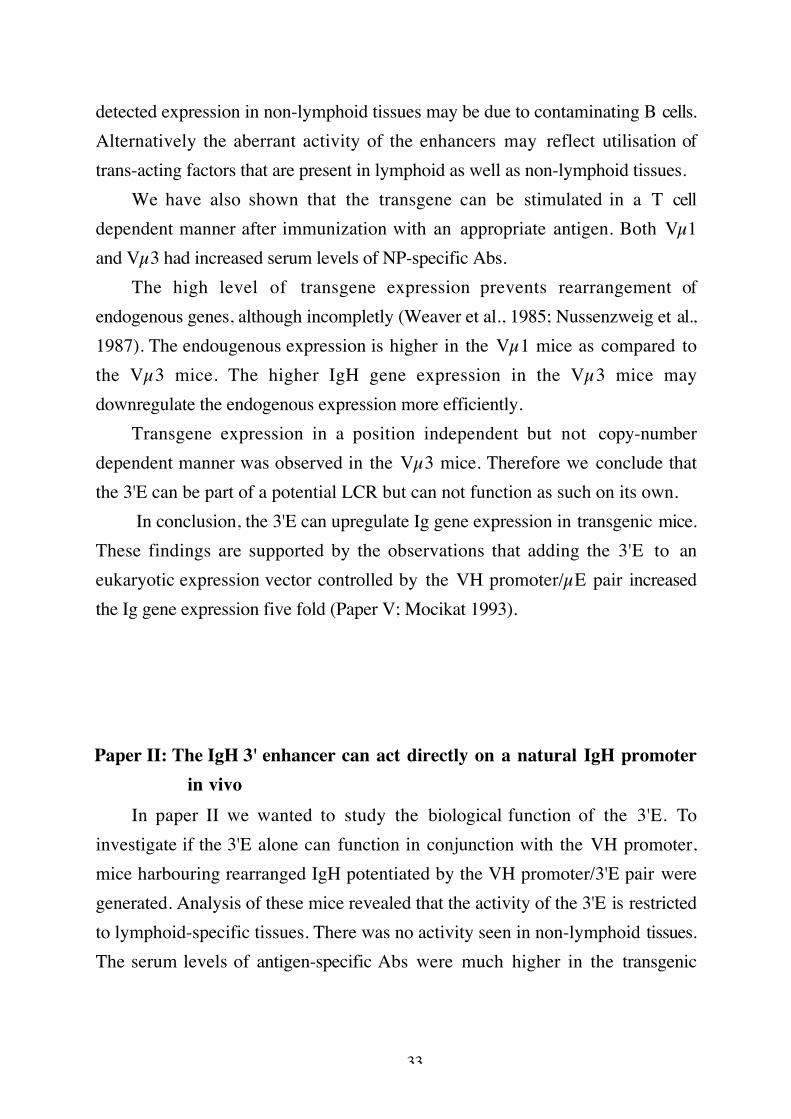
One possible mechanism whereby the 3'E could potentiate VH promoterinduced transcription over such long distances is represented by the loopingmodel (figure 9).
Loop
3' Enhancer
VH promoter
1. 2.
Figure 9. The looping model. Interaction of homologous (1) orheterologous (2) factors bound at the promoter and enhancer, with thelooping out of intervening DNA.
This suggests that activators bound at a distance interact with proteinsbound at the promoter, causing a looping out of intervening sequences. Membersof the bHLH-zip family can bind to DNA as homo or heterodimers. Theseproteins can also form tetramers (Ferre-D'Amare et al., 1994). This findingsuggests that bHLH-zip tetramers, by binding to DNA motifs in both the VHpromoter and the 3'E, may provide a physical bridge between the two controlregions.
Support for the possible VH promoter/3'E interaction is observations byLieberson et al. (1995). They showed that a cell line, lacking the µE, loses theability to express IgH gene upon the targeted deletion of the 3'E.
35
Paper III: Cloning of variable regions from three different humanAbs that differ in their reactivity against digoxin derivates.
The relation between Ab specificity and amino acid sequences of CDR1-3, inboth heavy and light chains of human monoclonal Abs, was investigated todetermine if a particular VH/VL gene segment is utilized during the primaryimmune response against a hapten. Human monoclonal IgM Abs against digoxinwere established after in vitro immunization of human peripheral bloodlymphocytes. Three human monoclonal Abs were generated, which differ in theirfine-specificity against digoxin and several digoxin analogues. The variable heavyand light chain regions were PCR amplified using leader sequence and constantregion specific primers, and subsequently sequenced. Comparison of the DNAsequences with those of the GenBank/EMBL databases revealed that thedifferent variable regions of the Abs belonged to different subfamilies. Thecomplementarity determining regions of the three Abs showed considerabledifferences, both in amino acid sequnce and in length. Thus, despite very similarreactivity of the three Abs against digoxin there are significant variations in theiramino acid sequences. We therefore conclude that binding specificity is not aconsequence of an apparent restriction of particular gene segments in theprimary human immune response against digoxin.
The PCR based protocol first used in this study can be a useful general toolfor studies on the relationship between the structure of an Ab variable regionsand its antigen binding function. The variable regions can be cloned intoprokaryotic expression vectors for studies of mutagenic variants of the variableregions.
Paper IV: Generation of human Ab fragments from single B cells.A method for generating human Ab fragments from single B cells is
described in paper IV (figure 10). Two different approaches were examined
36
for the generation of human Abs; either (i) the direct isolation of antigen-specific B cells followed by PCR-amplification of the variable genes from thissingle cell, or (ii) cloning antigen-specific B cells by limiting dilution in an EL-4/CD40 based cell culture and then PCR amplification of the variable regiongenes. Both these approaches allow us to immortalize antigen-specific variableregions and to retain the original pairing.
The antigen-specific sIg+ B cells were selected using antigen-coatedmagnetic beads. The magnetic bead rosetted B cells were isolated as single cellsusing a simple manual pipetting procedure. The immortalization procedure usedis based on PCR technology. To minimize the number of steps involved inobtaining the desired VL/VH genes, the mRNA was isolated with oligo dTmagnetic beads and thereafter the cDNA synthesis was performed directly on thebeads. The sensitivity of the PCR procedure was increased using nested PCR, ascompared to earlier protocols (Larrick 1989b). In the first step leader sequenceand constant region specific primers were used followed by FR1 and FR4specific primers in the second step.
For selection of specific B cells against recall antigen it is possible to use thissingle cell approach with direct PCR immortalization of the genes. In activated Bcells the mRNA level is much higher as compared to the level in resting B cells.Therefore we developed a cellular amplification step for rare binders, e. g.primary response specificities. A combination of the EL-4 system described byZubler et al. (1985) and the CD40 system described by Banchereau et al. (1991)was used for cellular amplification of antigen-selected B cells. This combination ofcell cultures resulted in specific clones from approximately 10% of themagnetically selected B cells. The positive clones were immortalized using thePCR protocoll described above. The VL/VH from a single cell and single cellclone respectively were cloned into a procaryotic expression vector andexpressed as soluble Fab fragments.
A further advantage in using the cellular amplification procedure is theopportunity to test for antigen-specificity in an ELISA. If cross-reactiveantibodies are selected, the directly isolated B cells can not be analysed until thegenes have been cloned into prokaryotic or eukaryotic expression vectors.
37
In conclusion, we have developed a method for generation of human Absselected from single antigen-specific B cells. The approach offers the possibility ofinvestigating the original variable region combinations and is very fast andefficient compared to conventional hybridoma technology.
Paper V: Design of an eukaryotic IgH expression vectorIn paper V an eukaryotic IgH expression vector was developed using
different regulatory elements. Conventional vectors rely on the VH promoter incombination with the µE. The Ig expression levels using these vectors aresubstantially lower than the levels achieved in the parental hybridomas. Recentfindings suggested that other control elements might be important for Igexpression. Therefore we evaluated if these novel enhancer elements couldcontribute to higher Ig gene expression in a myeloma cell line. A backbonevector was constructed, which contains the VH gene (from a NP-specific Ab), theγ3 constant region, the VH promoter and the µE. Different enhancer elementswere added to the backbone vector , such as the 3'E (1kb), 3'E (4kb) and finallythe combination of the 3'E, HS3 and HS4. Inclusion of the 3'E in the backbonevector resulted in a five fold increase in Ig expression, whereas including the 3'E(4kb) or the 3'E/HS3/HS4 did not upregulate Ig expression any further. Theabsence of increased expression using the combination 3'E/HS3/HS4 elements ismost probably related to the characteristics of the host cell used in theseexperiments. Observation by Michelson et al. (1995) indicated that the activity ofthe HS4 and 3'E are host cell dependent.
To analyse if there is a correlation between the number of enhancerelements and transcriptional enhancement, we cloned the 3'E on both sides of areporter gene. However, no copy-number dependent increase in transcription ofthe gene was observed. We also assessed if the position of the 3'E in the vectorwas of importance, since Mocikat et al. (1995) claim that the 3'E activity issuppressed if it is located in close proximity to the µE. In our vector the positionof the 3'E did not affect the expression levels.

38
Therefore we conclude that in our expression vector the 3'E upregulates theimmunoglobulin expression in a position-independent way. This vector,designated pTIF-1 (figure 11), can be used for expression of any antibody ofinterest, using PCR immortalization of the variable heavy chain region.Furthermore, the constant region can be exchanged which facilitates studies ofantibody effector functions.
BamHI
ClaI
ClaI
pTIF-13'E
VHγ3 constant
µE
Neo-marker
VH promoter
Figure 11. The eukaryotic IgH expression vector: pTIF-1.
39
Concluding remarksThis thesis describes studies for understanding the role of the 3' enhancer in
regulation of Ig gene expression. Although progress has been made, severalquestions remain to be solved, such as is the 3'E, HS3 and HS4 combination apotential LCR? This can be answered after generating transgenic animalsharbouring transgenes potentiated by these enhancer elements. Our eukaryoticexpression result was less elucidating regarding these control elements.Preliminary data, however, indicate that the expression is cell line dependent,since we do see an upregulation of Ig gene transcription in the cell line S194, butnot in J558L. Therefore I conclude that a careful investigation regarding the cellline used for transfection of eukaryotic expression vectors needs to be done. Theoptimal cell line would be a human cell line to achieve a proper glycosylationpattern. Unfortunately, the stable host cells available for transfection are of mousex human origin at best.
The different approaches possible for achieving human Abs are expandingall the time. In this thesis I have described some of the approaches to generateand produce human Abs. It would be a challenge to combine some of theseapproaches to achieve a high affinity Ab against desired antigen. For examplecombining the methods described in paper IV and in paper V could be oneapproach. If the Ab of interest is derived from a primary response, anintermediate step involving affinity maturation of the genes will be necessary. Theadvantage in using the approach in paper IV is that the Ab retains the originalVH/VL pairing which is nomally lost in combinatorial antibody libraries.
In conclusion, I do believe that in the near future a whole range of possibletherapeutic agents based on the Ab molecule will be available.

40
Acknowledgements
I wish to express my gratitude to all those who have supported and assisted meduring the course of this study. Especially I would like to thank:
my supervisor Carl Borrebaeck for introducing me to the fascinating field ofimmunotechnology and for your confidence in me
Sven Pettersson for his never-ceasing enthusiasm and for valuable discussions
Mats Ohlin for always taking time and showing interest in my problems
Ann Catrin for being the best of roommates, I really enjoyed all our "chats",journeys and hard work together
Anki and Helene for their friendship both inside and outside of the lab
To all past and present members of the department for creating such a niceatmosphere, I will miss all the coffeebreaks, pasta-lunches and beer times!
my parents and my sister for their encouragement, support and effort tounderstand my work
Torbjörn for your love, patience and understanding during all these years
Frida for being the lovely little person you are and for given me time to completethis thesis
41
Armitage, R.J., Fanslow, W.C., Strockbine, L., Sato, T.A., Clifford, K.N., Macduff,B.M., Anderson, D.M., Gimpel, S.D., Davis-Smith, T., Maliszewski, C.R.,Clark, E.A., Smith, C.A., Grabstein, K.H., Cosman, D. and Spriggs, M.K.(1992) Nature 357, 80-82.
Arulampalam, V., Grant, P.A., Samuelsson, A., Lendahl, U. and Pettersson, S.(1994) Eur. J. Immunol. 24, 1671-1677.
Arulampalam, V., Grant, P., Poellinger, L. and Pettersson, S. (1995) Mol.Immunol. 32, 1369-1375.
Baker, M.D., Karn, H.A. and Read, L.R. (1994) J. Immunological Meth. 168, 25-32.
Banchereau, J., de Paoli, P., Vallé, A., Garcia, E. and Rousset, F. (1991) Science251, 70-72.
Banerji, J., Olson, L. and Schaffner, W. (1985) Cell 33, 729-740.Barbas III, C.F., Kang, A.S., Lerner, R. and Benkovics, S.J. (1991) Proc. Natl.
Acad. Sci. USA 88, 7978-7982.Barbas III, C.F., Bain, J.D., Hoekstra, D.M. and Lerner, R. (1992) Proc. Natl.
Acad. Sci. USA 89, 4457-4461.Bebbington, C.R., Renner, G., Thomson, S., King, D., Abrams, D. and Yarranton,
G.T. (1992) Bio/Technology 10, 169-175.Blom van Assendelft, G., Hanscombe, O., Grosveld, F. and Greaves, D.R. (1989)
Cell 56, 969-977.Borrebaeck, C.A.K., Danielsson, L. and Möller, S. (1988) Proc. Natl. Acad. Sci.
USA 85, 3995-3999.Brekke, O.H., Michaelsen, T.E., Sandin, R. and Sandlie, I. (1993) Nature 363,
628-630.Capecchi, M.R. (1989) Trends Genet. 5, 70-Casson, L.P. and Manser, T. (1995) J. Immunol. 155, 5647-5654.Chen, J., Young, F., Bottaro, A., Stewart, V., Smith, R.K. and Alt, F.W. (1993)
EMBO J. 12, 4635-4645.Chin, L.-T., Malmborg, A.-C., Kristensson, K., Hinkula, J., Wahren, B. and
Borrebaeck, C.A.K. (1995) Eur. J. Immunol. 25, 657-663.Clackson, T., Hoogenboom, H.R., Griffiths, A.D. and Winter, G. (1991) Nature
352, 624-628.Clark, E.A. and Ledbetter, J.A. (1986) Proc. Natl. Acad. Sci. USA 83, 4494-
4498.Cockerill, P.N., Yuen, M.-H. and Garrad, W.T. (1987) J. Biol. Chem. 262, 5394-
5397.

42
Cogné, M., Lansford, R., Bottaro, A., Zhang, J., Gorman, J., Young, F., Cheng,H.-L. and Alt, F.W. (1994) Cell 77, 737-747.
Coloma, M.J., Larrick, J.W, Ayala, M. and Gavilondo-Cowley, J.V. (1991)BioTechniques 11, 152-156.
Corcoran, L.M., Karvelas, M., Nossal, G.J.V., Ye, Z.-S., Jacks, T. and Baltimore,D. (1993) Genes Dev. 7, 570-582.
Crossley, M. and Orkin, S.H. (1993) Curr. Opinion Genetics Dev. 3, 232-237.Dariavach, P., Williams, G.T., Campbell, K., Pettersson, S. and Neuberger, M.S.
(1991) Eur. J. Immunol. 21, 1499-1504.de Kruif, J., Boel, E. and Logtenberg, T. (1995a) J. Mol. Biol. 248, 97-105.de Kruif, J., Terstappen, L., Boel, E. and Logtenberg, T. (1995b) Proc. Natl.
Acad. Sci. USA 92, 3938-3942.Dickinson, L.A., Joh, T., Kohwi, Y. and Kohwi-Shigematsu, T. (1992) Cell 70,
631-645.Dorai, H. and Moore, G.P. (1987) J. Immunol. 139, 4232-4241.Dreyfus, M., Doyen, N. and Rougeon, F. (1987) EMBO J. 6, 1685-1690.Duenas, M. and Borrebaeck, C.A.K. (1994) Bio/Technology 12, 999-1002.Eaton, S. and Calame, K. (1987) Proc. Natl. Acad. Sci. USA 84, 7634-7638.Eckhardt, L.A. and Birshtein, B.K. (1985) Mol. Cell. Biol. 5, 856-868.Ephrussi, A., Church, G.M., Tonegawa, S. and Gilbert, W. (1985) Science 227,
134-140.Falkner, F.G. and Zachau, H.G (1984) Nature 310, 71-74.Ferré-d'Amaré, A.R., Pognonec, P., Roeder, R.G. and Burley, S.K. (1994) EMBO
J. 13, 180-189.Forrester, W.C., van Genderen, C., Jenuwein, T. and Grosschedl, R. (1994)
Sceince 265, 1221-1225.Francés, V., Pandrau-Garcia, D., Guret, C., Ho, S., Wang, Z., Duvert, V., Saeland,
S. and Martinez-Valdez, H. (1994) EMBO J. 13, 5937-5943.Glaser, S.M., Yelton, D.E. and Huse, W. (1992) J. Immunol. 149, 3903-3913.Giannini, S.L., Singh, M., Calvo, C.-F., Ding, G. and Birshtein, B.K. (1993) J.
Immunol. 150, 1772-1780.Gillies, S.D., Morrison, S.L., Oi, V.T. and Tonegawa, S. (1985) Cell 33, 717-728.Grant, P.A., Arulampalam, V., Ährlund-Richter, L. and Pettersson, S. (1992)
Nucl. Acids Res. 20, 4401-4408.Grant, P.A., Thompson, C.B. and Pettersson, S. (1995) EMBO J. 14, 4501-4513.Grosschedl, R. and Baltimore, D. (1985) Cell 41, 885-897.Grosschedl, R., Weaver, D., Baltimore, D. and Costantini, F. (1984) Cell 38, 647-
658.

43
Grosveld, F., Blom van Assendelft, G., Greaves, D.R. and Kollias, G. (1987) Cell51, 975-985.
Gstaiger, M., Knoepfel, L., Georgiev, O., Schaffner, W. and Hovens, C.M. (1995)Nature 373, 360-362.
Gu, H., Zou, Y-R. and Rajewsky, K. (1993) Cell 73, 1155-1164.Hermansson, G.G., Eisenberg, D., Kincade, P.W. and Wall, R. (1988) Proc. Natl.
Acad. Sci. USA. 85, 6890-6894.Hermes, J.D., Parekh, S.M., Blacklow, S.C., Köster, H. and Knowles, J.R. (1989)
Gene 84, 143-151.Holliger, P., Prospero and T. Winter, G. (1993) Proc. Natl. Acad. Sci. USA 90,
6444-6448.Hoogenboom, H.R., Marks, J.D., Griffiths, A.D. and Winter, G. (1992a)
Immunological Rev. 130, 41-67.Hoogenboom, H.R. and Winter, G. (1992b) J. Mol. Biol. 227, 381-388.Ingvarsson, S., Simonsson Lagerkvist, A.C., Mårtensson, C., Granberg, U.,
Ifversen, P., Borrebaeck, C.A.K. and Carlsson, R. (1995) Immunotechnology1, 29-39.
Jabara, H.H., Fu, S.M., Geha, R.S. and Vercilli, D. (1990) J. Exp. Med. 172, 1861-1864.
Jenuwein, T. and Grosschedl, R. (1991) Genes. Dev. 5, 932-943.Kadesch, T. (1992) Immunol. Today 13, 31-36.Kerr, W.G., Cooper, M.D., Feng, L., Burrows, P.D. and Hendershot, L.M. (1989)
Int. Immunol. 1, 355-Kitamura, D., Kudo, A., Schaal, S., Müller, W., Melchers, F. and Rajewsky, K.
(1992) Cell 69, 823-831.Kozbor, D., Lagarde, A.E. and Roder, J.C. (1982) Proc. Natl. Acad. Sci. USA 79,
6651-6655.Küppers, R., Zhao, M., Hansmann, M.-L. and Rajewsky, K. (1993) EMBO J. 12,
4955-4976.Köhler, G. and Milstein, C. (1975) Nature 256, 495-497.Larrick, J.W., Danielsson, L., Brenner, E.F., Abrahamsson, M., Fry, K.E. and
Borrebaeck, C.A.K. (1989a) Biochem. Biophys. Res. Commun. 160, 1250-1256.
Larrick, J.W., Danielsson, L., Brenner, C.A., Wallace, E.F., Abrahamsson, M.,Fry, K.E. and Borrebaeck, C.A.K. (1989b) Bio/Technology 7, 934-938.
Lenardo, M., Pierce, J.W. and Baltimore, D. (1987) Science 236, 1573-1577.Lennon, G.G. and Perry, R.P. (1990) J. Immunol. 5, 1983-87.
44
Liebermann, T.A., Lenardo, M. and Baltimore, D. (1990) Mol. Cell. Biol. 10,3155-3162.
Lieberson, R., Giannini, S.L., Birshtein, B.K. and Eckhardt, L.A. (1991) Nucl.Acids Res. 19, 933-937.
Lieberson, R., Ong, J., Shi, X. and Eckhardt, L.A. (1995) EMBO J. 14, 6229-6238.
Linsley, P.S., Brady, W., Grosmaire, L., Aruffo, A., Damle, N.K. and Ledbetter,J.A. (1991) J. Exp. Med. 173, 721-730.
Luo, Y., Fujii, H., Gerster, T. and Roeder, R.G. (1992) Cell 71, 231-241.Luo, Y. and Roeder, R.G. (1995) Mol. Cell. Biol. 15, 4115-4124.Madisen, L. and Groudine, M. (1994) Genes Dev. 8, 2212-2226.Marks, J.D., Tristem, M., Karpas, A. and Winter, G. (1991a) Eur. J. Immunol. 21,
985-991.Marks, J.D., Hoogenboom, H.R., Bonnert, T.P., McCafferty, J., Griffiths, A.D. and
Winter, G. (1991b) J. Mol. Biol. 222, 581-597.Marks, J.D., Hoogenboom, H.R., Griffiths, A.D. and Winter, G. (1992a) J. Biol.
Chem. 267, 16007-16010.Marks, J.D., Griffiths, A.D., Malmqvist, M., Clackson, T.P., Bye, J.M. and Winter,
G. (1992b) Bio/Technology 10, 779-783.Matsuuchi, L., Gold, M.R., Travis, A., Grosschdel, R., DeFranco, A.L. and Kelly,
R.B. (1992) Proc. Natl. Acad. Sci. USA. 89, 3304-3308.Meyer, K.B., Skogberg, M., Morgenfeld, C., Ireland, J. and Pettersson, S. (1995)
Eur. J. Immunol. 25, 1770-1777.Michaelson, J.S., Giannini, S.L. and Birshtein, B.K. (1995) Nucl. Acids Res. 23,
975-981.Misener, V., Downey, G.P. and Jongstra, J. (1991) Int. Immunol. 3, 1129-1136.Mason, J.O., Williams, G.T. and Neuberger, M.S. (1985) Cell 41, 479-487.Matthias, P. and Baltimore, D. (1993) Mol. Cell. Biol. 13, 1547-1553.Mocikat, R., Harloff, C. and Kütemeir, G. (1993) Gene 136, 349-353.Mocikat, R., Kardinal, C. and Klobeck, H.-G. (1995a) Eur. J. Immunol. 25, 3195-
3198.Mocikat, R., Kardinal, C., Lang, P., Zeidler, R. and Thierfelder, S. (1995b)
Immunology 84, 159-163.Morrison, S.L., Coloma, M.J., Espinoza, D., Hastings, A., Shin, S.-U., Wims, L.A.
and Wright, A. (1995) Antibody Engineering Ed. Borrebaeck, C.A.K.,Oxford University Press, N.Y., 267-294.
Nakamura, T., Sekar, M.C., Kubagawa, H. and Cooper, M.D. (1993) Int.Immunol. 5, 1309-1315.
45
Nakatani, T., Nomura, N., Horigome, K., Ohtsuka, H. and Noguchi, H. (1989)Bio/Technology 7, 805-810.
Nelson, B., Tian, G., Erman, B., Gregoire, J., Maki, R., Graves, B. and Sen, R.(1993) Science 261, 82-86.
Neuberger, M.S. (1983) EMBO J. 2, 1373-78.Neurath, M., Strober, M.F. and Wakatsuki, Y. (1994) J. Immunol. 153, 730-742.Noelle, R.J., Ledbetter, J.A. and Aruffo, A. (1992) Immunol. Today 13, 431-433.Nonoyama, S., Hollenbaugh, D., Aruffo, A., Ledbatter, J.A. and Ochs, H.D.
(1993) J. Exp. Med. 178, 1097-1102.Nussenzweig, M.C., Shaw, A.C., Sinn, E., Danner, D.B., Holmes, K.L., Morse III,
H.C. and Leder, P. (1987) Science 236, 816-819.Pack, P., Kujau, M., Schroeckh, V., Knüpfer, U., Wenderoth, R., Riesenberg, D.
and Plückthun, A. (1993) Bio/Technology 11, 1271-1276.Parvin, J.D., Timmers, M.H.Th. and Sharp, P.A. (1992) Cell 68, 1135-1144.Perez-Mutul, J., Macchi, M. and Wasylyk, B. (1988) Nucl. Acids Res. 16, 6085-
6096.Pettersson, S., Cook, G.P., Brüggemann, M., Williams, G.T. and Neuberger, M.S.
(1990) Nature 344, 165-168.Pike, B.L., Alderson, M.R. and Nossal, G.J. (1987) Immunol. Rev. 99, 119-152.Poellinger, L. and Roeder, R.G. (1989) Mol. Cell. Biol. 9, 747-756.Potter, H., Weir, L. and Leder, P. (1984) Proc. Natl. Acad. Sci. USA 81, 7161-
7165.Rothman, P., Li, S.C., Gorham, B., Glimcher, L., Alt, F. and Boothby, M. (1991)
Mol. Cell. Biol. 11, 5551-5561.Sakaguchi, N., Kashiwamura, S-I., Kimoto, M., Thalmann, P. and Melchers, F.
(1988) EMBO J. 7, 3457-3646.Sandri-Goldin, R.M., Goldin, A.L., Levine, M. and Glorioso, J.C. (1981) Mol. Cell.
Biol. 1, 743-Scheuermann, R.H. and Chen, U. (1989) Genes Dev. 3, 1255-1266.Schwarzenbach, H., Newell, J.W. and Matthias, P. (1995) J. Biol. Chem. 270, 898-
907.Serwe, M. and Sablitzky, F. (1993) EMBO J. 12, 2321-2327.Shin, S.-U., Wright, A., Bonagura, V. and Morrison, S.L. (1992) Immunological
Rev. 130, 87-107.Singh, M. and Birshtein, B.K. (1993) Mol. Cell. Biol. 13, 3611-3622.Smith, R.I.F. and Morrison, S.L. (1994) Bio/Technology 12, 683-688.Spalwski, J.B., Fu, S.M. and Lipsky, P.E. (1993) J. Immunol. 150, 1276-1285.Staudt, L.M. and Lenardo, M.J. (1991) Annu. Rev. Immunol. 9, 373-398.

46
Steinitz, M., Klein, G., Koskimies, S. and Makel, O. (1977) 269, 420-422.Strubin, M., Newell, J.W. and Matthias, P. (1995) Cell 80, 497-506.Sturb, U. (1987) Annu. Rev. Immunol. 5, 151-174.Söderlind, E., Vergeles, M. and Borrebaeck, C.A.K. (1995) Gene 160, 269-275.Wabl, M.R. and Burrows, P.D. (1984) Proc. Natl. Acad. Sci. USA 81, 2452-2455.Walls, A.M., Hsiao, K.-C. and Harris, L.J. (1993) Nucl.Acids Res. 21, 2921-2929.Wasylyk, B., Hahn, S.L. and Giovane, A. (1993) Eur. J. Biochem. 211, 7-18.Waterhouse, P., Penninger, J.M., Timms, E., Wakeham, A., Shahinian, A., Lee,
K.P., Thompson, C.B., Griesser, H. and Mak, T.W. (1995) Science 270, 985-988.
Weaver, D., Costantini, F., Imanishi-Kari, T. and Baltimore, D. (1985) Cell 42,117-127.
Wijgerde, M., Grosveld, F. and Fraser, P. (1995) Nature 377, 209-213.Zubler, R.H., Erard, F., Lees, R.K., van Laer, M., Mingari, C., Moretta, L. and
MacDonald, H.R. (1985) J. Immunol. 134, 3662-3668.