expression of fluorescent genes in trypanosoma cruzi and ... · expression of fluorescent genes in...
TRANSCRIPT

VECTOR/PATHOGEN/HOST INTERACTION, TRANSMISSION
Expression of Fluorescent Genes in Trypanosoma cruzi andTrypanosoma rangeli (Kinetoplastida: Trypanosomatidae):
Its Application to Parasite-Vector Biology
PALMIRA GUEVARA,1 MANUEL DIAS, AGUSTINA ROJAS,2 GLADYS CRISANTE,2
MARIA TERESA ABREU-BLANCO, EUFROZINA UMEZAWA,3 MARTIN VAZQUEZ,4
MARIANO LEVIN,4 NESTOR ANEZ,2 AND JOSE LUIS RAMIREZ5
Instituto de Biologõa Experimental, Universidad Central de Venezuela, Apartado Postal 48162, Caracas 1041A, Venezuela
J. Med. Entomol. 42(1): 48!56 (2005)
ABSTRACT Two Trypanosoma cruzi-derived cloning vectors, pTREX-n and pBs:CalB1/CUB01,were used to drive the expression of green ßuorescent protein (GFP) and DsRed in Trypanosomarangeli Tejera, 1920, and Trypanosoma cruzi Chagas, 1909, isolates, respectively. Regardless of thespecies, group, or strain, parasites harboring the transfected constructs as either episomes or stablechromosomal integrations showed high-level expression of ßuorescent proteins. Tagged ßagellates ofboth species were used to experimentally infectRhodnius prolixus Stal, 1953. In infected bugs, singleor mixed infections of T. cruzi and T. rangeli displayed the typical cycle of each species, with noapparent interspecies interactions. In addition, infection of kidney monkey cells (LLC-MK2) withGFP-T. cruzi showed that the parasite retained its ßuorescent tag while carrying out its life cycle withincultured cells. The use of GFP-tagged parasites as a tool for biological studies in experimental hostsis discussed, as is the application of this method for copopulation studies of same-host parasites.
KEYWORDS Trypanosoma cruzi, Trypanosoma rangeli, green ßuorescent protein, mixed infection
Trypanosoma cruzi Chagas, 1909, causes ChagasÕ dis-ease, a debilitating and often incurable ailment affect-ing nearly 20 million people in endemic areas of Southand Central America. In some of these areas, anotherprotozoa, Trypanosoma rangeli Tejera, 1920, sharesinsect vectors and mammalian hosts with T. cruzi(DÕAlessandro-Bacigalupo and Saravia 1992). UnlikeT. cruzi, T. rangelihas pathological effects on the insectvector (Tobie 1965, Watkins 1971, Anez 1984), but itis harmless to the mammalian host (DÕAlessandro-Bacigalupo and Saravia 1992). T. rangeli is transmittedto vertebrates by the bite of triatomine bugs (Tobie1965, Anez 1984), whereasT. cruzi transmission occursby fecal contamination. Mixed infections with bothspecies of parasites in mammalian and triatomine hostsare not uncommon (Hoare 1972).
In laboratory experiments, the innocuous, stronglyßuorescent green ßuorescent protein (GFP) is animportant tool for tagging cells. GFP expression doesnot require cofactors such as ATP or reduced coen-
zymes, and it has proved invaluable for the in vivovisualization of cell processes (Southward and Surette2002). However, there have been few reports on theuse of GFP for examining parasite!vector interactions(Bingle et al. 2001, Guevara et al. 2001). In the presentwork, the T. cruzi cloning vector pTREX-n (Vazquezand Levin 1999) was used to express GFP and DsRedin different T. cruzi and T. rangeli isolates. The result-ing tagged parasites were used to examine the lifecycles of T. cruzi and T. rangeli in the vectorRhodniusprolixusStal, 1859. In addition, in vitro cultured kidneymonkey cells were used to observe the invasion andintracellular multiplication of GFP-tagged T. cruzi.
Materials and Methods
Parasites. Venezuelan isolates of T. cruzi (MHOM/Ve/92/2-92-YBM and MHOM/Ve/91/1-91-JMP) andT. rangeli (MCAN/Ve/82/Dog-82, IRHO/Ve/98/Triat-1, IRHO/Ve/98/Triat-2, and MMAC/Ve/98/Mono) and a T. cruzi Brazilian reference strain (CLstrain clone-Brener) (Cano et al. 1995) were used.Parasites were grown in Liver Infusion Tryptose(LIT) medium until they reached a density of 5 ! 106
cells/ml. GFP- and DsRed-tagged T. cruzi andT. rangeliwere cultured in NNN media supplementedwith 500 !g/ml of geneticin G418 antibiotic.
1 E-mail: palmiragt@@hotmail.com.2 Universidad de los Andes, Facultad de Ciencias, Departamento de
Biologõa, Merida, 5101, Venezuela.3 Instituto de Medicina Tropical de Sao Paulo, Universidad Sao
Paulo, Sao Paulo, Brazil.4 Institute for Genetic Engineering and Molecular Biology, Buenos
Aires, Argentina.5 Instituto de Estudios Avanzados-MCT, Caracas, Venezuela.
0022-2585/05/0048!0056$04.00/0 ! 2005 Entomological Society of America

T. cruziCell InfectionAssays.Subconßuent culturesof LLC-MK2 cells were infected with metacyclicforms of T. cruzi isolate GFP-MHOM/Ve/92/2-92-YBM. After 48!72 h, free parasites were washed awayand the infected LLC-MK2 cells were maintained in2% fetal calf serum-RPMI-1640, at 37"C in 5% CO2.Trypomastigotes (TCT) were obtained from cell su-pernatants and used for subsequent infection of newLLC-MK2 cultures.Triatomine Bugs. Nymphs (fourth and "fth instar)
ofR. prolixuswere reared in closed colonies for use inthis work (Anez and East 1984).Triatomine Experimental Infections. Cultured
ßagellates were collected by centrifugation at 4,000 !g. The supernatants were discarded and the pelletswere resuspended in de"brinated rabbit blood to aconcentration of 5 ! 106 parasites per milliliter. Thismixture was then placed in an arti"cial feeding systemcoupled to a circulating water bath adjusted to 37"C(Garcia et al. 1984). Batches of 25 bugs each wereallowed to feed for 30 min. Engorged insects were keptat 25"C with 80% humidity and a photoperiod of 12:12(L:D) h. Systematic observations were performed atzero hour, daily up to day 15, and every 7 d thereafteruntil day 31. Hemolymph was sampled from the cutend of one leg per infected bug, smeared on a glassslide, and examined by ßuorescent and light micros-copy. Other specimens were dissected at differenttimes and parts of their digestive tracts were teasedapart and placed on glass slides for observation. Flu-orescence observations were performed on an Axio-scope ßuorescent microscope (Carl Zeiss, Jena, Ger-many) by using an excitation wavelength of 520 nmand observing with a "lter with a range between 450and 490 nm. In this way, a compromise emission wave-length was reached that allows the simultaneous ob-servation of green and red. Photographs were takenwith a fully automated MC-80 camera (Carl Zeiss).Because parasites were not"xed, their movement pro-duced blurred images.Fluorescence Level Determination. A total of 107
GFP-labeled parasites were adjusted to 500!l in salinesolution (0.85% NaCl), placed in quartz cuvettes, andanalyzed in a ßuorescence spectrophotometer (modelF 2000, Hitachi, Tokyo, Japan) with excitation at 495nm and detection at 515 nm. The background was setin comparison to the same concentration of nonßuo-rescent cells, and ßuorescence was expressed as arbi-trary units.Typing of T. cruzi and T. rangeli Isolates. Before
experimental infections,T. cruzi and T. rangeli isolateswere typed. For typing T. cruzi, we used two species-speci"c polymerase chain reaction (PCR) assays: the"rst targeted repeated sequences of the intergenicribosomal spacer (SER) (Novak et al. 1993, Gonzalezet al. 1994), and the second targeted the C6-inter-spersed repetitive DNA element (Araya et al. 1997).T. rangeli isolates were identi"ed with a species-spe-ci"c PCR assay directed to the P542 repetitive element(Vargas et al. 2000). All ampli"cation reactions werecarried out on 10 ng of genomic DNA by using the
primers, reaction conditions and ampli"cation param-eters described in the original publications.T. cruzi rDNA Group Typing. For this purpose, we
examined a “group-speci"c” PCR fragment found inthe 24S subunit ribosomal gene, as described previ-ously by Souto and Zingales (1993).Parasite DNA Isolation. Cultured parasites were
harvested at a cell density of 5 ! 106 ßagellates permilliliter and lysed by incubation in 10 mM Tris-HCl,pH 7.5, 100 mM NaCl, 0.5% SDS, 1 mM EDTA, fol-lowed by digestion with proteinase K (2 !g/ml). DNAwas isolated by phenol:chloroform extraction, and to-tal nucleic acids were recovered by ethanol precipi-tation. The DNA of T. rangeli strain San Agustin wasgenerously donated by Dr. John Swindle (InfectiousDisease Research Institute, Seattle, WA).GFP and DsRed Plasmid Constructs. A HindIII/XhoI fragment derived from pGFP5(S65T) containingthe mgfp5(S65T) gene version of GFP (Siemeringet al. 1996) was cloned into pTREX-n digested withHindIII/XhoI (Fig. 2A), resulting in plasmidpTREXn-GFP5(S65T). The DsRed construct wasmade by replacing the GFP gene in pTREXn-GFP5(S65T) with an EcoRI/Not fragment derived fromplasmid pDsRed1.1 (BD Biosciences Clontech, PaloAlto, CA). The "nal construct was named pTREXn-DsRed1.1 (Fig. 2A). pBsCalB1/CUB01 constructs ex-pressing GFP were obtained by replacing the CUBgene with an XbaI fragment derived from pEGFP,containing the EGFP version of GFP (BD BiosciencesClontech) (Fig. 4B). The resulting construct,pBs:CalB1/CUB01-EGFP, was converted to theDsRed vector by replacement of the EGFP genewith an XmaI/NotI fragment derived from plasmidpDsRed1.1 (Fig. 4B).Parasite Transfections and Selection of T. cruzi andT. rangeli Stable Fluorescent Cell Lines In Vivo.The electroporation protocol used for T. cruzi andT. rangeliwas as described by Hariharan et al. (1993).Cultured ßagellates were grown to mid-log phase,harvested by centrifugation, and washed with LITmedia minus hemin and serum (LIT-HS). Cells wereadjusted with LIT-HS to a "nal concentration of 8.5 !108/ml, 200 !g of plasmid DNA was added to 0.35 mlof cell suspension in a 2-mm gap electroporation cu-vette (BTX), and the mixtures were incubated at 4"Cfor 10 min. Cells were transfected by a single electricpulse of 300 V, 1000 !F, and 100 # by using a GenePulser II (Bio-Rad, Hercules, CA). Electroporatedcells were resuspended in 10 ml of complete LITmedium and incubated at 28"C. Forty-eight hourslater, 500 !g/ml of G418 was added to the media. After15 d, the antibiotic was withdrawn.Chromosomal Band Analysis. Pulse "eld gel elec-
trophoresis (PFGE) of transformedT. rangelicell lineswas performed on a CHEF-DR III System apparatus(Bio-Rad). Agarose blocks were prepared as de-scribed previously (Galindo and Ramõrez 1989). Apositive control agarose block was prepared with wild-typeT. rangeli cells and 0.1 !g of pTREX-GFP5(S65T)DNA. For PFGE, a 1% agarose gel was run at 14"C in0.5! Tris borate-EDTA buffer at 6 V/cm at 120" sep-
January 2005 GUEVARA ET AL.: EXPRESSION OF FLUORESCENT GENES IN Trypanosoma 49

aration angle with 60!120-s switching time for 20 h,followed by 200!200-s switching time for 14 h at4.5 V/cm, and 240!240-s switching time for 6 h at 4V/cm. The gel was stained with ethidium bromide (0.5!g/ml) and visualized in a UV transilluminator. Gelswere then blotted onto Hybond-N membranes (Am-ersham Biosciences UK, Ltd., Paisley, UK) by capillaryaction for 24 h, UV cross-linked, hybridized, washed,and autoradiographed, all using standard protocols(Sambrook et al. 1989).DNA Probes. To obtain the GFP probe, plasmid
pTREXn-GFP was digested with BamHI/NotI, and an800-base pair (bp) fragment was agarose gel puri"ed(Fig. 2A). The DsRed probe was isolated fromEcoRI/NotI digested pTREXn-DsRed (Fig. 2A) in the samemanner. The 26S rDNA probe was derived from a1.2 kb HindII/XhoI fragment cloned into pLma18.2(P.G., unpublished data). The T. rangeliGADPH 1-kbcoding sequence was ampli"ed from T. rangeli Triat-1genomic DNA by using primers GAPDH cod5$ F: 5$CCC ATC AAG GTC GGY ATC AAC GGC 3$ andGAPDH cod3$ R: 5$ AGG TCC ACC ACG CGG TGSGAG TA 3$, which were derived from consensus se-quences from the reported T. cruzi and Leishmaniamexicana Biagi, 1953, genes (Kendall et al. 1990, Han-naert et al. 1992). All probes were random primerlabeled with ["-32P]dCTP by using standard protocols(Sambrook et al. 1989).
Results
T. rangeli and T. cruzi Identification and Typing.Ampli"cation of DNA from T. cruzi isolates with spe-cies-speci"c PCR primers yielded the expected am-pli"cation products for the SER (130 bp; Fig. 1A) andC6 interspersed repeated sequences (330 bp; Fig. 1B).Also, the two T. cruzi Venezuelan isolates (JMP andYBM) yielded the typical 110-bp group-2 ribotypeampli"cation band, whereas the CL Brener isolateyielded the expected group-1 125-bp ampli"cationproduct (Fig. 1C). All isolates were then tested witha T. rangeli species-speci"c PCR assay. As shown inFig. 1D, only T. rangeli cultures showed positive am-pli"cation of the expected 450-bp product.T. rangeli and T. cruzi Transfection Experiments.
Fig. 2A shows the pTREX-n-based constructs used forthe transfection experiments. This vector contains theribosomal promoter (RP) of group-2 T. cruzi strain LaCruz (Martinez-Calvillo 1998), followed by an HX1transplicing region derived from theT. cruzi ribosomalprotein TcP2# gene (Vazquez and Levin 1999). Thegenes for GFP [version mgfp5(S65T)] or DsRed 1.1were inserted downstream of the RP. In pTREX-n, theinserted marker genes were followed by the antibioticmarker (NEO) and T. cruzi GADPH intergenic se-quences. Figure 2B shows a mix of GFP-T. rangeli andDsRed-T. cruzi-tagged cells. Fluorescence was evenlyspread throughout thecell body, including theßagella.
Fig. 1. T. cruzi andT. rangeliPCR species identi"cation and rDNA group typing. (A and B)T. cruzi species were identi"edby PCR assays for repeated SER sequences and the C6 interspersed repetitive DNA element respectively. (C)T. cruzi rDNAgroup typing by ampli"cation of the 24S subunit ribosomal gene. (D) T. rangeli species-speci"c PCR assays directed to theP542 repetitive element. Lanes: M, 100-bp ladder; 1, T. cruzi CL Brener; 2, T. cruzi JMP; 3, T. cruzi YBM; 4, T. rangeli Triat-1;5, T. rangeli Dog-82; 6, T. rangeli San Agustõn; 7, T. rangeli Mono; 8, T. rangeli Triat-2; and 9, H2O.
50 JOURNAL OF MEDICAL ENTOMOLOGY Vol. 42, no. 1

Figure 3 shows the time course of GFP expression inT. rangeli and T. cruzi cells transfected with pTREXn-GFP. At day 1, without G418, 5 to 10% of cells showeda strong transient expression of the green marker.Once the antibiotic was added at day 2, the number ofgreen ßuorescent cells steadily increased, reaching100% at days 7 and 10 for T. cruzi and T. rangeli,respectively. The antibiotic was removed 15 d post-electroporation, and there was no evident decrease inthe number of ßuorescent cells, indicating that theGFP was stably integrated into the chromosome. Allstrains transfected with pTREX-n vectors retained
their ßuorescence levels after 1 yr of culturing withoutantibiotic selection (Table 1).
The second type of constructs was based on vectorpBs:CalB1/CUB01 (Swindle, unpublished data).These included T. cruzi ubiquitin transcription regu-latory and transplicing sequences from the calmodu-lin-ubiquitin 2.65 locus (Ajioka and Swindle 1993),which had been previously tested in T. cruzi expres-sion vectors (Laurent and Swindle 1999) (Fig. 4A).Although theT. rangeli calmodulin-ubiquitin locus hasa similar gene organization to that of T. cruzi, there isno known sequence homology in the intergenic re-gions (J. Swindle, personal communication). Unlikethe pTREXn-GFP vectors, the pBs:CalB1/CUB01-de-rived plasmids required G418 for stability and contin-uous expression of the ßuorescent markers (Table 1).Chromosomal Integration of GFP Genes. The lo-
calization of the GFP genes in transfected cells wasdetermined by Southern blot analysis of PFGE-re-solved chromosomal bands hybridized with a radio-labeled GFP probe. In T. cruzi cells transfected withpTREXn-GFP, the probe recognized unique chromo-somal bands that ranged in size from 1.1 to 2.7 Mbp(according to the strain). These results coincidedwith the location of the ribosomal gene locus in thatstrain (Table 1; data not shown). A similar analysis forT. rangeli recognized a 0.915-Mbp band (Fig. 5, lanes6!11), and in some cases a second band of 0.945 Mbp(Fig. 5, lanes 8 and 9). Neither of these matched theexpected two Mbp ribosomal band. To test whether
Fig. 2. Stable expression of ßuorescent markers driven by the RP. (A) Map of GFP and DsRed pTREX-n constructs. Thewhite bar and the black ßag mark the RP and the transcriptional start point, respectively. Black bars with arrows indicatethe GFP and DsRed genes and the direction of transcription. Dashed bars show intergenic regions. White bars with arrowsshow the positions of the ampicillin and neomycin resistance genes. (B) DsRed-T. cruzi and GFP-T. rangeli-tagged epimas-tigotes in mixed cultures. Magni"cation, 400!.
Fig. 3. T. rangeli and T. cruzi expression of RP-drivenGFP over time. T. rangeli Triat-1 is indicated by empty dia-monds and T. cruzi CL Brener is denoted by "lled circles.
January 2005 GUEVARA ET AL.: EXPRESSION OF FLUORESCENT GENES IN Trypanosoma 51

integration had occurred at the GADPH locus, wehybridized T. rangeli PFGE blots with a probe con-sisting of the coding region of the T. rangeli GADPHgene. The T. rangeli GADPH gene was thus localizedto a 1.8-Mbp band (data not shown), which was in-consistent with the localization of the integrated con-struct. Our inability to concretely establish the inte-
gration point of the GFP gene inT. rangeliwas possiblydue to the existence of regions homologous to theT. cruzi RP elsewhere in the genome.
Except for CL Brener cells transfected with pBs:CalB1/CUB01EGFP, all experiments using the pBs:CalB1/CUB01 vector were unstable, with cells requir-ing continued antibiotic selection to maintain GFPexpression (Table 1). Because experiments in bugsdemand an absence of antibiotics, this construct wasnot used further.Life Cycles of Tagged T. cruzi and T. rangeli inR. prolixus. Fig. 6 summarizes the life cycle of GFP-T. rangeli in infectedR. prolixus.Details on the time ofdevelopment and the distribution of different forms ofthe parasite in various parts of the insectÕs body aregiven in the "gure. Similar to what is observed innontagged T. rangeli, GFP parasites were evident inthe digestive tract, the hemolymph and the salivaryglands ofR. prolixus (Fig. 6B, C, D, F, and insert), andin the insect feces (data not shown). In mixed infec-tions, GFP-T. rangeli and DsRed-T. cruzi ßagellatescould be observed in the bugÕs gut from 7 to 20 dpostinfection (Fig. 7B and C). In coinfected bugs,invasion into the hemolymph was only observed withGFP-T. rangeli. In these cases, both extracellular de-velopment in the hemolymph and intrahemocytic in-vasionbyGFP-T. rangeliwasdetectedatday21postin-fection (Fig. 7D and E).
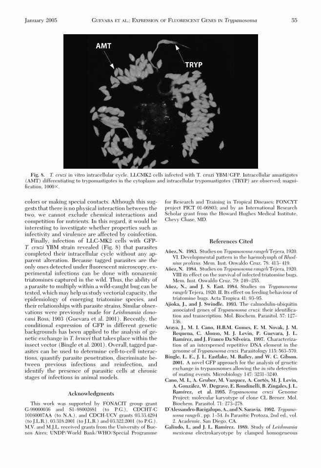
As in the case of single infections by T. rangeli, 1 wkafter the parasites reached the hemolymph, slenderGFP-ßagellates and GFP-metacyclic forms could beobserved inside the salivary glands (data not shown).Although green and red parasites were detected in theinsect hindgut, only T. cruzi red cells displayed thetypical trypomastigote infective forms (data notshown).T. cruzi In Vitro Intracellular Cycle. As shown in
Fig. 8, T. cruzi YBM-GFP trypomastigotes released
Fig. 4. Expression vector based on sequences in the cal-modulin/ubiquitin locus of T. cruzi. (A) Partial map of T. cruzicalmodulin/ubiquitin 2.65 locus. (B) Map of the pBS:CalB1/CUB01-EGFP expression construct. Intergenic untranslated se-quences in the 2.65 calmodulin/ubiquitin locus and in the ex-pression vector are indicated by dashed bars. Coding genes arerepresented by white boxes. Black bars with arrows indicateEGFPorDsRedgenesanddirectionoftranscription.Whitebarswith arrows show the position of the ampicillin and neomycinresistance genes. Transplicing site, ts.
Table 1. T. cruzi and T. rangeli analysis of transfected strains
Strain Expression vectorFluorescent
markerExpression
Fluorescencelevelsa
Chromosomallocation
T. cruzi CLBrener
pTREX-n GFP5(S65T) Stable 5.6 1.6 Mbp
T. cruzi JMP pTREX-n GFP5(S65T) Stable 14.2 1.1 MbpT. cruzi YBM pTREX-n GFP5(S65T) Stable 36.2 NDT. rangeli Triat-1 pTREX-n GFP5(S65T) Stable 16.2 0.915, 0945 Mbp and
extrachromosomalT. rangeli Dog-82 pTREX-n GFP5(S65T) Stable 10.8 NDT. cruzi CL
BrenerpBs:CalB1/CUB01 EGFP G418 required 0.7 2.7 Mbp and
extrachromosomalT. cruzi CL
BrenerpBs:CalB1/CUB01 DsRed 1.1 G418 required 0.8 2.7 Mbp and
extrachromosomalT. cruzi JMP pBs:CalB1/CUB01 EGFP G418 required 0.6 ExtrachromosomalT. cruzi JMP pBs:CalB1/CUB01 DsRed 1.1 G418 required 0.6 ExtrachromosomalT. cruzi YBM pBs:CalB1/CUB01 EGFP G418 required 0.7 ExtrachromosomalT. cruzi YBM pBs:CalB1/CUB01 DsRed 1.1 G418 required 0.9 ExtrachromosomalT. rangeli Triat-1 pBs:CalB1/CUB01 EGFP G418 required 3.3 ExtrachromosomalT. rangeli Triat-1 pBs:CalB1/CUB01 DsRed 1.1 G418 required 1.2 ExtrachromosomalT. rangeli Dog-82 pBs:CalB1/CUB01 EGFP G418 required 2.2 NDT. rangeli Dog-82 pBs:CalB1/CUB01 DsRed 1.1 G418 required 1.1 ND
ND, not determined.a Arbitrary units.
52 JOURNAL OF MEDICAL ENTOMOLOGY Vol. 42, no. 1

from LLC-MK2 cells and metacyclic trypomastigotesgrown inLITmediumcanef"ciently andcontinuouslyinfect tissue cultures in vitro. The ßuorescence couldbe seen in all intracellular developmental forms(amastigote and trypomastigotes), and in free meta-cyclic trypomastigotes. The GFP did not seem to betoxic to either the parasites or the cultured monkeycells.
Discussion
In the present work, we used theT. cruziRP to drivestable expression of green (GFP) and red (DsRed)ßuorescent proteins in different strains (and groups)of T. cruzi and T. rangeli. Although previous transientgene expression experiments driven by the RP cor-roborated the universality of group-2 T. cruzi se-quences (Nunes et al. 1997), the validity of these datafor establishing phylogenic relationships has beenquestioned (Laurent and Swindle 1999). Here, weused stable gene expression driven by a promoterderived from group-2 strain La Cruz (Martinez-Cal-villo 1998) and found that our results did not correlatewith species or rDNA group; indeed, we found largedifferences among T. cruzi strains (Table 1). For ex-ample, the CL Brener (group-1) strain showed poormarker expression, the JMP (group-2) strain and T.rangeli yielded intermediate marker expression, andthe YBM (group-2) strain showed the highest level ofmarker expression. We feel that the relatively highexpression of the marker in T. rangeli emphasized thephylogenetic proximity of the two species, and therobustnessof thegroup-2RP in this sortof experiment.
Similar results were observed for ubiquitin regula-tory sequences in the pBs:CalB1/CUB01 vector, al-
though in contrast to the pTREX-n-based constructs,integration was a rare event. This differential integra-tion of constructs is not easy to explain, because pre-vious reports indicated that RP-drivenT. cruzi expres-sion vectors rapidly integrated into the ribosomallocus even when transfected as circular plasmids(Martinez-Calvillo et al. 1997, Vazquez and Levin1999, Lorenzi et al. 2003). In addition, it is unclear howthe pTREX-n-constructs integrated into the T. rangeligenome, because this did not occur at the ribosomal orGADPH loci. In some strains (Fig. 5, lanes 8 and 9),integration was associated with a molecular weightincrease of some bands, which may indicate a multipletandem repeat insertion of pTREX-n GFP (Lorenzi etal. 2003).
In summary, the pTREX-n vector has several de-sirable features: ef"cient transcription and adequatetrans-splicing lead to high levels of expression, con-structs are rapidly and stably integrated, and the vec-tor is fairly universal. These properties allowed us toproduce stableT. cruziandT. rangeliGFP-tagged cells,which we used to follow the course of infection inR. prolixus. In single infections, both parasites com-pleted their expected developmental cycles(DÕAlessandro-Bacigalupo and Saravia 1992, Kollienand Schaub 2000) and were easy to visualize ßuores-cently. In bugs that were fed blood containing 2,000GFP-T. rangeli cells per milliliter, dividing epimastig-otes and round forms were concentrated in the in-sectÕs slender gut. At day 20 postinfection, a massivecrossing of GFP parasites toward the insect hemo-lymph occurred, and the high degree of pleiomor-phism of T. rangeli cells in culture was reduced to twoforms: elongated epimastigotes and ring-shaped intra-hemocyte cells. These results support those of Anez
Fig. 5. Genomic localization of GFP. (A) Gel of chromosomal bands from control and transfected T. rangeli cell lines.(B) Southern blot of A with GFP-speci"c probe. The arrows indicate the chromosomal bands identi"ed by the probe. Lanesare 1, Saccharomyces cerevisiaechromosomes; 2 and 3,T. rangeliTriat-1 wild type; 4 and 5,T. rangeliTriat-1 wild type % plasmidpTREXn-GFP5(S65T); 6 and 7, T. rangeliTriat-1 transfected with pTREXn-GFP5(S65T) grown with G418; 8 and 9,T. rangeliTriat-1 transfected with pTREXn-GFP5(S65T) present in hemolymph recovered fromR. prolixus; 10 and 11, T. rangeliTriat-1transfected with pTREXn-GFP5(S65T) grown without G418; and 12, Hansenula wingeiWickerham, 1950, chromosomes.
January 2005 GUEVARA ET AL.: EXPRESSION OF FLUORESCENT GENES IN Trypanosoma 53

(1983) by using nontagged parasites. A week later, theinsect salivary glands presented typical infective me-tacyclic forms and elongated epimastigotes. T. rangelicells were also observed at the insect rectum or infeces, but these were noninfective forms.
Single T. cruzi infections reproduced the develop-mental pattern of this parasite (data not shown). Dur-ing the mixed infection, parasites retained their de-
velopmental patterns. For example, no DsRed-T. cruzicells crossed the insectÕs gut epithelium, even whenlarge numbers of GFP-T. rangeli cells were activelydoing so. This evidence is consistent with the exis-tence of speci"c invasive mechanisms for T. rangeli,similar to those described for Plasmodium (Ghosh etal. 2001). Also, high numbers of T. cruzi and T. rangelicells were observed next to each other, without mixing
Fig. 6. GFP-tagged T. rangeli infections of R. prolixus. (A) T. rangeli-GFP epimastigotes in culture (used in arti"cialinfection); magni"cation, 400!. (B) Middle gut 7!19 d postinfection; magni"cation, 100!. (C and D) Hemolymph extra- andintracellular epimastigotes 19 d postinfection; magni"cation 100!, 400!. (E and F) Salivary glands (SG) 27 d postinfection;magni"cation 100!, insert 400!.
Fig. 7. T. cruzi-DsRed/T. rangeli-GFP mixed infections of R. prolixus. (A) 50:50 mix of T. cruzi-DsRed/T. rangeli-GFPcultured epimastigotes; magni"cation, 400!. (B and C) Mid gut mixed infection, 7!20 d; magni"cation, 400!. (D and E)T. rangeli in hemolymph at 21 d postinfection; magni"cation, 400!.
54 JOURNAL OF MEDICAL ENTOMOLOGY Vol. 42, no. 1
colors or making special contacts. Although this sug-gests that there is no physical interaction between thetwo, we cannot exclude chemical interactions andcompetition for nutrients. In this regard, it would beinteresting to investigate whether properties such asinfectivity and virulence are affected by coinfection.
Finally, infection of LLC-MK2 cells with GFP-T. cruzi YBM strain revealed (Fig. 8) that parasitescompleted their intracellular cycle without any ap-parent alteration. Because tagged parasites are theonly ones detected under ßuorescent microscopy, ex-perimental infections can be done with nonaxenictriatomines captured in the wild. Thus, the ability ofa parasite to multiply within a wild-caught bug can betested, which may help us study vectorial capacity, theepidemiology of emerging triatomine species, andtheir relationships with parasite strains. Similar obser-vations were previously made for Leishmania dono-vani Ross, 1903 (Guevara et al. 2001). Recently, theconditional expression of GFP in different geneticbackgrounds has been applied to the analysis of ge-netic exchange in T. brucei that takes place within theinsect vector (Bingle et al. 2001). Overall, tagged par-asites can be used to determine cell-to-cell interac-tions, quantify parasite penetration, discriminate be-tween previous infections and reinfection, andidentify the presence of parasitic cells at chronicstages of infections in animal models.
Acknowledgments
This work was supported by FONACIT group grantG-99000036 and S1!98002681 (to P.G.), CDCHT-C10160007AA (to N.A.), and CDCH-UCV grants 03.33.4294(to J.L.R.), 03.318.2001 (to J.L.R.) and 03.322.2001 (to P.G.).M.V. and M.J.L. received grants from the University of Bue-nos Aires; UNDP/World Bank/WHO/Special Programme
for Research and Training in Tropical Diseases; FONCYTproject PICT 01-06803; and by an International ResearchScholar grant from the Howard Hughes Medical Institute,Chevy Chase, MD.
References Cited
Anez, N. 1983. Studies onTrypanosoma rangeliTejera, 1920.VI. Developmental pattern in the haemolymph of Rhod-nius prolixus. Mem. Inst. Oswaldo Cruz. 78: 413!419.
Anez, N. 1984. Studies onTrypanosoma rangeliTejera, 1920.VIII its effect on the survival of infected triatomine bugs.Mem. Inst. Oswaldo Cruz. 79: 249!255.
Anez, N., and J. S. East. 1984. Studies on Trypanosomarangeli Tejera, 1920. II. Its effect on feeding behaviour oftriatomine bugs. Acta Tropica 41: 93!95.
Ajioka, J., and J. Swindle. 1993. The calmodulin-ubiquitinassociated genes of Trypanosoma cruzi: their identi"ca-tion and transcription. Mol. Biochem. Parasitol. 57: 127!136.
Araya, J., M. I. Cano, H.B.M. Gomes, E. M. Novak, J. M.Requena, C. Alonso, M. J. Levin, P. Guevara, J. L.Ramırez, and J. Franco Da Silveira. 1997. Characteriza-tion of an interspersed repetitive DNA element in thegenome of Trypanosoma cruzi. Parasitology 115: 563!570.
Bingle, L. E., J. L. Eastlake, M. Bailey, and W. C. Gibson.2001. A novel GFP approach for the analysis of geneticexchange in trypanosomes allowing the in situ detectionof mating events. Microbiology 147: 3231!3240.
Cano, M. I., A. Gruber, M. Vazquez, A. Cortes, M. J. Levin,A. Gonzalez,W.Degrave, E. Rondinelli, B. Zingales, J. L.Ramırez, et al. 1995. Trypanosoma cruzi GenomeProject: molecular karyotype of clone CL Brener. Mol.Biochem. Parasitol. 71: 273!278.
D’Alessandro-Bacigalupo,A., andN. Saravia. 1992. Trypano-soma rangeli., pp. 1!54. In Parasitic Protoza, 2nd ed., vol.2. Academic, San Diego, CA.
Galindo, I., and J. L. Ramırez. 1989. Study of Leishmaniamexicana electrokaryotype by clamped homogeneous
Fig. 8. T. cruzi in vitro intracellular cycle. LLCMK2 cells infected with T. cruzi YBM/GFP. Intracellular amastigotes(AMT) differentiating to trypomastigotes in the cytoplasm and intracellular trypomastigotes (TRYP) are observed; magni-"cation, 1000!.
January 2005 GUEVARA ET AL.: EXPRESSION OF FLUORESCENT GENES IN Trypanosoma 55
electric"eld electrophoresis. Mol. Biochem. Parasitol. 34:245!252.
Garcia, E., P. Azambuja, and V. T. Contreras. 1984. Large-scale rearing of Rhodnius prolixus and preparation ofmetacyclic trypomastigotes ofTrypanosoma cruzi,pp. 43!46. In C. Morel [ed.], Genes and antigens of parasites. Alaboratory manual, 2nd ed. UNPD/World bank/WHOspecial Programme for Research and training in TropicalDiseases, Rio de Janeiro, Brazil.
Ghosh, A. K., P. E. Ribolla, and M. Jacobs-Lorena. 2001.Targeting Plasmodium ligands on mosquito salivaryglands and midgut with a phage display peptide library.Proc. Natl. Acad. Sci. USA 98: 13278!13281.
Gonzalez, N., I. Galindo, P. Guevara, E. Novak, J. V. Scorza,N. Anez, J. F. Da Silveira, and J. L. Ramırez. 1994. Iden-ti"cation and detection of Trypanosoma cruzi by using aDNA ampli"cation "ngerprint obtained from the ribo-somal intergenic spacer. J. Clin. Microbiol. 32: 153!158.
Guevara, P., D. Pinto-Santini, A. Rojas, G. Crisante, N. Anez,and J. L. Ramırez. 2001. Green ßuorescent protein-tagged Leishmania in phlebotomine sand ßies. J. Med.Entomol. 38: 39!43.
Hannaert, V., M. Blaauw, L. Kohl, S. Allert, F. R. Opperdoes,and P. A. Michels. 1992. Molecular analysis of the cyto-solic and glycosomal glyceraldehyde-3-phosphatedehy-drogenase in Leishmania mexicana.Mol. Biochem. Para-sitol. 55: 115!126.
Hariharan, S., J. Ajioka, and J. Swindle. 1993. Stable trans-formation of Trypanosoma cruzi: inactivation of thePUB12.5 polyubiquitin gene by targeted disruption. Mol.Biochem. Parasitol. 57: 15!30.
Hoare, C. A. 1972. Herpetosoma from man and other mam-mals, pp. 288!314. In The trypanosomes of mammals: azoological monograph, Blackwell Scienti"c Publications,Oxford, England.
Kendall,G., A. F.Wilderspin, F. Ashall,M.A.Miles, and J.M.Kelly. 1990. Trypanosoma cruzi glycosomal glyceralde-hyde-3-phosphate dehydrogenase does not conform tothe ÔhotspotÕ topogenic signal model. EMBO J. 9: 2751!2758.
Kollien, A. H., and G. A. Schaub. 2000. The development ofTrypanosoma cruzi in triatominae. Parasitol. Today 16:381!387.
Laurent, J. P., and J. Swindle. 1999. Variation of transientgene expression within single lineages of Trypanosomacruzi. Parasitology 119: 583!589.
Lorenzi, H. A., M. P. Vazquez, and M. J. Levin. 2003. Inte-gration of expression vectors into the ribosomal locus ofTrypanosoma cruzi. Gene 310: 91!99.
Martinez-Calvillo, S. 1998. Analisis funcional de la secuen-cia promotora del gene de RNA ribosomal de Trypano-soma cruzi mediante tecnicas de transformacion molec-ular. Ph.D. dissertation, Universidad Autonoma deMexico, Mexico, DF.
Martinez-Calvillo, S., I. Lopez, and R. Hernandez. 1997.pRIBOTEX expression vector: a pTEX derivative for arapid selection of Trypanosoma cruzi transfectants. Gene199: 71!76.
Novak, E., M. De Mello, H.B.M. Gomes, I. Galindo, P. Gue-vara, J. L. Ramırez, and J. F. Da Silveira. 1993. Repeti-tive sequences in the ribosomal intergenic spacer ofTrypanosoma cruzi.Mol. Biochem. Parasitol. 60: 273!280.
Nunes, L. R., M. R. de Carvalho, and G. A. Buck. 1997.Trypanosoma cruzi strains partition into two groups basedon the structure and function of the spliced leader RNAand rRNA gene promoters. Mol. Biochem. Parasitol. 86:211!224.
Southward, C. M., and M. G. Surette. 2002. The dynamicmicrobe: green ßuorescent protein brings bacteria tolight. Mol. Microbiol. 45: 1191!1196.
Sambrook, J., E.F.Fritsch, andT.Maniatis. 1989. Molecularcloning: a laboratory manual. Cold Spring Harbor Labo-ratory Press, Cold Spring Harbor, NY.
Siemering, K. R., R. Golbik, R. Sever, and J. Haseloff. 1996.Mutations that suppress the thermosensitivity of greenßuorescent protein. Curr. Biol. 6: 1653!1663.
Souto, R. P., and B. Zingales. 1993. Sensitive detection andstrain classi"cation ofTrypanosoma cruziby ampli"cationof ribosomal RNA sequences. Mol. Biochem. Parasitol. 62:45!52.
Tobie, E. J. 1965. Biological factors inßuencing transmissionof Trypanosoma rangeli by Rhodnius prolixus. J. Parasitol.51: 837!841.
Vargas, N., R. P. Souto, J. C. Carranza, G. A. Vallejo, and B.Zingales. 2000. Ampli"cation of a speci"c repetitiveDNA sequences for Trypanosoma rangeli identi"cationand its potential application in epidemiological investi-gations. Exp. Parasitol. 96: 147!159.
Vazquez, M. P., and M. Levin. 1999. Functional analysis ofthe intergenic region of TcP2# gene loci allowed theconstruction of an improved Trypanosoma cruzi expres-sion vector. Gene 239: 217!225.
Watkins, R. 1971. Histology of Rhodnius prolixus infectedwith Trypanosoma rangeli. J. Invertebr. Pathol. 17: 59!66.
Received for publication 13 December 2002; accepted 13October 2003.
56 JOURNAL OF MEDICAL ENTOMOLOGY Vol. 42, no. 1