exploring printed drug formulations for inkjet and stencil
TRANSCRIPT
Henrika Wickström
Exploring Printed Drug Formulations for Inkjet and Stencil PrintingA study in Pharmaceutical Sciences
Henrika W
ickström
// Explo
ring Printed
Drug
Form
ulations fo
r Inkjet and Stencil Printing
// 20
20
Henrika Wickström
Exploring Printed Drug Formulations for Inkjet and Stencil Printing
A study in Pharmaceutical SciencesPharmaceutical ink formulation development for inkjet and stencil printing of oral solid dosage forms are described and discussed in this doctoral thesis.
ISBN 978-952-12-4008-9
9 789521 240089

Henrika WickströmBorn 1989 in Sibbo, Finland
The Ph.D. thesis project in Pharmaceutical Sciences was carried out as a joint supervision (co-tutelle) agreement between Åbo Akademi University and Ghent University during 2014-2020.
M.Sc. degree in Biosciences, 2014 B.Sc. degree in Pharmacy, 2011
Exploring printed drug formulations for inkjet and stencil printing
A study in Pharmaceutical Sciences
Henrika Wickström
Pharmaceutical Sciences Laboratory Faculty of Science and Engineering
Åbo Akademi University
Laboratory of Process Analytical Technology
Faculty of Pharmaceutical Sciences Ghent University
Åbo, Finland, 2020
Supervisors
Prof. Dr. Niklas O. Sandler Pharmaceutical Sciences Laboratory Faculty of Science and Engineering Åbo Akademi University Finland Prof. Dr. Thomas de Beer Laboratory of Process Analytical Technology Faculty of Pharmaceutical Sciences Ghent University Belgium
Opponent and Reviewer
Prof. Dr. Maike Windbergs Institute of Pharmaceutical Technology Buchmann Institute for Molecular Life Sciences Goethe University Frankfurt Germany
Reviewer
Prof. Dr. João Pinto Faculty of Pharmacy University of Lisbon Portugal ISBN (Print): 978-952-12-4008-9 ISBN (PDF): 978-952-12-4009-6 Painosalama Oy – Åbo, Finland, 2020
iii
The author and the promoters give the authorization to consult and to copy part of this thesis for personal use only. Any other use is limited by the Laws of Copyright, especially concerning the obligation to refer to the source whenever results are cited from this thesis.
Åbo, December 11th, 2020 The promoters The author Prof. Niklas Sandler Prof. Thomas de Beer Henrika Wickström
iv
Abstract (English)
Solid dosage forms are predominantly produced in large batches of a few and predetermined dose strengths. The current manufacturing methods do not enable high production flexibility and the product quality is assured mainly based on sampling. A need for novel manufacturing methods of medicines has arisen, which could enable manufacture of tailor-made doses according to personalized needs. To assure the quality of the product in-line/on-line quality controls should be implemented. This approach could offer decentralized production of medicines at the point of care.
The recent trend in the drug development is to focus on the patient. Up to 42% of the U.S. FDA approved medicines in 2018 were categorized as personalized medicines. These medicines are prescribed according to patent specific information such as gene or biomarker status. Patient-centered drug design focuses on the holistic treatment of an individual taking the specific needs and preferences of a patient or a patient group into account. Product-related characteristics considered in patient-centric drug product design are e.g. type of dosage form, dose to therapeutic effect, and product recognition.
Lately, the potential of utilizing printing technologies as manufacturing methods of pharmaceuticals has been studied. Printing technologies enable flexible deposits of materials according to manual or digital designs. This thesis aimed to explore drug formulations for inkjet and stencil printing technologies to manufacture flexible and tailored dosage forms, that could better meet patient needs. Pharmaceutical single and multicomponent inks were formulated and printed on edible paper or as orodispersible films to prepare flexible-dose strengths. Also, to address solubility challenges of poorly soluble drugs co-amorphous systems and mesoporous silica nanoparticle inks were formulated.
Liquid-based inkjet inks and semi-solid stencil inks were developed with favorable rheological properties for inkjet and stencil printing, respectively. Drug doses of a few micrograms to 1.1 mg were prepared by picolitre ink deposition by varying the printed layers and resolution. Dose strength flexibility was achieved for the stencil printed orodispersible films of 0.49 to 2.56 mg by varying the stencil area (i.e. aperture size of the print plate). The content uniformity of the prepared inkjet and stencil printed dosage forms were high. The obtained dose range was mainly dependent on the solubility of the active pharmaceutical ingredient in the ink solvents and on the absorptive properties of the edible substrate. Improved dissolution rates were achieved for the inkjet-printed formulations regardless of the amino acid addition, which would indicate that the drug was molecularly dispersed on the substrate or amorphous.
If printed drug doses are prepared on-demand and the dose is tuned according to the drug treatment response of an individual, a fast and non-destructive quality control method is needed to ensure the printed drug quality. Thus, an indirect dose quantification method based on color intensity measurements was evaluated for inkjet-printed dosage forms. The results show that different doses could be differentiated according to the measured color
v
intensities. However, the obtained dose intensities of print setting combinations (i.e. resolution and layers) should be validated with a direct dose quantification method, prior implementation as a quality control tool.
This thesis gives an overview of and insight into the potential of inkjet and stencil printing technologies as manufacturing methods of solid dosage forms. Important aspects worth considering during drug formulation development of liquid and semi-solid inks are discussed and utilizable features of the technologies for the manufacturing of flexible-dose strengths are pointed out.
vi
Abstrakt (Swedish)
I dag tillverkas de flesta fasta lakemedelsformer i stora satser och i endast nagra forutbestamda dosstyrkor. Detta innebar att produktionsflexibiliteten, med tanke pa doser och tillverkad mangd, ar snav. Lakemedelskvaliteten sakras pa basis av analyser gjorda pa en sampelmangd tagen fran den tillverkade satsen. Det har uppstatt ett behov av att framstalla lakemedel med nya metoder, som kunde sakerstalla tillverkning av skraddarsydda lakemedelsdoser enligt patientens behov. For att trygga kvaliteten av lakemedelsdoserna, som tillverkats decentraliserat, borde kvalitetskontrollen utgora en del av tillverkningsprocessen.
Den senaste trenden inom lakemedelsutveckling ar att patienten har fatt en mera central roll. Upp till 42% av de lakemedel som blivit beviljade forsaljningstillstand av den amerikanska lakemedelsmyndigheten FDA klassificerats som individualiserade lakemedel. Dessa lakemedel ordineras pa basis av individens geninformation eller biomarkorstatus. Patientcentrerad lakemedelsdesign fokuserar pa vard av individen fran ett helhetsmassigt vardperspektiv, vilket innebar att patientens specifika behov eller preferenser beaktas. Produktrelaterade egenskaper som tas i beaktande vid patientcentrerad lakemedelsdesign ar till exempel val av lakemedelsform, identifiering av produkt och sambandet mellan dos och terapeutisk effekt.
Utskriftsteknologiernas potential som tillverkningsmetod av lakemedel har studerats under den senaste aren. Teknologierna mojliggor flexibel utskrift av material enligt manuella eller digitala designs. Malsattningen med avhandlingen var att utforska lakemedelsformuleringar for tillverkning av fasta lakemedelsformer med hjalp av blackstrale- och schablonutskrifts-teknologier. Farmaceutiska uni- och multikomponentblack formulerades och skrevs ut pa atbart papper eller som munsonderfallande filmer, for att tillverka flexibla dosstyrkor. Framstallning av co-amorfa lakemedelsformuleringar med hjalp av utskriftteknologi utforskades, i forsok att forbattra biotillgangligheten av ett svart vattenlosligt lakemedel. AÄven framstallning av lakemedelsblack innehallande mesoporosa kiseldioxidnano-partiklar utforskades, eftersom de underlattar transporten av svart vattenlosliga lakemedel i kroppen.
Losningsbaserat black for blackstraleskrivare och halvfast black for schablonutskrivare med fordelaktiga reologiska egenskaper utvecklades for respektive utskriftteknologier. Lakemedelsdoser fran nagra mikrogram upp till 1.1 mg framstalldes genom deponering av picoliter droppar med blackstraleskrivare genom att variera antalet utskrivna lager och utskriftsresolution. Tillverkning av mun sonderfallande filmer med variation av dosstyrkan fran 0.49 till 2.56 mg uppnaddes genom att variera schablonarean. Dosstyrkan av de lakemedel som tillverkades med de tva olika utskriftsteknologierna var enhetlig. Lakemedlens loslighet och substratens absorptionsegenskaper korrelerade med de doser som kunde uppnas. Forbattrad upplosningshastighet uppnaddes for de blackstraleutskrivna doserna, vilket visade sig vara oberoende av aminosyra tillsatsen. Detta
vii
indikerade att lakemedlet i den framstallda dosformen var antingen molekylart fordelat pa substratet eller amorft.
Om lakemedelsdoser skulle tillverkas vid behov och om dosstyrkan skulle bestammas enligt individens respons till behandlingen, skulle snabba och icke-destruktiva metoder for kvalitetskontroll av de tillverkade doserna behovas. En indirekt doskvantifieringsmetod, som baserar sig pa bestamning av fargintensiteten, utvarderades for blackstraleutskriftsdoserna. Studien visade att bestamning av fargintensitet, kunde anvandas for att differentiera olika doser. Dos- och fargintensiteten och kombinationer av utskriftsinstallningar (till exempel resolution och utskrivna lager) borde ytterligare valideras av en direkt doskvantifieringsmetod, fore implementering som ett verktyg for kvalitetskontroll.
Denna avhandling presenterar och ger en inblick i blackstrale-och schablon utskriftsteknologiers potential som lakemedelstillverkningsmetoder. Viktiga aspekter vid formulering av de utskrivbara lakemedelslosningar och halvfasta material diskuteras och tillverkning av flexibla dosstyrkor genom att utnyttja utskriftmetodernas egenskaper redogors.
viii
Samenvatting (Dutch)
De meeste orale farmaceutische doseringsvormen worden tegenwoordig enkel in grote hoeveelheden vervaardigd en dit slechts in vooraf bepaalde doseringssterktes. De huidige fabricagemethodes zijn momenteel niet in staat om een hoge productieflexibiliteit en productkwaliteit te verschaffen aangezien ze voornamelijk gebaseerd zijn op de staalname van producten. Er is echter een behoefte aan innovatieve fabricagetechnieken die compatibel zijn met gepersonaliseerde geneeskunde en een in-line/on-line product kwaliteitscontrole. Deze aanpak kan een gedecentraliseerde productie-methode op de afgifteplaats faciliteren.
Er is een trend bezig in de geneesmiddelenontwikkeling die de patient centraal stelt. Reeds 42% van de geneesmiddelen die door het U.S. FDA voor de handel zijn goedgekeurd, zijn geclassificeerd als "gepersonaliseerde geneesmiddelen". Deze medicijnen worden voorgeschreven op basis van persoonsspecifieke informatie zoals genanalyze of biomarkerstatus. Een patientgerichte geneesmiddelenkeuze richt zich op het individu vanuit een holistisch zorgperspectief, waarbij rekening wordt gehouden met de specifieke behoeften of voorkeuren van een patient of patientengroep. De belangrijkste eigenschappen waarmee rekening gehouden moet worden bij een gepersonaliseerde geneesmiddelenkeuze zijn de geneesmiddelenvorm, de product herkenbaarheid en de relatie tussen de dosis en het therapeutisch effect.
Het potentieel van printtechnologieen bij de vervaardiging van geneesmiddelen is de afgelopen jaren uitgebreid onderzocht. Deze technologie maakt flexibele bedrukking van materialen mogelijk volgens een handmatig of digitaal vervaardigd ontwerp. Het doel van het proefschrift was om verschillende farmaceutische formulaties te onderzoeken voor de productie van vaste farmaceutische vormen met behulp van de inkjet- en sjabloon- printtechnologie. Enkelvoudige en meervoudige farmaceutische component-inkten werden geformuleerd en gedrukt op eetbaar papier of geformuleerd als orodispergeerbare film om flexibele doseringssterktes te vervaardigen. Verder werden bereidingen van co-amorfe geneesmiddel-formulaties met aminozuren onderzocht in een poging om de biologische beschikbaarheid van slecht-oplosbare medicijnen te verbeteren. Daarnaast werden inkten vervaardigd die mesoporeuze silica-nanodeeltjes bevatten. De nanodeeltjes werden geladen met slecht-oplosbare geneesmiddelen om de medicijnafgifte te verbeteren.
Vloeibare en half-vaste inkten met gunstige reologische eigenschappen werden ontwikkeld voor respectievelijk de inkjet- en sjabloon-printtechnologie. Medicijndosissen van een paar microgram tot 1.1 mg werden bereid door de het afzetten van picoliter druppels en een variatie van aantal afgedrukte lagen en de printresolutie. In het geval van sjabloon-printen werd het sjabloon oppervlakte (i.e. spleetgrootte van de printplaat) aangepast om orodispergeerbare films van 0.49 tot 2.56 mg te verkrijgen. De dosis uniformiteit van zowel de inkjet- als de sjabloon-geprinte dosisvormen waren uitstekend. Het potentieel dosisbereik was voornamelijk afhankelijk van de oplosbaarheid van het farmaceutische actief
ix
in het inkt-oplosmiddel en de absorptie-eigenschappen van het eetbaar substraat. Een verbeterde oplossnelheid werd bereikt voor inkjet-gedrukte dosissen, onafhankelijk van de toevoeging van aminozuren. Dit gaf aan dat het farmaceutisch actief moleculair gedispergeerd of amorf op het substraat aanwezig was.
Als geneesmiddelenvormen naar behoefte zouden worden geproduceerd en de dosissterkte zou kunnen worden bepaald op basis van individuele behandelingseisen dan is er een nood aan snelle en niet-destructieve methoden voor kwaliteitscontrole. Een indirecte dosis-kwantificatiemethode, die is gebaseerd op de bepaling van de kleurintensiteit, werd geevalueerd voor de inkjet-geprinte dosissen. Uit de studie bleek dat kleurintensiteit kan worden gebruikt om verschillende doses te differentieren waardoor het gebruikt kan worden als instrument voor de kwaliteitscontrole. Echter dient er een voorafgaande validatie te gebeuren tussen de dosis- en kleurintensiteit, gerelateerd aan de afdrukinstellingen (i.e. resolutie en aantal afgedrukte lagen), door middel van een directe doseringsbepalingsmethode.
Dit proefschrift presenteert het potentieel van inkjet- en sjabloon printtechnologieen als farmaceutische productiemethode. De belangrijke aspecten bij het formuleren van de printbare medicijnoplossingen en half-vaste inkten worden besproken en er wordt op de waardevolle eigenschappen van de flexibele dosissterkte bekwame printtechnologieen gewezen.

x
List of original publications
I. Wickström, H., Palo, M., Rijckaert, K., Kolakovic, R., Nyman, J.O., Määttänen, A., Ihalainen, P., Peltonen, J., Genina, N., de Beer, T., Löbmann, K., Rades, T. and Sandler, N. (2015). Improvement of dissolution rate of indomethacin by inkjet printing. European Journal of Pharmaceutical Sciences, 75, pp.91–100.
II. Wickström, H., Nyman, J.O., Indola, M., Sundelin, H., Kronberg, L., Preis,
M., Rantanen, J. and Sandler, N., (2017). Colorimetry as quality control tool for individual inkjet-printed pediatric formulations. AAPS PharmSciTech, 18(2), pp.293–302. Reprint license: 4862360902658
III. Wickström, H., Hilgert, E., Nyman, J.O., Desai, D., Şen Karaman, D., de
Beer, T., Sandler, N. and Rosenholm, J.M., (2017). Inkjet Printing of Drug-Loaded Mesoporous Silica Nanoparticles – A Platform for Drug Development. Molecules, 22(11), p.2020.
IV. Wickström, H., Koppolu, R., Mäkilä, E., Toivakka, M., & Sandler, N.
(2020). Stencil Printing – A Novel Manufacturing Platform for Orodispersible Discs. Pharmaceutics, 12(1), 33.
Contribution of Henrika Wickström to the original publications: I. Participated in the study design, performed a major part of the
experiments, data analysis & wrote the paper.
II. Data analysis & wrote the paper. III. Main role in the study design, performed a major part of the ink
development and characterization experiments, data analysis & wrote the paper.
IV. Main role in the study design, performed the experiments, data analysis &
wrote the paper.
xi
List of supporting publications
Varan, C., Wickström, H., Sandler, N., Aktaş, Y. and Bilensoy, E., (2017). Inkjet printing of antiviral PCL nanoparticles and anticancer cyclodextrin inclusion complexes on bioadhesive film for cervical administration. International Journal of Pharmaceutics. 531(2), 701–713. Vakili, H., Wickström, H., Desai, D., Preis, M., and Sandler, N., (2017). Application of a handheld NIR spectrometer in prediction of drug content in inkjet printed orodispersible formulations containing prednisolone and levothyroxine. International journal of pharmaceutics, 524(1), pp.414–423. Wickström, H., Anthoni, A., Palo, M., Nyman, J.O., Maattanen, A., Nurmi, M., Moritz, N., Oja, T., Preis, M. and Sandler, N., (2016) Application of antibacterial coatings on resin composite implant materials using inkjet printing technology. In NIP & Digital Fabrication Conference 2016(1)89–93. Society for Imaging Science and Technology. Fonteyne M, Wickström H, Peeters E, Vercruysse J, Ehlers H, Peters BH, Remon JP, Vervaet C, Ketolainen J, Sandler N, Rantanen J. (2014). Influence of raw material properties upon critical quality attributes of continuously produced granules and tablets. European Journal of Pharmaceutics and Biopharmaceutics, 87(2), 252–263.
xii
Abbreviations
a* Color component (green/red) ACN Acetonitrile b* Color component (blue/yellow) B1 Thiamine hydrochloride B2 Riboflavin 5′-monophosphate sodium salt B3 Nicotinamide B6 Pyridoxine hydrochloride BioRAM Biopharmaceutics risk assessment roadmap CAD Computer-aided design CIJ Continuous inkjet CSLM Confocal scanning laser microscopy CoPVP Crospovidone CPP Critical process parameters CQA Critical quality attributes DMSO Dimethyl sulfoxide DoD Drop-on-demand inkjet DCS Developability Classification System DS Docusate sodium salt DSC Differential scanning calorimetry dpi Droplets per inch ΔE*ab Numerical color illumination difference between sample and ref. EHD Electrohydrodynamic EtOH Ethanol EXT Semi-solid extrusion FA Formic acid FDA U.S. Food and Drug Administration FDM Fused deposition modeling FT-IR Fourier transform infrared spectrometer FUR Furosemide ICH International Council for Harmonization of Technical
Requirements for Pharmaceuticals for Human Use IMC Indomethacin IPA Isopropanol HMDSO Hexamethyldisiloxane HPC Hydroxypropyl cellulose HPLC High-performance liquid chromatography HPMC Hydroxypropyl methylcellulose L* Lightness component LA Lactic acid L-Arg L-Arginine LC-MS Liquid chromatography-mass spectrometry MeOH Methanol
xiii
MLS Multiple light scattering MSNs Mesoporous silica nanoparticles NAP Naproxen ODF Orodispersible film PAT Process analytical technology PCL Polycaprolactone PCL-LPS Acid-modified polycaprolactone PEI Polyethyleneimine PEO Polyethylene oxide PIJ Piezoelectric inkjet printer PG Propylene glycol Ph.Eur. European Pharmacopeia PLM Polarized light microscopy PMI Precision medicine initiative PTFE Poly(tetrafluoroethylene) PVP Polyvinylpyrrolidone QbD Quality by Design QTPP Quality Target Product Profile SCMC Carboxymethylcellulose sodium salt SEM Scanning electron microscopy SFF Solid free-form fabrication SLA Stereolithography SLS Selective laser sintering SOFT Structured orodispersible template substrates SWLI Scanning white-light interferometry UV Ultraviolet TBZ Tetrabenazine TFA Trifluoroacetic acid TIJ Thermal inkjet printer XRD X-ray Powder Diffraction 0D Zero dimensional 1D One dimensional 2D Two dimensional 3D Three dimensional 4D Four dimensional
xiv
Table of contents
Abstract (English) ......................................................................................................................... iv
Abstrakt (Swedish) ....................................................................................................................... vi
Samenvatting (Dutch) ............................................................................................................... viii
List of original publications ........................................................................................................ x
List of supporting publications ................................................................................................. xi
Abbreviations ................................................................................................................................. xii
Table of contents ......................................................................................................................... xiii
1. Introduction .................................................................................................................................. 1
2. Literature overview ................................................................................................................... 2
2.1 Pharmaceutical development ........................................................................................ 2
2.2 Personalized medicine and person-centered care ................................................ 3
2.2.1 Patient-centric dosage forms ................................................................................. 3
2.3 Coating and printing processes ..................................................................................... 4
2.3.1 Coating ............................................................................................................................ 4
2.3.1.1 Solvent casting ......................................................................................................... 5
2.3.2 Conventional printing methods ............................................................................ 5
2.3.2.1 Stencil and screen printing ................................................................................. 6
2.3.2.2 Flexography ............................................................................................................... 7
2.3.3 Digital printing technologies.................................................................................. 7
2.3.3.1 Inkjet printing .......................................................................................................... 9
2.4 Utilization of printing technologies in the field of pharmaceutics .............. 11
2.4.1 Printing – from labeling to manufacturing of pharmaceuticals ........... 11
xv
2.4.2 Printing of pharmaceuticals................................................................................ 11
2.5 Design and manufacture of dosage forms by inkjet printing ........................ 13
2.5.1 Pharmaceutical formulations for inkjet printing ....................................... 13
2.5.2 Substrates ................................................................................................................... 17
2.6 Design and manufacture of dosage forms using casting and conventional printing methods ..................................................................................................................... 18
2.6.1 Pharmaceutical formulations for casting & flexographic printing ..... 19
2.7 Quality of printed dosage forms ................................................................................ 19
2.7.1 Inkjet print quality .................................................................................................. 20
2.7.2 Stencil print quality ................................................................................................ 20
2.7.3 Solid-state forms ..................................................................................................... 21
2.7.4 Regulatory perspectives of printed dosage forms..................................... 21
2.8 Quality control methods of printed dosage forms ............................................. 22
2.8.1 Colorimetric method .............................................................................................. 22
3. Aims of the study ..................................................................................................................... 24
4. Materials and Method ........................................................................................................... 25
4.1 Materials .............................................................................................................................. 25
4.1.1 Active pharmaceutical ingredients .................................................................. 25
4.1.2 Substrates ................................................................................................................... 25
4.2 Methods ............................................................................................................................... 26
4.2.1 Preparation of pharmaceutical inks for inkjet printing .......................... 26
4.2.1.1 Indomethacin ink formulations (I)............................................................... 26
4.2.1.2 Vitamin B ink formulation (II) ....................................................................... 26
4.2.1.3 MSN ink formulations (III) .............................................................................. 26
xvi
4.2.1.4 T4 ink formulation............................................................................................... 27
4.2.2 Preparation of pharmaceutical inks for stencil printing ......................... 27
4.2.2.1 Haloperidol ink formulations (IV) ................................................................ 27
4.2.3 Viscosity (I, III, IV) ................................................................................................... 27
4.2.4 Surface tension (I, III) ............................................................................................ 27
4.2.5 Contact angle (III) .................................................................................................... 28
4.2.6 Multiple light scattering (MLS) (III) ................................................................. 28
4.2.7 Printing technology ................................................................................................ 28
4.2.7.1 Piezoelectric inkjet printing (I, III) ............................................................... 28
4.2.7.2 Thermal inkjet printing (II) ............................................................................. 28
4.2.7.3 Stencil printing (IV) ............................................................................................ 29
4.2.8 Content uniformity.................................................................................................. 29
4.2.8.1 UV-Vis Spectrophotometry (I, III) ................................................................. 29
4.2.8.2 Liquid chromatography-mass spectrometry (LC-MS) (II) .................. 29
4.2.8.3 High-performance liquid chromatography (HPLC) (IV) ...................... 30
4.2.9 Drug release (I, III) .................................................................................................. 30
4.2.10 Colorimetry (II)...................................................................................................... 31
4.2.11 Substrate thickness (II) ...................................................................................... 31
4.2.12 Scanning white-light interferometry (SWLI) (II, III) .............................. 31
4.2.13 Confocal scanning laser microscopy (CSLM) (III) ................................... 31
4.2.14 Optical microscopy (III) ..................................................................................... 32
4.2.15 pH (IV) ....................................................................................................................... 32
4.2.16 Disintegration (IV)................................................................................................ 32
4.2.17 Infrared Spectroscopy (I, IV) ............................................................................ 32
xvii
4.2.18 Scanning electron microscopy (SEM) (I, III) ............................................. 32
4.2.19 Polarized light microscopy (PLM) (IV) ........................................................ 33
4.2.20 Differential scanning calorimetry (DSC) (IV) ........................................... 33
4.2.21 X-ray Powder Diffraction (XRD) (IV) ........................................................... 33
4.2.22 Color stability ......................................................................................................... 33
5. Results and Discussion ......................................................................................................... 34
5.1 Formulation and characterization of pharmaceutical inks ............................ 34
5.1.1 APIs used for printing ........................................................................................... 34
5.1.2 Ink excipients ............................................................................................................ 35
5.1.3 Ink formulation development of poorly soluble drugs ........................... 37
5.2 Characterization of substrates and ink-substrate interactions .................... 39
5.3 Characterization of printed dosage forms............................................................. 41
5.3.1 Dosing flexibility and accuracy .......................................................................... 41
5.3.2 Solid state characterization ................................................................................ 43
5.3.3 Dissolution ................................................................................................................. 45
5.4 Analytical methods for the analysis of printed dosage forms ....................... 46
5.4.1 Colorimetry ................................................................................................................ 46
5.4.2 Drug and color stability ........................................................................................ 47
6. Conclusion .................................................................................................................................. 49
7. Future perspectives ............................................................................................................... 51
8. Acknowledgements ................................................................................................................ 52
9. References .................................................................................................................................. 54
Original publications .................................................................................................................. 65
Curriculum Vitae ........................................................................................................................ 123


1
1. Introduction
Solid dosage forms are predominantly produced in large volumes and in batches of a few predetermined dose strengths, which are chosen based on population-level information (Govender et al., 2020). There is a lack of patient-centered dosage forms, that would enable tailoring of the dose or formulation according to the patient’s needs. Characteristics such as patient’s age, weight, body surface, gender, genetic profile, or treatment response could be important to consider if a patient is treated with e.g. an active pharmaceutical ingredient (API) with a narrow therapeutic window or a varying pharmacokinetic or pharmacodynamic profile. Dosing according to a patient's needs is also a way to minimize adverse drug reactions (Cohen, 1999). Due to the lack of age-appropriate products on the market, compounded products are usually prescribed to the pediatric population (Van Riet-Nales et al., 2017). The quality of these products is often poorer due to the lack of quality standards for compounded products.
During the last decade, 2D and 3D printing technologies have emerged as promising manufacturing methods of pharmaceuticals. State-of-the-art technologies enable the deposition of liquid, semi-solid, or solid based pharmaceutical ink formulations according to predefined patterns or designs.
Inkjet printing is a non-contact method that enables controlled deposition of small (pl) drops onto a substrate according to a predefined digital design. The technology has been suggested to enable production of tailored dosing strengths of small dose APIs as orodispersible dosage forms. Stencil printing, on the other hand, is a contact method enabling deposition of semi-solid inks according to the stencil pattern and its applicability for the manufacturing of pharmaceuticals has not been studied before.
Alongside the exploration of printing technologies as potential manufacturing methods of pharmaceuticals, printable pharmaceutical formulations need to be developed. Since many of the APIs are poorly soluble and permeable, different formulation strategies need to be adopted to ensure the bioavailability of the API.
The purpose of this research was to formulate and characterize pharmaceutical liquid and semi-solid inks for preparation of inkjet and stencil printed solid dosage forms, that would be suitable for pediatrics and elderly with swallowing issues. Formulations of APIs with different physicochemical properties were developed by adapting different strategies to improve the bioavailability. Furthermore, the applicability of the technologies was studied in terms of the manufacturing of age-appropriate doses within specific dose ranges. On-demand colorimetric analysis was evaluated as a quality control method of the printed dose strengths.
2
2. Literature overview
2.1 Pharmaceutical development Pharmaceutical development aims to design a product and to develop a robust manufacturing process, which enables the production of qualitative pharmaceuticals (EMA, 2017b). The production process should preferably be built and controlled based on risk management and knowledge gained from the product and process development, ensuring that the produced drug product fulfills the specifications and manufacturing controls. Thus, the quality of the produced drug product today should be built in by design (Quality by Design, QbD) and not be achieved by testing.
The U.S. Food and Drug Administration (FDA) based initiative of QbD advice to determine a quality target product profile (QTPP), which “forms the basis of design for the development of the product” (EMA, 2017b). This means that the route of administration, dosage form, and the delivery system is determined. Also, the attributes affecting the pharmacokinetic characteristics (e.g. dissolution) as well as drug product quality criteria (e.g. sterility, stability, drug release) are defined. Critical quality attributes (CQA) of e.g. the materials used in the formulation as well as critical process parameters (CPP) of the manufacturing process are studied and the most optimal parameters are chosen. Process Analytical Technology (PAT) is another initiative that encourages to design, analyze, and control manufacturing processes (Simon et al., 2015). The increased process understanding and control are gained from the implementation of process analyzers (e.g. sensors and probes) into manufacturing lines. The identification and control of the CQA and CPP by timely measurements lead to an increase in product quality (Process Analytical Technology, PAT) (FDA, 2004). Other favorable approaches to utilize to aid formulation development are the Design of Experiments (DoE), In vitro-in vivo correlation (IVIVC) Biopharmaceutical Classification System (BCS), and Developability Classification System (DCS) (Butler and Dressman, 2010; Davanço, Campos and Carvalho, 2020). According to the BCS, the APIs are divided into different classes (I-IV) depending on their solubility and permeability (Amidon et al., 1995). The system is used in formulation development to predict the in vivo behavior of oral formulations and indicate (Davanço, Campos and Carvalho, 2020). However, the system should be used with caution, since physiological differences between patient groups might impact the solubility and permeability of the API in the individual. Consequently, the development of a pediatric BCS has been suggested to better suit the population characteristics to aid pediatric drug formulation development and treatment of the pediatrics (Abdel-Rahman et al., 2012; Batchelor, 2014). Also, the BCS was used as a starting point for the development of the DCS for oral drugs (Butler and Dressman, 2010). The DSC focuses on the development of the drug and takes the intestinal solubility, permeability, and particle size of the drug into consideration. Therapy-driven formulation
3
approaches (i.e. BioRAM), based on biopharmaceutics to optimize clinical drug product performance, have also been studied (Selen et al., 2014).
2.2 Personalized medicine and person-centered care Personalized medicine can be defined as “tailoring of medical treatment to the individual characteristics, needs and preferences of each patient“ (HHS and FDA, 2013). The development of the individualized healthcare concept has resulted in the use of the terms personalized medicine and patient-centered care (El-Alti, Sandman and Munthe, 2019). The terms can be distinguished from their origins: personalized medicine is based on biomedical information and diagnostics and person-centered care is based on the holistic treatment of an individual from a caring perspective.
Personalization of the treatment, as a concept, has already been implemented in the healthcare sector (Collins, 2015). Before blood transfusions, the patient’s blood group is determined. Determination of genes has also proven to be useful and has proven to indicate the drug response in the treatment of breast cancer (e.g. HER2) (Hamburg and Collins, 2010). Even though personalization has been adopted, it has not yet acquired significance throughout the health care sector.
Novel manufacturing solutions are still needed to enable the production of personalized dosage forms and tailored dose strengths, alongside the development of diagnostics tools and analytical devices to support personalized drug therapy (Gubala et al., 2012; Rantanen and Khinast, 2015). For instance, organ development and enzyme composition vary notably during the growth and maturing of a child and these factors have an impact on the administration, distribution, metabolism, and excretion (pharmacokinetics) as well as on the receptor and organ interaction (pharmacodynamics) of a compound (Kearns et al., 2003; Van Riet-Nales et al., 2017). Likewise, changes in the renal and hepatic function, gastric emptying, and pH occur in the body as adults age (Page, Coupe, and Barrett, 2016). It was recently highlighted that new dosage forms appropriate for children of different ages and stages of development are still needed (Rautamo et al., 2020). Also, it is important to bear in mind that a single dosage form does not answer the specific needs of every patient or population group (Sam et al., 2012). Also, a strategic reflection written by the regulatory body of medicine in the European Union for 2025, emphasizes the need of integrating science and technology in the development of pharmaceuticals to better answer patients’ needs (EMA, 2019a).
2.2.1 Patient-centric dosage forms
To facilitate the administration and delivery of the API to the site of action in a safe, efficient, reproducible, and convenient way, various dosage forms have been developed. A patient-centric dosage form offers e.g. dosing flexibility and ease of administration for the target population group without the need for formulation modification (Page, Coupe, and Barrett, 2016; Rautamo et al., 2020). However, it
4
is important to note that the dose preferences should be balanced among the needs of the patient, caregiver, prescriber, and payer. Currently, drug administration challenges exist, and they can be divided into dosage form-related or patient-related challenges (Stegemann et al., 2016; Rautamo et al., 2020).
Liquid preparations such as solutions and suspensions are flexible to administer in various doses based on e.g. weight or surface area. Liquid preparations are also easy to administer to newborns and people suffering from dysphagia if the volumes needed are low (Sam et al., 2012). The formulations should preferably be dosed using syringes, rather than spoons or cups, to ensure accurate dose administration and avoid dosing errors (Walsh et al., 2020). The main disadvantage of liquid preparations is the limited opportunity to modify the drug release (Sam et al., 2012).
Powders and granules also enable dosing flexibility and are more stable compared to liquid formulations (Sam et al., 2012). However, if the powders are produced by compounding, dose variations can occur if a tablet is crushed and weighed into a smaller dose (Liu et al., 2014). Medication errors can also be introduced if the powders are prepared in dose sachets and part of the powder is left in the container. If a controlled or extended-release tablet is compounded crushing or splitting most probably will impact the release kinetics of the API.
The size and shape of the solid dosage form are important to match the population group to be treated. Both mini tablets and orodispersible dosage forms (tablets/films) are considered as suitable dosage forms for pediatrics and elderly (Liu et al., 2014; Orlu et al., 2017). The minitablets (≤ 3 mm) can i.e. be administered to neonates older than 6 months. It is a single- or multi-unit oral dosage form enabling dosing flexibility as one or several tablets can be administered simultaneously (Aleksovski et al., 2015; Mitra et al., 2017). Regardless of the possibilities, appropriate dispensing devices are still needed for the handling/counting of the minitablets. Orodispersible films are single- or multiple-layer thin polymer sheets (Preis et al., 2013). The advantage with orodispersible formulations is the rapid disintegration in the oral cavity after which the dosage form is swallowed with saliva to the gastrointestinal tract. Dosing flexibility is achieved by varying the cut area of the ODF (Visser et al., 2015). The acceptability of ODFs (2 x 3cm) was recently studied among pediatrics from a couple of days to one year old and concluded to be superior over syrups (0.5- 3 ml) (Klingmann et al., 2020).
2.3 Coating and printing processes
2.3.1 Coating
The transfer of a liquid-based material onto a web to form a layer is called coating (Bishop, 2015). The viscosity of the coated material is of great importance and can be controlled by selecting low or high molecular weight polymers to be dissolved in the carrier fluid. Solvent casting is an example of a zero dimensional
5
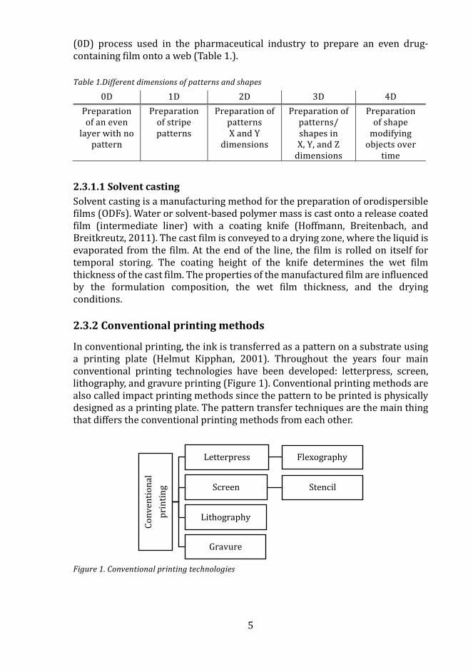
(0D) process used in the pharmaceutical industry to prepare an even drug-containing film onto a web (Table 1.).
Table 1.Different dimensions of patterns and shapes
0D 1D 2D 3D 4D Preparation of an even
layer with no pattern
Preparation of stripe patterns
Preparation of patterns X and Y
dimensions
Preparation of patterns/ shapes in X, Y, and Z
dimensions
Preparation of shape
modifying objects over
time
2.3.1.1 Solvent casting Solvent casting is a manufacturing method for the preparation of orodispersible films (ODFs). Water or solvent-based polymer mass is cast onto a release coated film (intermediate liner) with a coating knife (Hoffmann, Breitenbach, and Breitkreutz, 2011). The cast film is conveyed to a drying zone, where the liquid is evaporated from the film. At the end of the line, the film is rolled on itself for temporal storing. The coating height of the knife determines the wet film thickness of the cast film. The properties of the manufactured film are influenced by the formulation composition, the wet film thickness, and the drying conditions.
2.3.2 Conventional printing methods
In conventional printing, the ink is transferred as a pattern on a substrate using a printing plate (Helmut Kipphan, 2001). Throughout the years four main conventional printing technologies have been developed: letterpress, screen, lithography, and gravure printing (Figure 1). Conventional printing methods are also called impact printing methods since the pattern to be printed is physically designed as a printing plate. The pattern transfer techniques are the main thing that differs the conventional printing methods from each other.
Figure 1. Conventional printing technologies
Conv
entio
nal
prin
ting
Letterpress Flexography
Screen Stencil
Lithography
Gravure
6
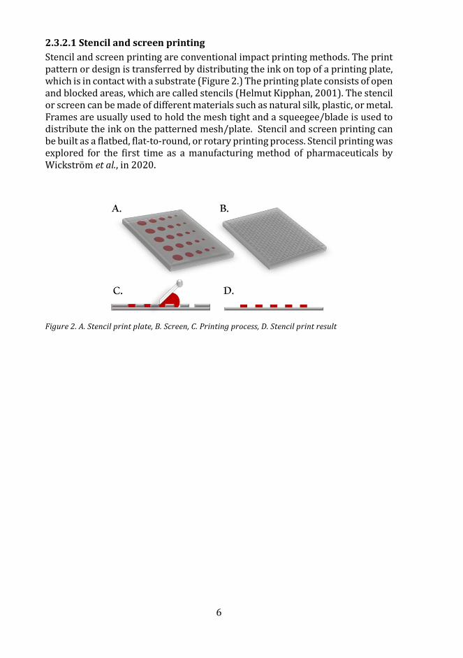
2.3.2.1 Stencil and screen printing Stencil and screen printing are conventional impact printing methods. The print pattern or design is transferred by distributing the ink on top of a printing plate, which is in contact with a substrate (Figure 2.) The printing plate consists of open and blocked areas, which are called stencils (Helmut Kipphan, 2001). The stencil or screen can be made of different materials such as natural silk, plastic, or metal. Frames are usually used to hold the mesh tight and a squeegee/blade is used to distribute the ink on the patterned mesh/plate. Stencil and screen printing can be built as a flatbed, flat-to-round, or rotary printing process. Stencil printing was explored for the first time as a manufacturing method of pharmaceuticals by Wickstrom et al., in 2020.
Figure 2. A. Stencil print plate, B. Screen, C. Printing process, D. Stencil print result
7
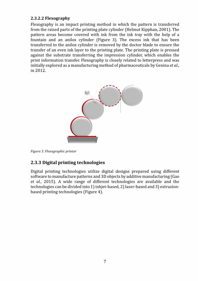
2.3.2.2 Flexography Flexography is an impact printing method in which the pattern is transferred from the raised parts of the printing plate cylinder (Helmut Kipphan, 2001). The pattern areas become covered with ink from the ink tray with the help of a fountain and an anilox cylinder (Figure 3). The excess ink that has been transferred to the anilox cylinder is removed by the doctor blade to ensure the transfer of an even ink layer to the printing plate. The printing plate is pressed against the substrate transferring the impression cylinder, which enables the print information transfer. Flexography is closely related to letterpress and was initially explored as a manufacturing method of pharmaceuticals by Genina et al., in 2012.
Figure 3. Flexographic printer
2.3.3 Digital printing technologies
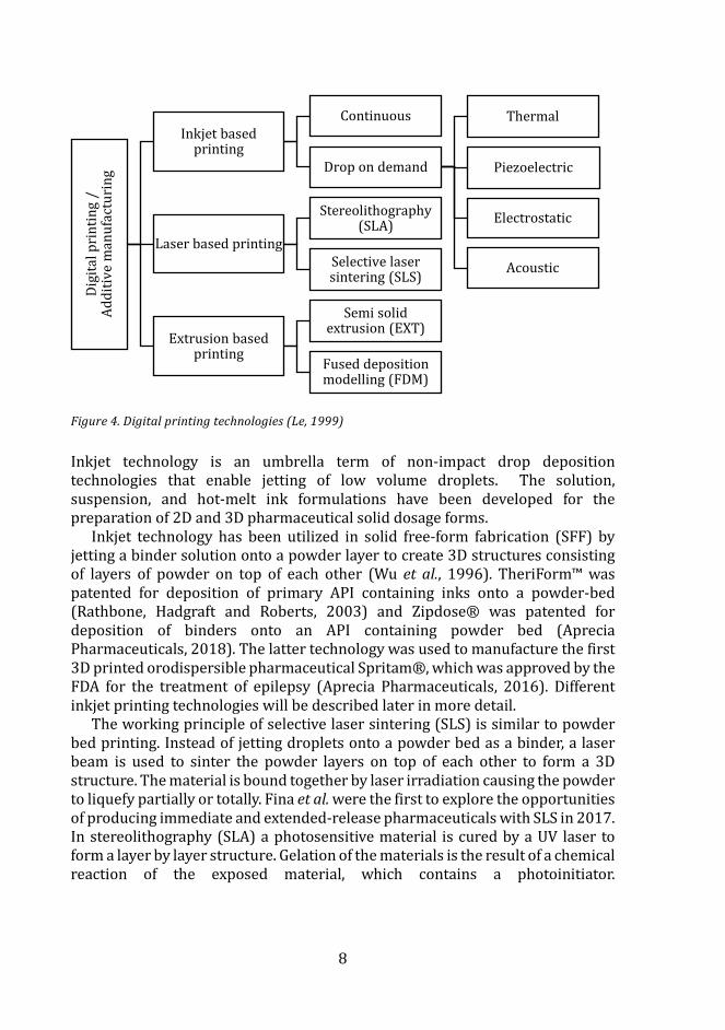
Digital printing technologies utilize digital designs prepared using different software to manufacture patterns and 3D objects by additive manufacturing (Gao et al., 2015). A wide range of different technologies are available and the technologies can be divided into 1) inkjet-based, 2) laser-based and 3) extrusion-based printing technologies (Figure 4).
8
Figure 4. Digital printing technologies (Le, 1999)
Inkjet technology is an umbrella term of non-impact drop deposition technologies that enable jetting of low volume droplets. The solution, suspension, and hot-melt ink formulations have been developed for the preparation of 2D and 3D pharmaceutical solid dosage forms.
Inkjet technology has been utilized in solid free-form fabrication (SFF) by jetting a binder solution onto a powder layer to create 3D structures consisting of layers of powder on top of each other (Wu et al., 1996). TheriForm™ was patented for deposition of primary API containing inks onto a powder-bed (Rathbone, Hadgraft and Roberts, 2003) and Zipdose® was patented for deposition of binders onto an API containing powder bed (Aprecia Pharmaceuticals, 2018). The latter technology was used to manufacture the first 3D printed orodispersible pharmaceutical Spritam®, which was approved by the FDA for the treatment of epilepsy (Aprecia Pharmaceuticals, 2016). Different inkjet printing technologies will be described later in more detail.
The working principle of selective laser sintering (SLS) is similar to powder bed printing. Instead of jetting droplets onto a powder bed as a binder, a laser beam is used to sinter the powder layers on top of each other to form a 3D structure. The material is bound together by laser irradiation causing the powder to liquefy partially or totally. Fina et al. were the first to explore the opportunities of producing immediate and extended-release pharmaceuticals with SLS in 2017. In stereolithography (SLA) a photosensitive material is cured by a UV laser to form a layer by layer structure. Gelation of the materials is the result of a chemical reaction of the exposed material, which contains a photoinitiator.
Digi
tal p
rint
ing
/
Addi
tive
man
ufac
turi
ng
Inkjet based printing
Continuous
Drop on demand
Thermal
Piezoelectric
Electrostatic
AcousticLaser based printing
Stereolithography (SLA)
Selective laser sintering (SLS)
Extrusion based printing
Semi solid extrusion (EXT)
Fused deposition modelling (FDM)
9
Pharmaceuticals with modified-release were manufactured using SLS by Wang et al. in 2016.
In extrusion-based printing, a semi-solid (EXT) gel or paste is extruded through a nozzle of a pressurized system at room temperature in a layer by layer manner to form a 3D structure (Khaled et al., 2014). The technology has also been called as pressure-assisted microsyringe printing (PAM). Extrusion-based printing of solid hotmelt formulations consisting of a thermoplastic polymer filament called fuse deposition modeling (FDM) has also been explored as a manufacturing method of pharmaceuticals (Goyanes et al., 2014). The polymer filament is heated above its softening point and extruded from a nozzle in a layer by layer print design resulting in immediate solidification after printing. Preparation of 4D printed pharmaceuticals by hotmelt extrusion and FDM was reported by Melocchi et al. in 2019. In 4D pharmaceuticals, time is the fourth dimension, which means that the extruded or the 3D printed pharmaceutical exhibits shape recovery over time.
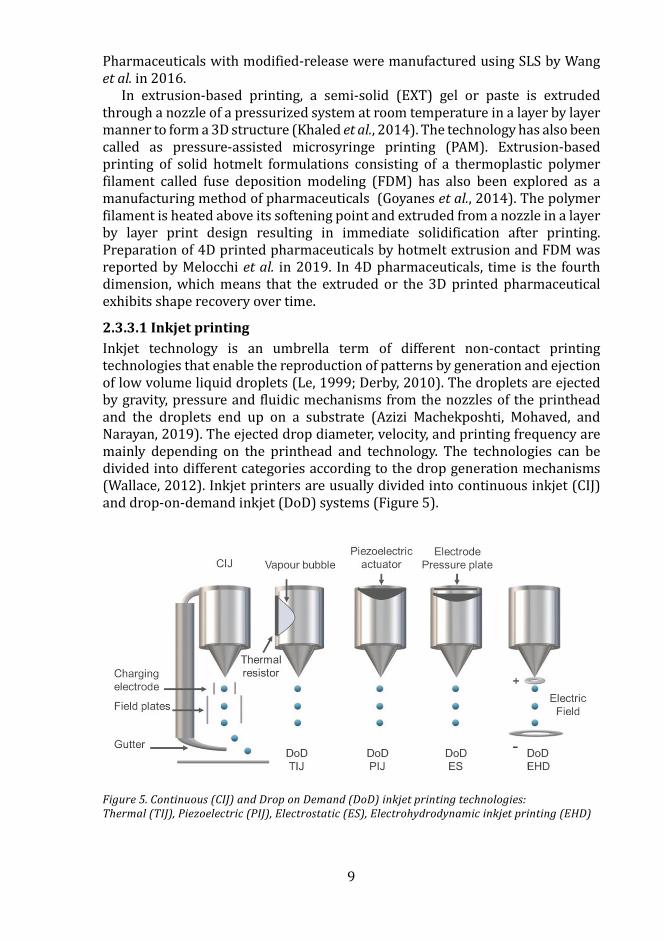
2.3.3.1 Inkjet printing Inkjet technology is an umbrella term of different non-contact printing technologies that enable the reproduction of patterns by generation and ejection of low volume liquid droplets (Le, 1999; Derby, 2010). The droplets are ejected by gravity, pressure and fluidic mechanisms from the nozzles of the printhead and the droplets end up on a substrate (Azizi Machekposhti, Mohaved, and Narayan, 2019). The ejected drop diameter, velocity, and printing frequency are mainly depending on the printhead and technology. The technologies can be divided into different categories according to the drop generation mechanisms (Wallace, 2012). Inkjet printers are usually divided into continuous inkjet (CIJ) and drop-on-demand inkjet (DoD) systems (Figure 5).
Figure 5. Continuous (CIJ) and Drop on Demand (DoD) inkjet printing technologies: Thermal (TIJ), Piezoelectric (PIJ), Electrostatic (ES), Electrohydrodynamic inkjet printing (EHD)
10
In the CIJ systems, the ink is continuously jetted from the printhead regardless of the activity of the printer (Derby, 2010). The pressurized system forces the ink out through the nozzle. Charged and uncharged droplets are formed from the continuous stream (binary deflection). The charged droplets are deflected by an electric field (field plates), which directs the ink to a gutter for recirculation, whereas the uncharged droplets end up at the substrate and form the printed pattern.
DoD inkjet printers generate droplets from a nozzle of a fluid-filled pathway as a response to a pressure pulse on demand (Jang, Kim and Moon, 2009). Droplets are formed as the surface tension threshold of the fluid at the nozzle is reached by a pressure pulse. The printing methods can be divided based on how the drop is generated. The most common are thermal (bubblejet) and piezoelectric technologies, which typically have nozzle diameters of 10-50 µm, resulting in 1-70 pl drop volumes (Jang, Kim, and Moon, 2009; Daly et al., 2015). DoD electrohydrodynamic (EHD), electromagnetic and micro-valve (valve jet) printing have also been studied (Elele et al., 2012; Bonhoeffer, Kwade, and Juhnke, 2017; Kollamaram, Faucher, et al., 2018; Zou et al., 2019).
Thermal inkjet (TIJ) printers have thin thermal resistors, which are activated and heated to 200-300°C as a response to a current (Le, 1999; Derby, 2010; Azizi Machekposhti, Mohaved, and Narayan, 2019). The heating lasts for only a few µs, which has been seen to slightly elevate the temperature (~4 -10°C) of the printed ink. The expansion and collapse of bubbles caused by the vaporization of the ink generate the pressure pulse, which forces droplets to be ejected from the nozzle of the thermal inkjet printer.
Piezoelectric inkjet (PIJ) printers have piezoelectric actuators, which deform as a response to a current. The deformation causes a pressure pulse, which reduces the space in the jet chamber forcing a drop to be ejected from the nozzle. The piezoelectric printheads can be built in different modes e.g. bend or shear.
Electrostatic (ES) inkjet printers work similarly to the piezoelectric printers, by increasing and reducing the space in the ink chamber. The print head contains a pressure plate, which deflects as a voltage pulse is formed between the plate and an electrode, which gives rise to droplet ejection (Kamisukil et al., 1998).
Electrohydrodynamic inkjet (EHD) printers work by electric field-induced ink flow. The electric field is obtained by constructing a high voltage between the nozzle and the substrate, which leads to droplet ejection from the nozzle (Elele et al., 2012; Gudapati, Dey, and Ozbolat, 2016). These printers enable printing of high viscous inks and droplet volumes of 0.2-2 µl.
Valve-based micro-dispensing enables the generation of drops from picoliter to nanoliter volume ranges (Planchette et al., 2016; Bonhoeffer, Kwade, and Juhnke, 2017; Kollamaram, Faucher, et al., 2018; Kollamaram, Hopkins, et al., 2018). The larger drop volumes are dispensed from larger nozzles (e.g. 150-300 µm). The systems are either CIJ or DoD depending on the working pressure and valve-opening time of the dispensers/printers (Gudapati, Dey and Ozbolat, 2016; Bonhoeffer et al., 2017). The opening of microvalves can, for instance, be controlled by electromagnetic or piezoelectric triggers, that move a plunger or a rod enabling droplet formation. (Horsnell, D. et al., 2009; McNestry, 2014).
11
2.4 Utilization of printing technologies in the field of pharmaceutics
2.4.1 Printing – from labeling to manufacturing of pharmaceuticals
Printing technologies are widely used in the pharmaceutical industry to label pharmaceutical packages with batch and expiration information. By the introduction of the EU directive 2011/62/EU, the union started to tackle the problem with counterfeit drugs. Since 2019, a unique 2D data matrix has been included on each product package for track and trace purposes throughout the supply chain in Europe.
Research regarding anticounterfeit actions has been made. Fluorescent nanoparticles have been possible to inkjet print as QR codes on capsules for anticounterfeit purposes (You et al., 2016). This approach has further been refined by combining the concept of personalized medicine with printing as a manufacturing method of pharmaceuticals; a colored pharmaceutical ink was printed as a QR code on an edible substrate to both manufacture and label a personalized dose using inkjet printing (Edinger et al., 2018). Labeling of tablets and capsules by inkjet printing was already patented by Voss in 1985. Also, the labeling of 3D-printed tablets with QR codes by inkjet printing technology has recently been reported (Trenfield et al., 2019). The first 3D printed medicine was approved by the FDA a few years ago (Aprecia Pharmaceuticals, 2016). All in all, printing has moved from only being a labeling technology to becoming a manufacturing method of pharmaceuticals enabling also unique and personalized labeling.
2.4.2 Printing of pharmaceuticals
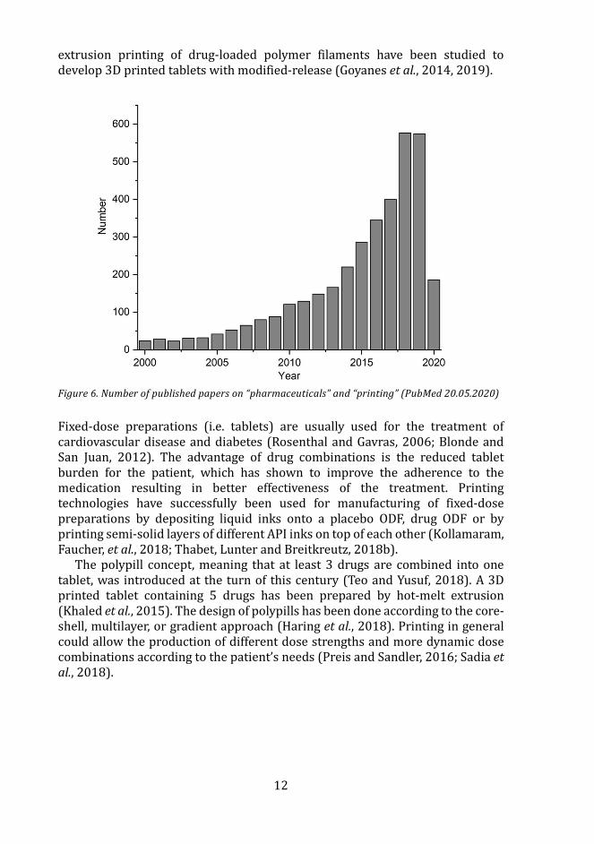
There has been an increasing interest in manufacturing pharmaceuticals by printing technologies and it is visualized by the number of published papers in Figure 6. Both immediate and modified release drug formulations have been developed using different printing technologies. Conventional and DoD printing technologies have been utilized for dose deposition on edible substrates (Hirshfield et al., 2014; Planchette et al., 2016). DoD technologies have also been used to prepare pharmaceuticals without a substrate, which has required the formulation of semi-solid and hot-melt inks (Kyobula et al., 2017). Semi-solid ink formulations for extrusion and pressure-assisted micro syringe printing have also been formulated to manufacture immediate-release tablets and ODFs, respectively (El Aita, Breitkreutz, and Quodbach, 2019; Sjoholm and Sandler, 2019). Release properties have successfully been tuned by inkjet printing of ink formulations according to different geometries or layers (Lee et al., 2012; Khaled et al., 2014; Kyobula et al., 2017; Gioumouxouzis et al., 2018). Also, liquid-based inks have been dispensed in immediate or extended-release capsules to modify the release (Okwuosa et al., 2018). Hot-melt extrusion and direct powder
12
extrusion printing of drug-loaded polymer filaments have been studied to develop 3D printed tablets with modified-release (Goyanes et al., 2014, 2019).
Figure 6. Number of published papers on “pharmaceuticals” and “printing” (PubMed 20.05.2020)
Fixed-dose preparations (i.e. tablets) are usually used for the treatment of cardiovascular disease and diabetes (Rosenthal and Gavras, 2006; Blonde and San Juan, 2012). The advantage of drug combinations is the reduced tablet burden for the patient, which has shown to improve the adherence to the medication resulting in better effectiveness of the treatment. Printing technologies have successfully been used for manufacturing of fixed-dose preparations by depositing liquid inks onto a placebo ODF, drug ODF or by printing semi-solid layers of different API inks on top of each other (Kollamaram, Faucher, et al., 2018; Thabet, Lunter and Breitkreutz, 2018b).
The polypill concept, meaning that at least 3 drugs are combined into one tablet, was introduced at the turn of this century (Teo and Yusuf, 2018). A 3D printed tablet containing 5 drugs has been prepared by hot-melt extrusion (Khaled et al., 2015). The design of polypills has been done according to the core-shell, multilayer, or gradient approach (Haring et al., 2018). Printing in general could allow the production of different dose strengths and more dynamic dose combinations according to the patient’s needs (Preis and Sandler, 2016; Sadia et al., 2018).
13
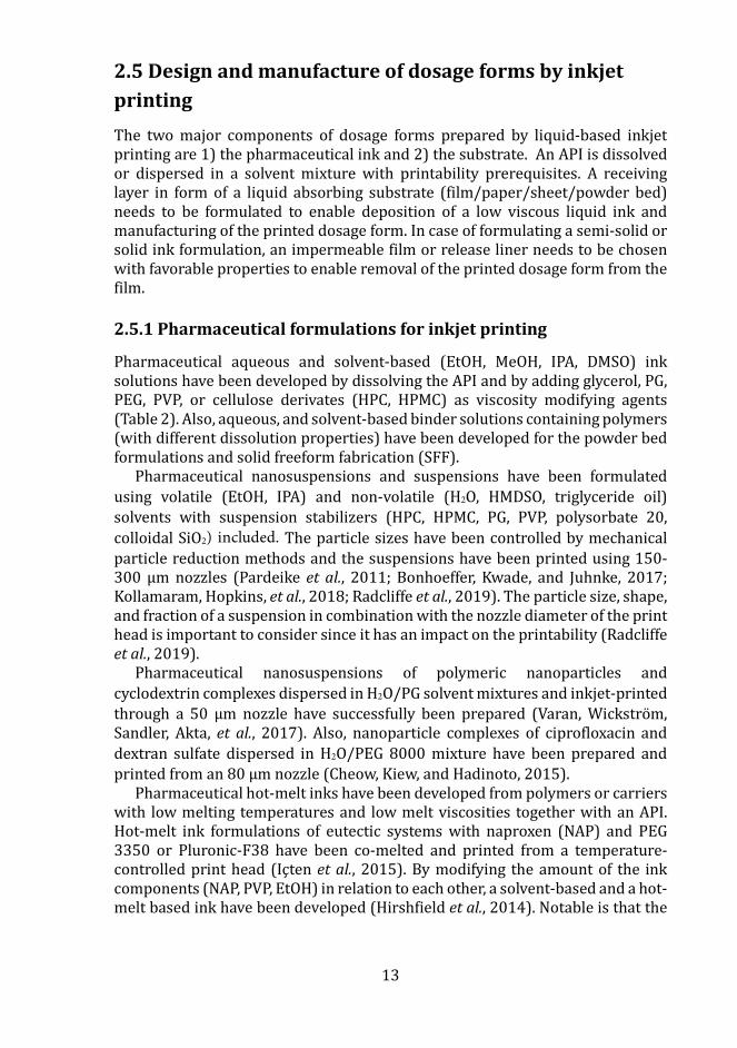
2.5 Design and manufacture of dosage forms by inkjet printing The two major components of dosage forms prepared by liquid-based inkjet printing are 1) the pharmaceutical ink and 2) the substrate. An API is dissolved or dispersed in a solvent mixture with printability prerequisites. A receiving layer in form of a liquid absorbing substrate (film/paper/sheet/powder bed) needs to be formulated to enable deposition of a low viscous liquid ink and manufacturing of the printed dosage form. In case of formulating a semi-solid or solid ink formulation, an impermeable film or release liner needs to be chosen with favorable properties to enable removal of the printed dosage form from the film.
2.5.1 Pharmaceutical formulations for inkjet printing
Pharmaceutical aqueous and solvent-based (EtOH, MeOH, IPA, DMSO) ink solutions have been developed by dissolving the API and by adding glycerol, PG, PEG, PVP, or cellulose derivates (HPC, HPMC) as viscosity modifying agents (Table 2). Also, aqueous, and solvent-based binder solutions containing polymers (with different dissolution properties) have been developed for the powder bed formulations and solid freeform fabrication (SFF).
Pharmaceutical nanosuspensions and suspensions have been formulated using volatile (EtOH, IPA) and non-volatile (H2O, HMDSO, triglyceride oil) solvents with suspension stabilizers (HPC, HPMC, PG, PVP, polysorbate 20, colloidal SiO2) included. The particle sizes have been controlled by mechanical particle reduction methods and the suspensions have been printed using 150-300 µm nozzles (Pardeike et al., 2011; Bonhoeffer, Kwade, and Juhnke, 2017; Kollamaram, Hopkins, et al., 2018; Radcliffe et al., 2019). The particle size, shape, and fraction of a suspension in combination with the nozzle diameter of the print head is important to consider since it has an impact on the printability (Radcliffe et al., 2019).
Pharmaceutical nanosuspensions of polymeric nanoparticles and cyclodextrin complexes dispersed in H2O/PG solvent mixtures and inkjet-printed through a 50 µm nozzle have successfully been prepared (Varan, Wickstrom, Sandler, Akta, et al., 2017). Also, nanoparticle complexes of ciprofloxacin and dextran sulfate dispersed in H2O/PEG 8000 mixture have been prepared and printed from an 80 µm nozzle (Cheow, Kiew, and Hadinoto, 2015).
Pharmaceutical hot-melt inks have been developed from polymers or carriers with low melting temperatures and low melt viscosities together with an API. Hot-melt ink formulations of eutectic systems with naproxen (NAP) and PEG 3350 or Pluronic-F38 have been co-melted and printed from a temperature-controlled print head (Içten et al., 2015). By modifying the amount of the ink components (NAP, PVP, EtOH) in relation to each other, a solvent-based and a hot-melt based ink have been developed (Hirshfield et al., 2014). Notable is that the
14
modification showed to have an impact on the solid-state of the final printed dosage form. A hot-melt ink was prepared by co-melting and printing a formulation consisting of beeswax and fenofibrate (Kyobula et al., 2017), and a hot-melt extruded formulation consisting of maltodextrin, glycerine, glycine, and paracetamol was also developed. The powders were wetted by glycerine and blended. Glycine was added to the formulation to improve the fluidity of the melt-blends (Musazzi et al., 2018).
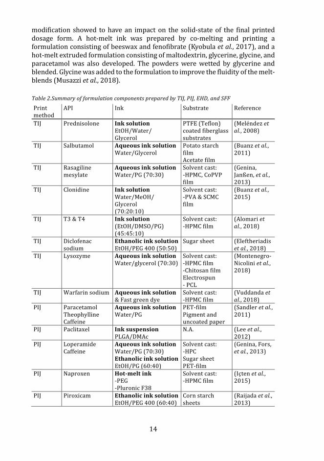
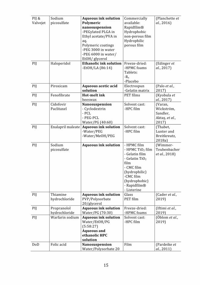
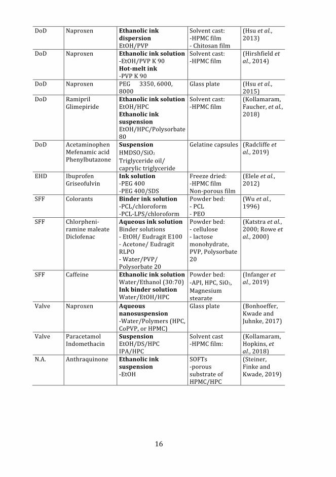
Table 2.Summary of formulation components prepared by TIJ, PIJ, EHD, and SFF
Print method
API Ink Substrate Reference
TIJ Prednisolone Ink solution EtOH/Water/ Glycerol
PTFE (Teflon) coated fiberglass substrates
(Meléndez et al., 2008)
TIJ Salbutamol
Aqueous ink solution Water/Glycerol
Potato starch film Acetate film
(Buanz et al., 2011)
TIJ Rasagiline mesylate
Aqueous ink solution Water/PG (70:30)
Solvent cast: -HPMC, CoPVP film
(Genina, Janßen, et al., 2013)
TIJ Clonidine
Ink solution Water/MeOH/ Glycerol (70:20:10)
Solvent cast: -PVA & SCMC film
(Buanz et al., 2015)
TIJ T3 & T4 Ink solution (EtOH/DMSO/PG) (45:45:10)
Solvent cast: -HPMC film
(Alomari et al., 2018)
TIJ Diclofenac sodium
Ethanolic ink solution EtOH/PEG 400 (50:50)
Sugar sheet (Eleftheriadis et al., 2018)
TIJ Lysozyme Aqueous ink solution Water/glycerol (70:30)
Solvent cast: -HPMC film -Chitosan film Electrospun - PCL
(Montenegro-Nicolini et al., 2018)
TIJ Warfarin sodium Aqueous ink solution & Fast green dye
Solvent cast: -HPMC film
(Vuddanda et al., 2018)
PIJ Paracetamol Theophylline Caffeine
Aqueous ink solution Water/PG
PET-film Pigment and uncoated paper
(Sandler et al., 2011)
PIJ Paclitaxel Ink suspension PLGA/DMAc
N.A. (Lee et al., 2012)
PIJ Loperamide Caffeine
Aqueous ink solution Water/PG (70:30) Ethanolic ink solution EtOH/PG (60:40)
Solvent cast: -HPC Sugar sheet PET-film
(Genina, Fors, et al., 2013)
PIJ Naproxen
Hot-melt ink -PEG -Pluronic F38
Solvent cast: -HPMC film
(Içten et al., 2015)
PIJ Piroxicam
Ethanolic ink solution EtOH/PEG 400 (60:40)
Corn starch sheets
(Raijada et al., 2013)
15
PIJ & Valvejet
Sodium picosulfate
Aqueous ink solution Polymeric nanosuspension -PEGylated PLGA in Ethyl acetate/PVA in aq. Polymeric coatings -PEG 3000 in water -PEG 6000 in water/ EtOH/ glycerol
Commercially available: Rapidfilm® Hydrophobic non-porous film Hydrophilic porous film
(Planchette et al., 2016)
PIJ Haloperidol Ethanolic ink solution -EtOH/LA (86:14)
Freeze-dried: -HPMC foams Tablets: -Rx -Placebo
(Edinger et al., 2017)
PIJ Piroxicam Aqueous acetic acid solution
Electrospun -Gelatin matrix
(Palo et al., 2017)
PIJ Fenofibrate Hot-melt ink beeswax
PET films (Kyobula et al., 2017)
PIJ Cidofovir Paclitaxel
Nanosuspension - Cyclodextrin - PCL - PEG-PCL Water/PG (40:60)
Solvent cast: -HPC film
(Varan, Wickström, Sandler, Aktaş, et al., 2017)
PIJ Enalapril maleate Aqueous ink solution -Water/PEG -Water/MeOH/PEG
Solvent cast: -HPC film
(Thabet, Lunter and Breitkreutz, 2018a)
PIJ Sodium picosulfate
Aqueous ink solution - HPMC film - HPMC TiO2 film - Gelatin film - Gelatin TiO2 film - CMC film (hydrophilic) -CMC film (hydrophobic) - Rapidfilm® - Listerine
(Wimmer-Teubenbacher et al., 2018)
PIJ Thiamine hydrochloride
Aqueous ink solution PVP/Polysorbate 20/glycerol
Glass PET film
(Cader et al., 2019)
PIJ Propranolol hydrochloride
Aqueous ink solution Water/PG (70:30)
Freeze-dried: -HPMC foams
(Iftimi et al., 2019)
PIJ Warfarin sodium
Aqueous ink solution Water/EtOH/PG (5:58:27) Aqueous and ethanolic HPC solution
Solvent cast: -HPC film
(Öblom et al., 2019)
DoD Folic acid Nanosuspension Water/Polysorbate 20
Film (Pardeike et al., 2011)
16
DoD Naproxen Ethanolic ink dispersion EtOH/PVP
Solvent cast: -HPMC film - Chitosan film
(Hsu et al., 2013)
DoD Naproxen Ethanolic ink solution -EtOH/PVP K 90 Hot-melt ink -PVP K 90
Solvent cast: -HPMC film
(Hirshfield et al., 2014)
DoD Naproxen
PEG 3350, 6000, 8000
Glass plate (Hsu et al., 2015)
DoD Ramipril Glimepiride
Ethanolic ink solution EtOH/HPC Ethanolic ink suspension EtOH/HPC/Polysorbate 80
Solvent cast: -HPMC film
(Kollamaram, Faucher, et al., 2018)
DoD Acetaminophen Mefenamic acid Phenylbutazone
Suspension HMDSO/SiO2
Triglyceride oil/ caprylic triglyceride
Gelatine capsules (Radcliffe et al., 2019)
EHD Ibuprofen Griseofulvin
Ink solution -PEG 400 -PEG 400/SDS
Freeze dried: -HPMC film Non-porous film
(Elele et al., 2012)
SFF Colorants Binder ink solution -PCL/chloroform -PCL-LPS/chloroform
Powder bed: - PCL - PEO
(Wu et al., 1996)
SFF Chlorpheni-ramine maleate Diclofenac
Aqueous ink solution Binder solutions - EtOH/ Eudragit E100 - Acetone/ Eudragit RLPO - Water/PVP/ Polysorbate 20
Powder bed: - cellulose - lactose monohydrate, PVP, Polysorbate 20
(Katstra et al., 2000; Rowe et al., 2000)
SFF Caffeine Ethanolic ink solution Water/Ethanol (30:70) Ink binder solution Water/EtOH/HPC
Powder bed: -API, HPC, SiO2, Magnesium stearate
(Infanger et al., 2019)
Valve Naproxen Aqueous nanosuspension -Water/Polymers (HPC, CoPVP, or HPMC)
Glass plate (Bonhoeffer, Kwade and Juhnke, 2017)
Valve Paracetamol Indomethacin
Suspension EtOH/DS/HPC IPA/HPC
Solvent cast -HPMC film:
(Kollamaram, Hopkins, et al., 2018)
N.A. Anthraquinone Ethanolic ink suspension -EtOH
SOFTs -porous substrate of HPMC/HPC
(Steiner, Finke and Kwade, 2019)
17
2.5.2 Substrates
A substrate is an edible sheet/film and the ink-receiving material, which is an important part of the inkjet-printed dosage form. Solvent cast films of typically cellulose derivates (HPMC, HPC) and sheets consisting of sugars have been used as substrates (Table 2.) Commercially available orodispersible films have also been studied as substrates for inkjet printing (Planchette et al., 2016; Wimmer-Teubenbacher et al., 2018). Since the orodispersible films and sheets have not been designed to absorb large ink amounts, porous films/foams of cellulose derivates have been developed as substrates for inkjet formulations. Also, the suitability of utilizing compacted placebo tablets was investigated by GSK and Edinger et al. (2017). The deposition of pharmaceutical ink slurries into gelatin capsules was successfully conducted by Radcliffe et al. (2019).
Different methods such as freeze-drying, foaming by blending, and electrospinning have been used for the preparation of porous substrates. The non-ionic cellulose sodium carboxymethylcellulose has been used to formulate substrates by freeze-drying and drying in air. Freeze-drying resulted in a film consisting of a porous network structure while drying in air resulted in smooth non-porous films (Boateng, Matthews, et al., 2009). Porous substrates based on polymers (HPMC, PEG 4000, polysorbate 20, poloxamer 188) were produced by freeze-drying and foaming of the solution using a hand blender before solvent casting (Iftimi et al., 2019). Steiner, Finke, and Kwade, (2019) developed a porous substrate with a closed bottom side and a protective top layer. The substrate was made by dispersing HPMC particle suspension in a binder solution consisting of HPC dissolved in EtOH. The film was cast by a casting knife and the protective top layer was achieved by spray coating. Another method of producing porous substrates is electrospinning. Electrospun fibrous substrates consisting of a natural polymer (gelatin) was prepared by dissolving the polymer in an acidic aqueous solution (acetic acid) and thermally crosslinking it with glucose as a crosslinking agent (Palo et al., 2017). A formulation consisting of a polyester (PCL) dissolved in a mixture of an ethyl acetate/acetone solution was developed for electrospinning by Montenegro-Nicolini et al., (2018).
Powder processing of cellulose (CMC, HPC), lactose, or polymers (PCL, PEO) in a layer-wise (100-250µm) manner was utilized in the fabrication of 3D printed tablets (Wu et al., 1996; Katstra et al., 2000; Rowe et al., 2000; Infanger et al., 2019). The powder bed layers served as a substrate onto which binder and drug solutions were deposited by inkjet technology.
Since the structure, thickness, and appearance of the edible sugar sheets, solvent cast films, freeze-dried porous substrates, fibrous rice sheets, fibrous electrospun substrates, placebo tablets, and powder bed tablets are different, the mechanical and the ink absorption properties of the substrates vary. Texture analyzers have been used to evaluate the mechanical properties of film substrates (Preis, Knop, and Breitkreutz, 2014). However, there are no guidelines regarding the measurement methods or requirements of the mechanical properties of the substrates. The only guideline in Ph Eur 9th Ed. about solvent cast ODFs is that the film should “possess suitable mechanical strength to resist
18
handling without being damaged”. In general, the mechanical properties of the substrates made by different methods are dependent on the composition of the substrate formulation. The addition of plasticizers to ODF formulations has improved the flexibility and decreased the rigidity of the solvent cast films (Boateng, Stevens, et al., 2009). The mechanical strength and the moisture sorption of the freeze-dried substrates have been shown to depend on the polymer amount of the film (Boateng et al., 2010). Crosslinking of electrospun substrates by the addition of a crosslinking agent has also been shown to improve the mechanical properties of the porous materials (Siimon, Siimon, and Jarvekulg, 2015). However, processing parameters, drying, and storage conditions have also an impact on the final substrate properties (Preis, Knop, and Breitkreutz, 2014). For instance, sugar films have been shown to become more fragile when stored at low relative humidity (Galdeano et al., 2009).
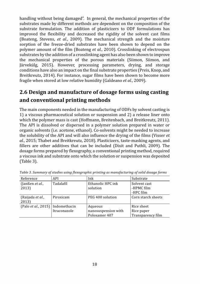
2.6 Design and manufacture of dosage forms using casting and conventional printing methods The main components needed in the manufacturing of ODFs by solvent casting is 1) a viscous pharmaceutical solution or suspension and 2) a release liner ontowhich the polymer mass is cast (Hoffmann, Breitenbach, and Breitkreutz, 2011).The API is dissolved or dispersed in a polymer solution prepared in water ororganic solvents (i.e. acetone, ethanol). Co-solvents might be needed to increasethe solubility of the API and will also influence the drying of the films (Visser etal., 2015; Thabet and Breitkreutz, 2018). Plasticizers, taste-masking agents, andfillers are other additives that can be included (Dixit and Puthli, 2009). Thedosage forms prepared by flexography, a conventional printing method, requireda viscous ink and substrate onto which the solution or suspension was deposited(Table 3).
Table 3. Summary of studies using flexographic printing as manufacturing of solid dosage forms
Reference API Ink Substrate (Janßen et al., 2013)
Tadalafil Ethanolic HPC ink solution
Solvent cast -HPMC film-HPC film
(Raijada et al., 2013)
Piroxicam PEG 400 solution Corn starch sheets
(Palo et al., 2015) Indomethacin Itraconazole
Aqueous nanosuspension with Poloxamer 407
Rice sheet Rice paper Transparency film
19
2.6.1 Pharmaceutical formulations for casting & flexographic printing
Solvent casting is a method used for the manufacturing of thin ODFs. Polymer-based solutions and suspensions have been prepared for the manufacturing of immediate release ODFs (Shimoda et al., 2009; Woertz and Kleinebudde, 2015a). The APIs have either been dissolved or dispersed in the polymer solution. Control of the particle size and preparation of micronized particles by e.g. spray drying or lyophilization before dispersion has improved the dissolution rate of the drug from the film (Brniak, Maslak, and Jachowicz, 2015; Manda et al., 2018). Similarly, the development of drug-loaded mesoporous silica nanoparticles (MSNs) dispersed into ODFs was seen to improve the dissolution of the API (Sen Karaman et al., 2018). Alternatively, modification of the release properties has also shown to be possible by incorporating drug-containing micropellets, prepared by wet extrusion and spheronization, to formulate prolonged-release ODFs (Speer et al., 2019). Preparation of bi- and multilayer ODFs has enabled the administration of two or more APIs at the same time (Preis et al., 2014; Thabet, Lunter, and Breitkreutz, 2018b). It was also found that a placebo layer between two films with APIs incompatible with each other did not give added value. The selection of two different polymers for the bi-layered film formulation was more feasible.
Flexographic printing was studied as an alternative manufacturing method of ODFs by depositing a viscous API solution onto placebo ODFs (Janßen et al., 2013). Another study explored the formulation of a poorly soluble drug by flexographic printing onto corn starch sheets, which showed increased dissolution (Raijada et al., 2013). The flexographic print uniformity was studied on a transparency film and a flexographic formulation of a nanosuspension on edible substrates was developed by Palo et al., 2015.
2.7 Quality of printed dosage forms The definition of the physicochemical properties of an API or a drug product during drug development is essential (EMA, 2000b, 2017b). Properties such as solubility in water and solvents, the dissociation constant, melting point, and solid-state are defined for an API in the preformulation stage and set the base for formulation development. The quality of printed dosage forms is closely linked to the ink composition, substrate choice, and processing parameters, as the formulation characteristics and the processing parameters, has shown to impact e.g. drug content and the solid-state of the printed dosage form (Melendez et al., 2008; Hirshfield et al., 2014; Kollamaram, Hopkins, et al., 2018; Cader et al., 2019). Both dosage amount and dosage form morphology has been identified as critical quality attributes of the dropwise printing process (Hirshfield et al., 2015).
20
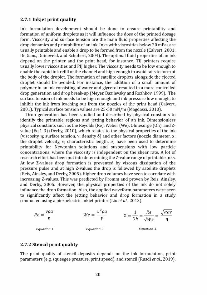
2.7.1 Inkjet print quality
Ink formulation development should be done to ensure printability and formation of uniform droplets as it will influence the dose of the printed dosage form. Viscosity and surface tension are the main fluid properties affecting the drop dynamics and printability of an ink. Inks with viscosities below 20 mPas are usually printable and enable a drop to be formed from the nozzle (Calvert, 2001; De Gans, Duineveld, and Schubert, 2004). The optimal fluid properties of an ink depend on the printer and the print head, for instance. TIJ printers require usually lower viscosities and PIJ higher. The viscosity needs to be low enough to enable the rapid ink refill of the channel and high enough to avoid tails to form at the body of the droplet. The formation of satellite droplets alongside the ejected droplet should be avoided. For instance, the addition of a small amount of polymer in an ink consisting of water and glycerol resulted in a more controlled drop generation and drop break-up (Meyer, Bazilevsky and Rozhkov, 1999). The surface tension of ink needs to be high enough and ink pressure low enough, to inhibit the ink from leaching out from the nozzles of the print head (Calvert, 2001). Typical surface tension values are 25-50 mN/m (Magdassi, 2010).
Drop generation has been studied and described by physical constants to identify the printable regions and jetting behavior of an ink. Dimensionless physical constants such as the Reyolds (Re), Weber (We), Ohnesorge (Oh), and Z-value (Eq.1-3) (Derby, 2010), which relates to the physical properties of the ink (viscosity, η; surface tension, γ; density δ) and other factors (nozzle diameter, α; the droplet velocity, v; characteristic length, a) have been used to determine printability for Newtonian solutions and suspensions with low particle concentrations, where the viscosity is independent on the shear rate. A lot of research effort has been put into determining the Z-value range of printable inks. At low Z-values drop formation is prevented by viscous dissipation of the pressure pulse and at high Z-values the drop is followed by satellite droplets (Reis, Ainsley, and Derby, 2005). Higher drop volumes have seen to correlate with increasing Z-values. This was predicted by Fromm and proven by Reis, Ainsley, and Derby, 2005. However, the physical properties of the ink do not solely influence the drop formation. Also, the applied waveform parameters were seen to significantly affect the jetting behavior and drop formation in a study conducted using a piezoelectric inkjet printer (Liu et al., 2013).
𝑅𝑅𝑒𝑒 =𝑣𝑣𝜌𝜌𝜌𝜌η
𝑊𝑊𝑒𝑒 = 𝑣𝑣2𝜌𝜌𝜌𝜌𝛾𝛾
𝑍𝑍 =1𝑂𝑂ℎ
=𝑅𝑅𝑒𝑒√𝑊𝑊𝑒𝑒
= �𝛼𝛼𝜌𝜌𝛾𝛾
η
Equation 1. Equation 2. Equation 3.
2.7.2 Stencil print quality
The print quality of stencil deposits depends on the ink formulation, print parameters (e.g. squeegee pressure, print speed), and stencil (Rusdi et al., 2019).
21
The stencil printing process begins by deposition of ink on the stencil, continues by printing the ink using a squeegee fixed at a certain angle, speed, and pressure, and ends by releasing the stencil from the printed product. Pasty inks have been printed for the manufacturing of electronic devices, but also liquid-based metal inks have been printed (Huang et al., 2011; Lazarus, Bedair and Kierzewski, 2017). The squeegee pressure and speed have been shown to determine the aperture filling, paste deposition height, and paste adherence to the substrate (Rusdi et al., 2019). The aspect ratio of stencil aperture width divided by stencil thickness can also indicate if qualitative print results can be achieved. The stencil has also an impact on the print performance, since the aperture orientation, sidewall roughness, and stencil material has been shown to affect the print transfer efficiency (Huang et al., 2011; Kay and Desmulliez, 2012).
2.7.3 Solid-state forms
The solid-state of the printed dosage form is important to characterize since the stability and bioavailability are different for crystalline (stable) and amorphous (instable) solids. Crystalline materials consist of molecules and atoms packed together in a well-defined order (Gavezzotti, 2007). The melting point and stability of a crystal depend on the forces holding the ordered molecules together. The molecules can be bound together via for instance weak van der Waals interactions and strong hydrogen bonds. Solubility and dissolution rates vary if the API has a different molecule packing (polymorphism) if water/solvent molecules are incorporated in the crystal structure (anhydrate, hydrate, solvate), if another solid molecular or ion is part of the crystal structure (cocrystal) or if the molecules lack long-range order structures (amorphous) (Blagden et al., 2007).
The deposition of pharmaceutical ink formulations by inkjet printing on substrates has shown to impact the solid-state of the drug. Polymorphs have been prepared from prednisolone, paracetamol and a non-stoichiometric hydrate have been prepared from thiamine hydrochloride by inkjet printing (Melendez et al., 2008; Kollamaram, Hopkins, et al., 2018; Cader et al., 2019). Control of the crystal formation has been reported to depend on the ink formulation and critical process parameters such as process temperature, product temperature, and drop volume (Hirshfield et al., 2014). Also, semi-crystalline and amorphous deposits have been formulated by varying the ink formulation (i.e. polymer amount). The addition of PG as a co-solvent in the ink has also been shown to inhibit the crystal growth of some APIs (Genina, Fors, et al., 2013). The substrates with different characteristics were observed to influence crystallization of the API; the ink deposit on the porous substrates did not crystallize, while the API crystallized on the impermeable and ODFs (Genina, Janßen, et al., 2013).
2.7.4 Regulatory perspectives of printed dosage forms
The regulatory challenges of implementing printing technologies as a manufacturing method of medicines arise if the aim is to deviate from mass
22
production and move towards the production of personalized dosage forms (Lim et al., 2018). Currently, no regulations about printed medicines exist. However, in 2017 FDA issued the guideline “Technical considerations for additive manufactured medical devices” stating recommendations about medical device design, software workflow, material controls, post-processing, process validation, and quality data. A statement regarding, that all considerations are not applicable for the manufactured products, due to the variety of used materials and printing technologies, was also made. Before printed personalized dosage forms can be manufactured, the regulatory requirements regarding preparation, quality control, and distribution need to be defined/adapted and fulfilled (European Commission, 2013, 2014a, 2014b). Lind et al., 2017, presented three different scenarios for future distribution chains of printed medicines. The pharmaceutical industry would have the best qualifications for manufacture and quality control of on-demand products. Pharmacies could also move towards having again a more central role in producing medicines. The most different scenario compared to the current practice, would be manufacturing of the medicine at patient’s homes.
2.8 Quality control methods of printed dosage forms Several analytical methods have been utilized to determine the quality of printed dosage forms. Dose quantification is traditionally performed by destructive HPLC, LC-MS, and UV-Vis methods. NIR spectroscopy, NIR, and Raman hyperspectral imaging have proven to be viable non-destructive dose quantification methods of printed formulations (Vakili et al., 2015, 2017; Edinger et al., 2017; Trenfield et al., 2018, 2020). The solid-state of the printed formulations have been determined by differential scanning calorimetry (DSC), X-ray diffraction, polarized microscopy, and Raman scanning microscopy (Jamroz et al., 2017; Trenfield et al., 2018). Texture analyzers have been used for determination of the mechanical properties of printed film/substrate formulations (Vakili et al., 2017; Thabet, Lunter and Breitkreutz, 2018a)
2.8.1 Colorimetric method
The human is a trichromatic organism with photoreceptors (cones) in the retina of the eye, which are sensitive to short (420–440 nm), middle (530–540 nm), and long (560–580 nm) wavelengths reflected or emitted by an item (Wu and Sun, 2013). The above-mentioned wavelengths are perceived by the individual as blue, green, and red, respectively, and are also defined as the tristimulus values.
Color coding is used in pharmaceutical formulations to aid dose strength differentiation and reduce the risk of medication errors (EMA, 2015). The color of the pharmaceutical is often specified and can be classified as a critical quality attribute if it is linked to e.g. contaminants or change of the solid-state (Zhou et al., 2011). Since colors are perceived differently by individuals, colorimeters have been developed to enable quantitative evaluation of colors (Hetrick et al., 2013).
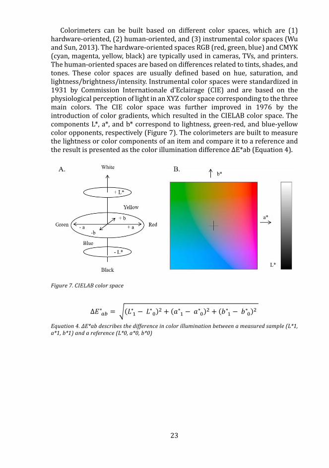
23
Colorimeters can be built based on different color spaces, which are (1) hardware-oriented, (2) human-oriented, and (3) instrumental color spaces (Wu and Sun, 2013). The hardware-oriented spaces RGB (red, green, blue) and CMYK (cyan, magenta, yellow, black) are typically used in cameras, TVs, and printers. The human-oriented spaces are based on differences related to tints, shades, and tones. These color spaces are usually defined based on hue, saturation, and lightness/brightness/intensity. Instrumental color spaces were standardized in 1931 by Commission Internationale d’Eclairage (CIE) and are based on the physiological perception of light in an XYZ color space corresponding to the three main colors. The CIE color space was further improved in 1976 by the introduction of color gradients, which resulted in the CIELAB color space. The components L*, a*, and b* correspond to lightness, green-red, and blue-yellow color opponents, respectively (Figure 7). The colorimeters are built to measure the lightness or color components of an item and compare it to a reference and the result is presented as the color illumination difference ΔE*ab (Equation 4).
Figure 7. CIELAB color space
∆𝐸𝐸 𝑎𝑎𝑎𝑎∗ = �(𝐿𝐿 1
∗ − 𝐿𝐿 0∗ )2 + (𝜌𝜌 1
∗ − 𝜌𝜌 0∗ )2 + (𝑏𝑏 1
∗ − 𝑏𝑏 0∗ )2
Equation 4. ΔE*ab describes the difference in color illumination between a measured sample (L*1, a*1, b*1) and a reference (L*0, a*0, b*0)
24
3. Aims of the study
This study aimed to formulate and study pharmaceutical inks for inkjet and stencil printing of oral solid dosage forms. The manufacturing flexibility of the formulated inkjet and stencil printed dosage forms dosing strength was evaluated. An in-direct colorimetric technique for on-demand quality control of printed dosage forms was also evaluated. The specific aims were to:
– Formulate and characterize low viscous pharmaceutical single-, multicomponent, and mesoporous silica nanoparticle (MSN) ink formulations for inkjet printing (I, II, III)
– Formulate and characterize a high viscous semi-solid polymer ink and orodispersible drug formulation by stencil printing (IV)
– Study and gain understanding about the ink formulation and substrate interactions (II, III)
– Study the dose amount and content uniformity of the inkjet and stencil printed doses (I, II, IV)
– Evaluate the effect of ink excipients on the dissolution rate and the solid-state properties of an active pharmaceutical ingredient (I)
– Evaluate if colorimetry could be utilized as an indirect quality control method for colored doses prepared by inkjet printing technology (II)
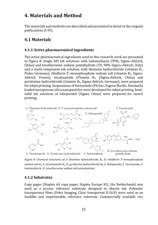
25
4. Materials and Method
The materials and methods are described and presented in detail in the original publications (I-IV).
4.1 Materials
4.1.1 Active pharmaceutical ingredients
The active pharmaceutical ingredients used in this research work are presented in Figure 8. Single API ink solutions with indomethacin (99%, Sigma–Aldrich, China) and levothyroxine sodium pentahydrate (T4, 98% Sigma–Aldrich, Italy) and a multi-component ink solution, with thiamine hydrochloride (vitamin B1, Fluka, Germany), riboflavin 5′-monophosphate sodium salt (vitamin B2, Sigma-Aldrich, France), nicotinamide (Vitamin B3, (Sigma-Aldrich, China) and pyridoxine hydrochloride (vitamin B6, Sigma-Aldrich, Germany), were prepared for inkjet printing. Suspensions of furosemide (Ph.Eur., Fagron Nordic, Denmark) loaded mesoporous silica nanoparticles were developed for inkjet printing. Semi-solid ink solutions of haloperidol (Sigma, China) were prepared for stencil printing.
Figure 8. Chemical structures of A. thiamine hydrochloride, B1, B. riboflavin 5′-monophosphate
sodium salt B2, C. nicotinamide B3, D. pyridoxine hydrochloride B6, E. Haloperidol, F. Furosemide, G. Indomethacin, H. Levothyroxine sodium salt pentahydrate
4.1.2 Substrates
Copy paper (Staples A4 copy paper, Staples Europe B.V., the Netherlands) was used as a porous reference substrate designed to absorb ink. Polyester transparency films (Folex Imaging, Clear transparent X-10.0) were used as an inedible and impermeable reference substrate. Commercially available rice
26
paper (Easybake®, N.J. Products Ltd., UK) and sugar paper (Sutton Valence, UK) were used as edible substrates for the colorimetry study of the inkjet-printed vitamin B doses. Polyester transparency films (Folex Imaging, Clear transparent X-10.0) were used and orodispersible hydroxypropyl methylcellulose (HPMC) films were prepared and used as substrates for the MSN nanosuspension deposits. The ODFs were prepared by casting the ODF solution onto the polyester transparency films using a film applicator (Multicator 411, Erichsen GmbH & Co. KG, Germany) having the wet thickness set at 500 µm. The solution consisted of HPMC (15 wt %, Pharmacoat 606, Shin Etsu, Tokyo, Japan) and glycerol (3 wt%, Sigma-Aldrich) dissolved in purified water and stirred on a magnetic stirrer overnight. Another polyester transparency film (Melinex, Dupont Teijin Films, USA) was used as a release liner in the stencil printing study. The T4 doses were prepared on a bilayer substrate, consisting of an HPC film (15% dissolved in EtOH, 500 µm) and a rice paper sheet. The HPC film was attached by spaying a small amount of isopropanol on the rice paper.
4.2 Methods
4.2.1 Preparation of pharmaceutical inks for inkjet printing
4.2.1.1 Indomethacin ink formulations (I) Ink formulations consisting of IMC (c=50 and 200 mg/ml) were prepared as such and having the amino acid L-arginine (c=24.3 mg/ml, L-Arg) or polymer polyvinylpyrrolidone (PVP) as additives. The formulations with only IMC were dissolved in a solvent mixture of DMSO and PG. The formulation containing IMC and L-Arg was dissolved in mixtures of DMSO, PG, and distilled water. The ink formulation containing only L-Arg was dissolved in distilled water and PG.
4.2.1.2 Vitamin B ink formulation (II) A multi-ink formulation was prepared by dissolving B1 (c= 10 mg/ml), B2 (c= 10 mg/ml), B3 (c= 20 mg/ml) and B6 (c= 10 mg/ml) in a commercially available yellow edible ink (Deco Enterprises Ltd., UK) designed for printing with Canon printers. The cartridge was emptied, washed, and refilled with the vitamin containing yellow ink. The yellow ink most likely contained water, glycerol (E422), tartrazine (E102), propylene glycol (E1520), and citric acid (E330).
4.2.1.3 MSN ink formulations (III) Nanosuspensions (c=1 and 5 mg/ml) consisting of furosemide (5 wt%, MSN–PEI–F5 and 15 wt%, MSN–PEI–F15) loaded and polyethyleneimine (PEI) surface-functionalized mesoporous silica nanoparticles (MSN) were developed. The nanoparticles were suspended in a solvent mixture consisting of deionized water and propylene glycol (PG).
27
4.2.1.4 T4 ink formulation A T4 ink was prepared by dissolving 20 mg/ml of the API in a DMSO/PG solvent mixture. Blue food colorant was added (1% v/v) to the ink.
4.2.2 Preparation of pharmaceutical inks for stencil printing
4.2.2.1 Haloperidol ink formulations (IV) Ink formulations of 12-18% of hydroxypropyl methylcellulose (HPMC) (Methocel E5 Premium LV, Dow, Germany) were prepared for stencil printing. The polymer was dissolved in a solvent mixture of deionized water and ethanol (EtOH) (Etax Aa 99.5%, Altia Oyj, Finland) under high-speed mixing. Glycerol (Fagron GmbH & Co. KG, Germany) was used as a plasticizer. Erythrosine B (spirit soluble 95%, Aldrich Chem. Co., USA) was added as a colorant and lactic acid (LA) (Sigma-Aldrich, Japan) was added to lower the pH of the formulation. HAL (1%, w/w) was the active pharmaceutical ingredients used in formulations. The following formulations were further studied HPMC 16%, HPMC 16% LA, HPMC 16% LA HAL and HPMC 16% HAL.
4.2.3 Viscosity (I, III, IV)
Rotational stress-controlled rheometers were used to determine the dynamic viscosity of the ink formulations. The dynamic viscosity of the temperature conditioned ink solutions was determined using a rheometer having a cone and plate geometry attached. Shear stress of 0.5 to 0.8 Pa was applied to the samples, which resulted in shear rates from 30 to 150 s-1 (Bohlin-CS, Malvern Instruments, UK) (I). A double-gap geometry was used to measure the temperature conditioned MSN ink suspensions by applying a stress ramp ranging from 10 to 1000 s-1 (Physica MCR 300, Anton Paar, Austria) (III). The semi-solid ink solutions were measured using the previously mentioned rheometer with a cone and plate geometry attached (IV). The solutions were pre-conditioned before applying a shear ramp ranging from 0.1 to 1000 s-1. The thixotropy of the solutions was also determined with a step test (IV). The samples were exposed to a high-shear rate (500 s-1) step change from a low constant shear rate (0.1 s-1) to determine the time-dependent recovery of the inks.
4.2.4 Surface tension (I, III)
A bubble tensiometer (Sensadyne PC 900, M&H Technologies Inc., USA) was used to determine the surface tension of temperature conditioned inks (I). A contact angle goniometer CAM 200 (KSV Instruments Ltd., Espoo, Finland, later Bohlin Scientific) was used to measure the surface tension of the MSN inks at room temperature (III). The pendant drop method was used, sand the recorded drop shape was fitted to the Young–Laplace equation using the OneAttension software (Theta1.4) to calculate the surface tension of the inks.
28
4.2.5 Contact angle (III)
The contact angle of the solvent mixtures and the 1 and 5 mg/ml nanosuspensions were performed with a contact angle goniometer CAM 200 (KSV Instruments Ltd., Espoo, Finland, later Bohlin Scientific) on transparency and orodispersible films using the sessile drop method. A 5 µl drop of liquid/suspension (23 ± 0.5°C) was ejected from the needle, placed onto the films (n=3), and monitored for 60 s.
4.2.6 Multiple light scattering (MLS) (III)
The colloidal suspension stability was studied by irradiating the nanoparticle suspensions (c=1&5 mg/ml) in the near-infrared region with an electroluminescent diode using multiple light scattering (MLS, Turbiscan MA2000, FormulAction, France). Data was recorded once a minute during a total of 180 min (n=1, 7 ml, 25°C). The acquired data were analyzed using the Turbisoft software (v 1.2.1, FormulAction, CIRTEM, France), which generated mean transmission profiles of the samples.
4.2.7 Printing technology
4.2.7.1 Piezoelectric inkjet printing (I, III) A piezoelectric inkjet printer (PixDro LP50, Roth & Rau, Eindhoven, the Netherlands) was used to deposit drug-containing inks (I, III). A printhead (Spectra SE 128 AA, Fujifilm, Dimatix Inc., USA) with 35 µm (Ø) nozzles was used to print the IMC solutions and a printhead (Spectra SL 128 AA, Fujifilm, Dimatix Inc., USA) with 50 µm (Ø) nozzles was used to print the T4 solution and MSN suspensions. The IMC doses were prepared by having a resolution of 500 x 500 dpi set and by depositing 1, 3, 6, and 9 layers of IMC ink onto an area of 1 cm2. The ARG-IMC-ARG formulation was printed by depositing 2 printed ARG layers, 4 IMC layers, and 2 ARG layers. The MSN suspensions were printed with resolutions of 150 x 150 dpi (1 layer), 500 x 500 dpi (1 & 10 layers) and a speed of 200 mm/s onto a 1 inch2 area. The substrate plate was heated at 30°C to facilitate the drying of the ink. The “print view” was calibrated according to a three-image calibration procedure given by the manufacturer of the printer before printing of the MSN suspension. No calibration was performed before the IMC doses were printed. Drop volume calculations were performed using the snapshot captured of the ink droplet using the PixDro software. Droplet formation was also studied using the advance droplet analysis (ADA, v.2.3, PixDro) software (III).
4.2.7.2 Thermal inkjet printing (II) An unmodified thermal desktop inkjet printer (Pixma iP3600, Canon Inc., Japan), with the paper loaded face-up, was used to print the yellow ink solution consisting of vitamin Bs (II). The color setting of the software was adjusted to enable ink ejection only from the yellow cartridge. Thus, the CMYK values were
29
set at 0, 0, 1, and 0 (0 = not in use, 1 = in use). An area of 4 cm2 was applied with ink and increasing doses were prepared by printing 1-10 layers.
4.2.7.3 Stencil printing (IV) A prototype stencil printing set-up was used to print the HPMC based ink formulations (IV). The set-up consisted of a drawdown coater (K202, RK Print-coat instruments Ltd., Royston, UK), a blade holder connected to a rod, a blade, and a frame. Stencils with varying stencil area and thickness were prepared to enable the printing of different doses.
4.2.8 Content uniformity
4.2.8.1 UV-Vis Spectrophotometry (I, III) The IMC does uniformity was studied by immersing a dose (n=3) in 100 ml of phosphate buffer pH 5.0 (Ph.Eur.) and leaving it stirring at 150 rpm. Sampling was performed after 3 h and was quantified using a UV-Vis spectrophotometer (Ultrospect 2100 pro, Biochrom Ltd., UK) at 265 nm.
The FUR dose incorporated in the nanoparticles (15wt% loading) and printed (10 layers, 1 inch2, n=3) on the HPMC film was immersed in 1 ml of EtOH. The samples were sonicated in a 25°C water bath for 30 min after which the samples were transferred to a rotating wheel for an additional 1.5 h. Before sampling and dose quantification with a UV-Vis spectrophotometer at λmax 273 nm, the samples were centrifuged for 10 min at 8000 rpm. Drug-free nanoparticle prints (10 layers, 1 inch2, n=3) were treated similarly and served as a blank for the UV-Vis- spectrophotometer measurements.
4.2.8.2 Liquid chromatography-mass spectrometry (LC-MS) (II) Liquid chromatography-mass spectrometry was used to quantify vitamin B doses. The setup consisted of high-performance liquid chromatography (Agilent 1100 series, USA) equipped with a binary pump, a vacuum degasser, an autosampler, and a column oven (30°C). Nitrogen 5.0 gas was used both as nebulizing gas (40 psi) and drying gas (10 l/min, 350°C). The compound separation was performed using a T3 analytical column (3 μm, 2.1 × 100 mm, Waters, Ireland) equipped with a pre-column (3 μm, 2.1 × 100 mm). The mobile phase consisted of 0.1% FA in deionized water (A) and 0.1% of FA in ACN/MeOH (50:50, B) and the flow rate was 0.3ml/min. The optimal separation was achieved using a gradient elution of B from 5% and ending in 95%. A 30 µl sample was injected and the run time was 25 min. Detection was performed in positive mode using an ion trap mass spectrometer equipped with an electrospray ionization source (Agilent Technologies, USA). The analytical ions (m/z) of the B1, B2, B3, B6, and paracetamol (internal standard) were 265, 457, 123, 170, and 152, respectively. The printed doses (n=3) were immersed in 10 ml of 0.1% FA and sonicated for 15 min at room temperature. Prior analysis the vitamin samples printed on sugar paper was centrifugated and filtrated (0.2 mm polytetrafluoroethylene membrane). The repeatability of the measurements was determined, and the matrix effect was evaluated.
30
4.2.8.3 High-performance liquid chromatography (HPLC) (IV) High-performance liquid chromatography (HPLC, LaChrome, Merck Hitachi, Tokyo, Japan) equipped with an isocratic solvent pump, a vacuum degasser, autosampler, a UV- detector, and a column oven was used to quantify the stencil printed HAL doses. The separation was performed using a pre-column (E 10 mm, GL Sciences, Japan) and an Intersil ODS-3.5 µm, 4.6 × 150 mm column (GL Sciences, Japan). The mobile phase (65:35, v/v) consisted of 0.05% TFA in deionized water (Trifluoroacetic acid, Sigma-Aldrich, Germany) and 0.05% TFA in ACN (Acetonitrile, HPLC grade, Fisher Chemicals, Loughborough, UK). The flow rate of the mobile phase was set at 1 mL/min. A 10µl sample was injected into the column and the drug was detected at a wavelength of 243 nm.
The T4 doses prepared by PIJ were analyzed with an HPLC system (Merck-Hitachi, Darmstadt, Germany) consisting of an isocratic solvent pump (L-7100) and an auto-sampler injection system (L-7200) with a 100 µL loop (Valco Instruments Corporation, USA). A reversed-phase column and a pre-column (LiChroCART 125-4 RP18 5µm and LiChrospher® 100 RP-18 5μm) were used to separate the drug, which was detected at 225nm using a UV-detector (L-7400). The flow rate and the injection volume were set at 0.6 ml/min and 10 µl, respectively. The mobile phase consisted of MeOH:H3PO4 0.1% (Phosphoric acid, 85 wt% solution in water, Acros Organics Germany) (70:30, V/V) with pH 2.92. Data analysis and peak integration were performed using the D-7000 HSM Chromatography Data Station software package.
4.2.9 Drug release (I, III)
IMC drug release of the printed samples (n=3) was studied using an in-house built dissolution set up. The IMC samples were put in metal sinkers and immersed in 100 ml of phosphate buffer pH 5.0 (T=37°C, 150 rpm). Samples were withdrawn for 60 min and analyzed using a UV/VIS-spectrophotometer (Ultrospect 2100 pro, Biochrom Ltd., UK) at 265 nm. IMC release rates of the printed samples were compared with crystalline IMC powder. The dissolution of crystalline IMC (2.0 mg) was conducted under sink conditions in 500 ml of phosphate buffer pH 5.0 (T=37°C, 150 rpm). The dissolution rates were expressed as the cumulative amount of drug (%) released over time. The average dose gained from the content uniformity measurements were used to calculate the cumulative amount released over time.
FUR drug release from the nanoparticles (c=1mg/ml, n=3) into the ink was studied. The ink consisted of water and propylene glycol into which the FUR loaded nanoparticles were dispersed and mixed at 50 rpm during 5h. A sample of 2 µl was withdrawn every hour from the ink supernatant, which was obtained by centrifuging the samples at 5000 rpm for 5 min. Re-dispersion of the ink was done by vortexing and sonicating the samples after each sampling time. The samples were analyzed in pendant mode using a UV-Vis spectrophotometer (Nanodrop 2000c spectrophotometer, Thermo Scientific) at λmax 273 nm.
31
4.2.10 Colorimetry (II)
The color illumination of the yellow vitamin B doses and blue T4 were studied using a digital handheld colorimeter (CLM-194, Eoptis, Italy). Each dose was measured in triplicate and the results were presented as L* (lightness) a* (green/red) b* (blue/yellow) values (± SD), according to the CIELAB system. The colorimeter consisted of integrated light-emitting diodes (LEDs) with standard illuminant D65, which corresponds to daylight, set. CIE1931- 2° was the standard observer. The device was built having a viewing angle of 0° and an illumination angle of 45°. Before the measurements, the colorimeter was calibrated using a white reference standard. The color illumination results were presented as CIELAB ΔE*ab, which was the illumination difference between the printed dose (L1, a1, b1) and the reference (L0, a0, b0). The illumination results of the printed layers were obtained by having the first layer as the measurement reference for the Vitamin B doses and the pure substrate as a measurement reference for T4. The ΔE*ab results were plotted against the number of printed layers and quantified doses.
4.2.11 Substrate thickness (II)
The substrate thickness of copy, rice, and sugar paper was measured (n=3) using a micrometer device (Lorentzen & Wettre, Sweden).
4.2.12 Scanning white-light interferometry (SWLI) (II, III)
A scanning white-light interferometer (SWLI) was utilized to determine the surface texture of copy, sugar, and rice paper substrates (14 x 14 cm2, n=3) (II). The vitamin B ink (5&10 layers, n=3) and MSN suspension (1 layer, 150 dpi, n=3) deposit impact on the substrates were also studied (II, III). The SWLI consisted of a reflective frame, interferometry objective, a piezoelectric z-scanner, a high-resolution camera, and two motorized translation stages. A standard halogen lamp was used as a light source. The samples were scanned using a ×6.3 magnification and the image data was acquired using in-house built software. A commercial MountainsMap® Imaging Topography 7.4 software was used to construct and analyze the acquired 3D data. The root mean square height of the surface (Sq) was calculated according to the ISO 25178 standard using the commercial software. The obtained surface data was further divided into waviness (Sq-W) and roughness (Sq-R) components to describe the large-scale surface texture changes and the nature of the substrate.
4.2.13 Confocal scanning laser microscopy (CSLM) (III)
A confocal scanning laser microscope (Leica TCS SP 5, Leica Microsystems GmbH, Germany) equipped with HCX PL APO 40×/1.15 and 63×/1.32 oil objective lenses was used to capture images of the fluorescence-labeled MSN suspension deposits on transparency and ODFs at an excitation wavelength of 488 nm.
32
4.2.14 Optical microscopy (III)
An optical microscopy imaging system (Evos XL Core Imaging System, Fisher Scientific GmbH, Germany) was used to visualize the MSN suspension deposits (×4, ×20, and ×40 magnification) printed using the piezoelectric inkjet printer.
4.2.15 pH (IV)
The pH of the ink formulations and the stencil printed haloperidol discs was measured using a pH meter (FE20, Mettler Toledo AG, Switzerland). The pH was measured by immersing the electrode into the ink formulations and by wetting the surface of the printed discs with distilled water (1 ml) to provide adequate contact with the electrode. The electrode equilibrated for 1 min before the pH of the discs was read.
4.2.16 Disintegration (IV)
The disintegration tests of the orodispersible discs (Ø 18 mm) was performed using a static test set-up consisting of a sample holder clamp, a 3 g clip weight, a beaker with 500 ml distilled water (37.0 ± 0.5 °C). The sample (n=10) was immersed into the distilled water and the endpoint was recorded as the 3 g weight reached the bottom of the beaker. Disintegration was performed of conditioned samples (25 °C, 60% RH) at ambient conditions. Placebo discs made using HPMC 12-18% inks and 250, 500, 750, 1000 µm thick stencils were analyzed.
4.2.17 Infrared Spectroscopy (I, IV)
An attenuated total reflectance infrared spectroscope (ATR–IR, Spectrum Two™, PerkinElmer, UK) equipped with a DiComp™ crystal was used to measure the IMC samples (n=3) at a force gauge of 140 N. The spectral region of 1000-1800 cm-1 was analyzed and the data was baselined corrected and normalized
A Fourier transform infrared spectrometer (FT-IR, Bruker Invenio R, Bruker Optics GmbH, Germany), equipped with a PA301 photoacoustic detector (Gasera Oy, Finland) using dried air as the carrier gas was used to measure the HAL samples (n=1). The spectral region of 600-1800 cm-1 was analyzed.
4.2.18 Scanning electron microscopy (SEM) (I, III)
A scanning electron microscope (SEM) (LEO Gemini 1530, Germany) supplied with a thermo scientific ultradry silicon drift detector was used to image the printed solution (I) and suspension (III) deposits. The samples were coated with an ultrathin layer of carbon using a vacuum evaporator. The printed samples were scanned at a working voltage of 2.7kV and x25 000 magnifications (I) and an acceleration voltage of 5 kV and x50, x100, x250, and x1000 magnifications (III).
33
An energy-dispersive X-ray spectroscope (EDX) (Thermo Scientific, USA), which is a SEM extension was used to identify the chemical elements on the surface of the highest IMC dose (I). The analysis was done at an accelerated voltage of 15 kV and a х250 time magnification.
4.2.19 Polarized light microscopy (PLM) (IV)
A microscope with polarized transmitted light (Leica DM IRB microskopie and Systeme GmbH, Germany) was used to study the starting materials and the stencil printed discs (×10/0.3 magnification, lens PL fluotar 506000). Images were captured using an OnePlus 5T mobile phone camera.
4.2.20 Differential scanning calorimetry (DSC) (IV)
A conventional DSC (Q2000, TA instruments, USA) was used to study the solid-state of the raw materials and stencil printed placebo and haloperidol formulations. The DSC was calibrated using sapphire crystals and indium. Tzero aluminum pans with perforated (discs) and intact (raw materials) lids were used to measure the 5 mg samples in triplicate. Thermograms of the raw materials and discs were recorded under a dynamic atmosphere of nitrogen (50 ml/min) and at a rate of 10°/min. The raw materials were subjected to a ramp from 0 to 200°C and the discs were subjected to heat (100°C)/ isothermal (100°C, 5 min)/ cool (-40°C)/ heat (200°C) cycle.
4.2.21 X-ray Powder Diffraction (XRD) (IV)
X-ray diffractograms of the raw materials, physical mixture, and printed stencil formulations were obtained by scanning the samples with an Empyrean diffractometer (Malvern Panalytical B.V., Almelo, the Netherlands) in θ/θ Bragg-Brentano geometry and by using Cu Kα radiation with a PIXcel3D detector in scanning line mode. The samples were attached to zero-background holders and scanned at 2θ from a range of 5 to 50° and with a step size of 0.013° and step time of 60 s.
4.2.22 Color stability
The color stability of the T4 samples was evaluated using a light chamber with daylight brightness set at 18 000 Lx, UV energy set at 3 W/m2, temperature set at 25°C and relative humidity set at 60% (Weiss technik Pharma 500L) during 7 days.
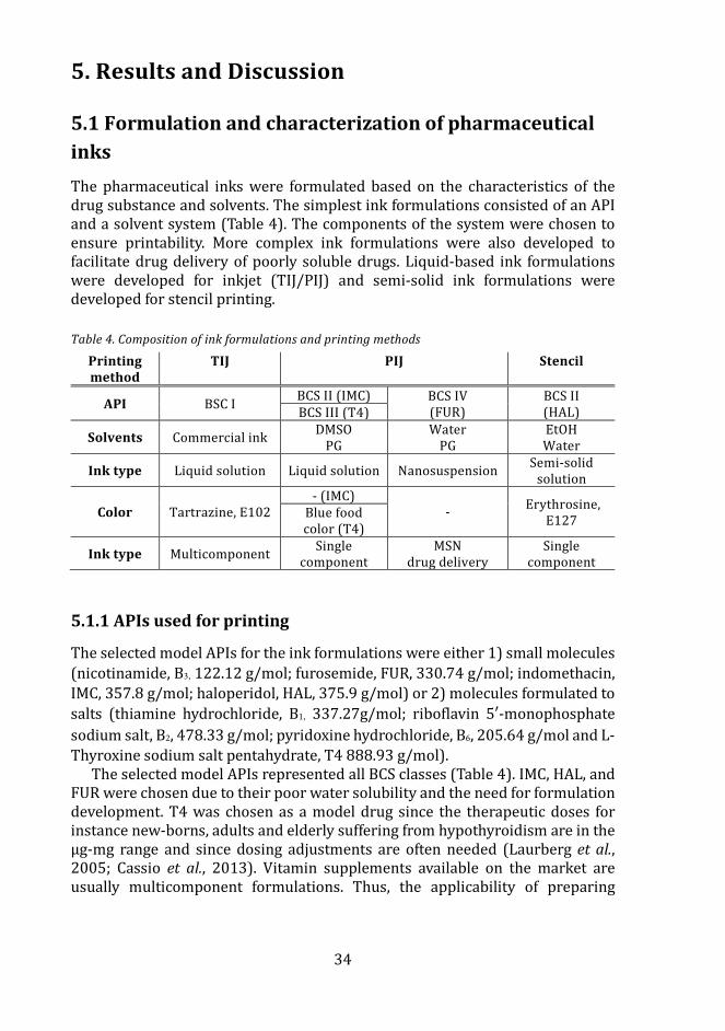
34
5. Results and Discussion
5.1 Formulation and characterization of pharmaceutical inks The pharmaceutical inks were formulated based on the characteristics of the drug substance and solvents. The simplest ink formulations consisted of an API and a solvent system (Table 4). The components of the system were chosen to ensure printability. More complex ink formulations were also developed to facilitate drug delivery of poorly soluble drugs. Liquid-based ink formulations were developed for inkjet (TIJ/PIJ) and semi-solid ink formulations were developed for stencil printing.
Table 4. Composition of ink formulations and printing methods
Printing method
TIJ PIJ Stencil
API BSC I BCS II (IMC) BCS IV (FUR)
BCS II (HAL) BCS III (T4)
Solvents Commercial ink DMSO PG
Water PG
EtOH Water
Ink type Liquid solution Liquid solution Nanosuspension Semi-solid solution
Color Tartrazine, E102 - (IMC)
- Erythrosine, E127 Blue food
color (T4)
Ink type Multicomponent Single component
MSN drug delivery
Single component
5.1.1 APIs used for printing
The selected model APIs for the ink formulations were either 1) small molecules (nicotinamide, B3, 122.12 g/mol; furosemide, FUR, 330.74 g/mol; indomethacin, IMC, 357.8 g/mol; haloperidol, HAL, 375.9 g/mol) or 2) molecules formulated to salts (thiamine hydrochloride, B1, 337.27g/mol; riboflavin 5′-monophosphate sodium salt, B2, 478.33 g/mol; pyridoxine hydrochloride, B6, 205.64 g/mol and L-Thyroxine sodium salt pentahydrate, T4 888.93 g/mol).
The selected model APIs represented all BCS classes (Table 4). IMC, HAL, and FUR were chosen due to their poor water solubility and the need for formulation development. T4 was chosen as a model drug since the therapeutic doses for instance new-borns, adults and elderly suffering from hypothyroidism are in the µg-mg range and since dosing adjustments are often needed (Laurberg et al., 2005; Cassio et al., 2013). Vitamin supplements available on the market are usually multicomponent formulations. Thus, the applicability of preparing
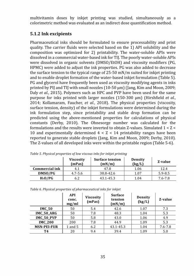
35
multivitamin doses by inkjet printing was studied, simultaneously as a colorimetric method was evaluated as an indirect dose quantification method.
5.1.2 Ink excipients
Pharmaceutical inks should be formulated to ensure processability and print quality. The carrier fluids were selected based on the 1) API solubility and the composition was optimized for 2) printability. The water-soluble APIs were dissolved in a commercial water-based ink for TIJ. The poorly water-soluble APIs were dissolved in organic solvents (DMSO/EtOH) and viscosity modifiers (PG, HPMC) were added to adjust the ink properties. PG was also added to decrease the surface tension to the typical range of 25-50 mN/m suited for inkjet printing and to enable droplet formation of the water-based inkjet formulation (Table 5). PG and glycerol have frequently been used as viscosity modifying agents in inks printed by PIJ and TIJ with small nozzles (10-50 µm) (Jang, Kim and Moon, 2009; Daly et al., 2015). Polymers such as HPC and PVP have been used for the same purpose for inks printed with larger nozzles (150-300 µm) (Hirshfield et al., 2014; Kollamaram, Faucher, et al., 2018). The physical properties (viscosity, surface tension, density) of the inkjet formulations were determined during the ink formulation step, since printability and stable drop formation can be predicted using the above-mentioned properties for calculations of physical constants (Derby, 2010). The Ohnesorge number was calculated for the formulations and the results were inverted to obtain Z-values. Simulated 1 < Z < 10 and experimentally determined 4 < Z < 14 printability ranges have been reported to generate stable droplets (Jang, Kim and Moon, 2009; Derby, 2010). The Z-values of all developed inks were within the printable region (Table 5-6).
Table 5. Physical properties of low viscous inks for inkjet printing
Viscosity (mPas)
Surface tension (mN/m)
Density (kg/L) Z-value
Commercial ink 4.1 47.8 1.06 12.4 DMSO/PG 4.7-5.6 38.8-42.6 1.07 5.9-8.5
H2O/PG 6.2 43.1-45.3 1.04 7.6-7.8
Table 6. Physical properties of pharmaceutical inks for inkjet
API
conc. mg/ml
Viscosity (mPas)
Surface tension (mN/m)
Density (kg/L) Z-value
IMC_50 50 5.4 42.6 1.07 7.3 IMC_50_ARG 50 7.8 48.3 1.04 5.3 IMC_50_PVP 50 5.8 43.0 1.06 4.9
IMC_200 200 7.8 44.9 1.09 5.3 MSN-PEI-FUR 1 and 5 6.2 43.1-45.3 1.04 7.6-7.8
T4 20 9.4 39.4 1.09 5.0
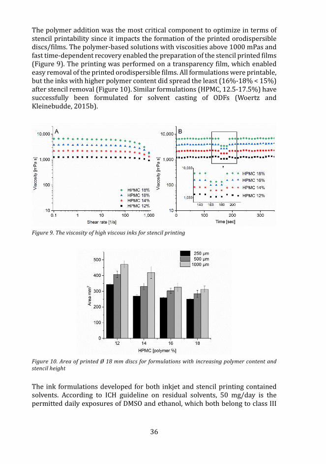
36
The polymer addition was the most critical component to optimize in terms of stencil printability since it impacts the formation of the printed orodispersible discs/films. The polymer-based solutions with viscosities above 1000 mPas and fast time-dependent recovery enabled the preparation of the stencil printed films (Figure 9). The printing was performed on a transparency film, which enabled easy removal of the printed orodispersible films. All formulations were printable, but the inks with higher polymer content did spread the least (16%-18% < 15%) after stencil removal (Figure 10). Similar formulations (HPMC, 12.5-17.5%) have successfully been formulated for solvent casting of ODFs (Woertz and Kleinebudde, 2015b).
Figure 9. The viscosity of high viscous inks for stencil printing
Figure 10. Area of printed Ø 18 mm discs for formulations with increasing polymer content and stencil height
The ink formulations developed for both inkjet and stencil printing contained solvents. According to ICH guideline on residual solvents, 50 mg/day is the permitted daily exposures of DMSO and ethanol, which both belong to class III
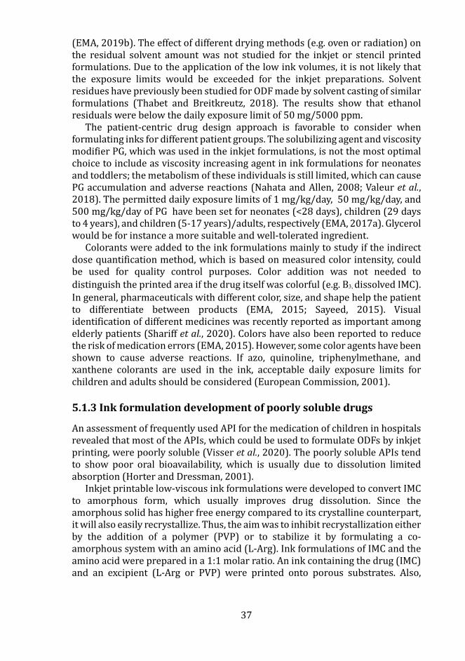
37
(EMA, 2019b). The effect of different drying methods (e.g. oven or radiation) on the residual solvent amount was not studied for the inkjet or stencil printed formulations. Due to the application of the low ink volumes, it is not likely that the exposure limits would be exceeded for the inkjet preparations. Solvent residues have previously been studied for ODF made by solvent casting of similar formulations (Thabet and Breitkreutz, 2018). The results show that ethanol residuals were below the daily exposure limit of 50 mg/5000 ppm.
The patient-centric drug design approach is favorable to consider when formulating inks for different patient groups. The solubilizing agent and viscosity modifier PG, which was used in the inkjet formulations, is not the most optimal choice to include as viscosity increasing agent in ink formulations for neonates and toddlers; the metabolism of these individuals is still limited, which can cause PG accumulation and adverse reactions (Nahata and Allen, 2008; Valeur et al., 2018). The permitted daily exposure limits of 1 mg/kg/day, 50 mg/kg/day, and 500 mg/kg/day of PG have been set for neonates (<28 days), children (29 days to 4 years), and children (5-17 years)/adults, respectively (EMA, 2017a). Glycerol would be for instance a more suitable and well-tolerated ingredient.
Colorants were added to the ink formulations mainly to study if the indirect dose quantification method, which is based on measured color intensity, could be used for quality control purposes. Color addition was not needed to distinguish the printed area if the drug itself was colorful (e.g. B3, dissolved IMC). In general, pharmaceuticals with different color, size, and shape help the patient to differentiate between products (EMA, 2015; Sayeed, 2015). Visual identification of different medicines was recently reported as important among elderly patients (Shariff et al., 2020). Colors have also been reported to reduce the risk of medication errors (EMA, 2015). However, some color agents have been shown to cause adverse reactions. If azo, quinoline, triphenylmethane, and xanthene colorants are used in the ink, acceptable daily exposure limits for children and adults should be considered (European Commission, 2001).
5.1.3 Ink formulation development of poorly soluble drugs
An assessment of frequently used API for the medication of children in hospitals revealed that most of the APIs, which could be used to formulate ODFs by inkjet printing, were poorly soluble (Visser et al., 2020). The poorly soluble APIs tend to show poor oral bioavailability, which is usually due to dissolution limited absorption (Horter and Dressman, 2001).
Inkjet printable low-viscous ink formulations were developed to convert IMC to amorphous form, which usually improves drug dissolution. Since the amorphous solid has higher free energy compared to its crystalline counterpart, it will also easily recrystallize. Thus, the aim was to inhibit recrystallization either by the addition of a polymer (PVP) or to stabilize it by formulating a co-amorphous system with an amino acid (L-Arg). Ink formulations of IMC and the amino acid were prepared in a 1:1 molar ratio. An ink containing the drug (IMC) and an excipient (L-Arg or PVP) were printed onto porous substrates. Also,
38
separate IMC (dissolved in DMSO and PG) and L-Arg (dissolved in H2O and PG) inks were prepared and layers of ink were applied on top of each other on a transparency film.
Co-amorphization of IMC has previously been achieved by solvent evaporation (Dengale et al., 2014). Similar solvent evaporation of small (pl) droplets is also expected to occur after printing. Co-melting and quench cooling of IMC and PVP have been shown to result in co-amorphization (Hirshfield et al., 2014). Large amounts of PVP could not be added to the solvent-based formulation prepared in this study. Thus, hotmelt inkjet formulations could better suit this preparation approach as larger polymer amounts have shown to be needed to achieve amorphous deposits (Hirshfield et al., 2014).
Formulation of amorphous solid dispersions (polymer-based glass solutions), which are single amorphous phase systems, have been shown to improve the dissolution of poorly soluble drugs (Van Den Mooter, 2012; Dengale et al., 2016). Stencil printed orodispersible films were prepared by dissolving the polymer HPMC and the poorly soluble drug HAL in an acidic (lactic acid) solvent system. pH modification has been considered as an alternative formulation approach for ionizable and poorly soluble drugs since the drug is present in a supersaturated form and can recrystallize over time of storage (Kawabata et al., 2011; Van Den Mooter, 2012). A similar formulation approach was taken for the amorphous solid dispersion with the poorly soluble drug tetrabenazine (TBZ), citric acid, and HPMC (Senta-Loys et al., 2017). The ODF formulation was amorphous and showed rapid drug dissolution still after 6 months of storage. No stability testing was performed for the HAL formulations.
Development of MSN drug formulations is another method to improve drug dissolution and enhance the permeation of poorly soluble APIs (Wang et al., 2015). The pharmaceutical ink consisted of drug-loaded MSN-PEI with a hydrodynamic particle size from 290 to 450 nm. Surface functionalization of the nanoparticles by PEI resulted in positively charged (39-47 mV) particles, which have been shown to facilitate cellular uptake (Xia et al., 2009). The surface functionalization protected the drug from leaching out into the ink for at least 5h and provided electrostatic stabilization of the nanosuspension. Water and PG were chosen as ink, based on the poor solubility of FUR in the solvents. The nanosuspension stability was confirmed by multiple light scattering and measuring mean transmittance of the suspension during 3h. Negatively charged (42-45 mV) polymeric or cyclodextrin complex nanosuspensions with particle sizes of less than 200 nm (Varan et al., 2017) and 300 nm (Cheow, Kiew, and Hadinoto, 2015) have been printed by inkjet technology. PG and PEG 8000 have been used to increase the viscosity of the ink. Nanosuspensions have also been formulated for inkjet printing by high-pressure homogenization and stabilized by the addition of polysorbate 20 (Pardeike et al., 2011). This method resulted in nanoparticles around 400 nm with a negative zeta potential (-39 mV).
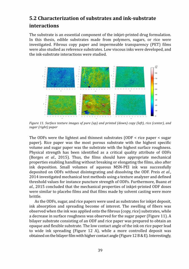
39
5.2 Characterization of substrates and ink-substrate interactions The substrate is an essential component of the inkjet-printed drug formulation. In this thesis, edible substrates made from polymers, sugars, or rice were investigated. Fibrous copy paper and impermeable transparency (PET) films were also studied as reference substrates. Low viscous inks were developed, and the ink-substrate interactions were studied.
Figure 11. Surface texture images of pure (up) and printed (down) copy (left), rice (center), and sugar (right) paper
The ODFs were the lightest and thinnest substrates (ODF < rice paper < sugar paper). Rice paper was the most porous substrate with the highest specific volume and sugar paper was the substrate with the highest surface roughness. Physical strength has been identified as a critical quality attribute of ODFs (Borges et al., 2015). Thus, the films should have appropriate mechanical properties enabling handling without breaking or elongating the films, also after ink deposition. Small volumes of aqueous MSN-PEI ink was successfully deposited on ODFs without disintegrating and dissolving the ODF. Preis et al., 2014 investigated mechanical test methods using a texture analyzer and defined threshold values for instance puncture strength of ODFs. Furthermore, Buanz et al., 2015 concluded that the mechanical properties of inkjet-printed ODF doses were similar to placebo films and that films made by solvent casting were more brittle.
As the ODFs, sugar, and rice papers were used as substrates for inkjet deposit, ink absorption and spreading become of interest. The swelling of fibers was observed when the ink was applied onto the fibrous (copy, rice) substrates, while a decrease in surface roughness was observed for the sugar paper (Figure 11). A bilayer substrate consisting of an ODF and rice paper was prepared to obtain an opaque and flexible substrate. The low contact angle of the ink on rice paper lead to wide ink spreading (Figure 12 A), while a more controlled deposit was obtained on the bilayer film with higher contact angle (Figure 12 B & E). Interestingly,
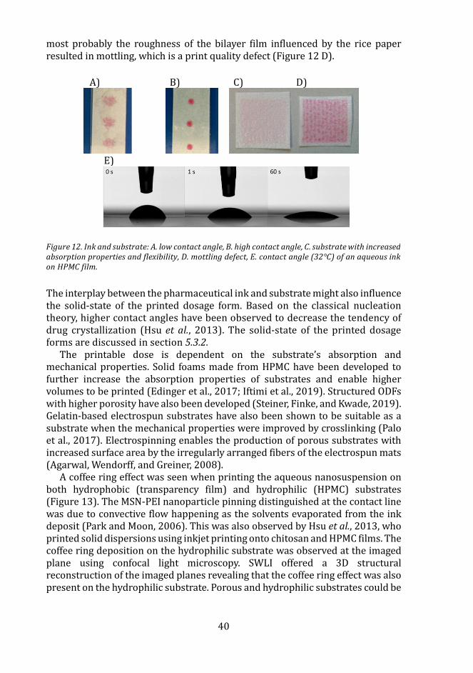
40
most probably the roughness of the bilayer film influenced by the rice paper resulted in mottling, which is a print quality defect (Figure 12 D).
A) B) C) D)
E)
Figure 12. Ink and substrate: A. low contact angle, B. high contact angle, C. substrate with increased absorption properties and flexibility, D. mottling defect, E. contact angle (32°C) of an aqueous ink on HPMC film.
The interplay between the pharmaceutical ink and substrate might also influence the solid-state of the printed dosage form. Based on the classical nucleation theory, higher contact angles have been observed to decrease the tendency of drug crystallization (Hsu et al., 2013). The solid-state of the printed dosage forms are discussed in section 5.3.2.
The printable dose is dependent on the substrate’s absorption and mechanical properties. Solid foams made from HPMC have been developed to further increase the absorption properties of substrates and enable higher volumes to be printed (Edinger et al., 2017; Iftimi et al., 2019). Structured ODFs with higher porosity have also been developed (Steiner, Finke, and Kwade, 2019). Gelatin-based electrospun substrates have also been shown to be suitable as a substrate when the mechanical properties were improved by crosslinking (Palo et al., 2017). Electrospinning enables the production of porous substrates with increased surface area by the irregularly arranged fibers of the electrospun mats (Agarwal, Wendorff, and Greiner, 2008).
A coffee ring effect was seen when printing the aqueous nanosuspension on both hydrophobic (transparency film) and hydrophilic (HPMC) substrates (Figure 13). The MSN-PEI nanoparticle pinning distinguished at the contact line was due to convective flow happening as the solvents evaporated from the ink deposit (Park and Moon, 2006). This was also observed by Hsu et al., 2013, who printed solid dispersions using inkjet printing onto chitosan and HPMC films. The coffee ring deposition on the hydrophilic substrate was observed at the imaged plane using confocal light microscopy. SWLI offered a 3D structural reconstruction of the imaged planes revealing that the coffee ring effect was also present on the hydrophilic substrate. Porous and hydrophilic substrates could be
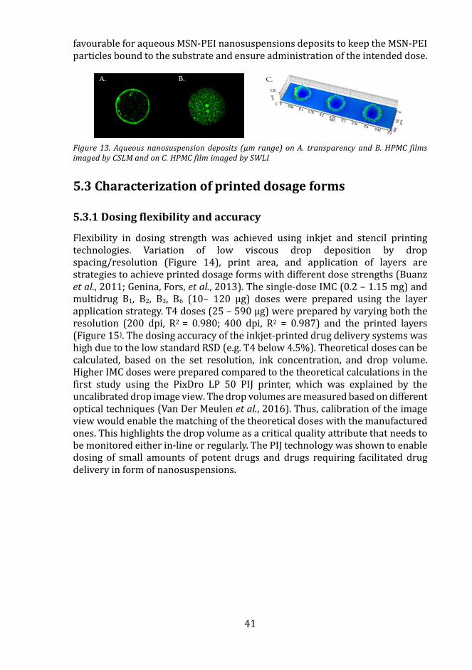
41
favourable for aqueous MSN-PEI nanosuspensions deposits to keep the MSN-PEI particles bound to the substrate and ensure administration of the intended dose.
Figure 13. Aqueous nanosuspension deposits (µm range) on A. transparency and B. HPMC films imaged by CSLM and on C. HPMC film imaged by SWLI
5.3 Characterization of printed dosage forms
5.3.1 Dosing flexibility and accuracy
Flexibility in dosing strength was achieved using inkjet and stencil printing technologies. Variation of low viscous drop deposition by drop spacing/resolution (Figure 14), print area, and application of layers are strategies to achieve printed dosage forms with different dose strengths (Buanz et al., 2011; Genina, Fors, et al., 2013). The single-dose IMC (0.2 – 1.15 mg) and multidrug B1, B2, B3, B6 (10– 120 µg) doses were prepared using the layer application strategy. T4 doses (25 – 590 µg) were prepared by varying both the resolution (200 dpi, R2 = 0.980; 400 dpi, R2 = 0.987) and the printed layers (Figure 15). The dosing accuracy of the inkjet-printed drug delivery systems was high due to the low standard RSD (e.g. T4 below 4.5%). Theoretical doses can be calculated, based on the set resolution, ink concentration, and drop volume. Higher IMC doses were prepared compared to the theoretical calculations in the first study using the PixDro LP 50 PIJ printer, which was explained by the uncalibrated drop image view. The drop volumes are measured based on different optical techniques (Van Der Meulen et al., 2016). Thus, calibration of the image view would enable the matching of the theoretical doses with the manufactured ones. This highlights the drop volume as a critical quality attribute that needs to be monitored either in-line or regularly. The PIJ technology was shown to enable dosing of small amounts of potent drugs and drugs requiring facilitated drug delivery in form of nanosuspensions.
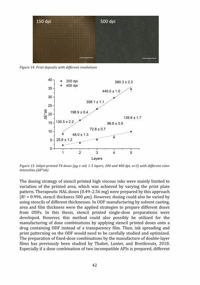
42
Figure 14. Print deposits with different resolutions
Figure 15. Inkjet-printed T4 doses (µg ± std; 1-5 layers, 200 and 400 dpi, n=3) with different color intensities (ΔE*ab)
The dosing strategy of stencil printed high viscous inks were mainly limited to variation of the printed area, which was achieved by varying the print plate pattern. Therapeutic HAL doses (0.49–2.56 mg) were prepared by this approach (R2 = 0.996, stencil thickness 500 µm). However, dosing could also be varied by using stencils of different thicknesses. In ODF manufacturing by solvent casting, area and film thickness were the applied strategies to prepare different doses from ODFs. In this thesis, stencil printed single-dose preparations were developed. However, this method could also possibly be utilized for the manufacturing of dose combinations by applying stencil printed doses onto a drug containing ODF instead of a transparency film. Then, ink spreading and print patterning on the ODF would need to be carefully studied and optimized. The preparation of fixed-dose combinations by the manufacture of double-layer films has previously been studied by Thabet, Lunter, and Breitkreutz, 2018. Especially if a dose combination of two incompatible APIs is prepared, different
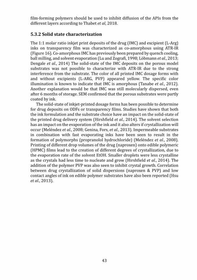
43
film-forming polymers should be used to inhibit diffusion of the APIs from the different layers according to Thabet et al, 2018.
5.3.2 Solid state characterization
The 1:1 molar ratio inkjet print deposits of the drug (IMC) and excipient (L-Arg) inks on transparency film was characterized as co-amorphous using ATR-IR (Figure 16). Co-amorphous IMC has previously been prepared by quench cooling, ball milling, and solvent evaporation (Lu and Zografi, 1998; Lobmann et al., 2013; Dengale et al., 2014) The solid-state of the IMC deposits on the porous model substrates was not possible to characterize with ATR-IR due to the strong interference from the substrate. The color of all printed IMC dosage forms with and without excipients (L-ARG, PVP) appeared yellow. The specific color illumination is known to indicate that IMC is amorphous (Tanabe et al., 2012). Another explanation would be that IMC was still molecularly dispersed, even after 6 months of storage. SEM confirmed that the porous substrates were partly coated by ink.
The solid-state of inkjet-printed dosage forms has been possible to determine for drug deposits on ODFs or transparency films. Studies have shown that both the ink formulation and the substrate choice have an impact on the solid-state of the printed drug delivery system (Hirshfield et al., 2014). The solvent selection has an impact on the evaporation of the ink and it also alters if crystallization will occur (Melendez et al., 2008; Genina, Fors, et al., 2013). Impermeable substrates in combination with fast evaporating inks have been seen to result in the formation of polymorphs (propranolol hydrochloride) (Melendez et al., 2008). Printing of different drop volumes of the drug (naproxen) onto edible polymeric (HPMC) films lead to the creation of different degrees of crystallization, due to the evaporation rate of the solvent EtOH. Smaller droplets were less crystalline as the crystals had less time to nucleate and grow (Hirshfield et al., 2014). The addition of the polymer PVP was also seen to inhibit crystal growth. Correlation between drug crystallization of solid dispersions (naproxen & PVP) and low contact angles of ink on edible polymer substrates have also been reported (Hsu et al., 2013).
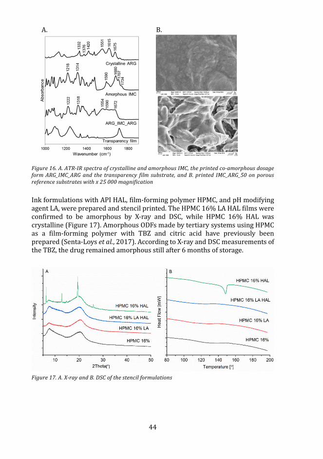
44
A. B.
Figure 16. A. ATR-IR spectra of crystalline and amorphous IMC, the printed co-amorphous dosage form ARG_IMC_ARG and the transparency film substrate, and B. printed IMC_ARG_50 on porous reference substrates with x 25 000 magnification
Ink formulations with API HAL, film-forming polymer HPMC, and pH modifying agent LA, were prepared and stencil printed. The HPMC 16% LA HAL films were confirmed to be amorphous by X-ray and DSC, while HPMC 16% HAL was crystalline (Figure 17). Amorphous ODFs made by tertiary systems using HPMC as a film-forming polymer with TBZ and citric acid have previously been prepared (Senta-Loys et al., 2017). According to X-ray and DSC measurements of the TBZ, the drug remained amorphous still after 6 months of storage.
Figure 17. A. X-ray and B. DSC of the stencil formulations
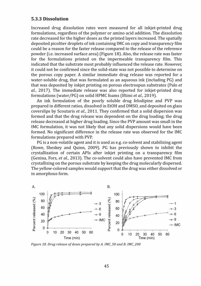
45
5.3.3 Dissolution
Increased drug dissolution rates were measured for all inkjet-printed drug formulations, regardless of the polymer or amino acid addition. The dissolution rate decreased for the higher doses as the printed layers increased. The spatially deposited picoliter droplets of ink containing IMC on copy and transparency film could be a reason for the faster release compared to the release of the reference powder (i.e. increased surface area) (Figure 18). Also, the release rate was faster for the formulations printed on the impermeable transparency film. This indicated that the substrate most probably influenced the release rate. However, it could not be confirmed since the solid-state was not possible to determine on the porous copy paper. A similar immediate drug release was reported for a water-soluble drug, that was formulated as an aqueous ink (including PG) and that was deposited by inkjet printing on porous electrospun substrates (Palo et al., 2017). The immediate release was also reported for inkjet-printed drug formulations (water/PG) on solid HPMC foams (Iftimi et al., 2019).
An ink formulation of the poorly soluble drug felodipine and PVP was prepared in different ratios, dissolved in EtOH and DMSO, and deposited on glass coverslips by Scoutaris et al., 2011. They confirmed that a solid dispersion was formed and that the drug release was dependent on the drug loading; the drug release decreased at higher drug loading. Since the PVP amount was small in the IMC formulation, it was not likely that any solid dispersions would have been formed. No significant difference in the release rate was observed for the IMC formulations prepared with PVP.
PG is a non-volatile agent and it is used as e.g. co-solvent and stabilizing agent (Rowe, Sheskey and Quinn, 2009). PG has previously shown to inhibit the crystallization of certain APIs after inkjet printing on a transparency film (Genina, Fors, et al., 2013). The co-solvent could also have prevented IMC from crystallizing on the porous substrate by keeping the drug molecularly dispersed. The yellow-colored samples would support that the drug was either dissolved or in amorphous form.
A. B.
Figure 18. Drug release of doses prepared by A. IMC_50 and B. IMC_200
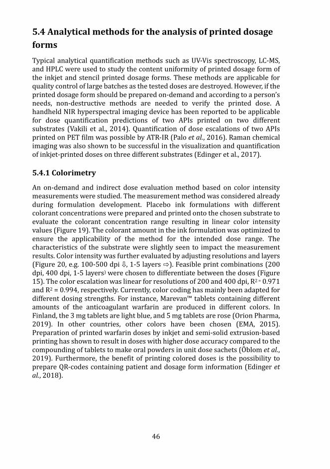
46
5.4 Analytical methods for the analysis of printed dosage forms Typical analytical quantification methods such as UV-Vis spectroscopy, LC-MS, and HPLC were used to study the content uniformity of printed dosage form of the inkjet and stencil printed dosage forms. These methods are applicable for quality control of large batches as the tested doses are destroyed. However, if the printed dosage form should be prepared on-demand and according to a person’s needs, non-destructive methods are needed to verify the printed dose. A handheld NIR hyperspectral imaging device has been reported to be applicable for dose quantification predictions of two APIs printed on two different substrates (Vakili et al., 2014). Quantification of dose escalations of two APIs printed on PET film was possible by ATR-IR (Palo et al., 2016). Raman chemical imaging was also shown to be successful in the visualization and quantification of inkjet-printed doses on three different substrates (Edinger et al., 2017).
5.4.1 Colorimetry
An on-demand and indirect dose evaluation method based on color intensity measurements were studied. The measurement method was considered already during formulation development. Placebo ink formulations with different colorant concentrations were prepared and printed onto the chosen substrate to evaluate the colorant concentration range resulting in linear color intensity values (Figure 19). The colorant amount in the ink formulation was optimized to ensure the applicability of the method for the intended dose range. The characteristics of the substrate were slightly seen to impact the measurement results. Color intensity was further evaluated by adjusting resolutions and layers (Figure 20, e.g. 100-500 dpi ⇩, 1-5 layers ⇨). Feasible print combinations (200 dpi, 400 dpi, 1-5 layers) were chosen to differentiate between the doses (Figure 15). The color escalation was linear for resolutions of 200 and 400 dpi, R2 = 0.971 and R2 = 0.994, respectively. Currently, color coding has mainly been adapted for different dosing strengths. For instance, Marevan™ tablets containing different amounts of the anticoagulant warfarin are produced in different colors. In Finland, the 3 mg tablets are light blue, and 5 mg tablets are rose (Orion Pharma, 2019). In other countries, other colors have been chosen (EMA, 2015). Preparation of printed warfarin doses by inkjet and semi-solid extrusion-based printing has shown to result in doses with higher dose accuracy compared to the compounding of tablets to make oral powders in unit dose sachets (OÄ blom et al., 2019). Furthermore, the benefit of printing colored doses is the possibility to prepare QR-codes containing patient and dosage form information (Edinger et al., 2018).
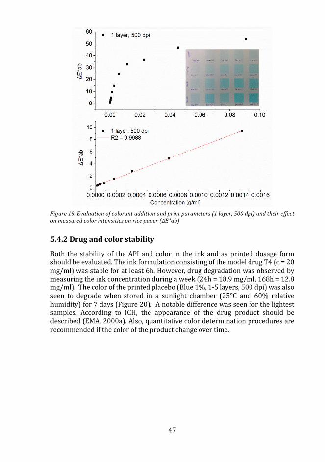
47
Figure 19. Evaluation of colorant addition and print parameters (1 layer, 500 dpi) and their effect on measured color intensities on rice paper (ΔE*ab)
5.4.2 Drug and color stability
Both the stability of the API and color in the ink and as printed dosage form should be evaluated. The ink formulation consisting of the model drug T4 (c = 20 mg/ml) was stable for at least 6h. However, drug degradation was observed by measuring the ink concentration during a week (24h = 18.9 mg/ml, 168h = 12.8 mg/ml). The color of the printed placebo (Blue 1%, 1-5 layers, 500 dpi) was also seen to degrade when stored in a sunlight chamber (25°C and 60% relative humidity) for 7 days (Figure 20). A notable difference was seen for the lightest samples. According to ICH, the appearance of the drug product should be described (EMA, 2000a). Also, quantitative color determination procedures are recommended if the color of the product change over time.
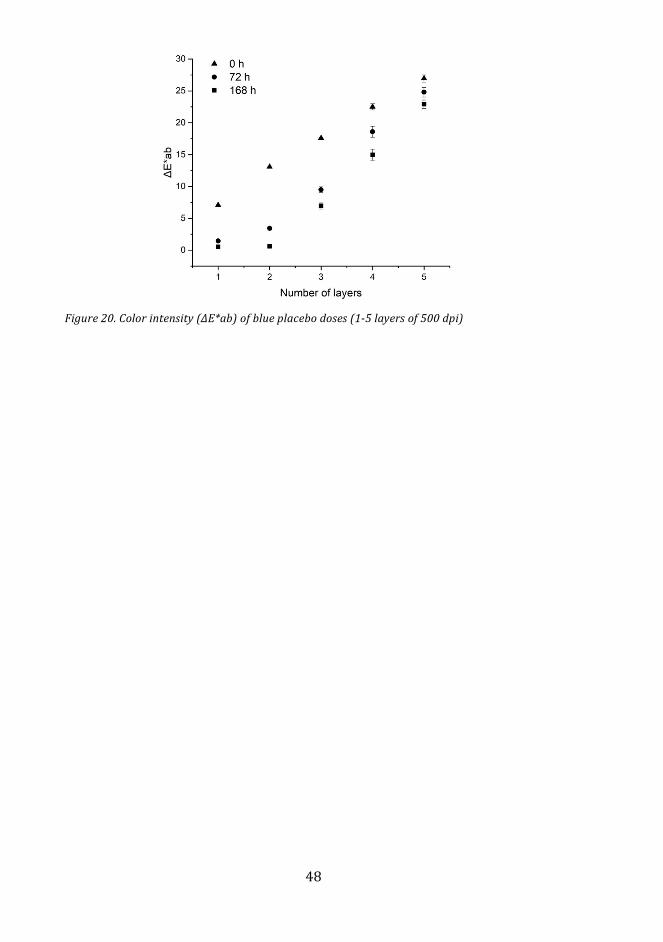
48
Figure 20. Color intensity (ΔE*ab) of blue placebo doses (1-5 layers of 500 dpi)
49
6. Conclusion
Pharmaceutical low viscous and high viscous inks were successfully formulated to study the potential of inkjet and stencil printing technologies as manufacturing methods of oral solid dosage forms. Active pharmaceutical ingredients with different characteristics were used as model drugs.
Single component ink formulations were developed for inkjet printing technology. IMC and T4 doses within intervals from 0.2 to 1.1 mg and from 25 to 590 µg, respectively, were prepared by inkjet printing. The doses showed high content uniformity.
A multicomponent ink consisting of four vitamin Bs dissolved in a commercially available yellow colored ink, for thermal inkjet printers, was prepared. Vitamin doses of up to 120 µg were made by applying 1-10 layers of the multivitamin ink on three different substrates. Both multivitamin (yellow) and levothyroxine doses (blue) were used to evaluate colorimetry as an on-demand dose quantification method. Optimization of the blue colorant addition was done to ensure the applicability of the method for the therapeutic levothyroxine dose range (1-5 layers, 100-500 dpi). Linear dose escalations and color intensity results were achieved. However, the compounds of the ink formulations and as printed solid dosage forms were seen to degrade by time. Also, the chosen colorant should remain stable if the color is to be used as an indirect dose quantification method. In conclusion, the colorimeter has potential to be used for on-demand dose quantification.
A drug-loaded MSN-PEI nanosuspension was successfully prepared and printed for the first time using piezoelectric inkjet technology. Inkjet printing showed to enable precise deposition of the nanosuspension and could be utilized for delivery of a precise dose of a potent drug with poor solubility and permeability. The antidiuretic drug furosemide was used as a model drug and it remained incorporated in the PEI coated MSN particles during dispersion in the solvent mixture (ink). The nanosuspension remained physically stable, according to the multiple light scattering results, for the time required for printing the doses.
The ink absorption and spreading (contact angle) was dependent on the ink and substrate characteristics. The opaque fibrous substrates rice and copy paper swelled upon ink deposition. Sugar paper was seen to smoothen as the ink was applied. A coffee ring phenomenon was observed for the MSN-PEI print deposits, which was caused by convective flow happening as the solvents evaporated from the ink deposit.
The printed indomethacin doses were deposited on porous copy paper and were seen to remain yellow, which would indicate that the drug was either dissolved or amorphous in the dosage form. The solid-state of the doses prepared on porous copy paper was not possible to characterize by ATR-IR, due to the strong interference from the substrate. Doses printed on a transparency film using a drug and an amino acid ink in a 1:1 molar ratio was shown to be co-amorphous. However, stabilization of indomethacin by the addition of an
50
amino acid or polymer could not be confirmed. The dissolution rate was increased for all indomethacin formulations compared to the crystalline reference, regardless of the added excipients.
Stencil printing was studied for the first time as a manufacturing method of pharmaceuticals. A semi-solid ink consisting of a film-forming polymer and a poorly soluble drug was prepared for the printing of ODFs with various dose strengths. Doses within a specific dose range (0.49–2.56 mg) of the orodispersible printed dosage forms were successfully prepared as a flat-bed process and the dosage formed fulfilled both mass and content uniformity requirements. Results obtained from the X-ray and DSC measurements suggest that the API was amorphous.
51
7. Future perspectives
Recent advancements in technology and manufacturing could facilitate the development of a more interactive healthcare system utilizing collected data and focusing on the needs of individual patients. Monitoring the individual’s response to treatment could offer the possibility to tailor the dose or drug delivery. Thus, the development of innovative manufacturing methods and quality control methods enabling flexible, on-demand manufacturing and real-time release are still needed.
The printing technologies explored in this thesis are examples of manual (stencil) and digital (inkjet) methods, that could be used to manufacture low dose pharmaceuticals on edible paper or orodispersible films. Inkjet could be utilized if dosing precision is critical, due to the possibility of depositing picolitre droplets on demand and high content uniformity. The printing method could be used for simultaneous personalized dose manufacturing and labeling (e.g. QR codes). If this approach is taken, more research about absorptive substrates and print quality is needed to ensure QR code scan accuracy. Research using an engineered stencil printer should still be conducted to study the applicability of producing pharmaceuticals by this conventional printing method. The potential for implementing stencil printing as part of a continuous ODF manufacturing line would be interesting to evaluate in the future.
If printing of medicines would be introduced as a manufacturing method in pharmacies, the printer could be viewed as a platform enabling the production and dosing of the drug delivery system and it would be subjected to specifications critical for product quality. The quality of the pharmaceutical ink and substrate quality would need to be ensured before use in the printer platform. Implementation of non-destructive quality control methods would also be needed to ensure the quality of the final personalized dosage form.
From an economical point of view, mainly potent niche products would be feasible to be prepared in the future by printing since the current quality control devices are quite expensive. However, large efforts are still required from society, industry, and regulatory bodies to move towards a decentralized, personalized, and affordable treatment of individuals using printed tailor-made medicines.
52
8. Acknowledgements
There are a lot of similarities in pursuing a Ph.D. as in being an entrepreneur. You need 1) a topic to investigate, 2) a vision to achieve, 3) to apply and gain funding, 4) build a network where ideas, challenges, failure, and success can be shared, and 5) to be persistent and believe in yourself.
The research presented in this Ph.D. thesis was carried out between 2014 and 2020 at the Pharmaceutical Sciences Laboratory at AÅ bo Akademi University, the Laboratory of Pharmaceutical Process Analytical Technology at Ghent University, and the Laboratory of Natural Materials Technology at AÅ bo Akademi University. I am grateful for having had the possibility to pursue a joint doctorate in collaboration with both universities.
I would like to express my gratitude and respect to my supervisors and mentors. You have been excellent role models and have taught me innovative and critical thinking, networking skills, project management skills, and how to be a pharma professional.
I would like to acknowledge all my co-authors, co-workers, and personnel in AÅ bo and Ghent, colleagues at Skanssin Apteekki and Bayer Oy. It has been nice to work together, acquire new skills and learn from each other.
I am also happy for being part of the Finnish Society of Physical Pharmacy, PSSRC, Nordic POP, and BBBB research clusters. In these communities, I have met nice cluster members and learned to present my research and to discuss different research topics. I am also grateful for being chosen to participate in the networking program Pharma Future Stars arranged by Pharma Industry Finland. It was a great opportunity to get to know inspiring fellow students with similar interests and learn from pharma professionals with different work experience.
I sincerely thank the opponent and the professors that have reviewed this thesis. Your valued opinions have contributed to this result and have strengthened my confidence as a researcher.
I would like to express my deepest gratitude to my family and friends for being there for me, for encouraging me to develop and to achieve my goals.
All in all, this journey has taught me how to investigate, innovate, network, write, communicate, fail, succeed, and believe in myself. I am thankful for the acquired competencies and for being able to learn new things every day. Without the help and support of all people involved, this journey would have been impossible to make. Thank you!

53
Henrika Wickstrom acknowledges the following organizations and foundations for funding her research and Ph.D. thesis:
- The Doctoral Network of Material Research (DNMR) at Åbo Akademi - Finnish Cultural Foundation (Grant #00181173) - Academy of Finland (Projects #284542, 309374) - Tor, Joe och Pentti Borg Foundation - Waldemar von Frenckell Foundation - Jane and Aatos Erkko Foundation - Joint Ph.D. network at Ghent University - Åbo Akademi University
54
9. References
Abdel-Rahman, S. M. et al. (2012) ‘Summary of the National Institute of Child Health and Human Development-Best Pharmaceuticals for Children Act Pediatric Formulation Initiatives Workshop-Pediatric Biopharmaceutics Classification System Working Group’, Clinical Therapeutics. doi: 10.1016/j.clinthera.2012.09.014.
Agarwal, S., Wendorff, J. H. and Greiner, A. (2008) ‘Use of electrospinning technique for biomedical applications’, Polymer. Elsevier BV, pp. 5603–5621. doi: 10.1016/j.polymer.2008.09.014.
El Aita, I., Breitkreutz, J. and Quodbach, J. (2019) ‘On-demand manufacturing of immediate release levetiracetam tablets using pressure-assisted microsyringe printing’, European Journal of Pharmaceutics and Biopharmaceutics. Elsevier, 134(November 2018), pp. 29–36. doi: 10.1016/j.ejpb.2018.11.008.
Aleksovski, A. et al. (2015) ‘Mini-tablets: A contemporary system for oral drug delivery in targeted patient groups’, Expert Opinion on Drug Delivery. Informa Healthcare, pp. 65–84. doi: 10.1517/17425247.2014.951633.
Alomari, M. et al. (2018) ‘Printing T3 and T4 oral drug combinations as a novel strategy for hypothyroidism’, International Journal of Pharmaceutics. Elsevier B.V., 549(1–2), pp. 363–369. doi: 10.1016/j.ijpharm.2018.07.062.
Amidon, G. L. et al. (1995) ‘A Theoretical Basis for a Biopharmaceutic Drug Classification: The Correlation of in Vitro Drug Product Dissolution and in Vivo Bioavailability’, Pharmaceutical Research: An Official Journal of the American Association of Pharmaceutical Scientists, 12(3), pp. 413–420. doi: 10.1023/A:1016212804288.
Aprecia Pharmaceuticals (2016) First FDA-approved medicine manufactured using 3D printing technology now available. doi: https://www.aprecia.com/news/first-fda-approved-medicine-manufactured-using-3d-printing-technology-now-available.
Aprecia Pharmaceuticals (2018) Zipdose. Available at:
https://www.aprecia.com/technology/zipdose (Accessed: 2 April 2020).
Azizi Machekposhti, S., Mohaved, S. and Narayan, R. J. (2019) ‘Inkjet dispensing technologies: recent advances for novel drug discovery’, Expert Opinion on Drug Discovery. Taylor and Francis Ltd, pp. 101–113. doi: 10.1080/17460441.2019.1567489.
Batchelor, H. (2014) ‘Paediatric biopharmaceutics classification system: Current status and future decisions’, International Journal of Pharmaceutics. Elsevier, 469(2), pp. 251–253. doi: 10.1016/j.ijpharm.2014.02.046.
Bishop, C. A. (2015) ‘Polymer Coating Basic Information’, in Vacuum Deposition Onto Webs, Films and Foils. Elsevier, pp. 221–239. doi: 10.1016/b978-0-323-29644-1.00011-6.
Blagden, N. et al. (2007) ‘Crystal engineering of active pharmaceutical ingredients to improve solubility and dissolution rates’, Advanced Drug Delivery Reviews, pp. 617–630. doi: 10.1016/j.addr.2007.05.011.
Blonde, L. and San Juan, Z. T. (2012) ‘Fixed-dose combinations for treatment of type 2 diabetes mellitus’, Advances in Therapy, pp. 1–13. doi: 10.1007/s12325-011-0094-1.
Boateng, J. S., Stevens, H. N. E., et al. (2009) ‘Development and mechanical characterization of solvent-cast polymeric films as potential drug delivery systems to mucosal surfaces’, Drug Development and Industrial Pharmacy, 35(8), pp. 986–996. doi: 10.1080/03639040902744704.
Boateng, J. S., Matthews, K. H., et al. (2009) ‘In vitro drug release studies of polymeric freeze-dried wafers and solvent-cast films using paracetamol as a model soluble drug’, International Journal of Pharmaceutics, 378(1–2), pp. 66–72. doi: 10.1016/j.ijpharm.2009.05.038.
Boateng, J. S. et al. (2010) ‘Characterisation of freeze-dried wafers and solvent evaporated films as potential drug delivery systems to mucosal surfaces’, International Journal of Pharmaceutics, 389(1–2), pp. 24–31. doi:
55
10.1016/j.ijpharm.2010.01.008. Bonhoeffer, B. et al. (2017) ‘Experimental
Characterization and Simulation of a Piezo-Actuated Micro Dispensing Valve’, Journal of Fluids Engineering, Transactions of the ASME. American Society of Mechanical Engineers (ASME), 139(5). doi: 10.1115/1.4035634.
Bonhoeffer, B., Kwade, A. and Juhnke, M. (2017) ‘Impact of Formulation Properties and Process Parameters on the Dispensing and Depositioning of Drug Nanosuspensions Using Micro-Valve Technology’, Journal of Pharmaceutical Sciences. Elsevier B.V., 106(4), pp. 1102–1110. doi: 10.1016/j.xphs.2016.12.019.
Borges, A. F. et al. (2015) ‘Oral films: Current status and future perspectives: I-Galenical development and quality attributes’, Journal of Controlled Release. Elsevier, pp. 1–19. doi: 10.1016/j.jconrel.2015.03.006.
Brniak, W., Maslak, E. and Jachowicz, R. (2015) ‘Orodispersible films and tablets with prednisolone microparticles’, European Journal of Pharmaceutical Sciences. doi: 10.1016/j.ejps.2015.04.006.
Buanz, A. B. M. et al. (2011) ‘Preparation of personalized-dose salbutamol sulphate oral films with thermal ink-jet printing’, Pharmaceutical Research, 28(10), pp. 2386–2392. doi: 10.1007/s11095-011-0450-5.
Buanz, A. B. M. et al. (2015) ‘Ink-jet printing versus solvent casting to prepare oral films: Effect on mechanical properties and physical stability’, International Journal of Pharmaceutics. Elsevier, 494(2), pp. 611–618. doi: 10.1016/j.ijpharm.2014.12.032.
Butler, J. M. and Dressman, J. B. (2010) ‘The developability classification system: Application of biopharmaceutics concepts to formulation development’, Journal of Pharmaceutical Sciences. John Wiley and Sons Inc., 99(12), pp. 4940–4954. doi: 10.1002/jps.22217.
Cader, H. K. et al. (2019) ‘Water-based 3D inkjet printing of an oral pharmaceutical dosage form’, International Journal of Pharmaceutics. Elsevier B.V., 564, pp. 359–368. doi: 10.1016/j.ijpharm.2019.04.026.
Calvert, P. (2001) ‘Inkjet printing for materials and devices’, Chemistry of Materials. doi: 10.1021/cm0101632.
Cassio, A. et al. (2013) ‘Comparison between liquid and tablet formulations of levothyroxine in the initial treatment of congenital hypothyroidism’, Journal of Pediatrics. Mosby Inc., 162(6). doi: 10.1016/j.jpeds.2012.11.070.
Cheow, W. S., Kiew, T. Y. and Hadinoto, K. (2015) ‘Combining inkjet printing and amorphous nanonization to prepare personalized dosage forms of poorly-soluble drugs’, European Journal of Pharmaceutics and Biopharmaceutics. Elsevier, 96, pp. 314–321. doi: 10.1016/j.ejpb.2015.08.012.
Cohen, J. S. (1999) ‘Ways to minimize adverse drug reactions: Individualized doses and common sense are key’, Postgraduate Medicine. McGraw-Hill Companies, pp. 163–172. doi: 10.3810/pgm.1999.09.688.
Collins, F. S. an. H. V. (2015) ‘A new initiative on precision medicine’, New England Journal of Medicine, 372(9), pp. 793–795. doi: 10.1056/NEJMp1500523.
Daly, R. et al. (2015) ‘Inkjet printing for pharmaceutics - A review of research and manufacturing’, International Journal of Pharmaceutics. Elsevier, 494(2), pp. 554–567. doi: 10.1016/j.ijpharm.2015.03.017.
Davanço, M. G., Campos, D. R. and Carvalho, P. de O. (2020) ‘In vitro – In vivo correlation in the development of oral drug formulation: A screenshot of the last two decades’, International Journal of Pharmaceutics. Elsevier B.V. doi: 10.1016/j.ijpharm.2020.119210.
Dengale, S. J. et al. (2014) ‘Preparation and characterization of co-amorphous Ritonavir-Indomethacin systems by solvent evaporation technique: Improved dissolution behavior and physical stability without evidence of intermolecular interactions’, European Journal of Pharmaceutical Sciences. Elsevier, 62, pp. 57–64. doi: 10.1016/j.ejps.2014.05.015.
Dengale, S. J. et al. (2016) ‘Recent advances in co-amorphous drug formulations’, Advanced Drug Delivery Reviews. Elsevier B.V., pp. 116–125. doi: 10.1016/j.addr.2015.12.009.
Derby, B. (2010) ‘Inkjet Printing of Functional and Structural Materials: Fluid Property Requirements, Feature Stability, and Resolution’, Annual Review of Materials
56
Research. Annual Reviews, 40(1), pp. 395–414. doi: 10.1146/annurev-matsci-070909-104502.
Dixit, R. P. and Puthli, S. P. (2009) ‘Oral strip technology: Overview and future potential’, Journal of Controlled Release, pp. 94–107. doi: 10.1016/j.jconrel.2009.06.014.
Edinger, M. et al. (2017) ‘Visualization and Non-Destructive Quantification of Inkjet-Printed Pharmaceuticals on Different Substrates Using Raman Spectroscopy and Raman Chemical Imaging’, Pharmaceutical Research. Springer New York LLC, 34(5), pp. 1023–1036. doi: 10.1007/s11095-017-2126-2.
Edinger, M. et al. (2018) ‘QR encoded smart oral dosage forms by inkjet printing’, International Journal of Pharmaceutics. Elsevier B.V., 536(1), pp. 138–145. doi: 10.1016/j.ijpharm.2017.11.052.
El-Alti, L., Sandman, L. and Munthe, C. (2019) ‘Person Centered Care and Personalized Medicine: Irreconcilable Opposites or Potential Companions?’, Health Care Analysis. Springer New York LLC, 27(1), pp. 45–59. doi: 10.1007/s10728-017-0347-5.
Eleftheriadis, G. K. et al. (2018) ‘In vitro evaluation of 2D-printed edible films for the buccal delivery of diclofenac sodium’, Materials. MDPI AG, 11(5). doi: 10.3390/ma11050864.
Elele, E. et al. (2012) ‘Electrodeless electrohydrodynamic drop-on-demand encapsulation of drugs into porous polymer films for fabrication of personalized dosage units’, Journal of Pharmaceutical Sciences. John Wiley and Sons Inc., 101(7), pp. 2523–2533. doi: 10.1002/jps.23165.
EMA (2000a) ICH Topic Q 6 A Specifications:Test Procedures and Acceptance Criteria for New Drug Substances and New Drug Products: Chemical Substances. Available at: https://www.ema.europa.eu/en/documents/scientific-guideline/ich-q-6-test-procedures-acceptance-criteria-new-drug-substances-new-drug-products-chemical_en.pdf.
EMA (2000b) ICH Topic Q6A Specifications: Test Procedures and Acceptance Criteria
for New Drug Substances and New Drug Products: Chemical Substances. Available at: http://www.emea.eu.int.
EMA (2015) Good practice guide medication error risk minimisation and prevention. Available at: https://www.ema.europa.eu/en/documents/regulatory-procedural-guideline/good-practice-guide-risk-minimisation-prevention-medication-errors_en.pdf.
EMA (2017a) Committee for Human Medicinal Products (CHMP) Propylene glycol used as an excipient. Available at: www.ema.europa.eu/contact.
EMA (2017b) ICH guideline Q8 (R2) on pharmaceutical development. Available at: www.ema.europa.eu/contact.
EMA (2019a) EMA Regulatory Science to 2025 Strategic reflection.
EMA (2019b) ICH guideline Q3C (R6) on impurities: guideline for residual solvents Step 5. Available at: www.ema.europa.eu/contactsTelephone+31.
European Commission (2001) Dietary Food Additive Intake in the European Union. Available at: https://ec.europa.eu/transparency/regdoc/rep/1/2001/EN/1-2001-542-EN-F1-1.Pdf.
European Commission (2013) Good Distribution Practice of medicinal products for human use. Available at: https://eur-lex.europa.eu/LexUriServ/LexUriServ.do?uri=OJ:C:2013:343:0001:0014:EN:PDF.
European Commission (2014a) Chapter 5 Production. Available at: https://ec.europa.eu/health/sites/health/files/files/eudralex/vol-4/chapter_5.pdf.
European Commission (2014b) Chapter 6 Quality control. Available at: https://ec.europa.eu/health/sites/health/files/files/eudralex/vol-4/2014-11_vol4_chapter_6.pdf.
FDA (2004) Guidance for Industry PAT - A Framework for Innovative Pharmaceutical Development, manufacturing, and Quality Assurance. Available at: http://www.fda.gov/cvm/guidance/published.html.
Fina, F. et al. (2017) ‘Selective laser sintering (SLS) 3D printing of medicines’, International Journal of Pharmaceutics.
57
Elsevier B.V., 529(1–2), pp. 285–293. doi: 10.1016/j.ijpharm.2017.06.082.
Galdeano, M. C. et al. (2009) ‘Effects of plasticizers on the properties of oat starch films’, Materials Science and Engineering C, 29(2), pp. 532–538. doi: 10.1016/j.msec.2008.09.034.
De Gans, B. J., Duineveld, P. C. and Schubert, U. S. (2004) ‘Inkjet printing of polymers: State of the art and future developments’, Advanced Materials. Wiley-VCH Verlag, pp. 203–213. doi: 10.1002/adma.200300385.
Gao, W. et al. (2015) ‘The status, challenges, and future of additive manufacturing in engineering’, CAD Computer Aided Design. Elsevier Ltd, 69, pp. 65–89. doi: 10.1016/j.cad.2015.04.001.
Gavezzotti, A. (2007) ‘A solid-state chemist’s view of the crystal polymorphism of organic compounds’, Journal of Pharmaceutical Sciences. John Wiley and Sons Inc., pp. 2232–2241. doi: 10.1002/jps.20870.
Genina, N., Fors, D., et al. (2013) ‘Behavior of printable formulations of loperamide and caffeine on different substrates - Effect of print density in inkjet printing’, International Journal of Pharmaceutics. doi: 10.1016/j.ijpharm.2013.06.003.
Genina, N., Janßen, E. M., et al. (2013) ‘Evaluation of different substrates for inkjet printing of rasagiline mesylate’, European Journal of Pharmaceutics and Biopharmaceutics. Elsevier B.V., 85(3 PART B), pp. 1075–1083. doi: 10.1016/j.ejpb.2013.03.017.
Gioumouxouzis, C. I. et al. (2018) ‘A 3D printed bilayer oral solid dosage form combining metformin for prolonged and glimepiride for immediate drug delivery’, European Journal of Pharmaceutical Sciences. Elsevier B.V., 120, pp. 40–52. doi: 10.1016/j.ejps.2018.04.020.
Govender, R. et al. (2020) ‘Therapy for the individual: Towards patient integration into the manufacturing and provision of pharmaceuticals’, European Journal of Pharmaceutics and Biopharmaceutics. Elsevier B.V., pp. 58–76. doi: 10.1016/j.ejpb.2020.01.001.
Goyanes, A. et al. (2014) ‘Fused-filament 3D printing (3DP) for fabrication of tablets’, International Journal of Pharmaceutics.
Elsevier, 476(1), pp. 88–92. doi: 10.1016/j.ijpharm.2014.09.044.
Goyanes, A. et al. (2019) ‘Direct powder extrusion 3D printing: Fabrication of drug products using a novel single-step process’, International Journal of Pharmaceutics. Elsevier B.V., 567. doi: 10.1016/j.ijpharm.2019.118471.
Gubala, V. et al. (2012) ‘Point of care diagnostics: Status and future’, Analytical Chemistry, pp. 487–515. doi: 10.1021/ac2030199.
Gudapati, H., Dey, M. and Ozbolat, I. (2016) ‘A comprehensive review on droplet-based bioprinting: Past, present and future’, Biomaterials. Elsevier Ltd, pp. 20–42. doi: 10.1016/j.biomaterials.2016.06.012.
Hamburg, M. A. and Collins, F. S. (2010) ‘The path to personalized medicine’, New England Journal of Medicine. Massachussetts Medical Society, pp. 301–304. doi: 10.1056/NEJMp1006304.
Haring, A. P. et al. (2018) ‘Programming of Multicomponent Temporal Release Profiles in 3D Printed Polypills via Core–Shell, Multilayer, and Gradient Concentration Profiles’, Advanced Healthcare Materials. Wiley-VCH Verlag, 7(16). doi: 10.1002/adhm.201800213.
Helmut Kipphan (ed.) (2001) Handbook of print media: technologies and production methods. Springer.
Hetrick, E. M. et al. (2013) ‘Integrating tristimulus colorimetry into pharmaceutical development for color selection and physical appearance control: A quality-by-design approach’, Journal of Pharmaceutical Sciences. John Wiley and Sons Inc., 102(8), pp. 2608–2621. doi: 10.1002/jps.23625.
HHS and FDA (2013) Paving the Way for Personalized Medicine FDA’s Role in a New Era of Medical Product Development FDA’s Role in a New Era of Medical Product Development.
Hirshfield, L. et al. (2014) ‘Dropwise additive manufacturing of pharmaceutical products for solvent-based dosage forms’, Journal of Pharmaceutical Sciences. John Wiley and Sons Inc., 103(2), pp. 496–506. doi: 10.1002/jps.23803.
Hirshfield, L. et al. (2015) ‘Real-Time Process Management Strategy for Dropwise
58
Additive Manufacturing of Pharmaceutical Products’, Journal of Pharmaceutical Innovation. Springer New York LLC, 10(2), pp. 140–155. doi: 10.1007/s12247-015-9218-5.
Hoffmann, E. M., Breitenbach, A. and Breitkreutz, J. (2011) ‘Advances in orodispersible films for drug delivery’, Expert Opinion on Drug Delivery. Informa Healthcare, 8(3), pp. 299–316. doi: 10.1517/17425247.2011.553217.
Horsnell, D., A. et al. (2009) ‘Solenoid valve fro a drop on demand inkjet printer’. Available at: https://patentimages.storage.googleapis.com/6a/af/08/28794dfbff1cad/US7571986.pdf.
Horter, D. and Dressman, J. B. (2001) Influence of physicochemical properties on dissolution of drugs q in the gastrointestinal tract, Advanced Drug Delivery Reviews. Available at: www.elsevier.com/locate/drugdeliv.
Hsu, H. Y. et al. (2015) ‘Drop printing of pharmaceuticals: Effect of molecular weight on PEG coated-naproxen/PEG 3350 solid dispersions’, AIChE Journal. John Wiley and Sons Inc., 61(12), pp. 4502–4508. doi: 10.1002/aic.14979.
Hsu, H. yun et al. (2013) ‘Effect of substrates on naproxen-polyvinylpyrrolidone solid dispersions formed via the drop printing technique’, Journal of Pharmaceutical Sciences. John Wiley and Sons Inc., 102(2), pp. 638–648. doi: 10.1002/jps.23397.
Huang, C. Y. et al. (2011) ‘The solder paste printing process: Critical parameters, defect scenarios, specifications, and cost reduction’, Soldering & Surface Mount Technology. doi: 10.1108/09540911111169057.
Içten, E. et al. (2015) ‘Dropwise additive manufacturing of pharmaceutical products for melt-based dosage forms’, Journal of Pharmaceutical Sciences. John Wiley and Sons Inc., 104(5), pp. 1641–1649. doi: 10.1002/jps.24367.
Iftimi, L. D. et al. (2019) ‘Edible solid foams as porous substrates for inkjet-printable pharmaceuticals’, European Journal of Pharmaceutics and Biopharmaceutics. Elsevier B.V., 136, pp. 38–47. doi: 10.1016/j.ejpb.2019.01.004.
Infanger, S. et al. (2019) ‘Powder bed 3D-printing of highly loaded drug delivery devices with hydroxypropyl cellulose as solid binder’, International Journal of Pharmaceutics. Elsevier B.V., 555, pp. 198–206. doi: 10.1016/j.ijpharm.2018.11.048.
Jamróz, W. et al. (2017) ‘3D printed orodispersible films with Aripiprazole’, International Journal of Pharmaceutics. Elsevier B.V., 533(2), pp. 413–420. doi: 10.1016/j.ijpharm.2017.05.052.
Jang, D., Kim, D. and Moon, J. (2009) ‘Influence of fluid physical properties on ink-jet printability’, Langmuir. doi: 10.1021/la900059m.
Janßen, E. M. et al. (2013) ‘Drug-printing by flexographic printing technology - A new manufacturing process for orodispersible films’, International Journal of Pharmaceutics. Elsevier B.V., 441(1–2), pp. 818–825. doi: 10.1016/j.ijpharm.2012.12.023.
Kamisukil, S. et al. (1998) A low power, small,electrostatically-driven commercial inkjet head, IEEE International Conference.
Katstra, W. E. et al. (2000) ‘Oral dosage forms fabricated by Three Dimensional PrintingE’, Journal of Controlled Release, 66, pp. 1–9. Available at: www.elsevier.com.
Kawabata, Y. et al. (2011) ‘Formulation design for poorly water-soluble drugs based on biopharmaceutics classification system: Basic approaches and practical applications’, International Journal of Pharmaceutics. Elsevier B.V., pp. 1–10. doi: 10.1016/j.ijpharm.2011.08.032.
Kay, R. and Desmulliez, M. (2012) ‘A review of stencil printing for microelectronic packaging’, Soldering and Surface Mount Technology, pp. 38–50. doi: 10.1108/09540911211198540.
Kearns, G. L. et al. (2003) ‘Developmental Pharmacology — Drug Disposition, Action, and Therapy in Infants and Children’, The new england journal of medicine, 12(18), p. 349. Available at: www.nejm.org.
Khaled, S. A. et al. (2014) ‘Desktop 3D printing of controlled release pharmaceutical bilayer tablets’, International Journal of Pharmaceutics, 461(1–2), pp. 105–111. doi: 10.1016/j.ijpharm.2013.11.021.
Khaled, S. A. et al. (2015) ‘3D printing of five-
59
in-one dose combination polypill with defined immediate and sustained release profiles’, Journal of Controlled Release. Elsevier, 217, pp. 308–314. doi: 10.1016/j.jconrel.2015.09.028.
Klingmann, V. et al. (2020) ‘Acceptability of an Orodispersible Film Compared to Syrup in Neonates and Infants: A Randomized Controlled Trial’, European Journal of Pharmaceutics and Biopharmaceutics. doi: 10.1016/j.ejpb.2020.03.018.
Kollamaram, G., Hopkins, S. C., et al. (2018) ‘Inkjet printing of paracetamol and indomethacin using electromagnetic technology: Rheological compatibility and polymorphic selectivity’, European Journal of Pharmaceutical Sciences. Elsevier B.V., 115, pp. 248–257. doi: 10.1016/j.ejps.2018.01.036.
Kollamaram, G., Faucher, A., et al. (2018) ‘Valvejet Technology for the Production of a Personalised Fixed Dose Combination of Ramipril and Glimepiride: an Investigative Study on the Stability of Ramipril’, Pharmaceutical Research. Springer New York LLC, 35(9). doi: 10.1007/s11095-018-2465-7.
Kyobula, M. et al. (2017) ‘3D inkjet printing of tablets exploiting bespoke complex geometries for controlled and tuneable drug release’, Journal of Controlled Release. doi: 10.1016/j.jconrel.2017.06.025.
Laurberg, P. et al. (2005) Hypothyroidism in the Elderly: Pathophysiology, Diagnosis and Treatment, Drugs Aging.
Lazarus, N., Bedair, S. S. and Kierzewski, I. M. (2017) ‘Ultrafine pitch stencil printing of liquid metal alloys’, ACS Applied Materials and Interfaces. American Chemical Society, 9(2), pp. 1178–1182. doi: 10.1021/acsami.6b13088.
Le, H. P. (1999) Progress and Trends in Ink-jet Printing Technology.
Lee, B. K. et al. (2012) ‘Fabrication of drug-loaded polymer microparticles with arbitrary geometries using a piezoelectric inkjet printing system’, International Journal of Pharmaceutics. doi: 10.1016/j.ijpharm.2012.02.011.
Lim, S. H. et al. (2018) ‘3D printed drug delivery and testing systems — a passing fad or the future?’, Advanced Drug Delivery Reviews. Elsevier B.V., 132, pp. 139–168.
doi: 10.1016/j.addr.2018.05.006. Lind, J. et al. (2017) ‘Social aspects in additive
manufacturing of pharmaceutical products’, Expert Opinion on Drug Delivery. Taylor and Francis Ltd, pp. 927–936. doi: 10.1080/17425247.2017.1266336.
Liu, F. et al. (2014) ‘Patient-centred pharmaceutical design to improve acceptability of medicines: Similarities and differences in paediatric and geriatric populations’, Drugs. Springer International Publishing, pp. 1871–1889. doi: 10.1007/s40265-014-0297-2.
Liu, Y. F. et al. (2013) ‘Control of droplet formation by operating waveform for inks with various viscosities in piezoelectric inkjet printing’, Applied Physics A: Materials Science and Processing, 111(2), pp. 509–516. doi: 10.1007/s00339-013-7569-7.
Löbmann, K. et al. (2013) ‘Amino acids as co-amorphous stabilizers for poorly water soluble drugs - Part 1: Preparation, stability and dissolution enhancement’, European Journal of Pharmaceutics and Biopharmaceutics. Elsevier B.V., 85(3 PART B), pp. 873–881. doi: 10.1016/j.ejpb.2013.03.014.
Lu, Q. and Zografi, G. (1998) ‘Phase Behavior of Binary and Ternary Amorphous Mixtures Containing Indomethacin, Citric Acid, and PVP’, Pharmacutical Research, 15(8), pp. 1202–1206.
Magdassi, S. (ed.) (2010) The chemistry of inkjet inks. World Scientific Publishing Co. Pte. Ltd.
Manda, P. et al. (2018) ‘Micronized Zaleplon Delivery via Orodispersible Film and Orodispersible Tablets’, AAPS PharmSciTech. Springer New York LLC, 19(3), pp. 1358–1366. doi: 10.1208/s12249-017-0924-9.
McNestry, M. (2014) ‘Print head with electromagnetic valve assembly’. Available at: https://patentimages.storage.googleapis.com/79/db/4b/7d6eaa2c3370be/US8454126.pdf.
Meléndez, P. A. et al. (2008) ‘Thermal inkjet application in the preparation of oral dosage forms: Dispensing of prednisolone solutions and polymorphic characterization by solid-state

60
spectroscopic techniques’, Journal of Pharmaceutical Sciences. John Wiley and Sons Inc., 97(7), pp. 2619–2636. doi: 10.1002/jps.21189.
Melocchi, A. et al. (2019) ‘Retentive device for intravesical drug delivery based on water-induced shape memory response of poly(vinyl alcohol): design concept and 4D printing feasibility’, International Journal of Pharmaceutics. Elsevier B.V., 559, pp. 299–311. doi: 10.1016/j.ijpharm.2019.01.045.
Van Der Meulen, M. J. et al. (2016) ‘Experimental techniques for retrieving flow information from within inkjet nozzles’, Journal of Imaging Science and Technology. Society for Imaging Science and Technology, 60(4). doi: 10.2352/J.ImagingSci.Technol.2016.60.4.040502.
Meyer, J. D., Bazilevsky, A. V and Rozhkov, A. N. (1999) Effects of Polymeric Additives on Thermal Ink Jets.
Mitra, B. et al. (2017) ‘Feasibility of mini-tablets as a flexible drug delivery tool’, International Journal of Pharmaceutics. Elsevier B.V., 525(1), pp. 149–159. doi: 10.1016/j.ijpharm.2017.04.037.
Montenegro-Nicolini, M. et al. (2018) ‘The Effect of Inkjet Printing over Polymeric Films as Potential Buccal Biologics Delivery Systems’, AAPS PharmSciTech. Springer New York LLC, 19(8), pp. 3376–3387. doi: 10.1208/s12249-018-1105-1.
Van Den Mooter, G. (2012) ‘The use of amorphous solid dispersions: A formulation strategy to overcome poor solubility and dissolution rate’, Drug Discovery Today: Technologies. Elsevier Ltd. doi: 10.1016/j.ddtec.2011.10.002.
Musazzi, U. M. et al. (2018) ‘Personalized orodispersible films by hot melt ram extrusion 3D printing’, International Journal of Pharmaceutics. Elsevier B.V., 551(1–2), pp. 52–59. doi: 10.1016/j.ijpharm.2018.09.013.
Nahata, M. C. and Allen, L. V (2008) Extemporaneous Drug Formulations, Clinical Therapeutics. doi: 10.1016/S0149-2918(08)00414-1.
Öblom, H. et al. (2019) ‘Towards printed pediatric medicines in hospital pharmacies: Comparison of 2d and 3d-
printed orodispersiblewarfarin films with conventional oral powders in unit dose sachets’, Pharmaceutics. MDPI AG, 11(7). doi: 10.3390/pharmaceutics11070334.
Okwuosa, T. C. et al. (2018) ‘On demand manufacturing of patient-specific liquid capsules via co-ordinated 3D printing and liquid dispensing’, European Journal of Pharmaceutical Sciences. Elsevier B.V., 118, pp. 134–143. doi: 10.1016/j.ejps.2018.03.010.
Orion Pharma (2019) MAREVAN tabletti 3 mg, MAREVAN FORTE tabletti 5 mg. Available at: https://laakeinfo.fi/Medicine.aspx?m=698&i=ORION+PHARMA_LIDOCAIN (Accessed: 17 May 2020).
Orlu, M. et al. (2017) ‘Acceptability of orodispersible films for delivery of medicines to infants and preschool children’, Drug Delivery. Taylor and Francis Ltd, 24(1), pp. 1243–1248. doi: 10.1080/10717544.2017.1370512.
Page, S., Coupe, A. and Barrett, A. (2016) ‘An industrial perspective on the design and development of medicines for older patients’, International Journal of Pharmaceutics. Elsevier B.V., 512(2), pp. 352–354. doi: 10.1016/j.ijpharm.2016.03.008.
Palo, M. et al. (2015) ‘Fabrication of drug-loaded edible carrier substrates from nanosuspensions by flexographic printing’, International Journal of Pharmaceutics. Elsevier, 494(2), pp. 603–610. doi: 10.1016/j.ijpharm.2015.01.027.
Palo, M. et al. (2016) ‘Quantification of caffeine and loperamide in printed formulations by infrared spectroscopy’, Journal of Drug Delivery Science and Technology. Editions de Sante, 34, pp. 60–70. doi: 10.1016/j.jddst.2016.02.007.
Palo, M. et al. (2017) ‘Development of Oromucosal Dosage Forms by Combining Electrospinning and Inkjet Printing’, Molecular Pharmaceutics. American Chemical Society, 14(3), pp. 808–820. doi: 10.1021/acs.molpharmaceut.6b01054.
Pardeike, J. et al. (2011) ‘Nanosuspensions as advanced printing ink for accurate dosing of poorly soluble drugs in personalized medicines’, International Journal of Pharmaceutics. doi:
61
10.1016/j.ijpharm.2011.08.033. Park, J. and Moon, J. (2006) ‘Control of
colloidal particle deposit patterns within picoliter droplets ejected by ink-jet printing’, Langmuir. doi: 10.1021/la053450j.
Planchette, C. et al. (2016) ‘Printing medicines as orodispersible dosage forms: Effect of substrate on the printed micro-structure’, International Journal of Pharmaceutics. doi: 10.1016/j.ijpharm.2015.10.054.
Preis, M. et al. (2013) ‘Oromucosal film preparations: Classification and characterization methods’, Expert Opinion on Drug Delivery, pp. 1303–1317. doi: 10.1517/17425247.2013.804058.
Preis, M. et al. (2014) ‘Design and evaluation of bilayered buccal film preparations for local administration of lidocaine hydrochloride’, European Journal of Pharmaceutics and Biopharmaceutics. doi: 10.1016/j.ejpb.2013.12.019.
Preis, M., Knop, K. and Breitkreutz, J. (2014) ‘Mechanical strength test for orodispersible and buccal films’, International Journal of Pharmaceutics. doi: 10.1016/j.ijpharm.2013.11.033.
Preis, M. and Sandler, N. (2016) ‘Printing technologies and tailored dosing’, Pharmacy & Therapeutics. Available at: http://www.hospitalpharmacyeurope.com/pharmacy-practice/printing-technologies-and-tailored-dosing.
Radcliffe, A. J. et al. (2019) ‘Dropwise Additive Manufacturing of Pharmaceutical Products Using Particle Suspensions’, Journal of Pharmaceutical Sciences. Elsevier B.V., 108(2), pp. 914–928. doi: 10.1016/j.xphs.2018.09.030.
Raijada, D. et al. (2013) ‘a step toward development of printable dosage forms for poorly soluble drugs’, Journal of Pharmaceutical Sciences. John Wiley and Sons Inc., 102(10), pp. 3694–3704. doi: 10.1002/jps.23678.
Rantanen, J. and Khinast, J. (2015) ‘The Future of Pharmaceutical Manufacturing Sciences’, Journal of Pharmaceutical Sciences. John Wiley and Sons Inc., pp. 3612–3638. doi: 10.1002/jps.24594.
Rathbone, M. J., Hadgraft, J. and Roberts, M. S. (2003) Modified-release drug delivery technology. Marcel Dekker.
Rautamo, M. et al. (2020) ‘A focus group study about oral drug administration practices at hospital wards—aspects to consider in drug development of age-appropriate formulations for children’, Pharmaceutics. MDPI AG, 12(2). doi: 10.3390/pharmaceutics12020109.
Reis, N., Ainsley, C. and Derby, B. (2005) ‘Ink-jet delivery of particle suspensions by piezoelectric droplet ejectors’, Journal of Applied Physics, 97(9). doi: 10.1063/1.1888026.
Van Riet-Nales, D. A. et al. (2017) ‘Paediatric Drug Development and Formulation Design—a European Perspective’, AAPS PharmSciTech, 18(2), pp. 241–249. doi: 10.1208/s12249-016-0558-3.
Rosenthal, T. and Gavras, I. (2006) ‘Fixed-Drug Combinations as First-Line Treatment for Hypertension’, Progress in Cardiovascular Diseases, 48(6), pp. 416–425. doi: 10.1016/j.pcad.2006.03.003.
Rowe, C. W. et al. (2000) Multimechanism oral dosage forms fabricated by three dimensional printingE, Journal of Controlled Release. Available at: www.elsevier.com/locate/jconrel.
Rowe, R. C., Sheskey, P. J. and Quinn, E. Q. (2009) Handbook of pharmaceutical excipients. 6th edn. London: Pharmaceutical Press and American Pharmacists Association.
Rusdi, M. S. et al. (2019) ‘Stencil printing process performance on various aperture size and optimization for lead-free solder paste’, International Journal of Advanced Manufacturing Technology. Springer London, 102(9–12), pp. 3369–3379. doi: 10.1007/s00170-019-03423-9.
Sadia, M. et al. (2018) ‘From “fixed dose combinations” to “a dynamic dose combiner”: 3D printed bi-layer antihypertensive tablets’, European Journal of Pharmaceutical Sciences. Elsevier B.V., 123, pp. 484–494. doi: 10.1016/j.ejps.2018.07.045.
Sam, T. et al. (2012) ‘A benefit/risk approach towards selecting appropriate pharmaceutical dosage forms - An application for paediatric dosage form selection’, in International Journal of Pharmaceutics, pp. 115–123. doi: 10.1016/j.ijpharm.2012.05.024.
62
Sandler, N. et al. (2011) ‘Inkjet printing of drug substances and use of porous substrates-towards individualized dosing’, Journal of Pharmaceutical Sciences. John Wiley and Sons Inc., 100(8), pp. 3386–3395. doi: 10.1002/jps.22526.
Sayeed, V. (2015) Size, Shape, and Other Physical Attributes of Generic Tablets and Capsules Guidance for Industry. Available at: http://www.fda.gov/Drugs/GuidanceComplianceRegulatoryInformation/Guidances/default.htm.
Scoutaris, N. et al. (2011) ‘Inkjet printing as a novel medicine formulation technique’, Journal of Controlled Release, 156(2), pp. 179–185. doi: 10.1016/j.jconrel.2011.07.033.
Selen, A. et al. (2014) ‘The biopharmaceutics risk assessment roadmap for optimizing clinical drug product performance’, Journal of Pharmaceutical Sciences. John Wiley and Sons Inc., pp. 3377–3397. doi: 10.1002/jps.24162.
Ṣen Karaman, D. et al. (2018) ‘Mesoporous silica nanoparticles facilitating the dissolution of poorly soluble drugs in orodispersible films’, European Journal of Pharmaceutical Sciences. Elsevier B.V., 122, pp. 152–159. doi: 10.1016/j.ejps.2018.06.027.
Senta-Loys, Z. et al. (2017) ‘Orodispersible films based on amorphous solid dispersions of tetrabenazine’, International Journal of Pharmaceutics. Elsevier B.V., 518(1–2), pp. 242–252. doi: 10.1016/j.ijpharm.2016.12.036.
Shariff, Z. B. et al. (2020) ‘Does the Formulation of Oral Solid Dosage Forms Affect Acceptance and Adherence in Older Patients? A Mixed Methods Systematic Review’, Journal of the American Medical Directors Association. doi: 10.1016/j.jamda.2020.01.108.
Shimoda, H. et al. (2009) ‘Preparation of a fast dissolving oral thin film containing dexamethasone: A possible application to antiemesis during cancer chemotherapy’, European Journal of Pharmaceutics and Biopharmaceutics, 73(3), pp. 361–365. doi: 10.1016/j.ejpb.2009.08.010.
Siimon, K., Siimon, H. and Järvekülg, M. (2015) ‘Mechanical characterization of
electrospun gelatin scaffolds cross-linked by glucose’, Journal of Materials Science: Materials in Medicine. Kluwer Academic Publishers, 26(1), pp. 1–9. doi: 10.1007/s10856-014-5375-1.
Simon, L. L. et al. (2015) ‘Assessment of recent process analytical technology (PAT) trends: A multiauthor review’, Organic Process Research and Development. American Chemical Society, pp. 3–62. doi: 10.1021/op500261y.
Sjöholm, E. and Sandler, N. (2019) ‘Additive manufacturing of personalized orodispersible warfarin films’, International Journal of Pharmaceutics. Elsevier B.V., 564, pp. 117–123. doi: 10.1016/j.ijpharm.2019.04.018.
Speer, I. et al. (2019) ‘Prolonged release from orodispersible films by incorporation of diclofenac-loaded micropellets’, International Journal of Pharmaceutics. Elsevier B.V., 554, pp. 149–160. doi: 10.1016/j.ijpharm.2018.11.013.
Stegemann, S. et al. (2016) ‘Defining Patient Centric Pharmaceutical Drug Product Design’, AAPS Journal. Springer New York LLC, 18(5), pp. 1047–1055. doi: 10.1208/s12248-016-9938-6.
Steiner, D., Finke, J. H. and Kwade, A. (2019) ‘SOFTs – Structured orodispersible film templates’, European Journal of Pharmaceutics and Biopharmaceutics. Elsevier B.V., 137, pp. 209–217. doi: 10.1016/j.ejpb.2019.03.001.
Teo, K. and Yusuf, S. (2018) ‘Polypill Variants (Quarter Pill Trials)’, American Journal of Hypertension. Oxford University Press, pp. 758–761. doi: 10.1093/ajh/hpy040.
Thabet, Y. and Breitkreutz, J. (2018) ‘Orodispersible films: Product transfer from lab-scale to continuous manufacturing’, International Journal of Pharmaceutics. Elsevier B.V., 535(1–2), pp. 285–292. doi: 10.1016/j.ijpharm.2017.11.021.
Thabet, Y., Lunter, D. and Breitkreutz, J. (2018a) ‘Continuous inkjet printing of enalapril maleate onto orodispersible film formulations’, International Journal of Pharmaceutics. Elsevier B.V., 546(1–2), pp. 180–187. doi: 10.1016/j.ijpharm.2018.04.064.
Thabet, Y., Lunter, D. and Breitkreutz, J.
63
(2018b) ‘Continuous manufacturing and analytical characterization of fixed-dose, multilayer orodispersible films’, European Journal of Pharmaceutical Sciences. Elsevier B.V., 117, pp. 236–244. doi: 10.1016/j.ejps.2018.02.030.
Trenfield, S. J. et al. (2018) ‘3D printed drug products: Non-destructive dose verification using a rapid point-and-shoot approach’, International Journal of Pharmaceutics. Elsevier B.V., 549(1–2), pp. 283–292. doi: 10.1016/j.ijpharm.2018.08.002.
Trenfield, S. J. et al. (2019) ‘Track-and-trace: Novel anti-counterfeit measures for 3D printed personalized drug products using smart material inks’, International Journal of Pharmaceutics. Elsevier B.V., 567. doi: 10.1016/j.ijpharm.2019.06.034.
Trenfield, S. J. et al. (2020) ‘Non-destructive dose verification of two drugs within 3D printed polyprintlets’, International Journal of Pharmaceutics. Elsevier B.V., 577. doi: 10.1016/j.ijpharm.2020.119066.
Vakili, H. et al. (2015) ‘Hyperspectral imaging in quality control of inkjet printed personalised dosage forms’, International Journal of Pharmaceutics. Elsevier, 483(1–2), pp. 244–249. doi: 10.1016/j.ijpharm.2014.12.034.
Vakili, H. et al. (2017) ‘Application of a handheld NIR spectrometer in prediction of drug content in inkjet printed orodispersible formulations containing prednisolone and levothyroxine’, International Journal of Pharmaceutics. doi: 10.1016/j.ijpharm.2017.04.014.
Valeur, K. S. et al. (2018) ‘The Cumulative Daily Tolerance Levels of Potentially Toxic Excipients Ethanol and Propylene Glycol Are Commonly Exceeded in Neonates and Infants’, Basic and Clinical Pharmacology and Toxicology. Blackwell Publishing Ltd, 122(5), pp. 523–530. doi: 10.1111/bcpt.12950.
Varan, C., Wickström, H., Sandler, N., Aktaş, Y., et al. (2017) ‘Inkjet printing of antiviral PCL nanoparticles and anticancer cyclodextrin inclusion complexes on bioadhesive film for cervical administration’. doi: 10.1016/j.ijpharm.2017.04.036.
Varan, C., Wickström, H., Sandler, N., Akta, Y.,
et al. (2017) ‘Title: Inkjet printing of antiviral PCL nanoparticles and anticancer cyclodextrin inclusion complexes on bioadhesive film for cervical administration Inkjet printing of antiviral PCL nanoparticles and anticancer cyclodextrin inclusion complexes on bioadhesive film for cervical administration’, International Journal of Pharmaceutics. doi: 10.1016/j.ijpharm.2017.04.036.
Visser, J. C. et al. (2015) ‘Orodispersible films in individualized pharmacotherapy: The development of a formulation for pharmacy preparations’, International Journal of Pharmaceutics. doi: 10.1016/j.ijpharm.2014.11.013.
Visser, J. C. et al. (2020) ‘A pediatrics utilization study in the Netherlands to identify active pharmaceutical ingredients suitable for inkjet printing on orodispersible films’, Pharmaceutics. MDPI AG, 12(2). doi: 10.3390/pharmaceutics12020164.
Voss, G. (1985) ‘Method for ink-jet printing on uncoated tablets or uncoated tablet cores’. Available at: https://patentimages.storage.googleapis.com/98/0a/23/38008c34d14788/US4548825.pdf.
Vuddanda, P. R. et al. (2018) ‘Personalisation of warfarin therapy using thermal ink-jet printing’, European Journal of Pharmaceutical Sciences. Elsevier B.V., 117, pp. 80–87. doi: 10.1016/j.ejps.2018.02.002.
Wallace, D. (2012) ‘Overview of Inkjet-Based Micromanufacturing’, in Korvink, Jan G. , Smith,Patrick J. , Brand, Oliver , Fedder, Gary K., Hierold, Christofer Tabata, Osamu, Shin, D. H. and S. D.-Y. (ed.) Inkjet-Based Micromanufacturing. John Wiley & Sons, Incorporated.
Walsh, J. et al. (2020) ‘European Paediatric Formulation Initiative Workshop Report: Improving the Administration of Oral Liquid Medicines in Paediatrics using Dosing Syringes and Enteral Accessories’, European Journal of Pharmaceutics and Biopharmaceutics. doi: 10.1016/j.ejpb.2020.04.007.
Wang, J. et al. (2016) ‘Stereolithographic (SLA) 3D printing of oral modified-release dosage forms’, International Journal of
64
Pharmaceutics. Elsevier B.V., 503(1–2), pp. 207–212. doi: 10.1016/j.ijpharm.2016.03.016.
Wang, Y. et al. (2015) ‘Mesoporous silica nanoparticles in drug delivery and biomedical applications’, Nanomedicine: Nanotechnology, Biology, and Medicine. Elsevier Inc., pp. 313–327. doi: 10.1016/j.nano.2014.09.014.
Wickström, H. et al. (2020) ‘Stencil printing—a novel manufacturing platform for orodispersible discs’, Pharmaceutics. MDPI AG, 12(1). doi: 10.3390/pharmaceutics12010033.
Wimmer-Teubenbacher, M. et al. (2018) ‘Pharmaceutical-grade oral films as substrates for printed medicine’, International Journal of Pharmaceutics. Elsevier B.V., 547(1–2), pp. 169–180. doi: 10.1016/j.ijpharm.2018.05.041.
Woertz, C. and Kleinebudde, P. (2015a) ‘Development of orodispersible polymer films containing poorly water soluble active pharmaceutical ingredients with focus on different drug loadings and storage stability’, International Journal of Pharmaceutics. doi: 10.1016/j.ijpharm.2015.07.032.
Woertz, C. and Kleinebudde, P. (2015b) ‘Development of orodispersible polymer films with focus on the solid state characterization of crystalline loperamide’, European Journal of
Pharmaceutics and Biopharmaceutics. doi: 10.1016/j.ejpb.2015.04.036.
Wu, B. M. et al. (1996) Solid free-form fabrication of drug delivery devices, Journal of Controlled Release.
Wu, D. and Sun, D. W. (2013) ‘Colour measurements by computer vision for food quality control - A review’, Trends in Food Science and Technology, pp. 5–20. doi: 10.1016/j.tifs.2012.08.004.
Xia, T. et al. (2009) ‘Polyethyleneimine coating enhances the cellular uptake of mesoporous silica nanoparticles and allows safe delivery of siRNA and DNA constructs’, ACS Nano. doi: 10.1021/nn900918w.
You, M. et al. (2016) ‘Three-dimensional quick response code based on inkjet printing of upconversion fluorescent nanoparticles for drug anti-counterfeiting’, Nanoscale, 8. doi: 10.1039/c6nr01353h.
Zhou, L. et al. (2011) ‘A systematic method development strategy for quantitative color measurement in drug substances, starting materials, and synthetic intermediates’, Journal of Pharmaceutical Innovation, 6(4), pp. 217–231. doi: 10.1007/s12247-011-9115-5.
Zou, W. et al. (2019) ‘Tip-assisted electrohydrodynamic jet printing for high-resolution microdroplet deposition’, Materials and Design. Elsevier Ltd, 166. doi: 10.1016/j.matdes.2019.107609.
65
Original publications
I Wickström, H., Palo, M., Rijckaert, K., Kolakovic, R., Nyman, J.O., Maattanen, A., Ihalainen, P., Peltonen, J., Genina, N., de Beer, T., Lobmann, K., Rades, T. and Sandler, N. 2015. Improvement of dissolution rate of indomethacin by inkjet printing. European Journal of Pharmaceutical Sciences, 75, pp.91–100. DOI: 10.1016/j.ejps.2015.03.009
II Wickström, H., Nyman, J.O., Indola, M., Sundelin, H., Kronberg, L., Preis, M., Rantanen, J. and Sandler, N., 2017. Colorimetry as quality control tool for individual inkjet-printed pediatric formulations. AAPS PharmSciTech, 18(2), pp.293–302. DOI: 10.1208/s12249-016-0620-1
III Wickström, H., Hilgert, E., Nyman, J.O., Desai, D., Şen Karaman, D., de Beer, T., Sandler, N. and Rosenholm, J.M., 2017. Inkjet Printing of Drug-Loaded Mesoporous Silica Nanoparticles – A Platform for Drug Development. Molecules, 22(11), p.2020. DOI: 10.3390/molecules22112020
IV
Wickström, H., Koppolu, R., Makila, E., Tovakka, M., and Sandler, N. (2020). Stencil Printing – A Novel Manufacturing Platform for Orodispersible Discs. Pharmaceutics, 12(1), 33. DOI: 10.3390/pharmaceutics12010033

Curriculum Vitae
Work experience
Production Pharmacist
Bayer Oy, Turku, 21.01.2019 – present My responsibilities are to maintain and improve the quality and GMP for the Mirena family production at the Bayer Turku manufacturing site. Main tasks include change and deviation management, standard operating procedure (SOP) writing and arranging and giving GMP trainings for the production staff.
Ph.D. Researcher in Pharmaceutical Technology Åbo Akademi University and Ghent University, 01.06.2014 - 21.01.2019 During my doctoral training I have gained project management skills. Collaboration with scientists from different laboratories and disciplines have broaden my general knowledge as a scientist and improved my communication skills.
University teacher in Pharmaceutics Åbo Akademi University (8 months)
Pharmacist Skanssin apteekki, Turku Part-time employment, 01.11.2012 – 21.01.2019 Full-time 27.05.2013 – 08.09.2013
R&D division trainee Bayer Oy, Turku, 20.08 -19.10.2012 The aim of the trainee period was to gain general knowledge about the work performed at the formulation development department.
Pharmacist Korson apteekki, Vantaa, Pharmacist, 10.06–31.07.2011 Pharmacy practice as a student, 15.03–15.10.2010 Technical assistant, Summer 2009
Technical assistant Sipoon apteekki, Sipoo Summer 2007&2008
Education Ph.D. in Pharmaceutical Sciences Ghent University and Åbo Akademi University, 2014 - 2020
M.Sc. in Biosciences (Biovetenskap med inriktning farmaci) Åbo Akademi University, 2012 - 2014 Master’s thesis: Development of printed co-amorphous drug delivery systems
B.Sc. in Pharmaceutical Sciences Åbo Akademi University, 2008 - 2011 Batchelor’s thesis: Läkemedelsbehandling av Alzheimers sjukdom
ERASMUS exchange Ghent University, 2012 Laboratory of Pharmaceutical Process Analytical Technology Project: Influence of raw material properties upon critical quality attributes of continuously produced granules and tablets
Matriculation exam Sibbo Gymnasium, 2005 - 2008
Position of trust and experience Pharma future stars (PIF), Autumn 2018 Pharma Industry Finland, Participant in networking program
Board member and Editor-in-Chief, 2017 & 2018 Finnish Society of Physical Pharmacy
Chair, 2012 Student nation Östra Finlands Nation vid Åbo Akademi r.f.
Treasurer, 2009 Pharmacy student association Ex Tempore vid Åbo Akademi r.f.

Henrika Wickström
Exploring Printed Drug Formulations for Inkjet and Stencil PrintingA study in Pharmaceutical Sciences
Henrika W
ickström
// Explo
ring Printed
Drug
Form
ulations fo
r Inkjet and Stencil Printing
// 20
20
Henrika Wickström
Exploring Printed Drug Formulations for Inkjet and Stencil Printing
A study in Pharmaceutical SciencesPharmaceutical ink formulation development for inkjet and stencil printing of oral solid dosage forms are described and discussed in this doctoral thesis.
ISBN 978-952-12-4009-6