ex vivo atherosclerotic plaque tissue cultures ex...
TRANSCRIPT

1
Materials and Methods Human atherosclerotic tissue For analysis of NOD2 expression, carotid plaques from the Atherosclerotic Plaque Expression (AtheroExpress) study were used. AtheroExpress is an ongoing longitudinal multicenter cohort study, initiated in 2002 and currently being executed in two Dutch hospitals: the University Medical Center Utrecht and Sint Antonius Hospital Nieuwegein. Recruitment of patients undergoing carotid endarterectomy started in April 2002. All cohort members are followed for the occurrence of adverse cardiovascular events for a minimum of 3 years. The objective of AtheroExpress is to evaluate differential atherosclerotic plaque expression of protein in relation to future cardiovascular events and patient characteristics.1 38 additional human atherosclerotic lesions were collected from patients undergoing carotid endarterectomy at Karolinska University Hospital, Sweden. And 10 human internal mammary arteries obtained from patients undergoing coronary artery bypass surgery were used as non-atherosclerotic control arteries. Written informed consent was obtained from all participants according to the declaration of Helsinki. The investigations were approved by the Ethical Committee of Northern Stockholm and by the Medical Ethical Committees of the participating hospitals in the Netherlands. Ex vivo atherosclerotic plaque tissue cultures Ex vivo atherosclerotic plaque tissue cultures were set up as previously described.2 In brief, fresh plaque tissues were processed to remove calcified tissue, cut into small pieces (about 1.5 mm3), and washed with cold PBS. The tissue was distributed equally in a 48-well plate (~ 0.1 g tissue/well). 4 h after incubation in RPMI with 10% FCS, the tissues were incubated with indicated reagents. Thereafter, supernatants and tissue were snap-frozen and kept at -80 oC. Macrophage cultures Human peripheral blood from healthy volunteers was obtained from the Blood Center, Karolinska University Hospital, Sweden. Peripheral blood mononuclear cells (PBMC) were prepared using Lymphoprep™ gradient medium (density 1.077 g/ml; Axis-Shield, Oslo, Norway) according to the manufacturer’s instructions. Monocytes purified from PBMC were cultured for 7 days in the medium that contained 60% AIM V® medium and 30% Iscove’s Modified Eagle’s Medium (IMDM) and was supplemented with 10% inactivated human AB+ serum. The media was changed every 3 days. Subsequently, 5 × 10⁵ macrophages/ml were plated in RPMI 1640 with 1% FCS and treated with different reagents for the indicated time and concentrations at 37°C, 5% CO2. In some experiments, THP-1 cells were cultured in RPM1 1640 containing 2 mM glutamine; 10 mM HEPES; 1mM sodium pyruvate; 4.5g/L glucose; 1.5g/L sodium bicarbonate and 10% FCS. RNA isolation and real time PCR Total RNA was isolated from carotid plaque tissue or macrophages with RNeasy Mini kits (QIAGEN, Germany) with an on-column DNase digestion step. RNA quantity and quality were assessed by NanoDrop (ND-1000) Spectrophotometer and an Agilent 2100 Bioanalyzer (Agilent Technologies), respectively. First-strand of cDNA was synthesized using random hexamers and SuperScript II (Invitrogen Life Technologies, Paisley, UK). Real-time PCR on cDNA was performed in an ABI 7900 Sequence Detector (Applied Biosystems, Carlsbad, CA) with Assay on
2
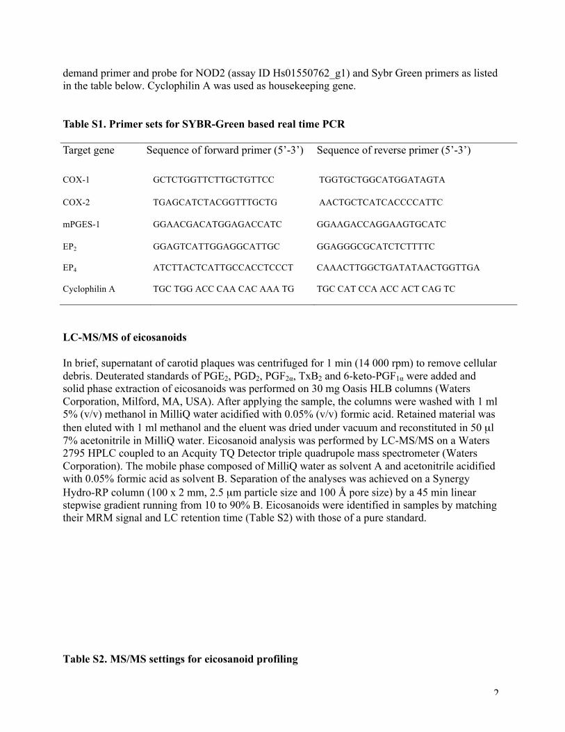
demand primer and probe for NOD2 (assay ID Hs01550762_g1) and Sybr Green primers as listed in the table below. Cyclophilin A was used as housekeeping gene.
Table S1. Primer sets for SYBR-Green based real time PCR
Target gene Sequence of forward primer (5’-3’) Sequence of reverse primer (5’-3’)
COX-1 GCTCTGGTTCTTGCTGTTCC TGGTGCTGGCATGGATAGTA
COX-2 TGAGCATCTACGGTTTGCTG AACTGCTCATCACCCCATTC
mPGES-1 GGAACGACATGGAGACCATC GGAAGACCAGGAAGTGCATC
EP2 GGAGTCATTGGAGGCATTGC GGAGGGCGCATCTCTTTTC
EP4 ATCTTACTCATTGCCACCTCCCT CAAACTTGGCTGATATAACTGGTTGA
Cyclophilin A TGC TGG ACC CAA CAC AAA TG TGC CAT CCA ACC ACT CAG TC
LC-MS/MS of eicosanoids
In brief, supernatant of carotid plaques was centrifuged for 1 min (14 000 rpm) to remove cellular debris. Deuterated standards of PGE2, PGD2, PGF2α, TxB2 and 6-keto-PGF1α were added and solid phase extraction of eicosanoids was performed on 30 mg Oasis HLB columns (Waters Corporation, Milford, MA, USA). After applying the sample, the columns were washed with 1 ml 5% (v/v) methanol in MilliQ water acidified with 0.05% (v/v) formic acid. Retained material was then eluted with 1 ml methanol and the eluent was dried under vacuum and reconstituted in 50 µl 7% acetonitrile in MilliQ water. Eicosanoid analysis was performed by LC-MS/MS on a Waters 2795 HPLC coupled to an Acquity TQ Detector triple quadrupole mass spectrometer (Waters Corporation). The mobile phase composed of MilliQ water as solvent A and acetonitrile acidified with 0.05% formic acid as solvent B. Separation of the analyses was achieved on a Synergy Hydro-RP column (100 x 2 mm, 2.5 µm particle size and 100 Å pore size) by a 45 min linear stepwise gradient running from 10 to 90% B. Eicosanoids were identified in samples by matching their MRM signal and LC retention time (Table S2) with those of a pure standard.
Table S2. MS/MS settings for eicosanoid profiling

3
Compound Retention time
(min)
Precursor ion
(m/z)
Product ion
(m/z)
Cone
(V)
Collision
(V)
6-keto-PGF1α-d4 16.9 373.1 249.2 53 21
6-keto-PGF1α 17.0 369.1 245.2 53 21
TxB2-d4 21.6 373.1 173.1 30 13
TxB2 21.6 369.1 169.1 30 13
PGF2α-d4 22.6 357.1 313.2 30 17
PGF2α 22.6 353.1 309.1 30 17
PGE2-d4 23.2 355.1 319.2 29 10
PGE2 23.3 351.1 315.1 29 10
PGD2-d4 24.2 355.1 319.1 17 10
PGD2 24.3 351.1 315.1 17 10
13-HODE1 40.0 295.2 277.1 45 25
15-HETE1 40.3 319.5 219.5 30 20
12-HETE1 40.9 319.5 179.5 30 20
5-HETE1 41.2 319.5 115.4 30 20
Western blotting
Protein of carotid plaque tissues or cells was washed with PBS and harvested following indicated treatments. Total proteins from carotid plaques or cells were extracted using T-PER tissue protein extraction reagent with protease inhibitor cocktail and EDTA (Thermo Scientific, Rockford, IL). Protein concentration was measured with the BCA assay (Bio Red, Hercules, CA). Protein samples were mixed with 2× loading buffer and boiled for 5 min. An equal amount of protein per lane was separated by SDS-PAGE (sodium dodecyl sulfate polyacrylamide gel electrophoresis) gel electrophoresis before being electroblotted onto PVDF (Polyvinylidene fluoride) membranes. The membranes were blocked in 5% non-fat milk in TBS-T (tris buffer PBS/0.1% Tween 20) and incubated with the following primary antibodies: mouse anti-human NOD2 (Cayman Chemical Co.), rabbit anti-COX-1 (Santa Cruz, CA,), mouse anti-human COX-2 (Dako, Glostrup, Denmark), mPGES-1 (provide by Dr. Per-Johan Jakobsson, Karolinska Institute), rabbit anti-human EP2 and rabbit anti-human EP4 (Cayman Chemical Co.), rabbit anti-human phospho-NF-

4
κB p65 (Ser468, Cell Signaling Technology, Beverly, MA, USA) and anti-IκBα (Cell Signaling Technology) in 3% non-fat milk overnight at 4°C, washed with TBS-T before incubation with the horseradish peroxidase-conjugated secondary antibody. To document the loading controls, the membrane was reprobed with a monoclonal antibody against α-smooth muscle actin (Sigma-Aldrich). After successive washes, the protein bands were detected with an enhanced chemiluminescence (ECL) kit (GE Healthcare, Piscataway, NJ). Optical densities of specific bands were measured with the use of the GelDoc system (Biorad, Hercules, CA) and expressed in arbitrary units.
Immunostaining Acetone-fixed 10 µm cryosections were preincubated for 30 min with 5% normal fetal calf serum or 2% BSA and 3% non-fat dry milk. Subsequently, sections were incubated with rabbit anti-NOD2 antibody (Cayman Chemical Co.) at 4°C overnight, followed with biotinylated goat-anti rabbit or horse anti-mouse secondary antibody followed by avidin-biotin peroxidase complex and developed with diaminobenzidine (all from Vector Laboratories, Burlingame, CA). The specificity of the NOD2 antibody was confirmed by incubation with isotype-matched control IgG. For double-staining, sections were first incubated with rabbit anti-NOD2 antibody (Cayman Chemical Co.) at 4°C overnight, followed with Alexa fluor 488 labeled goat anti-rabbit IgG (Life technologies, Stockholm, Sweden) for 30 min. Subsequently, the sections were incubated overnight with anti-human CD163 antibody (BioLegend, San Diego, CA), anti-CD68 antibody (BioLegend), mouse anti-human von Willebrand Factor antibody (DAKO) or anti-human COX-2 antibody (DAKO). Enzyme Immunoassays PGE2, LTB4 and LTE4 in supernatants of carotid plaques and macrophage cultures were determined by enzyme immunoassays (Cayman Chemical Co., Ann Arbor, MI) according to the manufacturer’s instructions. Data analysis Prism software (GraphPad) was used for statistical analysis. Results were analyzed by Student t test or One-way ANOVA followed by the appropriate post hoc comparison. p < 0.05 was regarded statistically significant.
References
1. de Kleijn DP, Moll FL, Hellings WE, Ozsarlak-Sozer G, de Bruin P, Doevendans PA, Vink A, Catanzariti LM, Schoneveld AH, Algra A, Daemen MJ, Biessen EA, de Jager W, Zhang H, de Vries JP, Falk E, Lim SK, van der Spek PJ, Sze SK, Pasterkamp G. Local atherosclerotic plaques are a source of prognostic biomarkers for adverse cardiovascular events. Arterioscler Thromb Vasc Biol. 2010;30:612-619 2. Ketelhuth DF, Rios FJ, Wang Y, Liu H, Johansson ME, Fredrikson GN, Hedin U, Gidlund M, Nilsson J, Hansson GK, Yan ZQ. Identification of a danger-associated peptide from

5
apolipoprotein b100 (apobds-1) that triggers innate proatherogenic responses. Circulation. 2011;124:2433-2443, 2431-2437