accelerated atherosclerosis in low-density lipoprotein...
TRANSCRIPT

Accelerated Atherosclerosis in Low-Density LipoproteinReceptor–Deficient Mice Lacking the Membrane-Bound
Complement Regulator CD59Sheng Yun, Viola W.Y. Leung, Marina Botto, Joseph J. Boyle, Dorian O. Haskard
Objective—Whereas studies in humans and animal models have suggested a role for complement activation inatherosclerosis, there has been little analysis of the importance of complement regulators. We tested the hypothesis thatthe terminal pathway inhibitor CD59 plays an essential role in limiting the proinflammatory effects ofcomplement activation.
Methods and Results—CD59 gene targeted mice (CD59a�/�) mice were crossed with low-density lipoprotein receptor–deficient (Ldlr�/�) mice. CD59-deficient Ldlr�/� mice had significantly more extensive en face Sudan IV staining ofthoracoabdominal aorta than Ldlr�/� single knock-outs, both after a low-fat diet (6.51�0.36% versus 2.63�0.56%,P�0.001) or a high-fat diet (17.05�2.15% versus 7.69�1.17%, P�0.004). Accelerated lesion formation inCD59a�/�/Ldlr�/� mice on a high-fat diet was associated with increased lesional vascular smooth muscle cell (VSMC)number and fibrous cap formation.
Conclusion—Our data show that CD59 deficiency accelerates the development of lesions and increases plaque VSMCcomposition. Assuming that the main function of CD59 is to prevent the development of C5b-9 membrane attackcomplexes, our observations are consistent with the terminal complement pathway having proatherogenic potential inthe Ldlr�/� mouse model, and highlight the importance of complement regulation. (Arterioscler Thromb Vasc Biol.2008;28:000-000)
Key Words: atherosclerosis � inflammation � immune system � complement � mice
Although inflammatory mechanisms are recognized asplaying critical roles in atherosclerosis and its clinical
complications,1 the contribution of complement to atherogen-esis is still poorly defined. Previous experimental studiesinvestigating the role of complement in atherogenesis havefocused on the effects of deficiencies of individual comple-ment pathway components.2 Whereas there is evidence thatthe classical pathway has protective functions, the terminalpathway has been shown in rabbits to have proatherogeniceffects.3,4
Normally the complement system is controlled by thebalance between complement activators and a variety offluid-phase and membrane-bound regulatory proteins. Astransport of plasma-derived inhibitors into the arterial wallmay be limited, it is possible that complement regulation inatherosclerotic plaques may depend particularly on cell sur-face inhibitors, such as protectin (CD59), decay acceleratingfactor (DAF, CD55), membrane cofactor protein (MCP,CD46), and, in the mouse, complement receptor 1 (CR1)-related gene y (Crry),
CD59 is a glycophosphoinositol lipid-anchored glyocopro-tein that protects cells from complement-mediated injury byinhibiting the insertion of C9 into cell membranes andthereby preventing the development of C5b-9 membraneattack complexes.5,6 It is known to be expressed by macro-phages, T lymphocytes, endothelial cells, and vascularsmooth muscle cells (VSMCs) in human atherosclerosis.7 TheCD59 gene in mice is duplicated, with CD59a being widelyexpressed and CD59b restricted to testis. CD59a�/� miceappear healthy but show exacerbated inflammation in variousdisease models.8–10 We report herein the effect on atherogen-esis of deleting CD59a in Ldlr�/� mice.
Materials and MethodsReagentsOil Red O, dextrin, gelatin, Mayer Hematoxylin, L-glutamic acid,glycerol, sodium azide, calcium chloride, magnesium sulfate, andsodium phosphate were obtained from Merck/BDH. Buffered formalsaline (4% w/w formaldehyde solution) was from Pioneer ResearchChemicals. OCT compound was from CellPath. Other reagents werefrom Sigma-Aldrich.
Original received January 31, 2008; final version accepted June 26, 2008.From the Bywaters Centre for Vascular Inflammation, National Heart and Lung Institute (S.Y., V.W.Y.L., J.J.B., D.O.H.), the Division of Investigative
Sciences (V.W.Y.L., J.J.B.), and the Molecular Genetics and Rheumatology Section (M.B.), Division of Medicine, Imperial College, London, UK.S.Y. and V.W.Y.L. contributed equally to this study.Correspondence to Professor Dorian O. Haskard, NHLI Cardiovascular Sciences, Imperial College, Hammersmith Hospital, Du Cane Road, London,
UK. E-mail [email protected]© 2008 American Heart Association, Inc.
Arterioscler Thromb Vasc Biol is available at http://atvb.ahajournals.org DOI: 10.1161/ATVBAHA.108.169912
1
by guest on June 7, 2018http://atvb.ahajournals.org/
Dow
nloaded from
by guest on June 7, 2018http://atvb.ahajournals.org/
Dow
nloaded from
by guest on June 7, 2018http://atvb.ahajournals.org/
Dow
nloaded from
by guest on June 7, 2018http://atvb.ahajournals.org/
Dow
nloaded from
by guest on June 7, 2018http://atvb.ahajournals.org/
Dow
nloaded from
by guest on June 7, 2018http://atvb.ahajournals.org/
Dow
nloaded from
by guest on June 7, 2018http://atvb.ahajournals.org/
Dow
nloaded from
by guest on June 7, 2018http://atvb.ahajournals.org/
Dow
nloaded from
by guest on June 7, 2018http://atvb.ahajournals.org/
Dow
nloaded from
by guest on June 7, 2018http://atvb.ahajournals.org/
Dow
nloaded from
by guest on June 7, 2018http://atvb.ahajournals.org/
Dow
nloaded from
by guest on June 7, 2018http://atvb.ahajournals.org/
Dow
nloaded from
by guest on June 7, 2018http://atvb.ahajournals.org/
Dow
nloaded from
by guest on June 7, 2018http://atvb.ahajournals.org/
Dow
nloaded from
by guest on June 7, 2018http://atvb.ahajournals.org/
Dow
nloaded from
by guest on June 7, 2018http://atvb.ahajournals.org/
Dow
nloaded from
by guest on June 7, 2018http://atvb.ahajournals.org/
Dow
nloaded from
by guest on June 7, 2018http://atvb.ahajournals.org/
Dow
nloaded from
by guest on June 7, 2018http://atvb.ahajournals.org/
Dow
nloaded from

Mice and DietsThe mice and diets used in the study are described in the supple-mental materials (please see http://atvb.ahajournals.org).
Lipoprotein, Cholesterol, andTriglyceride AnalysisAnalysis for lipoprotein profiles and serum total cholesterol andtriglycerides was as described.4
En Face Staining of AortaMethodology for en face staining of aortic lesions is in the supple-mental materials.
Aortic Root Histology and QuantificationCryosections of the aortic root were stained with Oil Red O andMayer hematoxylin and analyzed blind, as previously described.4
ImmunohistochemistryImmunohistochemistry and confocal microscopy techniques aredescribed in the supplemental materials.
StatisticsData handling is described in the supplemental materials.
ResultsThere was strong immunohistochemical staining of CD59 inthe aortic root of Ldlr�/� but not in CD59a�/�/Ldlr�/� mice(supplemental Figures I and II). Lesions were barely detect-able in the en face preparations of aortae of low-fat diet–fed Ldlr�/� mice but were significantly increased in theCD59a�/�/Ldlr�/� mice on this diet (CD59a�/�/Ldlr�/�
6.51�0.36% versus Ldlr�/� 2.63�0.56%, mean�SEM, P�0.001). Similarly, lesions in the aortic root were more than3-fold greater in CD59a�/�/ Ldlr�/� mice, either when ex-pressed as absolute lesion area (P�0.006) or as an areafraction (P�0.001). High-fat diet feeding enhanced en faceaortic lesion area in Ldlr�/� mice, and again CD59a�/�/Ldlr�/� mice had significantly larger lesions (CD59a�/�/Ldlr�/� 17.05�2.15% versus Ldlr�/� 7.69�1.17%, P�0.004). Aortic root lesion areas in high-fat–fed mice werenot different between groups (Figure 1 and supplementalFigure III).
Lesions in low-fat–fed mice consisted almost exclusivelyof macrophages and extracellular debris. In contrast, aorticroot lesions of high-fat–fed CD59a�/�/Ldlr�/� mice weremore complex than those in Ldlr�/� mice, despite the simi-larity in size. Thus there was a reduction in the proportion oflesional cells staining with the macrophage marker and a3-fold increased presence of alpha actin–positive VSMCs(47.7�3.7% versus 16.0�2.8% in Ldlr�/�, P�0.0001; Figure2). Furthermore, fibrous caps covered all lesions in high-fat–fed CD59a�/�/Ldlr�/� mice, compared with �25% of lesionsin Ldlr�/� mice. Further details of immunocytochemicalstaining, body weights and lipid profiles are given in thesupplemental materials.
DiscussionTo our knowledge this is the first experimental study address-ing the importance of an endogenous complement regulatorin atherosclerosis. Our data show that CD59 deficiencyaccelerates the development of lesions and increases plaque
VSMC composition. While we interpret this as evidence ofaccelerated plaque progression, the question arises as towhether the effects of CD59 deficiency might be to promotea relatively stable plaque phenotype characterized by a robustfibrous cap containing matrix and VSMC.11,12
The simplest explanation for our observations is that CD59inhibits the development of MAC in the arterial wall, but thisremains to be established. Whereas the assembly and inser-tion of C5b-9 into cell membranes may lyse nonnucleatedcells, sublytic levels can activate proliferation or proinflam-matory gene expression.13 It should be noted however that ourdata do not exclude the contribution of other mechanisms,such as an effect on the innate immune system of the mildhemolysis that has been reported in CD59a�/� mice.8
Our results need to be viewed alongside those showing anacceleration of atherosclerosis in Ldlr�/� mice that are defi-
Figure 1. CD59 deficiency accelerates aortic lipid depositionand atherosclerosis in the aortic root: Comparison betweenLdlr�/� and CD59a�/�/Ldlr�/�mice of (A) aortic en face lesionareas and (B) aortic root lesion area expressed as % fraction ofthe aortic root area.
2 Arterioscler Thromb Vasc Biol October 2008
by guest on June 7, 2018http://atvb.ahajournals.org/
Dow
nloaded from

cient in the classical complement pathway activator C1q.4
Taken together with previous reports,14,15 a paradigm isemerging in which the controlled activation of the classicaland possibly other upstream complement pathways is protec-tive through facilitation of the clearance of apoptotic cells andprobably also enzymatically-modified LDL and other debris,whereas complement regulators such as CD59 help preventthis upstream complement activation translating into theelaboration of downstream proinflammatory effects.
In summary, our data show that CD59 retards atheroscle-rosis. The relative roles of other fluid phase and membrane-bound complement regulators in atherosclerotic lesion devel-opment and in shaping plaque phenotype now deserve furtherinvestigation.
Sources of FundingThis study was funded by a Programme Grant from the British HeartFoundation.
DisclosuresNone.
References1. Hansson GK, Libby P. The immune response in atherosclerosis:
a double-edged sword. Nat Rev Immunol. 2006;6:508–519.2. Oksjoki R, Kovanen PT, Meri S, Pentikainen MO. Function and regu-
lation of the complement system in cardiovascular diseases. Front Biosci.2007;12:4696–4708.
3. Geertinger P, Sørensen H. On the reduced atherogenic effect of choles-terol feeding in rabbits with congenital complement (C6) deficiency.Artery. 1977;1:177–184.
4. Bhatia V, Yun S, Leung V, Grimsditch CE, Benson GM, Botto M, BoyleJJ, Haskard DO. Complement C1q reduces early atherosclerosis in low-density lipoprotein receptor-deficient mice. Am J Pathol. 2007;170:416–426.
5. Rollins SA, Sims PJ. The complement-inhibitory activity of CD59 residesin its capacity to block incorporation of C9 into membrane C5b-9.J Immunol. 1990;144:3478–3483.
6. Meri S, Morgan BP, Davies A, Daniels RH, Olavesen MG, Waldmann H,Lachmann PJ. Human protectin (CD59), an 18,000–20,000 MW com-plement lysis restricting factor, inhibits C5b-8 catalysed insertion of C9into lipid bilayers. Immunology. 1990;71:1–9.
7. Seifert PS, Roth I, Schmiedt W, Oelert H, Okada N, Okada H, Bhakdi S.CD59 (homologous restriction factor 20), a plasma membrane protein thatprotects against complement C5b-9 attack, in human atheroscleroticlesions. Atherosclerosis. 1992;96:135–145.
8. Holt DS, Botto M, Bygrave AE, Hanna SM, Walport MJ, Morgan BP.Targeted deletion of the CD59 gene causes spontaneous intravascularhemolysis and hemoglobinuria. Blood. 2001;98:442–449.
9. Turnberg D, Botto M, Warren J, Morgan BP, Walport MJ, Cook HT.CD59a deficiency exacerbates accelerated nephrotoxic nephritis in mice.J Am Soc Nephrol. 2003;14:2271–2279.
10. Mead RJ, Neal JW, Griffiths MR, Linington C, Botto M, Lassmann H,Morgan BP. Deficiency of the complement regulator CD59a enhancesdisease severity, demyelination and axonal injury in murine acute exper-imental allergic encephalomyelitis. Lab Invest. 2004;84:21–28.
11. Flugelman MY, Virmani R, Correa R, Yu ZX, Farb A, Leon MB, ElamiA, Fu YM, Casscells W, Epstein SE. Smooth muscle cell abundance andfibroblast growth factors in coronary lesions of patients with nonfatalunstable angina. A clue to the mechanism of transformation from thestable to the unstable clinical state. Circulation. 1993;88:2493–2500.
12. Weissberg PL, Clesham GL, Bennett MR. Is vascular smooth muscle cellproliferation beneficial? Lancet. 1996;347:305–307.
13. Niculescu F, Rus H. Mechanisms of signal transduction activated bysublytic assembly of terminal complement complexes on nucleated cells.Immunol Res. 2001;24:191–199.
14. Gershov D, Kim S, Brot N, Elkon KB. C-Reactive protein binds toapoptotic cells, protects the cells from assembly of the terminal com-plement components, and sustains an antiinflammatory innate immuneresponse: implications for systemic autoimmunity. J Exp Med. 2000;192:1353–1364.
15. Bhakdi S, Torzewski M, Paprotka K, Schmitt S, Barsoom H, SuriyapholP, Han SR, Lackner KJ, Husmann M. Possible protective role forC-reactive protein in atherogenesis: complement activation by modifiedlipoproteins halts before detrimental terminal sequence. Circulation.2004;109:1870–1876.
Figure 2. CD59 deficiency increases lesion complexity in high-fat–fed mice: Aortic root VSMCs (red) in (A) Ldlr�/� and (B)CD59a�/�/Ldlr�/� mice. L indicates lumen. Nuclei are stainedpurple with TOPRO-3. Green arrowheads illustrate increasedfibrous cap formation in CD59a�/�/Ldlr�/� mice. C, Quantifica-tion of VSMC.
Yun et al CD59 Protects From Atherosclerosis 3
by guest on June 7, 2018http://atvb.ahajournals.org/
Dow
nloaded from

Sheng Yun, Viola W.Y. Leung, Marina Botto, Joseph J. Boyle and Dorian O. Haskardthe Membrane-Bound Complement Regulator CD59
Deficient Mice Lacking−Accelerated Atherosclerosis in Low-Density Lipoprotein Receptor
Print ISSN: 1079-5642. Online ISSN: 1524-4636 Copyright © 2008 American Heart Association, Inc. All rights reserved.
Greenville Avenue, Dallas, TX 75231is published by the American Heart Association, 7272Arteriosclerosis, Thrombosis, and Vascular Biology published online July 10, 2008;Arterioscler Thromb Vasc Biol.
http://atvb.ahajournals.org/content/early/2008/07/10/ATVBAHA.108.169912.citationWorld Wide Web at:
The online version of this article, along with updated information and services, is located on the
http://atvb.ahajournals.org/content/suppl/2008/07/11/ATVBAHA.108.169912.DC1Data Supplement (unedited) at:
http://atvb.ahajournals.org//subscriptions/
at: is onlineArteriosclerosis, Thrombosis, and Vascular Biology Information about subscribing to Subscriptions:
http://www.lww.com/reprints
Information about reprints can be found online at: Reprints:
document. Question and AnswerPermissions and Rightspage under Services. Further information about this process is available in the
which permission is being requested is located, click Request Permissions in the middle column of the WebCopyright Clearance Center, not the Editorial Office. Once the online version of the published article for
can be obtained via RightsLink, a service of theArteriosclerosis, Thrombosis, and Vascular Biologyin Requests for permissions to reproduce figures, tables, or portions of articles originally publishedPermissions:
by guest on June 7, 2018http://atvb.ahajournals.org/
Dow
nloaded from

Yun et al - CD59 protects from atherosclerosis
Online Supplementary Material
MATERIAL AND METHODS
Reagents.
Oil Red O, dextrin, gelatin, Mayer’s Haematoxylin, L-glutamic acid, glycerol, sodium
azide, calcium chloride, magnesium sulphate and sodium phosphate were obtained
from Merck/BDH, Poole, UK. Buffered formal saline (4% w/w formaldehyde
solution) was from Pioneer Research Chemicals, Colchester, Essex. OCT compound
was from CellPath, Newtown, Powys, UK. Other reagents were from Sigma-Aldrich,
Poole, UK.
Mice and diets
CD59 gene-targeted mice (CD59a-/-) were generated in-house 1. Ldlr-/- mice were
obtained from Jackson Laboratories (Bar Harbor, Maine, USA). Both CD59a-/- and
Ldlr-/- mice were back-crossed for 10 generations on to the C57BL/6 background prior
to intercrossing to form CD59a-/-/Ldlr-/- double knockout mice. Mice genotypes were
determined by polymerase chain reaction. All mice in the study were female and
were studied at 22 weeks of age. Animals were housed in a specific pathogen-free
environment and studied according to UK Home Office regulations. The experimental
groups were gradually transferred onto high or low fat diets at 10 weeks of age. The
high fat diet (Arieblok Diet W, cat. 4021.06, Hope Farms, Woerden, The Netherlands)
consisted of 15% cocoa butter, 1% corn oil, 0.25% cholesterol, 40.5% sucrose, 10%
cornstarch, 20% casein, free of cholate, total fat content 16%. In contrast, the low fat
diet from the same supplier (Arieblok Reference Diet, cat. 4068.02) consisted of

54.3% glucose, 10% cornstarch, 5% soya oil, 20% casein, total fat content 5.2% and
no added cholesterol.
Lipoprotein, cholesterol and triglyceride analysis
Analysis for lipoprotein profiles and serum total cholesterol and triglycerides was as
described 2.
En face staining of aorta
Mice were killed by excess inhalation of carbon dioxide. Hearts and aortae were then
perfused in situ with oxygenated Krebs-Henseleit buffer at 37oC under a pressure of
~110 cm water via a cannula inserted in the left ventricle and an outlet created by
incision of the right atrium. After 10 min, the buffer was replaced with buffered 4%
formal saline at 37°C for 10 min, followed by a lipid staining solution containing
0.5% Sudan IV, 35% ethanol and 50% acetone for a further 10 min. The heart and
aorta were then removed and placed on ice cold PBS. For each specimen, the aorta
was cut at the arch start site. By using 85mm (0.025x 0.015 mm superfine tips)
microdissectiong scissors and 110mm Dumont, non-serrated dissection forceps, the
extraneous fatty and connective tissue around the aorta were carefully trimmed off
until a clear and transparent aorta was obtained. All small arteries were excised from
each aorta specimen, and the remaining intact aorta was removed and transferred into
a culture dish containing PBS. The entire aorta was then cut longitudinally from the
heart near the innominate artery to the iliac bifurcation. The dissected aorta was
destained with 80% ethanol for one minute to remove non-specific lipid staining, and
was then washed in PBS and allowed to lay flat onto a Superfrost slide. Quantification
was performed by taking images with a macroscopic CCD camera, drawing around

the en face aortic lesions and the entire aorta profile using the Image ProPlus TM
software (version 4.5, Media Cybernetics, USA). Red segmentation was used to assist
this procedure and make it more objective. The lesion area fraction was calculated by
dividing the mean lesion area by the mean area of the aorta and expressed as a
percentage.
Aortic root histology and quantification.
Cryosections of the aortic root were stained with Oil Red O and Mayer’s
haematoxylin and analysed blind, as previously described 2.
Immunohistochemistry
Immunohistochemistry was performed by standard procedures on residual sections
not required for analysis of lesion size. Primary antibodies, which were diluted as
appropriate in PBS, were rat mAb Moma-2 and rat anti-CD68 (both from Serotec,
Oxford, UK), alkaline phosphatase conjugated mouse anti-alpha actin (clone α1A4)
(Sigma-Aldrich, Poole, UK), rat monoclonal anti-mouse CD59 (clone MEL-4, a kind
gift from Prof BP Morgan, Cardiff, UK), rabbit anti-cleaved (activated) caspase-3
(Pharminger, Oxford, UK), rat anti-CD19, (Pharmingen, Oxford, UK), hamster anti-
CD3 (Pharmingen, Oxford, UK), goat anti-mouse IgM (Abcam, Cambridge, UK),
biotinylated goat anti-mouse IgG (Dako, Ely, UK) and goat anti-mouse C3 (ICN
Cappel, Irvine, CA). Primary antibodies were followed by biotinylated rabbit or goat
anti-rat immunoglobulin (Ig) secondary (Dako) and ABC-peroxidase system (Dako)
using 3,3'-diaminobenzidine tetrahydrochloride (DAB) as substrate. Results of
immunocytochemistry are presented as a percentage area fraction of the aortic root or
as the percentage of lesional cells, as analysed by Image ProPlus TM software above.

Confocal microscopy imaging
Confocal staining was performed on cryosections using the following reagents:
biotinylated Griffonia simplicifolia I lectin (Vector Laboratories, Cambridge, UK),
AlexaFluor 488-conjugated ant-CD68 (Serotec), FITC-labelled anti-alpha actin (clone
α1A4, Sigma-Aldrich, Poole, UK), alkaline-phosphatase-conjugated goat ant-rat Ig
(Serotec), goat-anti-rat IgG (Abcam), biotinylated goat ant-rabbit IgG (Dako),
AlexaFluor 488-conjugated streptavidin (Molecular Probes, Invitrogen, Paisley, UK),
and alkaline phosphatase-conjugated streptavidin (Dako). Alkaline phosphatase was
developed with Vector Red (Vector Laboratories), the fluorescence of which was
visualized at Alexa568 settings (Ex 543nm, Em 560-615nm). Nuclei in fluorescence
sections were counterstained with TOPRO-3 before mounting in 80% glycerol 20%
PBS. Sections were examined by confocal or by standard fluorescence microscopy
(for enumerating % actin-positive cells and fibrous caps). The confocal used was a
Zeiss LSM510 Meta using a standard trichannel set up using the Ar 488nm line, the
HeNe 543nm line and the HeNe 633nm line, a 1 Airy Unit pinhole (adjusted for light
wavelength) and 3 photomultipliers fed via green (505-530nm BP), orange-red (560-
615nm BP) and far red (LP638nm) filters. As advised by Zeiss, photomultiplier
(PMT) voltages and amplifier offset were adjusted online using fast scan to maximise
image clarity without saturation, and gain was left at the manufacturer’s default. PMT
voltages were typically green emission 600V, orange-red emission 400V, far red
emission 200V.

Statistics
Typically values for a given aortic root were the mean of five sections. Data were
expressed as mean ± SEM and tested by two-tailed Student’s t-test (Excel and
SigmaStat), with significance assumed at p < 0.05.

RESULTS
Immunocytochemisty
CD59 deficiency did not increase the number of apoptotic cells detectable with anti-
cleaved caspase 3. Very few T or B lymphocytes were detectable in lesions of either
strain, regardless of diet. We also observed diffuse lesional immunostaining of IgM,
IgG and C3, which was similar in Ldlr-/- and CD59a-/-/Ldlr-/- strains (not shown).
Body weights and lipid profiles
No differences were observed between Ldr-/- and CD59a-/-/Ldr-/- strains in final body
weight, or in total serum cholesterol and triglyceride levels (Table I:
http://atvb.ahajournals.org). There was no difference in the lipoprotein profile of Ldlr-
/- and CD59a-/-/Ldlr-/- mice, as determined by FPLC (not shown).

REFERENCES
(1) Holt DS, Botto M, Bygrave AE, Hanna SM, Walport MJ, Morgan BP. Targeted
deletion of the CD59 gene causes spontaneous intravascular hemolysis and
hemoglobinuria. Blood 200198:442-9.
(2) Bhatia V, Yun S, Leung V, Grimsditch CE, Benson GM, Botto M, Boyle JJ,
Haskard DO. Complement C1q reduces early atherosclerosis in low-density
lipoprotein receptor-deficient mice. Am J Pathol 2007;170:416-26.
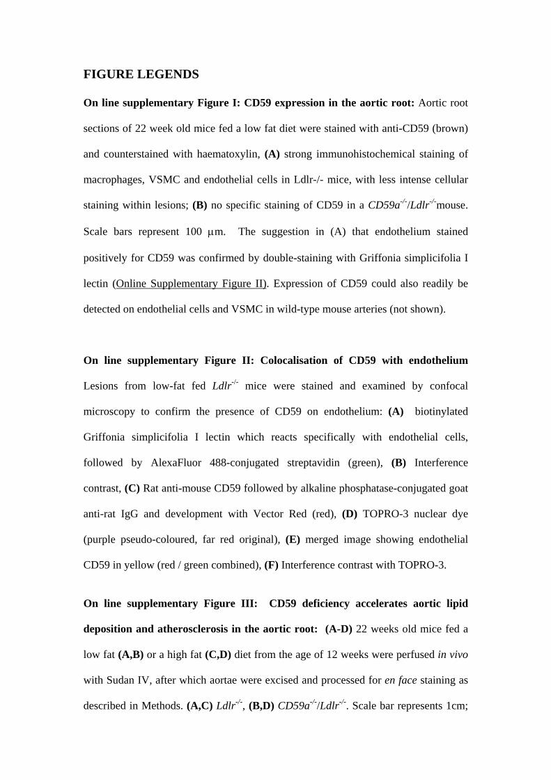
FIGURE LEGENDS
On line supplementary Figure I: CD59 expression in the aortic root: Aortic root
sections of 22 week old mice fed a low fat diet were stained with anti-CD59 (brown)
and counterstained with haematoxylin, (A) strong immunohistochemical staining of
macrophages, VSMC and endothelial cells in Ldlr-/- mice, with less intense cellular
staining within lesions; (B) no specific staining of CD59 in a CD59a-/-/Ldlr-/-mouse.
Scale bars represent 100 μm. The suggestion in (A) that endothelium stained
positively for CD59 was confirmed by double-staining with Griffonia simplicifolia I
lectin (Online Supplementary Figure II). Expression of CD59 could also readily be
detected on endothelial cells and VSMC in wild-type mouse arteries (not shown).
On line supplementary Figure II: Colocalisation of CD59 with endothelium
Lesions from low-fat fed Ldlr-/- mice were stained and examined by confocal
microscopy to confirm the presence of CD59 on endothelium: (A) biotinylated
Griffonia simplicifolia I lectin which reacts specifically with endothelial cells,
followed by AlexaFluor 488-conjugated streptavidin (green), (B) Interference
contrast, (C) Rat anti-mouse CD59 followed by alkaline phosphatase-conjugated goat
anti-rat IgG and development with Vector Red (red), (D) TOPRO-3 nuclear dye
(purple pseudo-coloured, far red original), (E) merged image showing endothelial
CD59 in yellow (red / green combined), (F) Interference contrast with TOPRO-3.
On line supplementary Figure III: CD59 deficiency accelerates aortic lipid
deposition and atherosclerosis in the aortic root: (A-D) 22 weeks old mice fed a
low fat (A,B) or a high fat (C,D) diet from the age of 12 weeks were perfused in vivo
with Sudan IV, after which aortae were excised and processed for en face staining as
described in Methods. (A,C) Ldlr-/-, (B,D) CD59a-/-/Ldlr-/-. Scale bar represents 1cm;

(E-H) photomicrographs of aortic roots in 22 weeks old mice following (E,F) low fat
diet or (G,H) high fat diet from aged 12 weeks. (E,G) Ldlr-/-, (F,H) CD59a-/-/Ldlr-/-.
Scale bars represent 1 mm.

Yun et al - CD59 protects from atherosclerosis
ONLINE SUPPLEMENTARY TABLE I
Body weights and total serum cholesterol and triglycerides in
22 week old Ldlr-/- and CD59a-/-/Ldlr-/- mice
Ldlr-/-
CD59a-/-/Ldlr-/-
mean + SEM (n)
mean +SEM (n)
Low fat diet Final body weight (g) 25.13 + 0.68 (23) 25.14 + 0.58 (20) Total cholesterol (mmol/l) 9.43 + 1.58 (23) 8.20 + 2.04 (20) Triglyceride (mmol/l) 1.76 + 0.54 (22) 1.76 + 0.35 (20) High fat diet Final body weight (g) 25.22 + 0.73 (18) 27.53 + 1.11 (18) Total cholesterol (mmol/l) 23.04 + 3.07 (12) 23.27 + 4.02 (15) Triglyceride (mmol/l) 3.266 + 0.73 (18)
4.12 + 1.78 (18)

A
B
E
Online Supplementary Figure I

Online Supplementary Figure II
A B C
D E F
A B C
D E F

Online Supplementary Figure III
A
B
C
D
G H
E F