evidence for glutamate, gaba and no in coordinating...
TRANSCRIPT
2310
INTRODUCTIONSponges are benthic suspension feeders that are often described aslacking a tissue level of organisation, sensory cells and coordinatedbehaviour – features that are characteristic of eumetazoans. Theabsence of these features is usually attributed to the early branchingof sponges from other metazoans, yet intriguingly, recent molecularand physiological studies generated by the new genome project onthe encrusting demosponge Amphimedon queenslandica suggest thatPorifera possess many of the molecules that are involved in cellsignalling systems in higher metazoans (for a review, see Adamskaet al., 2007; Richards et al., 2008; Sakarya et al., 2007). The genomicfindings force us to ask whether these components reflect a primitivepre-nervous system or whether sponges have a signalling andcoordination system similar to that of other animals but without theuse of conventional nerves and muscle.
While the absence of nerves and of rapid behaviour in spongeshas intrigued researchers for over a century, at the same time it hasposed difficulties for testing hypotheses using conventionaltechniques that require observation of responses to stimuli. Thegeneral activities of cellular sponges include slow contractions acrosspart and occasionally all of the body, closure of the osculum (thevent for water and the most distinct morphological region forobservation) and slowing or cessation of pumping (filtering). Threemechanisms of signalling have been proposed to explain thesebehaviours: electrical, mechanical or chemical (Jones, 1962).Electrical signalling is only known from glass sponges, presumablybecause they are syncytial, which means that action potentials can
move throughout the animal unimpeded by cell boundaries (Leys,1995; Leys and Mackie, 1997; Leys and Meech, 2006). Mechanicalsignalling, through tugging on each cell by neighbour cells (Emson,1966), and chemical signalling, through diffusible molecules eitherin the aquiferous canal system or through paracrine signalling inthe mesohyl have been investigated extensively in cellular spongeswith few concrete results except to show that sponges respond byslow contraction to touch, electric shock, temperature change,sediment and chemical agents (reviewed in Leys and Meech, 2006).It is unlikely that contractions propagate by mechanical tugging ofcell-on-cell because controlled inflation and contraction responsescan still propagate through sponges whose tissue is torn by theapplication of stimulants or whose osculum has been removed (thispaper; and S.P.L., personal observations). However, twoobservations strongly suggest that paracrine chemical signalling isthe most likely mechanism of signal transduction. First, cells in themesohyl stop crawling as contractions pass over a region (de Vosand Van de Vyver, 1981; Elliott and Leys, 2007), and second, thechoreography of a stereotypical behaviour of inflating and deflatingthe aquiferous system in response to clogging or agitation [calledthe inflation–contraction response (Elliott and Leys, 2007)] involvesthe coordination of regions hundreds of micrometres apart at thesame moment. Paracrine chemical signalling mechanisms in spongesrely on a molecule binding to a ligand-receptor system that controlsthe movement of ions directly (ionotropic) or indirectly(metabotropic) into and out of the cells via G-protein coupled
The Journal of Experimental Biology 213, 2310-2321© 2010. Published by The Company of Biologists Ltddoi:10.1242/jeb.039859
Evidence for glutamate, GABA and NO in coordinating behaviour in the sponge,Ephydatia muelleri (Demospongiae, Spongillidae)
Glen R. D. Elliott and Sally P. Leys*Department of Biological Sciences, University of Alberta, Edmonton, Alberta, Canada, T6G 2E9
*Author for correspondence ([email protected])
Accepted 26 March 2010
SUMMARYThe view that sponges lack tissue level organisation, epithelia, sensory cells and coordinated behaviour is challenged by recentmolecular studies showing the existence in Porifera of molecules and proteins that define cell signalling systems in higher ordermetazoans. Demonstration that freshwater sponges can contract their canals in an organised manner in response to both externaland endogenous stimuli prompted us to examine the physiology of the contraction behaviour. Using a combination of digital time-lapse microscopy, high-performance liquid chromatography–mass spectrometry (HPLC–MS) analysis, immunocytochemistry andpharmacological manipulations, we tested the role of the diffusible amino acids glutamate and g-aminobutyric acid (GABA) and ashort-lived diffusible gas, nitric oxide (NO), in triggering or modulating contractions in Ephydatia muelleri. We identified pools ofglutamate, glutamine and GABA used to maintain a metabotropic glutamate and GABA receptor signalling system. Glutamateinduced contractions and propagation of a stereotypical behaviour inflating and deflating the canal system, acting in a dose-dependent manner. Glutamate-triggered contractions were blocked by the metabatropic glutamate receptor inhibitor AP3 and byincubation of the sponge in an allosteric competitive inhibitor of glutamate, Kynurenic acid. Incubation in GABA inhibitedglutamate-triggered contractions of the sponge. Nitric oxide synthase, involved in the formation of the diffusible gas NO, waslocalised using NADPH-diaphorase to mesenchyme cells in the osculum and pinacoderm. A cGMP assay showed the same cellswere labelled suggesting that the NO system is functional. Our findings suggest sponges coordinate behaviour using chemicalmessenger systems common to other animals.
Supplementary material available online at http://jeb.biologists.org/cgi/content/full/213/13/2310/DC1
Key words: coordination, evolution of nervous systems, conduction, nitric oxide, GABA, glutamate.
THE JOURNAL OF EXPERIMENTAL BIOLOGY

2311Glutamate signalling in a sponge
pathways, possibly by an amino acid [e.g. glutamate, g-aminobutyricacid (GABA)], a biogenic amine (e.g. epinephrine), a peptide (e.g.RF-amides, melatonin) or a short-lived gas [e.g. nitric oxide (NO),carbon monoxide].
Glutamate, GABA and NO are important chemical messengersthat are found in plants, protists and metazoans where they functionin feeding, sensory systems, development and also act as neuro-active compounds (Lam et al., 1998; Moroz, 2001; Bouche et al.,1999). Molecular evidence suggests that receptors for glutamate,GABA and NO were present before plants and animals diverged(Lam et al., 1998; Moroz, 2001). The first characterisation of anyof these receptors from a sponge was a putative dualGlutamate/GABA receptor from Geodia cyndonium (Perovic et al.,1999). However, the recent work from the genome of thedemosponge A. queenslandica (http://www.jgi.doe.gov/sequencing/why/3161.html) has shown there to be metabotropic, but noionotropic, glutamate receptors, as well as GABA and NO receptors(Sakaraya et al., 2007). The post-synaptic scaffolding proteinsrequired for signalling have also been found in sponges, yetmorphological and behavioural demonstration of ‘proto-synaptic’complexes is still lacking (Sakaraya et al., 2007; Richards et al.,2008). Glutamate, GABA and NO have been found to play a rolein stimulating and modulating contractions in the demospongeTethya wilhelma (Ellwanger and Nickel, 2006). Furthermore,GABA-immunoreactive proteins are said to be localised in cellsassociated within the aquiferous canal system in Chondrilla nucula(Ramoino et al., 2007), and NO has been implicated in temperaturestress activation in Axinella polypoides and Petrosia ficiformis(Giovine et al., 2001). In the absence of more direct mechanismsof signalling between sponge cells, the slowness of the coordinatedbehaviour shown by sponges makes glutamate/GABA signallingsystems and modulation by a NO system a most likely hypothesis.
Ephydatia muelleri has coordinated contractions of the aquiferouscanal system that function to expel wastes and to flush the canalsystem on a regular basis (Elliott and Leys, 2007). We hypothesisedthat these sponges are able to coordinate or modulate contractionsby the use of small diffusible molecules such as amino acids(glutamate, GABA) or a short-lived diffusible gas (NO). Using acombination of microscopy techniques, immunocytochemistry andpharmacological manipulations we provide a description of possiblesignalling systems of the freshwater sponge E. muelleri. We havefound that glutamate triggers the inflation–contraction cycle of thesponge in a dose-dependent manner that varies with amplitude andduration. Glutamate-triggered contractions can be inhibited bygeneral metabotropic glutamate blockers and also by incubation ofthe sponge in GABA. Nitric oxide synthase (NOS) is found inchoanocytes, in dendritic cells of the apical pinacoderm, osculumand in cells that line the excurrent canal system. Upon stimulationby a NO donor, cGMP was localised to cells in the osculum, whichalso contracted. These results confirm a role for a chemicalmessenger signalling system in sponges and offer insight into themechanisms by which coordination of contractile behaviour occursin the absence of a conventional nervous system.
MATERIALS AND METHODSCollection and culturing of sponges
Pieces of the freshwater sponges Ephydatia muelleri (Lieberkuhn1855) and Spongilla lacustris (Linnaeus 1759) consisting of a spiculescaffold containing gemmules were collected in winter months fromsunken trees at a depth of 0–3m in Frederick and Rousseau Lakes,BC, Canada (48°47�51.7559�N, 125°2�58.5600�W), and stored insealed bags of unfiltered lake water at 4°C in the dark until ready
to use. The water was aerated once a month and gemmules kept inthis way were viable for up to one year. The gemmules weremechanically dissociated from the dead sponge skeleton, sterilisedwith 1% hydrogen peroxide, rinsed well in distilled water and platedon glass coverslips as described previously (Elliott and Leys, 2007).Sponges were grown in M-medium (Funayama et al., 2005), whichwas replaced every 48h. Whole-mount preparations consisted of asingle gemmule on an ethanol flamed sterilised 22mm2 coverslipin 60mm diameter Petri dishes, which allowed for easy transfer ofthe sponge into test substances and rinses. Only sponges 7–10 dayspost-hatching (d.p.h.) with a fully functional aquiferous canalsystem were used for experimentation, and care was taken not touse sponges if they had begun to regress due to lack of feeding.
High-performance liquid chromatography (HPLC)For HPLC, 150–200 sponges were grown in mass cultures on 6cmdiameter Petri dishes. The growth medium was changed every 24h.At 7 d.p.h. the medium was aspirated off and tissue was scrapedfrom the bottom of the Petri dishes with a sterile, plastic tissuescraper, and transferred using a sterile glass pipette to 1.6mlmicrofuge tubes. The tissue was centrifuged for 1min at 1000g toremove excess M-media. The final mass of each preparation was~200mg. The sponge tissue was immediately frozen in liquidnitrogen, weighed and stored at –80oC until analysed.
HPLC coupled with fluorimetric detection and a derivitisationprocedure (Grant et al., 2006) was used to separate and detect aminoacids at the Neurochemical Research Unit of the University ofAlberta. Sponge tissue samples of both E. muelleri and S. lacustriswere homogenised in 5 volumes of distilled water. The homogenatewas diluted 1:3 in methanol to precipitate proteins, immersed in icefor 10min and centrifuged (13,000g) at 4°C. Stock solutions ofamino acid standards [glutamate, L-glutamine, L-aspartate, L-asparagine, L-alanine, D-serine, L-serine, L-serine-O-phosphate, L-tryptophan, glycine, GABA, L-threonine, L-taurine, L-valine; Sigma-Aldrich, Oakville, ON, Canada) were prepared in 20% (v/v)methanol at a concentration of 1.0mgml–1 and were used to identifypeaks in the samples based on retention time and to quantify aminoacids in the samples based on a 9-point calibration curve. The actualstandard concentrations of glutamate, aspartate, serine, glutamine,alanine, glycine, taurine and tryptophan curves were 6, 4, 2, 1, 0.5,0.2, 0.1 and 0.04mgml–1; asparagine and GABA curves were 3, 2,1, 0.5, 0.25, 0.1, 0.5 and 0.2mgml–1; threonine and valine were 1.5,1, 0.5, 0.25, 0.125, 0.05, 0.025 and 0.01mgml–1. Derivitisationreagent solutions were prepared by dissolving 1mg of -phthaldialdehyde (OPA; Sigma-Aldrich) and 2mg of N-isobutyryl-L-cysteine (IBC; Sigma-Aldrich) in 0.1ml of methanol followed bythe addition of 0.9ml 0.2moll–1 of sodium borate buffer (pH10).Automated pre-column derivitisation was carried out on 5ml ofsample, standard or blank and 5ml of derivitising agent that wasinjected into a HPLC system for analysis. The HPLC systemconsisted of a Waters Alliance 2690XE instrument equipped witha Waters 474 programmable fluorescence detector (WatersCorporation, Milford, MA, USA) that pumped a mobile phasecontaining 15% methanol in 0.04moll–1 sodium phosphate buffer(pH6.2) through a Waters Symmetry C18 column (3.9mm�20mm,5mm). Samples were detected with an excitation wavelength of344nm and an emission wavelength of 443nm; spectra weredepicted by change in voltage.
Digital time-lapse and data acquisitionFor all pharmacology experiments, digital images of whole-mountsponges were viewed on a stereomicroscope (Olympus SZX-12,
THE JOURNAL OF EXPERIMENTAL BIOLOGY
2312
Markham, ON, Canada) captured with a QI-Cam monochrome CCDcamera (QImaging, Surrey, BC, Canada) with a colour filter.Images were captured using Northern Eclipse version 7 (EmpixImaging Inc., Mississauga, ON, Canada) from live video feed every5s. For image analysis, changes in the diameter of canals for everyfirst, fifth, tenth or twentieth image of the aquiferous canal system(perpendicular to the long axis of the canal) were measured intriplicate using either Northern Eclipse or Image J 1.31 (NIH,Washington, MA, USA) and data were logged into MS Excel 2003(Microsoft Corporation, Redmond, WA, USA). Centre, middle andperipheral canals were classed based on location and restingdiameter as characterised in Elliott and Leys (Elliott and Leys, 2007).Graphs were created using Sigma Plot (Systat Software, Inc., SanJose, CA, USA).
Test substance applicationThe contractile mechanism, response and coordination of theaquiferous canal system in E. muelleri was characterised and testedby the use of calcium and magnesium-free media [CF (calcium-free)and CMF (calcium–magnesium-free) media] (M-media withoutMgSO4 and CaCl2 but with 1mmoll–1 NMDG–Cl, 0.5mmoll–1
NMDG–SO4, 0.5mmoll–1 EGTA and/or 0.005mmoll–1 EDTA),glutamate (Sigma-Aldrich) and GABA (Sigma-Aldrich) with anantagonist of metabotropic glutamate receptors (m-GluR), 2-amino-3-phosphonopropionic acid (AP3, Sigma-Aldrich) (Young et al.,1994), and a non-competitive inhibitor of m-GluR, Kynurenic acid(KYN, Sigma-Aldrich). In order not to move sponges prior toexperiments, sponges in 60mm diameter Petri dishes containing 20mlof M-medium were left on the microscope stage to relax for one hourprior to each experiment. Stock solutions of 20mmoll–1 glutamate,50mmoll–1 GABA, 50mmoll–1 AP3 and 50mmoll–1 KYN wereprepared in distilled water or dimethylsulphoxide (DMSO) and wereadded directly to the medium to reach the final concentrations listedin each experiment. Great care was taken to always add solutions atthe side of the Petri dish opposite to the sponge and to mix by gentlypipetting 5–6 times for each test substance; equivalent mixing offluorescein dye confirmed the uniform distribution of the testsubstances in the dish. The sponges did not respond to equivalentadditions of M-medium and mixing. For blockers, sponges wereplaced into the final diluted medium in a Petri dish and allowed tosit undisturbed for 60min. For each experiment sponges were filmedfor 10min prior to any treatment to ensure that canals were at restingdiameter immediately prior to addition of the test substance.
NADPH-diaphorase histochemical detection of NOSJuvenile sponges were fixed by direct immersion into a mixture of3.7% paraformaldehyde and 0.3% glutaraldehyde in 0.1moll–1
phosphate buffered saline (PBS) for 24h at 4°C (Elliott and Leys,2007). After fixation, preparations were rinsed three times in coldPBS, twice in 0.1moll–1 Tris-HCl buffer (pH8.0) for 5min each andpermeabilised for 10min in 0.1moll–1 Tris-HCl buffer (pH8.0) with0.25% Triton X-100. For histochemical detection of NOS, spongeswere incubated with 1mmoll–1 -NADPH (Sigma-Aldrich),0.5mmoll–1 Nitro Blue Tetrazolium (Sigma-Aldrich), 0.1mmoll–1
dicumarol (Sigma-Aldrich), 0.25% Triton X-100 in 0.1moll–1 Tris-HCl buffer (pH8.0) in the dark until colour development was reached.The reaction was stopped by rinsing in cold PBS four times. Spongeswere post-fixed in 4% paraformaldehyde in methanol for 1h at 4°Cand rinsed in 100% dry ethanol. The ethanol was allowed to evaporate,and coverslips with sponges were inverted, mounted slightly raisedon Vaseline® legs in 100% glycerol. Edges were sealed with nailpolish. Images were captured as indicated above.
G. R. D. Elliott and S. P. Leys
cGMP assay for NO reactionJuvenile sponges were incubated in 1mmoll–1 3-isobutyl-1-methylxanthine (IBMX: phosphodiesterase inhibitor; Sigma-Aldrich) in Strekal’s medium, an equivalent culture medium to M-medium (Strekal and McDiffett, 1974), diluted from a 0.5moll–1
stock solution in DMSO for 30min. Sponges were exposed to0.1mmoll–1 S-nitroso-N-acetyl-DL-penicillamine (SNAP: NO donor;Sigma-Aldrich) for 2min and fixed in 4% paraformaldehyde in PBSfor 2h at 4°C. Coverslips with sponges were rinsed (30min) andinverted on a solution containing primary rabbit anti-cGMP (1:3000)(Millipore-Chemicon, Billerica, MA, USA) with 3% goat serum(Sigma-Aldrich) and 0.1% Triton X-100 in cold PBS and placedon a shaker at 4°C overnight. Preparations were rinsed in PBS andlabelled with a solution containing Alexa 488 goat anti-rabbit (1:100)(Molecular Probes-Invitrogen, Carlsbad, CA, USA) with 10% goatserum in cold PBS on a shaker for 3h. Sponges were rinsed in PBS,mounted in 100% glycerol, sealed with nail polish and imaged witha Zeiss Axioskop epifluorescence microscope with a QIcam CCDcamera operated by Northern Eclipse.
RESULTSEvidence for neurotransmitter molecules in sponge tissues:
HPLC–MS analysisSamples of sponge tissue analysed by HPLC revealed chromatographsof molecules that co-eluted with standards for the putative amino acidneurotransmitters aspartic acid, glutamic acid, asparagines, serine,glutamine, glycine, threonine, taurine, alanine, GABA, tryptophan andvaline (Fig.1). All amino acids were found in each sample with adetection limit ranging from 0.01 to 0.04mgml–1 of supernatant. Asshown in Table1, the amino acid levels were low except for glutamicacid, glutamine, aspartic acid, glycine and valine. Similar highconcentrations were found for glutamate in S. lacustris; however, allother amino acids in that sponge were low compared with E. muelleri(Table1, Fig.1). When single sponges were exposed to 30mmoll–1
KCl in an attempt to activate release of possible transmitters, nodetectable amino acids were released into the supernatant.
Evidence for a metabotropic glutamate signalling systemThe juvenile freshwater sponge has four regions that can be monitoredby stereomicroscopy for a response to stimulants: the osculum (vent),the tent (dermal tissues that lie above a space which surrounds thechoanosome), the choanosome (housing the choanocyte chamberswhich are the pumping and filtration units), and the incurrent andexcurrent canal systems (the aquiferous canal system). In the followingdescription we refer to each of these in turn.
Glutamate triggered both an instant contraction of the osculum andtent and also stimulated a full inflation and contraction of theaquiferous canal system with the same characteristics as thoseprovoked by vigorous mechanical stimulation (Fig.2) (Elliott andLeys, 2007). Both the initial contraction of the tent and triggering ofthe full inflation–contraction cycle were dependent on theconcentration of glutamate. The inflation–contraction cycle wastriggered at an agonist concentration of 70–90mmoll–1 of glutamate(Fig.3; supplementary material Movies1 and 2). All sponges wereundisturbed for 1h and then filmed for 10min prior to application ofglutamate in order to make sure that an inflation–contraction cyclewas not spontaneously occurring. Concentrations of agonist below30mmoll–1 had no effect on the sponge, and concentrations above100mmoll–1 were fatal. A sub-threshold agonist concentration(30–60mmoll–1) application of glutamate triggered a gradient ofbehaviours. The first response was an initial lowering of the dermaltissues or tent, pushing down on the subdermal cavity and
THE JOURNAL OF EXPERIMENTAL BIOLOGY

2313Glutamate signalling in a sponge
choanosome, and its return to normal diameter (supplementarymaterial Movie3). Following this the incurrent canals contractedthereby expanding the excurrent canals; however, it was not until theapplication of 70mmoll–1 of glutamate that a consistent inflation ofthe excurrent canals was generated by the contraction of the incurrentcanals, and at that threshold level of 70mmoll–1 of glutamate a fullinflation–contraction cycle occurred (Fig.3), which was similar to theresponse of sponges stimulated by ink and agitation (Elliott and Leys,
2007), except that the plateau phase was much reduced. At an agonistconcentration of 80–100mmoll–1 of glutamate the dermal tissuescontracted so severely that often either the tent or choanosome tore(supplementary material Movie4); interestingly however the canalsand osculum could still inflate and contract normally even withoutthe intact structure of the overlying dermal tissues. At an agonistconcentration of 80mmoll–1 of glutamate, two types of eventsoccurred: either the sponge choanosome began to fibrillate and thenfully contracted with no inflation–contraction cycle, or it began aninflation–contraction cycle ending with contraction of the excurrentcanals and did not recover for a long period. At an agonistconcentration of 100mmoll–1 of glutamate it appeared as if both theincurrent and excurrent canals contracted at the same time thuspreventing the normal inflation event (see Movie4 and Fig.S1 insupplementary material).
As the agonist concentration increased from 30mmoll–1 to100mmoll–1 the amplitude and duration of the inflation–contraction cycle increased (Fig.4A). The rate of contraction ofboth incurrent and excurrent canals (i.e. during the inflation andcontraction phases, respectively) was similar (Fig.4B) but bothphases contracted faster than when stimulated by agitation (Elliottand Leys, 2007).
Evidence for function of glutamate receptorsThe maximum concentration that elicited a full inflation–contractionresponse was 80mmoll–1 of glutamate, and so concentrations of
C
B
A
0
mV
10.00
20.00
30.00
40.00
50.00
0 5.00 10.00 15.00 20.00 25.00 30.00 35.00 40.00 45.00
Asp
Glu
Asn S
erG
ln
Gly
Thr
eo
Taur
Ala
Gab
a
Tryp
Val
0
10.00
20.00
30.00
40.00
50.00
Minutes
0 5.00 10.00 15.00 20.00 25.00 30.00 35.00 40.00 45.00
Asp Glu
Asn
Ser
Gln
Gly
Thr
eoTa
urA
laG
aba
Tryp
Val
Asp
Glu
Asn
Ser
Gln
Gly
Thr
eo
Taur
Ala
Gab
a
Tryp Val
0 5.00 10.00 15.00 20.00 25.00 30.00 35.00 40.00 45.00
0
10.00
20.00
30.00
40.00
50.00
60.00
70.00
60.00
70.00
Fig.1. High-performance liquidchromatography–massspectrometry (HPLC–MS)chromatographs of amino acids intissue of Ephydatia muelleri andSpongilla lacustris. Peaks representputative active neuronal aminoacids; the baseline indicates themobile (carrier) phase.(A)Ephydatia muelleri, (B) S.lacustris, (C) amino acid standards.Glu, Asp, Ser, Gln, Ala, Gly, Taurand Tryp were used at 2mgml–1,Asn and GABA were used at1mgml–1, and Threo and Val at0.5mgml–1. Aspartate (Asp),glutamate (Glu), asparagine (Asn),serine (Ser), glutamine (Gln),glycine (Gly), threonine (Threo),taurine (Taur), alanine (Ala),tryptophan (Tryp), g-aminobutyricacid (GABA) and valine (Val).
Table 1. Amino acid levels for tissue from Ephydatia muelleri andSpongilla lacustris analysed by HPLC
Amino acid E. muelleri S. lacustris
Aspartic acid 24.98±3.23 4.67±0.33Glutamic acid 48.21±6.03 116.94±3.94Asparagine 1.58±0.14 2.76±0.20Serine 4.67±1.09 16.40±2.39Glutamine 23.16±2.59 10.40±1.38Glycine 12.38±0.98 14.77±1.24Threonine 9.88±0.90 3.28±0.13Taurine 2.97±0.57 1.97±0.26Alanine 28.16±3.84 19.56±1.43g-Aminobutyric acid 6.09±1.28 2.67±0.16Tryptophan 7.13±1.94 2.59±0.11Valine 18.58±3.06 4.86±0.17
Values are expressed in mgml–1 of sample as means ± s.e. (N5 colonies of150–200 sponges). HPLC, high-performance liquid chromatography.
THE JOURNAL OF EXPERIMENTAL BIOLOGY

2314
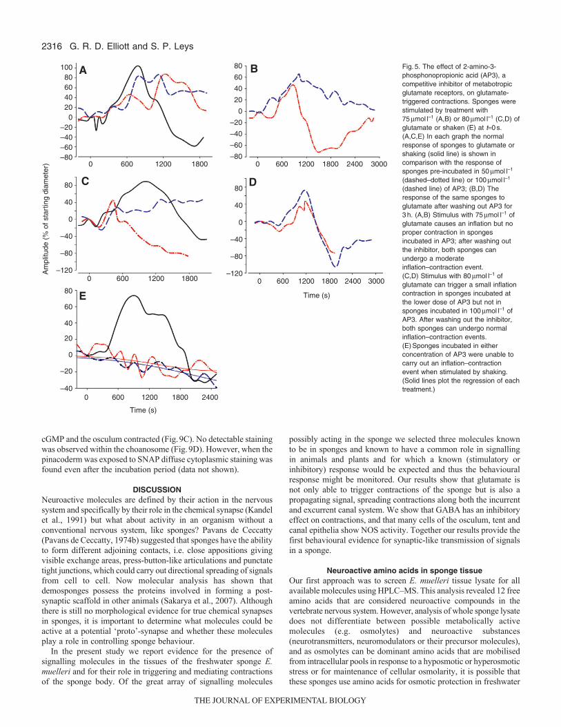
75mmoll–1 and 80mmoll–1 were used in the following experimentsto investigate the type of receptor involved, its competitive abilityand the role of calcium stores. The receptor type was evaluated byusing AP3, widely used as a general m-GluR antagonist. Whensponges were treated in control experiments with 75mmoll–1 and80mmoll–1 of glutamate, they exhibited normal glutamate-inducedinflation–contraction cycles (Fig.5A,C solid lines). AP3 eitherreduced or eliminated the inflation–contraction cycle depending onthe concentration of antagonist and agonist. When sponges wereincubated in either 50mmoll–1 or 100mmoll–1 of AP3 for 30min,stimulation with 75mmoll–1 of glutamate caused waves of inflationto ripple across the sponge body in different directions (Fig.5A);after the antagonist was washed out for 3h, sponges were able toboth inflate and contract in a more normal manner (Fig.5B). Whenstimulated by 80mmoll–1 of glutamate, sponges pre-incubated in100mmoll–1 of AP3 also carried out irregular inflation of theexcurrent canal but did not contract; however, sponges in the lowerconcentration of AP3 were able to carry out a much reduced butcomplete inflation–contraction event when stimulated by the higherconcentration of agonist (Fig.5C). After the AP3 was washed outfor 3h, both sponges carried out a normal inflation–contraction cyclein response to 80mmoll–1 of glutamate (Fig.5D). Sponges that wereincubated in either 50mmoll–1 or 100mmoll–1 of AP3 and thenstimulated by vigorous agitation, exhibited convulsions or twitchesbut no inflation–contraction cycle (Fig.5E).
To further test the functionality of the m-GluR system a non-competitive inhibitor KYN was applied to the sponges (Fig.6). KYNis thought to bind to an allosteric site of m-GluR changing the structureof the molecule, and as such it should reduce the receptor availabilityto glutamate. Sponges were incubated in 25, 50, 100, 150, 200mmoll–1
of KYN, and 80mmoll–1 of glutamate was added after 10, 20 or 30minto test for competition of the two molecules; in one set of experimentssponges in all concentrations of KYN were stimulated by vigorous
G. R. D. Elliott and S. P. Leys
shaking. Concentrations of KYN above 200mmoll–1 were cytotoxicand concentrations below 25mmoll–1 had little to no effect. After eachexperiment sponges were washed with fresh M-media and left for24h to confirm viability after experiments took place.
KYN eliminated the sponges’ inflation–contraction response in apattern that showed dependence on the concentration of inhibitor andlength of incubation. The longer the incubation and the higher theconcentration of inhibitor, the less likely it was that aninflation–contraction response could be stimulated by glutamate.Whereas 10min incubation in 25mmoll–1, 50mmoll–1 and 100mmoll–1
of KYN did not prevent an inflation–contraction response, 10min in150mmoll–1 and 200mmoll–1 KYN concentrations were sufficient toblock any response (Fig.6A). After 20min incubation in KYN,glutamate was only able to trigger an inflation–contraction responsein sponges incubated at the lower concentrations (25mmoll–1 and50mmoll–1) of KYN; no response occurred in preparations incubatedin 100mmoll–1, 150mmoll–1 or 200mmoll–1 KYN (Fig.6B). A 30minincubation in all concentrations eliminated all inflation–contractionresponses from sponges with the exception of 50mmoll–1, whichallowed an inflation–contraction response of slightly lower amplitudeand of lower duration (Fig.6C). However, all sponges incubated inKYN for 30min began to show small ripples of contractions indifferent regions of their choanosome. Sponges incubated in allconcentrations of KYN and stimulated by shaking immediatelycontracted and then gradually relaxed; there was noinflation–contraction behaviour (Fig.6D).
Role of calcium in contractionsCF media greatly reduced the inflation–contraction responsestimulated by 80mmoll–1 of glutamate, and CMF media abolishedall inflation–contraction responses stimulated by 80mmoll–1 ofglutamate (Fig.7). Sponges that were shaken in either CF or CMFmedia showed no inflation–contaction behaviour (data not shown).
Fig.2. Response of Ephydatia muelleri to application of 80mmoll–1 of glutamate. (A–D) Images showing the stages of contraction and the (a–d)corresponding areas of the excurrent aquiferous canal system. (E)A plot of the change in diameter of the largest excurrent canal (solid line) and the area ofthe excurrent aquiferous canal system (dashed line) in response to a stimulus of 80mmoll–1 of glutamate. The application of glutamate is shown as are theperiods of inflation and contraction and times at which each image (A–D) are indicated. Gemmule (g), osculum (osc), glutamate (glut), excurrent canals (ex,white arrow) and incurrent canals (black arrow). Scale bar, 1mm.
THE JOURNAL OF EXPERIMENTAL BIOLOGY

2315Glutamate signalling in a sponge
Evidence for a GABA signalling systemIn contrast to glutamate, application of GABA had barely any effectat concentrations as high as 1mmoll–1. In 3/18 sponges applicationof 250mmoll–1 and 500mmoll–1 of GABA triggered a contractionof the incurrent canals (e.g. Fig.8A). In two of these cases the drugalso triggered minor contractions of the choanosome, suggestingthe sponge tent had contracted; however, because this thin dermaltissue strung across the surface of the sponge is both taut and flatwhen viewed from the microscope above, this movement was noteasily quantifiable in the images captured.
The absence of any direct effect of GABA prompted us toexamine whether the molecule had an inhibitory role in the sponge.We used 1mmoll–1 of GABA because it had no detrimental effecton the sponges (they were perfectly normal the following day afterwashing out the drug). Remarkably, sponges soaked in 1mmoll–1
of GABA for 20min showed no response at all to application of75mmoll–1 of glutamate or to multiple applications of glutamate,
which brought the final concentration to 225mmoll–1, a concentrationthat would normally cause dramatic contractions and tearing of thedermal tissues in sponges not soaked in GABA (Fig.8B). After thedrug was washed out overnight, the sponges responded to challengeby 75mmoll–1 of glutamate with a normal inflation–contraction cycle(Fig.8B).
Evidence for a NO signalling systemNOS activity was visualised by the use of NAPDH-diaphorasestaining and cGMP accumulation assay (Fig.9). Using the NADPH-diaphorase staining protocol, cells that stained blue, indicatingpossible sites of active NOS, were found in the choanocytechambers, osculum, excurrent canals and apical pinacoderm(Fig.9A). Cells that lined the osculum stained the darkest andshowed a gradient of lighter staining at the base as compared withthe tip of the osculum by average grey level in that region. Thepinacocytes of the excurrent canals stained for NOS activity as well.Within the dermal tissues only a few cells that formed a loosenetwork in the mesohyl and the outer epithelium stained darkly forNOS activity (Fig.9B).
Using a cGMP live assay, cells immunoreactive for cGMP werefound within the osculum and in the dermal tissues, indicating theseare active targets for NO. Young sponges were treated with1mmoll–1 of IBMX to prevent the breakdown of cGMP producedin cells responsive to NO and then challenged by the NO donorSNAP. When exposed to SNAP cells in the osculum accumulated
B
A 75
80
70
90
60
10040
50
30
80
60
40
20
0
0
0 50 100
Time (s)
Am
plitu
de (
% c
hang
e fr
om s
tart
ing
diam
eter
)
150 200
600 1200 1800
–20
–40
–60
–80
20
10
0
–10
–20
Fig.3. Graphs showing the dose-dependent response of theinflation–contraction behaviour in Ephydatia muelleri to glutamate.(A)Concentrations of 70–90mmoll–1 of glutamate trigger a 20–30min-longinflation and contraction of the aquiferous system, the ‘inflation–contraction’response. The response is calculated as the percentage change indiameter of the largest excurrent canal over time, and each line representsthe mean of three experiments (see Fig.S1 in supplementary material foreach independent graph, and Movies 1–4 in supplementary material forexample experiments). (B)The initial contraction of the tent (reflected bythe quick contraction of the large excurrent canal) is also dependent on theconcentration of glutamate; solid lines show the response to concentrationsof 60–80mmoll–1; dashed lines show the response to lower and higherconcentrations.
B
A100
80
60
40
30
Concentration (μmol l–1)
Rat
e of
con
trac
tion
(ΔA
/t)
Δ τI–
C(s
)
Rel
ativ
e Δ
ampl
itude
40 50 60 70 80 90 100
30 40 50 60 70 80 90 100
20
0
0.30
0.25
0.20
0.10
0.15
0.05
0
1400
1200
1000
800
600
400
200
Fig.4. (A)Relative amplitude (left axis, circles and solid line) and duration(right axis, crosses and dashed line) of the inflation response stimulated bydifferent concentrations of glutamate. (B)Rate of contraction of theincurrent canals during inflation (solid circles and solid line), andcontraction (open circles, dashed line) of excurrent canals during deflation.Data analysed from Fig.3.
THE JOURNAL OF EXPERIMENTAL BIOLOGY
2316
cGMP and the osculum contracted (Fig.9C). No detectable stainingwas observed within the choanosome (Fig.9D). However, when thepinacoderm was exposed to SNAP diffuse cytoplasmic staining wasfound even after the incubation period (data not shown).
DISCUSSIONNeuroactive molecules are defined by their action in the nervoussystem and specifically by their role in the chemical synapse (Kandelet al., 1991) but what about activity in an organism without aconventional nervous system, like sponges? Pavans de Ceccatty(Pavans de Ceccatty, 1974b) suggested that sponges have the abilityto form different adjoining contacts, i.e. close appositions givingvisible exchange areas, press-button-like articulations and punctatetight junctions, which could carry out directional spreading of signalsfrom cell to cell. Now molecular analysis has shown thatdemosponges possess the proteins involved in forming a post-synaptic scaffold in other animals (Sakarya et al., 2007). Althoughthere is still no morphological evidence for true chemical synapsesin sponges, it is important to determine what molecules could beactive at a potential ‘proto’-synapse and whether these moleculesplay a role in controlling sponge behaviour.
In the present study we report evidence for the presence ofsignalling molecules in the tissues of the freshwater sponge E.muelleri and for their role in triggering and mediating contractionsof the sponge body. Of the great array of signalling molecules
G. R. D. Elliott and S. P. Leys
possibly acting in the sponge we selected three molecules knownto be in sponges and known to have a common role in signallingin animals and plants and for which a known (stimulatory orinhibitory) response would be expected and thus the behaviouralresponse might be monitored. Our results show that glutamate isnot only able to trigger contractions of the sponge but is also apropagating signal, spreading contractions along both the incurrentand excurrent canal system. We show that GABA has an inhibitoryeffect on contractions, and that many cells of the osculum, tent andcanal epithelia show NOS activity. Together our results provide thefirst behavioural evidence for synaptic-like transmission of signalsin a sponge.
Neuroactive amino acids in sponge tissueOur first approach was to screen E. muelleri tissue lysate for allavailable molecules using HPLC–MS. This analysis revealed 12 freeamino acids that are considered neuroactive compounds in thevertebrate nervous system. However, analysis of whole sponge lysatedoes not differentiate between possible metabolically activemolecules (e.g. osmolytes) and neuroactive substances(neurotransmitters, neuromodulators or their precursor molecules),and as osmolytes can be dominant amino acids that are mobilisedfrom intracellular pools in response to a hyposmotic or hyperosmoticstress or for maintenance of cellular osmolarity, it is possible thatthese sponges use amino acids for osmotic protection in freshwater
C
A
E
D
B100806040200
0
Am
plitu
de (
% o
f sta
rtin
g di
amet
er) 600 1200 1800 0 600 1200 1800 2400 3000
0 600 1200 1800
Time (s)
Time (s)
2400 30000 600 1200 1800
0 600 1200 24001800
–20–40
80
60
40
20
0
–20
–40
–60–80
80
60
40
20
0
–20
–40
–60
–80
80
40
0 0
–40
–80
–120
80
40
–40
–80
–120
Fig.5. The effect of 2-amino-3-phosphonopropionic acid (AP3), acompetitive inhibitor of metabotropicglutamate receptors, on glutamate-triggered contractions. Sponges werestimulated by treatment with75mmoll–1 (A,B) or 80mmoll–1 (C,D) ofglutamate or shaken (E) at t0s. (A,C,E) In each graph the normalresponse of sponges to glutamate orshaking (solid line) is shown incomparison with the response ofsponges pre-incubated in 50mmoll–1
(dashed–dotted line) or 100mmoll–1
(dashed line) of AP3; (B,D) Theresponse of the same sponges toglutamate after washing out AP3 for3h. (A,B) Stimulus with 75mmoll–1 ofglutamate causes an inflation but noproper contraction in spongesincubated in AP3; after washing outthe inhibitor, both sponges canundergo a moderateinflation–contraction event. (C,D) Stimulus with 80mmoll–1 ofglutamate can trigger a small inflationcontraction in sponges incubated atthe lower dose of AP3 but not insponges incubated in 100mmoll–1 ofAP3. After washing out the inhibitor,both sponges can undergo normalinflation–contraction events.(E)Sponges incubated in eitherconcentration of AP3 were unable tocarry out an inflation–contractionevent when stimulated by shaking.(Solid lines plot the regression of eachtreatment.)
THE JOURNAL OF EXPERIMENTAL BIOLOGY

2317Glutamate signalling in a sponge
systems; however, some of the free amino acids may also be usedfor signalling.
Eight potential neuroactive amino acids and metabolites werefound with the HPLC screen: glutamate, GABA, glycine, taurine,serine (D-serine), tryptophan (biogenic amines), aspartate (precursorfor NMDA: N-methyl-D-aspartic acid) and glutamine (precursor forGABA and glutamate) (Fig.1; Table1). Glutamate and GABA areneuroactive amino acids that are involved in both the central nervoussystem (CNS) and peripheral nervous system of vertebrates and in
invertebrates are typically associated with control of musclecontraction in either an excitatory action (glutamate) or an inhibitoryaction (GABA). Glutamine is the precursor molecule or reserve storefor glutamate and the GABA metabolic pathway. The high activityof GABA and glutamate as signalling molecules requires storageof an inactive glutamine for proper function within a cell. Glycineis a major inhibitory amino acid neurotransmitter in the vertebrateCNS that works by inducing a hyperpolarising chloride current whenbound to a post-synaptic receptor but it can also be a modulator inexcitatory ionotropic glutamate receptors. In Hydra, glycine has beenidentified and localised to the nerve net where it functions inpacemaker activity of peristaltic contractions (elongations andcontractions) of the muscle in the body column; it is also involvedin the chemosensory response during feeding by inhibiting theclosure of the mouth upon stimulation by glutathione (Pierobon etal., 2001; Ruggieri et al., 2004; Kass-Simon and Pierobon, 2006).Similar responses are observed by the application of alanine andtaurine in Hydra (Kass-Simon and Pierobon, 2006). In thedemosponge Tethya wilhelma, glycine has been shown to stimulatea contraction, increase contraction rhythm and decrease contractionamplitude (Ellwanger and Nickel, 2006). It was proposed that thisaction occurs via a metabotropic glycine receptor but unfortunatelyno antagonists (e.g. strychnine) were used to block the response toconfirm receptor-ligand binding.
Am
plitu
de (
% c
hang
e fr
om s
tart
ing
diam
eter
) 0
80
100
60
40
20
Time (s)
0 600 1200 1800
0 300 600 1200900
–20
0
60
40
20
–20
–40
–60
–80
–100
Fig.7. The effect of the absence of calcium and of calcium and magnesiumon the response of Ephydatia to glutamate and to shaking. (A)Theinflation–contraction response to stimulation by 75mmoll–1 of glutamate(open circles, blue) and 80mmoll–1 of glutamate (solid circles, black) innormal medium, in calcium-free medium (diamonds, red), and incalcium–magnesium-free medium (squares, green). The mean change±s.e. of three different sponges is shown. (B)The slopes of the inflationphase of 80mmoll–1 of glutamate treatment in normal (black), Ca-free (red)and Ca–Mg-free (green) media plotted with 95% confidence intervals.
10 min KYN
200 μmol l–1150 μmol l–1
100 μmol l–1
50 μmol l–125 μmol l–1
20 min KYN
30 min KYN
Am
plitu
de (
% c
hang
e fr
om s
tart
ing
diam
eter
)A
B
C
D
0
80604020
0 600 1200 1800
Time (s)
2400 3000
0 600 1200 1800 2400 3000
0 600 1200 1800 2400 3000
0 600 1200 1800 2400 3000
–20–40–60
0
80604020
–20–40–60
0
80604020
–20–40–60
0
20
–20
–40
–60
Fig.6. The effect of Kynurenic acid (KYN), a non-competitive (allosteric)inhibitor of glutamate receptors, on contractions triggered by glutamate(A–C) or shaking (D). (A–C) Sponges were incubated in 25-200mmoll–1 ofKYN for 10 (A), 20 (B) and 30 (C) minutes and then treated with to80mmoll–1 of glutamate; (D) sponges were treated similarly with KYN andshaken immediately for one minute. (A)When glutamate was applied10min after incubation in KYN, the inflation–contraction response wasdelayed by 2, 5, and 10min with a respective increase in the concentrationof KYN, and no inflation–contraction response occurred at the two highestconcentrations of KYN. (B)After 20min incubation in KYN theinflation–contraction response was delayed by 5 and 10min in 25 and50mmoll–1 KYN, respectively, but no contractions occurred inconcentrations of 100mmoll–1 and above. (C)After 30min incubation inKYN, an inflation–contraction response only occurred in 50mmoll–1 of KYN,all other preparations could not respond normally.
THE JOURNAL OF EXPERIMENTAL BIOLOGY

2318
Taurine is increasingly acknowledged to be a biologically activesubstance in both invertebrate and vertebrate nervous systems(Strang et al., 1990; Pirvola and Panula, 1992). Carlberg andcolleagues showed that taurine was abundant in many cell typesthat function both neurally and metabolically in cnidarians (Carlberget al., 1995). Anctil and Minh have shown that taurine is thedominant amino acid found in endodermal contractile cells in thesea pansy Renilla but within the spicule cells they suggest it functionsas an osmolyte (Anctil and Minh, 1997). In marine flatworms,taurine had the highest free amino acid concentration indicative ofan osmolyte (Barrett, 1981). Taurine was detected in E. muelleribut no functional data is available to suggest if it has a signallingor osmolyte function in the Porifera.
Other interesting metabolites found in the sponge tissue aretryptophan and aspartate both of which are used as precursormolecules for biogenic amine metabolism (Kandel et al., 1991).Trytophan is the precursor molecule for the production of serotonin,which one report suggests may be in sponge larvae (Weyrer et al.,1999). High concentrations of serotonin were found to inducecontractions in Tethya but there was no dose dependence of theeffect (Ellwanger and Nickel, 2006). Aspartate is a modulator ofNMDA glutamate receptors and its involvement as a chemicaltransmitter is tentative (Kandel et al., 1991), but because it is also
G. R. D. Elliott and S. P. Leys
a major product of the Krebs cycle its role in chemical transmissionin sponges is uncertain without further experimentation. We did notexamine the amount of free arginine but its presence would indicatea pool that could be used for NO production.
Although presence/absence data cannot confirm the function ofamino acids as chemical transmitters in sponges, the role of aminoacids as transmitter molecules has probably arisen from the simplegustatory behaviour of protists to control feeding. Cellular responsesto specific amino acids are speculated to have evolved into triggersfor feeding in cnidarians – stimulating the gut and entrances to thefeeding system; in sponges this is equivalent to the aquiferous canalsystem, i.e. ostia, canals and choanocytes. We focused ourexperimental work on two principal amino acids found in Ephydatiaand Spongilla tissue lysate, glutamate and GABA, because of theirubiquity in signalling in plant and animal systems.
Evidence for paracrine signalling by glutamate and GABAGlutamate is a chemical messenger that acts both throughmetabotropic and ionotropic receptors in sensory systems (Fagg andFoster, 1983) and is involved signalling in plant, protists,invertebrates and vertebrates (Van Houten, 1998; Nedergaard et al.,2002; Filleur et al., 2005). In Tethya wilhelma glutamate was foundto regulate body contractions in a dose-dependent manner(Ellwanger and Nickel, 2007) but desensitisation and spasm-likebehaviour were also observed; however, because Tethya is opaqueand the canals of that sponge cannot be viewed by light microscopy,it was impossible for those authors to determine the precise effectson dermal tissues, osculum and canals. We found that in E. muelleriglutamate triggered contractions of the tent and also, moreinterestingly, stimulated a propagated contraction along the entireincurrent and then excurrent canal systems to carry out astereotypical behaviour that we call the inflation–contraction cycle.Both types of contraction – the quick contraction of the tent andthe inflation–contraction response – were sensitive to glutamate ina dose-dependent manner. Contractions increased in duration andamplitude with increasing concentration of glutamate and could beinhibited by blockers and competitive agonists of glutamatereceptors, AP3 and KYN.
The blockers/inhibitors did not affect contraction of the osculumor tent but did prevent propagation of the inflation–contractionresponse; that they had long enough to act was demonstrated bytheir action in a time and concentration-dependent manner. Theaction of AP3 was to disrupt or at higher concentrations to preventthe inflation–contraction response but not to prevent all inflation ofexcurrent canals (contraction of incurrent canals). This suggests tous that control of the ‘inflation–contraction’ response is separatedregionally not only into primary (tent and osculum) and secondary(in- and excurrent canals) systems but that the incurrent andexcurrent canal epithelia probably have different populations ofreceptors. The addition of glutamate after the sponge was incubatedin AP3 caused the primary system to contract and some contractionof the incurrent canal but not of the excurrent canals, suggestingthat receptors in the excurrent canal system were blocked.
The action of KYN was even more distinct; lower concentrationswith short incubation times had no effect on the inflation–contractionresponse but longer incubations of low concentrations reduced thelikelihood there would be an inflation–contraction response and highconcentrations inhibited the inflation–contraction after only 10minin the competitive antagonist. The slightly delayed inflation–contraction response to glutamate at lower concentrations and shorterincubation periods of KYN suggests that glutamate eventually isable to out compete KYN and bind to its receptor. The complete
A
Con
trac
tion
of th
e ch
oano
som
e(%
cha
nge
from
sta
rtin
g va
lue)
Con
trac
tion
of th
e in
curr
ent c
anal
s(%
cha
nge
from
sta
rtin
g va
lue)
B
Am
plitu
de(%
cha
nge
from
sta
rtin
g ca
nal d
iam
eter
)
–80
–40
0
40
80
120
–10
–8
–6
–4
–2
0
2
4
–150
–125
–100
–75
–25
–50
0
25
–300 0 300 600 900
Time (s)
1200 1500 1800 2100
0 300 600 900 1200 1500 1800
Fig.8. Inhibition of contractions by g-aminobutyric acid (GABA). (A)GABA(250 and 500mmoll–1, applied at the black arrow) caused a quickcontraction of the choanosome (solid red line, left axis) and of the incurrentcanals (dotted and dashed blue lines, right axis) in E. muelleri. (B)Whensponges were incubated in 1mmoll–1 GABA for 20min (squares, black line)they did not respond to application of 75mmoll–1 of glutamate at t0 or toadditional doses of the same concentration of glutamate 10 and 20minafterwards (white arrows). After 24h washout in M-medium (circles, redline), all sponges responded normally to 75mmoll–1 glutamate (blackarrow). The mean (±s.e.) of three sponges is shown.
THE JOURNAL OF EXPERIMENTAL BIOLOGY

2319Glutamate signalling in a sponge
block of propagation of the inflation–contraction response at higherconcentrations of KYN however indicates that all sites for glutamatebinding were inhibited and therefore the sponge must depend onthe release of glutamate between cells to propagate contractions,generating a pool that eventually is able to bind to receptors onneighbouring cells, much like a chemical synapse.
Our findings with GABA are even more interesting. Whereas inT. wilhelma GABA was found to be 100-fold more potent thanglutamate in triggering contractions, we found that it in fact acts asan inhibitory molecule, preventing contraction of the osculum, tentand of any inflation–contraction events. At first we were surprisedthat no concentration of GABA could trigger a contraction – whichwe had expected based on the results from Tethya. However, ourresults are much more in line with what we would expect for thefunction of GABA in other animals as an inhibitory signal in feeding,growth, metamorphosis and as an inhibitory neurotransmittercausing relaxation of muscle (Fagg and Foster, 1983; White et al.,1986; Chebib and Johnston, 1999; Bouche et al., 2003).
Emson (Emson, 1966) also showed that GABA treatment had noeffect on the behaviour of the demosponge Cliona celata, andsuggested that its role was inhibitory, so it is unclear why GABAmight trigger a contraction in Tethya. The authors of that work(Ellewanger et al., 2007) suggested that since a sequence with putativeaffinity to both metabotropic glutamate and metabotropic GABAreceptor families was cloned from the demosponge Geodia cyndonium(Perovic et al., 1999), the glutamate/GABA system might not haveyet diverged in sponges. However, the new genome project on A.queenslandica has identified eight separate sequences of m-GluR andone GABA receptor (Sakarya et al., 2007), and given that demospongefamilies are equally primitive it is likely that early sponges alreadyhad distinct glutamate and GABA receptors. It would be interestingto assess the action of GABA on internal tissues of Tethya.
Our work is the first to identify physiologically distinct regionsin the sponge body and suggests that sponges may have differentreceptor populations on different tissues. Given the variety of
glutamate receptors now known to be in demosponges it would beinteresting to determine if they have regionalised expressionboundaries in Ephydatia, some on the osculum, others on the tentand yet others on the incurrent and excurrent canal systems. GABAreceptors may be more limited in type and more ubiquitous indistribution. One report shows that antibodies to mammalian GABAare localised to most cells of the sponge Chondrilla (Ramoino etal., 2007). Although cross-reactivity of the antibodies was confirmedby western blotting, the implication of the rather vagueimmunostaining obtained is difficult to interpret. It was shown thatChondrilla possesses the enzymes to synthesise and transportGABA, and HPLC–MS analysis showed that GABA was releasedfrom cells stimulated by KCl but as no experimental work wascarried out it cannot be speculated what function GABA might havein that sponge.
Requirement for calcium and magnesiumWe also tested for the ability of glutamate-triggered contractionsto propagate in the absence of calcium and of all divalent cations.In CF media, the effective dose of glutamate (75mmoll–1) causedthe osculum to contract and the incurrent canals to inflate but nofurther events occurred. In CMF media the incurrent canals didnot even inflate. These results confirm that Mg2+ can substitutefor Ca2+ in contractions, as shown by Prosser and colleagues(Prosser et al., 1962), but that an external calcium store is requiredfor propagation of contractions. A link between the glutamatereceptor and an internal calcium store is suggested by the fact thatan initial contraction of the osculum and apical pinacoderm canoccur in CMF media. However, no blockers have been identifiedthat will block the internal calcium stores in the Porifera (Lorenzet al., 1996).
A potential role for NO signallingWe also found using NADPH staining that in E. muelleri, NOactivity is located in the cells of the osculum, canals and choanocytes
Fig.9. Localisation of nitric oxide synthase(NOS) in Ephydatia muelleri by NADPH-diaphorase staining (A,B) and cGMPantibody assay (C,D). Cells showing NOSactivity (blue stained cells, white arrows)were located in the osculum (A), and in theepithelium lining the excurrent canals andthe mesohyl of the dermal tissues (B).(C)Many cells in the osculum (arrow) wereimmunoreactive for cGMP in the presenceof the nitric oxide donor S-nitroso-N-acetyl-DL-penicillamine (SNAP). (D)Control: in theabsence of the nitric oxide donor SNAPthere was fluorescence in the choanosomebut no cGMP accumulation in thepinacoderm. Scale bars, A,B,D: 500mm; C, 100mm. g, gemmule.
THE JOURNAL OF EXPERIMENTAL BIOLOGY
2320 G. R. D. Elliott and S. P. Leys
and particular cells in the mesohyl of the tent. Although NO activityin these cells could be involved in a stress (immune type) responsewe did find that the osculum in E. muelleri contracted withapplication of the NO donor SNAP but no inflation–contractionresponse was triggered. In T. wilhelma NO (produced by the NOdonor NOC-12) induced contractions and at the same timemodulated endogenous contraction rhythm and amplitude(Ellwanger and Nickel, 2006). These results are similar to theinvolvement of NO in the feeding response in Hydra (De Petrocelliset al., 1999) and in molluscs (Korneev et al., 1998), in the controlof swimming in Aglantha (Moroz et al., 2004) and in peristalticcontractions in Renilla (Anctil et al., 2005).
Another method to detect if a NO system is active is to use anassay to induce production of cGMP by activation of guanylatecyclase by a NO donor (SNAP). In E. muelleri the osculum labelledstrongly for cGMP, as did some mesohyl cells of the tent. Thus, inE. muelleri NO may function in modulating contraction of cells ofthe dermal tissues and the osculum. In A. queenslandica a gene forNOS has been found but it does not share similarities with eitherneuronal or immune NO signalling (Sakarya et al., 2007). Futurework is required to develop a protocol for a behavioural assay thatwill allow the dissection of the NO signalling system in sponges todetermine whether it is directly involved in, is responsible for ormodulates the propagation of contractions across the sponge.
Evolution of ligand-based coordination pathwaysOur results showing precise activation and inhibition ofphysiologically distinct regions of a sponge help to provide a clearerpicture of the properties and mechanisms of cell–cell signalling insponges. We can now say with certainty that glutamate and GABAwork as transmitters in excitatory–inhibitory roles to coordinatedistinct types of contractions in this sponge. Cells (some or manybut presumably not all) in the epithelia of the osculum, tent andcanal system can release and receive signals in a precise manner,functioning in an analogous manner to a suite of synapses acrossthese tissues. Because sponges lack a conventional nervous systemthis confirms opinions that the evolution of ligand-based receptorsystems predated the evolution of a nervous system (Parker, 1910;Jones, 1962; Pavans de Ceccatty, 1962; Mackie, 1970; Pavans deCeccatty, 1974a; Pavans de Ceccatty, 1974b; Mackie, 1979; Pavansde Ceccatty, 1979; Mackie, 1990; Nickel, 2004). As most spongesare cellular (only glass sponges are syncytial), it would probablyhave been easier for a mechanical or chemical messenger systemto evolve by adapting existing chemical molecules and membranereceptors instead of an electrical system that would requirecompletely novel proteins such as connexins or innexins to allowrapid transmission of signals between cells. Our results show thatthe inflation and contraction of the sponge aquiferous system (theinflation–contraction response) can continue despite tissues beingtorn apart by high doses of stimulant; this rules out the possibilitythat signals are simply transmitted mechanically from cell to cell.Rather we show that a chemical messenger (paracrine) system iscapable of providing discrete information transfer over longdistances and at the speed necessary to allow an effective behaviouralresponse by the animal. This system functions very effectivelywithout electrical signalling, the gain of which would have been amassive addition but one required by the co-evolution of rapidlycontracting (striated) muscle.
LIST OF ABBREVIATIONSAP3 2-amino-3-phosphonopropionic acidCF calcium free
CMF calcium–magnesium freeDMSO dimethylsulphoxideEDTA ethylene diamine tetraacetic acidEGTA ethylene glycol tetraacetic acidGABA g-aminobutyric acidHPLC–MS high-performance liquid chromatography–mass spectrometryIBMX 3-isobutyl-1-methylxanthineKYN Kynurenic acidm-GluR metabotropic glutamate receptorNO nitric oxideNOS nitric oxide synthaseSNAP S-nitro-N-acetyl-DL-penicillamine
ACKNOWLEDGEMENTSWe gratefully acknowledge G. Baker and the Neurochemical Research Unit,Department of Psychiatry and Centre for Neuroscience, at the University ofAlberta for help with HPLC, and the Director and staff at the Bamfield MarineSciences Centre where animals were collected. A. Waskiewicz, R. Palmer andtwo anonymous reviewers provided constructive suggestions on earlier drafts ofthe manuscript. This research was funded by an NSERC Discovery Grant toS.P.L.
REFERENCESAdamska, M., Degnan, S. M., Green, K. M., Adamski, M., Craigie, A., Larroux, C.
and Degnan, B. M. (2007). Wnt and TGF-b expression in the sponge Amphimedonqueenslandica and the origin of metazoan embryonic patterning. PLoS ONE 2,e1031.
Anctil, M. and Minh, N. C. (1997). Neuronal and nonneuronal taurine-likeimmunoreactivity in the sea pansy, Renilla koellikeri (Cnidaria, Anthozoa). CellTissue Res. 288, 127-134.
Anctil, M., Poulain, I. and Pelletier, C. (2005). Nitric oxide modulates peristalticmuscle activity associated with fluid circulation in the sea pansy Renilla koellikeri. J.Exp. Biol. 208, 2005-2017.
Barrett, J. (1981). Biochemistry of Parasitic Helminths. Baltimore: University ParkPress.
Bouche, N., Lacombe, B. and Fromm, H. (2003). GABA signaling: a conserved andubiquitous mechanism. Trends Cell Biol. 13, 807-810.
Carlberg, M., Alfredsson, K., Nielson, S.-O. and Anderson, P. A. V. (1995).Taurine-like immunoreactivity in the motor nerve net of the jellyfish Cyanea capillata.Biol. Bull. 188, 78-82.
Chebib, M. and Johnston, G. A. R. (1999). The ‘ABC’ of GABA Receptors: a briefreview. Clin. Exp. Pharmacol. Physiol. 26, 937-940.
De Petrocellis, L., Melck, D., Bisogno, T., Milone, A. and Di Marzo, V. (1999).Finding of the endocannabinoid signalling system in Hydra, a very primitiveorganism: Possible role in the feeding response. Neuroscience 92, 377-387.
de Vos, L. and Van de Vyver, G. (1981). Étude de la contraction spontanée chezl’éponge d’eau douce Ephydatia fluviatilis cultivée en vitro. Annales de la societeRoyale zoologique de Belgique 111, 21-31.
Elliott, G. and Leys, S. P. (2007). Coordinated contractions effectively expel waterfrom the aquiferous system of a fresh water sponge. J. Exp. Biol. 210, 3736-3748.
Ellwanger, K. and Nickel, M. (2006). Neuroactive substances specifically modulaterhythmic body contractions in the nerveless metazoon Tethya wilhelma(Demospongiae, Porifera). Front. Zool. 3, 7.
Ellwanger, K., Eich, A. and Nickel, M. (2007). GABA and glutamate specificallyinduce contractions in the sponge Tethya wilhelma. J. Comp. Physiol. A 193, 1-11.
Emson, R. H. (1966). The Reactions of the sponge Cliona celata to applied stimuli.Comp. Biochem. Physiol. 18, 805-827.
Fagg, G. E. and Foster, A. C. (1983). Amino acid neurotransmitters and theirpathways in the mammalian central nervous system. Neuroscience 9, 701-719.
Filleur, S., Walch-Liu, P., Gan, Y. and Forde, B. G. (2005). Nitrate and glutamatesensing by plant roots. Biochem. Soc. Transact. 33, 283-286.
Funayama, N., Nakatsukasa, M., Hayashi, T. and Agata, K. (2005). Isolation of thechoanocyte in the fresh water sponge, Ephydatia fluviatilis and its lineage marker, Efannexin. Dev. Growth Differ. 47, 243-253.
Giovine, M., Pozzolini, M., Favre, A., Bavestrello, G., Cerrano, C., Ottaviani, F.,Chiarantini, L., Cerasi, A., Cangiotti, M., Zocchi, E. et al. (2001). Heat stress-activated, calcium-dependent nitric oxide synthase in sponges. Nitric Oxide 5, 427-431.
Grant, S. L., Shulman, Y., Tibbo, P., Hampsonc, D. R. and Baker, G. B. (2006).Determination of D-serine and related neuroactive amino acids in human plasma byhigh-performance liquid chromatography with fluorimetric detection. J. Chromatogr. B844, 278-282.
Jones, W. C. (1962). Is there a nervous system in sponges? Biol. Rev. Biol. Proc.Cambridge Philos. Soc. 37, 1-50.
Kandel, E. R., Siegelbaum, S. A. and Schwartz, J. H. (1991). Synaptic Transmission.East Norwalk: Appleto and Lange.
Kass-Simon, G. and Pierobon, P. (2006). Cnidarian chemical neurotransmission, anupdated overview. Comp. Biochem. Physiol. A 146, 9-25.
Korneev, S. A., Piper, M. R., Picot, J., Phillips, R., Korneeva, E. I. and O’Shea, M.(1998). Molecular characteriztion of NOS in a mollusc: expression in a giantmodulatory neuron. J. Neurosci. 35, 65-75.
Lam, H.-M., Chiu, J., Hsieh, M.-H., Meisel, L., Oliveira, I. C., Shin, M. and Coruzzi,G. (1998). Glutamate-receptor genes in plants. Nature 396, 125-126.
Leys, S. P. (1995). Cytoskeletal architecture and organelle transport in giant. Biol. Bull.188, 241-254.
THE JOURNAL OF EXPERIMENTAL BIOLOGY

2321Glutamate signalling in a sponge
Leys, S. P. and Mackie, G. O. (1997). Electrical recording from a glass sponge.Nature 387, 29-30.
Leys, S. P. and Meech, R. W. (2006). Physiology of coordination in sponges. Can. J.Zool. 84, 288-306.
Lorenz, B., Bohnensack, R., Gamulin, V., Steffen, R. and Muller, W. E. G. (1996).Regulation of motility of cells from marine sponges by calcium ions. Cell. Signal. 8,517-524.
Mackie, G. O. (1970). Neuroid conduction and the evolution of conducting tissues. Q.Rev. Biol. 45, 319-332.
Mackie, G. O. (1979). Is there a conduction system in sponges? ColloquesInternationaux du CNRS. 291, 145-151.
Mackie, G. O. (1990). The elementary nervous system revisited. Am. Zool. 30, 907-920.
Moroz, L. (2001). Gaseous transmission across time and species. Am. Zool. 41, 304-320.
Moroz, L. L., Meech, R. W., Sweedler, J. V. and Mackie, G. O. (2004). Nitric oxideregulates swimming in the jellyfish Aglantha digitale. J. Comp. Neurol. 471, 26-36.
Nedergaard, M., Takano, T. and Hansen, A. J. (2002). Beyond the role of glutamateas a neurotransmitter. Nat. Rev. Neurosci. 3, 748-755.
Nickel, M. (2004). Kinetics and rhythm of body contractions in the sponge Tethyawilhelma (Porifera: Demospongiae). J. Exp. Biol. 207, 4515-4524.
Parker, G. H. (1910). The reactions of sponges with a consideration of the origin of thenervous system. J. Exp. Zool. 8, 765-805.
Pavans de Ceccatty, M. (1962). Systeme nerveux et integration chez les spongiaires.Annales des Sciences Naturelles, Zoologie et Biolologie animale 12, 127-137.
Pavans de Ceccatty, M. (1974a). Coordination in sponges. The foundations ofintegration. Am. Zool. 14, 895-903.
Pavans de Ceccatty, M. (1974b). The origin of the integrative systems: a change inview derived from research on coelenterates and sponges. Perspect. Biol. Med. 17,379-390.
Pavans de Ceccatty, M. (1979). Cell correlations and integration in Sponges. Colloq.Int. CNRS, Biologie des Spongiaires 291, 123-135.
Perovic, S., Krasko, A., Prokic, I., Muller, I. M. and Muller, W. E. G. (1999). Originof neuronal-like receptors in Metazoa: cloning of a metabotropic glutamate/GABA-like receptor from the marine sponge Geodia cydonium. Cell Tissue Res. 296, 395-404.
Pierobon, P., Minei, R., Porcu, P., Sogliano, C., Tino, A., Marino, G., Biggio, G.and Concas, A. (2001). Putative glycine receptors in Hydra: a biochemical andbehavioural study. Eur. J. Neurosci. 14, 1659-1666.
Pirvola, U. and Panula, P. (1992). Distribution of taurine in the rat cerebellum andinsect brain: application of a new antiserum against carbidiimide-conjugated taurine.Histochem. J. 24, 266-274.
Prosser, C. L., Nagai, T. and Nystrom, R. A. (1962). Oscular contractions insponges. Comp. Biochem. Physiol. 6, 69-74.
Ramoino, P., Gallus, L., Paluzzi, S., Raiteri, L., Diaspro, A., Fato, M., Bonanno, G.,Tagliafierro, G., Ferretti, C. and Manconi, R. (2007). The GABAergic-like system inthe marine demosponge Chondrilla nucula. Microsc. Res. Technique 70, 944-951.
Richards, G. S., Simionata, E., Perron, M., Adamska, M., Vervoort, M. andDegnan, B. M. (2008). Sponge genes provide new insight into the evolutionaryorigin of the neurogenic circuit. Curr. Biol. 18, 1156-1161.
Ruggieri, R. D., Pierobon, P. and Kass-Simon, G. (2004). Pacemaker activity inHydra is modulated by glycine receptor ligands. Comp. Biochem. Physiol. A 138,193-202.
Sakarya, O., Armstrong, K. A., Adamska, M., Adamski, M., Wang, I., Tidor, B.,Degnan, B. M., Oakley, T. H. and Kosik, K. S. (2007). A post-synaptic scaffold atthe origin of the animal kingdom. PLoS One 2, e506.
Strang, R. H. C., Whitton, P., Jabbar, A. and Nicholson, R. A. (1990). Taurine as aNeuromodulator in the Insect Central Nervous System. New York: Wiley-Liss.
Strekal, T. A. and McDiffett, W. (1974). Factors affecting germination, growth, anddistribution of the freshwater sponge, Spongilla fragilis Leidy (Porifera). Biol. Bull.146, 267-278.
Van Houten, J. (1998). Chemosensory transduction in Paramecium. Eur. J. Protistol.34, 301-307.
Weyrer, S., Rützler, K. and Rieger, R. (1999). Serotonin in Porifera? Evidence fromdeveloping Tedania ignis, the caribbean fire sponge (Demospongiae). Mem.Queensl. Mus. 44, 659-665.
White, J., Southgate, E., Thomson, J. and Brenner, S. (1986). The structure of thenervous system of the nematode Caenorhabditis elegans. Philos. Trans. R. Soc.Lond. B. Biol. Sci. 314, 1-340.
Young, M. R., Fleetwood-Walker, S. M., Mitchell, R. and Munro, F. E. (1994).Evidence for a role of metabotropic glutamate receptors in sustained nociceptiveinputs to rat dorsal horn neurons. Neuropharmacology 33, 141-144.
THE JOURNAL OF EXPERIMENTAL BIOLOGY