evaluating blood films€¦ · (wbc) malignancy, red blood cell (rbc) poikilocytosis, rbc...
TRANSCRIPT
1
www.vetmedpub.com DECEMBER 2004Clinical Solutions for Companion-Animal Practitioners
Idea Exchange • Mind Over Mi l ler • 2004 Annual Index
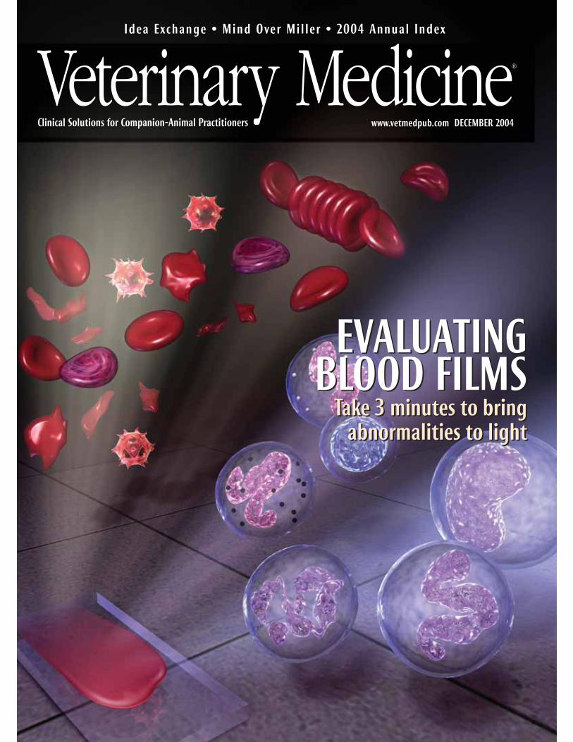
EVALUATING BLOOD FILMS
Take 3 minutes to bring
abnormalities to light
EVALUATING BLOOD FILMS
Take 3 minutes to bring
abnormalities to light
2
Many veterinarians and technicians do not routinely evaluate blood films microscopi-cally, largely because they lack confidence in either preparing a well-made bloodfilm or in being able to accurately identify important abnormalities. But blood films
should be evaluated whenever a complete blood count (CBC) is requested, regardless ofwhether the CBC is done in the clinic or at a reference laboratory. Blood film evaluation is asessential to a CBC as a microscopic examination of urine sediment is to a complete urinalysisor two views are to proper radiographic interpretation. No single hematology procedure pro-duces more valuable information yet requires so little additional time (we recommend threeminutes) and expense.
Even the most expensive hematology analyzers are not designed to eliminate peripheralblood film evaluation. Morphologic features that instruments cannot identify include left shifts(increased immature neutrophils), neutrophil toxicity, lymphocyte reactivity, white blood cell(WBC) malignancy, red blood cell (RBC) poikilocytosis, RBC inclusions, and platelet abnormali-ties. A quick blood film review will help validate certain numerical data including plateletcounts, WBC counts, WBC differentials, and RBC density, since even reference laboratory analyz-ers prove inaccurate with some of the more abnormal samples.
In this symposium, we suggest using a systematic method for evaluating blood films. Thismethod—which requires veterinarians and technicians to answer basic questions about RBC,WBC, and platelet numbers and morphology—maximizes hematologic information. And wecover the most common and important morphologic findings in the peripheral blood.
We hope this symposium will provide veterinary practitioners and technicians with theskills to properly prepare, stain, and evaluate a peripheral blood film and will encouragethem to use this vital hematology tool in their practices.
—Dr. Fred L. Metzger Jr.
P E E R - R E V I E W E D
Symposium on a three-minute peripheral blood film evaluation
MYR
IAM
KIR
KMAN
-OH
, KO
STU
DIO
S
3
8
14
Page 8
Page 3
Three-minute peripheral blood film evaluation: Preparing the film Fred L. Metzger Jr. and Alan Rebar
No single hematology procedure produces more valuable information yet requires so littletime and expense than a peripheral blood film. Proper preparation and staining of the filmare critical.
Three-minute peripheral blood film evaluation: The erythron and thrombonFred L. Metzger Jr. and Alan Rebar
When examining the red blood cells and platelets in a blood film, ask yourself thesequestions to quickly identify and further characterize conditions such as anemia andthrombocytopenia.
Three-minute peripheral blood film evaluation: The leukonFred L. Metzger Jr. and Alan Rebar
Taking a moment to briefly examine white blood cells will help you identify conditions suchas inflammation or stress that may indicate serious disease.
All articles have been reviewed by at least two board-certified specialists or recognized experts to ensure accuracy, thoroughness, and suitability.
Symposium on a three-minute peripheral blood film evaluation

3
P E E R - R E V I E W E D
Three-minute peripheral blood film evaluation:Preparing the filmNo single hematology procedure produces more valuable information yet requires so little time and expense than aperipheral blood film evaluation. Proper preparation and staining of the film are critical.
A PERIPHERAL BLOOD FILM evaluation should bepart of all complete blood counts (CBCs), re-gardless of whether hematology is performedin-house or at a reference laboratory. Propersample collection, slide preparation, andstaining are essential to accurately evaluate ablood film, as is the correct use of a high-quality microscope. This article describes thesteps in preparing a blood film and theequipment you’ll need.
Important components of a CBC include evalu-ating the erythron (hematocrit, total red bloodcell [RBC] count, hemoglobin concentration,absolute reticulocyte count, and RBC indices),the leukon (total white blood cell [WBC] count,five-part differential count including immatureneutrophils), the thrombon (platelet countand platelet indices), and the total proteinconcentration. As mentioned earlier, alwaysinclude a peripheral blood film evaluation inthe CBC.
A CBC should be included in evaluations ofevery sick patient, every patient with vaguesigns of disease, and every patient receivinglong-term medications. In addition, a CBC
should be performed as part of every preanes-thetic workup, for adult wellness and geriatricprofiles, and as a recheck test for patients inwhich RBC, WBC, or platelet abnormalities werepreviously diagnosed.
Artifacts must be avoided for proper hemato-logic interpretation. Causes of artifacts includepoor blood collection techniques, inadequatesample volumes, prolonged sample storage,and delayed sample analysis.
Proper blood collection is vital to prevent er-
roneous results from sample clotting and cellu-lar lysis. Obtain hematology samples from thelargest blood vessel possible to minimize cellu-lar trauma and to prevent the activation of clot-ting mechanisms. For accurate results, discardclotted samples and collect fresh samples. Com-mon venipuncture sites in dogs and cats in-clude the jugular, cephalic, and lateral and me-dial saphenous veins. Anticoagulants includeEDTA, heparin, and citrate. EDTA is the preferredanticoagulant for blood film preparation be-cause it preserves cellular detail better thanother anticoagulants do and does not interferewith Romanowsky staining of WBCs.
Inadequate sample volume is a commoncause of inaccurate hematologic results. Prop-erly fill anticoagulated blood collection tubes toavoid falsely decreased hematocrits and cellcounts and to prevent RBC shrinkage.
Hematologic samples must be analyzed assoon as possible to prevent artifacts created byexposure to anticoagulants and cell deteriora-tion due to storage and shipment. Analyze sam-ples within three hours or refrigerate them at39.2 F (4 C) to avoid an artificially increasedhematocrit, increased mean corpuscular vol-ume, and decreased mean corpuscular hemo-globin concentration.1 Prepare blood filmswithin one hour of collection to avoid morpho-logic artifacts. RBC crenation, neutrophil hyper-segmentation, lymphocytic nuclear distortion,and general WBC degeneration including vac-uolization in neutrophils may occur in agedsamples. In addition, monocyte vacuolization,monocyte cytoplasmic pseudopod formation,and platelet agglutination can be encounteredin stored samples.2 If you use a reference labo-ratory for primary hematologic analyses, werecommend submitting freshly prepared bloodfilms along with the anticoagulated blood.
Collecting a sample for the blood film
Components of and indications for a CBC
Fred L. Metzger Jr., DVM, DABVP
(canine and feline practice)Metzger Animal Hospital1044 Benner PikeState College, PA 16801
Alan Rebar, DVM, PhD, DACVPDepartment of Veterinary Pathobiology School of Veterinary Medicine Purdue UniversityWest Lafayette, IN 47907

4
Producing high-quality blood films begins byusing clean, new slides. Used slides fre-quently have imperfections such as scratchesand other physical defects. Slides must befree of fingerprints, dust, alcohol, detergents,and debris. Using a microhematocrit tube,place a small drop (2 to 3 mm in diameter)of well-mixed EDTA blood about 1 to 1.5 cmfrom the end of the slide. Next, draw aspreader slide back into the blood drop atabout a 30-degree angle until the spreaderslide edge contacts the sample drop and cap-illary action disperses the sample along theedge. Then, using a smooth steady motion,push the spreader slide away from the blooddrop, creating a uniform film that coversnearly the entire length of the slide (Figure1). Changing the angle of the spreader slide
will change the length and thickness of theblood film. For blood samples with lowhematocrits (severe anemia), you may needto decrease the angle of the spreader slide tomake a good-quality slide. In contrast, forsamples with high hematocrits (severe dehy-dration and polycythemia due to a variety ofconditions), you may need to increase theangle of the spreader slide.
Drying is an important step in the produc-tion of good-quality blood films. Allow theblood film to thoroughly air-dry before apply-ing stain, or use a heat block (at the low set-ting) or a hair dryer to hasten the drying time,thus minimizing refractile markings that candistort erythrocyte morphology. In addition,keep formalin and formalin-containing con-tainers away from all blood and cytologysmears to prevent staining artifacts such as in-creased cytoplasmic granularity and ba-sophilia.
Most veterinary practices use a modifiedWright’s stain (Romanowsky stain) for bothhematology and cytology. Modified Wright’sstains are available as either three- or two-so-lution kits. We prefer kits with three separatesolutions: an alcohol fixative, an eosinophilicstaining solution, and a dark-blue stainingsolution. Veterinary practices should havetwo separate Coplin stain jar sets—one forhematology and cytology samples and onefor contaminated samples such as those col-lected for ear and fecal cytology. For opti-mal results, replace staining solutions regu-larly.
Recommended blood staining protocolincludes dipping the air-dried slide five to10 times in each solution while blotting oneedge briefly in between each solution. Rinse
the slide with distilled water after the finalstaining step, and allow the blood film to air-dry before examination. Good staining tech-nique is critical to identifying polychro-matophils on a peripheral blood film. If thestaining is done improperly, many of thequick Romanowsky-type stains will not givethe needed tinctorial differences between ma-ture RBCs (orange-red) and polychromatophils(bluish-pink). Typically, poor staining withthe various quick stains results in all RBCs hav-ing a bluish or muddy tint.
The microscope
A high-quality microscope is essential forhematology. We recommend a binocular mi-croscope with a minimum of 10�, 20�, and100� (oil-immersion) objectives and wide-field 10� oculars. When evaluating the slide,make sure maximum light reaches the bloodfilm. This means positioning the substage con-denser as close as possible to the stage withthe iris wide open. The light source should beequipped with a variable rheostat to allowmaximal control of light intensity. To ensureoptimal focused light for the microscopic eval-uation, the microscope should be in Köhler il-
Evaluating the blood film
Staining the blood film
1. The steps in preparing a blood film. A small dropof blood is placed near the end of a slide (top). Aspreader slide is drawn back into the blood dropat a 30-degree angle (middle). Then the spreaderslide is pushed away from the blood drop,creating a uniform film across the slide (bottom).
F IGURE 1
Monolayer
Feathered edge Body of blood film
Application point
LabelMonolayer
F IGURE 2
Good staining technique is critical to identifying polychromatophils on a peripheral blood film.
2. The components of the peripheral blood film slide.
5
lumination. Contact your microscope vendor,or review the microscope manual to ensureproper illumination.
The three zones
A good-quality blood film has threezones: the body (near the point of blood ap-plication), the monolayer (the zone betweenthe body and feathered edge), and the feath-ered edge (the area most distant from thepoint of application) (Figure 2). The defini-tion of the monolayer varies from institutionto institution, but the definition used to en-sure consistent semiquantification of variousmorphologic abnormalities is the area whereabout half of the RBCs are touching one an-other without overlapping. The monolayer isthe area of the blood film where WBCs andplatelet numbers are estimated and cell mor-phology is examined. Although the mono-layer is the only zone where morphologicevaluation of individual cells is performed atoil-immersion magnification, systematicblood film evaluation includes assessing ofall three zones.
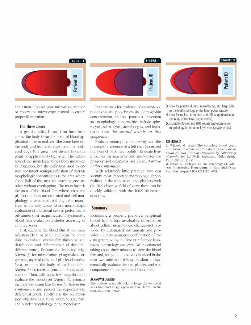
First, examine the blood film at low mag-nification (10� or 20�), and scan the entireslide to evaluate overall film thickness, celldistribution, and differentiation of the threedifferent zones. Evaluate the feathered edge(Figure 3) for microfilariae, phagocytized or-ganisms, atypical cells, and platelet clumping.Next, examine the body of the blood film(Figure 4 ) for rouleau formation or RBC agglu-tination. Then, still using low magnification,evaluate the monolayer (Figure 5), estimatethe total WBC count (see the third article in thissymposium), and predict the expected WBC
differential count. Finally, use the oil-immer-sion objective (100�) to examine RBC, WBC,and platelet morphology in the monolayer.
Evaluate RBCs for evidence of anisocytosis,poikilocytosis, polychromasia, hemoglobinconcentration, and RBC parasites. ImportantRBC morphologic abnormalities include sphe-rocytes, schistocytes, acanthocytes, and lepto-cytes (see the second article in thissymposium).
Evaluate neutrophils for toxicity and thepresence or absence of a left shift (increasednumbers of band neutrophils). Evaluate lym-phocytes for reactivity and monocytes forphagocytized organisms (see the third articlein this symposium).
With relatively little practice, you canidentify most important morphologic abnor-malities in the RBCs, WBCs, and platelets withthe 20� objective field of view; these can bequickly validated with the 100� oil-immer-sion view.
Examining a properly prepared peripheralblood film offers invaluable informationabout cellular morphologic changes not pro-vided by automated instruments and pro-vides a quality assurance confirmation of CBC
data generated by in-clinic or reference labo-ratory hematology analyzers. We recommendtaking about three minutes to view the bloodfilm and, using the questions discussed in thenext two articles of this symposium, to sys-tematically evaluate the RBC, platelet, and WBC
components of the peripheral blood film.
ACKNOWLEDGMENTThe authors gratefully acknowledge the technicalassistance and images provided by Dennis DeNi-cola, DVM, PhD, DACVP.
REFERENCES1. Willard, M. et al.: The complete blood countand bone marrow examination. Textbook ofSmall Animal Clinical Diagnosis by LaboratoryMethods, 3rd Ed. W.B. Saunders, Philadelphia,Pa., 1999; pp 11-30. 2. Rebar, A.; Metzger, F.: The Veterinary CE Advi-sor: Interpreting Hemograms in Cats and Dogs.Vet. Med. (suppl.) 96 (12):1-12; 2001.
Summary
F IGURE 3 F IGURE 4 F IGURE 5
Pat
ien
t ID
Pat
ien
t ID
Pat
ien
t ID
3. Look for platelet clumps, microfilariae, and large cellsin the feathered edge of the film ( purple section).
4. Look for rouleau formation and RBC agglutination inthe body of the film ( purple section).
5. Estimate platelet and WBC counts and examine cellmorphology in the monolayer zone ( purple section).


8
P E E R - R E V I E W E D
EVALUATING A PERIPHERAL blood film validates cellcounts performed by hematology analyzers,plus it offers valuable diagnostic information re-layed by erythrocytes, platelets, and leukocytes.The first article in this symposium describedhow to prepare a peripheral blood film. In thisarticle, we discuss important red blood cell(RBC) and platelet number and morphologicchanges. And in the next article, we discusswhite blood cell (WBC) alterations.
If a patient’s RBC mass is reduced, then thepatient is anemic. Once anemia is recognized,the next concern is bone marrow responsive-
ness: Is the anemia regenerative or nonre-generative? If the RBC bone marrow pre-cursor cells respond with increasedproduction of appropriate magnitude,
the anemia is regenerative (responsive). IfRBC production is not effectively increased,the anemia is nonregenerative (nonrespon-sive). Keep in mind that a lag of 48 to 72
hours exists before bone marrow responsive-ness is perceived in the peripheral blood, sobe careful in defining nonresponsiveness.
Evaluating the thrombon by first assessingplatelet numbers is another important part ofevery complete blood count (CBC). Thrombocy-topenia is more common than thrombocytosisand can be of great clinical importance. Plateletcounts below 40,000/µl can lead to spontaneousbleeding. Platelet clumping, which can be seenin any species but is particularly problematic incats, can give a falsely low platelet count.Platelet clumping frequently interferes with re-sults from impedance cell counters because ag-gregated platelets may be included in the RBC orWBC counts, resulting in artifactually decreasedplatelet counts.1
Our approach to peripheral blood film eval-uation uses a question-based format. This sys-
tematic approach allows veterinary practitionersand technicians to maximize important visualinformation.
Is there evidence of RBC regeneration? (Is
polychromasia
or reticulocytosis present?)
In dogs and cats, polychromasia is the princi-pal feature of RBC regeneration on a blood filmprepared with Wright’s or modified Wright’sstain. Polychromatophils are immature RBCs thatstain bluish because they contain RNA (Figure 1).Polychromatophils on blood smears preparedwith Wright’s or modified Wright’s stain roughlycorrespond to reticulocytes on smears preparedwith new methylene blue stain. If regeneration isstill questionable after you’ve evaluated Wright’s-stained blood films, perform a reticulocyte countwith new methylene blue stain.
When performing reticulocyte counts in cats,it is important to count aggregate reticulocytes(cells containing diffuse accumulations of reticu-lum) since they represent recent RBC production
Evaluating the RBCs
Fred L. Metzger Jr., DVM, DABVP
(canine and feline practice)Metzger Animal Hospital1044 Benner PikeState College, PA 16801
Alan Rebar, DVM, PhD, DACVPDepartment of Veterinary Pathobiology School of Veterinary MedicinePurdue UniversityWest Lafayette, IN 47907
Three-minute peripheral blood film evaluation: The erythron and thrombonWhen examining the red blood cells and platelets in a blood film, ask yourself these questionsto quickly identify and further characterize conditions such as anemia and thrombocytopenia.
F IGURE 1
1. Polychromatophils (arrows) in a dog (modifiedWright’s stain; 100�).

9
in response to relatively severe anemia (Fig-ure 2). In cats, aggregate reticulocytes matureinto punctuate reticulocytes within 12 hours,and punctuate reticulocytes mature into RBCsin about 10 days. Consequently, elevatedpunctuate reticulocytes represent RBC regener-ation two weeks earlier, whereas aggregatereticulocytes indicate recent regeneration.2 Indogs and cats, peak reticulocyte counts occurfour to eight days after the onset of anemia.
As a general guideline, a reticulocyte countabove 60,000/µl in cats (aggregate only) and80,000/µl in dogs indicates a regenerative ane-mia.3 Dogs with regenerative anemias havereticulocyte counts that are frequently 100,000to 300,000/µl (dogs with extremely regenera-tive anemia can have counts of 500,000/µl orgreater), while cats typically demonstrate aless dramatic response when only the aggre-
gate reticulocyte counts are evaluated.Reference laboratories can provide reticu-
locyte counts, and in some cases, they willautomatically add the reticulocyte count tothe CBC at an additional charge if anemia isidentified. The newer in-clinic, laser-basedhematology analyzers automatically provideabsolute reticulocyte counts with each CBC.4
Regenerative anemias can be further classi-fied as either blood loss or hemolytic (de-creased RBC lifespan) anemias. Whenever yoususpect hemolysis, closely examine RBC mor-phology to detect spherocytes, acanthocytes,schistocytes, RBC inclusions, and parasites (seebelow). Figure 3 is a flow chart for classifyinganemias in dogs and cats.
Are nucleated RBCs present?
Nucleated RBCs, or metarubricytes (Figure4 ), are not found in any appreciable numbers
in mammalian peripheral blood samples. Mostclinicians consider fewer than 4 nucleatedRBCs/100 WBCs to be insignificant when theWBC count is within the reference range. Nu-cleated RBCs may be seen in increased num-bers with strongly regenerative anemias, butthey should always be less numerous than thepolychromatophils or reticulocytes. This find-ing has been identified as an appropriate nu-cleated RBC response by some clinicians.
An inappropriate nucleated RBC responseoccurs when more than 5 nucleated RBCs/100WBCs are present in the absence of polychro-masia. Conditions associated with an inappro-priate nucleated RBC response include bonemarrow stromal damage, extramedullaryhematopoiesis, fractures, hyperadrenocorticism,feline leukemia virus infection, chemo-therapeutic drug administration, and lead toxi-
F IGURE 3
Classifying Anemias in Dogs and Cats
Blood loss anemia is apossible cause. Look forexternal or internal hem-orrhage or parasites (e.g. fleas,hookworms). Keep inmind that it takes oneto three days to see thehematocrit decrease.
Hemolytic anemia is apossible cause. Considerimmune-mediatedhemolytic anemia withspherocytosis or Heinzbody hemolytic anemia(e.g. onion ingestion,acetaminophen administration).
If bone marrow hypopla-sia is present, consideranemia of inflammation,anemia of chronic renaldisease, myelophthisisdue to bone marrow dis-ease, or chemotherapytoxicosis.
If bone marrow hyper-plasia with ineffectiveerythropoiesis is present,consider a nuclear matu-ration defect (FeLV infec-tion, drug toxicosis) or acytoplasmic maturationdefect (lead toxicosis,iron deficiency includingchronic blood loss).
When a patient has a decreased hematocrit, consider itshydration status and perform a reticulocyte count.
The anemia is regenerative if the reticulocyte count is morethan 80,000/µl in dogs and more than 60,000/µl in cats.
The anemia is nonregenerative if the reticulocyte count is less than80,000/µl in dogs and less than 60,000/µl in cats. In these cases,perform a serum chemistry profile, urinalysis, and other neededdiagnostic tests to rule out underlying diseases. If no such disease isfound, perform a bone marrow evaluation.
F IGURE 2
2. Aggregate (black arrows) and punctuate (red arrows)reticulocytes in a cat. Note that aggregatereticulocytes contain greater than or equal to fivebasophilic specks (new methylene blue stain; 100�).
4. A nucleated RBC (arrow) in a dog (modified Wright’sstain; 100�).
F IGURE 4

10
cosis, among others.5 Splenic dysfunction (e.g.decreased clearance of circulating nucleatedRBCs) is an important cause of an inappropri-ate release of nucleated RBCs and may occur inpatients in which the spleen has been re-moved and in patients with splenic neo-plasms, especially hemangiosarcoma.
Is autoagglutination present?
Agglutination is unorganized three-dimensional clumping of RBCs that must bedifferentiated from rouleau formation. It iscommon in dogs with immune-mediated he-molytic anemia. When confirmed, autoagglu-tination suggests an immune-mediatedprocess such as autoimmune-mediated he-molytic anemia or drug-induced hemolyticanemia (e.g. cephalosporins, penicillin) be-cause RBC surface antibodies cause cell cross-linking and the resulting agglutination.
Rouleau formation is organized linear ar-rays of RBCs caused by decreased zeta poten-tials from plasma proteins (e.g. globular pro-teins, fibrinogen) coating RBCs. This distinctiveformation is commonly described as stacks ofcoins. Unlike agglutination, which is a strongcross-linking between cells, rouleau is a resultof a weak binding between cells because ofdissimilar ionic charges on the RBC surface.
Rouleau formation is a common finding whenfibrinogenesis is increased and must be differ-entiated from agglutination. On routineWright’s-stained or modified-Wright’s-stainedblood films, marked rouleau formation andagglutination may be difficult to differentiate.
Whenever you suspect agglutination on ablood film, mix a drop of the well-mixed EDTA
anticoagulated blood on a new glass slidewith two or more drops of isotonic saline so-lution. Add a cover slip, and view the mixtureas an unstained wet preparation. Under theseconditions, most rouleaux formations dissipatebut autoagglutination typically persists and isrecognized as clumped RBCs (Figure 5).
Are poikilocytes present?
Normal canine RBCs are shaped like bicon-cave disks with prominent central pallor. Fe-line RBCs have much less apparent central pal-lor. Abnormally shaped RBCs are calledpoikilocytes and include artifactual changes(crenation) as well as true abnormalities (e.g.spherocytes, acanthocytes, schistocytes, lepto-cytes).
Crenation (Figure 6 ) can be confusedwith important RBC changes such as acantho-cytosis. Crenation is a shrinking artifact mostcommonly seen when less than optimal
amounts of blood are collected into the EDTA
tube and the peripheral blood film is notmade relatively quickly after the anticoagula-tion process.4 Some less common causes ofcrenation include electrolyte disturbances,uremia, and rattlesnake envenomation. Auseful differentiating feature for identifyingcrenation is that crenation typically affectslarge numbers of cells in a particular area ofthe slide, whereas true poikilocytosis typi-cally affects relatively lower numbers of cellsthroughout the peripheral blood film. Crena-tion can be minimized by preparing bloodfilms immediately after blood samples arecollected and properly anticoagulated.
Spherocytes are spherical RBCs that havelost their normal biconcave shape, resulting inmore intense staining than normal RBCs. Theyhave no central zone of pallor, and they ap-pear smaller than normal RBCs (Figure 7 ).More than four to six spherocytes per 100�field is considered elevated. Spherocytes arecommonly seen with many of the immune-mediated hemolytic anemias we encounter inveterinary medicine. Be careful when attempt-ing to identify spherocytes in feline RBCs be-cause feline RBCs are much less biconcave thancanine RBCs and, therefore, have much lesscentral pallor.
F IGURE 5
5. A saline wet preparation of canine blood showingautoagglutination (20�).
6. Crenation in a dog (modified Wright’s stain; 100�). 7. Spherocytes (black arrows) and a polychromatophil (red
arrow) in a dog (modified Wright’s stain; 100�).8. Acanthocytes (arrow) in a dog (modified Wright’s stain;
100�).9. Schistocytes (arrows) in a dog (modified Wright’s stain;
100�).
F IGURE 6 F IGURE 7
F IGURE 8 F IGURE 9

11
Acanthocytes are abnormally shaped RBCshaving two to 10 blunt, fingerlike surface pro-jections of varying sizes (Figure 8). Thesemorphologic changes are related to abnormalaccumulation of lipids within the RBC mem-brane when there is an abnormal plasmacholesterol:phospholipid ratio. Acanthocytesare seen occasionally in normal animals. Con-ditions most commonly associated with acan-thocyte formation include underlying meta-bolic diseases or diseases affecting normallipid metabolism. Nonneoplastic and neoplas-tic (hemangiosarcoma in particular) diseasesinvolving the liver, spleen, and kidney mayhave associated acanthocytosis in dogs and,occasionally, in cats.6 Acanthocytosis is mostfrequently associated with liver disease andsplenic hemangiosarcoma.
Schistocytes are RBC fragments formed bymechanical injury (Figure 9). Even in verylow numbers (one schistocyte in every threeto five 100� objective fields), schistocytes areclinically relevant. Finding even a few schisto-cytes may help you identify underlying orsubclinical disseminated intravascular coagu-lation (DIC). Microvascular mechanical frag-mentation of RBCs associated with diseasessuch as hemangiosarcoma and dirofilariasismay also result in schistocyte formation.7
A leptocyte (codocyte, target cell) is anRBC with excess cell membrane that forms ashape often referred to as having a Mexicanhat appearance (Figure 10). Leptocytes areseen occasionally in normal animals. In-creased numbers of leptocytes (> 3/100�oil-immersion field) are expected with poly-chromasia because polychromatophils haveexcess membranes compared with normalRBCs. Consequently, leptocytosis is commonwhen reticulocytosis is present, so most ref-erence laboratories do not report this mor-phologic finding. However, laboratories willreport leptocytes when they are seen in theabsence of polychromasia since the excesslipid membrane compared with cytoplasmicvolume is an abnormality. The two primarymechanisms causing this abnormal morphol-ogy are either an upset in thecholesterol:phospholipid ratio in plasma re-sulting in lipid loading (as might be seen
with liver disease and other metabolic disor-ders) or a decrease in cytoplasmic contentcompared with normal (as might be seenwith iron deficiency typically due tochronic blood loss).8 With iron deficiency,RBC hemoglobin content decreases, and inaddition to the leptocytosis, hypochromasiais often observed.
Are RBC inclusions present?
Accurately characterizing various inclu-sions is an important aspect of blood filmevaluation. Inclusions may include Heinzbodies, basophilic stippling, Howell-Jollybodies, and certain infectious agents.
Heinz bodies are localized accumulationsof denatured, oxidized, and precipitated he-moglobin in the RBC. They often affix to theinner RBC membrane and project from theRBC surface. Conditions associated withHeinz body hemolytic anemia include aceta-minophen toxicosis in cats and acute onionand zinc toxicosis in dogs. Diabetes mellitus,hyperthyroidism, and lymphosarcoma in catsand the use of oral benzocaine sprays arealso associated with increased numbers ofHeinz bodies; but anemia is not present inthese cases.8 The Heinz bodies observed incats may be small or not project from the cellsurface, making their identification difficult(Figure 11). New methylene blue stainingwill help confirm the presence of Heinzbodies; the Heinz bodies stain dark bluecompared with the rest of the erythrocyte(Figure 12). In contrast to Heinz bodies indogs, Heinz bodies can be an incidentalfinding in cats, and their relevance must bedetermined by evaluating clinical signs andother hemogram parameters.
Basophilic stippling is seen inRomanowsky-stained films as small, dark-bluepunctate aggregates of residual ribosomes inRBCs (Figure 13). It has traditionally been as-sociated with lead toxicosis in dogs but mayalso be associated with highly regenerativeanemias in any species. Further diagnostic in-vestigation is important when basophilic stip-pling is seen in the absence of reticulocytosis.
Howell-Jolly bodies are small nuclearremnants that may be increased with acceler-
F IGURE 10
F IGURE 12
F IGURE 13
F IGURE 11
10. Leptocytes (arrow) in a dog (modified Wright’s stain;100�).
11. Heinz bodies, which are noselike RBC projections,(arrows) in a cat (modified Wright’s stain; 100�).
12. A blood film from a cat showing Heinz bodies; they areeasier to see with new methylene blue stain (100�).
13. Basophilic stippling (black arrow) and apolychromatophil (red arrow) in a dog (modifiedWright’s stain; 100�).

12
ated RBC regeneration. In healthy animals, thelow numbers of RBCs with Howell-Jolly bodiesreleased from the bone marrow are quicklyremoved from circulation primarily throughthe action of fixed tissue macrophages in thespleen. If Howell-Jolly bodies are easily iden-tified and no reticulocytosis is noted, investi-gate underlying splenic disease. This inclu-sion can be an incidental finding in catsbecause of the open splenic architecture anddecreased erythrophagocytic properties inher-ent to the feline spleen.
Infectious agents including Babesia (Fig-ure 14), Cytauxzoon, and Mycoplasma (previ-ously known as Haemobartonella) speciesmay be identified in RBCs. Evaluation for My-coplasma species (Figure 15) should be per-formed on freshly collected blood samplesthat have not been refrigerated to improveidentification.
Are platelet numbers normal,
decreased, or increased?
Microscopic validation of platelet counts isan important component of blood film evalu-ation. Platelet clumping (Figure 16 ) inter-feres with accurate enumeration of plateletswith all hematology analyzers and with man-ual counting methods. Clumping occurs inmany samples because of platelet activationduring the collection process. Increasednumbers of large platelets may also result ininaccurate platelet counts when impedance
counting methods are used. On a well-prepared peripheral blood film
in which no marked platelet clumping at thefeathered edge is present, the average num-ber of platelets observed per 100� oil-immersion monolayer field multiplied by20,000 provides a good estimate of the num-ber of platelets/µl. As a general rule, youshould see a minimum of eight to 10 plateletsand a maximum of 35 to 40 platelets per100� oil-immersion monolayer field ofview.9
If the platelet numbers are decreased,
can the mechanism
be determined?
Thrombocytopenias occur through fourbasic mechanisms: sequestration, utilization(consumption), destruction, and decreased orineffective production. While clues as to theunderlying mechanism are found in the CBC,in most cases, bone marrow evaluation isneeded for complete interpretation.
Sequestration thrombocytopenias are un-common in veterinary medicine. They areusually the result of hypersplenism and,thus, are characterized by an enlargedspleen on physical or radiographic examina-tion.
Utilization, or consumption, thrombocy-topenias are caused by excessive activation ofthe coagulation cascade. These thrombocy-topenias are associated with inflammatory dis-ease and DIC. Utilization thrombocytopeniasare usually moderate, with platelet numbers
in the 75,000 to 150,000/µl range, but valuesbelow 40,000/µl accompanied by petechiaeare possible.10 Bone marrow evaluation re-veals normal to increased numbers ofmegakaryocytes.
Destruction thrombocytopenias are im-mune-mediated thrombocytopenias in whichnormal circulating platelets are destroyed bycirculating antiplatelet antibodies. Destructionthrombocytopenias are often extreme, with pe-ripheral platelet counts well below 50,000/µl.Bone marrow examination is characterized bynormal to increased numbers of megakary-ocytes.
Decreased or ineffective production is asso-ciated with extremely low platelet counts;counts well below 50,000/µl are common.Bone marrow evaluation will vary greatly inmost of these cases, although they will sharesimilar end results of deceased effective pro-duction by the bone marrow. With decreasedproduction thrombocytopenias, no identifiableto extremely low numbers of megakaryocytesare present. With ineffective production, mostbone marrow samples have normal to in-creased numbers of megakaryocytes, and theproduction of platelets is ineffective in manycases because of an immune-mediated de-structive process directed at an early stage ofplatelet development or at the megakaryocytepopulation itself.
Evaluating the platelets
F IGURE 16
16. A blood film from a cat showing clumped platelets(arrow) (modified Wright’s stain; 20�).
14. Babesia canis (arrow) in a dog (modified Wright’sstain; 100�).
F IGURE 15
15. Mycoplasma haemocanis (arrow) in a dog (modifiedWright’s stain; 100�).
F IGURE 14

13
If platelet numbers are increased,
is the thrombocytosis reactive
or neoplastic?
Most thrombocytosis is reactive or neo-plastic. Reactive thrombocytosis can occursecondary to exercise, hemorrhage,splenectomy, excitement, fractures, high cir-culating glucocorticoid concentrations,myelofibrosis, and iron deficiency anemiaas well as 24 hours or more after bloodloss.11 When the possible causes of reac-tive thrombocytosis have been ruled out,then the possibility of primary plateletleukemia must be considered. When ex-tremely high platelet counts are seen (>1,000,000/µl), thrombocytosis due to neo-plasia must be strongly considered.
Are enlarged platelets present?
The presence of enlarged platelets (Figure17 ), which correlates with increased meanplatelet volume (MPV), is supportive of an in-creased rate of thrombopoiesis in the bonemarrow in response to a peripheral demand.This interpretation can be used with mostspecies; however, in cats, enlarged platelets isan equivocal finding.
Determining if an anemia is regenerative ornonregenerative is critical when developing alist of differential diagnoses. In addition, auto-mated platelet counts should be verified byexamining blood films because plateletclumping is common, especially in cats,which results in artifactually decreasedplatelet counts.
ACKNOWLEDGMENTThe authors gratefully acknowledge the technicalassistance and images provided by Dennis DeNi-cola, DVM, PhD, DACVP.
REFERENCES 1. Rebar A.H. et al.: Laboratory methods in
hematology. A Guide to Hematology in Dogs andCats. Teton New Media, Jackson, Wyo., 2002; pp3-36. 2. Duncan, J. et al.: Erythrocytes. Veterinary
Laboratory Medicine, 3rd Ed. Iowa State Univer-sity Press, Ames, 1994; pp 3-61. 3. Feldman, B.F. et al.: Reticulocyte response.
Schalm’s Veterinary Hematology, 5th Ed. Lippin-cott Williams & Wilkins, Baltimore, Md., 2000; pp110-116. 4. Rebar A.H. et al.: Erythrocytes. A Guide to
Hematology in Dogs and Cats. Teton New Media,Jackson, Wyo., 2002; pp 30-68. 5. Rebar, A.; Metzger, F.: The Veterinary CE Advi-sor: Interpreting Hemograms in Cats and Dogs.Vet. Med. (suppl.) 96 (12):1-12; 2001.6. Feldman, B.F. et al.: Classification and labora-tory evaluation of anemia. Schalm’s VeterinaryHematology, 5th Ed. Lippincott Williams &Wilkins, Baltimore, Md., 2000; pp 140-150. 7. Rebar A.H. et al.: Platelets. A Guide to Hema-tology in Dogs and Cats. Teton New Media,Jackson, Wyo., 2002; pp 113-134.8. Thrall, M.A.: Erythrocyte morphology. Veteri-nary Hematology and Clinical Chemistry. Lip-pincott Williams & Wilkins, Philadelphia, Pa.,2004; pp 69-82.9. Duncan, J. et al.: Erythrocytes, leukocytes,hemostasis. Veterinary Laboratory Medicine, 3rdEd. Iowa State University Press, Ames, 1994; pp75-93.10. Harvey, J.W.: Platelets. Atlas of VeterinaryHematology: Blood and Bone Marrow of Domes-tic Animals. W.B. Saunders, Philadelphia, Pa.,2001; pp 75-80. 11. Feldman, B.F. et al.: Acquired platelet dys-function. Schalm’s Veterinary Hematology, 5th
Ed, Lippincott Williams & Wilkins, Baltimore,Md., 2000; pp 496-500.
Conclusion
FIGURE 17
17. A blood film from a dog with immune-mediatedhemolytic anemia. Note the enlarged platelets (blackarrows). Also note the polychromasia, spherocytosis, andHowell-Jolly body (red arrow) (modified Wright’s stain;100�).

14
P E E R - R E V I E W E D
Three-minute peripheral blood film evaluation:The leukonTaking a moment to briefly examine white blood cells will help you identify conditions such as inflammation or stress that may indicate serious disease.
IN THE PREVIOUS ARTICLE, we discussed how toexamine the erythron and thrombon compo-nents of peripheral blood films. In this arti-cle, we again use a question-based format toguide you in evaluating the leukon by as-sessing white blood cell (WBC) numbers andmorphology. All five WBC cell types are as-sessed. Table 1 lists the general patterns ofWBC response under a variety of circum-stances.
Experience is required to accurately estimatecell counts directly from blood films. Subjec-tive analysis of total WBC numbers may beperformed by counting three to five 20� ob-
jective fields; 10 to 20WBCs/20� field is con-sidered normal indogs and cats. An-other method in-volves counting sev-
eral 100�oil-immersion monolayer
fields and multiplying the av-erage number of WBCs/100�oil-immersion field by 2,000 to
obtain a final estimated totalWBC count.1
A left shift may be the only indicator of activeinflammation in veterinary patients becausetotal WBC and neutrophil counts are frequentlywithin the reference range. Left shifts are char-acterized by increased numbers of immature
neutrophils (e.g. band cells, metamyelocytes)in circulation. A left shift is a hallmark of in-flammation, so accurately identifying bandcells is extremely valuable. No hematology an-alyzer (in-house or reference laboratory instru-mentation) has been documented to accu-rately identify band neutrophils; consequently,they must be identified microscopically. Onblood films, the nucleus of a band neutrophiltypically has parallel sides (Figure 1), whereasthe nucleus of a mature neutrophil is distinctlysegmented. One useful approach to differenti-ate band neutrophils is to estimate the degreeof nuclear indentation. First, identify the nar-rowest and widest portions of the nucleus. Ifthe narrowest portion is less than one-third ofthe widest portion, the cell is classified as aband cell.1
The monocyte-macrophage continuum rep-resents the second major branch of the cir-culating phagocyte system (neutrophils arethe first). Monocytes (Figure 2), unlike gran-ulocytes (neutrophils, eosinophils, ba-sophils), are released into the peripheralblood as immature cells and then differenti-ate into phagocytic macrophages (Figure 3),epithelioid macrophages, or multinucleatedgiant cells.
Monocytosis can be another indicator ofinflammation. It may be seen in both acuteand chronic conditions but may also be acomponent of stress leukograms. Conditionsfrequently associated with monocytosis in-clude systemic fungal diseases (histoplasmo-sis, blastomycosis, cryptococcosis, coccid-ioidomycosis, aspergillosis),
Is there a monocytosis?
Is a left shift present?
Is the total WBC count elevated,
normal, or decreased?
Fred L. Metzger Jr., DVM, DABVP
(canine and feline practice)Metzger Animal Hospital 1044 Benner Pike State College, PA 16801
Alan Rebar, DVM, PhD, DACVPDepartment of Veterinary PathobiologySchool of Veterinary Medicine Purdue University West Lafayette, IN 47907

15
immune-mediated hemolytic anemia, bac-terial endocarditis, certain neoplastic dis-eases (particularly when tissue necrosis isnoted), and certain pyogranulomatous dis-eases (e.g. feline infectious peritonitis, tox-oplasmosis, foreign body reactions).2 Thebest interpretation of monocytosis in theabsence of a stress leukogram is tissue de-mand for macrophages.
Persistent eosinophilia indicates a systemichypersensitivity reaction and can be anotherindicator of inflammation. Conditions associ-ated with persistent eosinophilia includeheartworm disease in dogs and cats, felineasthma, canine atopic syndromes, felineeosinophilic granuloma complex (linearplaque form), hypereosinophilic syndrome,mast cell leukemia, and flea allergydermatitis.3 Parasitic infections confined tothe bowel, such as whipworm infections orascarid and hookworm infections in adultnonpregnant dogs and cats, do not cause per-sistent peripheral eosinophilia because theparasites do not have a systemic phase.4
The presence of reactive lymphocytes (Figure4 ) simply suggests systemic antigenic stimula-tion. This stimulation could be in response to arecent vaccination, an infectious disease, or an-other process stimulating the immune system.
The most commonly identified morphologicchange associated with lymphocyte reactivity isincreased amounts of basophilic cytoplasm.
Atypical lymphocytes (Figure 5) are abnor-mal lymphocytes characterized by increasedsize with indented or cleft nuclei and largeazurophilic cytoplasmic granules. These cellsare not disease-specific and may be present
with infectious and neoplastic conditions.Atypical lymphocytes require additional in-vestigation by a clinical pathologist. Bonemarrow examination and detailed cytologicand possible histologic evaluation of periph-eral and internal lymphoid tissues may alsobe recommended.
Are reactive or atypical
lymphocytes present?
Is a persistent eosinophilia present?
TABLE 1 Common Leukocyte Patterns in Dogs and Cats
Segmented
Condition Total WBC Count Neutrophil Count Band Neutrophil Count Lymphocyte Count
Overwhelming Decreased Decreased or no change Increased or no change Decreasedinflammation
Acute inflammation Increased or no change Increased or no change Increased or no change Decreased or no change
Chronic inflammation Increased or no change Increased or no change Increased or no change Increased or no change
Excitement Increased or no change Increased in dogs; increased No change No change in dogs; or no change in cats increased in cats
Stress Increased or no change Increased or no change No change Decreased
F IGURE 2
F IGURE 4F IGURE 3
F IGURE 1
1. Canine band neutrophils (modified Wright’s stain;100�).
2. A canine monocyte (modified Wright’s stain; 100�).
3. A buffy coat smear from a dog showing a macrophage(arrow) phagocytizing Histoplasma species (modifiedWright’s stain; 100�).
4. A reactive lymphocyte in a dog (modified Wright’sstain; 100�).

16
Toxic neutrophils are a result of an acceler-ated rate of neutrophil production in re-sponse to inflammatory signals received bythe bone marrow. Toxic neutrophil changesinclude retention of cytoplasmic features ofimmaturity and include foamy basophiliccytoplasm (Figure 6 ) and small basophilicprecipitates known as Döhle bodies (Figure7 ). Döhle bodies are often present in lownumbers in normal cats, so, by themselves,they are a relatively equivocal finding; butDöhle bodies indicate serious toxicity indogs.5 Other toxic changes include bizarrenuclear shapes and cellular giantism (Fig-ure 8).
Systemic toxemia is often associated withbacterial endotoxins, but noninfectiouscauses also occur. Infectious diseases com-monly accompanied by severe neutrophiltoxicity include feline pyothorax, pyometra,and severe canine prostatitis. Noninfectiouscauses associated with toxemia include im-mune-mediated hemolytic anemia, acutepancreatitis, tissue necrosis, zinc and leadtoxicosis, and cytotoxic drug therapy.6
Evaluating the five different WBC types iscritical when you develop a differential di-agnosis list. Increases or decreases in neu-trophils, eosinophils, basophils, lympho-cytes, and monocytes should alert you toinvestigate certain conditions. For example,neutrophilic left shifts, persistenteosinophilia, and monocytosis are indicatorsof inflammation: Left shifts (increased num-bers of immature, or band, neutrophils incirculation) indicate increased turnover andtissue use of neutrophils. Persistent periph-eral eosinophilia indicates a systemic allergicor hypersensitivity reaction. And monocyto-sis is seen in peripheral blood when a de-mand for phagocytosis is present.
ACKNOWLEDGMENTThe authors gratefully acknowledge the technicalassistance and images provided by Dennis DeNi-cola, DVM, PhD, DACVP.
REFERENCES1. Rebar, A.; Metzger, F.: The Veterinary CE Advi-sor: Interpreting Hemograms in Cats and Dogs.Vet. Med. (suppl.) 96 (12):1-12; 2001.2. Duncan, J. et al.: Erythrocytes, leukocytes, he-mostasis. Veterinary Laboratory Medicine, 3rd Ed.Iowa State University Press, Ames, 1994; pp 75-93. 3. Willard, M. et al.: Leukocyte disorders. Text-book of Small Animal Clinical Diagnosis by Labo-ratory Methods, 3rd Ed. W.B. Saunders, Philadel-phia, Pa., 1999; pp 53-79. 4. Rebar, A.H. et al.: Laboratory methods inhematology. A Guide to Hematology in Dogs andCats. Teton New Media, Jackson, Wyo., 2002; pp10-28. 5. Willard, M. et al.: The complete blood countand bone marrow examination. Textbook ofSmall Animal Clinical Diagnosis by LaboratoryMethods, 3rd Ed. W.B. Saunders, Philadelphia,Pa., 1999; pp 11-30. 6. Duncan, J. et al.: Erythrocytes, leukocytes, he-mostasis. Veterinary Laboratory Medicine, 3rd Ed.Iowa State University Press, Ames, 1994; pp 75-93.
ConclusionAre toxic neutrophils present?
5. Atypical lymphocytes in a dog (modified Wright’sstain; 100�).
6. Mild neutrophil toxicity in the form of cytoplasmicbasophilia in a cat (modified Wright’s stain; 100�).
7. Moderate neutrophil toxicity in the form of Döhlebodies (arrow) in a cat (modified Wright’s stain;100�).
8. Marked neutrophil toxicity in a dog. Note the cellulargiantism and basophilic, foamy, vacuolatedcytoplasm (modified Wright’s stain; 100�).
F IGURE 5 F IGURE 6
F IGURE 7 F IGURE 8
© Reprinted from VETERINARY MEDICINE, December 2004 Printed in U.S.A.
IDEXX Part # 09-65274-00
Copyright Notice Copyright by Advanstar Communications Inc. Advanstar Communications Inc. retains all rights to this article. This article may only be viewed or printed (1) for personal use. User may notactively save any text or graphics/photos to local hard drives or duplicate this article in whole or in part, in any medium. Advanstar Communications Inc. home page is located at http://www.advanstar.com.