european school of molecular medicine … · european school of molecular medicine . sede di napoli...
TRANSCRIPT

EUROPEAN SCHOOL OF MOLECULAR MEDICINE
SEDE DI NAPOLI
UNIVERSITA’ DEGLI STUDI DI NAPOLI “FEDERICO II”
Ph.D. in Molecular Medicine – Ciclo III/XXI
Human Genetics
“Mutation-independent treatment of autosomal dominant Retinitis Pigmentosa (adRP)”
Tutor: Enrico Maria Surace, DVM
Internal Supervisor:
Prof. Alberto Auricchio, MD
External Supervisor:
Prof. Toni Cathomen, Ph.D
Coordinator:
Prof. Francesco Salvatore
Ph.D. student:
Dr. Claudio Mussolino
Academic Year: 2008-2009
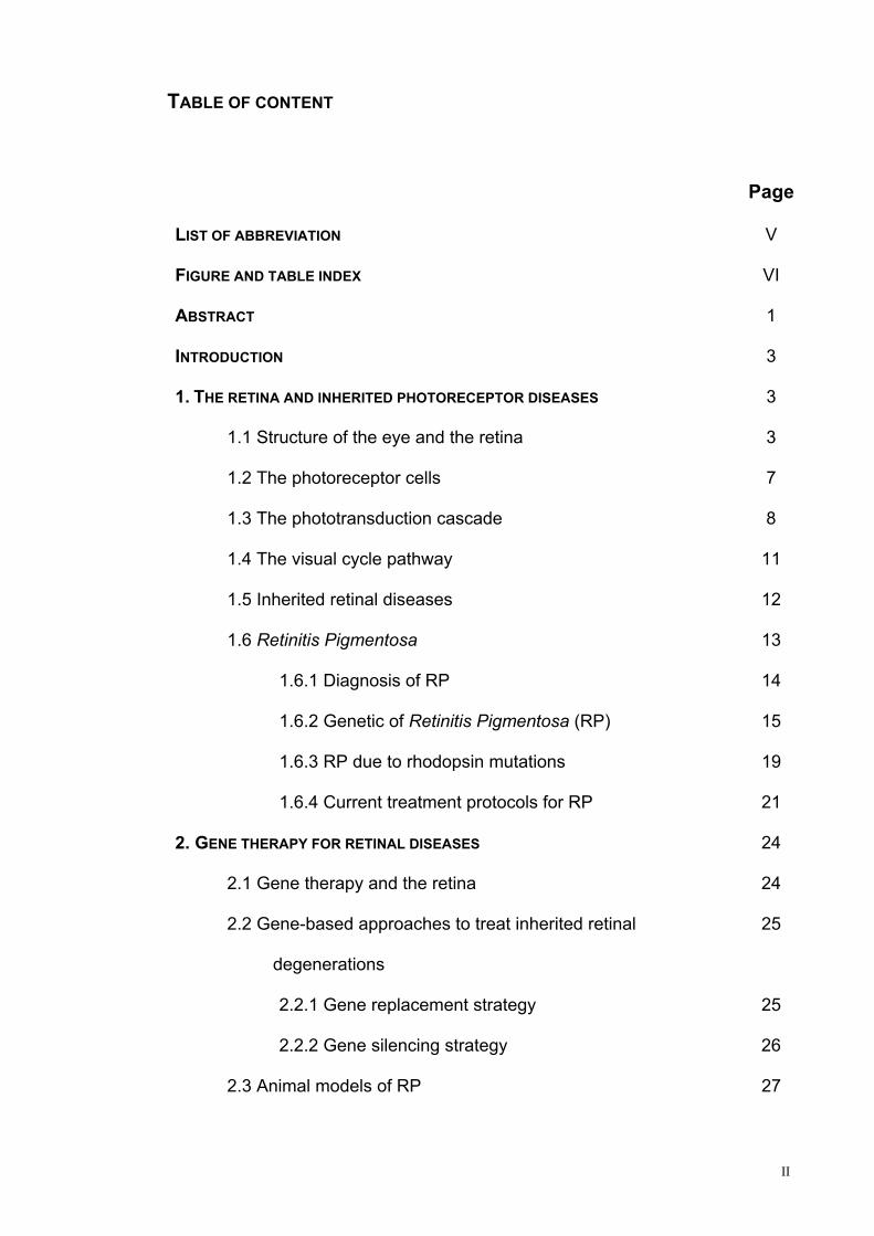
TABLE OF CONTENT
Page
LIST OF ABBREVIATION V
FIGURE AND TABLE INDEX VI
ABSTRACT 1
INTRODUCTION 3
1. THE RETINA AND INHERITED PHOTORECEPTOR DISEASES 3
1.1 Structure of the eye and the retina 3
1.2 The photoreceptor cells 7
1.3 The phototransduction cascade 8
1.4 The visual cycle pathway 11
1.5 Inherited retinal diseases 12
1.6 Retinitis Pigmentosa 13
1.6.1 Diagnosis of RP 14
1.6.2 Genetic of Retinitis Pigmentosa (RP) 15
1.6.3 RP due to rhodopsin mutations 19
1.6.4 Current treatment protocols for RP 21
2. GENE THERAPY FOR RETINAL DISEASES 24
2.1 Gene therapy and the retina 24
2.2 Gene-based approaches to treat inherited retinal
degenerations
25
2.2.1 Gene replacement strategy 25
2.2.2 Gene silencing strategy 26
2.3 Animal models of RP 27
II

3. ADENO-ASSOCIATED VIRUS (AAV) AS A TOOL FOR IN VIVO GENE
TRANSFER
31
3.1 General overview on AAV 31
3.2 Advantages and limitations of recombinant AAV (rAAV) 32
3.3 rAAV as a gene transfer vehicle for the retina 35
4. ZINC-FINGER-BASED ARTIFICIAL TRANSCRIPTION FACTORS (ZF-ATFS) 38
4.1 Generation of ZF-ATFs 40
4.2 ZF-ATFs applications 41
AIMS 43
MATERIALS AND METHODS 45
Rational design of the artificial Zinc-Finger based Transcription
Factors (ZF-TFs)
46
Reporter Constructs 47
Selection of the functional ZF-TFs in human cells by transient
transfection
48
Electromobility Shift Assay (EMSA) 48
AAV vector production and purification 49
RNA preparation and measurement of rhodopsin transcript levels by
Real Time PCR
49
Retinal stem cells culture and analysis 51
Animal model, vector administration, and tissue collection 52
Histological analysis 53
Electroretinogram measurements 53
Statistical analysis 54
RESULTS 55
1. GENERATION AND CHARACTERIZATION OF ENGINEERED ZINC FINGER 56
III

TRANSCRIPTION REPRESSORS TARGETED TO THE HUMAN RHODOPSIN
PROMOTER
1.1 Design and generation of Zinc Finger-based transcription
factors to control rhodopsin gene expression
56
1.2 In vitro selection of functional ZF-Rs 58
1.3 ZF-Rs mediated repression of human rhodopsin in retinal
stem cells
61
2. ASSESSMENT OF EFFICACY OF AAV-MEDIATED GENE TRASNFER OF
ARTIFICIAL ZF-REPRESSOR TO PHOTORECEPTORS OF ADRP MOUSE
MODEL
65
2.1 Delivery of ZF-Rs to murine photoreceptors decrease adRP
retinal progression
65
DISCUSSION 71
REFERENCES 78
IV

LIST OF ABBREVIATIONS
ZFP, zinc finger protein
ZFRs, zinc finger-based repressors
hRHO, human Rhodopsin
mRHO, murine Rhodopsin
adRP, autosomal dominant Retinitis Pigmentosa
arRP, autosomal recessive Retinitis Pigmentosa
miRNA, micro RNA
AAV, adeno-associated virus
LCA, leber congenital amaurosis
hRHO-P, human rhodopsin promoter
ZF-ATF, zinc finger-based artificial transcription factors
DBD, DNA-binding domain
NLS, nuclear localization signal
RSC, retinal stem cells
eGFP, enhanced green fluorescent protein
GC, genome copies
PDE6, phosphodiesterase-6
Cnga, cGMP-gated channel
RHOK, rhodopsin kinase
V

FIGURE AND TABLE INDEX
Page
Figure 1. Schematic view of the human eye anatomy 4
Figure 2. Drawing depicting the layered structure of the retina 5
Figure 3. Structure of the photoreceptor cells 7
Figure 4. Phototransduction pathway in rod photoreceptors 9
Figure 5. Rhodopsin regeneration during visual cycle 11
Figure 6. Fundus photograph of an healty individual and of a patient
with RP
14
Figure 7. Electroretinogram in normal and RP patient 15
Figure 8. Structure of the rhodopsin protein with the most common
mutations highlighted
20
Figure 9. Schematic representation of rAAV vector production 33
Figure 10. Schematic representation of typical Cys2His2-type zinc-
finger motif
39
Figure 11. Schematic view of the binding 39
Figure 12. DNA sequence of the human rhodopsin proximal promoter
region
56
Figure 13. Schematic representation of the zinc finger transcriptional
activator (ZF-A) and repressor (ZF-R)
58
Figure 14. Histograms representing the fold change upon transfection
of the transactivator constructs (ZF-As)
59
Figure 15. Transcriptional activation of the murine rhodopsin promoter 59
Figure 16. Histograms representing the extent of repression mediated
by ZF-Rs
60
VI

VII
Figure 17. The artificial transcription factors with shuffled DBD 60
Figure 18. Binding activity of ZF-R6 assayed by electromobility shift
assay (EMSA)
61
Figure 19. Apoptosis in P347S Retinal Stem Cells (RSC) 62
Figure 20. ZFP-mediated repression of Rhodopsin in Retinal Stem
Cells (RSC)
63
Figure 21. Morphology of P347S+/+ and P347S+/- at 1 month of age
(P30)
66
Figure 22. AAV-mediated photoreceptor ZFP gene transfer results in
the downregulation of hRHO transgene
67
Figure 23. Functional improvement measured by electroretinogram
(ERG) upon ZF-R6 delivery
68
Figure 24. Delay of disease progression upon ZF-R6 delivery in
diseases photoreceptors of adRP mouse model
69
Figure 25. C57BL/6 mice treated with ZF-R6 70
Table 1. Genes for retinitis pigmentosa and functions of their protein
products
17, 18
Table 2. Target sites in the human rhodopsin promoter recognized by
the artificial ZF-TFs generated
57

Abstract
ABSTRACT
Viral-mediated gene therapy holds great promise for the treatment of severe
inherited retinal diseases, such as Retintitis Pigmentosa (RP), which is caused
by mutations in genes preferentially expressed in photoreceptor cells. The
availability of vectors derived from the small adeno-associated virus (AAV)
which efficiently and stably transduce the retina of animal models after
intraocular administration strongly support the possibility to develop novel
strategies for the treatment of such severe retinal degenerations otherwise
incurable thus far.
The main goals of my PhD project were:
- generate artificial transcription repressors (ZFPs) targeted to the human
rhodopsin promoter to silence at the transcriptional level the rhodopsin
gene;
- assess the efficacy of the treatment and the impact on the disease
progression in the RP mouse model.
Retinitis pigmentosa is by far the most studied inherited retinal disease. It is
clinically and genetically heterogeneous recognizing autosomal recessive
(arRP), autosomal dominant (adRP), X-linked, and digenic patterns of
inheritance. More than 30 diseases genes have been identified so far and 12 of
these have been associated with (adRP), representing between 15% and 35%
of all cases. Despite recent success of the gene-based complementation
approach for genetic recessive traits, the development of therapeutic strategies
for gain-of-function mutations poses great challenges. General therapeutic
principles to correct these genetic defects mostly rely on post-transcriptional
gene regulation (RNA silencing). Engineered zinc finger protein (ZFP)-based-
1

Abstract
2
repression of transcription may represent a novel and alternative mutation
independent therapeutic approach for treating gain-of-function mutations, but
proof-of-concept of this use is still lacking. In my PhD project we used a novel
strategy to treat adRP based on zinc-finger-based artificial transcription factors
(ZF-ATFs). These molecules can be engineered to silence genes carrying gain-
of-function mutations that cause toxic effects into the cell where they are
expressed. We generated ten artificial transcriptional repressors targeted to the
human Rhodopsin which is the gene most commonly associated with adRP
(20–30% of cases) with more than 150 mutations identified throughout its
sequence, representing the most commonly mutated gene in RP.
We characterized in vitro the ability of artificial transcriptional repressors to bind
specifically the human rhodopsin promoter in order to exert a specific
transcriptional control and we selected two out of ten functional zinc-finger-
based repressors of rhodopsin. One of this was selected as the most efficient
and was enclosed in an AAV2/8 for in vivo experiments. We demonstrated that
the selected artificial zinc-finger-based repressors (ZFRs) resulted in a robust
transcriptional repression of hRHO impacting disease progression in a mouse
model of adRP over-expressing the P347S mutation.
The data obtained support the use of ZFP-mediated silencing as a potentially
relevant therapeutic strategy to treat gain of function mutations.

Introduction

- Introduction -
INTRODUCTION
1. THE RETINA AND INHERITED PHOTORECEPTOR DISEASES
1.1 Structure of the eye and the retina
The eye is perhaps the most important sensory organ for humans and it is the
first component of the visual system which allows assimilating information from
the environment. This ability is called “Visual Perception” and it is by far the
most complex sensory system. This is due to the fact that vision must handle
demands such as the
transduction of light stimuli to
neural impulses, the binocular
and more distant depth
perception and the colour
discrimination.
The structure of the human eye
(Fig. 1) can be divided into three
main layers or tunics whose
names reflect their basic
functions: the fibrous, the vascular, and the nervous tunics. The fibrous tunic,
also known as the tunica fibrosa oculi, is the outer layer of the eyeball
consisting of the cornea and sclera. It consists of dense connective tissue filled
with the collagen to both protect the inner components of the eye and maintain
its shape. The vascular tunic, also known as the tunica vasculosa oculi, is the
middle vascularized layer, which includes the iris, ciliary body, and choroids.
The iris sits between the anterior chamber which contains the aqueous humour
4

- Introduction -
essential for nourishing the lens and the cornea, and the posterior chamber
filled with the vitreous humour. This substance is jelly-like and, besides helping
the eye to keep its shape, it transmits the light to the back of the eye. The lens
is a clear, flexible structure responsible for sharpening of the image at the retina
and it is connected to the ciliary body (which contains the ciliary muscles). The
choroid contains blood vessels that supply the retinal cells with necessary
oxygen and remove the waste products of respiration. The nervous tunic, also
known as the tunica nervosa oculi, is the inner sensory structure, which
includes the retina.
ggggg
Retina is a light sensitive tissue lining the inner surface of the eye (Fig. 2). It is
a highly organized array of neurons, which serve as transducer for the
conversion of the light into neuronal signals which eventually reach the brain.
Due to the complexity of this process, retinal cells give rise to a variety of
neuronal cell types, which conduct and facilitate the entire cascade of events.
Retina is composed of seven classes of cells structured in layers: photoreceptor
5

- Introduction -
cells (rods and cones), bipolar cells, horizontal cells, amacrine cells, Müller glia
cells and retinal ganglion cells (Fig. 2).
The most outer layer is the retinal pigment epithelium (RPE), which is situated
between the choroid and the photoreceptors. RPE nourishes the retina and is
involved in the phagocytosis of the outer segment of photoreceptor cells and is
also involved in the chromophore regeneration (see section 1.3). The second
layer, the photoreceptor layer, comprises the outer and the inner segments of
photoreceptors (rods and cones), while the photoreceptors cell bodies form the
outer nuclear layer (ONL). At the synaptic terminals of photoreceptors, in a
region called the outer plexiform layer (OPL) light induced signals are
transferred from rods and cones to bipolar and horizontal cells which together
with Müller glia and amacrine cells form the inner nuclear layer (INL). Horizontal
cells provide lateral interaction in the OPL and aid in signal processing. One
type of rod bipolar cells and at least 10 different types of cone bipolar cells
transfer light-induced signals to the inner plexiform layer (IPL), which comprises
dendrites of amacrine cells and ganglion cells. Amacrine cells are inhibitory
interneurons; there are about 40 different subtypes. Müller cells are the main
glial cells of the retina. They form architectural support structures stretching
radially across the thickness of the retina and set the limits of the retina at the
outer and inner limiting membranes, respectively. Dendrites of amacrine cells,
bipolar and ganglion cells form the inner plexiform layer (IPL). The ganglion cell
layer (GCL) forms the innermost retinal layer. Ganglion cell dendrites collect the
signals of bipolar and amacrine cells and transmit these signals through their
axons, which form the optic nerve, to the visual centres of the brain1.
6
- Introduction -
1.2 The photoreceptor cells
Perception of light initiates in the highly specialized retinal cells called
photoreceptors (Fig. 3). Photoreceptors are highly polarized retinal neurons
with the unique property of transforming physical signals (photons of light) first
into biochemical messages and then
into electrical message “perceived” by
specialized brain structures (visual
cortex). Photoreceptors contain four
distinct compartments: the outer
segment (OS), a thin cilium, which
connects the outer to the inner
segment (IS), a cell body containing
the nucleus and the cytoplasm and a
short axon connecting the
photoreceptor cell to interneurons (Fig.
3). The OS is the compartment in which
the conversion of light energy into electrical signals (phototransduction
cascade) occurs. It consists of an array of flat membranous disks that arise
during development as a series of invagionations of the cell’s plasma
membrane. These discs disintegrate near the apical surface of the cells and the
cellular debris are removed through phagocytosis by the adjacent RPE2
following a diurnal rhythm. The old discs are gradually replaced by newly
formed ones that migrate to the base of the OS3. The IS contains most of the
photoreceptor metabolic machinery, including the ER, the Golgi apparatus, and
the mitochondria. Cellular components and metabolites are exchanged and/or
transported between the IS and OS through the narrow connecting cilium.
7

- Introduction -
There are two types of photoreceptor cells in the human retina: rods which
mediate dim-light vision and cones which function in bright light. Rods represent
95% of photoreceptor cells in the human retina and are responsible for sensing
contrast, brightness and motion. They contain a photopigment called rhodopsin
(RHO, max absorbance 500 nm) which is capable of trapping photons. The
cones perceive fine resolution, spatial resolution, and colour vision and they
contain three different colour pigments, blue-sensitive pigment (445 nm), green-
sensitive pigment (535 nm) and red-sensitive pigment (570 nm) sensitive to
different colours: blue, green and red respectively1. In humans, cone density is
maximal in the fovea, which contains about 10% of the cones of the retina; their
density decreases drastically across the macula, beyond the borders of which
density is relatively constant but asymmetric, with higher densities on the nasal
side of the retina. Rods are also distributed unevenly across the retina: there
are no rod photoreceptors within of the fovea. Beyond this rod-free zone, they
increase rapidly to reach a peak along an elliptical ring at the eccentricity of the
optic disc. In rodents, fovea is not present and photoreceptors are constantly
distributed through the retina.
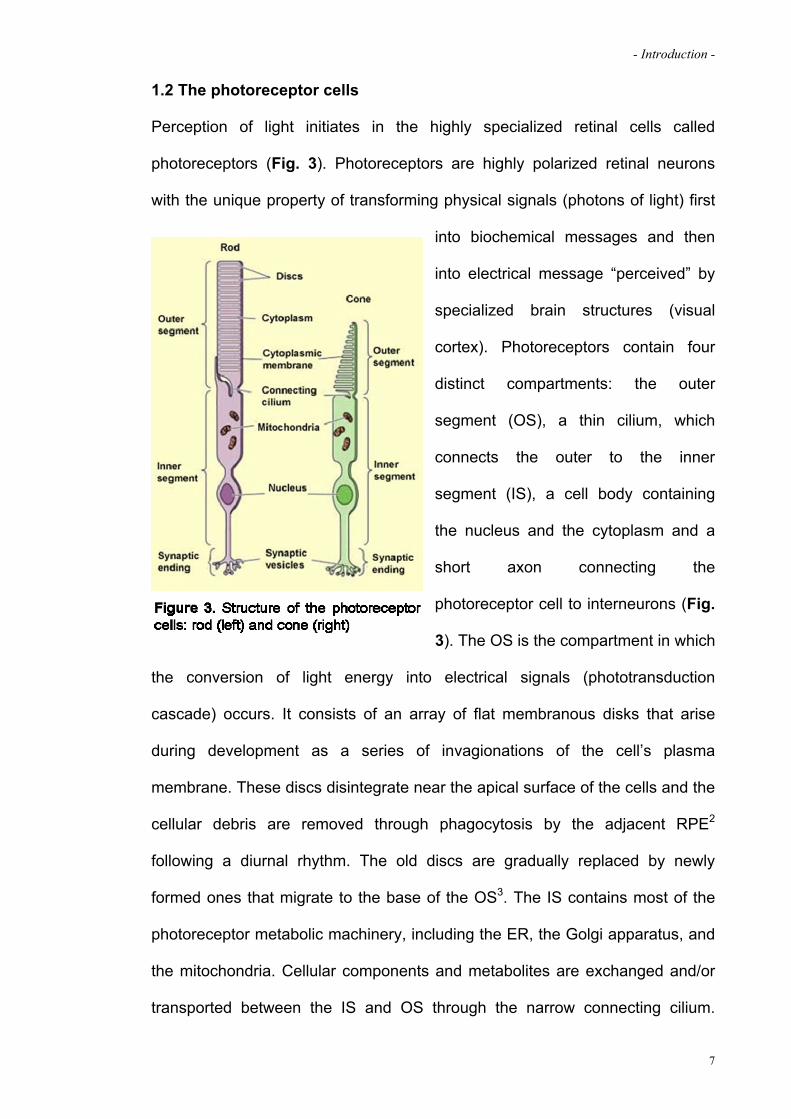
1.3 The phototransduction cascade
Transduction of absorbed light into electrical signal that is eventually perceived
as sight takes place within photoreceptor cells via a complex molecular process
called phototransduction4. Rod phototransduction has been more
comprehensively studied than cones one. It takes place in the rod OS discs
with the absorption of light by rhodopsin which is a photopigment that counts for
80% of total amount of rod outer segment proteins. It is composed of a
backbone, termed rod-specific opsin, a seven transmembrane G-protein-
8
- Introduction -
coupled receptor, bound to the light-sensitive chromophore 11-cis-retinal
(11cRAL) a derivative of vitamin A5. The phototransduction cascade initiates
when the visual pigment, rhodopsin, absorbs a photon (hv; Fig. 4). This induces
a conformational change of the chromophore to all-trans, which still has the
same chemical structure as the cis but a different physical form. Because the
all-trans-retinal (atRAL) no longer fits with the rhodopsin, it begins to pull away
from it until there is a complete split (within seconds). This in turn induces
conformational changes in the rhodopsin which is activated (R*). This
intermediate molecule (metarhodopsin II) interacts with the next member of the
cascade, a G-protein called Transducin (G) which is an heterotrimeric protein
(). This interaction induces the subunit of the transducin to exchange a
bound guanosine diphosphate (GDP) moiety to guanosine triphosphate (GTP).
The activated GTP-bounded subunit (G*) detaches from the and
subunits of the transducin and associated with the next member of the cascade,
the phosphodiesterase 6 (PDE6) which is a multisubunit complex, composed of
two tightly bound catalytic subunits, (99kDa, PDE) and (98kDa, PDE) in
addition to two identical inhibitory subunits of 11kDa. The enzyme is anchored
9

- Introduction -
to the rim membrane by an isoprenylic group at the C-terminus of the and
subunits. The G* subunit interacts directly with the inhibitory subunit of the
PDE6. An increase in the activity of the cGMP phosphodiesterase at this stage
induces a fall in the concentration of cGMP in the cytoplasm5 which leads to the
closure of cGMP-gated cation (Na+ and Ca++) channels on the plasma
membrane of the rod’s OSs with a consequent decline in calcium concentration
within the cell. This causes a graded hyperpolarization of the plasma
membrane which is conveyed, as in all neuronal cells, to the synaptic region of
the rod where it decreases the amount of neurotransmitter (glutamate) release.
This initial signal is transmitted via second-order retinal neurons to the optic
nerve and to the brain. The decline in calcium concentration mediates the
recovery of the photoreceptor cell after a bleach of light. This is as important for
maintaining sensitivity in vision as the cell’s ability to respond to a single
photon. Deactivation of rhodopsin starts with phosphorylation by rhodopsin
kinase (RHOK) and is followed by the capture of rhodopsin by the protein
arrestin6. The arrestin-binding prevents further activation of transducin and
releases the all-trans-retinal from rhodopsin. The concentration of cGMP within
the cell is restored by the increased synthesis of cGMP by a retinal guanylate
cyclase (GC). These pathways are triggered by the decline in the intracellular
calcium concentration and mediated by a family of calcium-binding proteins,
including recoverin and guanylyl cyclise activating protein (GCAP). The
calcium-bound recoverin inhibits the activity of rhodopsin kinase7. Sustained
phototransduction depends on replenishing the 11-cis-retinal lost as a result of
light activation of the visual pigments in a pathway which takes place between
the photoreceptor cells and the RPE in a process called visual cycle8.
10
- Introduction -
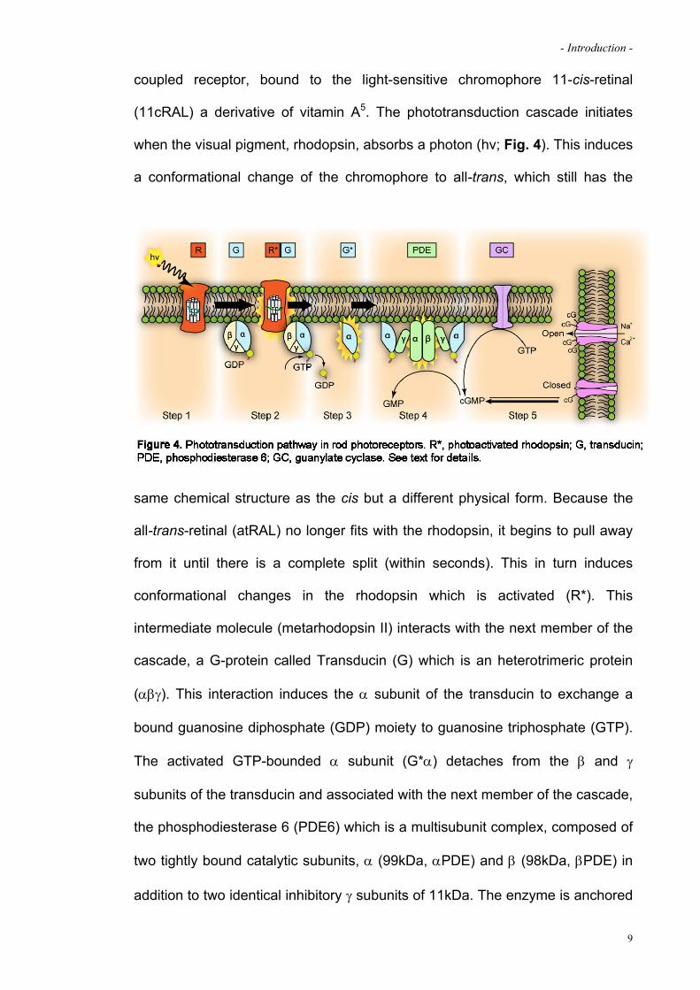
1.4 The visual cycle pathway
Absorption of a photon of light by rhodopsin causes isomerisation of the
chromophore from 11-cis-retinal to all-trans-retinal. In order to restore light
sensitivity of rhodopsin all-trans-retinal must be converted back to 11-cis-retinal
through a multistep pathway called visual cycle (Fig. 5). Visual cycle initiates in
f
the photoreceptor cells, precisely in the inner surface of the rod disks
membrane, with the release of all-trans-retinal which is subsequently
transferred to cytoplasmic surface of the disks by retina-specific ATP-binding
cassette transporter (ABCA4)9; 10. In the photoreceptor cytoplasm, all-trans-
retinal is reduced by an all-trans retinol dehydrogenase (atRDH) and the
resulting all-trans retinol (vitamin A) is transported across the subretinal space
to the RPE. Within the RPE, all-trans-retinol is bound to cellular retinal binding
protein (CRBP)11 and immediately esterified to all-trans-retinyl esters (atRE) by
11
- Introduction -
lecithin retinol acyltransferase (LRAT)12. atRE is isomerized to 11-cis-retinol by
isomerohydrolase (IMH) in conjunction with RPE65 protein. The resulting 11-
cis-retinol (11cROL) is oxidized by the retinol dehydrogenase (11cisRDH) to the
final product 11-cis retinal13. The 11-cis-retinal exits the RPE, traverses the
subretinal space, and enters the photoreceptor outer segments where it
combines with opsin protein to form a new molecule of light-sensitive
odopsin. rh
1.5 Inherited retinal diseases
The complex retinal structure and signalling network, which includes numerous
neurotransmitters, neuromodulators, phototransduction proteins, transcription
factors, etc., lead to a wide range of targets for potential events that may cause
pathogenic changes in its function. According to some estimates14 the eye is
the fourth system most commonly affected by genetic diseases in humans. At
the same time, genetic eye diseases, both monogenic and genetically complex,
comprise the commonest causes of blindness in children and adults
worldwide15. Out of different eye diseases, retinal disorders are especially
important since there are more then 100 diseases that include a form of retinal
dystrophy, as listed in the online database of human genetic diseases (Online
Mendelian Inheritance in Man-OMIM). In the industrialized world, the most
common diseases involving the retina are diabetic retinopathy, glaucoma, and
age-related macular degeneration (AMD), which together affect several percent
of the population16. Each of these diseases has both genetic and non-genetic
components. Contrary to complex diseases, the simple Mendelian retinal
diseases have an earlier onset and some have a more severe clinical course
than typically observed for the three more common disorders listed above and,
12

- Introduction -
for the most part, they are untreatable. These characteristics, together with the
possibility of exploiting genetic approaches to understand disease mechanisms,
have drawn attention on the Mendelian disorders. Retinitis pigmentosa is by far
the most studied inherited retinal degeneration and it may serve as an example
f remarkable genetic heterogeneity connected with retinal dystrophies.
f visual acuity and
efective colour vision are the prominent early symptoms.
o
1.6 Retinitis Pigmentosa
Retinitis Pigmentosa (RP) is the term given to a set of inherited retinal
degeneration with a prevalence of 1:3500 worldwide17; 18 neither preventable
nor curable19; 20. It is an highly variable disorder, indeed some patients develop
symptomatic visual loss in childhood whereas others remain asymptomatic until
mid-adulthood. Many patients fall into a classic pattern of difficulties with dark
adaptation and night blindness in adolescence and loss of mid-peripheral visual
field in young adulthood. As the disease advances, they experience a drop of
far peripheral vision which, develops in tunnel vision and eventually in central
vision lost, usually by age 60 years20. Visual symptoms indicate the gradual
loss of the two photoreceptor types: first loss of rods, which mediate achromatic
vision in starlight or moonlight and are more abundant in the peripheral retina,
then loss of cones, which are important for colour vision and fine acuity in
daylight. Most patients are legally blind by age 40 years because of severely
constricted visual field. In most form of RP, loss of rod function exceeds
reduction of cone sensitivity. In other types, rod and cone decline is similar.
Occasionally, the deficit of cones far exceeds that of rods; in this case the
disease is termed cone-rod degeneration, and loss o
d
13

- Introduction -
1.6.1 Diagnosis of RP
Clinically, RP patients are diagnosed based on three main abnormalities:
ges of the retina and RPE, abnormal
electroretinogram (ERG) and attenuation of
the retinal vasculature and changes to the
optic nerve head18. In addition, reduction of
the visual field, colour vision impairment, dark
adaptation or cataracts, which is showed in
about 50% of individuals with RP, can be
observed. Pigmentary changes in the retina
are evident during the fundus examination
and remain a common factor in RP diagnosis
(Fig. 6); they mainly consist in marked
pigment epithelial thinning, optic disc pallor
and the classical “bone spicule deposits which result from the release of
pigment by degenerating cells in the retinal pigment epithelium. The pigment
granules accumulate in perivascular clusters, known as “bone-spicule
formations” due to their morphological appearance, in the neural retina.
Consequently, early in the disease, the pigmented posterior pole of the eye, the
fundus, develops a granular appearance. This is followed by the development
of bone-spicule pigmentary deposits overlying the depigmented fundus21. The
main tool for diagnosis and classification of retinitis pigmentosa is the
Electroretinogram (ERG) which is an objective measure of retinal function and it
is useful for accurate diagnosis of disease, for assessment of severity, to follow
the course of the disease, to provide a visual prognosis and for measurement of
responses to treatments. In this procedure, retina is either dark adapted
atrophy and pigmentary chan
14
- Introduction -
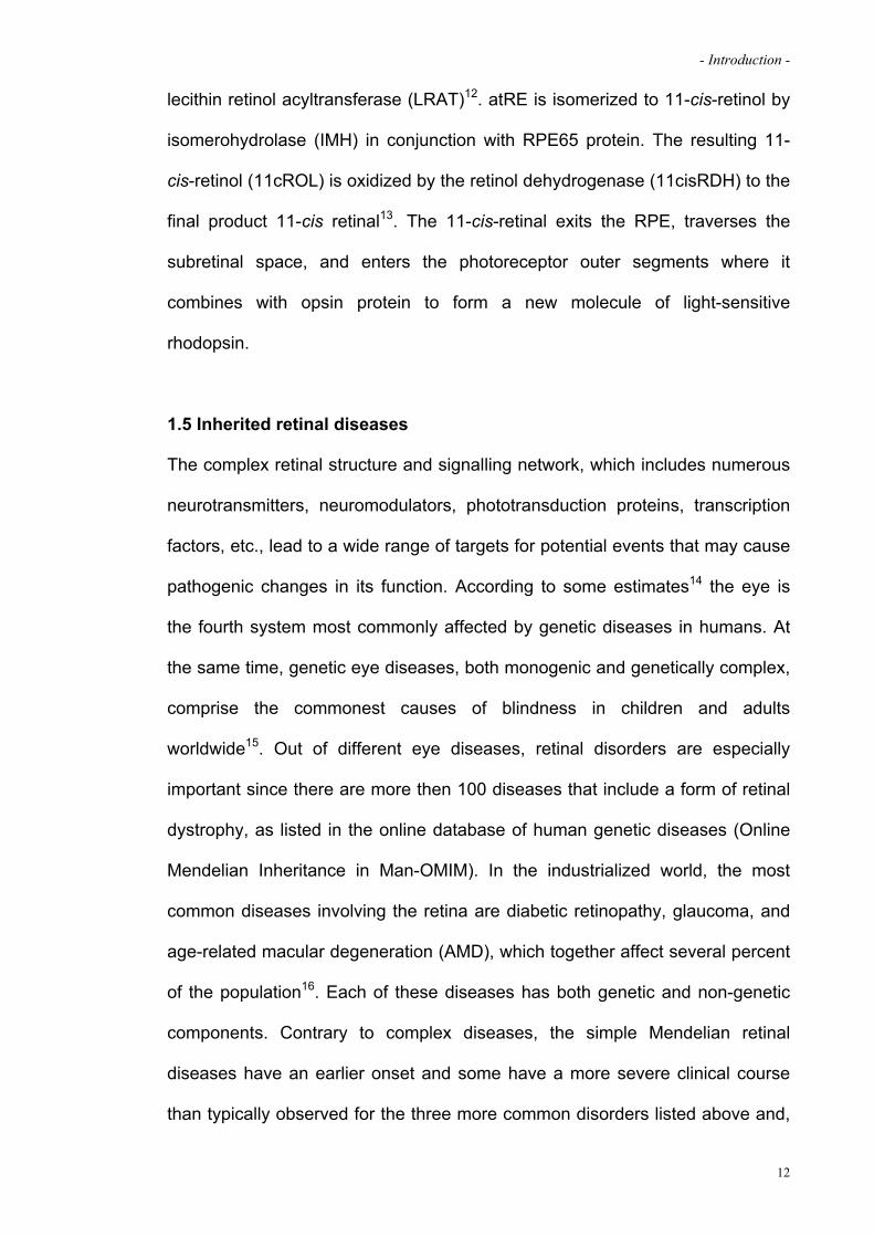
(scotopic ERG) or adapted to a specific level of light (photopic ERG), and then
stimulated with a brief flash of light. The summed electrical response of the
retina is recorded extraocularly with a contact lens electrode. A single-flash dim
blue light elicits a rod response, a brighter single-flash white light elicits a
combined rod-plus-cone response, and flickering (30Hz) white stimuli generate
cone-isolated response. With single flashes (0-5 Hz) of white light, an initial a-
wave shows hyperpolarisation of photoreceptors and a subsequent b-wave
result from depolarisation of cells in the inner nuclear layer. Typically, patients
with RP have reduced rod and cone response amplitudes and a delay in their
timing18 (Fig. 7).
g g
1.6.2 Genetic of Retinitis Pigmentosa (RP)
Retinitis Pigmentosa is due to many distinct causes and involves diverse
biological pathways but with overlapping symptoms and similar consequences.
Most forms of RP are monogenic and can be inherited with classical patterns18;
22: autosomal dominant (adRP) forms account for 30-40% of total cases,
autosomal recessive (arRP) forms are the most common variants with a
15

- Introduction -
frequency of 50-60% while X-linked (X-lRP) can be observed in 5-15% of
affected individuals. However, some unusual patterns as digenic inheritance23
or maternal (mitochondrial) inheritance24 have also been reported in the
literature. In most cases, patients with Retinitis Pigmentosa have no associated
systemic or extraocular abnormalities. Moreover, there are multisystem
diseases in which RP is accompanied by involvement of other tissues and
organs. Examples are Usher’s syndrome (USH), in which Retinitis Pigmentosa
is associated with hearing impairment and it is the most frequent syndromic
form (10-20% of total cases)25, or Bardet-Biedl syndrome, where RP is
associated with obesity, cognitive impairment, polydactyly, hypogenitalism, and
renal disfunction26. A remarkable feature of Retinitis Pigmentosa is its high
genetic heterogeneity: so far, more than 50 genes responsible for non-
syndromic forms have been identified that accounts for 60% of total cases20
(RetNet: http://www.sph.uth.tmc.edu/Retnet/) while about 40% of cases are due
to genes not yet mapped. Mutations in genes preferentially expressed in
photoreceptors are the most common cause of RP followed by RPE-specific
genes. In rare cases RP is caused by mutations in genes expressed in other
retinal cell types or outside the eye. Despite the genetic heterogeneity of RP,
photoreceptor cells mainly degenerate by apoptosis27 although the mechanisms
by which the genetic defect leads to cell death are still unclear28. According to
the known or presumed function of the encoded proteins, the genes responsible
for RP so far identified have been clearly grouped into functional categories as
schematically depicted in Table 120. Some of these genes encode proteins in
the rod photoreceptor cascade, the specific biochemical pathway that
transduces light stimuli and leads to changes in photoreceptor-cell polarisation.
Recessive null mutations in any of these genes would evidently interfere with
16

- Introduction -
rod function and produce night blindness from birth. Subsequent death of rod
photoreceptors is probably an outcome of the deranged physiology associated
with the defective or absent gene product. For example, without functional rod
cGMP phosphodiesterase, arising with recessive defects in PDE6 or PDE6,
cGMP concentrations in rod photoreceptor outer segment rise and this in turn
opens cGMP-gated channels in the plasma membrane. Rods apparently die
zz
17

- Introduction -
zz
from the rush of cation flowing into the cells through these open channels29.
Another relevant example is given by dominant mutations in rhodopsin gene
which are probably detrimental to rods because the mutant forms of the protein
are toxic to rod photoreceptors. The toxic effects are attributable to interference
with metabolism, perhaps by formation of intracellular protein aggregates, from
a defect in intracellular transport, or from a fault in the structure of the
photoreceptor outer segments30. The reason why mutations in genes
exclusively expressed in rod photoreceptors cause the death of both rod and
cone cells is not yet clear. The secondary death of cones might be due to
reliance on neighbouring rods for survival. The discovery of the RdCVF protein,
18
- Introduction -
a factor released from rods that promote cone survival, provides a possible
explanation to this question31. The second most common group of genes
mutated in RP are involved in visual cycle, in particular in recycling the
rhodopsin chromophore 11-cis-retinaldehyde18. An example is the gene RPE65
which is primarily expressed in the RPE and endowed with isomerise activity for
the rhodopsin ligand 11-cis-rtinal32. Mutations in genes encoding photoreceptor
structural proteins or transcription factors have also been identified. Moreover,
some genes for RP are expressed in tissue outside the eye, and others encode
proteins that are essential for life. For example, non syndromic RP is caused by
dominant mutations in genes PRPF31, PRPF8 and PRPF3 that encode
components of the spliceosome, a vital complex that excises introns from RNA
transcript33.
1.6.3 RP due to rhodopsin mutations
Mutations in the rhodopsin gene are the most common cause of Retinitis
Pigmentosa among human patients and account for 20-30% of adRP cases34-37
(Fig. 8). Rhodopsin is the light-absorbing protein that mediates vision at low
light levels. Like other visual pigments, it consists of a chromophore (11-cis-
retinal) covalently bound to an integral membrane protein (opsin).
Photoisomerization of retinal from 11-cis to all-trans induces a
conformational change in the apoprotein, leading to a conformation that is
competent to activate the photoreceptor-specific G-protein transducin,
thereby initiating the phototransduction cascade. In mammals, rhodopsin
accumulates to a level of 5x10⁷ molecules/rod outer segment and is
synthesized throughout life at a rate of 5x10⁶ molecules/rod/day38. Rhodopsin is
localized to the rods outer segments which contain hundreds of flattened
19

- Introduction -
membrane sacs (also called disks) stacked in close apposition and where it
represents the 80% of total proteins content39. Over 150 mutations in the
rhodopsin gene leading to autosomal dominant RP (adRP) have been
characterized so far which can be grouped in two distinct classes of
biochemical defect. Approximately 85% of the mutant proteins (class II) occurs
in the N-terminus of the protein and are produced at lower levels than the wild
zzz
type, accumulate predominantly in the endoplasmic reticulum, and bind 11-cis-
retinal variably or not at all. This class of proteins appears to be defective in
folding and/or stability. The remaining 15% of mutant proteins (class I) map
very close to the C-terminus, a region of the protein for which no function has
yet been assigned, and resemble the wild type protein, indeed are produced at
high levels, accumulate in the plasma membrane and efficiently bind to 11-cis-
retinal to form photolabile pigments40; 41. Because these rhodopsin mutations
were identified in the heterozygous condition in patients with adRP, it seems
20
- Introduction -
likely that the mutant proteins interfere with or participate aberrantly in some
physiological process. A second possibility is that they are physiologically silent
and that RP results from haploinsufficiency, a scenario that seems unlikely in
light of the finding that heterozygous carriers of one apparently null mutation in
the rhodopsin gene do not have RP42. Such mutational heterogeneity
represents a significant barrier to the development of therapies for adRP
associated with rhodopsin gene.
1.6.4 Current treatment protocols for RP
At the moment, no cure is available for RP but some pharmacological
treatments are recommended by clinicians to slow visual loss. Nutritional or
neuroprotective treatments that affect secondary biochemical pathways have
the advantage of being less dependent on the disease-causing mutation and
could therefore be widely applicable.
For example, based on a study of the natural course of Retinitis Pigmentosa,
patients assuming vitamin A, vitamin E, or both were recorded to have slower
declines in ERG amplitudes than those not taking such supplements43. This
observation prompted a randomised clinical trial of oral vitamin A and E
supplements in 601 patients with dominant, recessive and X-linked non-
syndromic Retinitis Pigmentosa and Usher’s syndrome type II. Patients
assigned high-dose vitamin A showed a significantly slower decline in cone
ERG amplitudes than did those in the other groups. Based on these results,
many clinicians recommend that adults with early or middle stages of Retinitis
Pigmentosa take 15,000 IU of oral vitamin A palmitate every day and avoid high
dose vitamin E supplements. Toxic effects have not been reported, even if older
individuals could also be monitored for bone health because a slight increased
21

- Introduction -
risk for hip fractures due to osteoporosis has been reported in postmenopausal
women and men older then 49 years who take vitamin A supplements43.
Another nutritional treatment assessed for patients with Retinitis Pigmentosa is
docosahexaenoic acid (DHA), an omega-3 fatty acid found in high
concentration in oil fish. DHA is apparently important for the photoreceptor
membrane, since membranes containing rhodopsin and cone-opsin in
photoreceptor cells have very high dose of this fatty acid. Amounts of DHA in
red-blood cells are on average lower in patients with RP than in unaffected
people. Nevertheless, results from two independent studies of oral DHA
supplements to individuals with Retinitis Pigmentosa did not show clear benefits
even if people with the highest concentrations of DHA in red-blood cells had the
slowest rates of retinal degeneration44.
Further, finding of work done in animals have shown that some neurotrophic
factors can promote photoreceptor survival45; 46. Results of a human phase I
study of an intravitreal capsule containing cells that release ciliary neurotrophic
factor have been reported. Small-molecule drugs are also being assessed as
possible treatments for RP. For example, in a study of a calcium-channel
blocker (diltiazem), researchers claimed a beneficial effect in a mouse model of
RP due to recessive mutations in the -subunit of rod phosphodiesterase.
However, three subsequent trials of this drug in mice and other animal models
by independent groups failed to confirm a benefit47; 48.
Lastly, nanotechnology and biotechnology leading company make great effort
in order to develop devices to electrically stimulate the retina, optic nerve, or
visual cortex. The first results obtained are encouraging since the few people
testing the first version of these devices have reported seeing phosphenes
(flash of light) in response to direct retinal stimulation49.
22

- Introduction -
It’s anyway clear that, increasing the knowledge of the biochemical defects
associated to RP is essential to develop diverse approaches for the treatment
of such challenging disease. Nevertheless, these data show that the above-
mentioned approaches cannot be considered a real therapeutic option to halt or
reverse RP thus far.
23
- Introduction -
2. GENE THERAPY FOR RETINAL DISEASES
2.1 Gene therapy and the retina
Gene therapy aims at delivering corrective genetic material to a cell, tissue or
target organ in order to prevent or cure a disease50. Nucleic acids do not readily
cross cell membranes; consequently, it is necessary to envelope the genetic
material in a lipidic-containing complex or incorporate it in a viral vector51; 52.
The former inserts genetic material directly into the target cells by fusing to the
host cell membrane. Unfortunately they are not selective for a target cell, have
low transduction efficiency and mediate short-lived gene expression51; 52. On
the other hand, viral delivery of therapeutic genes appears much more
promising since a prolonged transgene expression is obtained with the use of
particular recombinant viral vectors51.
The retina represents an ideal target organ for gene therapy approaches for a
number of reasons: i) it is easy to manipulate and the small size of the eye
allows the use of low doses of vector; ii) the partial immune privileged
properties of the eye can limit immune responses toward the transgene and the
vector; iii) the eye is enclosed and the presence of the blood–retinal-barrier, of
the RPE and of the intracellular junctions in the inner retina can help avoiding
unintentional spreading of vectors to neighbouring tissues as well as to the
general circulation; iv) non invasive in vivo techniques for ocular tissues
imaging and visual function evaluation are available (Electroretinogram, ERG;
Optical Coherence Tomography, OCT); v) since ocular diseases develop
bilaterally and symmetrically, if one eye is treated the other can be used as an
useful untreated internal control; vi) surgical procedures have been adapted for
24

- Introduction -
the transfer of genetic material into the two main ocular compartments
(Subretinal and intravitreal).
2.2 Gene-based approaches to treat inherited retinal degenerations
Gene-therapy approaches are dependent on the type of mutation to reverse
thus different strategies may be adopted in order to treat a gain- or a loss-of-
function mutation as well as for the treatment of a dominant-negative mutation
or in the case of haploinsufficiency. However, such strategies can be divided in
two distinct groups: gene replacement or gene silencing strategies.
2.2.1 Gene replacement strategy
Recessively inherited diseases typically result from alterations that eliminate the
encoded protein (loss-of-function mutations). They can give rise to dominant
inheritance if the remaining normal copy does not express sufficient protein to
meet cell’s needs. Dominance resulting from inadequate expression levels is
known as haploinsufficiency and it is extremely rare, thus for most genes, one
allele is sufficient to preserve retinal function. For the treatment of loss-of-
function mutations, the introduction of a normal copy of the gene into the
diseased tissue53 (gene-replacement approach) can supply the missing protein.
This approach, for example, has been applied to a form of Leber Congenital
Amaurosis (LCA)54, the most severe form of inherited childhood blindness. The
target gene in this case is RPE65, which is expressed in the RPE and encodes
for an isomerise that is essential for production of the photopigment 11-cis-
retinal32. Subretinal administration of an AAV vector containing a corrected copy
of the RPE65 gene has shown to restore vision in mice and dogs harbouring a
mutation in the gene55-61. The recent success of clinical trials for the treatment
25

- Introduction -
of this disease is providing sound evidence for the use of gene-based
complementation therapeutic strategy to treat genetic recessive traits62-68.
Gene-replacement approach has also been successfully used in animal models
of genetically identified forms of RP, however, in this case, it is essential to
intervene during early stages of the diseases progression in order to prevent
the loss of photoreceptor cells.
2.2.2 Gene silencing strategy
Dominant mutations typically alter the transcribed amino acid sequence and
result in toxic variants of the encoded protein (gain-of-function of dominant-
negative mutations). One strategy to treat these alterations is to eliminate the
mutant gene (gene silencing approach) and hope that the remaining normal
copy of the gene will provide sufficient functional protein. The main target of
genetic silencing strategies is the mRNA transcript whose function can be
inhibited by antisense-, ribozyme- and more recently by small interfering RNAs-
(siRNAs) and miRNA-based approaches. In particular, RNAi is holding great
promise for treating dominant diseases for its efficiency in mRNA transcripts
cleavage in animal models of adRP due to P23H or P347S53; 69-74. An emerging
alternative to such RNA-targeting approaches is the modulation of gene
expression at the transcriptional level by using artificial zinc finger-based
transcription factors (ZF-TFs) or the genomic inactivation of a disease gene by
using targeted zinc finger-based nucleases (ZFNs)75-77. The action of such
novel approaches is targeted to the genomic sequence, silencing the
downstream target gene independently by the causative mutation identified; as
a consequence they allow the development of economic viable therapeutics
since they circumvent the mutational heterogeneity associated to diseases,
26

- Introduction -
such as adRP due to rhodopsin mutations. Indeed, as mentioned before,
mutations in the rhodopsin gene are the most common cause of Retinitis
Pigmentosa (RP) among human patients and nearly all rhodopsin mutations are
dominant (The only characterized rhodopsin recessive allele is the mutation
E249ter42).
2.3 Animal models of RP
The study of animal models with naturally occurring degenerative retinal
diseases has been important for the discovery of numerous candidate genes,
some of which have led to the identification of new disease genes in humans.
These animals are useful models for understanding molecular pathways
activated during the disease and to test experimental therapies for this
otherwise incurable group of defects. Furthermore, the ability to generate gene-
specific transgenic and targeted (knock-out or knock-in) mice, allows the
creation of new models of human retinal disease and facilitate the elucidation of
gene dysfunction. Several animal models of RP are available, mostly in
rodents, pigs and canines78-80. Each of them shows peculiar phenotypical
features depending on the mutated gene/allele, although all RP models are
characterized by photoreceptors degeneration with the main differences
consisting in the onset and time course of the disease. In this paragraph I will
focus on the most common animal models of RP due to mutations in the
rhodopsin gene. In 1997, Humphries and colleagues generated mice carrying a
targeted disruption of the rhodopsin (RHO) gene81. The rod outer segments of
Rho-/- mice do not develop fully and photoreceptor cells are lost over a 3 month
period. No rod ERG response is present in 8-weeks-old animals. Rho+/-
animals retain the majority of their photoreceptors although the inner and outer
27
- Introduction -
segments of these cells display some structural disorganization and the outer
segments become shorter in older mice. The rhodopsin knock-out animals
provide a useful genetic background on which to express other mutant opsin
transgenes as well as assessing the therapeutic potential of re-introducing
functional rhodopsin genes into degenerating retinal tissues. The P23H
mutation is the most prevalent mutation in human adRP patients in US82.
Twelve percent of American patients with autosomal dominant Retinitis
Pigmentosa (adRP) carry a substitution of histidine for proline at codon 23
(P23H) in their rhodopsin gene, resulting in photoreceptor cell death from the
synthesis of the abnormal gene product. Transgenic rodents and pigs
overexpressing the dominant P23H rhodopsin mutations that recapitulate the
human disease have been generated83-87. Transgenic mice containing the
P23H mutation appear to develop normal photoreceptors, but their light-
sensitive outer segments never reach normal length85. With advancing age,
both rod and cone photoreceptors are progressively reduced in number. The
degeneration of the transgenic retina is associated with a gradual decrease of
light-evoked electroretinogram responses. The disease mechanism associated
with the P23H mutation remains unclear. Haploinsufficiency is unlikely, since
humans and mice heterozygous for rhodopsin null mutations do not show
clinical evidence of RP, although they may present with electrophysiological
abnormalities42; 81; 88; 89. In addition, transgenic animals overexpressing the
P23H allele on a wild-type rhodopsin background present with retinal
degeneration. Overexpression of wild-type rhodopsin in P23H transgenic mice
slows the progression of retinal degeneration, indirectly suggesting a dominant-
negative effect of the mutated allele90. Several independent observations point
to a gain-of-function effect exerted by the P23H mutation. Its overexpression in
28
- Introduction -
cultured cells results in its accumulation in the endoplasmic reticulum (ER) as a
consequence of the incorrect folding of the mutated protein91. In the ER,
misfolded P23H aggregates and overwhelms the proteasome system, leading
to cell death91; 92.
Mutations resulting in changes in the C-terminal domain of rhodopsin result in
some of the more severe forms of the disease, with total blindness occurring in
early adulthood. The rhodopsin C-terminal sequence is recognized by specific
factors in the trans-Golgi network. Mutations resulting in changes in this region
produce RP due to formation of abnormal post-Golgi membranes and from the
aberrant subcellular localization of rhodopsin93-95. Among the class I mutants,
those affecting the proline in position 347 near the C-terminus seems to be
extremely deleterious. This residue is conserved among all known visual
pigments. Six different missense mutations affecting this residue have been
identified among patients with RP, indicating that proline-347 mediates some
vital function of rhodopsin. Proline-347 mutants are of particular interest, since
no functional abnormalities of the mutant proteins have been observed in in
vitro studies, yet they tend to cause a more severe form of RP than that found
in patients with adRP due to other mutations96. A transgenic mouse carrying a
human P347S allele was generated to investigate the early pathogenic events
by which this rhodopsin mutant leads to photoreceptor cells death94. In the pre-
degenerate or early degenerating retinae of these mice there was no gross
perturbation of phototransduction, as monitored by ERGs, and no accumulation
of mutant rhodopsin. However, the mutant retinae exhibited a distinct
morphological phenotype (extracellular accumulation of small rhodopsin-
containing vesicles in the vicinity of basal outer segments and apical inner
segments) which suggests that the C-terminus of rhodopsin might have a role
29

- Introduction -
in rhodopsin transport to the outer segments95. Photoreceptor cell death in the
P347S mice can be thus a consequence of this primary defect.
To further elucidate the mechanisms of photoreceptor cells death induced by
rhodopsin mutations, also transgenic porcine model for the rhodopsin P347L
and P347S mutations have been produced and show a disease phenotype
similar to that found in humans86; 97; 98.
30

- Introduction -
3. ADENO-ASSOCIATED VIRUS (AAV) AS A TOOL FOR IN VIVO GENE
TRANSFER
The success of gene therapy approach is based on an appropriate amount of a
therapeutic gene delivered into the target tissue without substantial toxicity.
Each viral vector system is characterized by an inherent set of properties that
affect its suitability for specific gene therapy applications. For some disorders,
long-term expression from a relatively small proportion of cells would be
sufficient (for example, genetic disorders), whereas other pathologies might
require high, but transient, gene expression.
To date, three types of viruses have mainly proven useful for retinal transgene
delivery. They are Adeno-Associated Virus (AAV), Lentivirus and Adenovirus53;
99. These viruses are genetically modified to eliminate the inherent toxicity
caused by their ability to replicate and disseminate51. Among these vector
systems AAV-based vectors are very promising. AAVs are not associated with
human diseases and possess a number of properties that make them
particularly suitable for clinical gene therapy, including the efficiency to transfer
genetic material to a number of dividing and not dividing cells and a natural
propensity to persist in human cells. In addition, the development of AAV
vectors for gene therapy so far has shown an excellent safety record with data
accumulated from thousands of animal studies and hundreds of human
patients51; 100.
3.1 General overview on AAV
The adeno-associated virus (AAV) is a small (20-25 nm in diameter), non-
enveloped, icosahedric Dependovirus belonging to the Parvoviridae family101.
These viruses are non pathogens for humans and possess a linear, single-
31

- Introduction -
stranded DNA genome that can replicate in the presence of different helper
viruses such as adenovirus, herpesvirus or papillomavirus102. AAV was
originally isolated as a contaminant of adenoviral cultures and thus given the
name adeno-associated virus. It is native of humans and non-human primates
(NHP) and exists in nature in more than 100 distinct variants, including both
those defined serologically as serotypes and those defined by DNA sequence
as genomovars103; 104. The AAV genome (4.7 Kb) consists of two open reading
frames: i) rep, which codes for a family of multifunctional non-structural proteins
that are involved in viral genome replication, transcriptional control, integration
and encapsidation of rAAV genomes into preformed capsids105-109 and ii) cap
encoding for the three structural proteins VP1, VP2 and VP3110; 111. rep and cap
are flanked by viral T shaped palindromic elements, the inverted terminal
repeats (ITRs) which are 145 nucleotides in length102. In vitro experiments
demonstrated that, in the absence of an helper virus, AAV establishes latency
by integrating in a site-specific manner in the human chromosome 19q13.3-qter
(in a site called AAVS1)112 through an interaction between the ITRs and the
AAVS1 locus mediated by the rep proteins113. Despite the presence of a
preferential integration site, the status of AAV genomes from infected cells has
been shown to be mainly episomal114; 115.
3.2 Advantages and limitations of recombinant AAV (rAAV)
The conversion of an AAV isolate into recombinant vector (rAAV) to be used in
gene therapy is obtained by exchanging the viral coding sequence between the
ITRs with the therapeutic gene116. To produce rAAV, the rep and cap functions
as well as the helper genes needed are provided in trans117. In the absence of
rep, rAAV loses its site-specific integration ability118. The most commonly used
32

- Introduction -
strategy to produce rAAV vectors to be used in gene therapy is based on the
co-transfection into permissive cells (usually human embryonic kidney 293
cells) of three separate plasmids116; 117: i) a plasmid containing the viral ITRs
flanking the therapeutic gene cassette; ii) a packaging plasmid encoding for the
rep and cap proteins; iii) the helper plasmid encoding for the essential
adenoviral helper genes (Fig. 9)116; 117. The versatility of the rAAV vectors is
that
g
the cap genes in the packaging plasmid can be interchanged between different
AAV serotypes isolated (from AAV1 to AAVn) and cloned allowing the assembly
of hybrid rAAV with the vector genome (encoding the therapeutic gene) from
one serotype (the most studied and commonly used is for example the genome
from AAV2) and the capsid from a different AAV serotype119; 120. These hybrid
vectors are named rAAVx/y where x indicates the serotypes of origin of the
genome and the y is the capsid119. Since capsid proteins are the main
determinants of rAAV tropism and transduction characteristics (intensity and
33

- Introduction -
onset of gene expression)121; 122, vectors with different capsids have different
abilities to transduce target cells in vivo (Fig. 9). This can be partly explained by
the presence of specific receptors for AAV serotypes on the membrane of
target cells. For example in the case of rAAV2/2, capsid proteins interact with a
membrane receptor complex including heparin sulphate proteoglycans,
fibroblast growth factor receptor 1 and integrin123-125, while rAAV2/5 interacts
with O-linked sialic acid and platelet derived growth factor receptor126; 127. The
absence of the receptor complex for rAAV2/2 on the luminal surface of airways
epithelia and the presence of O-linked sialic acid explain the ability of rAAV2/5,
but not of rAAV2/2, to transduce the lung in vivo128; 129. It’s highly likely that
post-entry events can be additionally influenced by different AAV viral capsids.
Compared to other viral vectors, rAAV induces little or no innate immunity,
probably due to the lack of viral sequence other than the ITRs130. In addition,
rAAV generally elicits a reduced cellular immuno-response against the
transgene product, probably due to the inability of rAAV vectors to efficiently
transduce or activate mature antigen presenting cells (APCs)131. Both the
humoral and cell-mediated response to the delivered transgene depend on a
number of variables including the nature of the transgene itself, the promoter
used, the route and site of administration, the vector dose and the host
factors132-134. The majority of these variables can be suitably modified. Humoral
and, more recently, cell-mediated immune responses to the rAAV virion capsid
have been consistently detected in animals and humans following rAAV vector
delivery103; 104; 133; 135-137. The presence of neutralizing antibodies and cell-
mediated immunity against capsid proteins has been shown to prevent or
greatly reduce the success of vector re-administration and to limit the duration
of transgene expression133; 135-139. Several studies have suggested that evasion
34

- Introduction -
of the immune response against the rAAV capsid can be obtained by using
different serotypes by capsid modification or by immunosuppression132; 134; 136.
One of the major drawback of rAAV vectors as tools for in vivo gene transfer is
their relatively small packaging capacity (4.7 Kb) which limits the possibility to
develop therapies for diseases caused by mutations in large genes such as
Duchenne muscular dystrophy, cystis fibrosis, Stargardt’s disease and
others140; 141. Various strategies have been adopted to overcome this limitation
taking advantage of the propensity of AAV genomes to form head-to-tail
concatamers through intermolecular recombination108; 142-146.
Despite this limitation, the absence of human diseases associated with their
infection, the low toxicity and immunogenicity, the ability to transduce both
dividing and non-dividing cells and the possibility to use a specific serotype to
transduce a target tissue make rAAV an ideal candidate vehicle for gene
therapy applications in vivo.
3.3 rAAV as a gene transfer vehicle for the retina
rAAV are promising vectors for gene therapy in the retina since they can
transduce dividing and non-dividing cells102, mediate efficient and prolonged
transgene expression147; 148 and are able to transduce the retina with a different
cell tropism and efficiency119. To date, rAAV vectors have been used to improve
the efficiency of transduction in different retinal cell layers121; 149; 150 which are
affected in many inherited and non-inherited blinding diseases28. Subretinal
injections of rAAV2/2, rAAV2/5, rAAV2/7 and rAAV2/8 in rodents result in
efficient transduction of photoreceptor cells and RPE cells121; 151. rAAV2/5-
mediated transduction peaks 5 days post-treatment, when rAAV2/2 begins to
express. Another characteristic of rAAV2/5 is that it is able to transduce a
35

- Introduction -
considerably higher number of photoreceptors than rAAV2/2 (with a ratio of
400:1, 15 weeks after transduction) reaching a number of genomic copies per
eye 20 folds higher than rAAV2/2121; 150. Interestingly, rAAV2/7 and rAAV2/8
mediated six to eight fold higher levels of in vivo photoreceptor transduction
than rAAV2/5151. Many of the features of rAAV-mediated retinal transduction in
rodents have been validated in feline, canine and non-human primates (NHP)
models55; 56; 147; 152-155. In NHP, rAAV2/2 efficiently targets rod cells and RPE
and is not able to transduce cones, whereas rAAV2/5 appears to be more
efficient than rAAV2/2 in transducing rod photoreceptors147; 154. The RPE has
been efficiently transduced by subretinal injections of rAAV2/4 that seems
exclusive for this cell type, in which it allows a stable transgene expression in
rodents, canines and NHP150; 156. rAAV2/1 and rAAV2/6 exhibit a higher RPE-
transduction specificity and efficiency and a faster expression than rAAV2/2121;
150. rAAV2/3 poorly transduces the retina following subretinal administration,
possibly due to the absence of a specific receptor or co-receptor essential for
capsid interaction with cellular membrane156. rAAV2/2 is the only rAAV vector
able to efficiently transduce retinal ganglion cells, the trabecular meshwork and
different cells of the inner nuclear layer, upon intravitreal injections150; 157. rAAV
vectors can efficiently transduce neuroprogenital retinal cells, with transduction
characteristics depending on the time of administration. For example, subretinal
injection of rAAV2/1 at embryonic stage 14.5 (E14.5) results in expression of
the transgene in various cell types while if given at post-natal day 0 (P0), the
transgene expression is confined to the RPE and photoreceptors158. Similarly,
fetal retina is barely transduced by rAAV2/2 while the same vector can
transduce various retinal cell types if administered soon after birth; finally while
subretinal fetal administration of rAAV2/5 results in transduction of cone
36

- Introduction -
photoreceptors, amacrine and ganglion cells, when given at birth it transduces
both cones and rods as well as Mueller cells158.
In addition, to restrict transgene expression to particular cell types in the retina,
the use of tissue-specific promoters can be exploited. Among them, promoter
fragments as well as cis-acting elements from the RPE65 or VMD2 genes have
been coupled to the proper rAAV serotype to target RPE55; 159. In 1997 Flannery
and colleagues148 used the proximal promoter region of the mouse rod opsin
promoter located within -385 to +36 (RPPR) to restrict rAAV2/2 expression
specifically to rat photoreceptors. Moreover, Glushakova and collaborators160
have shown that this promoter is photoreceptor specific but not rod-specific:
subretinal injections in rats of rAAV2/5 expressing RPPR-driven eGFP resulted
in both rods and cones transduction, suggesting that new insights are
necessary in order to achieve specific transgene expression in photoreceptors.
Thus, in order to think to the development of a rAAV vector as gene therapy
vehicle to delivery transgene product to a target cell, it is extremely important to
consider which capsid to use, the promoter element and the route of
administration of the vector. To date, among the vectors tested, the best
combination in terms of transduction efficiency and transgene expression levels
into photoreceptor cells can be obtained using a rAAV2/8 and the human
rhodopsin promoter. These date rise from the experiments performed by
Allocca and colleagues, a study in which I took part151.
37
- Introduction -
4. ZINC-FINGER-BASED ARTIFICIAL TRANSCRIPTION FACTORS (ZF-ATFS)
A variety of biological processes including development, differentiation and
diseases are regulated through gene expression which is mainly modulated by
transcription factors. These proteins are able to bind to specific sites on the
promoter region of target genes thus controlling their expression. This in
principle suggests that the generation of “artificial” transcription factors (ATFs)
tailored to the promoter of a given gene enable the transcriptional control of the
target thus impacting on the downstream process. This idea paves the way for
the development of novel gene therapy approaches aimed at controlling the
expression of a disease gene.
Transcription factors are protein mainly composed by three elements: i) a DNA
binding domain (DBD); ii) an effector domain to control the expression of the
target; iii) a nuclear localization signal (NLS) to deliver the transcription factor to
the nucleus where the transcription mechanism occurs. Many proteins are able
to bind DNA but zinc finger proteins (ZFPs) have been chosen for the
generation of artificial transcription factors on the basis of their plasticity,
modularity and because they bind the DNA as monomer. This is a very
important characteristic since usually promoter regions have no palindromic
sequences. One of the most common zinc finger-based DBD in eukaryotes is
the Cys2His2 type zinc finger domain which comprises multiple repeats of
approximately 30 amino acids with a simple fold stabilized by hydrophobic
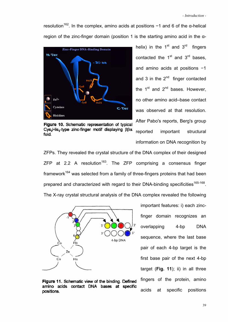
interactions and the chelation of a single zinc ion (Fig. 10). It was first
discovered in the transcription factor TFIIIA of Xenopus laevis in 1985161.
Classically, one unit of the Cys2His2 zinc-finger domain was thought to contact
3-bp of the DNA target. Pabo's group reported the X-ray crystal structure of the
DNA complex with the endogenous transcription factor Zif268 at 2.1 A
38
- Introduction -
resolution162. In the complex, amino acids at positions −1 and 6 of the α-helical
region of the zinc-finger domain (position 1 is the starting amino acid in the α-
helix) in the 1st and 3rd fingers
contacted the 1st and 3rd bases,
and amino acids at positions −1
and 3 in the 2nd finger contacted
the 1st and 2nd bases. However,
no other amino acid–base contact
was observed at that resolution.
After Pabo's reports, Berg's group
reported important structural
information on DNA recognition by
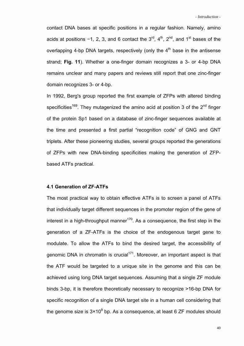
ZFPs. They revealed the crystal structure of the DNA complex of their designed
ZFP at 2.2 A resolution163. The ZFP comprising a consensus finger
framework164 was selected from a family of three-fingers proteins that had been
prepared and characterized with regard to their DNA-binding specificities165-168.
The X-ray crystal structural analysis of the DNA complex revealed the following
important features: i) each zinc-
finger domain recognizes an
overlapping 4-bp DNA
sequence, where the last base
pair of each 4-bp target is the
first base pair of the next 4-bp
target (Fig. 11); ii) in all three
fingers of the protein, amino
acids at specific positions
39
- Introduction -
contact DNA bases at specific positions in a regular fashion. Namely, amino
acids at positions −1, 2, 3, and 6 contact the 3rd, 4th, 2nd, and 1st bases of the
overlapping 4-bp DNA targets, respectively (only the 4th base in the antisense
strand; Fig. 11). Whether a one-finger domain recognizes a 3- or 4-bp DNA
remains unclear and many papers and reviews still report that one zinc-finger
domain recognizes 3- or 4-bp.
In 1992, Berg's group reported the first example of ZFPs with altered binding
specificities169. They mutagenized the amino acid at position 3 of the 2nd finger
of the protein Sp1 based on a database of zinc-finger sequences available at
the time and presented a first partial “recognition code” of GNG and GNT
triplets. After these pioneering studies, several groups reported the generations
of ZFPs with new DNA-binding specificities making the generation of ZFP-
based ATFs practical.
4.1 Generation of ZF-ATFs
The most practical way to obtain effective ATFs is to screen a panel of ATFs
that individually target different sequences in the promoter region of the gene of
interest in a high-throughput manner170. As a consequence, the first step in the
generation of a ZF-ATFs is the choice of the endogenous target gene to
modulate. To allow the ATFs to bind the desired target, the accessibility of
genomic DNA in chromatin is crucial171. Moreover, an important aspect is that
the ATF would be targeted to a unique site in the genome and this can be
achieved using long DNA target sequences. Assuming that a single ZF module
binds 3-bp, it is therefore theoretically necessary to recognize >16-bp DNA for
specific recognition of a single DNA target site in a human cell considering that
the genome size is 3×10⁹ bp. As a consequence, at least 6 ZF modules should
40

- Introduction -
be joined together in order to recognize a DNA target site of 18-bp. To date, a
recognition code table has been generated which allows to recognize 49 of the
64 DNA triplets172-176 and the modular assembly of the DBD can be also
facilitated through the use of the Zinc Finger Tools website based on Barbas’
modules (http://www.zincfingertool.org)177. Moreover, two-finger building blocks
made by Sangamo Bioscience are commercially available via Sigma-Aldrich76.
Once that a panel of ZF-based DBDs targeted to the desired gene are
obtained, they could be expressed and purified in order to examine their binding
properties determining the apparent dissociation constant (Kd) through
techniques such as Electromobility Shift Assay (EMSA). The best or better
DBDs are fused to a nuclear localization signal (NLS), to an epitope tag such as
FLAG, c-myc or HA to monitor ATF expression, and to an effector domain to
generate ATFs. The most frequently used transcriptional activator is the VP-64
domain178 while the most popular transcriptional repressor is a Kruppel-
associated box (KRAB) domain of KOX1179. Finally, the resulting ATFs are then
cloned into mammalian expression plasmids or viral vectors and their
functionality is investigated by transient reporter assays or more ideally by
analyzing the mRNA levels of target genes by Northern blotting or quantitative
real-time PCR. The efficacy and the specificity of the ATFs can be evaluated by
microarray analysis, DNase I footprinting assay or chromatin
immunoprecipitation (ChIP).
4.2 ZF-ATFs applications
The first study of gene regulation by altered transcription factors in living cells
(yeast) was reported in 1992180. Since then, several groups spent their effort in
order to develop this technology to achieve the transcriptional control at will of a
41

- Introduction -
42
given gene with therapeutic purposes. The first 6-finger ATF was reported in
1998178 to modulate the expression of the ErbB-2 which is overexpressed in a
high percentage of human adenocarcinomas. A 6-finger ATF targeted to the
promoter region of the ErbB-2 gene was generated and demonstrated a
successful and specific up- and down-regulation of the target gene in vitro. The
first example of ATF-mediated gene regulation in vivo was reported in 2002181
in a study sponsored by Sangamo Bioscience, a biotechnology company
worldwide leader in the design and development of engineered zinc finger
DNA-binding proteins (ZFPs) for gene regulation and gene modification). They
designed ZFPs to regulate the endogenous gene encoding vascular endothelial
growth factor-A (VEGF-A) which is an endothelial cell-specific mitogen that is a
key inducer of new blood vessel growth, both during embryogenesis and in later
processes such as wound healing. VEGF-A levels are dramatically increased
by hypoxia, triggering angiogenesis and microvascular permeability. Therefore,
both activation and repression of VEGF-A are attractive therapeutic
approaches. They showed that expression of these new ZFPs in vivo led to
induced expression of the protein VEGF-A, stimulation of angiogenesis and
acceleration of experimental wound healing181 thus establishing for the first time
the feasibility and potential utility of this approach as new tool for gene therapy.
Two years ago, ZF-based ATFs entered phase II clinical trials76.

- Aims -
AIMS
The aim of the project of my Ph.D. project was the development of a novel gene
therapy approach for the treatment of inherited retinal degenerations due to
gain of function mutations. We developed a strategy based on zinc finger
technology to repress the expression of the rhodopsin, the gene most
commonly associated with autosomal dominant Retinitis Pigmentosa, a severe
inherited retinal degeneration with a prevalence of 1:3500 worldwide. The
possibility to silence transcriptionally rhodopsin would allow to prevent the
pathological consequences of the expression of the mutated allele, thus halting
the patho-physiological cascade of events that lead to retinal degeneration. The
strategy employed is mutational-independent because transcriptional silencing
does not discriminate between the mutated and wild-type alleles. Thereby,
based on this characteristic feature, in principle any gain of function mutation
can be treatable; however, since also the wild type allele is silenced, a
combined repression and replacement system will be developed in the future.
The specific aims of my project were the following:
1. Generation and characterization of engineered zinc finger
transcriptional repressors targeted to the human rhodopsin promoter.
To control the expression of the rhodopsin gene we generated a series of
polydactyl ZF-based transcription factors fused either to an activator (VP64)
or to a repressor (KRAB) in order to modulate the expression of the target
gene.
2. Assessment of the efficacy of RHO transcriptional repression in vivo
and impact on the disease progression in an adRP mouse model.
43

- Aims -
44
Based on in vitro data in aim 1 I selected a ZFR and enclosed it in an
AAV2/8 vector for photoreceptor delivery in a mouse model of adRP
expressing a mutated human rhodopsin (P347S). This animal model
develops retinal degeneration with fast progression. The levels and
specificity of transcriptional repression were measured. In vivo expression of
the ZFR in the diseased photoreceptor resulted in the specific
downregulation of the transgenic human rhodopsin leaving unaltered the
expression levels of the endogenous murine allele. This in turn ameliorated
the retinal phenotype functionally and morphologically as measured by
electroretinograms and histological the progression of the disease.

Materials and Methods
- Materials and Methods -
MATERIALS AND METHODS
Rational design of the artificial Zinc-Finger based Transcription Factors
(ZF-TFs)
The protein sequences of the DNA Binding Domains (DBDs) targeted to the 10
different target sites (see Table 2 in Results section) were obtained using the
web-based Zinc Finger Tools (http://www.zincfingertools.org). Each DBD was
composed of two arms each recognizing a 9-bp half target site and fused
together through a linker sequence in order to overcome the gap sequence. We
used two different linkers according to the length of the gap: for gaps longer
than 3-bp (target sites from 1 to 4) we used a complex structured linker, as
reported by Moore186 (GRSSVESACVTSVLVALLPATSAPTQVSG) while for
longer gaps (target sites from 5 to 10) we used a flexible linker, as described by
Rebar181 (QNKKGGSGDGKKKQHA). We optimized the corresponding DNA
sequences to facilitate the subsequent cloning steps and purchased them as
transgene in the pLS-standard vector at “TOP Gene Technologies”, Quebec,
Canada. After the in vitro selection steps, we purchased an additional DBD with
the fingers of the DBD number 6 shuffled (from 1.2.3-4.5.6 to 5.1.6-3.4.2) to be
used as a negative control for a total of 11 DBDs. All the DBDs were flanked by
a ClaI and an XbaI site at the 5’ and 3’ end, respectively.
DBD number 1 was purchased as a complete protein fused at the N-Ter with
the repressor domain KRAB (Krüppel-associated box,
DAKSLTAWSRTLVTFKDVFVDFTREEWKLLDTAQQILYRNVMLENYKNLVSLG
YQLTKPDVILRLEKGEEPWLVEREIHQETHPDSETAFEIKSSV) and the nuclear
localization signal (NLS, PKKKRKV) from SV40 large T antigen and fused at
the C-Ter with the HA-tag (YPYDVPDYAS). Also DBD number 2 was
46

- Materials and Methods -
purchased as a complete protein but fused at the C-Ter with the NLS, the
activator domain VP64 (tetrameric repeat of herpes simplex VP16’s minimal
activation domain,
GRADALDDFDLDMLGSDALDDFDLDMLGSDALDDFDLDMLGSDALDDFDLD
MLIN) and the HA-tag. In the two proteins, the DBDs can be exchanged
through a double digestion with the restriction endonucleases ClaI and XbaI.
The 11 different transcription activators (ZF-As) and repressors (ZF-Rs) were
inserted in the pAAV2.1-CMV-EGFP NotI and HindIII sites, downstream of the
CMV (Cytomegalovirus) promoter by removing the EGFP coding sequence for
transfection experiments. For AAV preparations, ZF-R6 was inserted in the
pAAV2.1-RK-EGFP NotI and HindIII sites, downstream of the RK (Rhodopsin
Kinase) promoter by removing the EGFP coding sequence. For retrovirus
preparations used in Retinal Stem Cell experiments, coding sequences of ZF-
R2, ZF-R6 and ZF-R6 shuffled were cloned in LXSN plasmid EcoRI and HindIII
sites downstream of the CMV promoter.
Reporter Constructs
Luciferase reporter plasmids were generated from the pGL3-Basic vector
(Promega, Madison, WI) by cloning the proximal upstream regions of the
human or murine rhodopsin gene. The human rhodopsin promoter (hRHO-P)
extending from -330 to +92 relative to the transcription start site was PCR
amplified using the FastStart High Fidelity PCR System (Roche) from a human
genomic DNA sample using the following primers: hRHO-P.Nhe forward 5’-
TTTTGCTAGCTGTCCAGAGGACATAGCAC-3’ and hRHO-P.EcoRV reverse
5-AAAGATATCGGCTGTGGCCCTTGTGGCTGA-3’. For the murine rhodopsin
promoter (mRho-P) extending from -404 to +96 we used a murine genomic
47
- Materials and Methods -
DNA sample and the following primers: mRho-P.Nhe forward 5’-
TTTTGCTAGCTCAGATCTGTCAAGTGAGCC-3’ and mRho-P.HindIII reverse
5’-AAAAAGCTTGGCTGCGGCTCTCGAGGCTG-3’. The two fragments were
inserted within the NheI/SmaI or NheI/HindIII sites of pGL3-basic vector
obtaining pGL3-hRHO-P-Luciferase or pGL3-mRho-P-Luciferase, respectively.
Selection of the functional ZF-TFs in human cells by transient transfection
HEK293 cells were plated in 6-well plates at a density of 400.000 cells/well.
Twenty-four hours later, the cells were co-transfected using FUGENE 6 (Roche,
Basel, Switzerland) following the manufacturer’s instructions. Transfection
cocktails included 200ng of reporter plasmid (or pGL3basic as a negative
control), 300 ng of ZF-TF, 300 ng of a plasmid encoding for the murine CRX
(alone as a positive control in transactivation assay or in combination with the
ZF-Rs for the repression assays) and 10 ng of Renilla Luciferase reporter
plasmid control (Promega) to normalize for transfection efficiency. The amount
of DNA was kept constant by adding pAAV2.1-TBG-EGFP to a total of 1 g.
Cells were harvested 48 h after transfection in 1x PLB Lysis Buffer (Promega).
Firefly and Renilla Luciferases activities were measured in a GloMaxTM
luminometer (Promega) using Dual-Luciferase Reporter Assay System
(Promega).
Electromobility Shift Assay (EMSA)
HEK293 cells were transfected with a plasmid encoding the ZF-R2, ZF-R6 or
ZF-R6 shuffled using FUGENE 6 as described above. After 48 h, cells were
harvested and nuclear extracts were prepared using NE-PER Nuclear and
Cytoplasmic extraction kit (Pierce) according to the manufacturer’s instructions.
48

- Materials and Methods -
The presence of the proteins in the nuclear fractions was assessed by Western
Blotting analysis using as primary antibody a rabbit anti-HA (1:100; SIGMA,
Milan, Italy). Electromobility Shift Assay was performed using Lightshift
Chemiluminescent EMSA kit (Pierce) following the manufacturer’s instructions.
In brief, nuclear extracts (ZF-R2, ZF-R6 or ZF-R6 shuffled) were incubated with
equal amounts (20 fmol) of the specific byotinilated double stranded probe (5’-
TGTGGGGGTTAGAGGGTCTACGACTA-3’) to asses the specific shift for 20
min at room temperature. The competition control was performed adding a
molar excess of the unlabeled probe to the mix. The sequence of the unspecific
was the following: 5’-TGCGGCGGCTAAAGGCTCGACAACGA-3’. The
supershift was obtained adding a rabbit anti-HA (1:100; SIGMA, Milan, Italy)
primary antibody at the mix.
AAV vector production and purification
AAV vectors were produced by the TIGEM AAV Vector Core using pAAV2.1-
RK-ZF-R6 and pAAV2.1-CMV-EGFP. Recombinant AAV8 viruses were
produced by triple transfection of HEK293 cells followed by CsCl2 purification of
the vectors, as previously described121. For each viral preparation, physical
titers (GC/ml) were determined by both PCR quantification using TaqMan
(Perkin–Elmer, Life and Analytical Sciences, Inc.) and dot-blot analysis.
RNA preparation and measurement of rhodopsin transcript levels by Real
Time PCR
RNAs from tissues or cells were isolated using TRIZOL reagent (Invitrogen)
according to the manufacturer’s protocol. cDNA were amplified from 500 ng of
isolated RNA using the SuperScript™ III First-Strand Synthesis (Invitrogen) as
49

- Materials and Methods -
indicated in the attached instructions. Transcripts levels of human and murine
rhodopsin were measured by Real Time PCR using the ABI Prism 7000
Sequence Detection System (Applied Biosystems, Foster City,CA) and the
following primers: hRho_forward 5’…CCGGCTGGTCCAGGTACAT….3’ and
hRho_reverse 5’…TTGTTGACCTCCGGCTTGAG…3’; mRho_forward
5’…TGTTGAACAAGCAGTTCCGGA...3’ and mRho_reverse
5’…TCGTCATCTCCCAGTGGATTC...3’. The PCRs with cDNA were carried in
25 l total volume using 12.5 l SYBR Green Master Mix (Applied Biosystems)
and 400 nM primers under the following conditions: preheating, 50°C for 2 min
and 95°C for 10 min; cycling, 40 cycles of 95°C for 15 s and 60°C for 1 min. All
the reactions were standardized against murine GAPDH by using the following
primers: mGAPDH_forward 5’…CTTCACCACCATGGAGAAGGC…3’ and
mGAPDH_reverse 5’…CTCATGACCACAGTCCATGCC…3’. To quantify
expression levels of photoreceptor specific genes (Peripherin, PDE6and
Cnga) we used the following primers: for Peripherin: mPeriph.forward 5’-
CGGGACTGGTTCGAGATTC-3’ and mPeriph.reverse 5’-
ATCCACGTTGCTCTTGATGC-3’; for PDE6: PDE6.forward 5’-
TGAAGATGAAGATGTTTTCACG-3’ and PDE6.reverse 5’-
CTCTGCGTGTCTCACAGTTG-3’; for Cnga: Cnga.forward 5’-
ACTCGTACAAAAGGCGAGGAC-3’ and Cnga.reverse 5’-
CTTTGTTGCTGCTGTTGTTGAC-3’. Each sample was analyzed in triplicate in
two independent experiments.
50

- Materials and Methods -
Retinal stem cells culture and analysis
All procedures on mice (including their euthanasia) were performed in
accordance with the ARVO Statement for the Use of Animals in Ophthalmic and
Vision Research and with institutional guidelines for animal research.
Retinal stem cells were isolated from adult P347S mice and were incubated for
5 days until floating spheres formed in serum free medium (0.6% glucose and
N2 hormone mix in DMEM-F12) containing 20 ng/ml basic FGF supplemented
with 2 µg/ml heparin (Sigma, Milan, Italy), as previously described. Retinal
floating spheres were allowed to differentiate on eight well glass slides that
were coated with extracellular matrix (ECM, Sigma) in DMEM-F12,
supplemented with 1% FBS (Gibco, San Giuliano Milanese, Italy). Analysis was
performed 12 days after induction of differentiation. Apoptotic nuclei were
detected by TdT-dUTP terminal nick-end labeling kit (TUNEL, Roche
Diagnostics, Monza, Italy) according to the manufacturer’s protocol. For
immunofluorescence staining, cells were washed once with PBS and then fixed
for 10 min in PBS containing 4% PFA. After fixation, cells were washed and
incubated 5 min in sodium citrate buffer 0.1M containing 0.2 % Triton X100.
Blocking solution containing 3% bovine serum albumin (Sigma, Milan, Italy) was
applied for 1 h. Primary antibodies were diluted in PBS and incubated 90 min at
RT. The primary antibodies used were: rabbit anti-HA (1:100; SIGMA, Milan,
Italy) and mouse anti rhodopsin 1D4 (1:400; SIGMA, Milan, Italy). Cells were
then washed in PBS and incubated with secondary antibodies: 1:1000 Alexa
Fluor® 568 anti-rabbit, 1:1000 Alexa Fluor® 568 anti-mouse (Molecular
Probes), and 1:500 Cy5 anti-rabbit (Jackson Immunoresearch, West Grove,
PA, USA). Slides were coverslipped with Vectashield (Vector laboratories,
51
- Materials and Methods -
Burlingame, CA, USA) and photographed using either an Axioplan microscope
(Zeiss) or a Leica Laser Confocal Microscope System.
To allow ectopic expression in differentiated retinal cells, ZF-R2, ZF-R6 and ZF-
R6 shuffled were cloned in Moloney murine leukemia virus-based vector LXSN
as described above (see first paragraph of this section). Retinal cell cultures
were transduced at day 7 post explant with retroviruses and cells were
analyzed 4 days later.
Animal model, vector administration, and tissue collection
All procedures were performed in accordance with institutional guidelines for
animal research. P347S +/+ animals for breeding were kindly provided by Dr.
G. Jane Farrar (Smurfit Institute of Genetics, Trinity College Dublin, Dublin,
Ireland) and were bred in the animal facility of the Biotechnology Center of the
Cardarelli Hospital (Naples, Italy) with C57BL/6 mice (Charles Rivers
Laboratories, Calco, Italy) to obtain P347S +/- mice. At P4, animals were
anesthetized on ice and subretinally injected in the right eye with 1 l AAV2/8-
RHOK-ZF-R6 vector (6.3*1012 GC/ml) and 1 l of AAV2/1-CMV-EGFP (6.0*1012
GC/ml) in mix whereas the contralateral eye received, as control, the same
dose of AAV2/1-CMV-EGFP and AAV2/8-RHOK-EGFP vectors in mix. Mice
were sacrificed at postnatal day 18 or at 1 month of age (P18 or P30). For
morphological studies, eyecups were harvested, fixed by immersion in 4%
paraformaldehyde and then embedded in OCT (kaltek). For RNA extractions,
the isolated retinae were removed and snap-frozen in liquid nitrogen and
treated as described in the above RNA preparation section.
52

- Materials and Methods -
Histological analysis
For each eye, 150 to 200 serial sections (5-m thick) were cut along the
horizontal plane; the sections were progressively distributed on 10 glass slides
so that each slide contained 15 to 20 sections representative of the whole eye
at different levels. Slides were coverslipped with Vectashield containing DAPI
(4',6-diamidino-2-phenylindole; Vector laboratories, Burlingame, CA, USA) to
stain cells nuclei and retinal histology was analyzed an Axioplan microscope
(Zeiss). To quantify photoreceptor rescue, the number of nuclei in the outer
nuclear layer (ONL) of each eye was counted. For each section, the number of
nuclei in the ONL was separately counted on the nasal, central, and temporal
sides. The nasal, temporal, and central counts of each section were
independently averaged, therefore obtaining a number that was the average of
the three sides for each eye. The counts from each group (treated and
untreated) were then averaged and standard errors were calculated.
Electroretinogram measurements
ERGs were performed at P30, P60 and P90. Briefly, animals were adapted to
the dark for 3 h and then anesthetized and accommodated in a stereotaxic
apparatus under dim red light. ERGs were evoked by flashes of different light
intensities ranging from 10-4 to 20 cd.s/m2 generated through a Ganzfeld
stimulator (Lace). To minimize the noise, three different responses evoked by
light were averaged for each luminance step. The electrophysiological signals
were recorded by gold plate electrodes inserted under the lower eyelids.
Electrodes in each eye consisted of a needle electrode inserted subcutaneously
at the level of the corresponding frontal region. The different electrodes were
connected to a two-channel amplifier. Amplitudes of a-and b-waves were
53

- Materials and Methods -
54
plotted as a function of increasing light intensities. After completion of
responses obtained under dark-adapted conditions the recording session
continued with the aim to dissect the cone pathway mediating the light
response. To this end the ERG in response to light of 20 cd.s/m2 was recorded
in the presence of a continuous background light (background light set at 50
cd/m2). The amplitude of the b-wave for each eye was plotted as a function of
luminance under scotopic and photopic conditions. For each group the mean b-
wave amplitude was plotted.
Statistical analysis
Data are expressed as mean ± s.e.m. as indicated and compared by two-tailed
Student’s t-tests, two-sample equal variance.

Results

- Results -
RESULTS
1. GENERATION AND CHARACTERIZATION OF ENGINEERED ZINC FINGER
TRANSCRIPTION REPRESSORS TARGETED TO THE HUMAN RHODOPSIN
PROMOTER
1.1 Design and generation of Zinc Finger-based transcription factors to
control rhodopsin gene expression
To control the rhodopsin gene expression we designed Zinc Finger-based
Artificial Transcription Factors (ZF-ATFs) targeted to the human rhodopsin
promoter (hRHO-P). We first scanned the proximal hRHO-P to identify
amenable target regions applying the following criteria: (i) exclusivity of the 18-
bp DNA sequences in the human genome, (ii) putative accessibility to the
chromatin environment (in proximity of known endogenous transcription factors
binding sites)182, (iii) absence of annotated polymorphisms and (iv) divergence
between the human and murine rhodopsin promoter target sequences. We
selected 10 different continuous or discontinuous 18-bp target sites within the
proximal region of the hRHO-P (from -90 to -32, relative to the transcription
start site; Fig. 12 and Table 2) that fulfilled the 4 criteria described above. To
obtain a DNA Binding Domain (DBD) tailored to a unique target sequence in
56
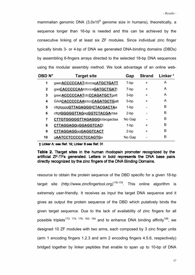
- Results -
mammalian genomic DNA (3,0x10⁹ genome size in humans), theoretically, a
sequence longer than 16-bp is needed and this can be achieved by the
consecutive linking of at least six ZF modules. Since individual zinc finger
typically binds 3- or 4-bp of DNA we generated DNA-binding domains (DBDs)
by assembling 6-fingers arrays directed to the selected 18-bp DNA sequences
using the modular assembly method. We took advantage of an online web-
resource to obtain the protein sequence of the DBD specific for a given 18-bp
target site (http://www.zincfingertool.org)176-178. This online algorithm is
extremely user-friendly. It receives as input the target DNA sequence and it
gives as output the protein sequence of the DBD which putatively binds the
given target sequence. Due to the lack of availability of zinc fingers for all
possible triplets173; 174; 176; 183; 184 and to enhance DNA binding affinity185, we
designed 10 ZF modules with two arms, each composed by 3 zinc finger units
(arm 1 encoding fingers 1.2.3 and arm 2 encoding fingers 4.5.6, respectively)
bridged together by linker peptides that enable to span up to 10-bp of DNA
57
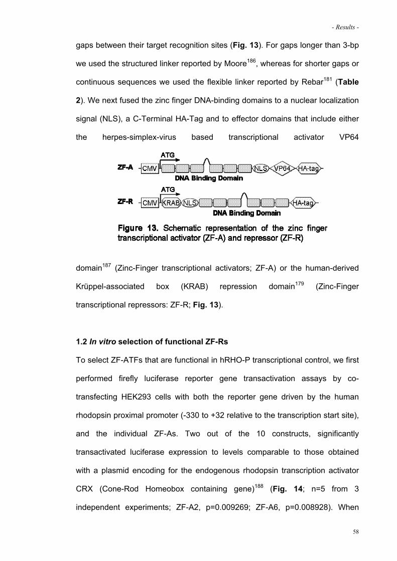
- Results -
gaps between their target recognition sites (Fig. 13). For gaps longer than 3-bp
we used the structured linker reported by Moore186, whereas for shorter gaps or
continuous sequences we used the flexible linker reported by Rebar181 (Table
2). We next fused the zinc finger DNA-binding domains to a nuclear localization
signal (NLS), a C-Terminal HA-Tag and to effector domains that include either
the herpes-simplex-virus based transcriptional activator VP64
dhdshfgdfhf jgfhjgf
domain187 (Zinc-Finger transcriptional activators; ZF-A) or the human-derived
Krüppel-associated box (KRAB) repression domain179 (Zinc-Finger
transcriptional repressors: ZF-R; Fig. 13).
1.2 In vitro selection of functional ZF-Rs
To select ZF-ATFs that are functional in hRHO-P transcriptional control, we first
performed firefly luciferase reporter gene transactivation assays by co-
transfecting HEK293 cells with both the reporter gene driven by the human
rhodopsin proximal promoter (-330 to +32 relative to the transcription start site),
and the individual ZF-As. Two out of the 10 constructs, significantly
transactivated luciferase expression to levels comparable to those obtained
with a plasmid encoding for the endogenous rhodopsin transcription activator
CRX (Cone-Rod Homeobox containing gene)188 (Fig. 14; n=5 from 3
independent experiments; ZF-A2, p=0.009269; ZF-A6, p=0.008928). When
58
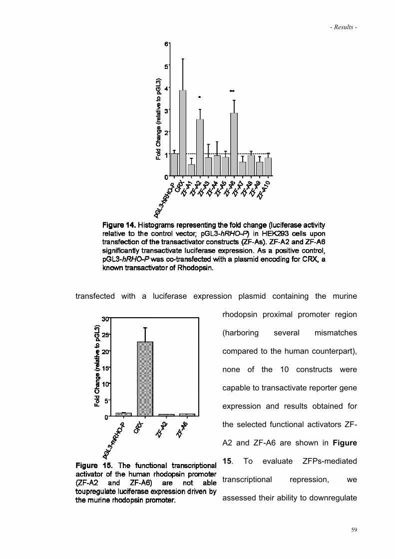
- Results -
ffffffffffff
transfected with a luciferase expression plasmid containing the murine
rhodopsin proximal promoter region
(harboring several mismatches
compared to the human counterpart),
none of the 10 constructs were
capable to transactivate reporter gene
expression and results obtained for
the selected functional activators ZF-
A2 and ZF-A6 are shown in Figure
15. To evaluate ZFPs-mediated
transcriptional repression, we
assessed their ability to downregulate
59

- Results -
ggggggggggg gggggggg
CRX-mediated transcription through triple transfection in HEK293 cells
including the ZF-Rs, the CRX and the reporter plasmids. Notably, ZF-R2 and
ZF-R6, containing the same DBDs
as ZF-A2 and ZF-A6, significantly
reduced luciferase expression
levels (81% and 64%, repression
relative to CRX transactivation,
respectively. Fig. 16; n=5 from 3
independent experiments; ZF-R2,
p=0.000480; ZF-R6, p=0.009053)
similarly to the repression obtained
with CRX DBD fused to KRAB used
as positive control189. As a further
60

- Results -
control, either activation or repression activity were completely abolished when
exchanging the relative position of individual ZF units of the DBD 6 (from 1.2.3-
4.5.6 to 5.1.6-3.4.2; ZF-A6- or ZF-R6-shuffled; Fig. 17). In addition,
electromobility shift assay (EMSA) showed binding specificity of ZF-R6 (Fig.
18) to the target promoter sequence in vitro.
hgfg
1.3 ZF-Rs mediated repression of human rhodopsin in retinal stem cells
To probe whether the two selected ZF-ATFs promote rhodopsin transcriptional
repression in the chromosomal context, we took advantage of an in vitro system
to differentiate retinal precursors (Retinal Stem Cells, RSC)190. Neurospheres
derived from the murine adult ocular ciliary margin, differentiate in vitro into
various retinal neuronal cell types, including photoreceptor precursors
61
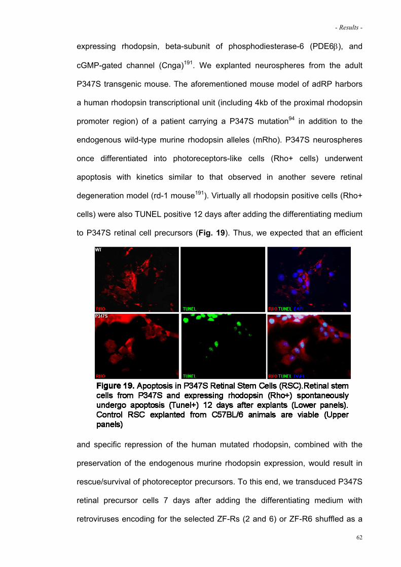
- Results -
expressing rhodopsin, beta-subunit of phosphodiesterase-6 (PDE6), and
cGMP-gated channel (Cnga)191. We explanted neurospheres from the adult
P347S transgenic mouse. The aforementioned mouse model of adRP harbors
a human rhodopsin transcriptional unit (including 4kb of the proximal rhodopsin
promoter region) of a patient carrying a P347S mutation94 in addition to the
endogenous wild-type murine rhodopsin alleles (mRho). P347S neurospheres
once differentiated into photoreceptors-like cells (Rho+ cells) underwent
apoptosis with kinetics similar to that observed in another severe retinal
degeneration model (rd-1 mouse191). Virtually all rhodopsin positive cells (Rho+
cells) were also TUNEL positive 12 days after adding the differentiating medium
to P347S retinal cell precursors (Fig. 19). Thus, we expected that an efficient
gggg
and specific repression of the human mutated rhodopsin, combined with the
preservation of the endogenous murine rhodopsin expression, would result in
rescue/survival of photoreceptor precursors. To this end, we transduced P347S
retinal precursor cells 7 days after adding the differentiating medium with
retroviruses encoding for the selected ZF-Rs (2 and 6) or ZF-R6 shuffled as a
62

- Results -
negative control. Twelve days after the induction of differentiation ~70% of cells
expressed rhodopsin (Rho+), 98% of which were also double positive (HA+;
Rho+) confirming that retroviruses transduce retinal cell precursors
efficiently191. ZF-R2 or ZF-R6 transduction produced a significant 77% and 92%
protection from cell death, respectively, whereas virtually all the ZF-R6 shuffled
transduced cells underwent apoptosis (Fig. 20, panels A,B and D; n=2 from 2
independent experiments; ZF-R2, p=4.54x10⁻¹¹; ZF-R6, p=1.18x10⁻¹¹).
Confocal images of triple stained (HA+; Rho+; TUNEL+) samples confirmed
63

- Results -
that only cells expressing ZF-R2 or ZF-R6 are protected from apoptosis (Fig.
20, panel C).
To assess whether protection from apoptosis was exerted through the specific
repression of human rhodopsin transcription, we measured hRHO mRNA
levels, 4 days after transduction. ZF-R2 and ZF-R6 transduced cells revealed a
51% and 88% reduction of human rhodopsin transcript levels, respectively,
compared to hRHO levels in ZF-R6 shuffled treated cells (Fig. 20, panel E; n=2
from 2 independent experiments; ZF-R2, p=0.0133; ZF-R6, p=0.0055). These
results show that ZFPs promote a strong human rhodopsin transcriptional
repression in a chromosomal context, and that this, in turn, can protect retinal
progenitors of an adRP model from apoptosis.
64

- Results -
2. ASSESSMENT OF EFFICACY OF AAV-MEDIATED GENE TRASNFER OF
ARTIFICIAL ZF-REPRESSOR TO PHOTORECEPTORS OF ADRP MOUSE
MODEL
2.1 Delivery of ZF-Rs to murine photoreceptors decrease adRP retinal
progression
The ZF-R6 construct, which provided robust human rhodopsin transcriptional
repression and apoptosis protection, was selected for in vivo experiments. We
generated an AAV2/8 vector currently considered the most efficient delivery
vehicle for photoreceptor gene transfer151; 153; 155; 182; 192, containing the ZF-R6
construct under the photoreceptor-specific Rhodopsin Kinase (RHOK) promoter
element151 (AAV2/8-RHOK-ZF-R6). In homozygous P347S mice the
overexpression of the mutated rhodopsin leads to a fast progression of retinal
degeneration. Indeed, by P21, only 1 row of photoreceptor nuclei was
detectable by histology. Thus, in order to decrease human P347S rhodopsin
expression levels we crossed P347S mice with a wild-type C57BL/6
background. The resulting progeny (P347S+/-) still overexpressed the human
rhodopsin transgene ~1.4 folds compared to the expression derived by the two
endogenous murine alleles (mRho) and displayed a milder retinal degeneration
(Fig. 21).
At P70, P347S+/- retinae still retained 5-6 rows of photoreceptor nuclei and
residual functionality, thus allowing the evaluation of disease progression over
time by both histology and electroretinography (ERG) analysis. We first
assessed transcriptional repression by subretinal co-injection in P4 P347S+/- of
1 x 10⁹ genome copies (GC) of AAV2/8-RHOK-ZF-R6 and a vector encoding
the Enhanced Green Fluorescent Protein (EGFP; AAV2/1-CMV-EGFP) to
65

- Results -
identify the transduced retinal area. The contralateral eye received a mix of
AAV2/1-CMV-EGFP and AAV2/8-RHOK-EGFP vectors as control. Western blot
ggggg gg
analysis showed expression of ZF-R6 protein in the retina twelve days post
injection (P18, before photoreceptor degeneration; Fig. 22, panel A).
Quantitative real-time PCR analysis demonstrated that the EGFP positive
portion of the retina treated with AAV2/8-RHOK-ZF-R6 displayed a significant
~26% reduction of the hRHO transcript relative to the endogenous mRho
compared to contralateral EGFP-treated retinae (Fig. 22, panel B; n=9 from 2
independent littermates; p=0.002643). In contrast, the expression profile of 3
photoreceptor specific genes was unchanged (Fig. 22, panel B; n=4). To
evaluate whether the specific downregulation of the human rhodopsin
transgene improved retinal function of the animal model, we recorded
66

- Results -
electroretinograms (ERGs) in treated and untreated eyes one month after
vector administration (P30). As shown (Fig. 23) the delivery of ZF-R6 to
diseased photoreceptors resulted in an amelioration of visual function
measured in scotopic and photopic conditions. We next evaluated the ability of
AAV2/8-RHOK-ZF-R6 to impact on P347S photoreceptor disease progression.
ggg gg
Since we noticed variability in retinal function assessed by ERG among
different littermates, despite similar P347S rhodopsin levels, we decided to
contextually measure base-line ERG parameters and subretinally treat the
animals (P30) and then monitor disease progression over time (P60 and P90).
At 1 month of age in two independent littermates both scotopic and photopic
ERG amplitudes were measured (a-wave max: pre-treatment EGFP=84.80 ±
67

- Results -
9.77; pre-treatment ZF-R6=80.52 ± 10.39; b-wave max: pre-treatment
EGFP=347.77 ± 30.73; pre-treatment ZF-R6=320.76 ± 23.98; n=12; Fig. 24,
panel A). At both 30 and 60 days following subretinal vector administration, the
eyes that received AAV2/8-RHOK-ZF-R6 showed a preservation of ERG
responses compared to control eyes, in which the degeneration led to a ~40%
reduction of the b-wave max amplitude (P60 data not shown; P90, a-wave max:
post-treatment EGFP=39.60 ± 7.78, p<0.01; post-treatment ZF-R6 =55.53 ±
gg
8.69; b-wave max: post-treatment EGFP=222.06 ± 24.54, p<0.01; post-
treatment ZF-R6 =286.82 ± 11.90; n=12; Fig. 24, panel A). In addition,
histological analysis of mice at P90 demonstrated that ZF-R6 expression was
also associated with a partial protection of photoreceptor degeneration (Fig. 24,
panel B; n=3). Finally, to evaluate potential toxicity on retinal function of a
healthy mouse retina due to ZF-R6 treatment, we subretinally injected AAV2/8-
RHOK-ZF-R6 in a cohort of 10 C57BL/6 animals. Two months after vector
delivery neither histological abnormalities nor reduction both in a- and b-wave
68

- Results -
could be measured in treated eyes compared to untreated parental eyes
supporting the safety of this approach (Fig. 25).
69
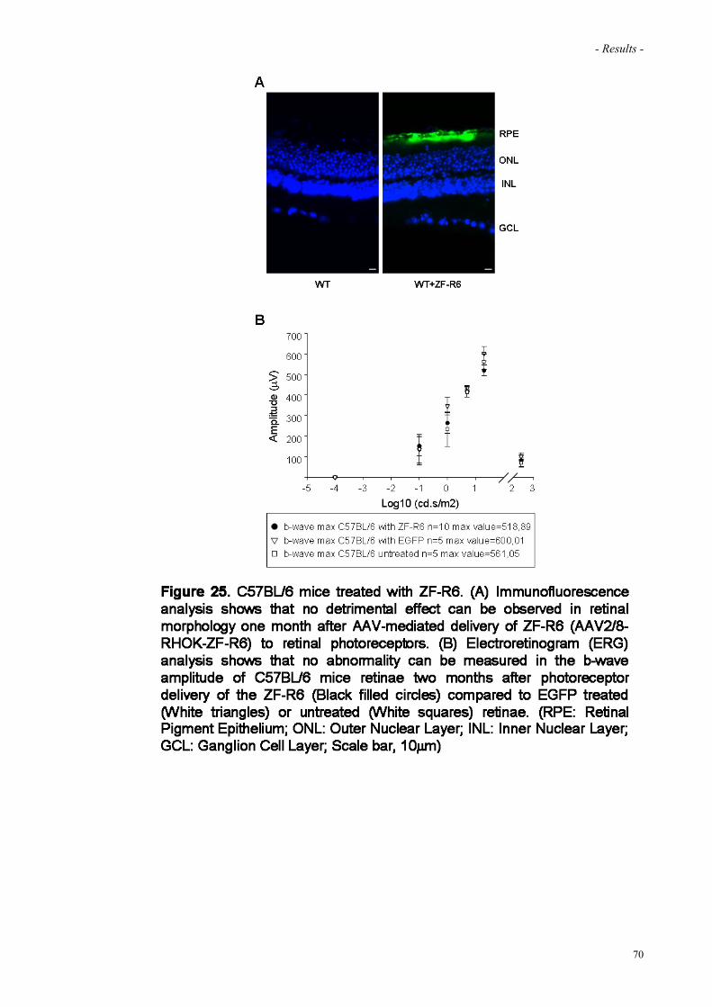
- Results -
70
ggg

Discussion
- Discussion -
DISCUSSION
Gene therapy for the treatment of recessive inherited ocular disorders has
proven successful. In 2008, the results published for three independent clinical
trials carried out to treat Leber’s congenital amaurosis (LCA) due to RPE65
mutations provided sound evidence for the use of gene therapy approaches to
treat recessive ocular diseases62-68. By contrast, the development of therapeutic
strategies to treat autosomal dominant retinal disorders has not proceeded at
the same pace, largely due to their complexity. The common approaches under
consideration for the therapy of these genetic disorders include strategies to
modulate secondary effects associated with disease, such as factors
influencing cell longevity or function193-195. However, many dominantly inherited
disorders may require correction of the underlying genetic defect and the
common strategies to this purpose mainly rely on the silencing of the disease
gene expression through RNAi-, ribozyme- or miRNA-based approaches. An
alternative strategy is based on the transcriptional repression of a disease gene
using artificial transcription factors which were developed in the last ten years
with the aim to create molecules tailored to control the expression profiles of a
disease-causative gene.
In this study we demonstrated that photoreceptor delivery of an AAV2/8 vector
containing an artificial zinc finger-based transcriptional repressor (ZF-R6)
targeted to the human rhodopsin promoter robustly repressed the target gene
expression, and this in turn was associated with improvement of photoreceptors
functionality and in a delay of the disease progression in P347S mouse model
of autosomal dominant Retinitis Pigmentosa (adRP).
72

- Discussion -
The first part of this PhD project was aimed at generating functional artificial
transcription factors (ATFs) targeted to human rhodopsin promoter. To this end,
we first identified the DNA sequence in the proximal rhodopsin promoter to
serve as target for the ATFs. We established two criteria to select the target
sequences: i) the absence of polymorphisms and ii) the accessibility of the
target site in the chromatin environment that can both affect the binding of the
ATFs. Respecting these criteria, we identified ten putative target sites in the
human rhodopsin proximal promoter in a region where no polymorphisms are
annotated and in proximity of binding sites for known transcriptional regulators
of the rhodopsin gene. In addition, for experimental needs, we chose target
sites in a region which is not conserved between rodents and humans
genomes. The reason of this choice derives by the animal model available for
our in vivo studies, the P347S mouse model. This animal carries a P347S
human mutated rhodopsin allele in addition to the murine endogenous alleles.
Targeting a sequence which is not conserved between mouse and man will
allow us to be selective in modulate human rhodopsin expression leaving
unaltered the endogenous counterpart.
We identified ten putative target sites in the proximal rhodopsin promoter
matching the criteria explained above and, using the modular assembly
method, we generated ten zinc-finger based transcriptional modulator of the
rhodopsin gene. We assessed in vitro the functionality of these ATFs by
reporter assays and we identified two out of ten activators and repressors able
to control rhodopsin promoter activity. Since our final goal is to downregulate
the expression of mutated human rhodopsin causing adRP, the functionality of
the selected repressors (ZF-R2 and ZF-R6) was further validated in another in
vitro system which better mimic the real condition: retinal stem cells (RSCs)
73

- Discussion -
explanted by the P347S mouse model. Indeed, the reporter system used in the
first screening had the main limitation to be based on an episomal system
transfected in the cells which is largely far from the condition in which the
promoter to be controlled is in its chromatin environment. On the other hand,
RSCs explanted by the P347S mouse model have the advantage to harbor the
human rhodopsin cassette in the chromosomal context thus allowing a more
accurate validation of the ATFs functionality as compared to the episomal
system used in the first round of selection. Furthermore, the binding ability to
the intended target site was demonstrated by Electromobility Shift Assay
(EMSA).
The following part of my PhD project was to evaluate the effect in vivo of the
delivery of the selected ZF-R6 to photoreceptors of both a mouse model of
adRP (P347S) and of a normal mouse model (C57BL/6) in order to address
both the efficacy and potential safety concerns, respectively. To this end, ZF-R6
was enclosed in an AAV2/8 vector, considered to date the most efficient in
murine photoreceptor transduction151 and delivered subretinally in diseased or
healthy retinae. We assessed no detrimental effects, both on the functionality
(measured by electroretinograms, ERG) and in the structure (evaluated
histologically) of normal retinae after the treatment, suggesting that the
approach is safe. Interestingly, the diseased retinae showed a preservation of
retinal function compared to controls upon injection of P347S animals at birth.
This result was also confirmed in adult animals in which we monitored the
disease progression over time upon ZF-R6 delivery at one month of age.
Indeed up to three months after the treatment, in the control-treated eyes we
recorded a 40% reduction of visual function compared to the baseline while in
the study-group eyes, the drop was only 10%.
74

- Discussion -
As a consequence, our findings extend the possible uses of artificial zinc finger-
based DNA binding proteins as a new paradigm in gene therapy to treat
dominant diseases based on the modulation of an endogenous gene
expression.
The common therapeutic approach to date for the treatment of gain-of-function
mutations mainly rely on RNAi-mediated downregulation of a gene-causing a
disease. The first example of such therapeutic strategy in vivo was assessed to
silence a dominant allele of ataxin-1 in order to treat a spinocerebellar ataxia
type 1 (SCA1) in mutational independent manner and then applied to other
mouse models of human disease including the P347S adRP mouse73; 74. Thus,
based on the data presented here, either transcriptional (artificial ZFP
technology) or post-transcriptional (RNAi) silencing strategies may be
considered alternatives to one another; however, a side-by-side comparison is
needed to determine which of the two systems is the safest and the most
efficacious. In principle, ZFP technology has the advantage that the target
sequences are present in one or two copies/diploid genome, whereas the
mRNA transcripts (the RNAi targets) are highly abundant. It is worth
emphasizing that rhodopsin is expressed extremely highly in the retina with 6.4
million rod photoreceptor cells, each producing a steady-state level of 60 million
of rhodopsin molecules (550–650 pmol)196. Moreover, besides the off-target
silencing events which potentially set hurdles for the development of a safe
ZFP- or RNAi-based system, studies have shown that high levels of siRNA may
cause cellular toxicity through various mechanisms. Fatality in mice was
associated with the downregulation of liver-derived microRNAs (miRNAs),
indicating possible competition of the latter with shRNAs for limiting cellular
factors required for the processing of various small RNAs197; 198. Also, an
75
- Discussion -
attractive and therapeutic valuable feature of ZFPs is the possibility to control
their expression levels through the use of inducible promoter systems which
have not yet been optimized to control RNAi-based repression systems.
Indeed, a key safety and efficacy element for the success of silencing strategies
relies on fine-tuning control of the expression levels of the repressor system. In
addition, a considerable potential advantage of ZF-mediated transcriptional
control of an endogenous gene includes the ability to regulate all the natural
splice variants of the target with a single therapeutic molecule. This of course is
not the case of rhodopsin but it has already been demonstrated to be an
important advantage in other therapeutic approaches181. Finally, the versatility
of ZFPs design theoretically allows to switch the repressor with an activator
domain (maintaining the same DBD) to treat also dominant diseases due to
haploinsufficiency.
Mutation-independent approaches such as the strategy exploited in this study
for rhodopsin linked adRP, are extremely valuable technically and economically
as a single therapeutic agent that may provide a remedy for many different
mutant alleles of a disease gene (for example in the case of rhodopsin, more
than 150 different mutations have been identified in its sequence).
However, although this strategy may provide a rational solution for many
dominant conditions, the application of the approach is complex. Indeed, one
limitation of the mutational-independent silencing is the simultaneous
repression of both the mutated and the normal alleles. As a consequence, a
therapeutic strategy based on such approach must consider the development of
a two-components system able to suppress both mutant and wild-type alleles of
a target gene and simultaneously to provide a replacement gene that encodes
wild-type protein but which is refractory to suppression.
76
- Discussion -
77
The results presented here represent an in vivo demonstration that suppression
through an artificial transcription factor and replacement given by the
endogenous murine gene may provide benefit in an animal model of a
dominant retinal disease. Thus, one future development will be to determine
whether, in an amenable mouse model (ideally, harboring both a mutated and a
normal allele of the human rhodopsin gene on a murine rhodopsin knock-out
background), down-regulation of both alleles will be sufficient to inhibit/delay the
disease progression or whether simultaneous replacement with a human
rhodopsin transgene not sensitive to the treatment would be necessary. In the
latter case, a vector incorporating both the artificial ZF-based repressor and the
replacement gene ensuring their simultaneous expression will be required (for
instance with bidirectional promoters enabling the coordinate expression of the
two transgenes199). Moreover, it has been shown that either high or low
rhodopsin expression levels can be deleterious to rod function, suggesting that
tight regulation of rhodopsin expression levels is required for successful
application of retinal repression-replacement strategy.
Our data demonstrate that the novel and generally applicable mutational-
independent strategy based on artificial zinc finger proteins enables the
transcriptional control of an endogenous causative gene and has perspectively
therapeutic relevance for the treatment of diseases due to gain of function
mutations.

References

- References -
REFERENCES
1. Blanks JC (2001) Morphology and topography of the retina. In: Mosby SL (ed) Retina, pp 32-53
2. Young RW (1976) Visual cells and the concept of renewal. Investigative
ophthalmology & visual science 15:700-725 3. Hinton GTDR (2001) Structure and functions of rod photoreceptors. In: Mosby SL
(ed) Retina, pp 104-121 4. Flannery JC (2001) Structure and functions of rod photoreceptors. In: Mosby SL (ed)
Retina, pp 122-137 5. Nathans J (1992) Rhodopsin: structure, function, and genetics. Biochemistry
31:4923-4931 6. Dhallan RS, Macke JP, Eddy RL, Shows TB, Reed RR, Yau KW, Nathans J (1992)
Human rod photoreceptor cGMP-gated channel: amino acid sequence, gene structure, and functional expression. J Neurosci 12:3248-3256
7. van Soest S, Westerveld A, de Jong PT, Bleeker-Wagemakers EM, Bergen AA
(1999) Retinitis pigmentosa: defined from a molecular point of view. Survey of ophthalmology 43:321-334
8. Thompson DA, Gal A (2003) Vitamin A metabolism in the retinal pigment
epithelium: genes, mutations, and diseases. Progress in retinal and eye research 22:683-703
9. Beharry S, Zhong M, Molday RS (2004) N-retinylidene-phosphatidylethanolamine is
the preferred retinoid substrate for the photoreceptor-specific ABC transporter ABCA4 (ABCR). The Journal of biological chemistry 279:53972-53979
10. Sun H, Molday RS, Nathans J (1999) Retinal stimulates ATP hydrolysis by purified
and reconstituted ABCR, the photoreceptor-specific ATP-binding cassette transporter responsible for Stargardt disease. The Journal of biological chemistry 274:8269-8281
11. Saari JC, Bredberg L, Garwin GG (1982) Identification of the endogenous retinoids
associated with three cellular retinoid-binding proteins from bovine retina and retinal pigment epithelium. The Journal of biological chemistry 257:13329-13333
12. Hinton GTDR (2001) Cell biology of the retinal pigment epithelium. In: Mosby SL
(ed) Retina, pp 104-121 13. Simon A, Hellman U, Wernstedt C, Eriksson U (1995) The retinal pigment
epithelial-specific 11-cis retinol dehydrogenase belongs to the family of short chain alcohol dehydrogenases. The Journal of biological chemistry 270:1107-1112
79

- References -
14. Hosford DJH, I. Freund, C. McInnes, R. R. Van Heyningen V. (2001) Transcription factors in eye disease and ocular development. New York: McGraw-Hill
15. Gregory-Evans K, Bhattacharya SS (1998) Genetic blindness: current concepts in
the pathogenesis of human outer retinal dystrophies. Trends Genet 14:103-108 16. Rattner A, Sun H, Nathans J (1999) Molecular genetics of human retinal disease.
Annual review of genetics 33:89-131 17. Farrar GJ, Kenna PF, Humphries P (2002) On the genetics of retinitis pigmentosa
and on mutation-independent approaches to therapeutic intervention. The EMBO journal 21:857-864
18. Phelan JK, Bok D (2000) A brief review of retinitis pigmentosa and the identified
retinitis pigmentosa genes. Molecular vision 6:116-124 19. Dryja T (2001) Retinitis pigmentosa and stationary night blindness. In: Scrivers C,
Beaudet, A., Sly, W., Valle D (ed) The metabolic & molecular bases of inherited diseases, pp 5903-5933
20. Hartong DT, Berson EL, Dryja TP (2006) Retinitis pigmentosa. Lancet 368:1795-
1809 21. Li ZY, Possin DE, Milam AH (1995) Histopathology of bone spicule pigmentation
in retinitis pigmentosa. Ophthalmology 102:805-816 22. Berson EL (1996) Retinitis pigmentosa: unfolding its mystery. Proceedings of the
National Academy of Sciences of the United States of America 93:4526-4528 23. Kajiwara K, Berson EL, Dryja TP (1994) Digenic retinitis pigmentosa due to
mutations at the unlinked peripherin/RDS and ROM1 loci. Science (New York, NY 264:1604-1608
24. Zeviani M, Moraes CT, DiMauro S, Nakase H, Bonilla E, Schon EA, Rowland LP
(1998) Deletions of mitochondrial DNA in Kearns-Sayre syndrome. 1988. Neurology 51:1525 and 1528 pages following
25. Boughman JA, Fishman GA (1983) A genetic analysis of retinitis pigmentosa. The
British journal of ophthalmology 67:449-454 26. Katsanis N (2004) The oligogenic properties of Bardet-Biedl syndrome. Human
molecular genetics 13 Spec No 1:R65-71 27. Portera-Cailliau C, Sung CH, Nathans J, Adler R (1994) Apoptotic photoreceptor
cell death in mouse models of retinitis pigmentosa. Proceedings of the National Academy of Sciences of the United States of America 91:974-978
28. Clarke G, Heon E, McInnes RR (2000) Recent advances in the molecular basis of
inherited photoreceptor degeneration. Clinical genetics 57:313-329
80

- References -
29. Farber DB, Lolley RN (1974) Cyclic guanosine monophosphate: elevation in degenerating photoreceptor cells of the C3H mouse retina. Science (New York, NY 186:449-451
30. Min KC, Zvyaga TA, Cypess AM, Sakmar TP (1993) Characterization of mutant
rhodopsins responsible for autosomal dominant retinitis pigmentosa. Mutations on the cytoplasmic surface affect transducin activation. The Journal of biological chemistry 268:9400-9404
31. Leveillard T, Mohand-Said S, Poch O, Sahel JA (2005) [Rod-derived cone viability
factor: a clue for therapy of retinitis pigmentosa?]. Med Sci (Paris) 21:22-24 32. Jin M, Li S, Moghrabi WN, Sun H, Travis GH (2005) Rpe65 is the retinoid
isomerase in bovine retinal pigment epithelium. Cell 122:449-459 33. Comitato A, Spampanato C, Chakarova C, Sanges D, Bhattacharya SS, Marigo V
(2007) Mutations in splicing factor PRPF3, causing retinal degeneration, form detrimental aggregates in photoreceptor cells. Human molecular genetics 16:1699-1707
34. Dryja TP, Hahn LB, Cowley GS, McGee TL, Berson EL (1991) Mutation spectrum
of the rhodopsin gene among patients with autosomal dominant retinitis pigmentosa. Proceedings of the National Academy of Sciences of the United States of America 88:9370-9374
35. Inglehearn CF, Keen TJ, Bashir R, Jay M, Fitzke F, Bird AC, Crombie A,
Bhattacharya S (1992) A completed screen for mutations of the rhodopsin gene in a panel of patients with autosomal dominant retinitis pigmentosa. Human molecular genetics 1:41-45
36. Sheffield VC, Fishman GA, Beck JS, Kimura AE, Stone EM (1991) Identification
of novel rhodopsin mutations associated with retinitis pigmentosa by GC-clamped denaturing gradient gel electrophoresis. American journal of human genetics 49:699-706
37. Sung CH, Davenport CM, Hennessey JC, Maumenee IH, Jacobson SG,
Heckenlively JR, Nowakowski R, Fishman G, Gouras P, Nathans J (1991) Rhodopsin mutations in autosomal dominant retinitis pigmentosa. Proceedings of the National Academy of Sciences of the United States of America 88:6481-6485
38. Knowles A, Dartnall HJA (1977) The Photobiology of vision. Academic Press,
New York 39. Pfister C, Kuhn H, Chabre M (1983) Interaction between photoexcited rhodopsin
and peripheral enzymes in frog retinal rods. Influence on the postmetarhodopsin II decay and phosphorylation rate of rhodopsin. European journal of biochemistry / FEBS 136:489-499
40. Sung CH, Davenport CM, Nathans J (1993) Rhodopsin mutations responsible for
autosomal dominant retinitis pigmentosa. Clustering of functional classes along the polypeptide chain. The Journal of biological chemistry 268:26645-26649
81

- References -
41. Sung CH, Schneider BG, Agarwal N, Papermaster DS, Nathans J (1991) Functional heterogeneity of mutant rhodopsins responsible for autosomal dominant retinitis pigmentosa. Proceedings of the National Academy of Sciences of the United States of America 88:8840-8844
42. Rosenfeld PJ, Cowley GS, McGee TL, Sandberg MA, Berson EL, Dryja TP (1992)
A null mutation in the rhodopsin gene causes rod photoreceptor dysfunction and autosomal recessive retinitis pigmentosa. Nature genetics 1:209-213
43. Feskanich D, Singh V, Willett WC, Colditz GA (2002) Vitamin A intake and hip
fractures among postmenopausal women. Jama 287:47-54 44. Hoffman DR, Locke KG, Wheaton DH, Fish GE, Spencer R, Birch DG (2004) A
randomized, placebo-controlled clinical trial of docosahexaenoic acid supplementation for X-linked retinitis pigmentosa. American journal of ophthalmology 137:704-718
45. LaVail MM, Yasumura D, Matthes MT, Lau-Villacorta C, Unoki K, Sung CH,
Steinberg RH (1998) Protection of mouse photoreceptors by survival factors in retinal degenerations. Investigative ophthalmology & visual science 39:592-602
46. Liang FQ, Aleman TS, Dejneka NS, Dudus L, Fisher KJ, Maguire AM, Jacobson
SG, Bennett J (2001) Long-term protection of retinal structure but not function using RAAV.CNTF in animal models of retinitis pigmentosa. Mol Ther 4:461-472
47. Frasson M, Sahel JA, Fabre M, Simonutti M, Dreyfus H, Picaud S (1999) Retinitis
pigmentosa: rod photoreceptor rescue by a calcium-channel blocker in the rd mouse. Nature medicine 5:1183-1187
48. Pearce-Kelling SE, Aleman TS, Nickle A, Laties AM, Aguirre GD, Jacobson SG,
Acland GM (2001) Calcium channel blocker D-cis-diltiazem does not slow retinal degeneration in the PDE6B mutant rcd1 canine model of retinitis pigmentosa. Molecular vision 7:42-47
49. Rizzo JF, 3rd, Wyatt J, Loewenstein J, Kelly S, Shire D (2003) Perceptual efficacy
of electrical stimulation of human retina with a microelectrode array during short-term surgical trials. Investigative ophthalmology & visual science 44:5362-5369
50. Nussbaum RLW (2001) The treatment of genetic disease. In: Mosby SL (ed)
Genetics in medicine, pp 255-276 51. Kay MA, Glorioso JC, Naldini L (2001) Viral vectors for gene therapy: the art of
turning infectious agents into vehicles of therapeutics. Nature medicine 7:33-40 52. Wolff J, Lewis DL, Herweijer H, Hegge J, Hagstrom J (2005) Non-viral approaches
for gene transfer. Acta Myol 24:202-208 53. Bainbridge JW, Tan MH, Ali RR (2006) Gene therapy progress and prospects: the
eye. Gene therapy 13:1191-1197
82

- References -
54. Cremers FP, van den Hurk JA, den Hollander AI (2002) Molecular genetics of Leber congenital amaurosis. Human molecular genetics 11:1169-1176
55. Acland GM, Aguirre GD, Bennett J, Aleman TS, Cideciyan AV, Bennicelli J,
Dejneka NS, Pearce-Kelling SE, Maguire AM, Palczewski K, Hauswirth WW, Jacobson SG (2005) Long-term restoration of rod and cone vision by single dose rAAV-mediated gene transfer to the retina in a canine model of childhood blindness. Mol Ther 12:1072-1082
56. Acland GM, Aguirre GD, Ray J, Zhang Q, Aleman TS, Cideciyan AV, Pearce-
Kelling SE, Anand V, Zeng Y, Maguire AM, Jacobson SG, Hauswirth WW, Bennett J (2001) Gene therapy restores vision in a canine model of childhood blindness. Nature genetics 28:92-95
57. Dejneka NS, Surace EM, Aleman TS, Cideciyan AV, Lyubarsky A, Savchenko A,
Redmond TM, Tang W, Wei Z, Rex TS, Glover E, Maguire AM, Pugh EN, Jr., Jacobson SG, Bennett J (2004) In utero gene therapy rescues vision in a murine model of congenital blindness. Mol Ther 9:182-188
58. Jacobson SG, Boye SL, Aleman TS, Conlon TJ, Zeiss CJ, Roman AJ, Cideciyan
AV, Schwartz SB, Komaromy AM, Doobrajh M, Cheung AY, Sumaroka A, Pearce-Kelling SE, Aguirre GD, Kaushal S, Maguire AM, Flotte TR, Hauswirth WW (2006) Safety in nonhuman primates of ocular AAV2-RPE65, a candidate treatment for blindness in Leber congenital amaurosis. Human gene therapy 17:845-858
59. Le Meur G, Stieger K, Smith AJ, Weber M, Deschamps JY, Nivard D, Mendes-
Madeira A, Provost N, Pereon Y, Cherel Y, Ali RR, Hamel C, Moullier P, Rolling F (2007) Restoration of vision in RPE65-deficient Briard dogs using an AAV serotype 4 vector that specifically targets the retinal pigmented epithelium. Gene therapy 14:292-303
60. Narfstrom K, Katz ML, Bragadottir R, Seeliger M, Boulanger A, Redmond TM,
Caro L, Lai CM, Rakoczy PE (2003) Functional and structural recovery of the retina after gene therapy in the RPE65 null mutation dog. Investigative ophthalmology & visual science 44:1663-1672
61. Pang JJ, Chang B, Kumar A, Nusinowitz S, Noorwez SM, Li J, Rani A, Foster TC,
Chiodo VA, Doyle T, Li H, Malhotra R, Teusner JT, McDowell JH, Min SH, Li Q, Kaushal S, Hauswirth WW (2006) Gene therapy restores vision-dependent behavior as well as retinal structure and function in a mouse model of RPE65 Leber congenital amaurosis. Mol Ther 13:565-572
62. Bainbridge JW, Smith AJ, Barker SS, Robbie S, Henderson R, Balaggan K,
Viswanathan A, Holder GE, Stockman A, Tyler N, Petersen-Jones S, Bhattacharya SS, Thrasher AJ, Fitzke FW, Carter BJ, Rubin GS, Moore AT, Ali RR (2008) Effect of gene therapy on visual function in Leber's congenital amaurosis. The New England journal of medicine 358:2231-2239
63. Cideciyan AV, Aleman TS, Boye SL, Schwartz SB, Kaushal S, Roman AJ, Pang JJ,
Sumaroka A, Windsor EA, Wilson JM, Flotte TR, Fishman GA, Heon E, Stone EM, Byrne BJ, Jacobson SG, Hauswirth WW (2008) Human gene therapy for
83
- References -
RPE65 isomerase deficiency activates the retinoid cycle of vision but with slow rod kinetics. Proceedings of the National Academy of Sciences of the United States of America 105:15112-15117
64. Cideciyan AV, Hauswirth WW, Aleman TS, Kaushal S, Schwartz SB, Boye SL,
Windsor EA, Conlon TJ, Sumaroka A, Pang JJ, Roman AJ, Byrne BJ, Jacobson SG (2009) Human RPE65 gene therapy for Leber congenital amaurosis: persistence of early visual improvements and safety at 1 year. Human gene therapy 20:999-1004
65. Cideciyan AV, Hauswirth WW, Aleman TS, Kaushal S, Schwartz SB, Boye SL,
Windsor EA, Conlon TJ, Sumaroka A, Roman AJ, Byrne BJ, Jacobson SG (2009) Vision 1 year after gene therapy for Leber's congenital amaurosis. The New England journal of medicine 361:725-727
66. Hauswirth WW, Aleman TS, Kaushal S, Cideciyan AV, Schwartz SB, Wang L,
Conlon TJ, Boye SL, Flotte TR, Byrne BJ, Jacobson SG (2008) Treatment of leber congenital amaurosis due to RPE65 mutations by ocular subretinal injection of adeno-associated virus gene vector: short-term results of a phase I trial. Human gene therapy 19:979-990
67. Maguire AM, High KA, Auricchio A, Wright JF, Pierce EA, Testa F, Mingozzi F,
et al. (2009) Age-dependent effects of RPE65 gene therapy for Leber's congenital amaurosis: a phase 1 dose-escalation trial. Lancet 374:1597-1605
68. Maguire AM, Simonelli F, Pierce EA, Pugh EN, Jr., Mingozzi F, Bennicelli J, Banfi
S, et al. (2008) Safety and efficacy of gene transfer for Leber's congenital amaurosis. The New England journal of medicine 358:2240-2248
69. Chadderton N, Millington-Ward S, Palfi A, O'Reilly M, Tuohy G, Humphries MM,
Li T, Humphries P, Kenna PF, Farrar GJ (2009) Improved retinal function in a mouse model of dominant retinitis pigmentosa following AAV-delivered gene therapy. Mol Ther 17:593-599
70. Kiang AS, Palfi A, Ader M, Kenna PF, Millington-Ward S, Clark G, Kennan A,
O'Reilly M, Tam LC, Aherne A, McNally N, Humphries P, Farrar GJ (2005) Toward a gene therapy for dominant disease: validation of an RNA interference-based mutation-independent approach. Mol Ther 12:555-561
71. LaVail MM, Yasumura D, Matthes MT, Drenser KA, Flannery JG, Lewin AS,
Hauswirth WW (2000) Ribozyme rescue of photoreceptor cells in P23H transgenic rats: long-term survival and late-stage therapy. Proceedings of the National Academy of Sciences of the United States of America 97:11488-11493
72. Lewin AS, Drenser KA, Hauswirth WW, Nishikawa S, Yasumura D, Flannery JG,
LaVail MM (1998) Ribozyme rescue of photoreceptor cells in a transgenic rat model of autosomal dominant retinitis pigmentosa. Nature medicine 4:967-971
73. O'Reilly M, Palfi A, Chadderton N, Millington-Ward S, Ader M, Cronin T, Tuohy
T, Auricchio A, Hildinger M, Tivnan A, McNally N, Humphries MM, Kiang AS, Humphries P, Kenna PF, Farrar GJ (2007) RNA interference-mediated
84

- References -
suppression and replacement of human rhodopsin in vivo. American journal of human genetics 81:127-135
74. Xia H, Mao Q, Eliason SL, Harper SQ, Martins IH, Orr HT, Paulson HL, Yang L,
Kotin RM, Davidson BL (2004) RNAi suppresses polyglutamine-induced neurodegeneration in a model of spinocerebellar ataxia. Nature medicine 10:816-820
75. Jamieson AC, Miller JC, Pabo CO (2003) Drug discovery with engineered zinc-
finger proteins. Nat Rev Drug Discov 2:361-368 76. Pearson H (2008) Protein engineering: The fate of fingers. Nature 455:160-164 77. Segal DJ, Barbas CF, 3rd (2001) Custom DNA-binding proteins come of age:
polydactyl zinc-finger proteins. Current opinion in biotechnology 12:632-637 78. Chader GJ (2002) Animal models in research on retinal degenerations: past progress
and future hope. Vision research 42:393-399 79. Chang B, Hawes NL, Hurd RE, Davisson MT, Nusinowitz S, Heckenlively JR
(2002) Retinal degeneration mutants in the mouse. Vision research 42:517-525 80. Lin CT, Gould DJ, Petersen-Jonest SM, Sargan DR (2002) Canine inherited retinal
degenerations: update on molecular genetic research and its clinical application. The Journal of small animal practice 43:426-432
81. Humphries MM, Rancourt D, Farrar GJ, Kenna P, Hazel M, Bush RA, Sieving PA,
Sheils DM, McNally N, Creighton P, Erven A, Boros A, Gulya K, Capecchi MR, Humphries P (1997) Retinopathy induced in mice by targeted disruption of the rhodopsin gene. Nature genetics 15:216-219
82. Wang DY, Chan WM, Tam PO, Baum L, Lam DS, Chong KK, Fan BJ, Pang CP
(2005) Gene mutations in retinitis pigmentosa and their clinical implications. Clinica chimica acta; international journal of clinical chemistry 351:5-16
83. Machida S, Kondo M, Jamison JA, Khan NW, Kononen LT, Sugawara T, Bush RA,
Sieving PA (2000) P23H rhodopsin transgenic rat: correlation of retinal function with histopathology. Investigative ophthalmology & visual science 41:3200-3209
84. Naash MI, Hollyfield JG, al-Ubaidi MR, Baehr W (1993) Simulation of human
autosomal dominant retinitis pigmentosa in transgenic mice expressing a mutated murine opsin gene. Proceedings of the National Academy of Sciences of the United States of America 90:5499-5503
85. Olsson JE, Gordon JW, Pawlyk BS, Roof D, Hayes A, Molday RS, Mukai S,
Cowley GS, Berson EL, Dryja TP (1992) Transgenic mice with a rhodopsin mutation (Pro23His): a mouse model of autosomal dominant retinitis pigmentosa. Neuron 9:815-830
86. Petters RM, Alexander CA, Wells KD, Collins EB, Sommer JR, Blanton MR, Rojas
G, Hao Y, Flowers WL, Banin E, Cideciyan AV, Jacobson SG, Wong F (1997)
85
- References -
Genetically engineered large animal model for studying cone photoreceptor survival and degeneration in retinitis pigmentosa. Nature biotechnology 15:965-970
87. Steinberg R (1996) Transgenic rat models of inherited retinal degeneration caused
by mutant opsin genes. ARVO Abstract No 3190 88. Greenberg J, Roberts L, Ramesar R (2003) A rare homozygous rhodopsin splice-
site mutation: the issue of when and whether to offer presymptomatic testing. Ophthalmic genetics 24:225-232
89. Rosenfeld PJ, Hahn LB, Sandberg MA, Dryja TP, Berson EL (1995) Low incidence
of retinitis pigmentosa among heterozygous carriers of a specific rhodopsin splice site mutation. Investigative ophthalmology & visual science 36:2186-2192
90. Tan E, Wang Q, Quiambao AB, Xu X, Qtaishat NM, Peachey NS, Lem J, Fliesler
SJ, Pepperberg DR, Naash MI, Al-Ubaidi MR (2001) The relationship between opsin overexpression and photoreceptor degeneration. Investigative ophthalmology & visual science 42:589-600
91. Saliba RS, Munro PM, Luthert PJ, Cheetham ME (2002) The cellular fate of mutant
rhodopsin: quality control, degradation and aggresome formation. Journal of cell science 115:2907-2918
92. Illing ME, Rajan RS, Bence NF, Kopito RR (2002) A rhodopsin mutant linked to
autosomal dominant retinitis pigmentosa is prone to aggregate and interacts with the ubiquitin proteasome system. The Journal of biological chemistry 277:34150-34160
93. Deretic D, Schmerl S, Hargrave PA, Arendt A, McDowell JH (1998) Regulation of
sorting and post-Golgi trafficking of rhodopsin by its C-terminal sequence QVS(A)PA. Proceedings of the National Academy of Sciences of the United States of America 95:10620-10625
94. Li T, Snyder WK, Olsson JE, Dryja TP (1996) Transgenic mice carrying the
dominant rhodopsin mutation P347S: evidence for defective vectorial transport of rhodopsin to the outer segments. Proceedings of the National Academy of Sciences of the United States of America 93:14176-14181
95. Sung CH, Makino C, Baylor D, Nathans J (1994) A rhodopsin gene mutation
responsible for autosomal dominant retinitis pigmentosa results in a protein that is defective in localization to the photoreceptor outer segment. J Neurosci 14:5818-5833
96. Berson EL, Rosner B, Sandberg MA, Weigel-DiFranco C, Dryja TP (1991) Ocular
findings in patients with autosomal dominant retinitis pigmentosa and rhodopsin, proline-347-leucine. American journal of ophthalmology 111:614-623
86

- References -
97. Kraft TW, Allen D, Petters RM, Hao Y, Peng YW, Wong F (2005) Altered light responses of single rod photoreceptors in transgenic pigs expressing P347L or P347S rhodopsin. Molecular vision 11:1246-1256
98. Tso MO, Li WW, Zhang C, Lam TT, Hao Y, Petters RM, Wong F (1997) A
pathologic study of degeneration of the rod and cone populations of the rhodopsin Pro347Leu transgenic pigs. Transactions of the American Ophthalmological Society 95:467-479; discussion 479-483
99. Dejneka NS, Bennett J (2001) Gene therapy and retinitis pigmentosa: advances and
future challenges. Bioessays 23:662-668 100. Mueller C, Flotte TR (2008) Clinical gene therapy using recombinant adeno-
associated virus vectors. Gene therapy 15:858-863 101. Berns K (1996) Parvoviridae: the viruses and their replication. In: Lippincot-
Raven (ed) Fundamental Virology, Philadelphia, pp 1017-1042 102. Muzyczka N (2001) Parvoviridae: the viruses and their replication. In: Lippincot-
Raven (ed) Fundamental Virology, Philadelphia, pp 2327–2359 103. Gao G, Vandenberghe LH, Alvira MR, Lu Y, Calcedo R, Zhou X, Wilson JM
(2004) Clades of Adeno-associated viruses are widely disseminated in human tissues. Journal of virology 78:6381-6388
104. Gao GP, Alvira MR, Wang L, Calcedo R, Johnston J, Wilson JM (2002) Novel
adeno-associated viruses from rhesus monkeys as vectors for human gene therapy. Proceedings of the National Academy of Sciences of the United States of America 99:11854-11859
105. Dubielzig R, King JA, Weger S, Kern A, Kleinschmidt JA (1999) Adeno-
associated virus type 2 protein interactions: formation of pre-encapsidation complexes. Journal of virology 73:8989-8998
106. King JA, Dubielzig R, Grimm D, Kleinschmidt JA (2001) DNA helicase-mediated
packaging of adeno-associated virus type 2 genomes into preformed capsids. The EMBO journal 20:3282-3291
107. Kyostio SR, Owens RA, Weitzman MD, Antoni BA, Chejanovsky N, Carter BJ
(1994) Analysis of adeno-associated virus (AAV) wild-type and mutant Rep proteins for their abilities to negatively regulate AAV p5 and p19 mRNA levels. Journal of virology 68:2947-2957
108. Lusby EW, Berns KI (1982) Mapping of the 5' termini of two adeno-associated
virus 2 RNAs in the left half of the genome. Journal of virology 41:518-526 109. Smith RH, Kotin RM (1998) The Rep52 gene product of adeno-associated virus is
a DNA helicase with 3'-to-5' polarity. Journal of virology 72:4874-4881 110. Kronenberg S, Kleinschmidt JA, Bottcher B (2001) Electron cryo-microscopy and
image reconstruction of adeno-associated virus type 2 empty capsids. EMBO reports 2:997-1002
87

- References -
111. Rabinowitz JE, Samulski RJ (2000) Building a better vector: the manipulation of AAV virions. Virology 278:301-308
112. Kotin RM, Linden RM, Berns KI (1992) Characterization of a preferred site on
human chromosome 19q for integration of adeno-associated virus DNA by non-homologous recombination. The EMBO journal 11:5071-5078
113. Linden RM, Ward P, Giraud C, Winocour E, Berns KI (1996) Site-specific
integration by adeno-associated virus. Proceedings of the National Academy of Sciences of the United States of America 93:11288-11294
114. Chen CL, Jensen RL, Schnepp BC, Connell MJ, Shell R, Sferra TJ, Bartlett JS,
Clark KR, Johnson PR (2005) Molecular characterization of adeno-associated viruses infecting children. Journal of virology 79:14781-14792
115. Schnepp BC, Jensen RL, Chen CL, Johnson PR, Clark KR (2005) Characterization
of adeno-associated virus genomes isolated from human tissues. Journal of virology 79:14793-14803
116. Rabinowitz JE, Samulski J (1998) Adeno-associated virus expression systems for
gene transfer. Current opinion in biotechnology 9:470-475 117. Zolotukhin S (2005) Production of recombinant adeno-associated virus vectors.
Human gene therapy 16:551-557 118. McCarty DM, Young SM, Jr., Samulski RJ (2004) Integration of adeno-associated
virus (AAV) and recombinant AAV vectors. Annual review of genetics 38:819-845
119. Auricchio A (2003) Pseudotyped AAV vectors for constitutive and regulated gene
expression in the eye. Vision research 43:913-918 120. Gao G, Vandenberghe LH, Wilson JM (2005) New recombinant serotypes of AAV
vectors. Current gene therapy 5:285-297 121. Auricchio A, Kobinger G, Anand V, Hildinger M, O'Connor E, Maguire AM,
Wilson JM, Bennett J (2001) Exchange of surface proteins impacts on viral vector cellular specificity and transduction characteristics: the retina as a model. Human molecular genetics 10:3075-3081
122. Chao H, Liu Y, Rabinowitz J, Li C, Samulski RJ, Walsh CE (2000) Several log
increase in therapeutic transgene delivery by distinct adeno-associated viral serotype vectors. Mol Ther 2:619-623
123. Qing K, Mah C, Hansen J, Zhou S, Dwarki V, Srivastava A (1999) Human
fibroblast growth factor receptor 1 is a co-receptor for infection by adeno-associated virus 2. Nature medicine 5:71-77
124. Summerford C, Bartlett JS, Samulski RJ (1999) AlphaVbeta5 integrin: a co-
receptor for adeno-associated virus type 2 infection. Nature medicine 5:78-82
88
- References -
125. Summerford C, Samulski RJ (1998) Membrane-associated heparan sulfate proteoglycan is a receptor for adeno-associated virus type 2 virions. Journal of virology 72:1438-1445
126. Di Pasquale G, Davidson BL, Stein CS, Martins I, Scudiero D, Monks A, Chiorini
JA (2003) Identification of PDGFR as a receptor for AAV-5 transduction. Nature medicine 9:1306-1312
127. Kaludov N, Brown KE, Walters RW, Zabner J, Chiorini JA (2001) Adeno-
associated virus serotype 4 (AAV4) and AAV5 both require sialic acid binding for hemagglutination and efficient transduction but differ in sialic acid linkage specificity. Journal of virology 75:6884-6893
128. Auricchio A, O'Connor E, Weiner D, Gao GP, Hildinger M, Wang L, Calcedo R,
Wilson JM (2002) Noninvasive gene transfer to the lung for systemic delivery of therapeutic proteins. The Journal of clinical investigation 110:499-504
129. Zabner J, Seiler M, Walters R, Kotin RM, Fulgeras W, Davidson BL, Chiorini JA
(2000) Adeno-associated virus type 5 (AAV5) but not AAV2 binds to the apical surfaces of airway epithelia and facilitates gene transfer. Journal of virology 74:3852-3858
130. Zaiss AK, Liu Q, Bowen GP, Wong NC, Bartlett JS, Muruve DA (2002)
Differential activation of innate immune responses by adenovirus and adeno-associated virus vectors. Journal of virology 76:4580-4590
131. Zhang Y, Chirmule N, Gao G, Wilson J (2000) CD40 ligand-dependent activation
of cytotoxic T lymphocytes by adeno-associated virus vectors in vivo: role of immature dendritic cells. Journal of virology 74:8003-8010
132. Bessis N, GarciaCozar FJ, Boissier MC (2004) Immune responses to gene therapy
vectors: influence on vector function and effector mechanisms. Gene therapy 11 Suppl 1:S10-17
133. Vandenberghe LH, Wilson JM (2007) AAV as an immunogen. Current gene
therapy 7:325-333 134. Zaiss AK, Muruve DA (2005) Immune responses to adeno-associated virus
vectors. Current gene therapy 5:323-331 135. Manno CS, Pierce GF, Arruda VR, Glader B, Ragni M, Rasko JJ, Ozelo MC, et al.
(2006) Successful transduction of liver in hemophilia by AAV-Factor IX and limitations imposed by the host immune response. Nature medicine 12:342-347
136. Mingozzi F, High KA (2007) Immune responses to AAV in clinical trials. Current
gene therapy 7:316-324 137. Riviere C, Danos O, Douar AM (2006) Long-term expression and repeated
administration of AAV type 1, 2 and 5 vectors in skeletal muscle of immunocompetent adult mice. Gene therapy 13:1300-1308
89

- References -
138. Chirmule N, Xiao W, Truneh A, Schnell MA, Hughes JV, Zoltick P, Wilson JM (2000) Humoral immunity to adeno-associated virus type 2 vectors following administration to murine and nonhuman primate muscle. Journal of virology 74:2420-2425
139. Xiao W, Chirmule N, Berta SC, McCullough B, Gao G, Wilson JM (1999) Gene
therapy vectors based on adeno-associated virus type 1. Journal of virology 73:3994-4003
140. Dong JY, Fan PD, Frizzell RA (1996) Quantitative analysis of the packaging
capacity of recombinant adeno-associated virus. Human gene therapy 7:2101-2112
141. Hermonat PL, Quirk JG, Bishop BM, Han L (1997) The packaging capacity of
adeno-associated virus (AAV) and the potential for wild-type-plus AAV gene therapy vectors. FEBS letters 407:78-84
142. Duan D, Fisher KJ, Burda JF, Engelhardt JF (1997) Structural and functional
heterogeneity of integrated recombinant AAV genomes. Virus research 48:41-56
143. Duan D, Yue Y, Engelhardt JF (2001) Expanding AAV packaging capacity with
trans-splicing or overlapping vectors: a quantitative comparison. Mol Ther 4:383-391
144. Ghosh A, Yue Y, Duan D (2006) Viral serotype and the transgene sequence
influence overlapping adeno-associated viral (AAV) vector-mediated gene transfer in skeletal muscle. The journal of gene medicine 8:298-305
145. Nakai H, Storm TA, Kay MA (2000) Increasing the size of rAAV-mediated
expression cassettes in vivo by intermolecular joining of two complementary vectors. Nature biotechnology 18:527-532
146. Yan Z, Zhang Y, Duan D, Engelhardt JF (2000) Trans-splicing vectors expand the
utility of adeno-associated virus for gene therapy. Proceedings of the National Academy of Sciences of the United States of America 97:6716-6721
147. Bennett J, Maguire AM, Cideciyan AV, Schnell M, Glover E, Anand V, Aleman
TS, Chirmule N, Gupta AR, Huang Y, Gao GP, Nyberg WC, Tazelaar J, Hughes J, Wilson JM, Jacobson SG (1999) Stable transgene expression in rod photoreceptors after recombinant adeno-associated virus-mediated gene transfer to monkey retina. Proceedings of the National Academy of Sciences of the United States of America 96:9920-9925
148. Flannery JG, Zolotukhin S, Vaquero MI, LaVail MM, Muzyczka N, Hauswirth
WW (1997) Efficient photoreceptor-targeted gene expression in vivo by recombinant adeno-associated virus. Proceedings of the National Academy of Sciences of the United States of America 94:6916-6921
149. Rabinowitz JE, Rolling F, Li C, Conrath H, Xiao W, Xiao X, Samulski RJ (2002)
Cross-packaging of a single adeno-associated virus (AAV) type 2 vector
90

- References -
genome into multiple AAV serotypes enables transduction with broad specificity. Journal of virology 76:791-801
150. Yang GS, Schmidt M, Yan Z, Lindbloom JD, Harding TC, Donahue BA,
Engelhardt JF, Kotin R, Davidson BL (2002) Virus-mediated transduction of murine retina with adeno-associated virus: effects of viral capsid and genome size. Journal of virology 76:7651-7660
151. Allocca M, Mussolino C, Garcia-Hoyos M, Sanges D, Iodice C, Petrillo M,
Vandenberghe LH, Wilson JM, Marigo V, Surace EM, Auricchio A (2007) Novel adeno-associated virus serotypes efficiently transduce murine photoreceptors. Journal of virology 81:11372-11380
152. Ho TT, Maguire AM, Aguirre GD, Surace EM, Anand V, Zeng Y, Salvetti A,
Hopwood JJ, Haskins ME, Bennett J (2002) Phenotypic rescue after adeno-associated virus-mediated delivery of 4-sulfatase to the retinal pigment epithelium of feline mucopolysaccharidosis VI. The journal of gene medicine 4:613-621
153. Lebherz C, Maguire A, Tang W, Bennett J, Wilson JM (2008) Novel AAV
serotypes for improved ocular gene transfer. The journal of gene medicine 10:375-382
154. Lotery AJ, Yang GS, Mullins RF, Russell SR, Schmidt M, Stone EM, Lindbloom
JD, Chiorini JA, Kotin RM, Davidson BL (2003) Adeno-associated virus type 5: transduction efficiency and cell-type specificity in the primate retina. Human gene therapy 14:1663-1671
155. Stieger K, Colle MA, Dubreil L, Mendes-Madeira A, Weber M, Le Meur G,
Deschamps JY, Provost N, Nivard D, Cherel Y, Moullier P, Rolling F (2008) Subretinal delivery of recombinant AAV serotype 8 vector in dogs results in gene transfer to neurons in the brain. Mol Ther 16:916-923
156. Weber M, Rabinowitz J, Provost N, Conrath H, Folliot S, Briot D, Cherel Y,
Chenuaud P, Samulski J, Moullier P, Rolling F (2003) Recombinant adeno-associated virus serotype 4 mediates unique and exclusive long-term transduction of retinal pigmented epithelium in rat, dog, and nonhuman primate after subretinal delivery. Mol Ther 7:774-781
157. Ali RR, Reichel MB, De Alwis M, Kanuga N, Kinnon C, Levinsky RJ, Hunt DM,
Bhattacharya SS, Thrasher AJ (1998) Adeno-associated virus gene transfer to mouse retina. Human gene therapy 9:81-86
158. Surace EM, Auricchio A, Reich SJ, Rex T, Glover E, Pineles S, Tang W,
O'Connor E, Lyubarsky A, Savchenko A, Pugh EN, Jr., Maguire AM, Wilson JM, Bennett J (2003) Delivery of adeno-associated virus vectors to the fetal retina: impact of viral capsid proteins on retinal neuronal progenitor transduction. Journal of virology 77:7957-7963
159. Dinculescu A, Glushakova L, Min SH, Hauswirth WW (2005) Adeno-associated
virus-vectored gene therapy for retinal disease. Human gene therapy 16:649-663
91
- References -
160. Glushakova LG, Timmers AM, Issa TM, Cortez NG, Pang J, Teusner JT, Hauswirth WW (2006) Does recombinant adeno-associated virus-vectored proximal region of mouse rhodopsin promoter support only rod-type specific expression in vivo? Molecular vision 12:298-309
161. Miller J, McLachlan AD, Klug A (1985) Repetitive zinc-binding domains in the
protein transcription factor IIIA from Xenopus oocytes. The EMBO journal 4:1609-1614
162. Pavletich NP, Pabo CO (1991) Zinc finger-DNA recognition: crystal structure of a
Zif268-DNA complex at 2.1 A. Science (New York, NY 252:809-817 163. Kim CA, Berg JM (1996) A 2.2 A resolution crystal structure of a designed zinc
finger protein bound to DNA. Nature structural biology 3:940-945 164. Krizek BA, Amann BT, Kilfoil VJ, Merkle DL, Berg JM (1991) A consensus zinc
finger peptide: design, high-affinity metal binding, a pH-dependent structure, and a His to Cys sequence variant. JAmChemSoc 113:4518–4523
165. Desjarlais JR, Berg JM (1993) Use of a zinc-finger consensus sequence framework
and specificity rules to design specific DNA binding proteins. Proceedings of the National Academy of Sciences of the United States of America 90:2256-2260
166. Desjarlais JR, Berg JM (1994) Length-encoded multiplex binding site
determination: application to zinc finger proteins. Proceedings of the National Academy of Sciences of the United States of America 91:11099-11103
167. Kim CA, Berg JM (1995) Serine at position 2 in the DNA recognition helix of a
Cys2-His2 zinc finger peptide is not, in general, responsible for base recognition. Journal of molecular biology 252:1-5
168. Shi Y, Berg JM (1995) A direct comparison of the properties of natural and
designed zinc-finger proteins. Chemistry & biology 2:83-89 169. Desjarlais JR, Berg JM (1992) Toward rules relating zinc finger protein sequences
and DNA binding site preferences. Proceedings of the National Academy of Sciences of the United States of America 89:7345-7349
170. Yokoi K, Zhang HS, Kachi S, Balaggan KS, Yu Q, Guschin D, Kunis M, Surosky
R, Africa LM, Bainbridge JW, Spratt SK, Gregory PD, Ali RR, Campochiaro PA (2007) Gene transfer of an engineered zinc finger protein enhances the anti-angiogenic defense system. Mol Ther 15:1917-1923
171. Liu PQ, Rebar EJ, Zhang L, Liu Q, Jamieson AC, Liang Y, Qi H, Li PX, Chen B,
Mendel MC, Zhong X, Lee YL, Eisenberg SP, Spratt SK, Case CC, Wolffe AP (2001) Regulation of an endogenous locus using a panel of designed zinc finger proteins targeted to accessible chromatin regions. Activation of vascular endothelial growth factor A. The Journal of biological chemistry 276:11323-11334
92

- References -
172. Bae KH, Kwon YD, Shin HC, Hwang MS, Ryu EH, Park KS, Yang HY, Lee DK, Lee Y, Park J, Kwon HS, Kim HW, Yeh BI, Lee HW, Sohn SH, Yoon J, Seol W, Kim JS (2003) Human zinc fingers as building blocks in the construction of artificial transcription factors. Nature biotechnology 21:275-280
173. Dreier B, Beerli RR, Segal DJ, Flippin JD, Barbas CF, 3rd (2001) Development of
zinc finger domains for recognition of the 5'-ANN-3' family of DNA sequences and their use in the construction of artificial transcription factors. The Journal of biological chemistry 276:29466-29478
174. Dreier B, Fuller RP, Segal DJ, Lund CV, Blancafort P, Huber A, Koksch B,
Barbas CF, 3rd (2005) Development of zinc finger domains for recognition of the 5'-CNN-3' family DNA sequences and their use in the construction of artificial transcription factors. The Journal of biological chemistry 280:35588-35597
175. Maeder ML, Thibodeau-Beganny S, Osiak A, Wright DA, Anthony RM,
Eichtinger M, Jiang T, et al. (2008) Rapid "open-source" engineering of customized zinc-finger nucleases for highly efficient gene modification. Molecular cell 31:294-301
176. Segal DJ, Dreier B, Beerli RR, Barbas CF, 3rd (1999) Toward controlling gene
expression at will: selection and design of zinc finger domains recognizing each of the 5'-GNN-3' DNA target sequences. Proceedings of the National Academy of Sciences of the United States of America 96:2758-2763
177. Mandell JG, Barbas CF, 3rd (2006) Zinc Finger Tools: custom DNA-binding
domains for transcription factors and nucleases. Nucleic Acids Res 34:W516-523
178. Beerli RR, Segal DJ, Dreier B, Barbas CF, 3rd (1998) Toward controlling gene
expression at will: specific regulation of the erbB-2/HER-2 promoter by using polydactyl zinc finger proteins constructed from modular building blocks. Proceedings of the National Academy of Sciences of the United States of America 95:14628-14633
179. Margolin JF, Friedman JR, Meyer WK, Vissing H, Thiesen HJ, Rauscher FJ, 3rd
(1994) Kruppel-associated boxes are potent transcriptional repression domains. Proceedings of the National Academy of Sciences of the United States of America 91:4509-4513
180. Thukral SK, Morrison ML, Young ET (1992) Mutations in the zinc fingers of
ADR1 that change the specificity of DNA binding and transactivation. Molecular and cellular biology 12:2784-2792
181. Rebar EJ, Huang Y, Hickey R, Nath AK, Meoli D, Nath S, Chen B, Xu L, Liang
Y, Jamieson AC, Zhang L, Spratt SK, Case CC, Wolffe A, Giordano FJ (2002) Induction of angiogenesis in a mouse model using engineered transcription factors. Nature medicine 8:1427-1432
182. Graslund T, Li X, Magnenat L, Popkov M, Barbas CF, 3rd (2005) Exploring
strategies for the design of artificial transcription factors: targeting sites
93
- References -
proximal to known regulatory regions for the induction of gamma-globin expression and the treatment of sickle cell disease. The Journal of biological chemistry 280:3707-3714
183. Blancafort P, Magnenat L, Barbas CF, 3rd (2003) Scanning the human genome
with combinatorial transcription factor libraries. Nature biotechnology 21:269-274
184. Dreier B, Segal DJ, Barbas CF, 3rd (2000) Insights into the molecular recognition
of the 5'-GNN-3' family of DNA sequences by zinc finger domains. Journal of molecular biology 303:489-502
185. Kim JS, Pabo CO (1998) Getting a handhold on DNA: design of poly-zinc finger
proteins with femtomolar dissociation constants. Proceedings of the National Academy of Sciences of the United States of America 95:2812-2817
186. Moore M, Choo Y, Klug A (2001) Design of polyzinc finger peptides with
structured linkers. Proceedings of the National Academy of Sciences of the United States of America 98:1432-1436
187. Seipel K, Georgiev O, Schaffner W (1992) Different activation domains stimulate
transcription from remote ('enhancer') and proximal ('promoter') positions. The EMBO journal 11:4961-4968
188. Chen S, Wang QL, Nie Z, Sun H, Lennon G, Copeland NG, Gilbert DJ, Jenkins
NA, Zack DJ (1997) Crx, a novel Otx-like paired-homeodomain protein, binds to and transactivates photoreceptor cell-specific genes. Neuron 19:1017-1030
189. Chau KY, Chen S, Zack DJ, Ono SJ (2000) Functional domains of the cone-rod
homeobox (CRX) transcription factor. The Journal of biological chemistry 275:37264-37270
190. Giordano F, De Marzo A, Vetrini F, Marigo V (2007) Fibroblast growth factor and
epidermal growth factor differently affect differentiation of murine retinal stem cells in vitro. Molecular vision 13:1842-1850
191. Sanges D, Comitato A, Tammaro R, Marigo V (2006) Apoptosis in retinal
degeneration involves cross-talk between apoptosis-inducing factor (AIF) and caspase-12 and is blocked by calpain inhibitors. Proceedings of the National Academy of Sciences of the United States of America 103:17366-17371
192. Natkunarajah M, Trittibach P, McIntosh J, Duran Y, Barker SE, Smith AJ,
Nathwani AC, Ali RR (2008) Assessment of ocular transduction using single-stranded and self-complementary recombinant adeno-associated virus serotype 2/8. Gene therapy 15:463-467
193. Buch PK, MacLaren RE, Duran Y, Balaggan KS, MacNeil A, Schlichtenbrede FC,
Smith AJ, Ali RR (2006) In contrast to AAV-mediated Cntf expression, AAV-mediated Gdnf expression enhances gene replacement therapy in rodent models of retinal degeneration. Mol Ther 14:700-709
94

- References -
95
194. Komeima K, Rogers BS, Lu L, Campochiaro PA (2006) Antioxidants reduce cone cell death in a model of retinitis pigmentosa. Proceedings of the National Academy of Sciences of the United States of America 103:11300-11305
195. Perche O, Doly M, Ranchon-Cole I (2007) Caspase-dependent apoptosis in light-
induced retinal degeneration. Investigative ophthalmology & visual science 48:2753-2759
196. Wensel TG, Gross AK, Chan F, Sykoudis K, Wilson JH (2005) Rhodopsin-EGFP
knock-ins for imaging quantal gene alterations. Vision research 45:3445-3453 197. Boudreau RL, Martins I, Davidson BL (2009) Artificial microRNAs as siRNA
shuttles: improved safety as compared to shRNAs in vitro and in vivo. Mol Ther 17:169-175
198. Grimm D, Streetz KL, Jopling CL, Storm TA, Pandey K, Davis CR, Marion P,
Salazar F, Kay MA (2006) Fatality in mice due to oversaturation of cellular microRNA/short hairpin RNA pathways. Nature 441:537-541
199. Amendola M, Venneri MA, Biffi A, Vigna E, Naldini L (2005) Coordinate dual-
gene transgenesis by lentiviral vectors carrying synthetic bidirectional promoters. Nature biotechnology 23:108-116