molecular aspects of medicine - ioz.ac.cn
TRANSCRIPT
Molecular Aspects of Medicine 34 (2013) 919–938
Contents lists available at SciVerse ScienceDirect
Molecular Aspects of Medicine
journal homepage: www.elsevier .com/locate /mam
Review
The maternal to zygotic transition in mammals
0098-2997/$ - see front matter � 2013 Elsevier Ltd. All rights reserved.http://dx.doi.org/10.1016/j.mam.2013.01.003
Abbreviations: MZT, maternal to zygotic transition; DNMTs, DNA methyltransferases; 5fC, 5-formylcytosine; 5caC, 5-carboxylcytosine; TDG,DNA glycosylase; ICR, imprinting control regions; DMR, differentially methylated regions; SCNT, somatic cell nuclear transfer; ART, assisted reprtechnology; RNAi, RNA interference; ZGA, zygotic genome activation; TRC, transcription required complex; SCMC, subcortical maternal complexmicroRNA; endo-siRNA, endo-small interfering RNA; piRNA, Piwi-interacting RNA; dsRNA, double strand RNA.⇑ Corresponding authors.
E-mail addresses: [email protected] (L. Li), [email protected] (J. Dean).
Lei Li a,⇑, Xukun Lu a,b, Jurrien Dean c,⇑a Division of Molecular Embryonic Development, State Key Laboratory of Reproductive Biology, Institute of Zoology/Chinese Academy of Sciences,Beijing 100101, PR Chinab University of Chinese Academy of Sciences, Beijing 100049, PR Chinac Laboratory of Cellular and Developmental Biology, NIDDK, National Institutes of Health, Bethesda, MD 20892, USA
a r t i c l e i n f o a b s t r a c t
Article history:Available online 23 January 2013
Keywords:Maternal to zygotic transition (MZT)Mouse fertilizationGamete recognitionDNA methylationHistone modificationZygotic genome activation (ZGA)Maternal effect genesSubcortical maternal complex (SCMC)
Prior to activation of the embryonic genome, the initiating events of mammalian develop-ment are under maternal control and include fertilization, the block to polyspermy andprocessing sperm DNA. Following gamete union, the transcriptionally inert sperm DNA isrepackaged into the male pronucleus which fuses with the female pronucleus to form a1-cell zygote. Embryonic transcription begins during the maternal to zygotic transfer ofcontrol in directing development. This transition occurs at species-specific times afterone or several rounds of blastomere cleavage and is essential for normal development.However, even after activation of the embryonic genome, successful development relieson stored maternal components without which embryos fail to progress beyond initial celldivisions. Better understanding of the molecular basis of maternal to zygotic transitionincluding fertilization, the activation of the embryonic genome and cleavage-stage devel-opment will provide insight into early human development that should translate into clin-ical applications for regenerative medicine and assisted reproductive technologies.
� 2013 Elsevier Ltd. All rights reserved.
Contents
1. Introduction . . . . . . . . . . . . . . . . . . . . . . . . . . . . . . . . . . . . . . . . . . . . . . . . . . . . . . . . . . . . . . . . . . . . . . . . . . . . . . . . . . . . . . . . . . . . 9202. Maternal contribution to the onset of development . . . . . . . . . . . . . . . . . . . . . . . . . . . . . . . . . . . . . . . . . . . . . . . . . . . . . . . . . . . . 920
2.1. Fertilization . . . . . . . . . . . . . . . . . . . . . . . . . . . . . . . . . . . . . . . . . . . . . . . . . . . . . . . . . . . . . . . . . . . . . . . . . . . . . . . . . . . . . . . 9202.2. Zona pellucida . . . . . . . . . . . . . . . . . . . . . . . . . . . . . . . . . . . . . . . . . . . . . . . . . . . . . . . . . . . . . . . . . . . . . . . . . . . . . . . . . . . . . 9222.3. Models of sperm-zona recognition. . . . . . . . . . . . . . . . . . . . . . . . . . . . . . . . . . . . . . . . . . . . . . . . . . . . . . . . . . . . . . . . . . . . . 922
2.3.1. Glycan-release models . . . . . . . . . . . . . . . . . . . . . . . . . . . . . . . . . . . . . . . . . . . . . . . . . . . . . . . . . . . . . . . . . . . . . . . 9222.3.2. ZP2 cleavage model . . . . . . . . . . . . . . . . . . . . . . . . . . . . . . . . . . . . . . . . . . . . . . . . . . . . . . . . . . . . . . . . . . . . . . . . . 923
2.4. The post-fertilization block to polyspermy . . . . . . . . . . . . . . . . . . . . . . . . . . . . . . . . . . . . . . . . . . . . . . . . . . . . . . . . . . . . . . 924
3. Epigenetic regulation of the maternal to zygotic transition . . . . . . . . . . . . . . . . . . . . . . . . . . . . . . . . . . . . . . . . . . . . . . . . . . . . . . 9243.1. Epigenetics. . . . . . . . . . . . . . . . . . . . . . . . . . . . . . . . . . . . . . . . . . . . . . . . . . . . . . . . . . . . . . . . . . . . . . . . . . . . . . . . . . . . . . . . 9243.2. Demethylation of DNA in the paternal genome . . . . . . . . . . . . . . . . . . . . . . . . . . . . . . . . . . . . . . . . . . . . . . . . . . . . . . . . . . 9253.3. Maternal and paternal imprinting . . . . . . . . . . . . . . . . . . . . . . . . . . . . . . . . . . . . . . . . . . . . . . . . . . . . . . . . . . . . . . . . . . . . . 926
thymine-oductive; miRNA,

920 L. Li et al. / Molecular Aspects of Medicine 34 (2013) 919–938
3.4. Histone modification. . . . . . . . . . . . . . . . . . . . . . . . . . . . . . . . . . . . . . . . . . . . . . . . . . . . . . . . . . . . . . . . . . . . . . . . . . . . . . . . 927
4. Activation of the embryonic genome . . . . . . . . . . . . . . . . . . . . . . . . . . . . . . . . . . . . . . . . . . . . . . . . . . . . . . . . . . . . . . . . . . . . . . . . 9274.1. Transcriptome profile of the maternal to zygotic transition. . . . . . . . . . . . . . . . . . . . . . . . . . . . . . . . . . . . . . . . . . . . . . . . . 9274.2. Proteome profile of the maternal to zygotic transition. . . . . . . . . . . . . . . . . . . . . . . . . . . . . . . . . . . . . . . . . . . . . . . . . . . . . 9294.3. Regulation of zygotic genome activation . . . . . . . . . . . . . . . . . . . . . . . . . . . . . . . . . . . . . . . . . . . . . . . . . . . . . . . . . . . . . . . . 929
5. Persistent maternal effects in early development . . . . . . . . . . . . . . . . . . . . . . . . . . . . . . . . . . . . . . . . . . . . . . . . . . . . . . . . . . . . . . 930
5.1. Proteins of the subcortical maternal complex . . . . . . . . . . . . . . . . . . . . . . . . . . . . . . . . . . . . . . . . . . . . . . . . . . . . . . . . . . . . 9305.2. Formation of the subcortical maternal complex . . . . . . . . . . . . . . . . . . . . . . . . . . . . . . . . . . . . . . . . . . . . . . . . . . . . . . . . . . 9325.3. The function of the subcortical maternal complex . . . . . . . . . . . . . . . . . . . . . . . . . . . . . . . . . . . . . . . . . . . . . . . . . . . . . . . . 9326. Conclusions. . . . . . . . . . . . . . . . . . . . . . . . . . . . . . . . . . . . . . . . . . . . . . . . . . . . . . . . . . . . . . . . . . . . . . . . . . . . . . . . . . . . . . . . . . . . . 932Acknowledgements . . . . . . . . . . . . . . . . . . . . . . . . . . . . . . . . . . . . . . . . . . . . . . . . . . . . . . . . . . . . . . . . . . . . . . . . . . . . . . . . . . . . . . 933References . . . . . . . . . . . . . . . . . . . . . . . . . . . . . . . . . . . . . . . . . . . . . . . . . . . . . . . . . . . . . . . . . . . . . . . . . . . . . . . . . . . . . . . . . . . . . 933
1. Introduction
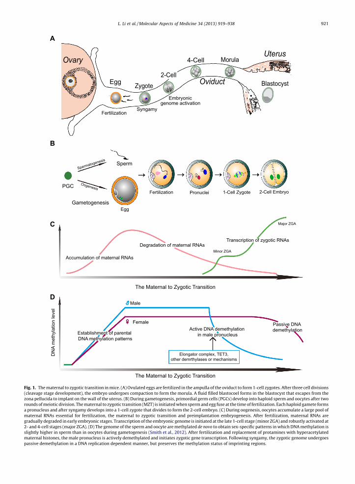
Mammalian development begins when the haploid sperm fuses with the haploid egg at fertilization to form the 1-celldiploid zygote (Fig. 1A). Both the maternal and paternal genomes are required for successful development (Mcgrath andSolter, 1984; Surani et al., 1984). At gamete fusion, the fertilizing sperm DNA is packaged with protamines and maturesperm are transcriptionally inert. Although the sperm provides DNA for the male pronucleus and is essential for egg acti-vation, the mitochondria, the microtubule-organizing center precursors and the stored cellular components in the spermplay minor roles in fertilization and early embryogenesis (Saunders et al., 2002; Schatten et al., 1985; Shitara et al.,1998; Sutovsky and Schatten, 2000). Thus, it is incumbent on the egg to provide a suitable environment for sperm–egg recognition, prevention of polyspermy, paternal genome remodeling and embryonic genome activation, ensuring asuccessful transition from maternal control to a shared responsibility with the male genome in directing early develop-ment (Fig. 1B).
At birth, the ovary contains a full complement of germ cells and after puberty, cohorts enter into a two week growthphase that culminates in meiotic maturation and ovulation into the oviduct. In mice, the female genome is transcriptionallyand translationally active as oocytes grow to �70 lm and the enlarging oocyte serves as a storehouse for maternal proteins(Fig. 1C) (De Leon et al., 1983). These proteins are critically important because transcription stops during meiotic maturationprior to ovulation and the embryonic genome is not robustly activated until the 2-cell stage (Fig. 1C). Thus, the maternal tozygotic transition (MZT) depends heavily on stored maternal components to form structures necessary to initiate develop-ment, eliminate redundant maternal materials and activate the embryonic genome to reprogram gene expression during theMZT (Tadros and Lipshitz, 2009).
Microarray and proteomic analyses have defined global patterns of gene expression in early development and trans-genesis provides insight into the role of specific genes during the MZT. Genetic studies have shown that maternal effectgenes affect multiple processes including pronuclear formation and fusion (Philipps et al., 2008; Wu et al., 2003), thefirst cell division (Burns et al., 2003; Tang et al., 2007), embryonic gene transcription (Bultman et al., 2006; Ramoset al., 2004) and cleavage-stage embryogenesis (Li et al., 2008a; Ma et al., 2006; Payer et al., 2003; Roest et al.,2004; Tong et al., 2000). Limited data from these studies suggest the important role of individual maternal effect geneson embryonic development, but the function and mechanisms of these genes in early embryogenesis are largelyunknown.
In this review, we focus on mice as a model to highlight molecular events in the MZT in mammals. Our discussionbegins with fertilization of the transcriptionally inert gametes, a process dependent on stored maternal proteins. Wethen examine the processing and activation of the embryonic genomes as a transition phase from maternal to zygoticcontrol and finish with the continued role of maternal components in cleavage stage embryogenesis. Finally we brieflyaddress future challenges associated with the MZT and the medical implications of understanding this process.
2. Maternal contribution to the onset of development
2.1. Fertilization
Fertilization heralds the onset of development in most species, including mammals. For successful embryonic develop-ment, sperm need to penetrate outer investments to fuse with the egg and there must be an effective post-fertilization blockto polyspermy. The absence of the former precludes development and the absence of the latter is an embryonic lethal.Although a multi-step process, a major arbiter of successful fertilization and preimplantation development in mammals isthe zona pellucida that surrounds ovulated eggs and preimplantation embryo (Fig. 2A and B). Encoded by maternal genes,
A
B
C
D
1-Cell Zygote 2-Cell EmbryoFertilization
Egg
Pronuclei
Ovary
Oviduct
Uterus
Zygote
Morula
Blastocyst
4-Cell
Egg2-Cell
SyngamyFertilization
Embryonicgenome activation
Degradation of maternal RNAs
Accumulation of maternal RNAs
Transcription of zygotic RNAs
The Maternal to Zygotic Transition
The Maternal to Zygotic Transition
Minor ZGA
Major ZGA
Spermatogenesis
Oogenesis
Sperm
PGC
Gametogenesis
DN
A m
ethy
latio
n le
vel
Establishment of parental DNA methylation patterns
Active DNA demethylation in male pronucleus
Passive DNA demethylation
Male
Female
♂
♀
Elongator complex, TET3, other demrthylases or mechanisms
Fig. 1. The maternal to zygotic transition in mice. (A) Ovulated eggs are fertilized in the ampulla of the oviduct to form 1-cell zygotes. After three cell divisions(cleavage stage development), the embryo undergoes compaction to form the morula. A fluid filled blastocoel forms in the blastocyst that escapes from thezona pellucida to implant on the wall of the uterus. (B) During gametogenesis, primordial germ cells (PGCs) develop into haploid sperm and oocytes after tworounds of meiotic division. The maternal to zygotic transition (MZT) is initiated when sperm and egg fuse at the time of fertilization. Each haploid gamete formsa pronucleus and after syngamy develops into a 1-cell zygote that divides to form the 2-cell embryo. (C) During oogenesis, oocytes accumulate a large pool ofmaternal RNAs essential for fertilization, the maternal to zygotic transition and preimplantation embryogenesis. After fertilization, maternal RNAs aregradually degraded in early embryonic stages. Transcription of the embryonic genome is initiated at the late 1-cell stage (minor ZGA) and robustly activated at2- and 4-cell stages (major ZGA). (D) The genome of the sperm and oocyte are methylated de novo to obtain sex-specific patterns in which DNA methylation isslightly higher in sperm than in oocytes during gametogenesis (Smith et al., 2012). After fertilization and replacement of protamines with hyperacetylatedmaternal histones, the male pronucleus is actively demethylated and initiates zygotic gene transcription. Following syngamy, the zygotic genome undergoespassive demethylation in a DNA replication dependent manner, but preserves the methylation status of imprinting regions.
L. Li et al. / Molecular Aspects of Medicine 34 (2013) 919–938 921

A
20 µm
Ovulated eggDiameter: 80 µmZona: 7 µm
Relative sizeof mouse sperm
C
s
ZP3ZP2ZP1
ZP3ZP2ZP1
Glycans
Egg
Embr
yo
Glycosidase(Release Glycan)
s
D
ZP3ZP2ZP1
ZP3ZP2ZP1
Egg
Embr
yo
Ovastacin(Cleave ZP2)
Mouse
BOvulated eggDiameter: 120 µmZona: 15 µm
Relative sizeof human sperm
Human
Fig. 2. Fertilization, the initiation of development. (A) The �7 lm wide mouse zona pellucida surrounds a �80 lm egg. As observed by scanning electronmicroscopy, the zona matrix has multiple pores which may facilitate sperm penetration (Familiari et al., 2008). Mouse sperm are �125 lm long with a thinacrosome overlying a distinctive falciform (hook-like) head. (B) The thicker (�15 lm) human zona pellucida surrounds a larger (�120 lm) egg and yet itsthree-dimensional structure is similar to mouse (Familiari et. al., 2006). Human sperm are half as long (�60 lm) as mouse sperm with a smaller, flattened,spatulate head. Of note, human sperm are fastidious and will not bind to mouse eggs, although mouse sperm bind to human (and most other) eggs. (C)Glycan release models postulate a carbohydrate ligand attached to a zona pellucida protein that interacts with a sperm surface receptor. AlthoughN-glycans have been proposed, most attention has been focused on O-glycans attached to ZP3. Following fertilization, the removal of the glycan by a corticalgranule glycosidase would account for the inability of sperm to bind to 2-cell embryos. However, recent biochemical investigations and genetic ablationstudies have not confirmed the candidacy of proposed glycan ligands or receptors as essential for sperm-egg recognition. (D) The ZP2 cleavage modelproposes that sperm bind with taxon-specificity to the N-terminus of ZP2 and the cleavage status of ZP2 is crucial for sperm-zona recognition and binding.Following fertilization, ovastacin, a metalloendoprotease, is exocytosed from egg cortical granules to cleave the N-terminus of ZP2 and prevent post-fertilization sperm binding. Implicit in the model is the presence of a cognate receptor(s) on the sperm surface which has not as yet been molecularlydefined.
922 L. Li et al. / Molecular Aspects of Medicine 34 (2013) 919–938
the zona pellucida not only mediates relative taxon-specific gamete recognition and provides a definitive block to poly-spermy, it also ensures passage of the early embryo down the oviduct prior to implantation in the uterus.
2.2. Zona pellucida
The genomes of eutherian mammals (including human, rat and mouse) contain four loci (Zp1, Zp2, Zp3, Zp4) on separate,but syntenic chromosomes that potentially encode zona proteins. Although all four proteins are detected in the zonapellucida of human (Lefievre et al., 2004) and rat (Hoodbhoy et al., 2005), only ZP1, ZP2 and ZP3 are present in mouse (Bleiland Wassarman, 1980b; Boja et al., 2003) because of multiple stop codons in transcripts expressed at the Zp4 locus (Lefievreet al., 2004). Each mouse gene is single copy in the genome (Chamberlin and Dean, 1989; Epifano et al., 1995; Kinloch et al.,1988; Liang et al., 1990) and mouse lines with null mutations for Zp1, Zp2 and Zp3 have been established. Mice lacking ZP1form a zona pellucida to which sperm bind and these mice are fertile, albeit with decreased fecundity (Rankin et al., 1999).The absence of either ZP2 or ZP3 precludes formation of a stable zona pellucida around eggs which are resorbed afterovulation and these mice are sterile (Liu et al., 1996; Rankin et al., 1996, 2001). However, this phenotype can be rescuedby transgenic expression of homologous human proteins, suggesting that human ZP2 and ZP3 are structurally comparableto the corresponding mouse proteins (Rankin et al., 1998, 2003).
2.3. Models of sperm-zona recognition
The molecular basis of mammalian gamete recognition has perplexed investigators for decades. Candidate glycans andproteins that were initially proposed based on biochemical or cell biology assays have not proven to be essential for fertilitywhen ablated in transgenic mouse models.
2.3.1. Glycan-release modelsThe most widely embraced models for sperm–egg recognition have been based on zona glycan ligands binding to a sperm
surface receptor (Fig. 2C). In these models, the post-fertilization release of a glycosidase from the egg’s cortical granules is

L. Li et al. / Molecular Aspects of Medicine 34 (2013) 919–938 923
hypothesized to cleave the candidate glycan and account for the inability of sperm to bind to the zona surrounding 2-cellembryos. Mouse and human taxon-specific binding is ascribed to differences in the glycosylation of their respective zonaproteins.
ZP3, initially described as a sperm ‘receptor’, but more seemingly a ‘ligand’, was first implicated in sperm–egg recog-nition based on the ability of gel-purified, re-natured, soluble ZP3 to inhibit (�80%) sperm binding to ovulated eggsin vitro (Bleil and Wassarman, 1980a). This model was extended to specify O-glycans on ZP3 as mediators of sperm–egg recognition (Florman and Wassarman, 1985), specifically a terminal a1,3 galactose residue (Bleil and Wassarman,1988), and further refined to implicate ZP3 Ser332 and Ser334 as attachment sites for the glycan ligand (Chen et al.,1998a). An independent line of investigation identified a second candidate glycan, N-acetylglucosamine, as the essentialZP3 ligand that was bound by sperm surface b1,4 galactosyl transferase acting as a lectin (Miller et al., 1992). In thismodel, the subsequent release by cortical granule N-acetylglucosaminidase accounted for the inability of sperm to bindto 2-cell embryos (Miller et al., 1993).
However, subsequent biochemical and genetic data have not supported these early models. In particular, geneticallymodified mice lacking a1,3 galactose (Liu et al., 1997; Thall et al., 1995) or N-acetylglucosamine (Williams et al., 2007) re-mained fertile and the absence of the candidate sperm receptor, b1,4 galactosyl transferase minimally affects fertility (Asanoet al., 1997; Lu and Shur, 1997). Turning to the putative attachment sites, mass spectrometric analysis, sensitive to low fem-tomole levels, did not detect glycosylation on either ZP3 Ser332 or Ser334 (Boja et al., 2003). Moreover, specific genetic muta-tion of the implicated serine residues to preclude attachment of O-glycans did not affect sperm recognition or fertility intransgenic mice (Liu et al., 1995) even when crossed into Zp3 null mice to reconstitute a zona pellucida in which ZP3Mut com-pletely replaces endogenous mouse ZP3. Sperm bound to ovulated eggs with a zona pellucida containing mutant ZP3 in theabsence of normal ZP3 and the otherwise normal appearing Zp3Mut female mice were fertile (Gahlay et al., 2010). Thus, cur-rent biochemical and genetic data are not consistent with ‘glycan-release’ models in which O-glycans attached to ZP3 Ser332
or Ser334 play an essential role in sperm–egg recognition.
2.3.2. ZP2 cleavage modelIn a series of loss of function assays, sperm binding and fertility was assessed in Zp1, Zp2 or Zp3 null mice. Mice lacking
ZP1 form a zona matrix with ZP2 and ZP3 that is structurally abnormal with increased porosity when viewed by electronmicroscopy. Sperm bind and fertilize Zp1 null eggs, but exhibit decreased fecundity with litter sizes half that of normal mice.This continued fertility documents that ZP1 is not essential for gamete recognition (Rankin et al., 1999). Mice lacking eitherZP2 or ZP3 have a more striking phenotype. No zona pellucida surrounds ovulated eggs in the absence of either protein andthe zona-free eggs are quickly resorbed into the epithelial lining of the oviduct. Although Zp2 and Zp3 null mice are sterile(Liu et al., 1996; Rankin et al., 1996, 2001), no assessment of the role of either ZP2 or ZP3 in sperm binding is possible in theabsence of a zona matrix.
Therefore, a series of gain-of-function assays were established to take advantage of the observation that, although indi-vidual proteins are conserved among mammals, human sperm do not bind to the mouse zona pellucida (Bedford, 1977).Using transgenesis, mouse lines were established in which human ZP1, ZP2, ZP3 and ZP4 replaced (or added to) endogenousmouse proteins (Rankin et al., 1998, 1999, 2003; Yauger et al., 2011). In a taxon-specific sperm binding assay, only humanZP2 supported human sperm binding after which sperm penetrated the ‘humanized’ zona pellucida and accumulated in theperivitelline space unable to fuse with mouse eggs (Baibakov et al., 2012). Normally ZP2 is cleaved after fertilization andsperm will not bind to the zona pellucida (Bauskin et al., 1999; Bleil et al., 1981). The cleavage site has been biochemicallydefined in mice (166LA;DE169) and, when mutated to prevent cleavage, sperm bind to the zona pellucida after fertilization andcortical granule exocytosis (Gahlay et al., 2010). Taken together, these observations support a model for gamete recognitionin which sperm bind to ZP2 and the post-fertilization cleavage of ZP2 accounts for the inability of sperm to bind to 2-cellembryos (Fig. 2D).
The sperm binding site on ZP2 was further defined in a bead binding assay in which human sperm bound well torecombinant human ZP239–154 or ZP239–267 peptides, but poorly to beads coated with the homologous mouse peptides.Reduction of disulfide bonds significantly decreased human sperm binding to the human peptides indicating a depen-dence on secondary structures. The binding of human sperm to huZP2 rescued eggs was inhibited by excess huZP239–
154, but not moZP235–149 peptides, which corroborated the specificity of the binding site on ZP2. Although fewer in num-ber, mouse sperm bound comparably to mouse and human ZP2 peptide beads consistent with its observed lack of taxonspecificity in binding to mouse and human eggs (Bedford, 1977). From these results, it was concluded that the N-termi-nus of human ZP2 plays an important role in gamete recognition on the surface of the zona pellucida (Baibakov et al.,2012).
In this rapidly changing landscape, a significant amount of investigation will be required to obtain wide-spread con-sensus for the molecular basis of gamete recognition. For glycan-release models to be revived, a specific glycan ligand(s)and sperm-surface receptor will need to be identified that, when genetically ablated, prevents fertilization. In addition,expression of the human glycan ligand in transgenic mice should break taxon-specific, sperm-egg recognition. The ZP2cleavage model requires further validation in transgenic mice expressing chimeric mouse-human and truncated forms ofmouse ZP2. The identification of a cognate sperm surface receptor with taxon-specificity that could be manipulated intransgenic mouse models would be particularly compelling. The incompleteness of our understanding of this critical

924 L. Li et al. / Molecular Aspects of Medicine 34 (2013) 919–938
event should inspire renewed research into the molecular basis of sperm-egg recognition that is required for fertilizationand the onset of development.
2.4. The post-fertilization block to polyspermy
Polyspermy is lethal for early embryos and at least three post-fertilization blocks to gamete interactions have evolved inmice. The first two occur rapidly after fertilization and prevent additional sperm from fusing with the egg’s plasmamembrane or penetrating the extracellular zona pellucida surrounding eggs and preimplantation embryos (Sato, 1979;Stewart-Savage and Bavister, 1988). The third and definitive block occurs over several hours and ensures that sperm donot bind to the surface of the zona pellucida (Baibakov et al., 2007; Inoue and Wolf, 1975). The molecular basis of the firsttwo blocks remains largely unknown, and the third correlates with egg cortical granule exocytosis (Barros and Yanagimachi,1971).
As described above, ZP2 is cleaved and sperm do not bind to the zona pellucida surrounding 2-cell embryos afterfertilization (Bauskin et al., 1999; Bleil et al., 1981). Ovastacin, a member of the astacin family of metalloendoproteases(Quesada et al., 2004), is released from cortical granules following fertilization and normally cleaves ZP2 (Burkart et al.,2012) over several hours (Baibakov et al., 2007). The ensuing proteolysis effectively precludes sperm binding to the zonapellucida (Burkart et al., 2012; Greenhouse et al., 1999). Either mutation of the ZP2 cleavage site (Gahlay et al., 2010) orablation of the gene encoding ovastacin (Burkart et al., 2012) prevents proteolysis (Fig. 2D). Thus, when ZP2 remains intact,sperm will bind to the zona pellucida surrounding the early embryo independent of fertilization and cortical granuleexocytosis.
3. Epigenetic regulation of the maternal to zygotic transition
During gametogenesis, mouse primordial germ cells (PGCs) establish sex-specific epigenetic marks with distinct DNAmethylation patterns that can occur globally and in specific imprinted regions (Fig. 1D) (Hayashi and Surani, 2009; Lees-Murdock and Walsh, 2008). As gametes mature, the haploid male and female genomes, packaged respectively with prota-mines and histones, become transcriptionally quiescent. At fertilization, the highly differentiated, haploid maternal eggand paternal sperm fuse to establish the totipotent 1-cell zygote in which both genomes undergo dynamic changes inDNA methylation (Fig. 1D) and are repackaged with histones in a specific manner to initiate embryonic development. Thereis substantial research that documents the critical importance of epigenetic modifications during the MZT.
3.1.Epigenetics
Epigenetics refers to heritable changes in gene expression or cellular phenotypes that occur without alterations in DNAsequence, but rather through DNA methylation, histone modification and regulation by non-coding RNAs (e.g. miRNA, siRNA,piRNA) (Berger et al., 2009). By adding a methyl group to the fifth carbon of cytosine residues in the context of CpG dinu-cleotides (Holliday and Pugh, 1975), DNA methylation plays vital roles in various cellular processes including regulationof gene expression, establishment of imprinting (Li et al., 1993), X chromosome inactivation in females (Robertson and Wolf-fe, 2000), silencing of parasitic retrotransposons (Yoder et al., 1997) and maintaining the stability and integrity of the gen-ome (Chen et al., 1998b). The patterns of DNA methylation can be divided into two categories: de novo DNA methylation andmaintenance DNA methylation, each with a set of well-characterized DNA methyltransferases (DNMTs) that are conserved inboth animals and plants (Goll and Bestor, 2005; Law and Jacobsen, 2010). Enzymes responsible for de novo DNA methylationare DNMT3 family members and include DNMT3a, DNMT3b and DNMT3L (Aapola et al., 2000; Okano et al., 1998, 1999;).DNMT3L lacks the key methyltransferase motifs and has no enzymatic activity, but acts as a regulator of both DNMT3aand DNMT3b in de novo DNA methylation (Gowher et al., 2005; Suetake et al., 2004). DNMT1 methylates DNA with a strongpreference for hemimethylated target sites and maintains methylated DNA during DNA replication (Hermann et al., 2004). Ingeneral, the status of DNA methylation is associated with gene transcription in which hypermethylation indicates repressionand hypomethylation specifies activation (Reik et al., 2001; Santos and Dean, 2004).
In the nucleus, DNA is packaged with histones to form core nucleosomes (two each of H2A, H2B, H3, and H4) which areseparated by linker histones (H1) (Luger et al., 1997; Turner, 2002) and further arranged into higher-order chromatin struc-tures. Post-translational modifications of individual histones (i.e., the histone code) in their amino termini include acetyla-tion, phosphorylation, ubiquitylation, ADP-ribosylation and methylation in which specific lysine or arginine residues can bemodified by mono-, di- and trimethylation. These modifications afford greater subtlety than DNA methylation alone and areessential in regulating cellular and molecular events including chromatin remodeling and gene expression (Berger, 2002;Kouzarides, 2007; Peterson and Laniel, 2004). More recent investigations of histone variants are providing additional insightinto the histone code, making these modulatory epigenetic events ever more intriguing (Kamakaka and Biggins, 2005; Sarmaand Reinberg, 2005).

L. Li et al. / Molecular Aspects of Medicine 34 (2013) 919–938 925
3.2. Demethylation of DNA in the paternal genome
Just prior to ovulation, oocytes undergo nuclear envelope breakdown and complete the first meiotic division. Ovulatedeggs are arrested at metaphase of the second meiotic division (MII) with their DNA packaged with maternal histones. Incontrast, the haploid sperm DNA is tightly packaged with protamines which must be removed after gamete fusion. Themechanisms of this removal during sperm nuclear decondensation are incompletely understood, but are accompanied bywide-spread demethylation (Abdalla et al., 2009; Mayer et al., 2000; Oswald et al., 2000) and followed by repackaging withhyperacetylated maternal histones to form the male pronucleus (Fig. 1D) (Adenot et al., 1997; Santos et al., 2002). Followingsyngamy of the two pronuclei, the zygotic genome undergoes passive demethylation in a DNA replication dependent manneruntil the morula stage (Fig. 1D). Thereafter, de novo methylation patterns are established to sustain successful cell lineagedifferentiation (Corry et al., 2009; Rougier et al., 1998; Santos et al., 2002).
DNA demethylation of the paternal genome is rapidly completed within 4–6 h after fertilization and occurs before thecommencement of the first round of DNA replication (Fig. 1D) (Santos and Dean, 2004; Santos et al., 2002). This indicatesan active process (Abdalla et al., 2009; Mayer et al., 2000; Oswald et al., 2000) in which the methyl group from 5-methylcytosine is enzymatically removed (Wu and Zhang, 2010). One candidate, MBD2 (methyl-CpG-binding domain protein 2)was reported to have demethylase activity and to directly remove methyl groups from 5-methylcytosine in vitro (Bhattach-arya et al., 1999). However, Mbd2 null mice are viable, fertile and lack abnormal DNA methylation patterns (Hendrich et al.,2001; Santos et al., 2002) and, thus, other candidates must be sought.
Using a siRNA-mediated knockdown screening strategy coupled with live cell imaging of mouse zygote, ELP3, a submit ofthe elongator complex (composed of ELP1 to ELP6), was reported to play important roles in paternal genome demethylation(Okada et al., 2010). ELP3 knockdown prevented paternal DNA demethylation as shown by cytosine-5 methylation (5mC)staining and bisulphite sequencing. The same effect was observed with knockdowns of ELP1 and ELP4 which suggests thatthe entire elongator complex may be involved in demethylation (Okada et al., 2010). ELP3 contains a histone acetyltransfer-ase (HAT) domain and a cysteine-rich Fe–S radical S-adenosylmethionine (SAM) domain which can catalyze paternal DNAdemethylation as shown by dominant-negative mutants using domain-specific mRNAs (Okada et al., 2010). Based on theknown catalytic mechanism of radical SAM domains (Wang and Frey, 2007), it is postulated that ELP3-mediated DNAdemethylation is accomplished by a series of radical reactions starting from the generation of a powerful oxidizing agent,the 50-deoxyadenosyl (50-dA) radical from SAM, and ending with the production of 5-hydroxymethyl-cytosine (5hmC) whichwould be further resolved into cytosine (Wu and Zhang, 2010).
Recently, the Tet family has been discovered to mediate DNA demethylation via an oxidative process catalyzing theconversion of 5mC to 5hmC (Ito et al., 2010; Tahiliani et al., 2009; Veron and Peters, 2011). The Tet family includesTet1, Tet2 and Tet3 in mice. Tet1 and Tet2 play a role in embryonic stem cell self-renewal and human myeloid malignan-cies, respectively, and their perturbation is accompanied by defects in DNA methylation (Ito et al., 2010; Nolte and Hof-mann, 2008; Xu et al., 2011). Elevation of 5hmC and reduction of 5mC is associated with enrichment of TET3 in themammalian paternal pronucleus, raising the possibility of a role of TET3 in active DNA demethylation of the paternal gen-ome (Gu et al., 2011; Iqbal et al., 2011; Wossidlo et al., 2011). Indeed, knocking down maternal Tet3 with specific siRNA(Wossidlo et al., 2011) or through transgenesis in murine oocytes and zygotes (Gu et al., 2011) leads to aberrant methyl-ation patterns in the male genome. The observed decrease in 5hmC and increase in 5mC levels in the late male pronucleusindicates that disruption of 5mC to 5hmC conversion causes abnormal DNA demethylation. However, there is little per-turbation in the maternal genome when Tet3 is disrupted which suggests specific targeting to the male (Gu et al.,2011). 5hmC can be further oxidized to 5-formylcytosine (5fC) and 5-carboxylcytosine (5caC), and the latter could be ex-cised by thymine-DNA glycosylase (TDG)–mediated base excision repair (He et al., 2011; Ito et al., 2011). However,whether 5hmC is an intermediate of demethylation or an end-product in the male pronucleus remains to be determined.The observation of replication-dependent dilution of 5hmC during preimplantation mouse development indicates that TETproteins may also facilitate passive demethylation of the embryonic genome after active demethylation of the male pro-nucleus (Inoue and Zhang, 2011).
Active demethylation of the paternal genome has been observed in many mammalian embryos including those of mouse,rat, pig, bovine and human (Abdalla et al., 2009; Dean et al., 2001; Fulka et al., 2004; Mayer et al., 2000) which indicates abiological imperative in early development. One hypothesis is that active DNA demethylation de-represses the paternal gen-ome. This could enable minor transcriptional activation of the paternal genome at the late 1-cell stage and prime the robusttranscription essential for the MZT. In support of this hypothesis, BrUTP incorporation, indicative of de novo gene transcrip-tion, is initially detected in the male pronucleus 5 h after pronuclear formation and is always higher than that observed inthe female pronucleus (Aoki et al., 1997). Alternatively, active demethylation may be required for reprogramming paternalchromatin into a totipotent state, as has been observed during somatic nuclear transferred into enucleated oocyte (Deanet al., 2003; Reik et al., 2001). However, neither hypothesis adequately explains why only the paternal genome undergoesactive demethylation and in such a narrow developmental window. Based on the kinship theory of imprinting (Wilkinsand Haig, 2003), demethylation of the paternal genome by maternal factors during the protamine-histone exchange (Reikand Walter, 2001) could maximize the female genome fitness by reactivating genes in paternal genome (Wilkins and Haig,2002). This evolutionary conflict based notion is consistent with the absence of active DNA demethylation in species withoutgenomic imprinting, such as zebrafish (Macleod et al., 1999) and Xenopus (Stancheva et al., 2002).

926 L. Li et al. / Molecular Aspects of Medicine 34 (2013) 919–938
Global and active DNA demethylation of the paternal genome in the one-cell zygotes is widely considered to occur shortlyafter fertilization. However, a recent study suggested that the weak staining of 5meC in the male nucleus during the first cellcycle may result from a progressive acid-resistant antigenic masking of 5meC (Li and O’Neill, 2012). When antigen retrievalwas performed using tryptic digestion, no loss of DNA methylation in male pronuclei was observed. Rather, there was per-sistence of 5meC in both pronuclei, an observation that was supported by the almost equal staining with MBD1 (methylbinding domain 1 protein), a methyl group binding protein (Li and O’Neill, 2012). Considering the difficulty in identifyinga specific demethylase or mechanism to account for active DNA demethylation in the male pronucleus, this general para-digm might need to be reconsidered and further investigated.
3.3. Maternal and paternal imprinting
While there is active DNA demethylation of the male pronucleus shortly after fertilization followed by passive, replicationdependent DNA demethylation of the entire genome (Santos and Dean, 2004; Santos et al., 2002), the methylation status ofimprinted regions in maternal and paternal genome remains constant. Genomic imprinting was discovered 30 years agowhen investigators using pronuclear transplantation demonstrated that diploid mouse embryos with two pronuclei originat-ing from the same parent (gynogenones or androgenones) failed to develop to term (Mcgrath and Solter, 1984; Surani et al.,1984). This was the first documentation that, although each pronucleus carried the same genetic information, maternal andpaternal alleles are not functionally equivalent in mammalian development.
In mammals, genomic imprinting gives rise to parental-specific monoallelic expression of particular genes that are onlyexpressed from either the maternal or the paternal allele. Since the first two paternally imprinted genes, Igf2r and H19, andthe first maternally imprinted gene, Igf2, were reported (Barlow et al., 1991; Bartolomei et al., 1991; Dechiara et al., 1991),more than 150 imprinted genes have been identified in mice (http://www.mousebook.org/catalog.php?catalog=imprinting).Most imprinted genes are organized in clusters that can span more than 1 Mb and are scattered throughout the genome(Verona et al., 2003). The parent-of-origin expression pattern of these genes results from different DNA methylation incis-acting regulatory elements termed imprinting control regions (ICRs) functioning through differentially methylated re-gions (DMRs), in which only one allele, either maternal or paternal, is methylated. Notably, dysfunction of ICRs leads to lossof proper expression of imprinted genes in the cluster (Fitzpatrick et al., 2002; Thorvaldsen et al., 1998) and improperexpression of imprinted genes can affect embryogenesis leading to growth defects and, in some cases, severe disease includ-ing cancer. The low efficiency in obtaining SCNT (somatic cell nuclear transfer) derived clones and the unpredictable abnor-malities in survivors are likely a result of aberrant genomic imprinting (Humpherys et al., 2001). In humans, severalmalformation syndromes, including Prader-Willi, Angelman, Beckwith-Wiedemann and Silver-Russell as well as cancerssuch as neuroblastoma (maternal chromosome 1p36 and paternal chromosome 2), Wilms’ tumour (maternal chromosome11p15.5) and acute myeblastic leukemia (paternal chromosome 7), have documented associations with imprinting defects(Butler, 2009; Lim and Maher, 2009; Walter and Paulsen, 2003). Clinical studies have reported that human malformationsassociated with imprinting disorders occur with greater frequency in babies obtained by assisted reproductive technology(ART) (Cox et al., 2002; DeBaun et al., 2003; Gicquel et al., 2003). Because of these impacts on mammalian development, pat-terns of genomic imprinting must be sustained faithfully throughout embryogenesis and on into adulthood (Li and Sasaki,2011).
DNA methyltransferases have been shown to control the methylation status of imprinted regions (Cirio et al., 2008; Hiras-awa et al., 2008; Howell et al., 2001; Kurihara et al., 2008). Depletion of DNMT1o, which is present in the oocyte and trans-locates to the nucleus of the 8-cell embryo, results in abnormal, allele-specific expression and half of the ICR methylation islost (Howell et al., 2001). DNMT1s are associated with MII oocyte chromatin and are present in the nucleus throughout pre-implantation development. Inhibiting DNMT1s activity with antibodies or RNAi (RNA interference) knockdowns decreasesDNA methylation in specific repetitive sequences and imprinted loci. Thus, DNMT1s is also responsible for methylationmaintenance (Cirio et al., 2008; Kurihara et al., 2008). Deletion of both maternal and zygotic DNMT1 results in complete lossof methylation at imprinted regions, suggesting that both maternal and zygotic DNMT1 are required to maintain DNA meth-ylation patterns at imprinted loci (Hirasawa et al., 2008).
However, DNA methyltransferases are not sufficient for to maintain imprints. Stella (official name, Dppa3 and also knownas Pgc7), first characterized as a maternal effect gene (Payer et al., 2003), plays important roles in maintaining methylation ofmost of the maternal genome and two paternally imprinted genes, H19 and Rasgrf1 (Nakamura et al., 2007). STELLA is pres-ent in both maternal and paternal pronuclei, but seems to preferentially bind to a specific ‘architecture’ in maternal chro-matin (Nakamura et al., 2012), namely that provided by dimethylated histone H3 lysine 9 (H3K9me2). When theH3K9me2 mark was removed with ectopic expression of an H3K9 methylation/dimethylation-specific demethylase (Jhdm2a,official name Kdm3a) in mouse zygotes, STELLA staining was no longer observed in the maternal pronucleus and maternalDNA methylation was not maintained (Nakamura et al., 2012). In addition, the protection of DNA methylation patterns inpaternally imprinted H19 and Rasgrf1 DMRs was associated with enrichment of H3K9me2 at these two loci, further support-ing a role for H3K9me2 in mediating protection of DNA methylation by STELLA (Nakamura et al., 2012). DNA demethylationis accompanied by the TET3-mediated conversion of 5mC to 5hmC in the paternal genome, but not in the maternal genome(Gu et al., 2011; Iqbal et al., 2011). However, strong staining of TET3 anomalously occurred in maternal pronucleus in Stellanull zygotes. Thus, the binding of STELLA to H3K9me2 containing chromatin seems to inhibit the binding of TET3 to its chro-

L. Li et al. / Molecular Aspects of Medicine 34 (2013) 919–938 927
matin targets, which prevents the conversion of 5mC to 5hmC and protects DNA methylation in normal mouse zygotes(Nakamura et al., 2012).
ZFP57, a KRAB-zinc finger protein, also has been shown to play a role in maintenance of both parental imprints (Li et al.,2008b). While zygotic depletion of ZFP57 results in partial lethality and partial disruption of several imprinted loci, lack ofboth maternal and zygotic ZFP57 leads to a more severe phenotype with greater embryonic lethality and complete loss ofmethylation at multiple loci (Snrpn, Peg1, Peg3, Peg5, and Dlk1 DMRs) (Li et al., 2008b). In addition, ZFP57 plays a role inestablishing maternal imprints at the Snrpn locus and maternal loss of ZFP57 precludes establishment of Snrpn imprintsin mouse oocytes (Li et al., 2008b). Consistent with these observations, a clinical study reported that ZFP57 mutations werefrequently found in patients with transient neonatal diabetes, a disease caused by hypomethylation of the promoter of theimprinted gene Plagl1, coupled with abnormal hypomethylation at multiple other imprinted loci (Mackay et al., 2008).Recently, Trim28 (also known as KAP-1 or Tif1b), which is highly expressed in the nuclei of oocytes and preimplantation em-bryos, was found to be involved in methylation maintenance at imprinted loci (Messerschmidt et al., 2012). When Trim28was depleted in oocytes, no viable offspring were obtained, although embryos could successfully develop into blastocysts(Messerschmidt et al., 2012). Upon further analysis, it was proposed that this severe phenotype might be a result of dysreg-ulation of genomic imprinting. Notably, the ICR of the paternally imprinted H19 was hypomethylated with up-regulation ofH19 expression in maternal Trim28 mutants (Messerschmidt et al., 2012), indicating the importance of maternal TRIM28 insafeguarding paternal H19 imprints from aberrant demethylation.
Taken together, these data suggest that multiple mechanisms have evolved to counteract global DNA demethylation andthus maintain proper imprints at specific genetic loci at particular times in development.
3.4. Histone modification
Histone modifications are dramatically different in maternal and paternal pronuclei of zygotes and undergo dynamicchanges during the MZT. After fertilization, hyperacetylated histones are incorporated into the paternal genome and mayaccount for its relatively higher transcriptional activity compared to the maternal pronucleus (Aoki et al., 1997). H3.3 is areplication-independent variant of H3 and indicative of a transcriptionally permissive state. Following fertilization, this var-iant is preferentially incorporated into the paternal pronucleus and enriched in paternal chromatin by means of a specifichistone chaperone, HIRA (Torres-Padilla et al., 2006; van der Heijden et al., 2005). On the other hand, while H3 di- andtri- K9 and K27 methylation are readily detected in the maternal pronucleus and may provide protection of the maternalgenome from active DNA demethylation, they are not present in the paternal pronucleus (Arney et al., 2002; Feil, 2009; Reiket al., 2003; Santos et al., 2005). Trimethylation of H3K4 is readily observed in the maternal pronucleus before pronuclearstage PN4, but the extent of this modification becomes indistinguishable between the two pronuclei by PN5 (Lepikhovand Walter, 2004). Considering the complexity of histone modifications, no single set of rules can be followed to explainthese phenomena and deciphering the detailed dynamics of various histone modifications during the MZT remains anintriguing challenge.
A further challenge is the need to coordinate histone modifications with DNA methylation for proper regulation of geneexpression in early embryonic development. For example, transcriptionally repressive H3K9 methyltransferases G9a (officialname, EHMT2), SUV39H1 and SETDB1 (also known as ESET) interact with DNA methyltransferases, including DNMT1,DNMT3A and DNMT3B. This cooperation establishes particular methylation patterns and can regulate gene expression atspecific time and sites in early development (Cedar and Bergman, 2009; Saitou et al., 2012). It seems likely that similar coor-dination between histone modifications and DNA methylation is involved in regulation of additional genes in mammaliandevelopment. To date this remains an under-investigated area of research, but high-throughput ChIP experiments shouldprovide insight into the relationship between histone modification and the MZT.
4. Activation of the embryonic genome
Transcription of the embryonic genome (zygotic genome activation, ZGA) occurs after fertilization and is critical for nor-mal embryonic development. The initiation of ZGA varies depending on the species. In mouse, ZGA is observed as early as theS/G2 phase in the male pronucleus of the 1-cell zygote and becomes robust at the 2-cell stage (Fig. 1C) (Bouniol et al., 1995;Latham et al., 1991; Ram and Schultz, 1993); in human (Davis, 1985) and pig (Braude et al., 1988) the ZGA occur and at 4–8-cell stage; and in cow (Frei et al., 1989) and sheep (Crosby et al., 1988) it is delayed until the 8–16-cell stage. Because of itsessential role in the onset of development, there has been considerable interest in the regulation of ZGA and changes in geneexpression profiles during the MZT.
4.1. Transcriptome profile of the maternal to zygotic transition
Conventional approaches including RT-PCR (reverse transcription; polymerase chain reaction), quantitative RT-RCR andnorthern blot analyses have been used to examine gene expression during the MZT (Christians et al., 1995; Davis et al.,1996). In addition, mRNA differential display (Zimmermann and Schultz, 1994), expressed sequence tag (EST) based analysis(Ko et al., 2000) and suppression subtractive hybridization (Zeng and Schultz, 2003) have been used to dissect stage specific

928 L. Li et al. / Molecular Aspects of Medicine 34 (2013) 919–938
gene expression during the MZT and preimplantation mouse development. In recent years, large-scale gene expression pro-files from microarrays and deep RNA sequencing have systematically provided enormous data sets of global gene expressionduring the MZT and preimplantation development in various mammals, including mice, cow, pigs and humans (Hamataniet al., 2004; Misirlioglu et al., 2006; Sirard et al., 2005; Vallee et al., 2008; Wang et al., 2004; Whitworth et al., 2005; Zenget al., 2004; Zeng and Schultz, 2005). Of particular note is the dynamic wave of gene expression corresponding to the MZT,during which maternal transcripts are degraded and zygotic genes are activated (Fig. 1C).
Three groups separately have reported large-scale gene expression patterns in mouse oocytes and preimplantation em-bryos using different microarray platforms (Hamatani et al., 2004; Wang et al., 2004; Zeng et al., 2004; Zeng and Schultz,2005). Employing NIA 22 K 60-mer oligo microarrays, 12,179 out of 21,939 genes were observed to exhibit significantchange (Hamatani et al., 2004). These genes were classified into nine groups (clusters 1–9) and, using a k-meansnonhierarchical clustering method, were further partitioned into three groups according to their expression patterns.The first group (clusters 7 and 9) comprises 1589 genes highly enriched in oocytes, but progressively degraded in earlydevelopment; the second group (clusters 1, 4, 5, and 8) contains 5126 genes that are initially transcribed from the embry-onic genome; and the third group (clusters 2, 3 and 6) is composed of 5464 genes that are both inherited as maternallystored transcripts and transcribed from the embryonic genome after ZGA at the 1–2-cell stage. Maternal transcripts thatare gradually degraded around ovulation and after fertilization are mainly represented in clusters 3, 6, 7 and 9, which in-clude a total of 4063 genes. Among these four clusters, only genes in cluster three are reactivated after ZGA. Gene expres-sion patterns corresponding to the ZGA are grouped in clusters 1, 5 and 8, with genes in cluster one increasing throughoutpreimplantation development and those in cluster 5 and 8 peaking at 2-cell and 4-cell stage, respectively, and then declin-ing as development progresses. Intriguingly, a set of genes in cluster 2 are firstly dramatically up-regulated from the 4-cellto 8-cell stage before compaction and then significantly decreased later in development. This novel gene expression patternis designated ‘mid-preimplantation gene activation’ (MGA), which might play crucial roles in cell polarity and the first celllineage specification.
In a second study using Affymetrix Murine Genome Array U74Av2, more than 12,000 genes, over 1/3 of which exhibited=5-fold change in their expression, were identified in the MZT (Wang et al., 2004). It was found that 737 genes increased by=3-fold, while 1082 genes decreased by =3-fold during meiotic maturation, indicating that accumulation of transcripts iscoupled with degradation and/or deadenylation of many maternal transcripts (Bachvarova, 1985; Paynton et al., 1988). Afterfertilization and initiation of ZGA, embryonic RNA increases in complexity because multiple genes are activated. The genes inwhich the 12 time points were examined from GV oocyte to blastocyst stage were clustered into two large temporal groups,one of which, the ‘oocyte-to-embryo’ phase, documents the dramatic changes of gene expression during the MZT (Wanget al., 2004).
In a third study based on MOE430 GeneChips, 14,119 genes were identified in mouse oocytes and early embryos including1-cell, 2-cell, 8-cell and blastocyst embryos (Zeng et al., 2004). Among all the identified genes, 13,378 genes were found dif-ferentially expressed in at least one stage and were further grouped into 6 classes (‘maternal’, ‘maternal-to-zygotic’, ‘1-celltransient’, ‘2-cell transient’, ‘8-cell transient’, and ‘blastocyst’) according to their temporal expression patterns. Members inthe ‘maternal’ group, such as Mos and Zp3, represent gene products accumulated in oogenesis that progressively vanish inlater development. ‘Maternal-to-zygotic’ genes usually are expressed in both oocytes and early embryos having first beendegraded and then replaced by zygotic transcripts. Expression Analysis Systematic Explorer (EASE) analysis of the 809 genestransiently expressed in the 2-cell embryos documented that these genes are involved in transcription and RNA metabolism,indicating potential roles in the MZT. Genes associated with chromatin assembly and disassembly are enriched in 1-cell and2-cell embryos and may be critical for chromatin structure changes observed during the ZGA and the MZT (Zeng et al., 2004).Pharmacological studies documented that 1819 genes (�17% of genes detected in the 2-cell embryos) are a-amanitin sen-sitive (inhibits RNA Polymerase II and III), indicating their initial expression after ZGA (Zeng and Schultz, 2005). EASE anal-ysis of these a-amanitin sensitive 2-cell transcripts revealed that bioprocesses, such as ribosome biogenesis and assembly,protein synthesis, RNA metabolism and transcription, are overrepresented, which further substantiates the selective andbiased nature of gene expression patterns at ZGA (Zeng et al., 2004; Zeng and Schultz, 2005). Moreover, the observed a-ama-nitin sensitive 2-cell transcripts are concordant with the NIA data (Hamatani et al., 2004), suggesting the reproducibility andvalidity of the data obtained in the two different platform.
In recent years, investigative attention has turned to analyzing profiles of small, non-coding RNAs including miRNAs(microRNA), endo-siRNAs (small interfering RNA) and piRNAs (Piwi-interacting RNA) during the MZT (Ma et al., 2010;Suh et al., 2010; Svoboda and Flemr, 2010; Tam et al., 2008; Tang et al., 2007; Watanabe et al., 2008). The expression patternsof miRNAs are divided into three classes: maternal, maternal-to-zygote and zygote patterns (Tang et al., 2007). Maternallyinherited miRNAs (e.g. the let-7 family members) are degraded and de novo miRNAs synthesis starts at the 2-cell stage (e.g.the mir-290 cluster). However, maternal miRNA appears necessary for early development. Embryos derived from Dicer nulloocytes in which almost all miRNAs are depleted, lack a functional meiotic spindle and fail to develop beyond the first celldivision (Tang et al., 2007). In other model organisms, including Drosophila (Bushati et al., 2008), zebrafish (Giraldez et al.,2006) and Xenopus (Lund et al., 2009), miRNAs that mediate silencing pathways deplete maternal mRNAs and are critical forthe MZT. However, global suppression of miRNA pathways in growing oocytes and preimplantation embryos has limited ef-fect on the MZT in mice (Ma et al., 2010; Suh et al., 2010).
Endo-siRNAs in mouse oocytes are derived from naturally occurring dsRNAs (double strand RNAs) that are produced byinverted repeat structures, bidirectional transcription, antisense transcripts and some expressed pseudogenes (Tam et al.,

L. Li et al. / Molecular Aspects of Medicine 34 (2013) 919–938 929
2008; Watanabe et al., 2008). Reduction of siRNA from the loss of DICER or AGO2 in oocytes leads to increase of mobile andrepetitive sequences and complementary endogenous transcripts (Watanabe et al., 2008). In addition, many up-regulatedtranscripts in Dicer1�/� oocytes have complementary sequences to endo-siRNAs (Ma et al., 2010). Given the minimal affectobserved after suppression of the miRNA pathways in mouse oocyte and preimplantation embryos, the siRNA pathwaymight serve as the dominant RNA silencing mechanism controlling the MZT.
With the large scale and global gene expression data during the MZT, the next step will be screening specific factors thatpotentially play critical roles in the MZT, such as the degradation of maternal factors, fertilization, the initiation of ZGA andeven the whole process of preimplantation development, and deciphering the molecular regulatory mechanisms of thesefunctional genes.
4.2. Proteome profile of the maternal to zygotic transition
Although RNA profiles provide essential data, they may not accurately reflect abundance of cognate proteins becausesome RNAs may be un-coupled (non-coding RNAs) or not immediately coupled (delayed translation) with protein synthesis(Nothias et al., 1996). Therefore, high-resolution 1-dimentional or 2-dimensional protein gel electrophoresis, polysomalmRNA microarray and protein microarray, coupled with mass spectrometry and bioinformatic analyses have been used toinventory proteins during the MZT (Flach et al., 1982; Latham et al., 1991; Potireddy et al., 2006; Wang et al., 2010; Yurttaset al., 2010).
Growing oocytes accumulate large pools of stored maternal RNAs and, although many are degraded after fertilization,some are stable and persist in early development (Potireddy et al., 2006; Yurttas et al., 2010; Zhang et al., 2009). Duringthe MZT, maternal mRNAs are recruited for translation in support of particular developmental events that occur beforeZGA. To identify these mRNAs, polysomal mRNAs in MII stage oocytes and late 1-cell zygotes were identified on microarrays(Potireddy et al., 2006). Maternal mRNAs recruited for translation depended on cis-regulatory elements within the tran-scripts and changed significantly during transition from oocyte to embryo. While most polysomal maternal mRNA isolatedfrom oocyte are associated with cellular homeostasis, those over-represented in late 1-cell embryos are related to biosyn-thesis, especially protein biosynthesis (Potireddy et al., 2006) which is consistent with data obtained by analyzing totalmRNA (Zeng et al., 2004). Of note, one class of proteins encoded by genes recruited for translation in late 1-cell embryosis associated with gene transcription, which may contribute to de novo zygotic gene transcription at 2-cell stage.
Using semi-quantitative mass spectrometry, 2781, 2973, and 2082 proteins were respectively identified in germinal ves-icle (GV), MII oocytes and zygotes (Wang et al., 2010). Proteins relating to metabolism and transport that may support oocytematuration were over-represented in GV oocytes and those associated with cell cycle, epigenetic modification, DNA repairand pluripotency regulation were over-represented in MII oocytes. Unexpectedly, zygotes were highly enriched in proteinsof the ubiquitination pathway including ubiquitin B (UBB), ubiquitin C (UBC) and proteasome proteins. This may account forthe observed decrease in peptides from MII oocytes (185,643) and to zygotes (85,369) and suggest a role for the ubiquitin–proteasome pathway in active degradation of maternal proteins after fertilization.
Although important strides have been made in cataloging the proteome, de novo zygotic protein synthesis during the ZGAremains inadequately understood. Thus, the next challenge seemingly lies in developing high-resolution, high-throughputprotein analyses from the limited biomaterials available from early embryos. Once catalogued, advances can be anticipatedin deciphering the potential molecular functions of maternal proteins that have been degraded and, more importantly, zy-gotic proteins that have been synthesized de novo and play critical roles in the MZT.
4.3. Regulation of zygotic genome activation
ZGA is indispensable for preimplantation development and was investigated in the early 1970s using a-amanitin, a spe-cific inhibitor of RNA polymerase II and III (Golbus et al., 1973; Warner and Versteegh, 1974). When a-amanitin was added tothe medium at the 1–2 cell stage, mouse embryos could not develop beyond 2-cells. In addition, fertilized eggs culturedin vitro tend to arrest at the 2-cell stage, a phenomenon termed the ‘2-cell block’ (Goddard and Pratt, 1983) which may re-flected delayed ZGA (Qiu et al., 2003) and depends on culture condidtions (Summers et al., 1995). Given the crucial roles ofZGA in embryonic development, multiple molecular mechanisms and levels of control may exist including maternal effectgenes, chromatin remodeling and DNA replication (Minami et al., 2007; Schultz, 1993).
Maternal factors, including Mater (official name, Nlrp5) (Tong et al., 2000), Padi6 (Yurttas et al., 2008), Hsf1 (Christianset al., 2000), Zar1 (Wu et al., 2003), Npm2 (Burns et al., 2003), Ctcf (Wan et al., 2008), Zfp36l2 (Ramos et al., 2004), Atg5 (Tsu-kamoto et al., 2008), Ago2 (official name, Eif2c2) (Lykke-Andersen et al., 2008), Basonuclin (Ma et al., 2006), Zag1 (officialname, N4bp2l2) (Matsuoka et al., 2008), Zar1l (Hu et al., 2010), Granzyme G (official name, Gzmg) (Tsai et al., 2010), Ring1,Rnf2 (Posfai et al., 2012), the maternal pluripotency transcription factors Oct4 (official name, Pou5f1) (Foygel et al., 2008)and Sox2 (Pan and Schultz, 2011) have been implicated in regulation of ZGA. Ablation of the gene encoding any one of theseproteins results in embryonic arrest at cleavage-stage development and is reminiscent of cycloheximide inhibition of the ini-tiation of ZGA (Cho et al., 2002; Li et al., 2010; Minami et al., 2007; Wang and Latham, 1997).
Chromatin remodeling has long been considered to play a critical role in the MZT by ensuring specific patterns of geneexpression (Cho et al., 2002; Kanka, 2003; Thompson et al., 1998). Several lines of evidence have documented that a chro-matin mediated transcriptionally repressive state is superimposed on the ZGA at the onset of mouse development. For exam-

930 L. Li et al. / Molecular Aspects of Medicine 34 (2013) 919–938
ple, enhancers are needed to express exogenous genes injected into the maternal pronucleus of 1-cell zygotes as well as thenuclei of 2-cell embryos (Henery et al., 1995; Wiekowski et al., 1991, 1993), and treatment with butyrate, an inhibitor ofhistone deacetylase, significantly increases exogenous gene expression in early embryos (Wiekowski et al., 1993). Therepressive transcriptional state of the chromatin structure might serve to selectively modulate promoter activity to ensureproper spatiotemporal expression of genes that are time-critical in triggering normal developmental while repressing non-required or deleterious genes (Schultz, 2002).
A chromatin mediated repressive state could account for the observation that genes involved in transcription and RNAprocessing are preferentially expressed at the 2-cell stage of development (Zeng et al., 2004). Employing in situ DNase I sen-sitivity assays (indicative of open chromatin structures) and in vitro transcription analysis (indicative of the total transcrip-tional activity), a direct association between the chromatin structure and the ZGA is observed (Cho et al., 2002). By and large,increased DNase I sensitivity corresponds with increased transcriptional activity from the late 1-cell to the late 2-cell stagewhile both are decreased from the early to late 2-cell stage. However, DNase I sensitivity and transcriptional activity exhib-ited opposite changes from early 1-cell to late 1-cell where initial transcriptional activity is associated with decreased DNaseI sensitivity. Thus, it seems that the involvement of chromatin structure in the regulation of ZGA begins at the late 1-cellstage, and that maternal proteins (Wang and Latham, 1997), rather than chromatin structure, may account for the initiationof ZGA and gene expression patterns at the early 1-cell stage.
Given that chromatin structure is critical for ZGA, it is reasonable to speculate that chromatin remodeling factors are in-volved in the MZT. Indeed, abnormality of factors involved in chromatin remodeling tends to result in prevention of ZGA.BRG1, a catalytic subunit of SWI/SNF chromatin remodeling complex, has been shown to be involved in this transition (Bult-man et al., 2006). Maternal mutation of Brg1 blocks embryonic development at early cleavage stages and reduces transcrip-tion for �30% of genes. In addition, the depletion of Brg1 reduces the level of dimethyl H3K4, a mark for transcriptionallyactive chromatin (Bultman et al., 2006). Another factor, TIF1a (transcription intermediary factor 1a), also modulates geneexpression during the first wave of transcription activation (Torres-Padilla and Zernicka-Goetz, 2006). TIF1a has been shownto translocate from the cytoplasm to the pronucleus and accumulate in specific regions that are enriched with chromatinremodelers including SNF2H and BRG1. Ablation of TIF1a, using either RNAi or anti-TIF1a antibody, results in arrest of em-bryos at the 2–4-cell stage, misallocation of RNA polymerase II, SNF2H and BRG1 as well as dysregulation of a subset of genesnecessary for proper ZGA (Torres-Padilla and Zernicka-Goetz, 2006).
The observation that embryonic gene expression is initially detected as early as mid-S-phase in 1-cell embryos (Aokiet al., 1997) and the subtle correlation between DNA replication and active or repressive transcription (Wolffe, 1991)raises the possibility that DNA replication might take part in the regulation of ZGA. The inhibition of the first round ofDNA synthesis does not prevent initiation of ZGA which is consistent with an internal ‘zygotic clock’ regulating ZGA inde-pendent of embryonic progression to the 2-cell stage (Wiekowski et al., 1991). However, failure of the first round of DNAreplication leads to a pronounced reduction in abundance of two transiently expressed genes in 2-cell embryos, TRC(Transcription Required Complex) and eIF-1A (Davis et al., 1996), and inhibits incorporation of BrUTP by �35% (Aokiet al., 1997). The inhibition of the second round of DNA replication in 2-cell embryos prevents the expression ofHsp70 (Christians et al., 1995), the TRC and elF-1A (Davis et al., 1996). Furthermore, BrUTP incorporation in aphidicolin(blocks DNA replication) treated 2-cell embryos at G2 of the cell cycle is �4-fold more than controls (Aoki et al., 1997).This suggests that the second round of DNA replication could exert influence on ZGA by establishing a transcriptionallyrepression architecture. Thus, it seems that both the first and second round of DNA replication are involved in the estab-lishment of repressive chromatin structure, which would further regulate gene expression patterns during ZGA (Cho et al.,2002).
5. Persistent maternal effects in early development
In additional to individual proteins, complexes of maternal proteins assembled during oogenesis can persist and playcritical roles in preimplantation development. For example, the zona pellucida that surrounds ovulated eggs to mediatefertilization also protects the dividing embryo as it passes through the oviduct prior to implantation in the uterus. If thezona pellucida is removed at the cleavage stage of development either biochemically (Bronson and McLaren, 1970; Mod-linski, 1970) or by ablation of Zp2 (Rankin et al., 2001) or Zp3 (Liu et al., 1996; Rankin et al., 1996), the zona-free embryosare resorbed into the epithelial lining of the oviduct and no offspring are observed. More recently a second maternalstructure has been identified and designated the subcortical maternal complex (SCMC). Like the extracellular zona matrix,the intracellular SCMC matrix is assembled during oogenesis and is essential for successful preimplantation development(Fig. 3).
5.1. Proteins of the subcortical maternal complex
The founding member of the SCMC is FLOPED, a maternal protein that was first identified as a downstream target of FIGLAand defined by its localization in the subcortex of the egg and 1-cell zygote (Li et al., 2008a). In an earlier investigation, FIGLAhad been identified as a germ-cell specific, basic helix-loop-helix transcription factor that coordinated expression of Zp1, Zp2and Zp3 (Liang et al., 1997). The further observation that FIGLA was required for neonatal formation of primordial follicles

C
A
MATER
Filia
FLOPED
TLE6Pl
asm
aM
embr
an
e
DIC
Egg 2-Cell1-Cell 4-Cell 8-Cell Morula Blastocyst
FLO
PED
F-ac
tin/D
NA
B
FLOPED (18 kD; 164 amino acids)
10X Repeat
Filia (38 kD; 346 amino acids)
WD Repeat (5X)
NLS
TLE6 (65 kD; 581 amino acids)
MATER (131 kD; 1,163 amino acids)
ATP
Leucine Rich Repeat (13X)5X Repeat NACHT
PADI6 (77 kD; 682 amino acids)Deiminase
KH
KH
Fig. 3. The Subcortical Maternal Complex. (A) A model of the SCMC (subcortical maternal complex). The SCMC includes at least four proteins: MATER,FLOPED, TLE6, Filia and, likely, PADI6. The first three proteins interacts each other, while Filia only binds to MATER (modified from Li et al., 2008a). (B)Protein domains in the members of the SCMC. MATER includes a novel 5X N-terminal repeat, a NACHT domain and a 13-fold leucine-rich domain. FLOPEDcontains an atypical KH domain and PADI6 has a deiminase domain that converts arginine to citrulline. TLE6 belongs to the Groucho co-repressor family andhas a nuclear localization signal (NLS) and a WD domain, but lacks the N-terminal Q domain required for DNA binding. Filia has an atypical KH domain in itsN-terminal and a novel 23 amino acid 10-fold repeat (modified from Li et al., 2010). (C) Localization of the SCMC in egg and early embryos. Mouse eggs andearly embryos were stained with phalloidin labeled with fluorescence (F-actin, green), DAPI (DNA, blue) and rabbit anti-FLOPED (red) and imaged byconfocal microscopy (modified from Li et al., 2008a).
L. Li et al. / Molecular Aspects of Medicine 34 (2013) 919–938 931
(Soyal et al., 2000) suggested a role in the activation of multiple downstream oocyte-specific genes. Using microarrays andSAGE to compare the transcriptome of newborn normal and FiglaNull ovaries, additional potential gene targets were identified(Joshi et al., 2007).
Floped (official name, Ooep), characterized as a maternal effect gene from this screen (Li et al., 2008a), was also identifiedin a proteomic screen of ovulated eggs and by digital differential display analysis of pre-existing cDNA libraries (Herr et al.,2008; Pierre et al., 2007). To discover potential binding partners, ovarian lysates were immunoprecipitated with antibodiesto FLOPED and analyzed by tandem mass spectrometry. Numerous candidate proteins, including those encoded by Mater,Filia, Padi6 and Tle6 were detected. The interactions of these proteins were confirmed by co-expression of Myc- or HA-taggedrecombinant proteins in heterologous cell lines (Li et al., 2008a). Mater (official name, Nlrp5) had been characterized as agene encoding an antigen associated with a mouse model of autoimmune oophoritis (Tong and Nelson, 1999). Unexpectedly,its ablation in female (but not male) mice prevented embryonic progression past cleavage-stage development and resulted infemale sterility (Tong et al., 2000). In further functional investigations of this maternal effect gene, Filia (official name,Khdc3), was identified as a binding partner of MATER (Ohsugi et al., 2008).
The phenotype of the MaterNull prompted a search for additional maternal effect genes in these data sets. To date, mul-tiple independent approaches now have identified four protein components in the SCMC (Fig. 3B) and based on its sub-cortical localization, immunoprecipitation with antibodies to FLOPED and a similar phenotype in gene-targeted mice, it islikely that PADI6 also participates in the SCMC (Li et al., 2008a; Yurttas et al., 2008). Sizing by gel filtration revealed thatthe SCMC has a molecular mass between �669 kDa and �2000 kDa, which is much larger than the total mass (�325 kDa)of the known proteins (Li et al., 2008a) (Fig. 3A). This discrepancy raises several possibilities regarding to the organizationand composition of the SCMC. First, additional proteins other than the aforementioned are present in the SCMC. Second,components in the SCMC oligomerize or polymerize with each other to conjointly form the �MDa complex, which issupported by recent crystallographic observation that the N-terminal fragment of Filia (Filia-N) forms a stable dimerin both solution and crystal (Wang et al., 2012). Third, both possibilities may contribute to the supramolecularorganization of the SCMC.

932 L. Li et al. / Molecular Aspects of Medicine 34 (2013) 919–938
5.2. Formation of the subcortical maternal complex
Mater (Chr7:24170908–24226941 bp), Floped (Chr9:78223916–78226532 bp), Padi6 (Chr4:140283270–140298558 bp),Tle6 (Chr10:81053649–81063818 bp) and Filia (Chr9:72950268–72952220 bp) are conserved, single copy genes locatedon four different chromosomes in the mouse genome and yet their expression is coordinately regulated during oogenesisto establish the SCMC (Li et al., 2008a; Ohsugi et al., 2008). Accumulation of transcripts of each gene is specific or enhancedin growing oocytes and, based on microarray and SAGE comparison of normal and null newborn ovaries, at least four out ofthese five genes are regulated by FIGLA. The regulation of Filia was indeterminate in these assays which lacked an appropri-ate element on the microarray or a tag in the SAGE libraries for Figla transcripts (Joshi et al., 2007).
MATER, FLOPED, PADI6, TLE6 and Filia are synthesized during oogenesis, and co-localize in the subcortex of growing oo-cytes, ovulated eggs and one-cell zygotes (Fig. 3C). As embryos complete their first divisions, these proteins are excludedfrom the region of cell–cell contact and segregate to the outer cells of the morula and blastocyst where they are asymmet-rically restricted to the apical cortex. This exclusion from cell–cell contact (Fig. 3C) is reversible (Herr et al., 2008; Li et al.,2008a; Ohsugi et al., 2008) and suggests a dynamic equilibrium between a cytoplasmic pool of preassembled complex (orcomponent parts) and the subcortical complex. Cell divisions non-parallel to the apical-basal axis of the polarized blasto-meres result in cell populations marked by the presence or absence of the SCMC (Fig. 3C). These observations are consistentwith a model in which the early embryo, although subject to regulative development, differentially accumulates a mater-nally expressed protein complex in topologically distinct blastomeres. The progeny of those containing the SCMC complexpreferentially form the trophectoderm; those without the complex preferentially become the inner cell mass of the blasto-cyst (Fig. 3C).
5.3. The function of the subcortical maternal complex
Ablation of the genes encoding individual components of the SCMC documented that each had maternal effect on earlyembryogenesis. For example, adult male and female MaterNull mice appeared normal and, although males were fertile, fe-males produced no offspring. MaterNull mice have normal ovarian histology and ovulate eggs that can be fertilized in vitroand in vivo, but embryos progress poorly past the first cell division and uniformly perish prior to implantation (Tong et al.,2000). A similar phenotype is observed in FlopedNull and Padi6Null mice, but a milder phenotype with delayed embryonicprogression and decreased fecundity was reported for FiliaNull female mice (Li et al., 2008a; Tong et al., 2000; Yurttaset al., 2008; Zheng and Dean, 2009). The observed 2-cell arrest might be a consequence of incompletion of ZGA, as de novoRNA and protein synthesis are significantly decreased in 2-cell embryos lacking the SCMC and the marker of ZGA, the TRC,is significantly reduced (Tong et al., 2000; Yurttas et al., 2008). Nonetheless, how the SCMC might modulate the initiationof ZGA is unknown. Although there is no obvious evidence that the SCMC is involved in the clearance of maternal factors,the potential roles of Filia (Wang et al., 2012) and FLOPED (Li et al., 2010; Pierre et al., 2007) in RNA interaction raise thepossibility that the SCMC might participate in RNA metabolism, localization and regulation of transcription andtranslation.
Thus, the SCMC joins the zona pellucida as a maternal complex that is vital in preimplantation mouse development, butour understanding of its function is far from complete. Each component of the SCMC has a human homologue with humanand mouse MATER (1200 aa) sharing 46% identity, FLOPED (149 aa) sharing 39% identity, Filia (217) sharing 41% identity,TLE6 (449 aa) sharing 44% identity and PADI6 (694 aa) sharing 67% (Li et al., 2010). Thus, exploring the molecular functionof the SCMC in mouse models systems may provide therapeutic insight to causes of human infertility and recurrent spon-taneous abortions.
6. Conclusions
Developmental control of the early mammalian embryo is gradually transferred from stored maternal proteins to thenewly activated embryonic genome through a series of carefully orchestrated steps. There appear to be multiple threadsin this gradual transfer rather than an abrupt change and maternal proteins can participate at least until the blastocyst stage.Development is initiated by gamete recognition, fertilization and the initial processing of the sperm genome, all of whichdepend heavily on stored maternal components. With intricate modulation of genetic and epigenetic regulators, the mater-nal to zygotic transition is concomitant with a dramatic reprogramming of gene expression and a series of cellular andmolecular events, including degradation of maternal factors, DNA demethylation, changes of histone modification and even-tual zygotic genome activation.
Despite significant progress, comprehensive understanding of the molecular basis and the corresponding consequences ofthe mammalian MZT remains a yet to be attained goal. Particularly intriguing among the unanswered questions are: whatare the downstream effects triggered by fertilization that facilitate processing of the male genome; what processes controldegradation of maternal RNAs and proteins and the initiation of ZGA; how and why is DNA actively and globally demethy-lated in the paternal pronucleus while specific regions in both parent genomes avoid demethylation to preserve imprinting;how does the histone code function in the two pronuclei and what is its effect on zygotic genome activation? To answerthese questions, more powerful technologies and systemic analyses, including high-throughput sequencing of transcripto-

L. Li et al. / Molecular Aspects of Medicine 34 (2013) 919–938 933
mes and epigenomes (e.g. bisulphite genomic sequencing and ChIP experiments) are required to investigate qualitative andquantitative changes in oocytes and early embryos. Considering the persistence of maternal proteins during the MZT andinto preimplantation development, specific protein knockout technologies, which relies on the conserved eukaryotic ubiq-uitin pathway and has been used in cultured cell lines and Drosophila (Caussinus et al., 2012; Zhang et al., 2003), need tobe developed in oocytes and early embryos to determine specific protein functions during the MZT.
Beyond its intrinsic importance, the maternal to zygotic transition also represents a paradigm of reverting terminally dif-ferentiated cells (gametes) to totipotency (1-cell zygotes) under physiological conditions which could provide insight intoregenerative medicine. It is likely that improved understanding of the molecular and systematic basis of the maternal to zy-gotic transition also will provide useful diagnostic and therapeutic information that can be translated to the clinical withimplications for improved medical technologies and reproductive choices for infertile couples.
Acknowledgements
We thank Professor Giuseppe Familiari for the scanning electron microscopy images of the mouse and human zonae pel-lucidae as well as our scientific colleagues for the many discussions that helped shape this review. This work is supported bythe National Basic Research Program of China (2012CB944401, 2011CB944501), the National Natural Science Foundation ofChina (30971655, 31171382), the Strategic Priority Research Program of the Chinese Academy of Sciences (XDA01010103),the Hundred Talents Program of Chinese Academy of Sciences and the Intramural Research Program of the National Insti-tutes of Health, NIDDK.
References
Aapola, U., Kawasaki, K., Scott, H.S., Ollila, J., Vihinen, M., Heino, M., Shintani, A., Minoshima, S., Krohn, K., Antonarakis, S.E., Shimizu, N., Kudoh, J., Peterson,P., 2000. Isolation and initial characterization of a novel zinc finger gene, DNMT3L, on 21q22.3, related to the cytosine-5-methyltransferase 3 genefamily. Genomics 65 (3), 293–298.
Abdalla, H., Yoshizawa, Y., Hochi, S., 2009. Active demethylation of paternal genome in mammalian zygotes. J. Reprod. Dev. 55 (4), 356–360.Adenot, P.G., Mercier, Y., Renard, J.P., Thompson, E.M., 1997. Differential H4 acetylation of paternal and maternal chromatin precedes DNA replication and
differential transcriptional activity in pronuclei of 1-cell mouse embryos. Development 124 (22), 4615–4625.Aoki, F., Worrad, D.M., Schultz, R.M., 1997. Regulation of transcriptional activity during the first and second cell cycles in the preimplantation mouse
embryo. Dev. Biol. 181 (2), 296–307.Arney, K.L., Bao, S., Bannister, A.J., Kouzarides, T., Surani, M.A., 2002. Histone methylation defines epigenetic asymmetry in the mouse zygote. Int. J. Dev. Biol.
46 (3), 317–320.Asano, M., Furukawa, K., Kido, M., Matsumoto, S., Umesaki, Y., Kochibe, N., Iwakura, Y., 1997. Growth retardation and early death of beta-1,4-
galactosyltransferase knockout mice with augmented proliferation and abnormal differentiation of epithelial cells. EMBO J. 16 (8), 1850–1857.Bachvarova, R., 1985. Gene expression during oogenesis and oocyte development in mammals. Dev. Biol. (NY 1985) 1, 453–524.Baibakov, B., Boggs, N.A., Yauger, B., Baibakov, G., Dean, J., 2012. Human sperm bind to the N-terminal domain of ZP2 in humanized zonae pellucidae in
transgenic mice. J. Cell Biol. 197 (7), 897–905.Baibakov, B., Gauthier, L., Talbot, P., Rankin, T.L., Dean, J., 2007. Sperm binding to the zona pellucida is not sufficient to induce acrosome exocytosis.
Development 134 (5), 933–943.Barlow, D.P., Stoger, R., Herrmann, B.G., Saito, K., Schweifer, N., 1991. The mouse insulin-like growth-factor type-2 receptor is imprinted and closely linked
to the tme locus. Nature 349 (6304), 84–87.Barros, C., Yanagimachi, R., 1971. Induction of zona reaction in golden hamster eggs by cortical granule material. Nature 233 (5317), 268–269.Bartolomei, M.S., Zemel, S., Tilghman, S.M., 1991. Parental imprinting of the mouse H19 gene. Nature 351 (6322), 153–155.Bauskin, A.R., Franken, D.R., Eberspaecher, U., Donner, P., 1999. Characterization of human zona pellucida glycoproteins. Mol. Hum. Reprod. 5 (6), 534–540.Bedford, J.M., 1977. Sperm/egg interaction: the specificity of human spermatozoa. Anat. Rec. 188 (4), 477–487.Berger, S.L., 2002. Histone modifications in transcriptional regulation. Curr. Opin. Genet. Dev. 12 (2), 142–148.Berger, S.L., Kouzarides, T., Shiekhattar, R., Shilatifard, A., 2009. An operational definition of epigenetics. Genes Dev. 23 (7), 781–783.Bhattacharya, S.K., Ramchandani, S., Cervoni, N., Szyf, M., 1999. A mammalian protein with specific demethylase activity for mCpG DNA. Nature 397 (6720),
579–583.Bleil, J.D., Beall, C.F., Wassarman, P.M., 1981. Mammalian sperm–egg interaction: fertilization of mouse eggs triggers modification of the major zona
pellucida glycoprotein, ZP2. Dev. Biol. 86 (1), 189–197.Bleil, J.D., Wassarman, P.M., 1980a. Mammalian sperm–egg interaction: identification of a glycoprotein in mouse egg zonae pellucidae possessing receptor
activity for sperm. Cell 20 (3), 873–882.Bleil, J.D., Wassarman, P.M., 1980b. Structure and function of the zona pellucida: identification and characterization of the proteins of the mouse oocyte’s
zona pellucida. Dev. Biol. 76 (1), 185–202.Bleil, J.D., Wassarman, P.M., 1988. Galactose at the nonreducing terminus of O-linked oligosaccharides of mouse egg zona pellucida glycoprotein zp3 is
essential for the glycoproteins sperm receptor activity. Proc. Natl. Acad. Sci. USA 85 (18), 6778–6782.Boja, E.S., Hoodbhoy, T., Fales, H.M., Dean, J., 2003. Structural characterization of native mouse zona pellucida proteins using mass spectrometry. J. Biol.
Chem. 278 (36), 34189–34202.Bouniol, C., Nguyen, E., Debey, P., 1995. Endogenous transcription occurs at the 1-cell stage in the mouse embryo. Exp. Cell Res. 218 (1), 57–62.Braude, P., Bolton, V., Moore, S., 1988. Human gene expression first occurs between the four- and eight-cell stages of preimplantation development. Nature
332 (6163), 459–461.Bronson, R.A., McLaren, A., 1970. Transfer to the mouse oviduct of eggs with and without the zona pellucida. J. Reprod. Fertil. 22 (1), 129–137.Bultman, S.J., Gebuhr, T.C., Pan, H., Svoboda, P., Schultz, R.M., Magnuson, T., 2006. Maternal BRG1 regulates zygotic genome activation in the mouse. Genes
Dev. 20 (13), 1744–1754.Burkart, A.D., Xiong, B., Baibakov, B., Jimenez-Movilla, M., Dean, J., 2012. Ovastacin, a cortical granule protease, cleaves ZP2 in the zona pellucida to prevent
polyspermy. J. Cell Biol. 197 (1), 37–44.Burns, K.H., Viveiros, M.M., Ren, Y.S., Wang, P., DeMayo, F.J., Frail, D.E., Eppig, J.J., Matzuk, M.M., 2003. Roles of NPM2 in chromatin and nucleolar
organization in oocytes and embryos. Science 300 (5619), 633–636.Bushati, N., Stark, A., Brennecke, J., Cohen, S.M., 2008. Temporal reciprocity of miRNAs and their targets during the maternal-to-zygotic transition in
Drosophila. Curr. Biol. 18 (7), 501–506.Butler, M.G., 2009. Genomic imprinting disorders in humans: a mini-review. J. Assist. Reprod. Genet. 26 (9–10), 477–486.Caussinus, E., Kanca, O., Affolter, M., 2012. Fluorescent fusion protein knockout mediated by anti-GFP nanobody. Nat. Struct. Mol. Biol. 19 (1), 117–121.

934 L. Li et al. / Molecular Aspects of Medicine 34 (2013) 919–938
Cedar, H., Bergman, Y., 2009. Linking DNA methylation and histone modification: patterns and paradigms. Nat. Rev. Genet. 10 (5), 295–304.Chamberlin, M.E., Dean, J., 1989. Genomic organization of a sex specific gene: the primary sperm receptor of the mouse zona pellucida. Dev. Biol. 131 (1),
207–214.Chen, J., Litscher, E.S., Wassarman, P.M., 1998a. Inactivation of the mouse sperm receptor, mZP3, by site-directed mutagenesis of individual serine residues
located at the combining site for sperm. Proc. Natl. Acad. Sci. USA 95 (11), 6193–6197.Chen, R.Z., Pettersson, U., Beard, C., Jackson-Grusby, L., Jaenisch, R., 1998b. DNA hypomethylation leads to elevated mutation rates. Nature 395 (6697), 89–
93.Cho, T., Sakai, S., Nagata, M., Aoki, F., 2002. Involvement of chromatin structure in the regulation of mouse zygotic gene activation. Anim. Sci. J. 73 (2), 113–
122.Christians, E., Campion, E., Thompson, E.M., Renard, J.P., 1995. Expression of the hsp-70.1 gene, a landmark of early zygotic activity in the mouse embryo, is
restricted to the first burst of transcription. Development 121 (1), 113–122.Christians, E., Davis, A.A., Thomas, S.D., Benjamin, I.J., 2000. Maternal effect of Hsf1 on reproductive success. Nature 407 (6805), 693–694.Cirio, M.C., Ratnam, S., Ding, F., Reinhart, B., Navara, C., Chaillet, J.R., 2008. Preimplantation expression of the somatic form of Dnmt1 suggests a role in the
inheritance of genomic imprints. BMC Dev. Biol. 8, 9.Corry, G.N., Tanasijevic, B., Barry, E.R., Krueger, W., Rasmussen, T.P., 2009. Epigenetic regulatory mechanisms during preimplantation development. Birth
Defects Res. C Embryo Today 87 (4), 297–313.Cox, G.F., Burger, J., Lip, V., Mau, U.A., Sperling, K., Wu, B.L., Horsthemke, B., 2002. Intracytoplasmic sperm injection may increase the risk of imprinting
defects. Am. J. Hum. Genet. 71 (1), 162–164.Crosby, I.M., Gandolfi, F., Moor, R.M., 1988. Control of protein-synthesis during early cleavage of sheep embryos. J. Reprod. Fertil. 82 (2), 769–775.Davis, D.L., 1985. Culture and storage of pig embryos. J. Reprod. Fertil. Suppl. 33, 115–124.Davis, W., DeSousa, P.A., Schultz, R.M., 1996. Transient expression of translation initiation factor eIF-4C during the 2-cell stage of the preimplantation mouse
embryo: Identification by mRNA differential display and the role of DNA replication in zygotic gene activation. Dev. Biol. 174 (2), 190–201.De Leon, V., Johnson, A., Bachvarova, R., 1983. Half-lives and relative amounts of stored and polysomal ribosomes and poly(A) + RNA in mouse oocytes. Dev.
Biol. 98 (2), 400–408.Dean, W., Santos, F., Reik, W., 2003. Epigenetic reprogramming in early mammalian development and following somatic nuclear transfer. Semin. Cell Dev.
Biol. 14 (1), 93–100.Dean, W., Santos, F., Stojkovic, M., Zakhartchenko, V., Walter, J., Wolf, E., Reik, W., 2001. Conservation of methylation reprogramming in mammalian
development: aberrant reprogramming in cloned embryos. Proc. Natl. Acad. Sci. USA 98 (24), 13734–13738.DeBaun, M.R., Niemitz, E.L., Feinberg, A.P., 2003. Association of in vitro fertilization with Beckwith–Wiedemann syndrome and epigenetic alterations of LIT1
and H19. Am. J. Hum. Genet. 72 (1), 156–160.Dechiara, T.M., Robertson, E.J., Efstratiadis, A., 1991. Parental imprinting of the mouse insulin-like growth factor-Ii gene. Cell 64 (4), 849–859.Epifano, O., Liang, L.F., Dean, J., 1995. Mouse Zp1 encodes a zona pellucida protein homologous to egg envelope proteins in mammals and fish. J. Biol. Chem.
270 (45), 27254–27258.Familiari, G., Heyn, R., Relucenti, M., Sathananthan, H., 2008. Structural changes of the zona pellucida during fertilization and embryo development. Front.
Biosci. 13, 6730–6751.Familiari, G., Relucenti, M., Heyn, R., Micara, G., Correr, S., 2006. Three-dimensional structure of the zona pellucida at ovulation. Microsc. Res. Tech. 69 (6),
415–426.Feil, R., 2009. Epigenetic asymmetry in the zygote and mammalian development. Int. J. Dev. Biol. 53 (2–3), 191–201.Fitzpatrick, G.V., Soloway, P.D., Higgins, M.J., 2002. Regional loss of imprinting and growth deficiency in mice with a targeted deletion of KvDMR1. Nat.
Genet. 32 (3), 426–431.Flach, G., Johnson, M.H., Braude, P.R., Taylor, R.A., Bolton, V.N., 1982. The transition from maternal to embryonic control in the 2-cell mouse embryo. EMBO J.
1 (6), 681–686.Florman, H.M., Wassarman, P.M., 1985. O-linked oligosaccharides of mouse egg ZP3 account for its sperm receptor activity. Cell 41 (1), 313–324.Foygel, K., Choi, B., Jun, S., Leong, D.E., Lee, A., Wong, C.C., Zuo, E., Eckart, M., Pera, R.A.R., Wong, W.H., Yao, M.W.M., 2008. A novel and critical role for oct4 as
a regulator of the maternal-embryonic transition. PLoS One 3 (12).Frei, R.E., Schultz, G.A., Church, R.B., 1989. Qualitative and quantitative changes in protein-synthesis occur at the 8–16-cell stage of embryogenesis in the
cow. J. Reprod. Fertil. 86 (2), 637–641.Fulka, H., Mrazek, M., Tepla, O., Fulka Jr., J., 2004. DNA methylation pattern in human zygotes and developing embryos. Reproduction 128 (6), 703–708.Gahlay, G., Gauthier, L., Baibakov, B., Epifano, O., Dean, J., 2010. Gamete recognition in mice depends on the cleavage status of an egg’s zona pellucida
protein. Science 329 (5988), 216–219.Gicquel, C., Gaston, V., Mandelbaum, J., Siffroi, J.P., Flahault, A., Le Bouc, Y., 2003. In vitro fertilization may increase the risk of Beckwith–Wiedemann
syndrome related to the abnormal imprinting of the KCNQ1OT gene. Am. J. Hum. Genet. 72 (5), 1338–1341.Giraldez, A.J., Mishima, Y., Rihel, J., Grocock, R.J., Van Dongen, S., Inoue, K., Enright, A.J., Schier, A.F., 2006. Zebrafish MiR-430 promotes deadenylation and
clearance of maternal mRNAs. Science 312 (5770), 75–79.Goddard, M.J., Pratt, H.P., 1983. Control of events during early cleavage of the mouse embryo: an analysis of the ‘2-cell block’. J. Embryol. Exp. Morphol. 73,
111–133.Golbus, M.S., Calarco, P.G., Epstein, C.J., 1973. The effects of inhibitors of RNA synthesis (alpha-amanitin and actinomycin D) on preimplantation mouse
embryogenesis. J. Exp. Zool. 186 (2), 207–216.Goll, M.G., Bestor, T.H., 2005. Eukaryotic cytosine methyltransferases. Annu. Rev. Biochem. 74, 481–514.Gowher, H., Liebert, K., Hermann, A., Xu, G., Jeltsch, A., 2005. Mechanism of stimulation of catalytic activity of Dnmt3A and Dnmt3B DNA-(cytosine-C5)-
methyltransferases by Dnmt3L. J. Biol. Chem. 280 (14), 13341–13348.Greenhouse, S., Castle, P.E., Dean, J., 1999. Antibodies to human ZP3 induce reversible contraception in transgenic mice with ‘humanized’ zonae pellucidae.
Hum. Reprod. 14 (3), 593–600.Gu, T.P., Guo, F., Yang, H., Wu, H.P., Xu, G.F., Liu, W., Xie, Z.G., Shi, L., He, X., Jin, S.G., Iqbal, K., Shi, Y.G., Deng, Z., Szabo, P.E., Pfeifer, G.P., Li, J., Xu, G.L., 2011.
The role of Tet3 DNA dioxygenase in epigenetic reprogramming by oocytes. Nature 477 (7366), 606–610.Hamatani, T., Carter, M.G., Sharov, A.A., Ko, M.S., 2004. Dynamics of global gene expression changes during mouse preimplantation development. Dev. Cell 6
(1), 117–131.Hayashi, K., Surani, M.A., 2009. Resetting the epigenome beyond pluripotency in the germline. Cell Stem Cell 4 (6), 493–498.He, Y.F., Li, B.Z., Li, Z., Liu, P., Wang, Y., Tang, Q.Y., Ding, J.P., Jia, Y.Y., Chen, Z.C., Li, L., Sun, Y., Li, X.X., Dai, Q., Song, C.X., Zhang, K.L., He, C., Xu, G.L., 2011. Tet-
mediated formation of 5-carboxylcytosine and its excision by TDG in mammalian DNA. Science 333 (6047), 1303–1307.Hendrich, B., Guy, J., Ramsahoye, B., Wilson, V.A., Bird, A., 2001. Closely related proteins MBD2 and MBD3 play distinctive but interacting roles in mouse
development. Genes Dev. 15 (6), 710–723.Henery, C.C., Miranda, M., Wiekowski, M., Wilmut, I., Depamphilis, M.L., 1995. Repression of gene-expression at the beginning of mouse development. Dev.
Biol. 169 (2), 448–460.Hermann, A., Goyal, R., Jeltsch, A., 2004. The Dnmt1 DNA-(cytosine-C5)-methyltransferase methylates DNA processively with high preference for
hemimethylated target sites. J. Biol. Chem. 279 (46), 48350–48359.Herr, J.C., Chertihin, O., Digilio, L., Jha, K.N., Vemuganti, S., Flickinger, C.J., 2008. Distribution of RNA binding protein MOEP19 in the oocyte cortex and early
embryo indicates pre-patterning related to blastomere polarity and trophectoderm specification. Dev. Biol. 314 (2), 300–316.

L. Li et al. / Molecular Aspects of Medicine 34 (2013) 919–938 935
Hirasawa, R., Chiba, H., Kaneda, M., Tajima, S., Li, E., Jaenisch, R., Sasaki, H., 2008. Maternal and zygotic Dnmt1 are necessary and sufficient for themaintenance of DNA methylation imprints during preimplantation development. Genes Dev. 22 (12), 1607–1616.
Holliday, R., Pugh, J.E., 1975. DNA modification mechanisms and gene activity during development. Science 187 (4173), 226–232.Hoodbhoy, T., Joshi, S., Boja, E.S., Williams, S.A., Stanley, P., Dean, J., 2005. Human sperm do not bind to rat zonae pellucidae despite the presence of four
homologous glycoproteins. J. Biol. Chem. 280 (13), 12721–12731.Howell, C.Y., Bestor, T.H., Ding, F., Latham, K.E., Mertineit, C., Trasler, J.M., Chaillet, J.R., 2001. Genomic imprinting disrupted by a maternal effect mutation in
the Dnmt1 gene. Cell 104 (6), 829–838.Hu, J.J., Wan, F.C., Zhu, X.Q., Yuan, Y., Ding, M.X., Ga, S.R., 2010. Mouse ZAR1-like (XM_359149) colocalizes with mRNA processing components and its
dominant-negative mutant caused two-cell-stage embryonic arrest. Dev. Dynam. 239 (2), 407–424.Humpherys, D., Eggan, K., Akutsu, H., Hochedlinger, K., Rideout 3rd, W.M., Biniszkiewicz, D., Yanagimachi, R., Jaenisch, R., 2001. Epigenetic instability in ES
cells and cloned mice. Science 293 (5527), 95–97.Inoue, A., Zhang, Y., 2011. Replication-dependent loss of 5-hydroxymethylcytosine in mouse preimplantation embryos. Science 334 (6053), 194.Inoue, M., Wolf, D.P., 1975. Fertilization-associated changes in the murine zona pellucida: a time sequence study. Biol. Reprod. 13 (5), 546–551.Iqbal, K., Jin, S.G., Pfeifer, G.P., Szabo, P.E., 2011. Reprogramming of the paternal genome upon fertilization involves genome-wide oxidation of 5-
methylcytosine. Proc. Natl. Acad. Sci. USA 108 (9), 3642–3647.Ito, S., D’Alessio, A.C., Taranova, O.V., Hong, K., Sowers, L.C., Zhang, Y., 2010. Role of Tet proteins in 5mC to 5hmC conversion, ES-cell self-renewal and inner
cell mass specification. Nature 466 (7310), 1129–1133.Ito, S., Shen, L., Dai, Q., Wu, S.C., Collins, L.B., Swenberg, J.A., He, C., Zhang, Y., 2011. Tet proteins can convert 5-methylcytosine to 5-formylcytosine and 5-
carboxylcytosine. Science 333 (6047), 1300–1303.Joshi, S., Davies, H., Sims, L.P., Levy, S.E., Dean, J., 2007. Ovarian gene expression in the absence of FIGLA, an oocyte-specific transcription factor. BMC Dev.
Biol. 7, 67.Kamakaka, R.T., Biggins, S., 2005. Histone variants: deviants? Genes Dev. 19 (3), 295–310.Kanka, J., 2003. Gene expression and chromatin structure in the pre-implantation embryo. Theriogenology 59 (1), 3–19.Kinloch, R.A., Roller, R.J., Fimiani, C.M., Wassarman, D.A., Wassarman, P.M., 1988. Primary structure of the mouse sperm receptor polypeptide determined by
genomic cloning. Proc. Natl. Acad. Sci. USA 85 (17), 6409–6413.Ko, M.S., Kitchen, J.R., Wang, X., Threat, T.A., Hasegawa, A., Sun, T., Grahovac, M.J., Kargul, G.J., Lim, M.K., Cui, Y., Sano, Y., Tanaka, T., Liang, Y., Mason, S.,
Paonessa, P.D., Sauls, A.D., DePalma, G.E., Sharara, R., Rowe, L.B., Eppig, J., Morrell, C., Doi, H., 2000. Large-scale cDNA analysis reveals phased geneexpression patterns during preimplantation mouse development. Development 127 (8), 1737–1749.
Kouzarides, T., 2007. Chromatin modifications and their function. Cell 128 (4), 693–705.Kurihara, Y., Kawamura, Y., Uchijima, Y., Amamo, T., Kobayashi, H., Asano, T., Kurihara, H., 2008. Maintenance of genomic methylation patterns during
preimplantation development require’s the somatic form of DNA methyltransferase 1. Dev. Biol. 313 (1), 335–346.Latham, K.E., Garrels, J.I., Chang, C., Solter, D., 1991. Quantitative-analysis of protein-synthesis in mouse embryos.1. Extensive reprogramming at the one-
cell and 2-cell stages. Development 112 (4), 921–932.Law, J.A., Jacobsen, S.E., 2010. Establishing, maintaining and modifying DNA methylation patterns in plants and animals. Nat. Rev. Genet. 11 (3), 204–220.Lees-Murdock, D.J., Walsh, C.P., 2008. DNA methylation reprogramming in the germ line. Epigenetics 3 (1), 5–13.Lefievre, L., Conner, S.J., Salpekar, A., Olufowobi, O., Ashton, P., Pavlovic, B., Lenton, W., Afnan, M., Brewis, I.A., Monk, M., Hughes, D.C., Barratt, C.L., 2004. Four
zona pellucida glycoproteins are expressed in the human. Hum. Reprod. 19 (7), 1580–1586.Lepikhov, K., Walter, J., 2004. Differential dynamics of histone H3 methylation at positions K4 and K9 in the mouse zygote. BMC Dev. Biol. 4, 12.Li, E., Beard, C., Jaenisch, R., 1993. Role for DNA methylation in genomic imprinting. Nature 366 (6453), 362–365.Li, L., Baibakov, B., Dean, J., 2008a. A subcortical maternal complex essential for preimplantation mouse embryogenesis. Dev. Cell 15 (3), 416–425.Li, L., Zheng, P., Dean, J., 2010. Maternal control of early mouse development. Development 137 (6), 859–870.Li, X.J., Ito, M., Zhou, F., Youngson, N., Zuo, X.P., Leder, P., Ferguson-Smith, A.C., 2008b. A maternal-zygotic effect gene, Zfp57, maintains both maternal and
paternal imprints. Dev. Cell 15 (4), 547–557.Li, Y., O’Neill, C., 2012. Persistence of cytosine methylation of DNA following fertilisation in the mouse. PLoS One 7 (1), e30687.Li, Y.F., Sasaki, H., 2011. Genomic imprinting in mammals: its life cycle, molecular mechanisms and reprogramming. Cell Res. 21 (3), 466–473.Liang, L., Soyal, S.M., Dean, J., 1997. FIGalpha, a germ cell specific transcription factor involved in the coordinate expression of the zona pellucida genes.
Development 124 (24), 4939–4947.Liang, L.F., Chamow, S.M., Dean, J., 1990. Oocyte-specific expression of mouse Zp-2: developmental regulation of the zona pellucida genes. Mol. Cell. Biol. 10
(4), 1507–1515.Lim, D.H., Maher, E.R., 2009. Human imprinting syndromes. Epigenomics 1 (2), 347–369.Liu, C., Litscher, E.S., Mortillo, S., Sakai, Y., Kinloch, R.A., Stewart, C.L., Wassarman, P.M., 1996. Targeted disruption of the mZP3 gene results in production of
eggs lacking a zona pellucida and infertility in female mice. Proc. Natl. Acad. Sci. USA 93 (11), 5431–5436.Liu, C.Y., Litscher, E.S., Wassarman, P.M., 1995. Transgenic mice with reduced numbers of functional sperm receptors on their eggs reproduce normally. Mol.
Biol. Cell 6 (5), 577–585.Liu, D.Y., Baker, H.W., Pearse, M.J., d’Apice, A.J., 1997. Normal sperm-zona pellucida interaction and fertilization in vitro in alpha-1,3-galactosyltransferase
gene knockout mice. Mol. Hum. Reprod. 3 (11), 1015–1016.Lu, Q., Shur, B.D., 1997. Sperm from beta 1,4-galactosyltransferase-null mice are refractory to ZP3-induced acrosome reactions and penetrate the zona
pellucida poorly. Development 124 (20), 4121–4131.Luger, K., Mader, A.W., Richmond, R.K., Sargent, D.F., Richmond, T.J., 1997. Crystal structure of the nucleosome core particle at 2.8 angstrom resolution.
Nature 389 (6648), 251–260.Lund, E., Liu, M.Z., Hartley, R.S., Sheets, M.D., Dahlberg, J.E., 2009. Deadenylation of maternal mRNAs mediated by miR-427 in Xenopus laevis embryos. Rna 15
(12), 2351–2363.Lykke-Andersen, K., Gilchrist, M.J., Grabarek, J.B., Das, P., Miska, E., Zernicka-Goetz, M., 2008. Maternal argonaute 2 is essential for early mouse development
at the maternal-zygotic transition. Mol. Biol. Cell 19 (10), 4383–4392.Ma, J., Flemr, M., Stein, P., Berninger, P., Malik, R., Zavolan, M., Svoboda, P., Schultz, R.M., 2010. MicroRNA activity is suppressed in mouse oocytes. Curr. Biol.
20 (3), 265–270.Ma, J., Zeng, F.Y., Schultz, R.M., Tseng, H., 2006. Basonuclin: a novel mammalian maternal-effect gene. Development 133 (10), 2053–2062.Mackay, D.J.G., Callaway, J.L.A., Marks, S.M., White, H.E., Acerini, C.L., Boonen, S.E., Dayanikli, P., Firth, H.V., Goodship, J.A., Haemers, A.P., Hahnemann, J.M.D.,
Kordonouri, O., Masoud, A.F., Oestergaard, E., Storr, J., Ellard, S., Hattersley, A.T., Robinson, D.O., Temple, I.K., 2008. Hypomethylation of multipleimprinted loci in individuals with transient neonatal diabetes is associated with mutations in ZFP57. Nat. Genet. 40 (8), 949–951.
Macleod, D., Clark, V.H., Bird, A., 1999. Absence of genome-wide changes in DNA methylation during development of the zebrafish. Nat. Genet. 23 (2), 139–140.
Matsuoka, T., Sato, M., Tokoro, M., Shin, S.W., Uenoyama, A., Ito, K., Hitomi, S., Amano, T., Anzai, M., Kato, H., Mitani, T., Saeki, K., Hosoi, Y., Iritani, A.,Matsumoto, K., 2008. Identification of ZAG1, a novel protein expressed in mouse prei rn plantation, and its putative roles in zygotic genome activation. J.Reprod. Develop. 54 (3), 192–197.
Mayer, W., Niveleau, A., Walter, J., Fundele, R., Haaf, T., 2000. Demethylation of the zygotic paternal genome. Nature 403 (6769), 501–502.Mcgrath, J., Solter, D., 1984. Completion of mouse embryogenesis requires both the maternal and paternal genomes. Cell 37 (1), 179–183.Messerschmidt, D.M., de Vries, W., Ito, M., Solter, D., Ferguson-Smith, A., Knowles, B.B., 2012. Trim28 is required for epigenetic stability during mouse oocyte
to embryo transition. Science 335 (6075), 1499–1502.
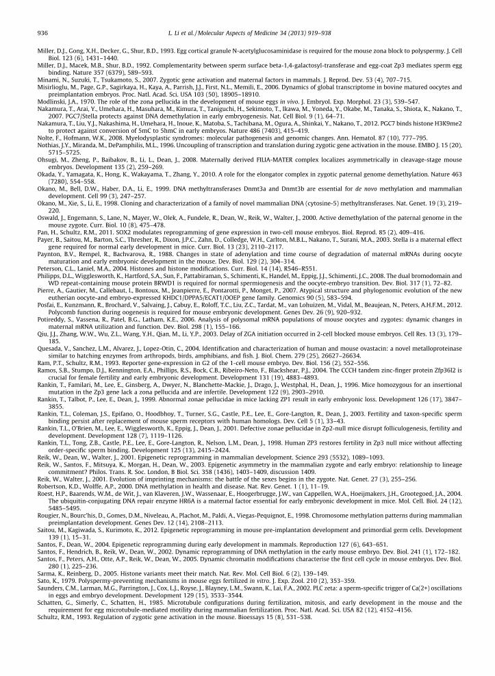
936 L. Li et al. / Molecular Aspects of Medicine 34 (2013) 919–938
Miller, D.J., Gong, X.H., Decker, G., Shur, B.D., 1993. Egg cortical granule N-acetylglucosaminidase is required for the mouse zona block to polyspermy. J. CellBiol. 123 (6), 1431–1440.
Miller, D.J., Macek, M.B., Shur, B.D., 1992. Complementarity between sperm surface beta-1,4-galactosyl-transferase and egg-coat Zp3 mediates sperm eggbinding. Nature 357 (6379), 589–593.
Minami, N., Suzuki, T., Tsukamoto, S., 2007. Zygotic gene activation and maternal factors in mammals. J. Reprod. Dev. 53 (4), 707–715.Misirlioglu, M., Page, G.P., Sagirkaya, H., Kaya, A., Parrish, J.J., First, N.L., Memili, E., 2006. Dynamics of global transcriptome in bovine matured oocytes and
preimplantation embryos. Proc. Natl. Acad. Sci. USA 103 (50), 18905–18910.Modlinski, J.A., 1970. The role of the zona pellucida in the development of mouse eggs in vivo. J. Embryol. Exp. Morphol. 23 (3), 539–547.Nakamura, T., Arai, Y., Umehara, H., Masuhara, M., Kimura, T., Taniguchi, H., Sekimoto, T., Ikawa, M., Yoneda, Y., Okabe, M., Tanaka, S., Shiota, K., Nakano, T.,
2007. PGC7/Stella protects against DNA demethylation in early embryogenesis. Nat. Cell Biol. 9 (1), 64–71.Nakamura, T., Liu, Y.J., Nakashima, H., Umehara, H., Inoue, K., Matoba, S., Tachibana, M., Ogura, A., Shinkai, Y., Nakano, T., 2012. PGC7 binds histone H3K9me2
to protect against conversion of 5mC to 5hmC in early embryos. Nature 486 (7403), 415–419.Nolte, F., Hofmann, W.K., 2008. Myelodysplastic syndromes: molecular pathogenesis and genomic changes. Ann. Hematol. 87 (10), 777–795.Nothias, J.Y., Miranda, M., DePamphilis, M.L., 1996. Uncoupling of transcription and translation during zygotic gene activation in the mouse. EMBO J. 15 (20),
5715–5725.Ohsugi, M., Zheng, P., Baibakov, B., Li, L., Dean, J., 2008. Maternally derived FILIA-MATER complex localizes asymmetrically in cleavage-stage mouse
embryos. Development 135 (2), 259–269.Okada, Y., Yamagata, K., Hong, K., Wakayama, T., Zhang, Y., 2010. A role for the elongator complex in zygotic paternal genome demethylation. Nature 463
(7280), 554–558.Okano, M., Bell, D.W., Haber, D.A., Li, E., 1999. DNA methyltransferases Dnmt3a and Dnmt3b are essential for de novo methylation and mammalian
development. Cell 99 (3), 247–257.Okano, M., Xie, S., Li, E., 1998. Cloning and characterization of a family of novel mammalian DNA (cytosine-5) methyltransferases. Nat. Genet. 19 (3), 219–
220.Oswald, J., Engemann, S., Lane, N., Mayer, W., Olek, A., Fundele, R., Dean, W., Reik, W., Walter, J., 2000. Active demethylation of the paternal genome in the
mouse zygote. Curr. Biol. 10 (8), 475–478.Pan, H., Schultz, R.M., 2011. SOX2 modulates reprogramming of gene expression in two-cell mouse embryos. Biol. Reprod. 85 (2), 409–416.Payer, B., Saitou, M., Barton, S.C., Thresher, R., Dixon, J.P.C., Zahn, D., Colledge, W.H., Carlton, M.B.L., Nakano, T., Surani, M.A., 2003. Stella is a maternal effect
gene required for normal early development in mice. Curr. Biol. 13 (23), 2110–2117.Paynton, B.V., Rempel, R., Bachvarova, R., 1988. Changes in state of adenylation and time course of degradation of maternal mRNAs during oocyte
maturation and early embryonic development in the mouse. Dev. Biol. 129 (2), 304–314.Peterson, C.L., Laniel, M.A., 2004. Histones and histone modifications. Curr. Biol. 14 (14), R546–R551.Philipps, D.L., Wigglesworth, K., Hartford, S.A., Sun, F., Pattabiraman, S., Schimenti, K., Handel, M., Eppig, J.J., Schimenti, J.C., 2008. The dual bromodomain and
WD repeat-containing mouse protein BRWD1 is required for normal spermiogenesis and the oocyte-embryo transition. Dev. Biol. 317 (1), 72–82.Pierre, A., Gautier, M., Callebaut, I., Bontoux, M., Jeanpierre, E., Pontarotti, P., Monget, P., 2007. Atypical structure and phylogenomic evolution of the new
eutherian oocyte-and embryo-expressed KHDC1/DPPA5/ECAT1/OOEP gene family. Genomics 90 (5), 583–594.Posfai, E., Kunzmann, R., Brochard, V., Salvaing, J., Cabuy, E., Roloff, T.C., Liu, Z.C., Tardat, M., van Lohuizen, M., Vidal, M., Beaujean, N., Peters, A.H.F.M., 2012.
Polycomb function during oogenesis is required for mouse embryonic development. Genes Dev. 26 (9), 920–932.Potireddy, S., Vassena, R., Patel, B.G., Latham, K.E., 2006. Analysis of polysomal mRNA populations of mouse oocytes and zygotes: dynamic changes in
maternal mRNA utilization and function. Dev. Biol. 298 (1), 155–166.Qiu, J.J., Zhang, W.W., Wu, Z.L., Wang, Y.H., Qian, M., Li, Y.P., 2003. Delay of ZGA initiation occurred in 2-cell blocked mouse embryos. Cell Res. 13 (3), 179–
185.Quesada, V., Sanchez, L.M., Alvarez, J., Lopez-Otin, C., 2004. Identification and characterization of human and mouse ovastacin: a novel metalloproteinase
similar to hatching enzymes from arthropods, birds, amphibians, and fish. J. Biol. Chem. 279 (25), 26627–26634.Ram, P.T., Schultz, R.M., 1993. Reporter gene-expression in G2 of the 1-cell mouse embryo. Dev. Biol. 156 (2), 552–556.Ramos, S.B., Stumpo, D.J., Kennington, E.A., Phillips, R.S., Bock, C.B., Ribeiro-Neto, F., Blackshear, P.J., 2004. The CCCH tandem zinc-finger protein Zfp36l2 is
crucial for female fertility and early embryonic development. Development 131 (19), 4883–4893.Rankin, T., Familari, M., Lee, E., Ginsberg, A., Dwyer, N., Blanchette-Mackie, J., Drago, J., Westphal, H., Dean, J., 1996. Mice homozygous for an insertional
mutation in the Zp3 gene lack a zona pellucida and are infertile. Development 122 (9), 2903–2910.Rankin, T., Talbot, P., Lee, E., Dean, J., 1999. Abnormal zonae pellucidae in mice lacking ZP1 result in early embryonic loss. Development 126 (17), 3847–
3855.Rankin, T.L., Coleman, J.S., Epifano, O., Hoodbhoy, T., Turner, S.G., Castle, P.E., Lee, E., Gore-Langton, R., Dean, J., 2003. Fertility and taxon-specific sperm
binding persist after replacement of mouse sperm receptors with human homologs. Dev. Cell 5 (1), 33–43.Rankin, T.L., O’Brien, M., Lee, E., Wigglesworth, K., Eppig, J., Dean, J., 2001. Defective zonae pellucidae in Zp2-null mice disrupt folliculogenesis, fertility and
development. Development 128 (7), 1119–1126.Rankin, T.L., Tong, Z.B., Castle, P.E., Lee, E., Gore-Langton, R., Nelson, L.M., Dean, J., 1998. Human ZP3 restores fertility in Zp3 null mice without affecting
order-specific sperm binding. Development 125 (13), 2415–2424.Reik, W., Dean, W., Walter, J., 2001. Epigenetic reprogramming in mammalian development. Science 293 (5532), 1089–1093.Reik, W., Santos, F., Mitsuya, K., Morgan, H., Dean, W., 2003. Epigenetic asymmetry in the mammalian zygote and early embryo: relationship to lineage
commitment? Philos. Trans. R. Soc. London, B Biol. Sci. 358 (1436), 1403–1409, discussion 1409.Reik, W., Walter, J., 2001. Evolution of imprinting mechanisms: the battle of the sexes begins in the zygote. Nat. Genet. 27 (3), 255–256.Robertson, K.D., Wolffe, A.P., 2000. DNA methylation in health and disease. Nat. Rev. Genet. 1 (1), 11–19.Roest, H.P., Baarends, W.M., de Wit, J., van Klaveren, J.W., Wassenaar, E., Hoogerbrugge, J.W., van Cappellen, W.A., Hoeijmakers, J.H., Grootegoed, J.A., 2004.
The ubiquitin-conjugating DNA repair enzyme HR6A is a maternal factor essential for early embryonic development in mice. Mol. Cell. Biol. 24 (12),5485–5495.
Rougier, N., Bourc’his, D., Gomes, D.M., Niveleau, A., Plachot, M., Paldi, A., Viegas-Pequignot, E., 1998. Chromosome methylation patterns during mammalianpreimplantation development. Genes Dev. 12 (14), 2108–2113.
Saitou, M., Kagiwada, S., Kurimoto, K., 2012. Epigenetic reprogramming in mouse pre-implantation development and primordial germ cells. Development139 (1), 15–31.
Santos, F., Dean, W., 2004. Epigenetic reprogramming during early development in mammals. Reproduction 127 (6), 643–651.Santos, F., Hendrich, B., Reik, W., Dean, W., 2002. Dynamic reprogramming of DNA methylation in the early mouse embryo. Dev. Biol. 241 (1), 172–182.Santos, F., Peters, A.H., Otte, A.P., Reik, W., Dean, W., 2005. Dynamic chromatin modifications characterise the first cell cycle in mouse embryos. Dev. Biol.
280 (1), 225–236.Sarma, K., Reinberg, D., 2005. Histone variants meet their match. Nat. Rev. Mol. Cell Biol. 6 (2), 139–149.Sato, K., 1979. Polyspermy-preventing mechanisms in mouse eggs fertilized in vitro. J. Exp. Zool. 210 (2), 353–359.Saunders, C.M., Larman, M.G., Parrington, J., Cox, L.J., Royse, J., Blayney, L.M., Swann, K., Lai, F.A., 2002. PLC zeta: a sperm-specific trigger of Ca(2+) oscillations
in eggs and embryo development. Development 129 (15), 3533–3544.Schatten, G., Simerly, C., Schatten, H., 1985. Microtubule configurations during fertilization, mitosis, and early development in the mouse and the
requirement for egg microtubule-mediated motility during mammalian fertilization. Proc. Natl. Acad. Sci. USA 82 (12), 4152–4156.Schultz, R.M., 1993. Regulation of zygotic gene activation in the mouse. Bioessays 15 (8), 531–538.

L. Li et al. / Molecular Aspects of Medicine 34 (2013) 919–938 937
Schultz, R.M., 2002. The molecular foundations of the maternal to zygotic transition in the preimplantation embryo. Hum. Reprod. Update 8 (4), 323–331.Shitara, H., Hayashi, J.I., Takahama, S., Kaneda, H., Yonekawa, H., 1998. Maternal inheritance of mouse mtDNA in interspecific hybrids: segregation of the
leaked paternal mtDNA followed by the prevention of subsequent paternal leakage. Genetics 148 (2), 851–857.Sirard, M.A., Dufort, I., Vallee, M., Massicotte, L., Gravel, C., Reghenas, H., Watson, A.J., King, W.A., Robert, C., 2005. Potential and limitations of bovine-specific
arrays for the analysis of mRNA levels in early development: preliminary analysis using a bovine embryonic array. Reprod. Fertil. Dev. 17 (1–2), 47–57.Smith, Z.D., Chan, M.M., Mikkelsen, T.S., Gu, H.C., Gnirke, A., Regev, A., Meissner, A., 2012. A unique regulatory phase of DNA methylation in the early
mammalian embryo. Nature 484 (7394), U339–374.Soyal, S.M., Amleh, A., Dean, J., 2000. FIGalpha, a germ cell-specific transcription factor required for ovarian follicle formation. Development 127 (21), 4645–
4654.Stancheva, I., El-Maarri, O., Walter, J., Niveleau, A., Meehan, R.R., 2002. DNA methylation at promoter regions regulates the timing of gene activation in
Xenopus laevis embryos. Dev. Biol. 243 (1), 155–165.Stewart-Savage, J., Bavister, B.D., 1988. A cell surface block to polyspermy occurs in golden hamster eggs. Dev. Biol. 128 (1), 150–157.Suetake, I., Shinozaki, F., Miyagawa, J., Takeshima, H., Tajima, S., 2004. DNMT3L stimulates the DNA methylation activity of Dnmt3a and Dnmt3b through a
direct interaction. J. Biol. Chem. 279 (26), 27816–27823.Suh, N., Baehner, L., Moltzahn, F., Melton, C., Shenoy, A., Chen, J., Blelloch, R., 2010. MicroRNA function is globally suppressed in mouse oocytes and early
embryos. Curr. Biol. 20 (3), 271–277.Summers, M.C., Bhatnagar, P.R., Lawitts, J.A., Biggers, J.D., 1995. Fertilization in vitro of mouse ova from inbred and outbred strains: complete
preimplantation embryo development in glucose-supplemented KSOM. Biol. Reprod. 53 (2), 431–437.Surani, M.A., Barton, S.C., Norris, M.L., 1984. Development of reconstituted mouse eggs suggests imprinting of the genome during gametogenesis. Nature
308 (5959), 548–550.Sutovsky, P., Schatten, G., 2000. Paternal contributions to the mammalian zygote: fertilization after sperm-egg fusion. Int. Rev. Cytol. 195, 1–65.Svoboda, P., Flemr, M., 2010. The role of miRNAs and endogenous siRNAs in maternal-to-zygotic reprogramming and the establishment of pluripotency.
EMBO Rep. 11 (8), 590–597.Tadros, W., Lipshitz, H.D., 2009. The maternal-to-zygotic transition: a play in two acts. Development 136 (18), 3033–3042.Tahiliani, M., Koh, K.P., Shen, Y., Pastor, W.A., Bandukwala, H., Brudno, Y., Agarwal, S., Iyer, L.M., Liu, D.R., Aravind, L., Rao, A., 2009. Conversion of 5-
methylcytosine to 5-hydroxymethylcytosine in mammalian DNA by MLL partner TET1. Science 324 (5929), 930–935.Tam, O.H., Aravin, A.A., Stein, P., Girard, A., Murchison, E.P., Cheloufi, S., Hodges, E., Anger, M., Sachidanandam, R., Schultz, R.M., Hannon, G.J., 2008.
Pseudogene-derived small interfering RNAs regulate gene expression in mouse oocytes. Nature 453 (7194), U534–U538.Tang, F., Kaneda, M., O’Carroll, D., Hajkova, P., Barton, S.C., Sun, Y.A., Lee, C., Tarakhovsky, A., Lao, K., Surani, M.A., 2007. Maternal microRNAs are essential for
mouse zygotic development. Genes Dev. 21 (6), 644–648.Thall, A.D., Maly, P., Lowe, J.B., 1995. Oocyte gal-alpha-1,3gal epitopes implicated in sperm adhesion to the zona-pellucida glycoprotein Zp3 are not required
for fertilization in the mouse. J. Biol. Chem. 270 (37), 21437–21440.Thompson, E.M., Legouy, E., Renard, J.P., 1998. Mouse embryos do not wait for the MBT: Chromatin and RNA polymerase remodeling in genome activation at
the onset of development. Dev. Genet. 22 (1), 31–42.Thorvaldsen, J.L., Duran, K.L., Bartolomei, M.S., 1998. Deletion of the H19 differentially methylated domain results in loss of imprinted expression of H19 and
Igf2. Genes Dev. 12 (23), 3693–3702.Tong, Z.B., Gold, L., Pfeifer, K.E., Dorward, H., Lee, E., Bondy, C.A., Dean, J., Nelson, L.M., 2000. Mater, a maternal effect gene required for early embryonic
development in mice. Nat. Genet. 26 (3), 267–268.Tong, Z.B., Nelson, L.M., 1999. A mouse gene encoding an oocyte antigen associated with autoimmune premature ovarian failure. Endocrinology 140 (8),
3720–3726.Torres-Padilla, M.E., Bannister, A.J., Hurd, P.J., Kouzarides, T., Zernicka-Goetz, M., 2006. Dynamic distribution of the replacement histone variant H3.3 in the
mouse oocyte and preimplantation embryos. Int. J. Dev. Biol. 50 (5), 455–461.Torres-Padilla, M.E., Zernicka-Goetz, M., 2006. Role of TIF1 alpha as a modulator of embryonic transcription in the mouse zygote. J. Cell Biol. 174 (3), 329–
338.Tsai, T.C., Lin, W., Yang, S.H., Cheng, W.T.K., Cheng, E.H., Lee, M.S., Chong, K.Y., Chen, C.M., 2010. Granzyme G is expressed in the two-cell stage mouse
embryo and is required for the maternal-zygotic transition. BMC Dev. Biol. 10, 88.Tsukamoto, S., Kuma, A., Mizushima, N., 2008. The role of autophagy during the oocyte-to-embryo transition. Autophagy 4 (8), 1076–1078.Turner, B.M., 2002. Cellular memory and the histone code. Cell 111 (3), 285–291.Vallee, M., Aiba, K., Piao, Y., Palin, M.F., Ko, M.S.H., Sirard, M.A., 2008. Comparative analysis of oocyte transcript profiles reveals a high degree of conservation
among species. Reproduction 135 (4), 439–448.van der Heijden, G.W., Dieker, J.W., Derijck, A.A.H.A., Muller, S., Berden, J.H.M., Braat, D.D.M., van der Vlag, J., de Boer, P., 2005. Asymmetry in histone H3
variants and lysine methylation between paternal and maternal chromatin of the early mouse zygote. Mech. Develop. 122 (9), 1008–1022.Veron, N., Peters, A.H., 2011. Epigenetics: Tet proteins in the limelight. Nature 473 (7347), 293–294.Verona, R.I., Mann, M.R., Bartolomei, M.S., 2003. Genomic imprinting: intricacies of epigenetic regulation in clusters. Annu. Rev. Cell Dev. Biol. 19, 237–259.Walter, J., Paulsen, M., 2003. Imprinting and disease. Semin. Cell Dev. Biol. 14 (1), 101–110.Wan, L.B., Pan, H., Hannenhalli, S., Cheng, Y., Ma, J., Fedoriw, A., Lobanenkov, V., Latham, K.E., Schultz, R.M., Bartolomei, M.S., 2008. Maternal depletion of
CTCF reveals multiple functions during oocyte and preimplantation embryo development. Development 135 (16), 2729–2738.Wang, J., Xu, M., Zhu, K., Li, L., Liu, X., 2012. The N-terminus of FILIA forms an atypical KH domain with a unique extension involved in interaction with RNA.
PLoS One 7 (1), e30209.Wang, Q.T., Piotrowska, K., Ciemerych, M.A., Milenkovic, L., Scott, M.P., Davis, R.W., Zernicka-Goetz, M., 2004. A genome-wide study of gene activity reveals
developmental signaling pathways in the preimplantation mouse embryo. Dev. Cell 6 (1), 133–144.Wang, Q.X., Latham, K.E., 1997. Requirement for protein synthesis during embryonic genome activation in mice. Mol. Reprod. Dev. 47 (3), 265–270.Wang, S.C., Frey, P.A., 2007. S-adenosylmethionine as an oxidant: the radical SAM superfamily. Trends Biochem. Sci. 32 (3), 101–110.Wang, S.F., Kou, Z.H., Jing, Z.Y., Zhang, Y., Guo, X.Z., Dong, M.Q., Wilmut, I., Gao, S.R., 2010. Proteome of mouse oocytes at different developmental stages.
Proc. Natl. Acad. Sci. USA 107 (41), 17639–17644.Warner, C.M., Versteegh, L.R., 1974. In vivo and in vitro effect of alpha-amanitin on preimplantation mouse embryo RNA polymerase. Nature 248 (450), 678–
680.Watanabe, T., Totoki, Y., Toyoda, A., Kaneda, M., Kuramochi-Miyagawa, S., Obata, Y., Chiba, H., Kohara, Y., Kono, T., Nakano, T., Surani, M.A., Sakaki, Y., Sasaki,
H., 2008. Endogenous siRNAs from naturally formed dsRNAs regulate transcripts in mouse oocytes. Nature 453 (7194), 539–543.Whitworth, K.M., Agca, C., Kim, J.G., Patel, R.V., Springer, G.K., Bivens, N.J., Forrester, L.J., Mathialagan, N., Green, J.A., Prather, R.S., 2005. Transcriptional
profiling of pig embryogenesis by using a 15-K member unigene set specific for pig reproductive tissues and embryos. Biol. Reprod. 72 (6), 1437–1451.Wiekowski, M., Miranda, M., Depamphilis, M.L., 1991. Regulation of gene-expression in preimplantation mouse embryos - effects of the zygotic clock and
the 1st mitosis on promoter and enhancer activities. Dev. Biol. 147 (2), 403–414.Wiekowski, M., Miranda, M., Depamphilis, M.L., 1993. Requirements for promoter activity in mouse oocytes and embryos distinguish paternal pronuclei
from maternal and zygotic nuclei. Dev. Biol. 159 (1), 366–378.Wilkins, J.F., Haig, D., 2002. Parental modifiers, antisense transcripts and loss of imprinting. Proc. Biol. Sci. 269 (1502), 1841–1846.Wilkins, J.F., Haig, D., 2003. What good is genomic imprinting: the function of parent-specific gene expression. Nat. Rev. Genet. 4 (5), 359–368.Williams, S.A., Xia, L., Cummings, R.D., McEver, R.P., Stanley, P., 2007. Fertilization in mouse does not require terminal galactose or N-acetylglucosamine on
the zona pellucida glycans. J. Cell Sci. 120 (Pt 8), 1341–1349.

938 L. Li et al. / Molecular Aspects of Medicine 34 (2013) 919–938
Wolffe, A.P., 1991. Implications of DNA replication for eukaryotic gene expression. J. Cell Sci. 99 (Pt 2), 201–206.Wossidlo, M., Nakamura, T., Lepikhov, K., Marques, C.J., Zakhartchenko, V., Boiani, M., Arand, J., Nakano, T., Reik, W., Walter, J., 2011. 5-
Hydroxymethylcytosine in the mammalian zygote is linked with epigenetic reprogramming. Nat. Commun. 2, 241.Wu, S.C., Zhang, Y., 2010. Active DNA demethylation: many roads lead to rome. Nat. Rev. Mol. Cell Biol. 11 (9), 607–620.Wu, X.M., Viveiros, M.M., Eppig, J.J., Bai, Y.C., Fitzpatrick, S.L., Matzuk, M.M., 2003. Zygote arrest 1 (Zar1) is a novel maternal-effect gene critical for the
oocyte-to-embryo transition. Nat. Genet. 33 (2), 187–191.Xu, Y., Wu, F., Tan, L., Kong, L., Xiong, L., Deng, J., Barbera, A.J., Zheng, L., Zhang, H., Huang, S., Min, J., Nicholson, T., Chen, T., Xu, G., Shi, Y., Zhang, K., Shi, Y.G.,
2011. Genome-wide regulation of 5hmC, 5mC, and gene expression by Tet1 hydroxylase in mouse embryonic stem cells. Mol. Cell 42 (4), 451–464.Yauger, B., Boggs, N.A., Dean, J., 2011. Human ZP4 is not sufficient for taxon-specific sperm recognition of the zona pellucida in transgenic mice.
Reproduction 141 (3), 313–319.Yoder, J.A., Walsh, C.P., Bestor, T.H., 1997. Cytosine methylation and the ecology of intragenomic parasites. Trends Genet. 13 (8), 335–340.Yurttas, P., Morency, E., Coonrod, S.A., 2010. Use of proteomics to identify highly abundant maternal factors that drive the egg-to-embryo transition.
Reproduction 139 (5), 809–823.Yurttas, P., Vitale, A.M., Fitzhenry, R.J., Cohen-Gould, L., Wu, W., Gossen, J.A., Coonrod, S.A., 2008. Role for PADI6 and the cytoplasmic lattices in ribosomal
storage in oocytes and translational control in the early mouse embryo. Development 135 (15), 2627–2636.Zeng, F., Baldwin, D.A., Schultz, R.M., 2004. Transcript profiling during preimplantation mouse development. Dev. Biol. 272 (2), 483–496.Zeng, F., Schultz, R.M., 2003. Gene expression in mouse oocytes and preimplantation embryos: use of suppression subtractive hybridization to identify
oocyte- and embryo-specific genes. Biol. Reprod. 68 (1), 31–39.Zeng, F.Y., Schultz, R.M., 2005. RNA transcript profiling during zygotic gene activation in the preimplantation mouse embryo. Dev. Biol. 283 (1), 40–57.Zhang, J., Zheng, N., Zhou, P., 2003. Exploring the functional complexity of cellular proteins by protein knockout. Proc. Natl. Acad. Sci. USA 100 (24), 14127–
14132.Zhang, P., Ni, X., Guo, Y., Guo, X., Wang, Y., Zhou, Z., Huo, R., Sha, J., 2009. Proteomic-based identification of maternal proteins in mature mouse oocytes. BMC
Genom. 10, 348.Zheng, P., Dean, J., 2009. Role of Filia, a maternal effect gene, in maintaining euploidy during cleavage-stage mouse embryogenesis. Proc. Natl. Acad. Sci. USA
106 (18), 7473–7478.Zimmermann, J.W., Schultz, R.M., 1994. Analysis of gene expression in the preimplantation mouse embryo: use of mRNA differential display. Proc. Natl.
Acad. Sci. USA 91 (12), 5456–5460.