etiology, differential diagnosis, and treatment of rotator
TRANSCRIPT
Anjali Paintal
1
Etiology, Differential Diagnosis, and Treatment of Rotator Cuff Tears
Introduction
The rotator cuff consists of four muscles: supraspinatus, infraspinatus, teres minor, and
subscapularis (see appendix A, figure 1).1 These muscles originate from the scapula and insert
onto the humerus to provide the glenohumeral joint with stability.1 The rotator cuff muscles
collectively prevent the humeral head from gliding superiorly during shoulder abduction,
allowing for improved joint alignment and range of motion during deltoid activation.1 These
muscles act by facilitating shoulder abduction, internal rotation, external rotation, and dynamic
stability. The subscapularis is an internal rotator and aids in anterior stability of the glenohumeral
joint. The supraspinatus is primarily an abductor, but also facilitates external rotation. The
infraspinatus and teres minor are primarily external rotators, as well.
Unfortunately, these muscles and associated tendons are often subject to many damaging
forces and injuries. The supraspinatus tendon is particularly susceptible to damage and is the
most commonly torn muscle in the group.2 For this reason, it is critically important to understand
the architecture of this muscle. The supraspinatus originates from the supraspinous fossa,
superior to the scapular spine, and passes through the subacromial space to insert onto the
superior facet of the greater tubercle.2 The subacromial bursa lies superiorly to the supraspinatus
muscle and plays an important role in protecting the musculotendinous unit from excessive
forces caused by the humeral head and the acromion (see appendix A, figure 2).3 Interestingly,
the supraspinatus is often described as having two muscle bellies – anterior and posterior.2 The
tendon of the posterior belly is flat and wide in structure while the anterior belly is thicker and
more tubular.2 In comparison to the posterior belly, the anterior belly distributes force through a
smaller cross-sectional area. Additionally, it was found that the anterior supraspinatus tendon can
Anjali Paintal
2
be subject to 288% more stress than the posterior tendon.2 The anterior supraspinatus tendon,
because of its position and structure, is subject to particularly high compressive, tensile, and
shearing forces, making it more likely to sustain an injury.2,4 Moreover, the supraspinatus tendon
is critically hypovascular where it traverses the subacromial space, resulting in increased
susceptibility to damage, degeneration, and delayed healing.4
Prevalence
Rotator cuff injuries are considered the most common tendon injuries in adults.5 Overall,
consensus is lacking regarding the true prevalence of rotator cuff tears. The occurrence of full
thickness rotator cuff tears is estimated to be around 22.1% in the general population, but
increases with age.6 Evidence suggests that the prevalence of rotator cuff tears range from
around 9.7% in patients under 20 years old to approximately 65% in adults over 70.7,8
Asymptomatic tears are more common than symptomatic tears; approximately 34.7% of tears are
symptomatic while 65.3% are asymptomatic.6 A study by Yamaguchi et al. analyzed 588 patients
with unilateral shoulder pain and found that 33.7% of these patients had a unilateral rotator cuff
tear, 30.1% had bilateral rotator cuff tears, and 36.1% had a bilaterally intact rotator cuff.9 This
study demonstrated that bilateral rotator cuff tears are relatively common in individuals
complaining of unilateral symptoms.9
Etiology and Risk Factors
Rotator cuff tears usually have a multifactorial etiology, including both intrinsic and
extrinsic factors.10 Neer describes one main extrinsic factor contributing to rotator cuff tears—
chronic impingement syndrome.10,11 Impingement of the supraspinatus tendon occurs by the

Anjali Paintal
3
coraco-acromial ligament and the anterior undersurface of the acromion.10,11 Bigliani et al. first
proposed that there are three different acromial shapes, described as type I, II, and II. Type I is a
flat acromion, type II is curved, and type III is hooked. It was found that type III hooked
acromions lead to rotator cuff tears in 70% of cases.10,11 Refuting this claim, a 2013 Balke et al.
study found that acromion type as described by Bigliani was not significantly associated with
rotator cuff injury, but a “lower lateral acromial angle and a large lateral extension of the
acromion” significantly correlated with a higher prevalence of rotator cuff tears and
impingement.12 This study found that 100% of individuals with an acromial slope of more than
43° and a lateral acromial angle of less than 70° had a rotator cuff tear.12 The Balke et al. study
suggests that acromial slope, acromial tilt, lateral acromial angle, and acromion index can be
used to assess risk for impingement and rotator cuff tears, and may be superior than Bigliani’s
acromion types for assessing risk.12
Other extrinsic factors contributing to rotator cuff tears include mechanical overuse,
repetitive microtrauma, traumatic shoulder injury, shoulder dislocations, and greater tuberosity
fractures.10 The most common cause of traumatic rotator cuff injury is from falling on an
outstretched arm, and they are more likely to occur when the arm is in an abducted and
externally rotated position.13 A 2013 systematic review found that in traumatic rotator cuff tears,
there is supraspinatus involvement in 84% of tears, subscapularis involvement in 78%, and
infraspinatus involvement in 39%.13 Unlike degenerative rotator cuff tears, traumatic tears
typically involve the subscapularis.13
Repetitive microtrauma can often occur due to daily tasks and occupational related risk
factors. Manual handling; including heavy lifting, pushing, pulling, holding, carrying; above
shoulder-height activities; repetitive work; vibration; and work that requires awkward postures
Anjali Paintal
4
all contribute to increased risk.14 An accumulation of multiple workplace exposures further
increases the risk.14 A 2008 Silverstein et al. study analyzed 733 workers in 12 different
worksites and found that 7.5% of them had a rotator cuff tear. Rotator cuff tears were
significantly associated with low job security, high job structural constraints, long duration of
shoulder flexion, forceful exertion (especially forceful pinch).15 Although most rotator cuff
injuries occur in adults above 40, young overhead sport athletes may also sustain rotator cuff
injuries due to overuse.16 The shoulder joint can experience physiological loads of up to 108% of
body weight and angular velocities of around 7,000 degrees during overhead throwing
activities.16 Additionally, the critical area of hypovascularity along the supraspinatus tendon is
particularly stressed during acceleration and deceleration activities associated with overhead
throwing, leading to repetitive tendon damage.16
Other significant risk factors for rotator cuff tears include smoking, diabetes,
hypercholesterolemia, and use of nonsteroidal anti-inflammatory drug (NSAID).8 Smoking slows
tendon healing and has been found to contribute to musculoskeletal pain and dysfunction.17 A
2010 study by Baumgarten et al. found that a history of smoking, increased duration of smoking,
increased mean packs per day, and increased mean pack-years of smoking are significantly
associated with increased risk of rotator cuff tears.17 Expanding upon the Baumgarten et al.’s
findings, a large systematic review by Bishop et al. found that smoking accelerates rotator cuff
degeneration and causes larger rotator cuff tears.18 Although many rotator cuff injuries are
asymptomatic, smoking leads to an increase in symptomatic tears, often times requiring surgical
intervention.18 Nicotine, found in cigarettes, is associated with fibroblast degeneration, irregular
fibril organization, and decreased tensile strength in tendon tissue.19,20 Additionally, smoking
results in more severe degenerative changes in the supraspinatus tendons earlier on in life,
Anjali Paintal
5
increased apoptosis, delayed tendon bone healing, and chronic inflammation.21,22 Overall,
smoking threatens the integrity of the rotator cuff through several different pathways and is a
significant modifiable risk factor associated with rotator cuff tears.
Evidence suggests that diabetes contributes to rotator cuff pathology. Possible
mechanisms behind this include impaired circulation as well as non-enzymatic glycosylation
which results in glycosylation end-products (AGEs).23 Accumulation of AGEs around the joint
causes an increase in tendon crosslinking, making the tissue stiffer and weaker.23 Diabetes
contributes to tissue hypoxia, free radical damage, and apoptosis through impaired circulation.23
Diabetes also increases the risk of requiring surgical repair and typically leads to poorer tendon-
bone healing post-surgery.24,25 Managing blood glucose is important to support the overall health
of the rotator cuff.
Evidence is mixed regarding the use of NSAIDs and rotator cuff pathology.26 Current
evidence supports that NSAID use can impair tendon-to-bone healing, which is an important
consideration to note for post rotator cuff repair treatment.26 It is suggested that the mechanism
behind this is related to NSAIDs’ role in blocking the production of prostaglandins, thus
decreasing inflammation, but leading to increased arachidonic acid which results in tissue
damage. 27 A 2014 study by Chechik et al. studied the effects of NSAIDs (meloxicam) on the
healing of RC tendons post-surgery and found that meloxicam use decreased that strength of the
repaired rotator cuff when it was given between 11 and 20 days after surgical repair, but not
when it was given within the first 10 days or not used at all.28 Although NSAIDs have been
shown to cause negative effects on rotator cuff healing, they also have been shown to decrease
tendon adhesion formation and pain, thus facilitating increased range of motion.26 The risks of
NSAID use should be carefully weighed against the potential benefits. Overall, caution should be

Anjali Paintal
6
used when prescribing NSAIDs to patients with rotator cuff pathology (especially when they
have multiple risk factors), since a growing body of evidence suggests that they may lead to a
decreased healing response.
Hypercholesterolemia is yet another risk factor that has been associated with increased
incidence of rotator cuff tears.29 Research has shown that hypercholesterolemia leads to reduced
tendon stiffness, decreased elasticity, and decreased maximum stress.30 The potential mechanism
behind the association of hypercholesterolemia and rotator cuff pathology is related to the effects
of increased fatty infiltrates within the tendon, resulting in suboptimal tendon composition.31
Additionally, patients with dyslipidemia tend to experience more pain after treatment for rotator
cuff tendinopathy.31 Increased age is associated with increased prevalence of many risk factors
associated with rotator cuff injuries such as hypercholesterolemia, diabetes, accumulated
microtraumas, oxidative stress, decreased vascularity, etc.8 Individuals over 60 years old are
approximately two times more likely to sustain a rotator cuff tear compared to their younger
counterparts and are significantly more likely to experience a massive tear.8,32
Differential Diagnosis and Clinical Exam
Symptoms of rotator cuff pathology vary among individuals and may include pain along
the anterolateral shoulder margin and lateral surface of the arm, pain upon abduction, pain when
sleeping on the injured shoulder, decreased range of motion, and muscle weakness.8 Because the
glenohumeral joint has many different components to it, the symptoms associated with rotator
cuff tears may present similar to other shoulder pathologies. For this reason, it is imperative to
have an extensive differential diagnosis list when assessing a patient with a primary complaint of
shoulder pain. The differential diagnosis list should include labral tears, ligament tears or sprains,

Anjali Paintal
7
coracoacromial and acromioclavicular ligament injuries, biceps tendon pathology, osteoarthritis,
adhesive capsulitis, neuropathies, cervical radiculopathy, among other potential pathologies.33
Magnetic resonance imaging (MRI) and ultrasonography (US) remain the primary
noninvasive methods to diagnose a rotator cuff tear, while intraoperative findings remain the
gold standard.34 US was found to have a sensitivity of 0.88 and specificity of 0.89, while MRI
was found to have a sensitivity of 0.91 and specificity of 0.84.34 With MRI and US
demonstrating comparable accuracy, US may be preferred by many patients and clinicians since
it is generally better tolerated by patients, less costly, and does not involve radiation exposure.34
In addition to imaging, a physical therapy clinical exam is warranted to address potential
shoulder pathology and detect rotator cuff tears. A comprehensive clinical exam should start with
a through subjective exam followed by inspection and palpation of the rotator cuff.33 Clinicians
should utilize observation and palpation to detect potential atrophy along the suprascapular and
infrascapular fossae, presenting as loss of muscle bulk on one side compared to the other.33 The
long head of the biceps as well as the acromioclavicular joint should also be palpated to help rule
out other potential pathologies.33 After passive and active ROM has been measured and manual
muscle strength has been assessed, special tests can be used to help rule in or out a rotator cuff
tear.33
Although shoulder special tests are a widely covered topic in literature, there is
heterogenous and inconclusive evidence regarding their diagnostic accuracy. A 2013 article by
Jain et al. describes select special tests that have “been more rigorously assessed for sensitivity
and specificity.”33 The special tests for the subscapularis include the life-off test and lag sign
(sensitivity: 17-100; specificity: 60-98), belly press test (sensitivity: 40-43; specificity: 93-98),
belly-off sign (sensitivity: 14-86; specificity: 91-95), and bear hug test (sensitivity:60;

Anjali Paintal
8
specificity: 92).33 Jain et al.’s chosen special tests for the supraspinatus and infraspinatus include
the external rotation lag sign (sensitivity: 48-98; specificity: 72-98), Jobe’s test (sensitivity: 53-
89; specificity: 65-82), and the drop arm test (sensitivity: 10-73; specificity: 77-98).33 To test the
teres minor and biceps tendon, the Hornblower’s sign (sensitivity:100; specificity 93) and
speed’s test (sensitivity: 53; specificity: 67) were selected.33 For impingement signs Jain et al.
recommend the Neer’s sign (sensitivity: 68-89; specificity: 48-98) and Hawkin’s sign
(sensitivity: 72-92; specificity: 44-78).33
Biederwolf conducted a systematic review of literature regarding the statistical and
clinical utility of shoulder special tests.34 The results from the review indicate that the external
rotation lag sign, the dropping sign, the Hornblower’s sign, and the internal rotation lag sign
demonstrated the best statistical utility, with post-test probabilities of 88.8%, 100%, 87.7%, and
92.4% respectively.34 Interestingly, the review found that special tests that do not consistently
demonstrate statistical utility for rotator cuff tears include the empty can test, the full can test, the
Neer test, the Hawkins-Kennedy, the Rent test, the Gilcrest palm-up test, drop sign in 90 degrees
abduction in scapular plane and 90 degrees of external rotation, the lift-off test, the belly-off test,
the Napolean test, the bear hug test, the supine impingement sign, the infraspinatus muscle test,
the painful arc sign, among others.34
Yet another more recent 2017 article by Jain et al., including 208 patients with shoulder
pain, analyzed the diagnostic accuracy of 15 different shoulder special tests. The authors found
that for supraspinatus tears, the Jobe’s test was 88% sensitive, 62% specific, and had a likelihood
ratio of 2.30, while the full can test had a sensitivity of 70% and specificity of 81%.35 For
infraspinatus tears, the external rotation lag sign at 0° fared the best with a specificity of 98% and
likelihood ratio of 6.06, followed by the Hornblowers sign which had a specificity of 96% and
Anjali Paintal
9
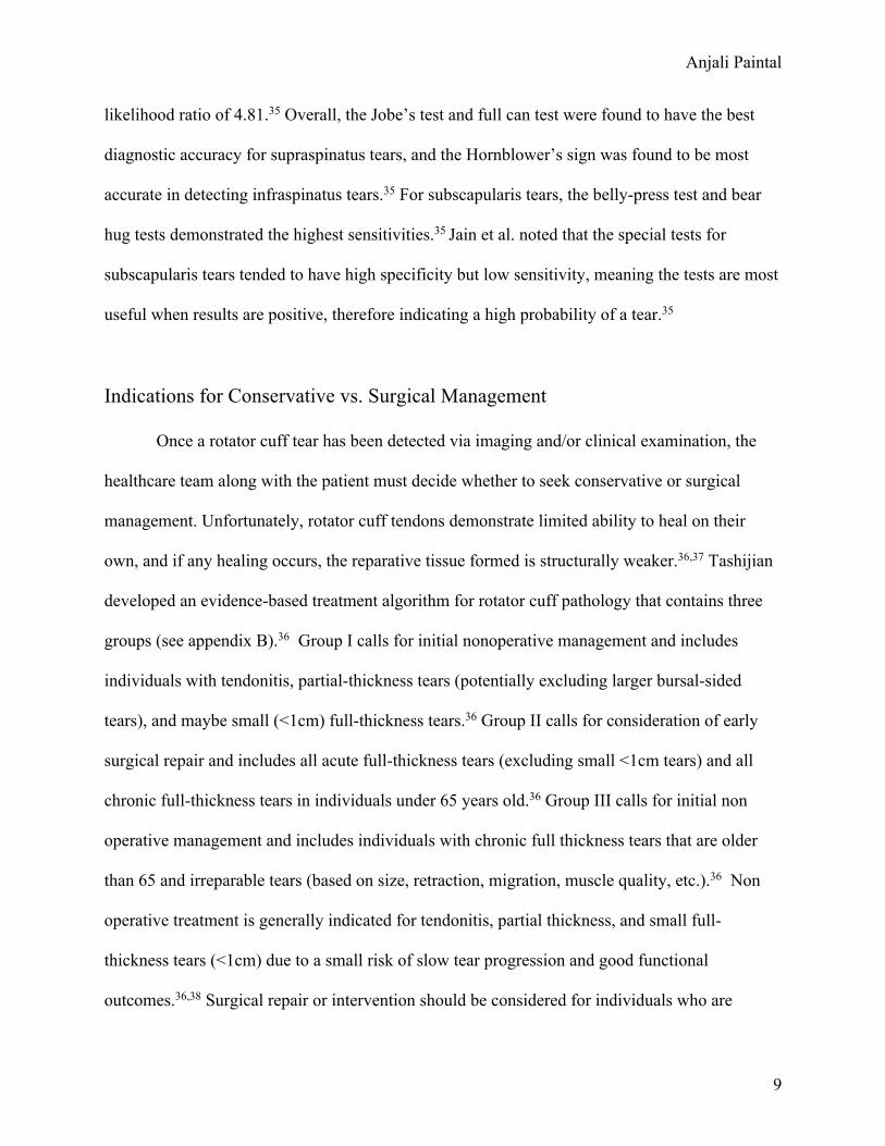
likelihood ratio of 4.81.35 Overall, the Jobe’s test and full can test were found to have the best
diagnostic accuracy for supraspinatus tears, and the Hornblower’s sign was found to be most
accurate in detecting infraspinatus tears.35 For subscapularis tears, the belly-press test and bear
hug tests demonstrated the highest sensitivities.35 Jain et al. noted that the special tests for
subscapularis tears tended to have high specificity but low sensitivity, meaning the tests are most
useful when results are positive, therefore indicating a high probability of a tear.35
Indications for Conservative vs. Surgical Management
Once a rotator cuff tear has been detected via imaging and/or clinical examination, the
healthcare team along with the patient must decide whether to seek conservative or surgical
management. Unfortunately, rotator cuff tendons demonstrate limited ability to heal on their
own, and if any healing occurs, the reparative tissue formed is structurally weaker.36,37 Tashijian
developed an evidence-based treatment algorithm for rotator cuff pathology that contains three
groups (see appendix B).36 Group I calls for initial nonoperative management and includes
individuals with tendonitis, partial-thickness tears (potentially excluding larger bursal-sided
tears), and maybe small (<1cm) full-thickness tears.36 Group II calls for consideration of early
surgical repair and includes all acute full-thickness tears (excluding small <1cm tears) and all
chronic full-thickness tears in individuals under 65 years old.36 Group III calls for initial non
operative management and includes individuals with chronic full thickness tears that are older
than 65 and irreparable tears (based on size, retraction, migration, muscle quality, etc.).36 Non
operative treatment is generally indicated for tendonitis, partial thickness, and small full-
thickness tears (<1cm) due to a small risk of slow tear progression and good functional
outcomes.36,38 Surgical repair or intervention should be considered for individuals who are

Anjali Paintal
10
younger than 65 and have full thickness tears (>1-1.5cm).36 Evidence suggests that these
individuals are highly likely to experience negative effects from conservative treatment alone
and surgical intervention tends to yield positive results if performed within 4 months of
injury.36,39 Individuals less than 65 years of age with significant rotator cuff tears may experience
tear progression, fatty infiltrates of the muscle, and tendon retraction if they do not attain prompt
surgical intervention.36,40 For individuals over 65, most injuries are irreparable, thus physical
therapy management is recommended over surgical intervention. Evidence suggests that only
about 43% of patients above 65 demonstrate evidence of healing after arthroscopic repair at 18
months postop. Although patients may not achieve rotator cuff healing from arthroscopic repair,
surgery can still be considered as a last treatment option to decrease symptoms.36
Surgical Intervention
The majority of surgeons perform rotator cuff repairs arthroscopically, with 90% of
patients reporting satisfaction at 6 months post operation.41 Three different operative techniques
were compared in a study by Millar et al.: an open technique, arthroscopic knotted, and
arthroscopic knotless.42 The study found that arthroscopic groups had a 20% better American
Shoulder and Elbow Surgeons score at 6 months and retears were more frequent in the open
repairs group (39%) compared to arthroscopic knotted (25%) and arthroscopic knotless (16%)
groups.42 Arthroscopic techniques generally demonstrate better clinical outcomes with greater
patient satisfaction, fewer retear rates, less postoperative pain, decreased risk of adhesions, and a
potentially more rapid recovery.42 Other surgical techniques include transosseous equivalent
suture anchor repair, single row, and double row techniques.43 Although these three techniques
have overall demonstrated similar clinical results, it is important to note that the single row

Anjali Paintal
11
technique demonstrated higher reprepture rates after two years, the transosseous equivalent
suture anchor repair demonstrated the smallest rerupture rate, and the double row and
transosseous equivalent lead to improved structural healing.43 Evidence suggests that the single
row technique may appropriate for small to medium size tears while the double row and
transosseous equivalent suture may fare better for larger tears.43
The main goals of surgical repair include the following: repair of the deltoid origin,
subacromial decompression, release of the rotator cuff to facilitate freely moving
musculotendinous units, a secure repair of tendon (via sutures and anchors or by transosseous
technique), and a supervised rehabilitation program.41 There is debate whether acromioplasty is
beneficial in all cases.43 Those in favor propose that acromioplasty prevents further compression
and allows for greater healing due to the bleeding of the bone in the subacromial space.43 Those
against acromioplasty propose that it can lead to further superior migration of the humeral head
due to removal of the coracoacromial ligament.43 It is generally accepted that acromioplasty is
indicated to maintain the integrity of the rotator cuff repair when a patient presents with a type 3
acromion, excessively sloped acromion angle, or a downward projecting spur.43
Outcomes Post-Surgical Intervention
A study by Cho and Rhee found that 77.5% of patients completely healed from the
arthroscopic rotator cuff repair while 22.5% experienced recurrent tears.44 Upon two year follow
up, there was significant pain relief and improvements in function.44 Many of the patient factors
that contribute to increase risk of rotator cuff tears, also lead to poorer post-surgical outcomes.
These factors include smoking, diabetes, osteoporosis, age, size of tear, and presence of fatty
degeneration in the rotator cuff muscle.8,44 The same study by Choo and Rhee found that groups
Anjali Paintal
12
less than 50 years of age, over 51 but less than 60, and over 61 had different rates of complete
healing; 87.8%, 79.4%, and 65.4% respectively.44 Additionally, the authors found that tears with
substantial fatty degeneration had recurrent tears.44 Another study by Rossi et al. analyzed long
term outcomes after arthroscopic rotator cuff repairs in 62 adults between ages 32 and 67, with a
mean follow-up duration of 10.4 years.45 This study found that 87% returned to sport and 80%
were able to return to the same preinjury level of activity.45 Overall, rotator cuff repairs tend to
have high patient satisfaction and lead to improved functional outcomes.
Post-Operative Rehabilitation
The primary goal of postoperative rehabilitation is to restore function and maintain repair
integrity.8 Although consensus is lacking regarding rehabilitation protocols following rotator cuff
repair surgery, rehabilitation protocols generally break the process down into 4 phases (as
described by Jung et al., 2018): phase 1 lasts from day 1 up to 6 weeks, phase 2 is from week 6
to week 12, phase 3 is from 3 months to 4 months, and phase 4 is from 4 months to 6 months.46
The goals for the first phase include pain reduction, facilitating tendon healing,
preserving range of motion, preventing adhesions, etc. During the first phase, the focus is on
progressing from immobilization to passive range of motion, then to active assisted range of
motion (by around 4 weeks). The second phase emphasizes active range of motion activities
against gravity and addressing any scapulothoracic dysfunction.46 By the third phase patients
progress to strengthening exercises and dynamic shoulder stability activities. The fourth phase
includes dynamic work/sports specific training and addressing any imbalances.46 A more detailed
protocol from the Jung et al. article can be found in appendix C.

Anjali Paintal
13
Non-Operative Management
Conservative management outcomes, especially for older patients, are generally positive.
A 2014 study by Kukkonen et al. analyzed outcomes of patients above 55 years of age with non-
traumatic supraspinatus tendon tears randomly divided into 3 groups: (i) physical therapy; (ii)
acromioplasty and physical therapy; (iii) and rotator cuff repair, acromioplasty and physical
therapy.50 At one year follow up, no differences in outcomes were seen between groups,
suggesting that conservative treatment should be the primary treatment method rather than
surgical intervention for non-traumatic tears in older adults.50 Another study including 452
patients with an average age of 62 found that physical therapy was effective in treating
nontraumatic rotator cuff tears in 75% of patients.51 The goal of non-operative physical therapy
management is to address modifiable impairments that lead to pain and disability.52 Consensus
has not been reached regarding the optimal duration of conservative care, but evidence suggests
that a minimum of 12 weeks is required for clinically relevant outcomes. Edwards et al. provides
a detailed evidence-based protocol for conservative management of rotator cuff tears.52 The
protocol, similarly to the Jung et al. protocol, is divided into 4 phases: range of motion,
flexibility, strengthening, and strengthening proprioception. The focus of each phase is very
similar to the post-operative protocol described by Jung et al., as well. The first phase is focused
on passive and active assisted range of motion exercises, the second phase addresses anterior and
posterior capsular tightness via stretching, the third phase emphasizes strengthening, and the
final phase focuses on more dynamic and work/sports specific strengthening activities.
Additional details regarding the conservative rehab protocol by Edwards et al. can be found in
appendix C.

Anjali Paintal
14
Conclusion
Overall, rotator cuff tears are a relatively common injuries, primarily affecting older
adults. It is important that physical therapists and other healthcare professionals perform a
thorough subjective and objective exam as well as follow evidence-based algorithms for a
suspected rotator cuff tear, in order for patients to attain prompt treatment and improved
outcomes. Whether patients opt for conservative or surgical interventions, physical therapists
play a vital role in the rehabilitation of rotator cuff injuries. To achieve optimal outcomes, PT’s
should follow an evidence-based rehabilitation protocol (such as the ones discussed in this paper)
and collaborate with their patients to decrease modifiable risk factors.

Anjali Paintal
15
Appendix A – Anatomical Review
Figure 1: Anterior and posterior view of the rotator cuff muscles. Image reprinted from May et al. 2019.5
Figure 2: Note the location of the supraspinatus muscle in relation to the subacromial bursa, acromion, and coracoacromial ligament. Image reprinted from Faruqi et al. 2019.3

Anjali Paintal
16
Appendix B – Treatment Algorithm
Figure 3: Treatment based algorithm for rotator cuff tears. Figure reprinted from 2016 Edwards
et al. article.52 Information from Tashijan et al.36
Appendix C – Protocols
Rehabilitation Protocol Following Rotator Cuff Repair Surgery
THE FOLLOWING REHAB PROTOCOL IS DIRECTLY FROM THE 2018 JUNG ET AL. ARTICLE46
Phase I: Day 1- Week 1
• Targets according to ICF:
Anjali Paintal
17
o Body functions: § Reducing pain, facilitating resorption § Preserving/ improving joint mobility § Regulating affected vegetative and neuromuscular functions § Improving joint stability § Tendon healing and preventing post-operative adhesions § Preventing structural damage § Improving functions affecting the sensory system § Learning the optimal positioning of scapula and centering of humeral head
o Activities/Participation: § Going about daily routine while alleviating the arm that has been operated
on § Facilitating mobility § Breaking down barriers that make ADLs difficult
• Contents: o Immobilization (as a form of protection) in 15-45° ABD o ABD orthosis/sling/brace can be removed during showers, while eating and for
physiotherapy o Pendulum exercise o Aquatic therapy if wounds are intect o CPM if favored o No active shoulder joint movement against resistance o Limitation: 30° ER, flex and ABD 90° in a pain-free range, avoid ADD PROM o Assistive active exercise in a pain-free range can begin in week 4, taking into
account the ROM limitations • Milestones before transition to next phase:
o Symmetrical and pain-free movement compared to opposite side: § PROM flexion 90° § PROM ER and IR with adjacent scapula 45° § PROM ABD with adjacent scapula 90°
• ADL and core exercises: o Pendulum exercise in elevation o Elev. In closed chain: stand in front of the table and stretch out arms o Active movement of elbow, wrist and fingers o Keeping posture erect and controlling scapula o Isolated scapula depression and protraction o At the end of the phase: aqua training
Phase II: Week 6-12
• Targets according to ICF: o Body functions:
§ Tissue healing, full PROM, developing dynamic shoulder stabilization, reducing pain, reducing inflammation
Anjali Paintal
18
§ Tendon healing and remodeling phase “low level loading” is permitted § Scar mobilization to prevent adhesions § Promoting resorption § Improving functions affecting the sensory motor system § Regulating affected vegetative and neuromuscular functions § Improving functions of muscle strength § Preventing structural damage § Full AAROM transitioning to AROM against force of gravity § Improved kinematics of the shoulder joint and scapula setting
o Activities/ participation: § Carrying out daily routine (household, personal hygiene) § Correcting posture (developing ergonomic posture) § Mobility (carrying/lifting objects, using arm-hand) § Participating in social activities § Following an independent home training program
• Contents: o Full AAROM transitioning to AROM against force of gravity o Scar mobilization o Aqua/gymnastics/aquatic therapy o CPM if favored o Training in closed chain to build up strength o Training in open chain to improve intramuscular coordination o Limitations: up to pain threshold o No resistance or strengthening exercises
• Milestones before transitioning to next phase: o Active achievement of all possible active range of movemens o No scapulothoracic dysfunction o Sufficient glenohumeral and scapulothoracic functionality
• ADL and core exercises: o Back position: support affected side with non-affected side and move arm above
head o Training of everyday movements o Eating, combing hair, getting dressed, etc. o Stabilization in closed chain o Proprioceptive training in an open chain o Isometric strengthening of RC to a max. of 50% of strength
Phase III: Month 3-4
• Targets According to ICF: o Body functions:
§ Full AROM § Dynamic shoulder stabilization, regaining strength and flexibility,
regaining functional activities
Anjali Paintal
19
§ Improved kinematics of the shoulder joint § Participating in work and social life § Improving the functions affecting the sensory motor system
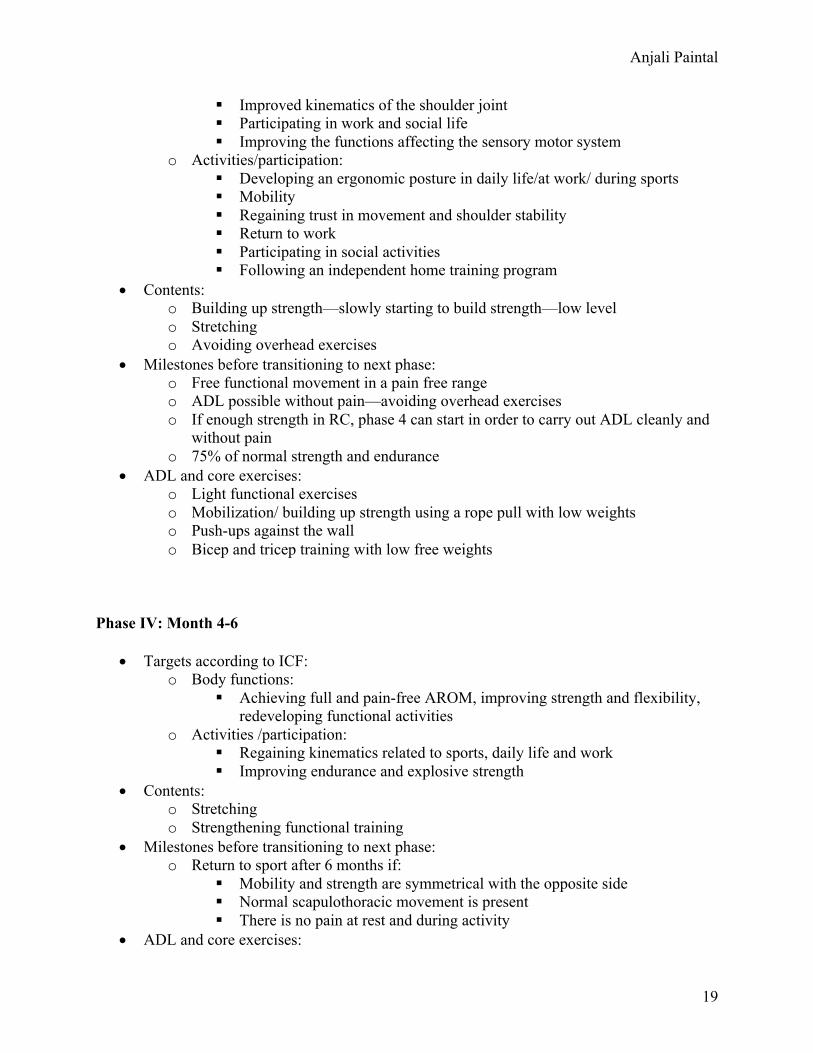
o Activities/participation: § Developing an ergonomic posture in daily life/at work/ during sports § Mobility § Regaining trust in movement and shoulder stability § Return to work § Participating in social activities § Following an independent home training program
• Contents: o Building up strength—slowly starting to build strength—low level o Stretching o Avoiding overhead exercises
• Milestones before transitioning to next phase: o Free functional movement in a pain free range o ADL possible without pain—avoiding overhead exercises o If enough strength in RC, phase 4 can start in order to carry out ADL cleanly and
without pain o 75% of normal strength and endurance
• ADL and core exercises: o Light functional exercises o Mobilization/ building up strength using a rope pull with low weights o Push-ups against the wall o Bicep and tricep training with low free weights
Phase IV: Month 4-6
• Targets according to ICF: o Body functions:
§ Achieving full and pain-free AROM, improving strength and flexibility, redeveloping functional activities
o Activities /participation: § Regaining kinematics related to sports, daily life and work § Improving endurance and explosive strength
• Contents: o Stretching o Strengthening functional training
• Milestones before transitioning to next phase: o Return to sport after 6 months if:
§ Mobility and strength are symmetrical with the opposite side § Normal scapulothoracic movement is present § There is no pain at rest and during activity
• ADL and core exercises:

Anjali Paintal
20
o PNF against resistance o Explosive strength training o Training in a specific sport in the pain free ranges
Conservative Management Rehabilitation Protocol
THE FOLLOWING REHAB PROTOCOL IS DIRECTLY FROM THE 2016 EDWARDS ET AL. ARTICLE52
Phase I: Range of Motion (ROM)
• Goals: o Improve glenohumeral motion (forward flexion, abduction & external rotation) o Improve shoulder and thoracic posture
• Exercises: o Passive ROM (PROM)
§ Forward flexion, internal/external rotation § Pendulum
o Posture § Postural education § Scapula setting exercises
o Active-assisted ROM (AAROM) § Wand exercises: elevation, abduction, adduction, internal/external rotation § Pulley assisted elevation
o Active ROM (AROM) § Wall slides
• Dose: o 3x15 reps daily
• Progression: o ROM should begin with PROM and pendulum exercises, progressing to AAROM
& AROM as comfort dictates
Phase II: Flexibility
• Goals: o Improve flexibility and reduce tightness of anterior and posterior capsule
• Exercises: o Anterior apsule (pectoralis minor) stretch
§ Supine bear hugs § Door frame stretch
o Posterior capsule stretch § Cross-body stretch § Towel stretch
o Upper trapezius stretch • Dose: 3x30 sec stretches, daily
Anjali Paintal
21
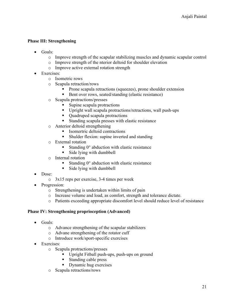
Phase III: Strengthening
• Goals: o Improve strength of the scapular stabilizing muscles and dynamic scapular control o Improve strength of the nterior deltoid for shoulder elevation o Improve active external rotation strength
• Exercises: o Isometric rows o Scapula retraction/rows
§ Prone scapula retractions (squeezes), prone shoulder extension § Bent over rows, seated/standing (elastic resistance)
o Scapula protractions/presses § Supine scapula protractions § Upright wall scapula protractions/retractions, wall push-ups § Quadruped scapula protractions § Standing scapula presses with elastic resistance
o Anterior deltoid strengthening § Isomertric deltoid contractions § Shulder flexion: supine inverted and standing
o External rotation § Standing 0° abduction with elastic resistance § Side lying with dumbbell
o Internal rotation § Standing 0° abduction with elastic resistance § Side lying with dumbbell
• Dose: o 3x15 reps per exercise, 3-4 times per week
• Progression: o Strengthening is undertaken within limits of pain o Increase volume and load, as comfort, strength and tolerance dictate. o Patients exceeding appropriate discomfort level should reduce level of resistance
Phase IV: Strengthening proprioception (Advanced)
• Goals: o Advance strengthening of the scapular stabilizers o Advane strengthening of the rotator cuff o Introduce work/sport-specific exercises
• Exercises: o Scapula protractions/presses
§ Upright Fitball push-ups, push-ups on ground § Standing cable press § Dynamic hug exercises
o Scapula retractions/rows
Anjali Paintal
22
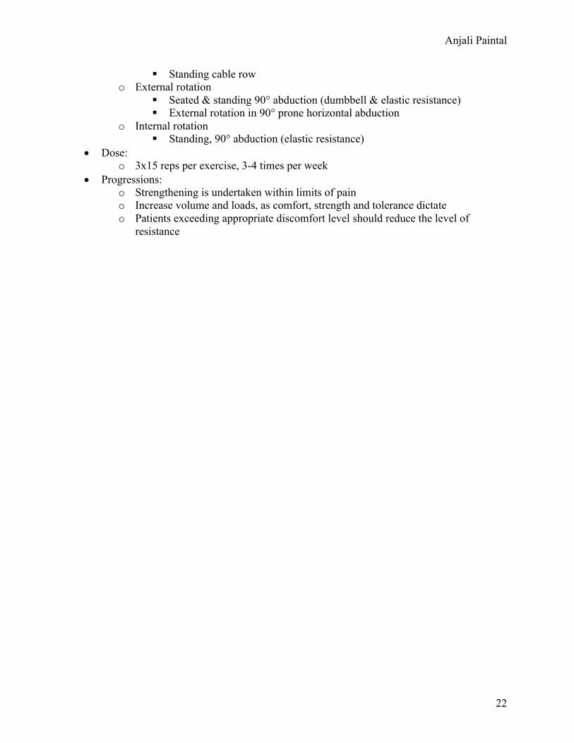
§ Standing cable row o External rotation
§ Seated & standing 90° abduction (dumbbell & elastic resistance) § External rotation in 90° prone horizontal abduction
o Internal rotation § Standing, 90° abduction (elastic resistance)
• Dose: o 3x15 reps per exercise, 3-4 times per week
• Progressions: o Strengthening is undertaken within limits of pain o Increase volume and loads, as comfort, strength and tolerance dictate o Patients exceeding appropriate discomfort level should reduce the level of
resistance

Anjali Paintal
23
References
1. Maruvada S, Varacallo M. Anatomy, Rotator Cuff. [Updated 2018 Nov 14]. In: StatPearls [Internet]. Treasure Island (FL): StatPearls Publishing; 2019 Jan-. Available from: https://www.ncbi.nlm.nih.gov/books/NBK441844/
2. Jeno SH, Schindler GS. Anatomy, Shoulder and Upper Limb, Arm Supraspinatus Muscle. [Updated 2019 Jan 4]. In: StatPearls [Internet]. Treasure Island (FL): StatPearls Publishing; 2019 Jan-. Available from: https://www.ncbi.nlm.nih.gov/books/NBK537202/
3. Faruqi T, Rizvi TJ. Subacromial Bursitis. [Updated 2019 Jun 4]. In: StatPearls [Internet]. Treasure Island (FL): StatPearls Publishing; 2019 Jan-. Available from: https://www.ncbi.nlm.nih.gov/books/NBK541096/
4. Gross MT. Voicethread on Tendon, 2019. 5. May T, Garmel GM. Rotator Cuff Injury. [Updated 2019 Oct 11]. In: StatPearls
[Internet]. Treasure Island (FL): StatPearls Publishing; 2019 Jan-. Available from: https://www.ncbi.nlm.nih.gov/books/NBK547664/
6. Minagawa H, Yamamoto N, Abe H, et al. Prevalence of symptomatic and asymptomatic rotator cuff tears in the general population: From mass-screening in one village. J Orthop. 2013;10(1):8–12. Published 2013 Feb 26. doi:10.1016/j.jor.2013.01.008
7. Via AG, De Cupis M, Spoliti M, Oliva F. Clinical and biological aspects of rotator cuff tears [published correction appears in Muscles Ligaments Tendons J. 2014 Oct;3(4):359]. Muscles Ligaments Tendons J. 2013;3(2):70–79. Published 2013 Jul 9. doi:10.11138/mltj/2013.3.2.070
8. Sambandam SN, Khanna V, Gul A, Mounasamy V. Rotator cuff tears: An evidence based approach. World J Orthop. 2015;6(11):902–918. Published 2015 Dec 18. doi:10.5312/wjo.v6.i11.902
9. Yamaguchi K. The demographic and morphological features of rotator cuff disease. A comparison of asymptomatic and symptomatic shoulders. Journal of bone and joint surgery. American volume. 08/2006;88(8):1699-1704. doi: 10.2106/JBJS.E.00835.
10. Via AG, De Cupis M, Spoliti M, Oliva F. Clinical and biological aspects of rotator cuff tears [published correction appears in Muscles Ligaments Tendons J. 2014 Oct;3(4):359]. Muscles Ligaments Tendons J. 2013;3(2):70–79. Published 2013 Jul 9. doi:10.11138/mltj/2013.3.2.070
11. Neer CS. Anterior acromioplasty for the chronic impingement syndrome in the shoulder: A preliminary report. The Journal of bone and joint surgery. American volume. 01/1972;54(1):41-50.
12. Balke M, Schmidt C, Dedy N, Banerjee M, Bouillon B, Liem D. Correlation of acromial morphology with impingement syndrome and rotator cuff tears. Acta Orthop. 2013;84(2):178–183. doi:10.3109/17453674.2013.773413
13. Mall NA. An evidenced-based examination of the epidemiology and outcomes of traumatic rotator cuff tears. Arthroscopy. 02/2013;29(2):366-376. doi: 10.1016/j.arthro.2012.06.024.
14. Linaker CH, Walker-Bone K. Shoulder disorders and occupation. Best Pract Res Clin Rheumatol. 2015;29(3):405–423. doi:10.1016/j.berh.2015.04.001
Anjali Paintal
24
15. Silverstein BA, Bao SS, Fan ZJ, et al.Rotator Cuff Syndrome: Personal, Work-Related Psychosocial and Physical Load Factors. Journal of Occupational and Environmental Medicine. 2008;50(9):1062-1076. doi: 10.1097/JOM.0b013e31817e7bdd.
16. Muto T, Inui H, Ninomiya H, Tanaka H, Nobuhara K. Characteristics and Clinical Outcomes in Overhead Sports Athletes after Rotator Cuff Repair. J Sports Med (Hindawi Publ Corp). 2017;2017:5476293. doi:10.1155/2017/5476293
17. Baumgarten KM, Gerlach D, Galatz LM, et al. Cigarette smoking increases the risk for rotator cuff tears. Clin Orthop Relat Res. 2010;468(6):1534–1541. doi:10.1007/s11999-009-0781-2
18. Bishop JY. Smoking predisposes to rotator cuff pathology and shoulder dysfunction: A systematic review. Arthroscopy. 08/2015;31(8):1598-1605. doi: 10.1016/j.arthro.2015.01.026.
19. Abate M, Vanni D, Pantalone A, Salini V. Cigarette smoking and musculoskeletal disorders. Muscles Ligaments Tendons J. 2013;3(2):63–69. Published 2013 Jul 9. doi:10.11138/mltj/2013.3.2.063
20. Duygulu F. The effect of subcutaneously injected nicotine on achilles tendon healing in rabbits. Knee surgery, sports traumatology, arthroscopy : official journal of the ESSKA. 08/2006;14(8):756-761. doi: 10.1007/s00167-006-0046-5.
21. Lundgreen K, Lian OB, Scott A, Nassab P, Fearon A, Engebretsen L. Rotator cuff tear degeneration and cell apoptosis in smokers versus nonsmokers. Arthroscopy. 2014;30(8):936–941. doi:10.1016/j.arthro.2014.03.027
22. Galatz LM. Nicotine delays tendon-to-bone healing in a rat shoulder model. Journal of bone and joint surgery. American volume. 09/2006;88(9):2027-2034. doi: 10.2106/JBJS.E.00899.
23. Hsu CL, Sheu WH. Diabetes and shoulder disorders. J Diabetes Investig. 2016;7(5):649–651. doi:10.1111/jdi.12491
24. Huang S. Diabetes mellitus increases the risk of rotator cuff tear repair surgery: A population-based cohort study. Journal of diabetes and its complications. 11/2016;30(8):1473-1477. doi: 10.1016/j.jdiacomp.2016.07.015.
25. Bedi A, Fox AJ, Harris PE, et al. Diabetes mellitus impairs tendon-bone healing after rotator cuff repair. J Shoulder Elbow Surg. 2010;19(7):978–988. doi:10.1016/j.jse.2009.11.045
26. Su B, O'Connor JP. NSAID therapy effects on healing of bone, tendon, and the enthesis. J Appl Physiol (1985). 2013;115(6):892–899. doi:10.1152/japplphysiol.00053.2013
27. Abraham AC, Shah SA, Thomopoulos S. Targeting Inflammation in Rotator Cuff Tendon Degeneration and Repair. Tech Shoulder Elb Surg. 2017;18(3):84–90. doi:10.1097/BTE.0000000000000124
28. Chechik O. Timing matters: NSAIDs interfere with the late proliferation stage of a repaired rotator cuff tendon healing in rats. Archives of orthopaedic and trauma surgery. 04/2014;134(4):515-520. doi: 10.1007/s00402-014-1928-5.
29. Yang Y, Qu J. The effects of hyperlipidemia on rotator cuff diseases: a systematic review. J Orthop Surg Res. 2018;13(1):204. Published 2018 Aug 17. doi:10.1186/s13018-018-0912-0
Anjali Paintal
25
30. Hast MW, Abboud JA, Soslowsky LJ. Exploring the role of hypercholesterolemia in tendon health and repair. Muscles Ligaments Tendons J. 2014;4(3):275–279. Published 2014 Nov 17.
31. Lai J, Gagnier JJ. The Effect of Lipid Disorders on the Risk of Rotator Cuff Disease: A Systematic Review and Meta-Analysis. JB JS Open Access. 2018;3(3):e0018. Published 2018 Sep 13. doi:10.2106/JBJS.OA.18.00018
32. Gumina S. The impact of aging on rotator cuff tear size. Musculoskeletal surgery. 06/2013;97 Suppl 1(s1):69-72. doi: 10.1007/s12306-013-0263-2.
33. Jain NB, Wilcox RB 3rd, Katz JN, Higgins LD. Clinical examination of the rotator cuff. PM R. 2013;5(1):45–56. doi:10.1016/j.pmrj.2012.08.019
34. Naqvi GA, Jadaan M, Harrington P. Accuracy of ultrasonography and magnetic resonance imaging for detection of full thickness rotator cuff tears. Int J Shoulder Surg. 2009;3(4):94–97. doi:10.4103/0973-6042.63218
35. Jain NB, Luz J, Higgins LD, et al. The Diagnostic Accuracy of Special Tests for Rotator Cuff Tear: The ROW Cohort Study. Am J Phys Med Rehabil. 2017;96(3):176–183. doi:10.1097/PHM.0000000000000566
36. Tashjian RZ. Epidemiology, natural history, and indications for treatment of rotator cuff tears. Clinics in sports medicine. 10/2012;31(4):589-604. doi: 10.1016/j.csm.2012.07.001.
37. Carpenter JE. Rotator cuff defect healing: A biomechanical and histologic analysis in an animal model. Journal of shoulder and elbow surgery. 11/1998;7(6):599-605. doi: 10.1016/s1058-2746(98)90007-6.
38. Fucentese SF. Evolution of nonoperatively treated symptomatic isolated full-thickness supraspinatus tears. Journal of bone and joint surgery. American volume. 05/2012;94(9):801-808. doi: 10.2106/JBJS.I.01286.
39. Petersen SA. The timing of rotator cuff repair for the restoration of function. Journal of shoulder and elbow surgery. 01/2011;20(1):62-68. doi: 10.1016/j.jse.2010.04.045.
40. Maman E. Outcome of nonoperative treatment of symptomatic rotator cuff tears monitored by magnetic resonance imaging. Journal of bone and joint surgery. American volume. 08/2009;91(8):1898-1906. doi: 10.2106/JBJS.G.01335.
41. Novoa-Boldo A, Gulotta LV. Expectations Following Rotator Cuff Surgery. Curr Rev Musculoskelet Med. 2018;11(1):162–166. doi:10.1007/s12178-018-9470-7
42. Millar NL, Wu X, Tantau R, Silverstone E, Murrell GA. Open versus two forms of arthroscopic rotator cuff repair. Clin Orthop Relat Res. 2009;467(4):966–978. doi:10.1007/s11999-009-0706-0
43. Pandey V, Jaap Willems W. Rotator cuff tear: A detailed update. Asia Pac J Sports Med Arthrosc Rehabil Technol. 2015;2(1):1–14. Published 2015 Feb 11. doi:10.1016/j.asmart.2014.11.003
44. Cho NS. The factors affecting the clinical outcome and integrity of arthroscopically repaired rotator cuff tears of the shoulder. Clinics in orthopedic surgery. 06/2009;1(2):96-104. doi: 10.4055/cios.2009.1.2.96.
45. Rossi LA. Long-term outcomes after in situ arthroscopic repair of partial rotator cuff tears. Arthroscopy. 03/2019;35(3):698-702. doi: 10.1016/j.arthro.2018.09.026.
46. Jung C, Tepohl L, Tholen R, et al. Rehabilitation following rotator cuff repair: A work of the Commission Rehabilitation of the German Society of Shoulder and Elbow Surgery e. V. (DVSE) in collaboration with the German Association for Physiotherapy (ZVK)

Anjali Paintal
26
e. V., the Association Physical Therapy, Association for Physical Professions (VPT) e. V. and the Section Rehabilitation-Physical Therapy of the German Society for Orthopaedics and Trauma e. V. (DGOU). Obere Extrem. 2018;13(1):45–61. doi:10.1007/s11678-018-0448-2
47. Lee BG. Effect of two rehabilitation protocols on range of motion and healing rates after arthroscopic rotator cuff repair: Aggressive versus limited early passive exercises. Arthroscopy. 01/2012;28(1):34-42. doi: 10.1016/j.arthro.2011.07.012.
48. Gross, MT. Myotendinous Junction and Muscle Voicethread, 2019. 49. Weiss LJ, Wang D, Hendel M, Buzzerio P, Rodeo SA. Management of Rotator Cuff
Injuries in the Elite Athlete. Curr Rev Musculoskelet Med. 2018;11(1):102–112. doi:10.1007/s12178-018-9464-5
50. Kukkonen J. Treatment of non-traumatic rotator cuff tears: A randomised controlled trial with one-year clinical results. The bone & joint journal. 01/2014;96-B(1):75-81. doi: 10.1302/0301-620X.96B1.32168.
51. Kuhn JE. Effectiveness of physical therapy in treating atraumatic full-thickness rotator cuff tears: A multicenter prospective cohort study. Journal of shoulder and elbow surgery. 10/2013;22(10):1371-1379. doi: 10.1016/j.jse.2013.01.026.
52. Edwards P, Ebert J, Joss B, Bhabra G, Ackland T, Wang A. EXERCISE REHABILITATION IN THE NON-OPERATIVE MANAGEMENT OF ROTATOR CUFF TEARS: A REVIEW OF THE LITERATURE. Int J Sports Phys Ther. 2016;11(2):279–301.