esthetic considerations related to bone and soft tissue
TRANSCRIPT

8/18/2019 Esthetic Considerations Related to Bone and Soft Tissue
http://slidepdf.com/reader/full/esthetic-considerations-related-to-bone-and-soft-tissue 1/9Rodriguez and Rosenstiel
In recent years the frequency of and esthetic demand for implant restorations in the esthetic zone has increased.Recent literature has revealed numerous consistent trends which may aid the clinician in achieving predictable esthet-ics. Maintaining generous facial bone by judicious placement as well as by using implants with diameters of less than4 mm appears to be beneficial. Avoiding adjacent implants in the esthetic zone while maintaining an implant to toothdistance of between 2 mm and 4 mm seems to aid in bone and soft tissue maintenance. Abutment connections inwhich the abutment is narrower than the implant offer distinct advantages, most notably increased bone heights.Also, provisional restoration, especially early in treatment provides long-term esthetic benefits. (J Prosthet Dent2012;108:259-267)
Esthetic considerations related to bone and
soft tissue maintenance and development
around dental implants: Report of the
Committee on Research in Fixed Prosthodontics
of the American Academy of Fixed Prosthodontics
Arthur M. Rodriguez, DMD, MSa and Stephen F. Rosenstiel
BDS, MSDb University of Pittsburgh School of Dental Medicine and VeteransAffairs Medical Center, Pittsburgh, Pa; The Ohio State University,College of Dentistry, Columbus, Ohio
aStaff Prosthodontist and Assistant Program Director, Veterans Affairs Medical Center; and Assistant Professor, Department ofProsthodontics, University of Pittsburgh School of Dental Medicine.bProfessor Emeritus, Division of Restorative and Prosthetic Dentistry, The Ohio State University.
Implant-retained restoration in the
esthetic zone have long been difficult
for reasons which are well document-
ed in the literature and anecdotally
acknowledged by many clinicians.1-4
Most prevalent among these are a
lack of facial bone and inadequate
interproximal tissue. However, the es-
thetic demands of clinicians as well as
the expectations of patients continue
to grow along with advances in com-
ponent design and clinical technique.
The following discussion summarizes
relevant current literature in an at-
tempt to establish evidence-based
guidelines for esthetic implant resto-
rations.
Bone availability and maintenance
Inadequate facial bone is a com-
mon problem that can present itself
at any time following extraction and
often leads to a more lingual implant
placement and an anterior ridge lap
restoration.5 In an attempt to avoid
this situation, osseous grafting or
regeneration techniques are used.6,7
However, these can be both time
consuming and unpredictable. Even
when an implant can be placed, a thin
plate of facial bone is often all that re-
mains, leading to thread exposure or
unpredictable future tissue height.8
Huynh et al9 found the average max-
illary anterior bony wall thicknesses
following maxillary anterior extrac-
tion to be approximately 0.8 mm with
87% less than 1 mm. Recent trends
by clinicians to use wider diameter
implants (4 mm or greater) have also
contributed to inadequate facial bone
thickness or facial tissue recession.10
Recession often occurs following the
initiation of the restorative phase as
thin attached tissue supported byinadequate bone cannot withstand
the stress created by restorative con-
tours. The theoretical advantage of
wider diameter implants is not well
supported in the literature. A finite
element analysis by Petrie et al11 did
show a decrease in crestal bone strain
as implant diameter and length in-
creased while modeling a 20 mm
mandibular section. However, clinical
data demonstrating the advantages
of wide diameter implants are scarce.
In fact clinical studies showed similar
or somewhat lower success rates for
wider diameter implants12-15 Degidi et
al15 showed a 99.4% success rate for
510 narrow diameter implants (<3.75
mm) over 8 years, with some evidence
of increased success for implants
wider than 3.4 mm. A clinical study
of 182 3.3-mm-diameter implants in
severely resorbed maxillae revealed a
survival rate of 99.4%.16 Zinsli et al17
reported a 98.7% 5-year survival rate
for 298 of the same reduced diameter
implants, over a 10-year period. In
this study the implants were restoredwith a variety of restorations, includ-
ing single teeth, fixed prostheses, and
overdentures.
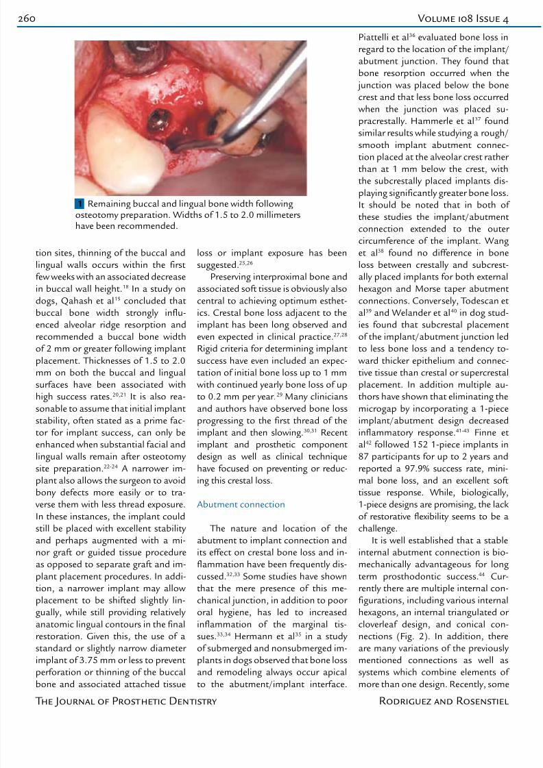
It has also been suggested that the
amount of remaining facial or lingual
bone following osteotomy is critical
for implant success1 (Fig. 1). Even af-
ter implant placement in fresh extrac-
8/18/2019 Esthetic Considerations Related to Bone and Soft Tissue
http://slidepdf.com/reader/full/esthetic-considerations-related-to-bone-and-soft-tissue 2/9
60 Volume 108 Issue 4
The Journal of Prosthetic Dentistry Rodriguez and Rosenstiel
tion sites, thinning of the buccal and
lingual walls occurs within the firstfew weeks with an associated decrease
in buccal wall height.18 In a study on
dogs, Qahash et al19 concluded that
buccal bone width strongly influ-
enced alveolar ridge resorption and
recommended a buccal bone width
of 2 mm or greater following implant
placement. Thicknesses of 1.5 to 2.0
mm on both the buccal and lingual
surfaces have been associated with
high success rates.20,21 It is also rea-
sonable to assume that initial implant
stability, often stated as a prime fac-
tor for implant success, can only be
enhanced when substantial facial and
lingual walls remain after osteotomy
site preparation.22-24 A narrower im-
plant also allows the surgeon to avoid
bony defects more easily or to tra-
verse them with less thread exposure.
In these instances, the implant could
still be placed with excellent stability
and perhaps augmented with a mi-nor graft or guided tissue procedure
as opposed to separate graft and im-
plant placement procedures. In addi-
tion, a narrower implant may allow
placement to be shifted slightly lin-
gually, while still providing relatively
anatomic lingual contours in the final
restoration. Given this, the use of a
standard or slightly narrow diameter
implant of 3.75 mm or less to prevent
perforation or thinning of the buccalbone and associated attached tissue
loss or implant exposure has been
suggested.25,26
Preserving interproximal bone and
associated soft tissue is obviously also
central to achieving optimum esthet-
ics. Crestal bone loss adjacent to the
implant has been long observed and
even expected in clinical practice.27,28
Rigid criteria for determining implant
success have even included an expec-
tation of initial bone loss up to 1 mm
with continued yearly bone loss of up
to 0.2 mm per year.29 Many clinicians
and authors have observed bone loss
progressing to the first thread of the
implant and then slowing.30,31 Recent
implant and prosthetic component
design as well as clinical technique
have focused on preventing or reduc-
ing this crestal loss.
Abutment connection
The nature and location of the
abutment to implant connection andits effect on crestal bone loss and in-
flammation have been frequently dis-
cussed.32,33 Some studies have shown
that the mere presence of this me-
chanical junction, in addition to poor
oral hygiene, has led to increased
inflammation of the marginal tis-
sues.33,34 Hermann et al35 in a study
of submerged and nonsubmerged im-
plants in dogs observed that bone loss
and remodeling always occur apicalto the abutment/implant interface.
Piattelli et al36 evaluated bone loss in
regard to the location of the implant/
abutment junction. They found that
bone resorption occurred when the
junction was placed below the bone
crest and that less bone loss occurred
when the junction was placed su-
pracrestally. Hammerle et al37 found
similar results while studying a rough/smooth implant abutment connec-
tion placed at the alveolar crest rather
than at 1 mm below the crest, with
the subcrestally placed implants dis-
playing significantly greater bone loss.
It should be noted that in both of
these studies the implant/abutment
connection extended to the outer
circumference of the implant. Wang
et al38 found no difference in bone
loss between crestally and subcrest-ally placed implants for both external
hexagon and Morse taper abutment
connections. Conversely, Todescan et
al39 and Welander et al40 in dog stud-
ies found that subcrestal placement
of the implant/abutment junction led
to less bone loss and a tendency to-
ward thicker epithelium and connec-
tive tissue than crestal or supercrestal
placement. In addition multiple au-
thors have shown that eliminating the
microgap by incorporating a 1-piece
implant/abutment design decreased
inflammatory response.41-43 Finne et
al42 followed 152 1-piece implants in
87 participants for up to 2 years and
reported a 97.9% success rate, mini-
mal bone loss, and an excellent soft
tissue response. While, biologically,
1-piece designs are promising, the lack
of restorative flexibility seems to be a
challenge.
It is well established that a stableinternal abutment connection is bio-
mechanically advantageous for long
term prosthodontic success.44 Cur-
rently there are multiple internal con-
figurations, including various internal
hexagons, an internal triangulated or
cloverleaf design, and conical con-
nections (Fig. 2). In addition, there
are many variations of the previously
mentioned connections as well as
systems which combine elements ofmore than one design. Recently, some
1 Remaining buccal and lingual bone width followingosteotomy preparation. Widths of 1.5 to 2.0 millimetershave been recommended.

8/18/2019 Esthetic Considerations Related to Bone and Soft Tissue
http://slidepdf.com/reader/full/esthetic-considerations-related-to-bone-and-soft-tissue 3/9
261October 2012
Rodriguez and Rosenstiel
definite advantages of the conical
connection have surfaced.45-50 Tesmer
et al45 have shown a decreased ten-
dency for bacterial colonization given
the intimate fit afforded by the coni-
cal connection. Biomechanical com-
plications, such as screw loosening or
component fracture, also seem to oc-
cur less with the conical connection,
theoretically because of decreased
micromovement at the implant/abut-
ment junction.46-48 Schwarz49 notedthat the conical Morse taper has virtu-
ally eliminated abutment screw loos-
ening and fracture. In a finite element
analysis study comparing multiple
systems and examining stress trans-
mission to the crestal bone during
multiple loading conditions, the coni-
cal Morse taper connection with plat-
form switch demonstrated the most
favorable stress transmission during all
nonaxial loading simulations.50
In addition to the specific configu-
ration of internal fit, the intimacy of
fit and, therefore, the movement of
the abutment relative to the implant
may also affect stress transmission
as well as tissue health and subse-
quent crestal bone loss.51,52 In fact,
connection stability may have a more
important role than the presence of
a microgap with regard to inflamma-
tion and bone loss. Hermann et al53
showed that welded 2-piece implants
displayed less bone loss than conven-tionally joined 2-piece implant/abut-
ment connections regardless of the
size of the microgap. King et al,52 in a
follow-up study, also concluded that
stability or lack of movement at the
abutment/implant junction was more
important than microgap size. They
compared welded 1-piece connec-
tions to 2-piece connections, while
varying the microgaps for each group
between 10 and 100 µm and foundthat the size of the microgap had no
significant effect on bone loss, while
the nonwelded groups consistently
showed higher levels of bone loss.
Heijdenrijk et al51 compared 2-piece
abutment/implant connections with
the connections placed above and
below the tissues to a 1-piece system
and found no association between
the presence of a microgap and in-flammation.
Platform switching
Locating the abutment/implant
connection internally or away from
the bone/tissue to implant interface
has also been attempted. This con-
cept, which involves an abutment of
decreased diameter relative to the
implant, is sometimes referred to asplatform switching.54, 55 While many
consider platform switching to be
a new concept, it has been incorpo-
rated into various systems for over 20
years.20,47,48 Platform switching moves
the abutment-implant interface in-
ward, away from the outer circum-
ference of the implant (Fig. 3). This
has been reported to decrease crestal
bone loss, preserve tissue height,
and promote soft tissue health.56-74
Whether this advantage is gained me-
chanically or biologically is debatable.
Some theorize that this configuration
shifts the inflammatory infiltrate in-
ward, encouraging soft tissue and/or
bone to biologically seal off the outer
circumference of the implant from
the oral environment.56 Becker et al74
found no histological advantage to a
0.3-mm platform switch with respect
to bone remodeling in a dog study
over 24 weeks, suggesting that theadvantage may be a consequence of
a physical barrier. Other authors75,76
have reported a more favorable stress
distribution within the bone for plat-
form switched connections.
Hurzeler et al62 showed signifi-
cantly less bone loss 1 year after res-
toration placement for 14 platform
switched implant single crowns than
for 8 similarly restored implants
that did not incorporate a platformswitch. In a pair of clinical studies in-
2 A, Conical internal abutment connection; B, Internalhexagon abutment connection.
A
B

8/18/2019 Esthetic Considerations Related to Bone and Soft Tissue
http://slidepdf.com/reader/full/esthetic-considerations-related-to-bone-and-soft-tissue 4/9
62 Volume 108 Issue 4
The Journal of Prosthetic Dentistry Rodriguez and Rosenstiel
volving implants placed immediately
into extraction sites, Canullo et al63,64
showed generally less bone loss than
previously described in the literature
after a mean follow-up of 22 months.
Also they noted significantly less buc-
cal tissue recession and greater papil-
la height in an experimental platform
switched group than in a nonplat-
form switched group. The treatment
protocol for both groups included
immediate provisional restoration
and placement of the definitive res-
toration 2 months following implant
placement. Vela-Nebot et al77 also
reported dramatically less bone loss
when using implant systems incorpo-
rating a narrower diameter of restor-
ative components relative to implant
diameter. A comprehensive literature
review of 10 studies and 1239 im-
plants by Atieh et al67 showed strong
evidence of less bone loss around the
platform switched implants, although
there was no difference in overall im-plant survival. The authors also noted
that a more favorable bone response
was seen when the platform switch
was 0.4 mm or greater. Wagenberg
and Froum68 followed 94 platform
switched connections over 11 years
and found that approximately 75%
showed no radiographic bone loss
with 88% displaying 0.8 mm or less
bone loss. Conversely a 2 year study
by Crespi et al,78 which compared ex-ternal hexagon regular platform im-
plants to platform switched implants
placed in fresh extraction sites andimmediately loaded, showed no dif-
ference in bone loss.
In an attempt to mimic platform
switching, some implant manufac-
tures offer implant designs in which the
coronal portion of the implant flares
to accommodate an abutment that is
roughly the same diameter as the im-
plant body but narrower than the flared
neck.54 Using this concept, it has been
stated that a residual ridge width of at
least 6.3 mm is necessary to accommo-
date the flared design.79 For example,
Cocchetto et al80 followed 15 implants
with a 5.8 mm diameter receiving a 4.1
mm abutment healing cap and showed
an average bone loss of only 0.3 mm
at 18 months. However, this study re-
quired a minimum bone width of 8
mm for inclusion. Because bone width
of this dimension is exceptional in the
esthetic zone, this design may have lim-
ited application. Neck diameters of thisdimension will most likely necessitate
removal of the same critical coronal
bone that the clinician is trying to pre-
serve. Also the resulting distance from
the implant to the adjacent tooth will
be minimal at best, preventing the es-
tablishment of a desirable interproximal
distance. Therefore, given the above, a
parallel walled implant of standard or
slightly narrow diameter with reduced
diameter restorative componentsseems preferable.
A distinct advantage of the im-
plant/abutment diameter difference
is the ability to place the implant
subcrestally, encouraging bone and
soft tissue at the superior edge of the
implant circumference to remain and
even grow to or over the edge of the
implant and remain there undisturbed
by restorative components.59,60,81,82
Novaes et al59,60 found this to be true,
while noting greater papilla fill and
less bone loss for subcrestal place-
ment than for crestal placement in
2 dog studies of platform switched
implants. They also concluded that
the presence of a microgap did not
adversely affect tissue health. Weng
et al81 compared a platform switched,
Morse taper design placed subcrest-
ally to an external hexagon design. They demonstrated bony overgrowth
only in the former. Veis et al82 com-
pared 193 regular platform implants
to 89 platform switched implants
and found that the platform switched
design resulted in less bone loss only
when placed subcrestally. Conversely,
Cochran et al83 found slightly more
bone loss for platform switched im-
plants placed 1 mm subcrestally than
for those placed crestally or 1 mm su-
pracrestally.
Tenenbaum et al84 showed a great-
er number and greater length of con-
nective tissue fibers around implants
which incorporated a nonflared neck
with reduced diameter abutments
than other designs. Attached tissue
thickness of greater than 2 mm has
been associated with a decreased ten-
dency for buccal bone loss and tissue
recession around platform switched
implants.85- 87 This effect was even moredramatic when implants were placed
supracrestally. Interestingly, when the
mucosa was considered thin (less than
2 mm), the authors found no advan-
tage with regard to bone level for the
platform switched design.88 Subcrestal
placement leads to other esthetic
advantages. The distance from the
top of the implant to the emergence
of the restoration is maximized, al-
lowing adequate vertical length forthe development of anatomic con-
3 Platform switched implant-abutment connection. Notenarrower diameter of abutment relative to implant allowingbone to remain undisturbed throughout restorative phase.

8/18/2019 Esthetic Considerations Related to Bone and Soft Tissue
http://slidepdf.com/reader/full/esthetic-considerations-related-to-bone-and-soft-tissue 5/9
263October 2012
Rodriguez and Rosenstiel
tours and proper emergence profiles.
Priest 61 recommended providing at
least 3 mm from the implant margin
to the zenith of the facial margins
of adjacent teeth, while noting that
provisional restoration is essential in
developing optimum esthetics. This
is especially critical when soft tissue
thickness is minimal.
Provisional Restoration
As in conventional restorative and
fixed prosthodontics, provisional res-
torations are an invaluable esthetic
and diagnostic tool. Provisional resto-
ration of implants is often neglected
probably because provisional compo-
nents are often time consuming and
cumbersome. Also many provisionalcomponents do not readily accom-
modate angle correction. Attached
gingiva can be sculpted and molded
by means of the provisional restora-
tion to help create the illusion of inter-
dental papillae as well as contributing
to more stable, predictable overall es-
thetics and providing increased func-
tion.89-92 Providing anatomic fixed
provisional restorations also avoids
the introduction of dramatically differ-
ent restorative contours at the time of
final restoration placement, decreas-
ing the likelihood of postplacement
recession.63,93 Lai et al94 and Gallucci
et al95 showed that dramatic soft tis-
sue changes, both in dimension as well
as health, occur following the initial
establishment of anatomic contours.
Although bone response was similar
to delayed provisional restoration,
Block et al92 found an average increase
in tissue height of 1 mm for immedi-ately provisionally restored implants
following their placement in fresh ex-
traction sites. Establishing these con-
tours immediately, either at placement
or during second stage uncovering
has been shown to be especially effec-
tive.63,79,93,95 DeRouck et al93 compared
the postrestoration soft tissue con-
tours of 24 immediately provisionally
restored implants to 25 restored with
a conventional 2-stage approach. Theyfound 2.5 to 3 times less midfacial re-
cession and better predictability of pa-
pilla heights in the immediately provi-
sionally restored group.
The use of definitive stock abut-
ments placed intraorally by the clini-
cian as opposed to laboratory pre-
pared custom abutments would seem
to offer distinct advantages in the es-
thetic zone. Provisional restoration issimplified, allowing the establishment
of a proximal contact point early in
the restorative phase to initiate pa-
pilla formation. The use of cumber-
some, interim, partial removable den-
tal prostheses is also obviated. Most
of all this technique eliminates the
multiple switching of various compo-
nents during the restorative phase. Al-
though such evidence in the literature
could not be found, it is these authors’opinion that the techniques afforded
by this type of abutment offer distinct
biologic and clinical advantages.
Parameters influencing papillary
tissue development
The possibility of papilla fill is
greatly enhanced when interproximal
bone is preserved, creating a reason-
able opportunity to provide a desir-
able distance of 5 mm or less from
the proximal contact to the intersep-
tal bone as described in the literature
for natural teeth as well as recently
for implants immediately adjacent
to natural teeth.96-99 Lops et al98 and
Gastaldo et al99 recommend 3 to 4
mm and 3 to 5 mm, respectively, be-
tween proximal contacts and bone
crest when the implant is adjacent
to a natural tooth. However, papilla
fill was observed, in some instances,where the bone to contact distance
was as great as 9 mm, suggesting, not
surprisingly, that multiple factors in-
fluence soft tissue response.97 Palmer
et al100 also found, in a study of 46
participants with platform switched
implants, that papilla fill was ob-
served when the proximal contact to
bone distance exceeded 4 to 5 mm.
It should be noted that both of these
studies involved implants adjacentto natural teeth. In addition, Kwon
et al66 observed that in the tooth-im-
plant-tooth clinical scenario, the criti-
cal distance is that from the proximal
contact to the bone level adjacent to
the natural tooth, termed tooth side
bone level. In this study of 17 im-
plants and 37 papillae, they found
virtually complete papilla fill when the
average bone to contact distance was4.88 mm or less and a lack of papilla
fill when this distance exceeded 6 mm.
The situation becomes much more
challenging when adjacent implants
are present. In a dog study of plat-
form-switched implants, de Oliveira
et al101 found that a 5 mm bone to
proximal contact distance proved to
be the critical factor for papilla devel-
opment between implants, regardless
of interimplant distances, which var-ied between 1 and 3 mm. Other inves-
tigators have observed that to achieve
papilla fill between implants, a proxi-
mal contact to bone crest distance
of 3 mm or less is desirable, while a
distance of 3 to 5 mm can provide
papilla fill between an implant and a
natural tooth.98
As previously mentioned, interim-
plant distance and tooth-to-implant
distance have been shown to play a
role and possibly interact with bone
to proximal contact distance. In a
more recent study involving the same
type of implants, de Oliveira et al102
reported increased bone remodeling
when interimplant distance was 1 mm
or less than in groups with 2 to 3 mm
interimplant distance. In the natural
dentition, Martegani et al103 found
that papilla loss increased dramatical-
ly when interradicular distances were
2.4 mm or less and that this factorhad both an independent and com-
bined effect with proximal contact
distance. Degidi et al104 examined 152
implants and 99 interimplant sites
and found that optimum interim-
plant distance for papilla develop-
ment was greater than 2 mm but less
than 4 mm. They also found that op-
timum bone to contact height should
be 3 to 4 mm and that papilla height
decreased dramatically when this dis-tance exceeded 6 mm. Tarnow et al25
8/18/2019 Esthetic Considerations Related to Bone and Soft Tissue
http://slidepdf.com/reader/full/esthetic-considerations-related-to-bone-and-soft-tissue 6/9
64 Volume 108 Issue 4
The Journal of Prosthetic Dentistry Rodriguez and Rosenstiel
also concluded that less interproximal
bone loss occurs as interimplant dis-
tance increases, while recommending a
minimum of 3 mm, lending further sup-
port to their earlier mentioned recom-
mendation of using standard to narrow
diameter implants in the esthetic zone.
Garber et al105 have recommended in
order to maintain interproximal boneheight and the accompanying papilla,
that the implant should be placed at
least 1.5 mm from adjacent teeth and
3 mm from adjacent implants. Traini et
al106 also recommend a minimum of 3
mm interimplant distance to provide
optimum alveolar bone health. They
observed a more desirable orienta-
tion of collagen fibers and significantly
increased marrow spaces for interim-
plant distances of 3 mm in a histologi-cal dog study of 48 implants, sacrificed
24 months after implant placement. In
a similar follow-up study, they observed
that an interimplant distance of 3 mm
allowed for an increase in the develop-
ment of blood vessels.107
While it is difficult to isolate clini-
cal variables scientifically, the litera-
ture seems to indicate that the risk
to interproximal bone increases for
tooth-to-implant distances less than
3 mm.108 Conversely, in a 5-year study
of interproximal bone changes be-
tween implants, Chang et al109 found
that interimplant distance played a
minor role in final bone levels and
that the coronal height of bone to
implant contact was a better predic-
tor. Lee et al110 found that both inter-
implant distance and bone to implant
contact level had minimal effect on
papilla height and that the width of
keratinized tissue exerted a greaterinfluence. However, the literature is
fairly consistent in showing that the
maintenance of proximal bone height
and associated papilla height seems
to be more difficult between adjacent
implants than in the tooth-implant
situation.111-113 In a retrospective study
evaluating previously mentioned vari-
ables, Kourkouta et al112 followed 35
implants in 15 participants and found
bone crest and papilla heights to be4.6 mm and 2 mm lower, respectively,
for the implant-implant clinical situa-
tion than for the tooth-implant situ-
ation. They also found only 1 mm of
missing papilla height when the inter-
implant distance was 3 mm and that
immediate provisional restoration
reduced papilla loss from 2 mm to
1 mm. It is also noteworthy that, in
this study, 87.5% of participants werepleased with their esthetic result even
with a papilla height discrepancy of 2
mm. Because most of these changes
have been found to occur within the
first 6 months following placement, the
advantages of early and long term pro-
visional restoration seem clear. Tarnow
et al113 also recommended caution,
while reporting mean papillary tissue
heights of only 3.4 mm for 136 adja-
cent implant sites in 33 participants. While 3 to 4 mm appears to be the
ideal interimplant distance, the use of
a platform switched design may allow
more bone and soft tissue maintenance
for enhanced esthetics, effectively in-
creasing the interabutment distances
for implants in close proximity 65,105 In a
study of 41 pairs of platform switched
implants in 37 participants, Rodri-
guez-Ciurana et al65 demonstrated
bone maintenance to be an average of
2.4 mm above the implant/abutment
interface at interimplant distances
less than 3 mm. As stated previously,
Novaes et al59,60 also found improved
papilla fill for subcrestally placed plat-
form switched implants than for those
placed crestally. However, the papilla
height was not influenced by interim-
plant distance, which varied from 1
to 3 mm.59,60 In addition to his previ-
ously mentioned recommendation of
a 3 mm distance from implant to ad- jacent gingival margin zeniths, Priest 61
similarly recommended 3 mm of in-
terimplant distance as well as orient-
ing the center of the implant 3 mm
palatal to the future facial restorative
margin. Therefore, because of the es-
thetic challenges detailed above, the
placement of adjacent implants in the
esthetic zone should be avoided when-
ever possible.114
SUMMARY
A review of the current literature
suggests that the use of standard to
narrow diameter implants, a plat-
form switched design, and early pro-
visional restoration should be con-
sidered when long term esthetics are
paramount. Also, maintaining inter-implant and tooth-to-implant dis-
tances of approximately 3 mm is desir-
able with regard to crestal bone main-
tenance and papilla development.
Subcrestal placement may afford ad-
equate space to develop restorative
contours from the top of the implant
to restoration emergence while avoid-
ing facial thread exposure. This may
be even more effective in combina-
tion with a platform switched design.In addition, abutment to implant
connections, which are extremely in-
timate and allow little to no relative
movement, seem to offer distinct ad-
vantages with regard to tissue health.
Lastly, the use of noncustomizable
abutments and abutment level restor-
ative techniques provides for ease of
provisional restoration and treatment
sequencing while avoiding multiple
exposures of the implant/abutment
junction, although the biological ad-
vantages this may offer have yet to be
investigated.
REFERENCES
1. Sammartino G, Marenzi G, di Lauro AE,Paolantoni G. Aesthetics in oral implantol-ogy: biological, clinical, surgical and pros-thetic aspects. Implant Dent 2007;16:54-59.
2. Sunitha RV, Ramakrishnan T, Kumar S, Em-madi P. Soft tissue preservation and crestalbone loss around single-tooth implants. J
Oral Implantol 2008;34:223-9.3. Lai HC, Zhang ZY, Wang F, Zhuang LF, Liu
X, Pu YP. Evaluation of soft tissue altera-tion around implant supported single toothrestoration in the anterior maxilla: thepink esthetic score. Clin Oral Implants Res2008;19:560-4.
4. Wheeler SL. Implant complications inthe esthetic zone. J Oral Maxillofac Surg2007;65:93-102.
5. Leblebicioglu B, Rawal S, Mariotti A. A re-view of the functional and esthetic require-ments for dental implants. J Am Dent Assoc2007;138:321-9.

8/18/2019 Esthetic Considerations Related to Bone and Soft Tissue
http://slidepdf.com/reader/full/esthetic-considerations-related-to-bone-and-soft-tissue 7/9
265October 2012
Rodriguez and Rosenstiel
6. Fagan MC, Owens H, Smaha J, Kao RT.Simultaneous hard and soft tissue augmen-tation for implants in the esthetic zone:report of 37 consecutive cases. J Periodon-tol 2008;79:1782-8.
7. Buser D, Chen ST, Weber HP, Belsar UC.Early implant placement following single-tooth extraction in the esthetic zone:biologic rationale and surgical proce-dures. Int J Periodontics Restorative Dent2008;28:441-51.
8. Chen ST, Darby IB, Reynolds EC. A pro-spective clinical study of non-submergedimmediate implants: clinical outcomes andesthetic results. Clin Oral Implants Res2007;18:552-62.
9. Huynh-Ba G, Pjetursson BE, Sanz M, Cec-chinato D, Ferrus J, Lindhe J, et al. Analysisof the socket bone wall dimensions in theupper maxilla in relation to immediateimplant placement. Clin Oral Implants Res2010;21:37-42.
10.Evans CD, Chen ST. Esthetic outcomes ofimmediate implant placements. Clin OralImplants Res 2008;19:73-80.
11.Petrie CS, Williams JL. Comparative
evaluation of implant designs: influence ofdiameter, length, and taper on strains in thealveolar crest. A three-dimensional finite-element analysis. Clin Oral Implants Res2005;16:486-94.
12.Shin SW, Bryant SR, Zarb GA. A retrospec-tive study on the treatment outcome ofwide-bodied implants. Int J Prosthodont2004;17:52-8.
13.Eckert SE, Meraw SJ, Weaver AL, LohseCM. Early experience with wide-platformMk II implants. Part I: Implant survival.Part II: Evaluation of risk factors involv-ing implant survival. Int J Oral MaxillofacImplants 2001;16:208-16.
14.Ivanoff CJ, Grondahl K, Sennerby L, Berg-strom C, Lekholm U. Influence of variationsin implant diameters: a 3- to 5-year retro-spective clinical report. Int J Oral Maxillo-fac Implants 1999;14:173-80.
15.Degidi M, Piattelli A, Carinci F. Clinicaloutcome of narrow diameter implants: aretrospective study of 510 implants. J Peri-odontol 2008;79:49-54.
16.Hallman M. A prospective study of treat-ment of severely resorbed maxillae withnarrow nonsubmerged implants: resultsafter 1 year of loading. Int J Oral MaxillofacImplants 2001;16:731-6.
17.Zinsli B, Sagesser T, Mericske E, Mericske-Stern R. Clinical evaluation of small-diam-
eter ITI implants: a prospective study. Int JOral Maxillofac Implants 2004;19:92-9.
18.Araújo MG, Sukekava F, Wennström JL,Lindhe J. Tissue modeling following implantplacement in fresh extraction sockets. ClinOral Implants Res 2006;17:615-24.
19.Qahash M, Susin C, Polimeni G, Hall J, Wikesjö UM. Bone healing dynamics atbuccal peri-implant sites. Clin Oral Im-plants Res 2008;19:166-72.
20.Morris HF, Ocho S, Orenstein IH, Petraz-zuolo V. AICRG, Part V: Factors influencingimplant stability at placement and theirinfluence on survival of Ankylos implants. JOral Implantol 2004;30:162-170.
21.Spray JR, Black CG, Morris HF, Ochi S. The influence of bone thickness on facialmarginal bone response: stage 1 placementthrough stage 2 uncovering. Ann Periodon-tol 2000;5:119-128.
22.Listgarten MA. Clinical trials of endosseousimplants: issues in analysis and interpreta-tion. Ann Periodontol 1997;2:299-313.
23.Kohn DH. Overview of factors impor-tant in implant design. J Oral Implantol1992;18:204-19.
24.Steigenga JT, al-Shammari KF, Nociti FH,Misch CE, Wang HL. Dental implant designand its relationship to long term implantsuccess. Implant Dent 2003;12:306-17.
25.Tarnow DP, Cho SC, Wallace SS. The effectof inter-implant distance on the height ofinter-implant bone crest. J Periodontol2000;71:546-9.
26.Covani U, Cornelini R, Calvo JL, TonelliP, Barone A. Bone remodeling aroundimplants placed in fresh extraction sock-ets. Int J Periodontics Restorative Dent2010;30:601-7.
27.Naert I, Quirynen M, Theuniers G, vanSteenburghe D. Prosthetic aspects of os-
seointegrated fixtures supporting over-dentures. A 4 year report. J Prosthet Dent1991;65:671-80.
28.Becker W, Becker BE, Newman MG, NymanS. Clinical and microbiological findings thatmay contribute to dental implant failure.Int J Oral Maxillofac Implants 1990;5:31-38.
29.Smith DE, Zarb GA. Criteria for successof osseointegrated endosseous implants. JProsthet Dent 1989;62:567-72.
30.Khayay PG, Hallage PG, Toledo RA. Aninvestigation of 131 consecutively placedwide screw-vent implants. Int J Oral Maxil-lofac Implants 2001;16:827-32.
31.Nissan J, Romanos GE, Mardinger O,Chaushu G. Immediate nonfunctionalloading of single-tooth implants in theanterior maxilla following augmentationwith freeze dried cancellous block allograft:a case series. Int J Oral Maxillofac Implants2008;23:709-16.
32.Assenza B, Scarano A, Petrone G, Iezzi G, Thams U, San Roman F, et al. Crestal boneremodeling in loaded and unloaded im-plants and the microgap: a histologic study.Implant Dent 2003;12:235-41.
33.Broggini N, McManus LM, Hermann JS, Medina R, Schenk RK, Buser D, et al.Peri-implant inflammation defined by theimplant-abutment interface. J Dent Res
2006;85:473-8.34.Hänggi MP, Hänggi DC, Schoolfield JD,
Meyer J, Cochran DL, Hermann JS. Crestalbone changes around titanium implants.Part I: A retrospective radiographicevaluation in humans comparing two non-submerged implant designs with differentmachined collar lengths. J Periodontol2005;76:791-802.
35.Hermann JS, Cochran DL, Nummikoski P V,Buser D. Crestal bone changes around tita-nium implants. A radiographic evaluationof unloaded nonsubmerged and submergedimplants in the canine mandible. J Peri-odontal 1997;68:1117-30.
36.Piatelli A, Vrespa G, Petrone G, Iezzi G, An-nibali S, Scarano A. Role of the microgapbetween implant and abutment: a retro-spective histologic evaluation in monkeys. JPeriodontol 2003;74:346-52.
37.Hämmerle CH, Bragger U, Bürgin W, LangNP. The effect of subcrestal placement ofthe polished surface of ITI implants onmarginal soft and hard tissues. Clin OralImplants Res 1996;7:111-9.
38.Wang D, Nagata MJ, Bell M, de Melo LG,
Bosco AF. Influence of microgap locationand configuration on peri-implant bonemorphology in nonsubmerged implants:an experimental study in dogs. Int J OralMaxillofac Implants 2010;25:540-7.
39.Todescan FF, Pustiglioni FE, Imbronito AV,Albrektsson T, Gioso M. Influence of themicrogap in the peri-implant hard and soft tis-sues: a histomorphometric study in dogs. Int JOral Maxillofac Implants 2002;17:467-72.
40.Welander M, Abrahamsson I, Berglundh T. Placement of two-part implants in siteswith different buccal and lingual boneheights. J Periodontol 2009;80:324-9.
41.Broggini N, McManus LM, Hermann JS,
Medina RU, Oates TW, Schenk RK, etal. Persistent acute inflammation at theimplant-abutment interface. J Dent Res2003;82:232-7.
42.Finne K, Rompen E, Toljanic J. Clinicalevaluation of a prospective multicenterstudy on 1-piece implants. part 1: Marginalbone level evaluation after 1 year of fol-low up. Int J Oral Maxillofac Implants2007;22:226-34.
43.Fiorellini JP, Buser D, Paquette DW, Williams RC, Haghighi D, Weber HP. Aradiographic evaluation of bone healingaround submerged and non-submergeddental implants in beagle dogs. J Periodon-tol 1999;70:248-254.
44.Stanford CM. Achieving and maintain-ing predictable implant esthetics throughthe maintenance of bone around dentalimplants. Compend Contin Educ Dent2002;23:13-20.
45.Tesmer M, Wallet S, Koutouzia T, Lundgren T. Bacterial colonization of the dental im-plant fixture-abutment interface: an in v itrostudy. J Periodontol 2009;80:1991-7.
46.Khraisat A, Stegaroiu R, Nomura S,Miyakawa O. Fatigue resistance of twoimplant/abutment joint designs. J ProsthetDent 2002;88:604-10.
47.Norton MR. In vitro evaluation of thestrength of the conical implant to abutment
joint in two commercially available implantsystems. J Prosthet Dent 2000;83:567-71.
48.Mangano C, Mangano F, Piatelli A, Iezzi G,Mangano A, La Colla L. Prospective clini-cal evaluation of 307 single-tooth morsetaper-connection implants: a multicenterstudy. Int J Oral Maxillofac Implants2010;25:394-400.
49.Schwarz MS. Mechanical complicationsof dental implants. Clin Oral Implants Res2000;11:156-8.
50.Bozkaya D, Mufta S, Muftu A. Evaluationof load transfer characteristics of five differ-ent implants in compact bone at differentload levels by finite element analysis. J
Prosthet Dent 2004;92:523-30.

8/18/2019 Esthetic Considerations Related to Bone and Soft Tissue
http://slidepdf.com/reader/full/esthetic-considerations-related-to-bone-and-soft-tissue 8/9
66 Volume 108 Issue 4
The Journal of Prosthetic Dentistry Rodriguez and Rosenstiel
51.Heijdenrijk K, Raghoebar GM, Meijer HJ,Stegenga B, van der Reijden WA. Feasibil-ity and influence of the microgap of twoimplants placed in a non-submerged pro-cedure: a five-year follow up clinical trial. JPeriodontol 2006;77:1051-60.
52.King GN, Hermann JS, Schoolfield JD, Bus-er D, Cochran DL. Influence of the size ofthe microgap on crestal bone levels in non-submerged dental implants: a radiographicstudy in the canine mandible. J Periodontol
2002;73:1111-7.53.Hermann JS, Schoolfield JD, Schenk RK,
Buser D, Cochran DL. Influence of the sizeof the microgap on crestal bone changesaround titanium implants. A histometricevaluation of unloaded non-submerged im-plants in the canine mandible. J Periodontol2001;72:1372-83.
54.Lazzara RJ, Porter SS. Platform switching:A new concept in implant dentistry forcontrolling post restorative crestal bonelevels. Int J Periodontics Restorative Dent2006;26:9-17.
55.Prosper L, Redaelli S, Pasi M, Zarone F,Radaelli G, Gherlone EF. A randomized
prospective multicenter trial evaluat-ing the platform-switching technique forthe prevention of postrestorative crestalbone loss. Int J Oral Maxillofac Implants2009;24:299-308.
56.Luongo R, Traini T, Guidone PC, BiancoG, Cocchetto R, Celletti R. Hard and softtissue responses to the platform-switchingtechnique. Int J Periodontics RestorativeDent 2010;30:6-17.
57.Weigl P. New prosthetic restorative featuresof Ankylos implant system, J Oral Implantol2004;30:178-88.
58.Doring K, Eisenmann E, Stiller M. Func-tional and esthetic considerations for singletooth Ankylos implant crowns: 8 yeasrsof clinical performance. J Oral Implantol2004;30:198-209.
59.Novaes AB Jr, de Oliveira RR, Muglia VA, Papalexiou V, Taba M. The effects ofinterimplant distances on papilla forma-tion and crestal resorbtion in implants witha morse cone connection and a platformswitch: a histomorphometric study in dogs.
J Periodontol 2006;77:1839-49.60.Novaes AB Jr, Barros RR, Muglia VA, Borges
GJ. Influence of interimplant distances andplacement depth on papilla formation andcrestal resorbtion: a clinical and radio-graphic study in dogs. J Oral Implantol2009;35:18-27.
61.Priest GF. The esthetic challenge ofadjacent implants. J Oral Maxillofac Surg2007;65:2-12.
62.Hürzeler M, Ficki S, Zuhr O, Wachtel HC.Peri-implant bone level around implantswith platform-switched abutments: prelimi-nary data from a prospective study. J OralMaxillofac Surg 2007;65:33-9.
63.Canullo L, Rasperini G. Preservation ofperi-implant soft and hard tissues using plat-form switching of implants placed in immedi-ate extraction sockets: a proof-of-conceptstudy with 12-36-month follow-up. Int J OralMaxillofac Implants 2007;22:995-1000.
64.Canullo L, Iuriaro G, Iannello G. Double-blind randomized controlled trial on post-extraction immediately restored implantsusing the switching platform concept: softtissue response. Preliminary report. ClinOral Implants Res 2009;20:414-20.
65.Rodriguez-Ciurana X, Vela-Nebot X,Segala-Torres M, Calvo-Guirado JL, Cam-bra J, Mendez-Blanco V, et al. The effectof interimplant distance on the height ofthe interimplant bone crest when using
platform-switched implants. Int J Periodon-tics Restorative Dent 2009;29:141-51.
66.Kwon HJ, Lee DW, Park KH, Moon IS. Influ-ence of the tooth and implant side marginalbone level on the interproximal papilladimension in a single implant with a micro-thread, conical seal, and platform-switcheddesign. J Periodontal 2009;80:1541-47.
67.Atieh MA, Ibrahim HM, Atieh AH. Platformswitching for marginal bone preserva-tion around dental implants: a systematicreview and meta-analysis. J Periodontol2010;81:1350-66.
68.Wagenberg B, Forum SJ. Prospective studyof 94 platform switched implants observed
from 1992 to 2006. Int J PeriodonticsRestorative Dent 2010;30:9-17.69.Bilhan H, Mumcu E, Erol S, Kutay O. Influ-
ence of platform-switching on marginalbone levels for implants with mandibularoverdentures: a retrospective clinical study.Implant Dent 2010;19:250-8.
70.Trammell K, Geurs NC, O’Neal SJ, Liu PR,Haigh SJ, McNeal S, et al. A prospective,randomized, controlled comparison ofplatform-switched and matched-abutmentimplants in short-span partial denture situ-ations. Int J Periodontics Restorative Dent2009;29:599-605.
71.Ficki S, Zuhr O, Stein JM, Hürzeler MB.Peri-implant bone level around implantswith platform switched abutments. Int JOral Maxillofac Implants 2010;25:577-81.
72.Vigolo P, Givani A. Platform switchedrestorations on wide-diameter implants: a5-year clinical prospective study. Int J OralMaxillofac Implants 2009;24:103-9.
73.Luongo R, Traini T, Guidone PC, BiancoG, Cocchetto R, Celletti R. Hard and softtissue responses to the platform-switchingtechnique. Int J Periodontics RestorativeDent 2010;30:6-17.
74.Becker J, Ferrari D, Mihatovic I, Sahm N,Schaer A, Schwarz F. Stability of crestalbone level at platform-switched non-sub-merged titanium implants: a histomorpho-
metrical study in dogs. J Clin Periodontol2009;36:532-9.
75.Baggi L, Cappelloni I, DiGirolamo M,Maceri F, Vairo G. The influence of implantdiameter and length on stress distributionof osseointegrated implants related tocrestal bone geometry: a three-dimensionalfinite element analysis. J Prosthet Dent2008;100:422-31.
76.Tabata LF, Assunção WG, Adelino RicardoBarão V, de Sousa EA, Gomes EA, Delben
JA. Implant platform switching: biome-chanical approach using two-dimensionalfinite element analysis. J Craniofac Surg2010;21:182-7.
77.Vela-Nebot X, Rodriguez-Ciurana X,Rodado-Alonso C, Segalá-Torres M. Ben-efits of an implant platform modificationtechnique to reduce crestal bone resorb-tion. Implant Dent 2006;15:313-20.
78.Crespi R, Capparè P, Gherlone E. Radio-graphic evaluation of marginal bone levelsaround platform-switched and non-plat-form switched implants used in an immedi-ate loading protocol. Int J Oral MaxillofacImplants 2009;24:920-6.
79.Calvo Guirado JL, Saez Yugero MR, PardoZamora G, Muñoz Bario E. Immediate pro-visionalization on a new implant design foresthetic restoration and preserving crestalbone. Implant Dent 2007;16:155-159.
80.Cocchetto R, Traini T, Caddeo F, Cel-letti R. Evaluation of hard tissue responsearound wider platform-switched im-plants. Int J Periodontics Restorative Dent2010;30:163-71.
81.Weng D, Nagata MJ, Bell M, Bosco AF, deMelo LG Richter EJ. Influence of microgaplocation and configuration on the peri-implant bone morphology in submergedimplants. An experimental study in dogs.
Clin Oral implants Res 2008;19:1141-7.82.Veis A, Parissis N, Tsirlis A, Papadeli C,Marinis G, Zogakis A. Evaluation of peri-implant marginal bone loss using modifiedabutment connections at various crestallevel placements. Int J Periodontics Restor-ative Dent 2010;30:609-17.
83.Cochran DL, Bosshardt DD, Grize L, Hig-ginbottom FL, Jones AA, Jung RE, et al.Bone response to loaded implants withnon-matching implant-abutment diam-eters in the canine mandible. J Periodontol2009;80:609-17.
84.Tennenbaum H, Schaaf JF, Cuisiner FJ. His-tological analysis of the Ankylos peri-im-plant soft tissues in a dog model. ImplantDent 2003;12:259-65.
85.Jung RE, Jones AA, Higginbottom FL, Wilson TG, Schoolfield J, Buser D, et al. The influence of non-matching implantand abutment diameters on radiographiccrestal bone levels in dogs. J Periodontol2008;79:260-70.
86.Linkevicius T, Apse P, Grybauskas S, PuisysA. The influence of soft tissue thickness oncrestal bone changes around implants: a1-year prospective controlled clinical trial. Int JOral Maxillofac Implants 2009;24:712-9.
87.Schrott AR, Jimenez M, Hwang JW, Fiorel-lini J, Weber HP. Five year evaluation ofthe influence of keratinized mucosa on
peri-implant soft-tissue health and stabilityaround implants supporting full-arch man-dibular fixed prostheses. Clin Oral ImplantsRes 2009;20:1170-7.
88.Linkevicius T, Apse P, Grybauskas S, PuisysA. Influence of thin mucosal tissues oncrestal bone stability around implants withplatform switching: a 1 year pilot study. JOral Maxillofac Surg 2010;68:2272-7.
89.Kourtis S, Psarri C, Andritsakis P, Douk-oudakis A. Provisional restorations foroptimizing esthetics in anterior maxillaryimplants: a case report. J Esthet RestorDent 2007;19:6-17.
8/18/2019 Esthetic Considerations Related to Bone and Soft Tissue
http://slidepdf.com/reader/full/esthetic-considerations-related-to-bone-and-soft-tissue 9/9
267October 2012
Rodriguez and Rosenstiel
90.DeKok IJ, Chang SS, Moriarty JD, CooperLF. A retrospective analysis of peri-implanttissue responses at immediate load/pro-visionalized microthreaded implants. Int JOral Maxillofac Implants 2006;21:405-12.
91.Comlekoglu ME, Parlar AY, Gokce B,Dundar M, Kays E, Gunbay T. Immediateprovisional restoration fabrication for im-mediate implant loading using a modifiedtechnique: a clinical report. Gen Dent2010;58:140-3.
92.Block MS, Mercante DE, Lirette D, Mo-hamed W, Ryser M, Castellon P. Prospec-tive evaluation of immediate and delayedprovisional single tooth restorations. J OralMaxillofac Surg 2009;67:89-107.
93.DeRouck T, Collys K, Wyn I, Cosyn J.Instant provisionalization of immediatesingle-tooth implants is essential to opti-mize esthetic treatment outcome. Clin OralImplants Res 2009;20:566-70.
94.Lai HC, Zhang ZY, Wang F, Zhuang LF, Liu X, Pu YP. Evaluation of soft tissue alterationaround single-tooth restoration in the an-terior maxilla: the pink esthetic score. ClinOral Implants Res 2008;19:560-4.
95.Gallucci GO, Mavropoulos A, Bernard JP, Belser UC. Influence of immediateimplant loading on peri-implant soft tissuemorphology in the edentulous maxilla. Int JOral Maxillofac Implants 2007;22:595-602.
96.Tarnow DP, Magner AW, Fletcher P. The ef-fect of the distance from the contact pointto the crest of bone on the presence orabsence of the interproximal dental papilla.
J Periodontol 1992;63:995-6.97.Choquet V, Hermans M, Adriaenssens
P, Daelemans P, Tarnow DP, Malevez C.Clinical and radiographic evaluation ofthe papilla level adjacent to single toothdental implants. A retrospective study inthe maxillary anterior region. J Periodontol2001;72:1364-71.
98.Gastaldo JF, Curry PR, Sendyk WR. Effectof the vertical and horizontal distancesbetween adjacent implants and betweena tooth and an implant on the incidenceof interproximal papilla. J Periodontol2004;75:1242-6.
99.Lops D, Chiapasco M, Rossi A, BressanE, Romeo E. Incidence of inter-proximalpapilla between a tooth and an adjacentimmediate implant placed into a freshextraction socket: 1-year prospective study.Clin Oral Implants Res 2008;19:1135-40.
100.Palmer RM, Farkondeh N, Palmer PJ, Wil-son RF. Astra Tech single tooth Implants: anaudit of patient satisfaction and soft tissueform. J Clin Periodontol 2007;34:633-8.
101.de Oliveira RR, Novaes AB Jr, Papalex-
iou V, Muglia VA, Taba M Jr. Influence ofinterimplant distance on papilla formationand bone resorbtion: a clinical-radio-graphic study in dogs. J Oral Implantol2006;32:218-27.
102.deOliveira RR, Novaes AB, Taba M Jr,Papalexiou V, Muglia VA. Bone remodelingadjacent to Morse cone-connection im-plants with platform switch: a fluorescencestudy in the dog mandible. Int J Oral Maxil-lofac Implants 2009; 24(2):257-66.
103.Martegani P, Silvestri M, MascarelloF, Scipioni T, Ghezzi C, Roat C, et al.Morphometric study of the interproximalunit in the esthetic region to correlate
anatomic variables affecting the aspect ofsoft tissue embrasure space. J Periodontol2007;78:2260-5.
104.Degidi M, Novaes AB, Nardi D, Piattelli A.Outcome analysis of immediately placed,immediately restored implants in theesthetic area: the clinical relevance of dif-ferent interimplant distances. J Periodontol2008;79:1056-61.
105.Garber DA, Salama MS, Salama H. Im-mediate total tooth replacement. CompendContin Educ Dent 2001;3:21-60, 218.
106.Traini T, Novaes AB Jr, Papalexiou V,Piatelli A. Influence of interimplant distanceon bone microstructure: a histomorpho-metric study in dogs. Clin Implant DentRelat Res 2008;10:1-10.
107.Traini T, Novaes AB Jr, Piatelli A, Papalex-iou V, Muglis VA. The relationship betweeninterimplant distances and vasculariza-tion of the interimplant bone. Clin OralImplants Res 2010;21:822-9.
108.Martin W, Lewis E, Nicol A. Local riskfactors for implant therapy. Int J Oral Max-illofac Implants 2009;24:28-38.
109.Chang M, Wennstrom JL. Bone altera-tions at implant supported FDPs in relationto inter-unit distances: a 5-year radio-graphic study. Clin Oral Implants Res 2010;21:735-40.
110.Lee DW, Park KH, Moon IS. Dimensionof keratinized mucosa and the interproxi-mal papilla between adjacent implants. J
Periodontol 2005;76:1856-60.111.Chang M, Wennström JL. Peri-implant
soft tissue and bone crest alterations atfixed dental prostheses: a 3-year pro-spective study. Clin Oral Implants Res2010;21:527-34.
112.Kourkouta S, Dedi KD, Paquette DW,Mol A. Interproximal tissue dimensions inrelation to adjacent implants in the anteriormaxilla: clinical observations and patientaesthetic evaluation. Clin Oral Implants Res2009;20:1375-85.
113.Tarnow D, Elian N, Fletcher P, Froum S,Magner A, Cho SC, et al. Vertical distancefrom the crest of bone to the height of the
interproximal papilla between adjacentimplants. J Periodontol 2003;74:1785-8.114.Belser UC, Schmid B, Higginbottom F,
Buser D. Outcome analysis of implant res-torations located in the anterior maxilla: areview of the literature. Int J Oral MaxillofacImplants 2004;19:30-42.
Corresponding author:Dr Arthur M. Rodriguez1834 Liberty Way
Valencia, PA 16059Fax: 412-527-4616E-mail: [email protected]
Copyright © 2012 by the Editorial Council forThe Journal of Prosthetic Dentistry.