esophageal motility and gastroesophageal reflux · esophageal motility and gastroesophageal reflux...
TRANSCRIPT
3
Esophageal Motility and Gastroesophageal Reflux
Michele Grande, Massimo Villa and Federica Cadeddu University of Rome Tor Vergata,
Italy
1. Introduction
Gastroesophageal reflux disease (GERD) represents a real social problem in the western world. About 20% of population has at least once a week, typical symptoms of this disease (heartburn and acid regurgitation); this incidence is probably underestimated because many patients have symptoms referable to extra-esofageal locations (asthma, cough, hoarseness, , non cardiogenic chest pain). The Montreal consensus conference defined GERD as “a condition which develops when the reflux of gastric contents causes troublesome symptoms and/or complications” (Vakil et al.,2006) But this definition does not take into account all possible pathogenetic causes and their therapeutic implications. Therefore seems more relevant to the definition of Brazilian consensus conference who considered GERD to be “a chronic disorder related to the retrograde flow of gastro-duodenal contents into the esophagus and/or adjacent organs, resulting in a spectrum of symptoms, with or without tissue damage”(Moraes-Filho et al.,2002). This definition recognizes the chronic character of the disease, and acknowledges that the refluxate can be gastric and duodenal in origin, with important implications for the treatment of this disease (Herbella & Patti, 2010).
Gastric hydrochloric acid has long been recognized as harmful to the esophagus (Herbella et al. 2009). However, gastro-esophageal refluxate contains a variety of other noxious agents, including pepsin. Currently, it is recognized that this component of the refluxate (commonly called bile reflux and identified by the Bilitec bile reflux monitor using bilirubin as a marker) is composed of bile salts and pancreatic enzymes, and is also injurious to the esophageal mucosa (Tack, 2004). It causes symptoms, and could be linked to the development of Barrett’s esophagus and esophageal adenocarcinoma (Herbella & Patti, 2010).
Besides the constituents of the refluxate, symptom perception and mucosal damage also appear to be linked to the patterns of esophageal exposure and the volume of the release. Individuals are more likely to perceive a reflux event if the refluxate has a high proximal extent and a large volume (Tack, 2004; Herbella & Patti, 2010).
A highly efficient barrier exists between the stomach and the esophagus formed by the lower esophageal sphincter (LES), the diaphragm, the His angle, the Gubaroff valve and the phrenoesophageal membrane (Herbella & Patti, 2010).
The most important factors at work in preventing reflux include, well the lower esophageal sphincter, esophageal clearance mechanisms that limit contact time with noxious substances, and mucosal protective factors intrinsic to the esophageal mucosa.
www.intechopen.com

Gastroesophageal Reflux Disease
62
The LES, a 3- to 4-cm-long region of smooth muscle located at the esophagogastric junction, creates a zone of high pressure separating the esophageal and gastric compartments between swallows. The diaphragmatic crura assist the LES in the maintenance of a tonically closed sphincter. The hiatus hernia eliminates the contribution of the crural diaphragm to LES function and thereby promotes gastroesophageal reflux. The severity of reflux disease in patients with hiatal hernia has been positively correlated with the size of the hernia sac (Lowe,2006; Katz,2003).
The most common cause of gastroesophageal reflux is transient lower esophageal sphincter relaxation (TLESR) with an excessive exposure of the esophagus to gastric secretions as consequence of it. The initial event is in a sharp decrease in the tone pressure not triggered by swallowing or esophageal contractions. The duration of TLESR (about 10 seconds) is greater than those induced by swallowing (about 6-8 seconds) and is accompanied by gastroesophageal reflux.
Has been shown that TLESR occur with a frequency of 2-6 episodes for hour in normal subjects and increased in patients with GERD (3-8 episodes). In normal subjects, in fact, only 40-50% of such releases is followed by acid reflux while the percentage rises to 60-70% in patients with GERD (Mittal et al.,1995).
In healthy subjects showed reduced LES pressure in the postprandial period and during exercise; most reflux episodes (82%) occur during TLESR. The mechanism behind this release inappropriate is not yet clarified; some results suggest that this release occur in response to gastric distention and vagal stimulation.
The gastric distension is probably able to trigger such releases through the stimulation of mechanoreceptors located in the proximal stomach in the vicinity of the LES (Mittal et al.,1995).
Each time that gastric contents refluxing into the esophagus the extent of esophageal
mucosal injury depends on several factors including the contact time between refluxate and
the mucosa, the composition of refluxate and the intrinsic ability to resist damage the
esophageal epithelium (Pope, 1994). As the capacity of the refluxate to cause inflammation
and then symptoms depends on the time of contact between the esophageal mucosa and the
acid content of the refluxate a prompt and speedy clearance of the refluxate is of primary
importance. Acid clearance normally occurs as a two step process. At first most of the
refluxed volume is cleared quickly by one or two peristaltic contractions, thereafter the
remaining acid is neutralised by swallowed saliva (Timmer, 1994). Secondary peristalsis is
triggered by oesophageal distension and contributes to oesophageal volume clearance after
reflux (Schoeman & Holloway, 1995). It is the initial oesophageal motor event after most
reflux episodes in normal subjects.
In fact, pH-metric studies in healthy subjects have shown that primary peristalsis is the most
important mechanism of clearing after acid reflux in orthostatic position. When the subject is
in supine position, however, most reflux is neutralized by means clearance produced by
secondary peristalsis. The contact time between the esophageal mucosa and a acid reflux
potentially damaging increase during sleep when esophageal clearance is greatly reduced
due to the decrease in the number of swallowing, the volume and alkalinity of the saliva
and the absence of gravity (Achem et al.,1997).
www.intechopen.com
Esophageal Motility and Gastroesophageal Reflux
63
The esophageal acid clearance is a process that takes place in two stages. On one hand, the volume of the refluxate is removed by esophageal peristalsis, the other the acid pH is neutralized by bicarbonate rich saliva delivered by primary peristaltis.
Thus secondary peristalsis would not by itself be expected to restore oesophageal pH, but to complement and accelerate the effects of the primary peristalsis that follows (Schoeman & Holloway, 1995).
In normal subjects during concurrent ambulatory manometry and pH monitoring that while primary peristalsis was the most common initial oesophageal clearance event overall, secondary peristalsis was the important initial motor event when the subjects were supine or asleep, or both (Schoeman et al.,1995).
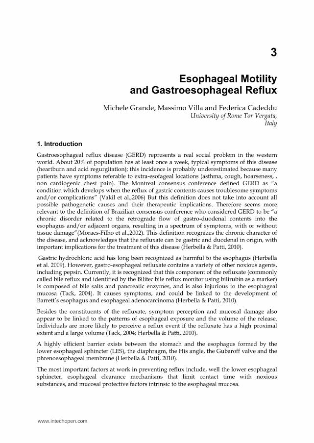
Several studies have shown that oesophageal function is impaired in patients with reflux oesophagitis, especially in high grade oesophagitis. Patients with reflux oesophagitis have reduced lower oesophageal sphincter pressures (figure 1), an increased incidence of failed peristalsis (figure 2), reduced distal peristaltic amplitudes, slower velocity of propagation and in some studies shorter duration of contractions (Timmer et al.,1994). Two groups have reported that healing of oesophagitis does not improve impaired oesophageal motility (Katz et al.,1986, Singh et al.,1992).
Fig. 1. Esophageal manometry in patients with gastroesophageal reflux with perfusion catheter to 6-way, three of which radial. Presence of low pressure LES and waves of low amplitude in the distal esofagus (45 cm).
www.intechopen.com
Gastroesophageal Reflux Disease
64
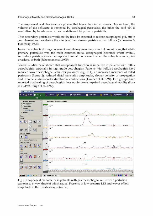
An extension of the clearance time has been reported in about 50% of patients with esophagitis (Kahrilas, 1986). The frequency of abnormalities of peristalsis increases with the severity of reflux reaching 20% in patients with GERD without esophageal lesions, 25% in those with moderate esophagitis, and 48% in those with severe esophagitis (Kahrilas, 1986). A weak or ineffective peristalsis (waves of amplitude less than 30/40 mm Hg) is not able to eliminate acid reflux from the esophagus (Kahrilas, 1986).
Fig. 2. Esophageal manometry with perfusion catheter to 6-way, three of which radial. Failed peristaltis in patients with gastroesophageal reflux.
Even lack of salivary function, characterized by reduced secretion or a reduced capacity for neutralization by saliva may result in a prolongation of esophageal clearance (Achem, 1997). For example, smokers have a reduced salivary secretion than nonsmokers and therefore have a higher incidence of GERD.
The velocity of propagation has been shown to be slower in patients with reflux oesophagitis. Gill et al have reported shorter durations of contraction in this condition (Gill et al.,1986). On the other hand, Singh et al have found a longer durations of contraction in patients with GERD compared with the controls (Singh et al.,1992). Oesophageal transit and acid clearance have also been shown to be slower in these patients (Singh et al.,1992). In agreement with those observations Timmer et al found, comparing oesophageal motility in patients with low grade oesophagitis with motility data obtained in a matched normal control group, reduced propagation velocity and duration of peristaltic contractions, with
www.intechopen.com
Esophageal Motility and Gastroesophageal Reflux
65
increase in the number of non transmitted contractions in patients with grade I and II oesophagitis. Peristaltic amplitude was not shown to be impaired (Timmer et al.,1994).
Defective peristalsis is associated with severe GERD, both in terms of symptoms and of
mucosal damage (Diener et al.,2001). As matter of fact, the composite reflux score
(DeMeester score) includes in its calculation two indirect measurements of esophageal
clearance (number of reflux episodes longer than 5 min and length of the longest episode).
In addition, the average esophageal clearance time can be calculated by dividing the total
minutes the pH is below 4 by the number of reflux episodes (Johnson & DeMeester, 1974).
This association also explains the high prevalence and severity of GERD in systemic diseases
that affects peristalsis, such as connective tissue disorders (Patti et al.,2008).
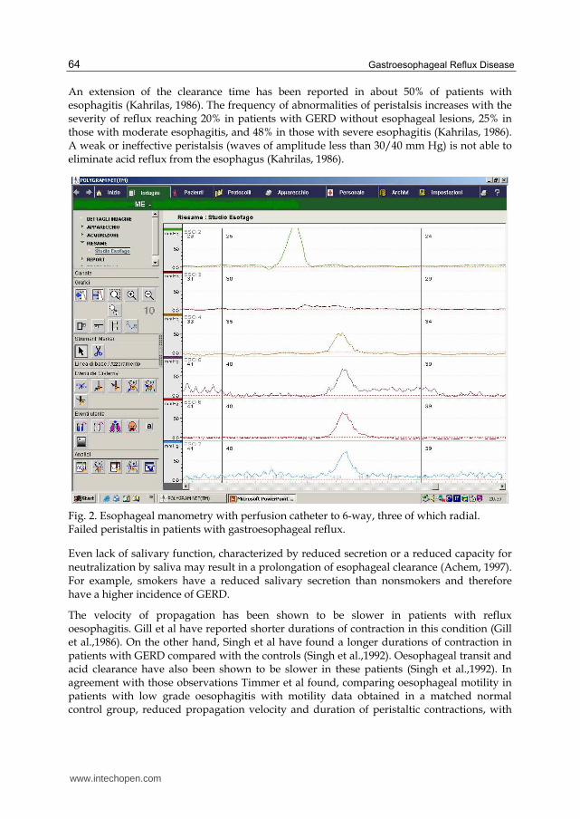
Fig. 3. Track condensed 24-hour pH-metry with antimony probe, the heart indicates the
presence of reflux symptoms. Patients with pathological acid reflux (pH <4 lasting more
than 5 minutes) in erect position (Number total reflux : 450; total reflux > 5 min : 19;
duration of longest reflux : 80 min; total reflux time pH<4 : 414 min).
It is known that 40%-50% of patients with GERD have abnormal peristalsis (Diener et
al.,2001). This dysmotility is particularly severe in about 20% of patients because of very low
amplitude of peristalsis and/or abnormal propagation of the peristaltic waves (ineffective
esophageal motility) (Patti & Perretta, 2003). Esophageal clearance is slower than normal,
therefore, the refluxate is in contact with the esophageal mucosa for a longer period of time
and it is able to reach more often the upper esophagus and pharynx (figures 3-5). Thus,
these patients are prone to severe mucosal injury (including Barrett’s esophagus) and
frequent extra-esophageal symptoms such as cough (Herbella & Patti, 2010; Patti & Perretta,
2003; Meneghetti et al.,2005).
N acid refluxes 450; N refluxes>5 min 19 Longest reflux 80 min; Interval pH<4: 414 min
www.intechopen.com
Gastroesophageal Reflux Disease
66
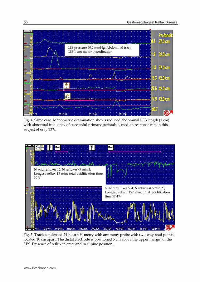
Fig. 4. Same case. Manometric examination shows reduced abdominal LES length (1 cm) with abnormal frequency of successful primary peristalsis, median response rate in this subject of only 33%.
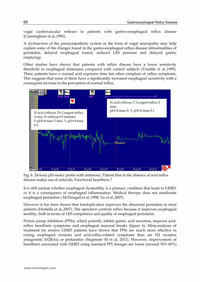
Fig. 5. Track condensed 24-hour pH-metry with antimony probe with two-way read points located 10 cm apart. The distal electrode is positioned 5 cm above the upper margin of the LES. Presence of reflux in erect and in supine position.
LES pressure 40.2 mmHg; Abdominal tract LES 1 cm; motor incordination
N acid refluxes 16; N refluxes>5 min 2; Longest reflux 13 min; total acidification time 30%
N acid refluxes 594; N refluxes>5 min 28; Longest reflux 157 min; total acidification time 57.4%
www.intechopen.com

Esophageal Motility and Gastroesophageal Reflux
67
In addition to primary peristalsis alterations, patients with GERD have secondary peristalsis impairments and in most of them the esophageal distension is not capable of triggering secondary peristaltic contractions (Williams et al.,1992). As this deficit can occur even in subjects with normal primary peristalsis has been suggested that the phenomenon is due to an altered response to esophageal acid reflux and / or relaxing (Schoeman & Holloway, 1995).
Patients with reflux disease have considerably lower secondary peristaltic response rates
than have aged matched controls with most patients failing to trigger any peristaltic
response at all (Schoeman & Holloway, 1995). This finding supports and extends earlier
findings on spontaneous reflux episodes, which showed that secondary peristalsis occurred
less frequently after reflux in patients with reflux oesophagitis compared with normal
subjects (Dodds et al.,1990).
Secondary peristalsis is a reflex response to oesophageal distension, the defect may lie in the
oesophageal motor nerves or muscles oesophageal sensation, the central integrative
mechanisms or a combination of these. Most patients with abnormal primary peristalsis also
had abnormal secondary peristalsis and in these patients we postulate that the defect lies in
the efferent limb of the motor pathway (Schoeman & Holloway, 1995). Most patients with
abnormal secondary peristalsis, however, had normal primary peristalsis. Because
secondary peristalsis seems to share a common motor pathway with primary peristalsis this
side of the reflex would seem to be intact, implying that the defect in secondary peristalsis is
due either to an abnormality of oesophageal sensation or in the integration of sensory
information with the motor component of the reflex (Schoeman & Holloway, 1995). This
hypothesis is supported by the findings of Williams et al who noted that the distension
threshold required to trigger a motor response was higher in patients with oesophagitis than
in healthy controls (Williams et al.,1992). Others, however, have found no difference in the
threshold volume required to trigger oesophageal motor responses using slow (1 ml/s)
infusions (Corazziari et al.,1986). Differences in the methods of these studies, however,
make direct comparisons of these results difficult. Secondary peristalsis can effectively clear
almost all of an injected acid bolus from the oesophagus leaving a negligible residual
volume (Schoeman & Holloway, 1995). However, changes in oesophageal pH would be
unlikely until neutralisation of the residual acid by bicarbonate rich saliva delivered by
primary peristalsis (Schoeman & Holloway, 1995). Thus secondary peristalsis would not by
itself be expected to restore oesophageal pH, but to complement and accelerate the effects of
the primary peristalsis that follows. During the day when patients are awake, any effect of
defective secondary peristalsis on acid clearance will be minimized by frequent primary
peristalsis. Secondary peristalsis is likely to be more important, however, during sleep when
the rate of primary peristalsis is substantially reduced (Orr et al.,1981).
While there is no doubt that these abnormalities are commonly present in patients with reflux oesophagitis, it’s debated whether these are primary phenomena or the consequences of repetitive injury and inflammation caused by acid reflux. Currently, the most reliable data is that the abnormalities of oesophageal motor function in patients with reflux oesophagitis do not improve after complete healing of oesophagitis (Singh et al.,1992). This suggests that oesophageal dysmotility is a primary phenomenon and not a consequence of injury and inflammation. In that regard were detected an high prevalence of impairment of
www.intechopen.com
Gastroesophageal Reflux Disease
68
vagal cardiovascular reflexes in patients with gastro-oesophageal reflux disease (Cunningham et al.,1991).
A dysfunction of the parasympathetic system in the form of vagal neuropathy may help explain some of the changes found in the gastro-esophageal reflux disease (abnormalities of peristalsis, delayed esophageal transit, reduced LES pressure and delayed gastric emptying).
Other studies have shown that patients with reflux disease have a lower sensitivity threshold to esophageal distension compared with control subjects (Trimble et al.,1995). These patients have a normal acid exposure time but often complain of reflux symptoms. This suggests that some of them have a significantly increased esophageal sensitivity with a consequent increase in the perception of normal reflux.
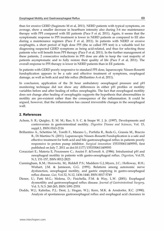
Fig. 6. 24-hour pH-metry probe with antimony. Patient that in the absence of acid reflux disease makes use of antacids. Functional heartburn ?
It is still unclear whether esophageal dysmotility is a primary condition that leads to GERD, or it is a consequence of esophageal inflammation. Medical therapy does not ameliorate esophageal peristalsis ( McDougall et al.,1998; Xu et al.,2007).
However it has been shown that fundoplication improves the abnormal peristalsis in most patients (Herbella et al.,2007). The operation controls reflux because it improves esophageal motility, both in terms of LES competence and quality of esophageal peristalsis.
Proton pump inhibitors (PPIs), which potently inhibit gastric acid secretion, improve acid-reflux heartburn symptoms and esophageal mucosal breaks (figure 6). Meta-analyses of treatment for erosive GERD patients have shown that PPIs are much more effective in curing esophageal erosions and acid-reflux-related symptoms than are H2 receptor antagonists (H2RAs) or prokinetics (Sugimoto M et al, 2011). However, improvement of heartburn associated with NERD using standard PPI dosages are lower (around 30%-60%)
N acid refluxes 7; Longest reflux 2 min; pH>4 time 0; % pH<4 time 0.1
N acid refluxes 29; Longest reflux 2 min; N refluxes>5 minutes 0 ;pH>4 time 5 min; % pH<4 time 0.4
www.intechopen.com
Esophageal Motility and Gastroesophageal Reflux
69
than for erosive GERD (Sugimoto M et al, 2011). NERD patients with typical symptoms, on average, show a smaller decrease in heartburn intensity also during 3-6 mo maintenance therapy with PPI compared with EE patients (Pace F et al, 2011). Again, it seems that the symptomatic response to PPI treatment is lower in NERD patients as compared to EE also during a maintenance regimen (Pace F et al, 2011). In patients with NERD or erosive esophagitis, a short period of high dose PPI (the so called PPI test) is a valuable tool for diagnosing suspected GERD symptoms as being acid-related, and thus for selecting those patients who will benefit from PPI therapy (Pace F et al, 2011). In the further management of these patients, 2 consecutive reductions in PPI dose are able to keep the vast majority of patients asymptomatic and to fully restore their quality of life (Pace F et al, 2011). The overall response to PPI therapy is lower in NERD patients than in EE patients.
In patients with GERD poorly responsive to standard PPI dose, laparoscopic Nissen-Rossetti fundoplication appears to be a safe and effective treatment of symptoms, esophageal damage, as well as both acid and bile reflux (Brillantino A et al, 2011).
In conclusion, application of the 24 hour ambulatory oesophageal pressure and pH monitoring technique did not show any differences in either pH profiles or motility variables before and after healing of reflux oesophagitis. The fact that oesophageal motility does not change after healing of oesophagitis supports the hypothesis that abnormalities in motility are pre-existent rather than the consequence of the inflammation. It could be argued, however, that the inflammation has caused irreversible changes in the oesophageal wall.
2. References
Achem, S. R.; Quigley, E. M. M.; Rao, S. S. C. & Snape W. J. Jr. (1997). Developments and
controversies in gastrointestinal motility. Digestive Disease and Sciences, Vol. 15,
suppl.1, ISSN 0163-2116
Brillantino A., Schettino M., Torelli F., Marano L., Porfidia R., Reda G., Grassia M., Braccio
B., Di Martino N. (2011). Laparoscopic Nissen–Rossetti Fundoplication is a safe and
effective treatment for both acid and bile gastroesophageal reflux in patients poorly
responsive to proton pump inhibitor. Surgical innovation 1553350611409593, first
published on July 7, 2011 as doi:10.1177/1553350611409593
Corazziari E.; Materia E, Pozzessere C.; Anzini F. &Torsoli A. (1986). Intraluminal pH and
oesophageal motility in patients with gastro-oesophageal reflux. Digestion, Vol.35,
N.3, 151-157, ISSN 0012-2823
Cunningham, K.M.; Horowitz, M,; Riddell P.S.; Maddern G.J.;Myers, J.C.; Holloway, R.H.;
Wishart, J.M. & Jamieson, G.G. (1991). Relations among autonomic nerve
dysfunction, oesophageal motility, and gastric emptying in gastro-oesophageal
reflux disease. Gut, Vol.32, N.12, 1436-1440, ISSN 0017-5749
Diener, U.; Patti M.G.; Molena, D.; Fisichella, P.M. & Way, L.W. (2001). Esophageal
dysmotility and gastroesophageal reflux disease. Journal of Gastrointestinal Surgery,
Vol. 5, N.3: 260-265, ISSN 1091-255X
Dodds, W.J.; Kahrilas, P.J.; Dent, J.; Hogan, W.J.; Kern, M.K. & Arndorfer, R.C. (1990).
Analysis of spontaneous gastroesophageal reflux and esophageal acid clearance in
www.intechopen.com

Gastroesophageal Reflux Disease
70
patients with reflux esophagitis. Journal of Gastrointestinal Motility,Vol 2, 79-89,
ISBN 9783923022199
Gill, R.C.; Bowes, K.L.; Murphy, P.D. & Kingma, Y.J. (1986). Esophageal motor abnormalities
in gastroesophageal reflux and the effects of fundoplication. Gastroenterology, Vol.
91, N.2, 364-369, ISSN 0016-5085
Herbella, F.A.; Nipominick, I. & Patti, M.G. (2009). From sponges to capsules. The history of
esophageal pH monitoring. Disease of the Esophagus; Vol.22, N.2, 99-103, ISSN 1120-
8694
Herbella, F.A. & Patti, M. G. (2010). Gastroesophageal reflux disease: From pathophysiology
to treatment. World Journal of Gastroenterology, Vol.16, N.30, 3745-3749, ISSN 1007-
9327
Herbella, F.A.; Tedesco, P.; Nipomnick, I.; Fisichella, P.M. & Patti, M.G. (2007). Effect of
partial and total laparoscopic fundoplication on esophageal body motility. Surgical
Endoscopy, Vol.21, N.2, 285-288, ISSN 0930-2794
Lowe, R. C. (2006). Medical management of gastroesophageal reflux disease. GI Motility
online doi:10.1038/gimo54
Johnson, L.F. & Demeester, T.R. (1974). Twenty-four-hour pH monitoring of the distal
esophagus. A quantitative measure of gastroesophageal reflux. American Journal of
Gastroenterology, Vol.62, N.4, 325-332, ISSN 1948-9498
Kahrilas, P.J.; Dodds, W.J.; Hogan, W.J.; Kern, M.; Arndorfer, R.C. & Reece, A. (1986).
Esophageal peristaltic dysfunction in peptic esophagitis. Gastroenterology, Vol.91,
N.4, 897-904, ISSN 0016-5085
Katz, P.O. (2003). Optimizing medical therapy for gastroesophageal reflux disease: state of
the art. Reviews in Gastroenterological Disorders, Vol.3, N.2, 59–69, ISSN 1533-001X
Katz, P.O.; Knuff, T.E.; Benjamin, S.B. & Castell, D.O. (1986). Abnormal esophageal
pressures in reflux esophagitis: cause or effect? American Journal of Gastroenterology,
Vol.81, N.9, 744-746, ISSN 0002-9270
McDougall, N.I.; Mooney, R.B.; Ferguson, W.R.; Collins, J.S.; Mc-Farland, R.J. & Love, A.H.
(1998). The effect of healing oesophagitis on oesophageal motor function as
determined by oesophageal scintigraphy and ambulatory oesophageal motility/pH
monitoring. Alimentary Pharmacology & Therapeutics, 12, N.9, 899-907, ISSN 0269-
2813
Meneghetti, A.T.; Tedesco, P.; Damani, T. & Patti, M.G. (2005). Esophageal mucosal damage
may promote dysmotility and worsen esophageal acid exposure. Journal of
Gastrointestinal Surgery, Vol.9, N.9, 1313-1317, ISSN 1091-255X.
Mittal, R.K.; Holloway, R.H.; Penagini, R.; Blackshaw, L.A. & Dent, J. (1995). Transient lower
esophageal sphincter relaxation. Gastroenterology, Vol.109, N.2, 601-610, ISSN 0016-
5085
Moraes-Filho, J.; Cecconello, I.; Gama-Rodrigues, J.; Castro, L.; Henry, M.A.; Meneghelli,
U.G. & Quigley, E. (2002). Brazilian consensus on gastroesophageal reflux disease:
proposals for assessment, classification, and management. American Journal of
Gastroenterology, Vol.97, N.2, 241-248, ISSN 0002-9270
Orr, W.C.; Robinson, M.G. & Iohnson, L.F. (1981). Acid clearance during sleep in the
pathogenesis of reflux esophagitis. Digestive Diseases and Sciences,Vol.26. N.5, 423-
427, ISSN 0163-2116
www.intechopen.com

Esophageal Motility and Gastroesophageal Reflux
71
Pace F, Riegler G, de Leone A, Dominici P, Grossi E, the EMERGE Study Group (2011).
Gastroesophageal reflux disease management according to contemporary
international guidelines: A translational study. World J Gastroenterol, Vol. 17, N. 9,
1160-1166, ISSN 1007-9327 (print) ISSN 2219-2840 (online)
Patti, M.G.; Gasper, W.J.; Fisichella, P.M.; Nipomnick, I. & Palazzo, F. (2008).
Gastroesophageal reflux disease and connective tissue disorders: pathophysiology
and implications for treatment. Journal of Gastrointestinal Surgery, 12(11): 1900-1906,
ISSN 1091-255X
Patti, M.G. & Perretta, S. (2003). Gastro-oesophageal reflux disease: a decade of changes.
Asian Journal of Surgery,Vol.26, N.1, pp. 4-6, ISSN 1015-9584
Pope, C. E. (1994). Acid-reflux disorders. New England Journal of Medicine, Vol.331, N.10, pp.
656-660, ISSN 0028-4793
Tack, J.; Koek, G.; Demedts, I.; Sifrim, D. & Janssens, J. (2004). Gastroesophageal reflux
disease poorly responsive to single-dose proton pump inhibitors in patients
without Barrett's esophagus: acid reflux, bile reflux, or both? American Journal of
Gastroenterology, Vol. 99, N.6,pp. 981-988, ISSN 0002-9270
Schoeman, M.N. & Holloway, R.H. (1995). Integrity and characteristics of secondary
oesophageal peristalsis in patients with gastro-oesophageal reflux disease. Gut,
Vol.36, N.4, pp. 499-504, ISSN 0017-5749
Schoeman, M.N.; Tippett, M.D.; Akkermans, L.M.; Dent, J. & Holloway, R.H. (1995).
Mechanisms of gastroesophageal reflux in ambulant healthy human subjects.
Gastroenterology, Vol.108, N.1, pp. 83-91, ISSN 0016-0012
Singh, P.; Adamopoulos, A.; Taylor, R.H. & Colin-Jones, D.G. (1992). Oesophageal motor
function before and after healing of oesophagitis. Gut, Vol.33, N.12, pp.1590-1596,
ISSN 0017-5749
Sugimoto M.; Nishino M.; Kodaira C.; Yamade M.; Uotani T.; Ikuma M.; Umemura K.;
Furuta T. (2011). Characteristics of non-erosive gastroesophageal reflux disease
refractory to proton pump inhibitor therapy. World J Gastroenterol, Vol. 17, N. 14:
1858-1865, ISSN 1007-9327 (print) ISSN 2219-2840 (online)
Timmer, R.; Breumelhof, R.; Nadorp, J.H. & Smout, A.J. (1994). Oesophageal motility and
gastro-oesophageal reflux before and after healing of reflux oesophagitis. A study
using 24 hour ambulatory pH and pressure monitoring. Gut, Vol.35, N.11, 1519-
1522, ISSN 0017-5749
Trimble, K.C.; Pryde, A. & Heading, R.C. (1995). Lowered oesophageal sensory thresholds
in patients with symptomatic but not excess gastro-oesophageal reflux: evidence
for a spectrum of visceral sensitivity in GORD. Gut, Vol.37, N.1, 7-12, ISSN 0017-
5749.
Vakil, N.; van Zanten, S.V.; Kahrilas, P.; Dent, J. & Jones, R. (2006). The Montreal definition
and classification of gastroesophageal reflux disease: a global evidence-based
consensus. American Journal of Gastroenterology, Vol.101, N.8, pp. 1900-1920, ISSN
0002-9270
Williams, D.; Thompson, D.G.; Marples, M.; Heggie, L.; O'Hanrahan, T.; Mani, V. &
Bancewicz, J. (1992). Identification of an abnormal esophageal clearance response to
intraluminal distention in patients with esophagitis. Gastroenterology, Vol.103, N.3,
pp. 943-953, ISSN 0016-0012
www.intechopen.com

Gastroesophageal Reflux Disease
72
Xu, J.Y.; Xie, X.P.; Song , G.Q. & Hou, X.H. (2007). Healing of severe reflux esophagitis with
PPI does not improve esophageal dysmotility. Diseases of the Esophagus, Vol.20, N.4,
pp. 346-352, ISSN 1120-8694
www.intechopen.com
Gastroesophageal Reflux DiseaseEdited by Prof. Mauro Bortolotti
ISBN 978-953-51-0314-1Hard cover, 186 pagesPublisher InTechPublished online 16, March, 2012Published in print edition March, 2012
InTech EuropeUniversity Campus STeP Ri Slavka Krautzeka 83/A 51000 Rijeka, Croatia Phone: +385 (51) 770 447 Fax: +385 (51) 686 166www.intechopen.com
InTech ChinaUnit 405, Office Block, Hotel Equatorial Shanghai No.65, Yan An Road (West), Shanghai, 200040, China
Phone: +86-21-62489820 Fax: +86-21-62489821
Gastroesophageal reflux disease affects many patients. This disease not only lowers their quality of life, but italso threatens some of them with an underhand risk of cancer. Additionally, it becomes an economic burdenfor the patients and society. The aim of this book on gastroesophageal reflux disease is to provide advice andguidance to gastroenterologists to help them understand and manage some aspects of this proteiformdisease.
How to referenceIn order to correctly reference this scholarly work, feel free to copy and paste the following:
Michele Grande, Massimo Villa and Federica Cadeddu (2012). Esophageal Motility and GastroesophagealReflux, Gastroesophageal Reflux Disease, Prof. Mauro Bortolotti (Ed.), ISBN: 978-953-51-0314-1, InTech,Available from: http://www.intechopen.com/books/gastroesophageal-reflux-disease/motor-impairments-in-gastroesophageal-reflux-disease

© 2012 The Author(s). Licensee IntechOpen. This is an open access articledistributed under the terms of the Creative Commons Attribution 3.0License, which permits unrestricted use, distribution, and reproduction inany medium, provided the original work is properly cited.