04308440 achkar esophageal motility...
TRANSCRIPT

1

Esophageal motility disorders, are defined really based on x-ray or motility testing. But, the
clinical context is one of chest pain, dysphagia, in the absence of any obstruction
2
There is a case history to start us off. This is a 36 year old woman who presents with chest
pain occurring occasionally at night, heart burn, regurgitation of food and acid, and a rare
trouble swallowing.
3
So, she is managed with PPI, told to elevate the head of the bed, and some of the
recommendations we talked about when we discussed management of GERD, told to avoid
late meals, and she returns two months later and says, “I’m not any better.”
4
What to do next? Is it an endoscopy, an x-ray, or should we double the PPI dose?
5

None of the above. What to do next is to take a more detailed history, because this is not
your classic GERD. There is not even partial improvement. So now, you take a more detailed
history, and what you find out. is that the dysphagia that was in your history initially as rare
has actually occurred to solids and liquids equally. It has led to regurgitated food, and when
you ask the patient, it looks undigested, therefore it has sat in the esophagus and come up
without reaching the stomach. And, when she eats she occasionally reports chest pain. So
now, you are on the right track because you suspect a motor disorder
6

So, there are key questions to be asked when you’re trying to diagnose dysphagia. The first
is, is it truly dysphagia? Patient will say, “I eat and it sits there. And, you translate that as
food sticking, and in fact they are describing post prandial fullness. And, what I ask patient,
I don’t say, “Do you have trouble swallowing?” Because most patients swallowing is a
voluntary phase. And, they will say no, because they don’t perceive that the food sticking
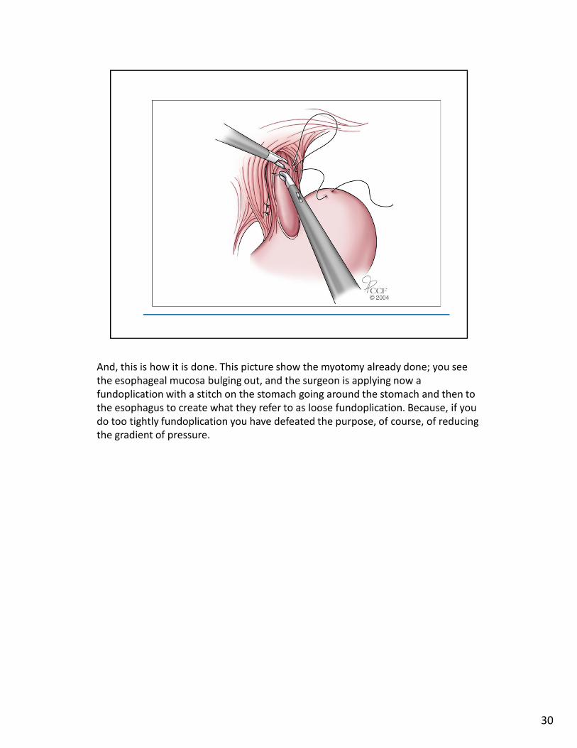
below in the retro sternal area is actually trouble swallowing. So I say, I ask patient, “Do you
have food sticking?” And, I always get the answer I’m looking for.
The second question is, is it dysphagia for solids only, liquids or both? Because if it’s solids
only, it’s mostly mechanical cause. Is it intermittent or progressive? Again, a stricture will
cause sticking of a steak from time to time whereas achalasia will cause dysphagia on a
regular basis and in increasing frequency. And, finally, at what level is the obstacle? Now,
this is strictly again because for oropharyngeal dysphasia it is very easy clinically to
recognize that the trouble is in the throat. But, beyond that, patients will tell you that the
food stick in their throat when it’s actually stuck at the GE junction because of the referred
discomfort
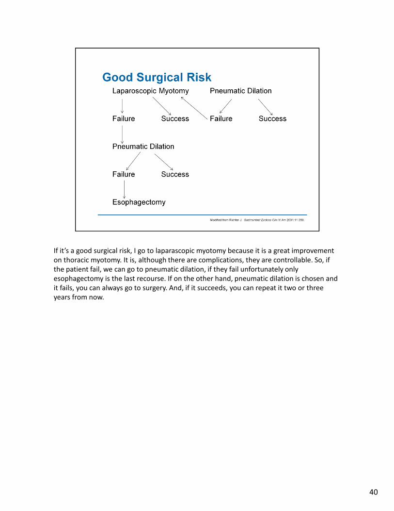
7

So, the other debate is should we obtain EGD or x-ray first? And, those debates go on and
on between surgeon, the gastroenterologist, and from a practical standpoint the answer to
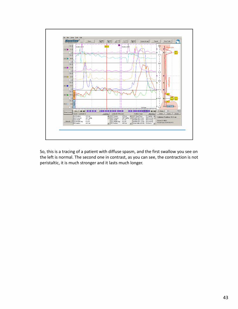
my, to me is very simple. If the patient is complaining of solids food dysphagia only, and it’s
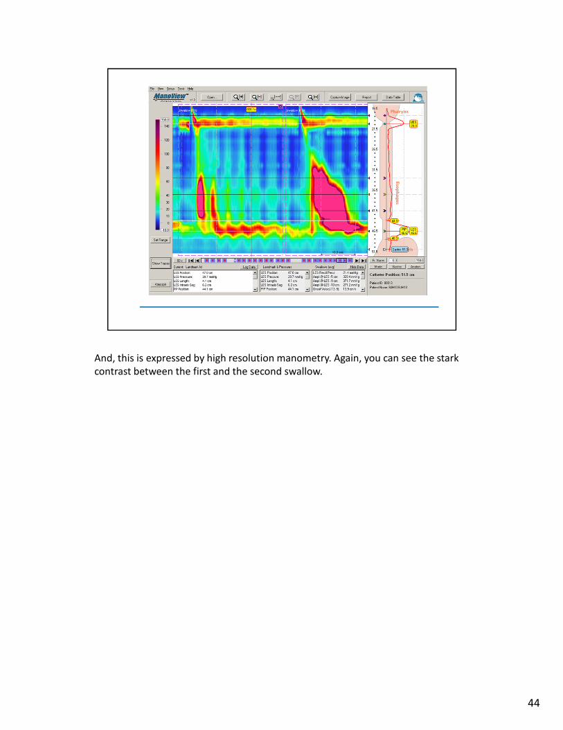
intermittent, the diagnosis is going to be either a stricture or a cancer, or a Schatzki ring
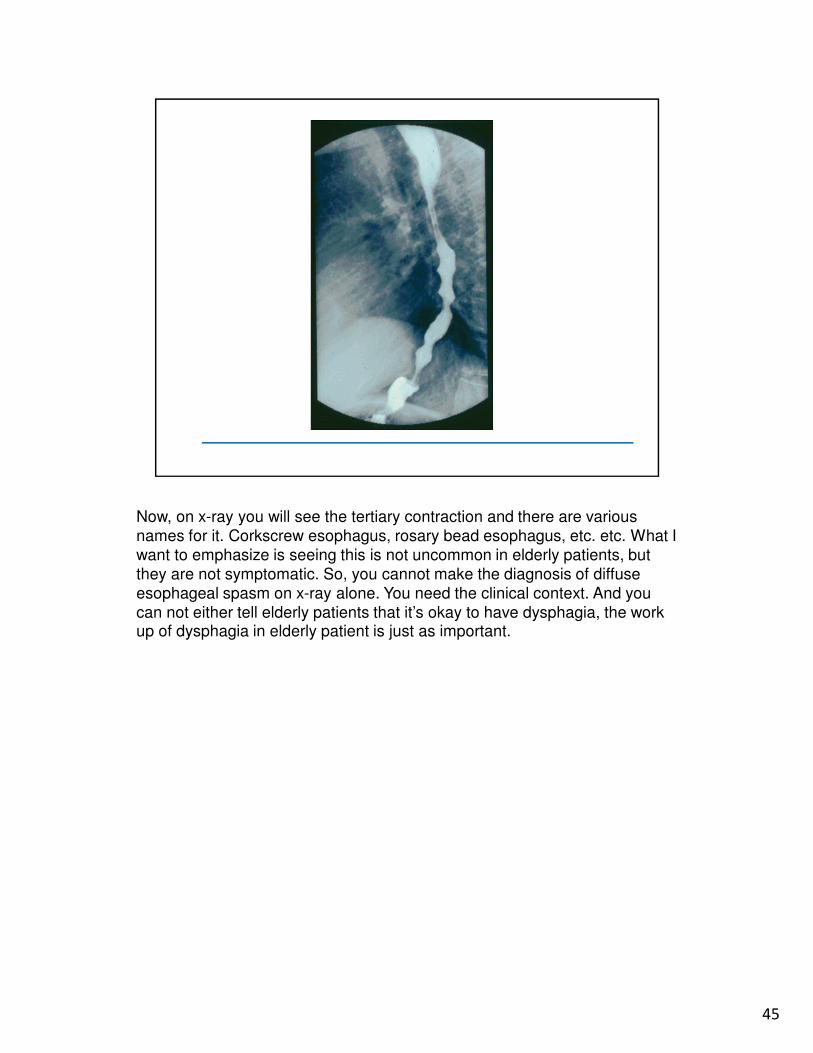
And, I go for EGD first because not only will I make the diagnosis, but I can apply treatment
at the same time and the patient consent can be obtained beforehand. If, on the other
hand, patients are complaining now of solids and liquid dysphagia, and it is getting worse
with time, I would start with an x-ray because it’s going to give me an idea of where the
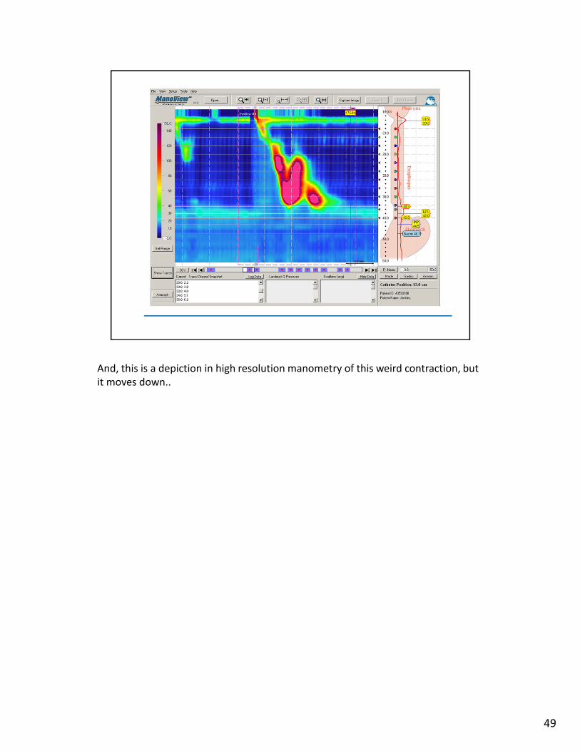
obstacle is, and if it’s a motor disorder, what the esophagus looks like.
8

How do we classify esophageal motor disorders? There are various ways to do it, and the
simplest to my mind is to follow the anatomy and the physiology. The esophagus is divided
in an upper third, which is striated muscle, under our own control, and lower two thirds,
out of our hands, smooth muscle and it transitions on between the two. So, if you separate
them this way, you will end up then in two sets of disease.
9

Striated motor disorders are basically what was called in the good old days transfer
dysphagia, meaning the transfer of the bolus from the mouth to the esophagus, or what we
call today oropharyngeal dysphagia. So, the trouble is in the upper part. Patient cannot
form a bolus, or cannot propel the bolus, or chokes on his food. And, the causes are
multiple. Any neurological disease can do it, trauma can do it, but the major cause is going
to be a stroke. And, you will see this in patients in nursing homes, or in hospitals after a
stroke.
10
On the other hand, smooth muscle disorders, there are four major categories which we will
address. Achalasia, which by far is more frequent, diffuse spasm, nutcracker esophagus,
and schleroderma. So, I will say a word about each one of those
11
Let’s start with achalasia: what is it? A-chalasia—no movement. It’s aperistalsis of the
esophagus by definition, not a single peristaltic contraction, poor relaxation of the lower
esophageal sphincter, but the lower esophageal sphincter can be high, pressure can be
high, or normal. In the old definition of the disease, we insisted on having a high pressure.
It does not matter what the pressure you start with, it’s the fact that the pressure gradient
does not break when you eat.
12

What’s the pathogenesis of the disease? All we know is that there is a patchy inflammation
of the myenteric plexus of the esophagus with decreased ganglion cells. Therefore, there is
a loss of latency gradient across the esophagus, because what the myenteric plexus does is
make the esophagus when we swallow and we initiate a swallow, the esophagus follows in
sequence, number one, two three as you go down the esophagus. And, when you lose that
progression, then the bolus is, stagnates in the esophagus and leads to aperistalsis. And,
the defect of inhibitory neurons in the LES prevent it from relaxing. In other words, when
we swallow, we inhibit the tonic contraction of the LES, and when that doesn’t occur, the
LES creates now an obstacle to the passage of the bolus.
13
What about symptoms? Well, achalasia affects all age groups, although there is a higher
preponderance in young people in their 20’s and 30’s and then a second peak in later years
in their 70’s. But, it affects all age groups, it’s typically dysphagia to both solids and liquids,
with food regurgitation, chest pain when we looked at our patient at the Cleveland Clinic
it’s present in 40 – 50%. Respiratory symptoms have been reported but are fortunately
rare, and weight loss, interestingly, is mild. You can actually have obese patients with
achalasia. And, this is a testimony to how patients can really work around that problem to
continue to eat.
14

This is a typical x-ray of a patient with achalasia. The esophagus looks atonic. Why?
Because you have barium up to the throat, and this is hard to achieve when patient
is a normal patient is swallowing barium. This esophagus is dilated, and the typical
finding is the deformity at the lower esophagus. If you look carefully you will see
the esophagus tapering to what is referred to as the “bird beak like deformity”
because it’s reminiscent of a bird’s beak.
15
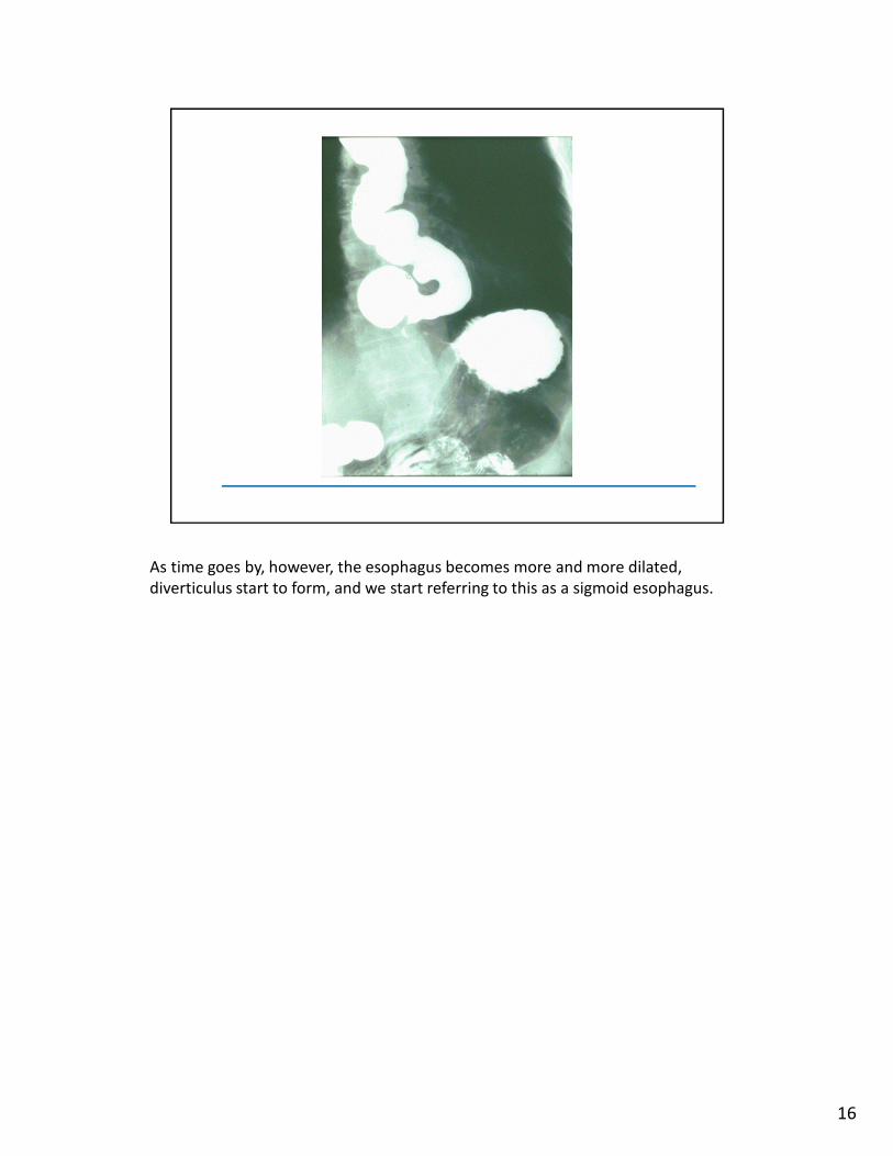
As time goes by, however, the esophagus becomes more and more dilated,
diverticulus start to form, and we start referring to this as a sigmoid esophagus.
16
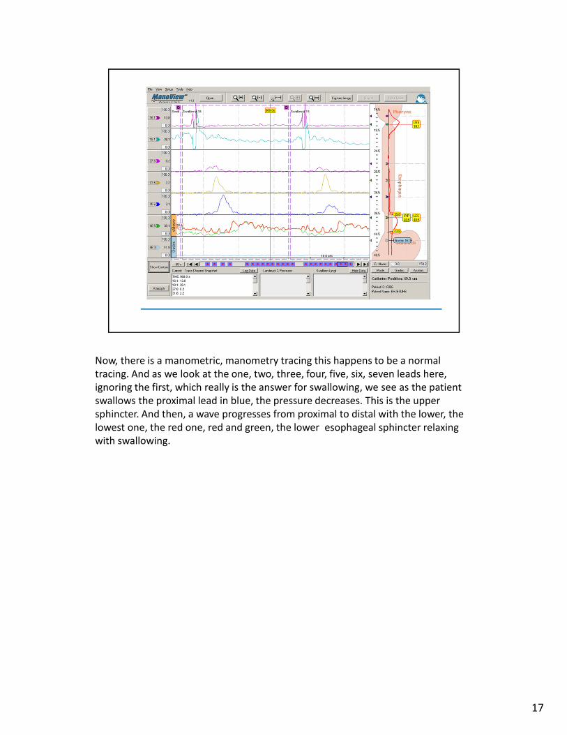
Now, there is a manometric, manometry tracing this happens to be a normal
tracing. And as we look at the one, two, three, four, five, six, seven leads here,
ignoring the first, which really is the answer for swallowing, we see as the patient
swallows the proximal lead in blue, the pressure decreases. This is the upper
sphincter. And then, a wave progresses from proximal to distal with the lower, the
lowest one, the red one, red and green, the lower esophageal sphincter relaxing
with swallowing.
17
And, this is a more modern tracing using high resolution manometry where we
have 35 sensors throughout the esophagus and it’s expressed in colors showing you
here the intensity of pressure melded by the color and the progression in time. So,
these are two normal swallows they progress nicely down the esophagus.
18

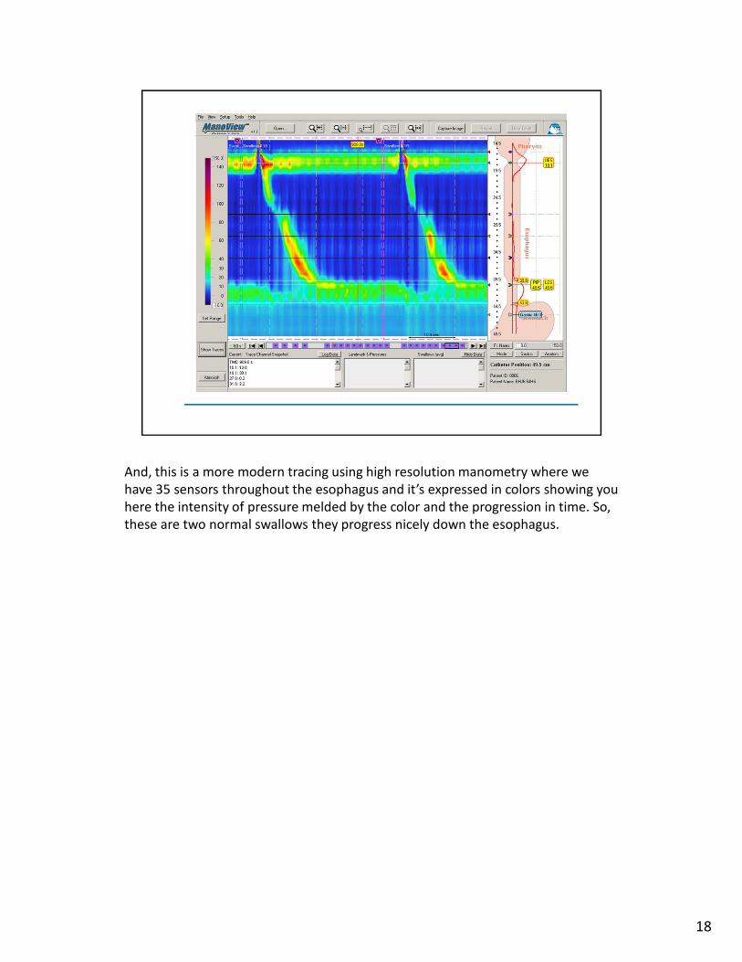
In contrast, this is a patient with achalasia Now, unlike two slides away, you see
now that the progression is not existent, the lower esophageal sphincter is not
relaxing as nicely, and this is shown better in the next slide
19

where you have now a straight line coming down, no progression, the esophagus
contracting at the same time. And, the lower esophageal sphincter pressure is not
dropping.
20
Now, what’s the treatment in achalasia? We unfortunately cannot restore the myenteric
plexus; we cannot regenerate the nerves. Our goal is limited really to reducing the gradient
of pressure represented by the LES. And, this can be achieved with one of few things
21
We can use a pharmacologic agent such as nitrates or calcium channel blockers to do it. We
can forcefully dilate the esophagus endoscopically or surgically by myotomy, and I will say a
word about botulinum toxin.
22

Now, this is a slide showing the effect of nitrate and calcium channel blocker on the
LES pressure. Once you administer either a calcium channel blocker in solid, in
broken line, or a nitrate in solid line, you will see that LES pressure decreases
significantly and the effect lasts for 40 minutes. So, presumably, the patient could
take a nitrate under the tongue before eating, and for the next half hour the LES
will be open enough to allow the presence of food.
23

Does this work? Yes, it works briefly; unfortunately there are common side effects which
particularly in the elderly, namely hypotension, make this not a practical solution. The
symptom relief is partial and in time they stop working. So, this, the pharmacotherapy of
achalasia really should be reserved for patient who are ineligible or refuse any other
treatment. And frankly, we have not used it much lately.
24

Well, how about pneumatic dilation? Pneumatic dilation is the forceful breakup actually of
the LES; you can not achieve that with a maloney or savary dilator, although some patient
do report, and there are series reporting benefit even from savary dilation. But, for more
lasting effect you have to forcefully break the fibers of the esophagus.
25
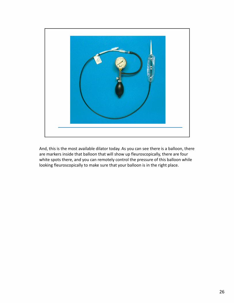
And, this is the most available dilator today. As you can see there is a balloon, there
are markers inside that balloon that will show up fleuroscopically, there are four
white spots there, and you can remotely control the pressure of this balloon while
looking fleuroscopically to make sure that your balloon is in the right place.
26

So, it’s an outpatient procedure; it’s combined with endoscopy. You need fleuroscopic
assistance, and while the technique is variable there are all kinds of recommendation as to
the time and the pressure of the balloon. I think the most prudent thing is to, while looking
under fleuroscopy as the balloon creates a waist at the LES and the LES gets effaced, you
stop, you hold it for 30 seconds, and you let down
27

Now, the perforation unfortunately is the main concern with the pneumatic dilation. And in
the best of hands it’s still 2 to 3%. It’s independent of age, I’ve seen it in young people, I’ve
seen it in 82 year old people. The role of the technique is questionable; I don’t think it’s
what you do, I think it’s the thinness of the esophageal wall. Unfortunately, when it occurs
it’s going to require thoracotomy in most patients. And so, that’s the concern of pneumatic
dilation.
28

Surgical myotomy comes up looking better than pneumatic dilation at least initially. And,
the misconception is once that you have operated, pneumatic dilation cannot be done.
That is not true; we have dilated patient after surgery as well as sent patient who have
failed pneumatic dilation to surgery. It’s of affected and accompanied by a fundoplication.
Today, it’s done laparoscopically which obviously improves the outcome and makes the
hospital stay much shorter. But, however simple it is, it should be done by an experienced
surgeon. This is not a surgery to be done by a thoracic surgeon who has done one before in
their lifetime.
29
And, this is how it is done. This picture show the myotomy already done; you see
the esophageal mucosa bulging out, and the surgeon is applying now a
fundoplication with a stitch on the stomach going around the stomach and then to
the esophagus to create what they refer to as loose fundoplication. Because, if you
do too tightly fundoplication you have defeated the purpose, of course, of reducing
the gradient of pressure.
30

And, this is now the stitches are in place and the surgeon is about to tie them up.
31
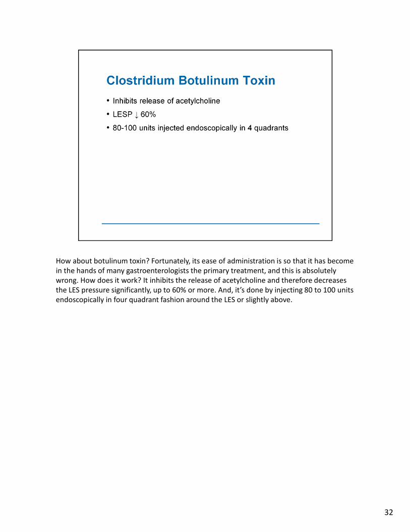
How about botulinum toxin? Fortunately, its ease of administration is so that it has become
in the hands of many gastroenterologists the primary treatment, and this is absolutely
wrong. How does it work? It inhibits the release of acetylcholine and therefore decreases
the LES pressure significantly, up to 60% or more. And, it’s done by injecting 80 to 100 units
endoscopically in four quadrant fashion around the LES or slightly above.
32

So, you mix the solution, and by the way once you’ve mixed it the shelf life is so
short that you do not mix it until the patient shows up because it is expensive.
33

And, this is a picture showing how the needle is being inserted through the
scope, and the medication is being applied around the esophagus. So, it’s fairly simple to do.
34

But, the indication really should be appropriately in elderly, frail patient in whom no other
treatment is being considered, who are a high surgical risk, who refuse other treatment or
who have pseudoachalasia, which is a condition created by cancer. And, in whom the
longevity is going to be very short. It’s inappropriate in young patients. It’s inappropriate in
healthy patients or when the diagnosis in doubt. Why? Because in long term, botulinum
toxin stops working. And, if you are going to consider surgery, the surgeon tells us that
botulinum toxin causes scarring in the area and apparently makes their surgery more
difficult. So, the first line of treatment in young, healthy patient is not botox.
35
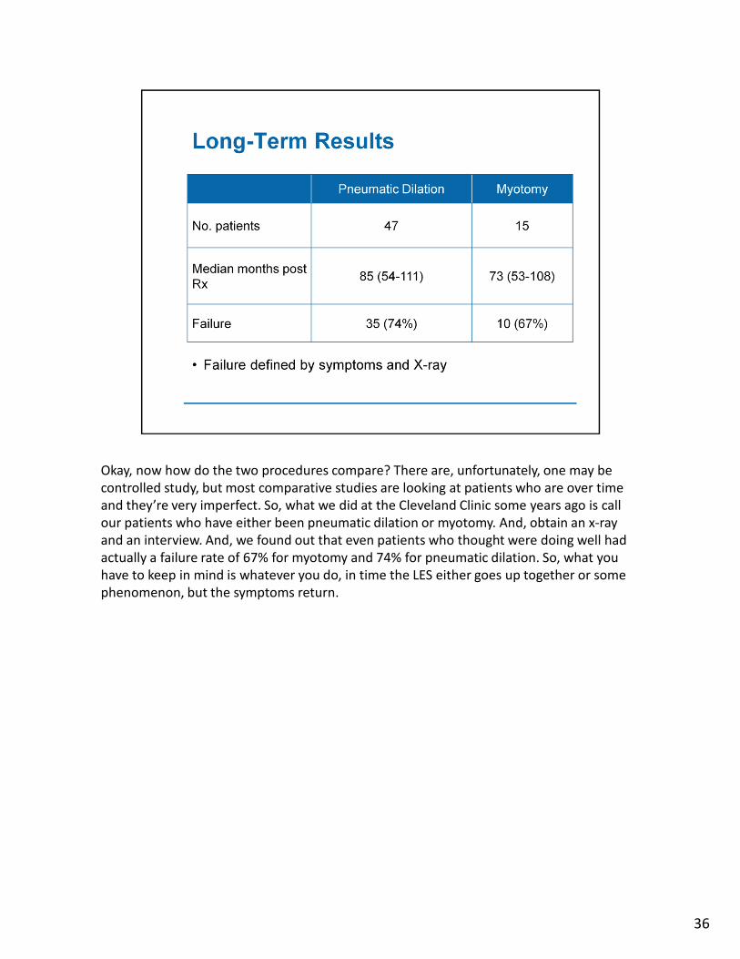
Okay, now how do the two procedures compare? There are, unfortunately, one may be
controlled study, but most comparative studies are looking at patients who are over time
and they’re very imperfect. So, what we did at the Cleveland Clinic some years ago is call
our patients who have either been pneumatic dilation or myotomy. And, obtain an x-ray
and an interview. And, we found out that even patients who thought were doing well had
actually a failure rate of 67% for myotomy and 74% for pneumatic dilation. So, what you
have to keep in mind is whatever you do, in time the LES either goes up together or some
phenomenon, but the symptoms return.
36

And, so what we do with our patient is obtain a timed barium swallow, and I will explain
the technique in a minute briefly, and we ask them to come periodically, namely once a
year to have this done, and we ask them how they’re doing and we look at the x-ray as well
in order to stay ahead of the deterioration that occurs over time. And, (slide 34) the timed
barium swallow is to give a certain amount of barium, the patient is asked to swallow it
quickly over 30 seconds and upright left posterior oblique position. X-rays at one, two, five
minutes we measured the width and the height of the barium column. And, this use is a
simple way of judging the esophageal emptying over time by comparing x-ray to the other
year after year
37
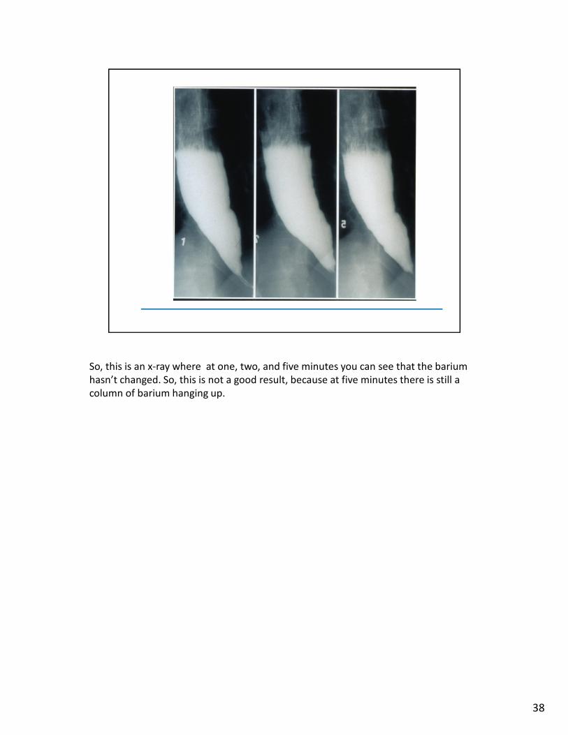
So, this is an x-ray where at one, two, and five minutes you can see that the barium
hasn’t changed. So, this is not a good result, because at five minutes there is still a
column of barium hanging up.
38

So, how do we approach the treatment of achalasia? I think you have to ask your question,
the first question, is the patient a good surgical risk or a poor surgical risk?
39
If it’s a good surgical risk, I go to laparascopic myotomy because it is a great improvement
on thoracic myotomy. It is, although there are complications, they are controllable. So, if
the patient fail, we can go to pneumatic dilation, if they fail unfortunately only
esophagectomy is the last recourse. If on the other hand, pneumatic dilation is chosen and
it fails, you can always go to surgery. And, if it succeeds, you can repeat it two or three
years from now.
40

If it’s a poor surgical risk, on the other hand, this is when I start with botox. If it succeeds,
you repeat it as needed, and most patients will need it every six months to a year. If it fails,
on the other hand, you can repeat it again, and if it fails again then you are left to either
palliation or reconsidering again intervention and assessing the risk and seeing if the
patient can submit to surgery or to pneumatic dilation.
41

A word about diffuse esophageal spasm. It’s a clinical entity expressed by dysphagia, chest
pain, spastic contraction, but in the presence of peristaltic contraction. And, this is a main
distinction with achalasia, where there is absolutely no peristalsis.
42
So, this is a tracing of a patient with diffuse spasm, and the first swallow you see on
the left is normal. The second one in contrast, as you can see, the contraction is not
peristaltic, it is much stronger and it lasts much longer.
43
And, this is expressed by high resolution manometry. Again, you can see the stark
contrast between the first and the second swallow.
44
Now, on x-ray you will see the tertiary contraction and there are various
names for it. Corkscrew esophagus, rosary bead esophagus, etc. etc. What I
want to emphasize is seeing this is not uncommon in elderly patients, but
they are not symptomatic. So, you cannot make the diagnosis of diffuse
esophageal spasm on x-ray alone. You need the clinical context. And you
can not either tell elderly patients that it’s okay to have dysphagia, the work up of dysphagia in elderly patient is just as important.
45

Now, diffuse non cardiac chest pain has been touted as a major cause of
esophageal problem. In fact, it’s not true. This is an old study taking 100 patients
with non cardiac chest pain, they’ve already seen a cardiologist, come to your clinic
and you suspect the esophagus. You do a motility testing and as you can see in
blue, about 72% have a normal motility. Only 28% have abnormal motility. Of these
28%, 48% have nutcracker esophagus, which I will talk about shortly. Non
esophageal, non specific motor disorder about a third, 36% in purple, only 10%, the
orange quadrant, 10% have diffuse spasm. So, it’s 10% of 28%. So, if you take again
100 patients with non cardiac chest pain, only 2 – 3% have diffuse spasm. So, it’s
not such a common condition.
46

The nutcracker esophagus was recognized about 20 years ago, and it’s a manometric entity.
It’s as if I’m by high amplitude, higher than 180 mg of mercury on average in the distal
esophagus, with a duration of more than 6 seconds. The contraction by definition are
peristaltic; there is no diffuse spasm here. And, it’s been one of the differential diagnosis of
chest pain.
47

It turns out with time, and this is an example of nutcracker esophagus the
contractions look wild, but they progress. The food is progressing. There may be
dysphagia rarely, but usually there isn’t.
48
And, this is a depiction in high resolution manometry of this weird contraction, but
it moves down..
49
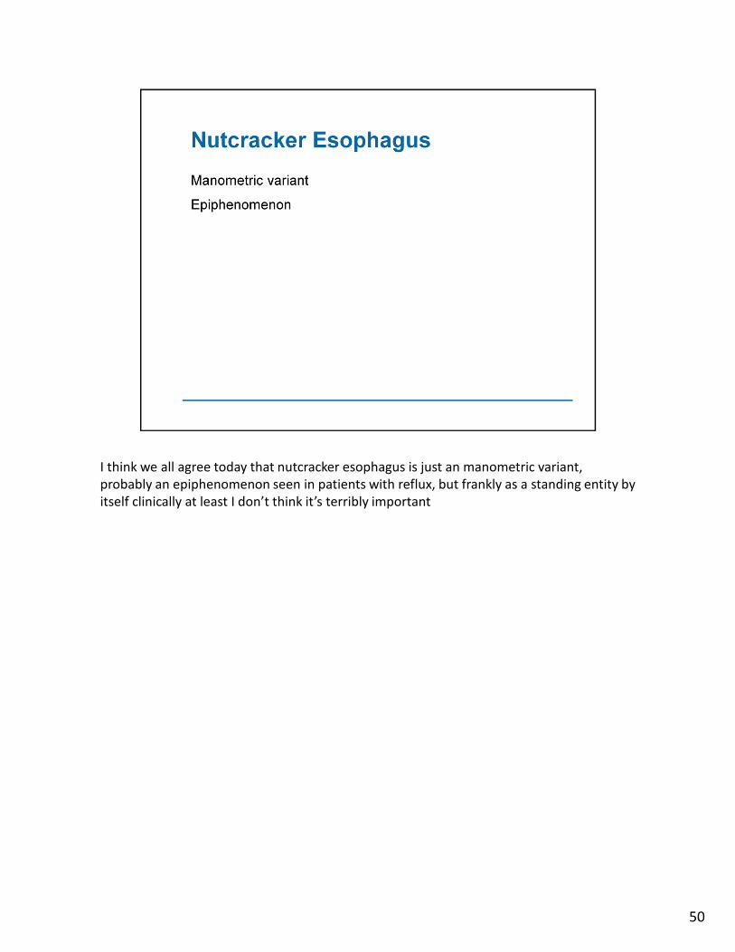
I think we all agree today that nutcracker esophagus is just an manometric variant,
probably an epiphenomenon seen in patients with reflux, but frankly as a standing entity by
itself clinically at least I don’t think it’s terribly important
50

Scleroderma, on the other hand, is obviously quite a distressing disorder, and it affects the
esophagus by creating an inadequate LES with a distal loss of peristalsis.
51

And, this is an image of an esophagus with scleroderma. As you can see, it
absolutely eight on esophagus, the lower esophageal sphincter is almost non
existent down in orange and green.
52

And if you depict that in high resolution manometry it looks like nothing is
happening.
53
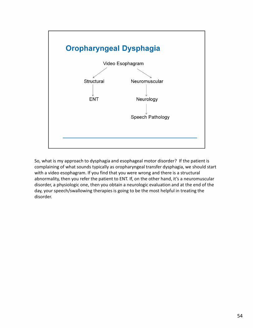
So, what is my approach to dysphagia and esophageal motor disorder? If the patient is
complaining of what sounds typically as oropharyngeal transfer dysphagia, we should start
with a video esophagram. If you find that you were wrong and there is a structural
abnormality, then you refer the patient to ENT. If, on the other hand, it’s a neuromuscular
disorder, a physiologic one, then you obtain a neurologic evaluation and at the end of the
day, your speech/swallowing therapies is going to be the most helpful in treating the
disorder.
54

If on the other hand, it’s an esophageal dysphagia, and it’s solids alone, you proceed to
endoscopy. If it’s solids and liquids, obtain an esophagram, it’s normal, proceed to
manometry. It’s abnormal, it’s a structural disorder, you go to EGD. If it’s neuromuscular,
you proceed to manometry.
55

So, my take home message is that dysphagia deserves attention at any age, x-ray and EGD
reliably and effectively predict most cases, and a carefully obtained history is absolutely
essential. Thank you.
56

57