escherichiacoli cellsurfaceperturbationanddisruption ... · luria-bertani agar (laboratorios conda,...
TRANSCRIPT
Escherichia coli Cell Surface Perturbation and DisruptionInduced by Antimicrobial Peptides BP100 and pepR*
Received for publication, April 6, 2010, and in revised form, May 21, 2010 Published, JBC Papers in Press, June 21, 2010, DOI 10.1074/jbc.M110.130955
Carla S. Alves‡, Manuel N. Melo§, Henri G. Franquelim§, Rafael Ferre¶, Marta Planas¶, Lidia Feliu¶, Eduard Bardají¶,Wioleta Kowalczyk�1, David Andreu�, Nuno C. Santos§, Miguel X. Fernandes‡, and Miguel A. R. B. Castanho§2
From the ‡Centro de Química da Madeira, Universidade da Madeira, Campus Universitario da Penteada, 9000-390 Funchal,Portugal, the §Instituto de Medicina Molecular, Faculdade de Medicina, Universidade de Lisboa, 1649-028 Lisbon, Portugal, the¶Laboratori d’Innovacio en Processos i Productes de Síntesi Organica, Departament de Química, Universitat de Girona, CampusMontilivi, 17071 Girona, Spain, and the �Department of Experimental and Health Sciences, Pompeu Fabra University, BarcelonaBiomedical Research Park, E-08003 Barcelona, Spain
The potential of antimicrobial peptides (AMPs) as an alter-native to conventional therapies iswell recognized. Insights intothe biological and biophysical properties of AMPs are thus keyto understanding their mode of action. In this study, the mech-anisms adopted by two AMPs in disrupting the Gram-negativeEscherichia coli bacterial envelope were explored. BP100 is ashort cecropin A-melittin hybrid peptide known to inhibit thegrowth of phytopathogenic Gram-negative bacteria. pepR, onthe other hand, is a novel AMP derived from the dengue viruscapsid protein. Both BP100 and pepR were found to inhibit thegrowth of E. coli at micromolar concentrations. Zeta potentialmeasurements of E. coli incubated with increasing peptideconcentrations allowed for the establishment of a correlationbetween the minimal inhibitory concentration (MIC) of eachAMP and membrane surface charge neutralization. While aneutralization-mediated killing mechanism adopted by eitherAMP is not necessarily implied, the hypothesis that surface neu-tralization occurs close to MIC values was confirmed. Atomicforce microscopy (AFM) was then employed to visualize thestructural effect of the interaction of each AMP with the E. colicell envelope. At their MICs, BP100 and pepR progressivelydestroyed the bacterial envelope, with extensive damage alreadyoccurring 2 h after peptide addition to the bacteria. A similareffect was observed for each AMP in the concentration-depen-dent studies. At peptide concentrations belowMIC values, onlyminor disruptions of the bacterial surface occurred.
Antimicrobial peptides (AMPs)3 represent a group of natu-rally occurring molecules that play a key role in the innatedefense system of virtually all organisms (1). Their robustmodeof action (2, 3), as well as their broad activity toward bacteria,
fungi, protozoa, and viruses (1, 4), makes AMPs appealing can-didates for the development of new and more efficient antimi-crobial agents. In fact, they provide an alternative to conven-tional antibiotics for the treatment of resistant pathogens (2, 3).An understanding of the mechanisms adopted by the AMPs isthus central to the advancement of thesemolecules to the statusof a new group of broad-spectrum antimicrobial agents.Characterization of AMPs has revealed a highly heterogene-
ous group of molecules, which differ in sequence, length, andstructural conformation (�-helical, �-sheet, extended, andlooped) (3, 5). Despite these variations, two functionally impor-tant features are shared by most AMPs: a net positive chargeand the ability to adopt an amphipathic structure. The net pos-itive charge allows for the electrostatic binding of the peptide tothe anionic microbial surface, while the amphipathic structurepromotes peptide insertion into the hydrophobic core of thecell membrane (2, 3). Severalmodels describingmembrane dis-ruption as a result of these direct peptide-lipid interactionshave been proposed: the barrel-stave pore model (6, 7), thetoroidal pore model (6–9), the disordered toroidal pore model(7, 10), and the carpet model (7, 9, 11). Regardless of the mech-anism adopted, a threshold peptide concentration needs to bereached for disruption of the membrane structure to occur (6,7). Alternativemechanisms involving cytoplasmic invasion andinterference of coremetabolic functions have also been consid-ered to account for the antimicrobial properties of some AMPs(2). In these cases, the microbial outer membrane must still betraversed to allow peptide penetration into the cell. Analysis ofthe peptide interactions at themembrane level is therefore cen-tral to the understanding of the mode of action of AMPs.In this study, the modes of action of two distinctly differ-
ent AMPs on the cell envelope of E. coli are explored. Thefirst peptide, BP100 (KKLFKKILKYL-NH2), is a short cati-onic cecropin A-melittin hybrid (12) obtained through acombinatorial chemistry approach (13). It has been estab-lished as an effective AMP, capable of inhibiting in vitro thegrowth of the economically important plant pathogenicGram-negative bacteria Erwinia amylovora, Pseudomonassyringae pv. Syringae, andXanthomonas axonopodis pv. vesica-toria, as well as in vivo the growth of E. amylovora (13). BP100has also been reported to display minimal cytotoxicity and lowsusceptibility to proteinase K degradation (13). Details of themembrane perturbation mechanisms adopted by this peptide
* This work was supported by the Fundacao para Ciencia e Tecnologia, Por-tugal (Fellowships SFRH/BD/24547/2005, SFRH/BD/24778/2005, andSFRH/BD/39039/2007, to C. S. A., M. N. M., and H. G. F., respectively, andProjects PTDC/QUI/69937/2006 and REEQ/140/BIO/2005).
1 Fellow in the Juan de la Cierva Program of the Spanish Ministry of Scienceand Innovation.
2 To whom correspondence should be addressed: Unidade Bioquímica Física,Instituto de Medicina Molecular, Faculdade de Medicina da Universidadede Lisboa, Av. Professor Egas Moniz, 1649-028, Lisboa, Portugal. Tel.: 351-217985136; Fax: 351-217999477; E-mail: [email protected].
3 The abbreviations used are: AMP, antimicrobial peptide; MIC, minimal inhib-itory concentration; AFM, atomic force microscopy; MHB, Mueller HintonBroth; PLL, poly-L-lysine.
THE JOURNAL OF BIOLOGICAL CHEMISTRY VOL. 285, NO. 36, pp. 27536 –27544, September 3, 2010© 2010 by The American Society for Biochemistry and Molecular Biology, Inc. Printed in the U.S.A.
27536 JOURNAL OF BIOLOGICAL CHEMISTRY VOLUME 285 • NUMBER 36 • SEPTEMBER 3, 2010
by guest on October 1, 2018
http://ww
w.jbc.org/
Dow
nloaded from
have largely been revealed through biophysical studies (14). Astrong selectivity toward anionic bacterial membrane modelswas detected for BP100, with a strong correlation between theclosely coupled processes of charge neutralization, permeabili-zation, and translocation being identified. The second peptide,pepR (LKRWGTIKKSKAINVLRGFRKEIGRMLNILNRRRR),is derived from the putative RNA-binding domain of the denguevirus capsid protein (15). The structural features of this peptide asobserved in the capsid protein (i.e. cationic, amphipathic �-helix)seem to point to a potentially potent AMP.Here, we present the characterization of BP100 and pepR
effects toward E. coli, both at cellular and molecular levels.Antimicrobial susceptibility and surface charge studies wereused to explore the concept of a neutralization-mediated killingmechanism adopted by either AMP. In addition to this, AFMwas used to assess the bactericidal effect of each AMP on thecell envelope morphology of E. coli, a representative Gram-negative bacterium. It is worth stressing that both peptides arenot effective against Gram-positive bacteria, for which otherconsiderations may apply.
EXPERIMENTAL PROCEDURES
Peptide Syntheses—BP100 and pepR were synthesized asC-terminal carboxamides on Rink amide MBHA resin (Nova-biochem, Laufelfingen, Switzerland) using standard 9-fluore-nylmethyloxycarbonyl (Fmoc) solid-phase synthesis methods(13, 16) in a model 433 automated synthesizer (Applied Bio-systems, Foster City, CA) running FastMoc protocols. Afterdeprotection and cleavage with trifluoroacetic acid/water/ethanedithiol/triisopropylsilane (94:2.5:2.5:1, v/v, 90 min,25 °C), the peptides were isolated by precipitation with chilleddiethyl ether, taken up in aqueous acetic acid and lyophilized.The synthetic material was purified to �95% homogeneity byreverse-phase HPLC and further characterized for identity byelectrospray or MALDI-TOF mass spectrometry.Preparation of Bacterial Cells—E. coli (ATCC 25922), main-
tained as stock cultures at �80 °C, were revived by growing onLuria-Bertani agar (Laboratorios CONDA, Madrid, Spain)plates overnight at 37 °C. An isolated bacterial colony was usedto inoculate Mueller Hinton Broth (MHB; OXOID LTD,Hampshire, England), and the bacterial culture was allowed togrow overnight at 37 °C. A 100-�l volume of the culture wasused to freshly inoculate 5ml ofMHB.The suspensionwas thenallowed to grow at 37 °C for 105 min, where a final bacterialconcentration of �3 � 108 colony-forming units/ml (cfu/ml)was reached (A600 � 0.1). Bacterial suspensions were dilutedusing freshMHB to 3� 105 cfu/ml for the antimicrobial activityand zeta potential studies, and to 3 � 106 cfu/ml for the AFMimaging experiments. For the latter two methodologies, cellswere centrifuged at 11,400 � g for 8 min, and washed twiceunder the same conditions using 10 mM HEPES buffer, pH 7.4,containing 150 mM NaCl.Antimicrobial Susceptibility Assay—The antimicrobial activ-
ities of BP100 and pepR against E. coli were monitored using aslightly modified microtiter broth dilution method (17). Inbrief, the lyophilized peptideswere solubilized in sterileMilli-Qwater to a final concentration of 1mM and filter sterilized usinga 0.22-�m pore size filter. Dilutions of the synthetic peptides
were then prepared to obtain final concentrations of 5, 10, 20,40, and 80 �M for BP100, and of 6.3, 12.5, 25, 50, 100, and 200�M for pepR. Each dilution (11.1 �l) was dispensed into apolypropylene microtiter plate well already containing 100 �lof the prepared 3 � 105 cfu/ml E. coli inoculum. Final peptideconcentrations of 0.5, 1, 2, 4, and 8 �M were tested for BP100,and 0.63, 1.25, 2.5, 5, 10, and 20 �M for pepR. Two replicateswere performed for each peptide and concentration used. Pos-itive controls contained filter-sterilized Milli-Q water insteadof peptide. The plate was incubated for 18 h at 37 °C withoutshaking. The growth of the bacterial suspension in each wellwas then quantified by A600 measurement. The lowest peptideconcentration to inhibit �50% growth was defined as theminimal inhibitory concentration (MIC). The experiment wasrepeated twice.Surface Charge Measurements—The zeta potential studies
were performed at 25 °C on a Zetasizer Nano ZS (MalvernInstruments, Worcestershire, UK) equipped with a 633-nmHeNe laser. Dilutions of the synthetic peptides were preparedto final concentrations of 5, 10, 20, 40, and 80�M for BP100, andof 6.3, 12.5, 25, 50, 100, and 200 �M for pepR using 10 mM
HEPES buffer, pH 7.4, containing 150 mM NaCl, and then fil-tered using a 0.22-�mpore-size filter. A 100-�l volume of eachpeptide stock dilution was added to 900 �l of the E. coli cells.Positive controls contained filtered buffer instead of peptide.The bacterial suspensions were dispensed into disposable zetacells with gold electrodes and allowed to equilibrate for 15 minat 25 °C. The zeta potential for each samplewas calculated fromthe measured value of the electrophoretic mobility using theSmoluchowski equation (18). The complete experiment wascarried out twice for each peptide using independently growncultures.Atomic ForceMicroscopy Imaging—In theAFMexperiments,
images were collected under different conditions. The E. colicells were incubated at 37 °C with 3 �M BP100 and 5 �M pepRfor 0.5, 2, and 5 h, and imaged. Bacterial cells incubated at 37 °Cfor 2 h with 0.3, 3 and 8 �M BP100, and with 0.5, 5, and 20 �M
pepR, were also imaged. Control samples were not treated withthe peptides. A 100-�l droplet of each test sample was appliedonto a poly-L-lysine (PLL) coated glass slide and allowed tostand at 25 °C for 20 min. After deposition, the sample wasrinsed 10 times with Milli-Q water, and air-dried at 25 °C. Onaverage, five individual bacterial cells were imaged at high res-olution for each peptide concentration and incubation timetested. All experiments were performedwith duplicate culturesfor each peptide.The AFM images were acquired using a JPK NanoWizard II
(Berlin, Germany) mounted on a Zeiss Axiovert 200 invertedmicroscope (Gottingen, Germany). Measurements were car-ried out in intermittent contact mode using uncoated siliconACL and ACT cantilevers from Applied NanoStructure (SantaClara, CA). ACL cantilevers had typical resonance frequenciesof 190 kHz and a spring constant of 45 N/m, while ACT canti-levers displayed typical frequencies of 300 kHz and a springconstant of 40 N/m. No significant differences in the imageacquisition were retrieved whenever ACT or ACL cantileverswere used.Height, error, and phase-shift imageswere recorded,
E. coli Disruption by BP100 and pepR
SEPTEMBER 3, 2010 • VOLUME 285 • NUMBER 36 JOURNAL OF BIOLOGICAL CHEMISTRY 27537
by guest on October 1, 2018
http://ww
w.jbc.org/
Dow
nloaded from

and images were line-fitted as required. Height and size infor-mation were acquired with the imaging software from JPK.Surface Roughness Analysis—The data generated from some
of the AFM height images were used to calculate the surfaceroughness of the bacterial cell exterior. Using Gwyddion v2.19(Czech Metrology Institute, Brno, Czech Republic), the bacte-rial cell form was estimated through the application of a meanfilter to the raw data. Subtraction of the treated image from theoriginal height image generated a flattened representation ofthe bacterial cell surface; the surface roughness of a selectedarea of this flattened image was then calculated from the heightstandard deviation, i.e. the root-mean-square value (Rrms) ofthe height distribution in Equation 1,
Rrms � ��i�1
N �zi � zm�2
�N � 1�(Eq. 1)
where,N is the total number of data points, zi is the height of theith point, and zm is themeanheight (19). Roughness valuesweremeasured over the entire bacterial cell surface on areas with afixed size of 125 � 125 nm2. The average surface roughness ofthe untreated andAMP-treated E. coli cells was then calculatedusing an unpaired t test.
RESULTS
In Vitro Antimicrobial Susceptibility—The antimicrobialactivities of BP100 and pepR against Staphylococcus aureus(ATCC 25923) and E. coli were determined using a modifiedmicrotiter broth dilution method (17). Both peptides werefound to be inactive against theGram-positive bacterium S. au-reus, at concentrations up to 200�M (data not shown). As such,S. aureus was excluded from the rest of the study. Each AMPwas observed to inhibit E. coli growth to varying degrees. Bac-terial growth inhibition by �50% was induced by BP100 usingpeptide concentrations ranging from 2 to 4 �M (Fig. 1A). Thesevalues are comparable to those obtained when treating theGram-negative phytopathogenic bacteria E. amylovora, P. sy-ringae pv. Syringae, and X. axonopodis pv. vesicatoria with thispeptide (13). Peptide concentrations in the range of 2.5–10 �M
were necessary for pepR to achieve the same level of inhibitionas BP100 (Fig. 1B).Surface Charge Neutralization of E. coli Cells by BP100 and
pepR—Zeta potential studies were carried out to monitor theeffect of each AMP on the membrane surface charge of E. coli.As shown in Fig. 1, A and B, E. coli in the absence of eitherpeptide displayed a zeta potential of�21.9� 3.0mV. Upon theaddition of increasing concentrations of BP100, the E. coli zetapotential values increased and then stabilized at approximately�0.8 mV (Fig. 1A). For pepR, a peptide concentration of 0.63�M was sufficient to promote negative surface charge neutral-ization (Fig. 1B). At pepR concentrations in the range of 2.5–10�M, stabilization of the zeta potential at �9.2 mVwas observed(Fig. 1B) reflecting an overcompensation in the E. coli surfacecharge. For BP100, the peptide concentration required toinduce membrane surface charge neutralization correspondswell to the MIC. In contrast, zero-potential precedes the MICfor pepR.
Atomic Force Microscopy Imaging of Untreated and AMP-treated E. coli Cells—To monitor the effect of AMP treatmenton the Gram-negative bacterial cell envelope, AFM images ofE. coli under various conditions were acquired. Images of a typ-ical untreated E. coli bacterium dried in air are presented in Fig.2. From the lock-in-amplitude image (Fig. 2A), it is clear thatthemembrane surface of the untreated bacterium is reasonablystructured. A corrugated surface with no visible pores or rup-tures was observed in all the examined cells. A cross-section ofthe acquired images was used to establish the dimensions of theuntreated bacterial cells (Fig. 2,B andC). The averagemeasuredlength, width, and height of the untreated cells (n � 23) were3.42 � 0.78 �m, 1.18 � 0.18 �m and 0.22 � 0.05 �m, respec-tively. The dimensions of the air-dried E. coli cells reportedhere compare well with those found in the literature (20, 21).E. coli cells were then treated with the minimum concentra-
tion of peptide required to inhibit bacterial growth by 50% for a
FIGURE 1. Effect of AMP treatment on the bacterial viability and zetapotential properties of E. coli. A and B, E. coli was treated with BP100 (A) andpepR (B). Peptide concentrations of 0.5, 1, 2, 4, and 8 �M were tested forBP100, and 0.63, 1.25, 2.5, 5, 10, and 20 �M for pepR. Dashed lines (–●–) cor-respond to the percentage of viable bacterial cells in the presence of increas-ing peptide concentrations, while the zeta potential is indicated by the solidlines (—E—). The dotted line both in A and B indicates a neutral surface netcharge, to highlight the peptide concentration range at which E. coli surfaceneutrality and possible overcompensation are achieved. In each case, eachvalue represents the mean of duplicate determinations. Error bars representthe S.E.
E. coli Disruption by BP100 and pepR
27538 JOURNAL OF BIOLOGICAL CHEMISTRY VOLUME 285 • NUMBER 36 • SEPTEMBER 3, 2010
by guest on October 1, 2018
http://ww
w.jbc.org/
Dow
nloaded from

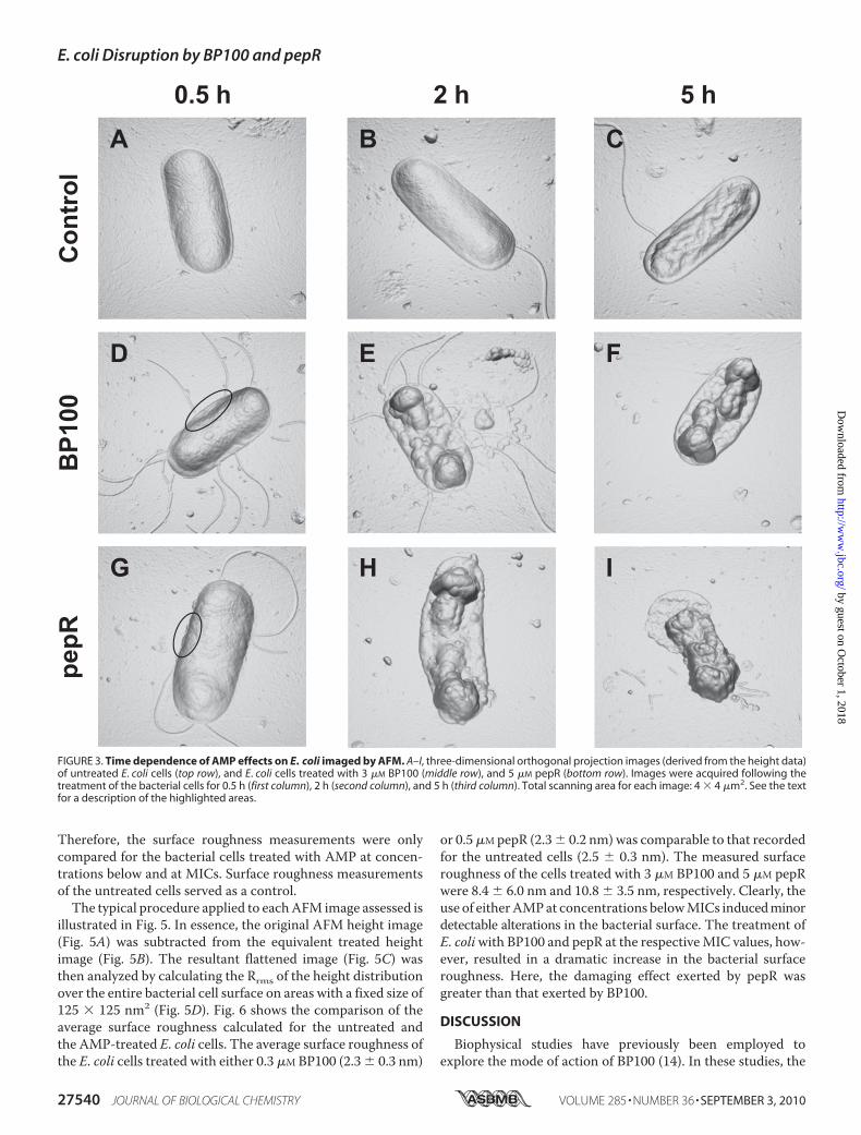
period of 0.5, 2, and 5 h, and imaged. Based on the antimicrobialactivity studies, final peptide concentrations of 3 and 5�Mwereselected for BP100 and pepR, respectively. Bacteria undergoingnoAMP treatment were also imaged at the same time intervals.The images acquired in this study are shown in Fig. 3, presentedin three-dimensional orthogonal projection. From Fig. 3, Aand B, it is clear that the untreated E. coli cells experiencedno morphological changes over the period of 2 h. The typicalrod-shaped structure was preserved, and the surface topog-raphy was comparable to that of the untreated cells imagedimmediately after sample preparation. After 5 h of incubation,however, an alteration in the morphology of the untreated cells
was observed (Fig. 3C). Keeping E. coli in the nutrient-freebuffer led to the eventual starvation of the bacterial cells andthe consequent shriveling of their overall structure. Thenature of the surface corrugation detected here is markedlydifferent from that of the bacterial cells incubated witheither AMP (Fig. 3, D–I).
The effect of BP100 and pepR onE. coli, following incubationfor the different time intervals, was comparable (Fig. 3,D–I). Inall cases, the treated bacterial cells retained their rod-like form.However, changes in the membrane surface corrugation couldalready be distinguished for the bacterial cells incubated for0.5 hwith either 3�MBP100 (Fig. 3D) or 5�MpepR (Fig. 3G). Aminor collapse in the outer membrane of the bacterial celltreated with BP100 was evident (see highlighted region in Fig.3D). Also, in the case of pepR, treatment seemed to inducemembrane blebbing (see highlighted region in Fig. 3G). Expo-sure of the cells to either AMP for 2 h or longer led to greatermembrane disruption (Fig. 3, E, F, H, and I). Broadly speaking,the action of both BP100 and pepR over time resulted in a col-lapse of the bacterial envelope, particularly at the septal region.This was associated with the formation of vesicle-like struc-tures on the membrane surface. Some leaked contents anddebris could also be detected around the partially disintegratedcells (Fig. 3, E and I). The events described here were observedfor almost all cells imaged under the same conditions.The effect of AMP concentration on bacterial cell morphol-
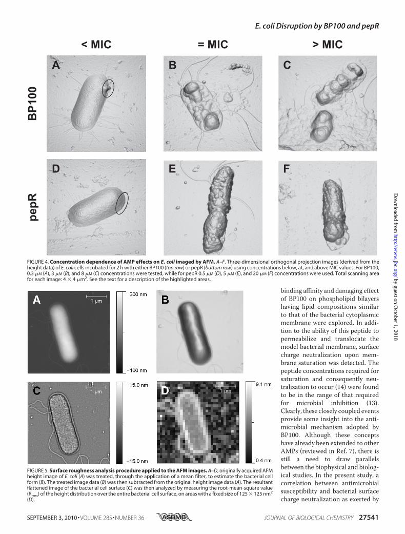
ogy was also assessed by AFM, as shown in Fig. 4. E. coli cellswere incubated for 2 h with 0.3, 3, and 8 �M BP100 and 0.5, 5,and 20 �M pepR. From all the acquired images, it is clear thatthe typical rod-shaped structure of the E. coli cells was main-tained following peptide treatment. However, characteristicphenomena associatedwith the exposure ofE. coli to increasingconcentrations of either BP100 or pepR were detected (Fig. 4,A–F). Exposure of the cells to 0.3�MBP100 (Fig. 4A) and 0.5�M
pepR (Fig. 4D) already inducedminor perturbations on the bac-terial envelope in comparison to the untreated cells (Fig. 3B).Membrane blebbing and a minor collapse at the apical end ofthe bacterial envelope were observed. Upon incubation ofE. coli with 3 �M BP100 (Fig. 4B), a pronounced collapse in themid-region of the envelope was detected. This was accompa-nied by the leakage of the cytoplasm contents of the bacterialcell. A similar event was registered when treating E. coli cellswith 8 �M BP100 (Fig. 4C). Only here a copious amount of fluidleaked contents and debris around the apical and septal regionsof the dividing cell was released. For pepR, treatment of thebacterial cells with 5 �M (Fig. 4E) and 20 �M (Fig. 4F) peptideconcentrations resulted in comparable alterations in the mem-brane surface. In both cases, the formation of vesicle-like struc-tures on the membrane surface was visible. For all the AMP-damagedE. coli cells imagedhere, different combinations of theabove-described phenomena were recorded.Surface Roughness Analysis of the AFM Imaged, Untreated,
and AMP-treated E. coli Cells—To quantify the damageexerted by each AMP, the roughness of the treated E. coli bac-terial cell surface was measured. The AFM concentration-de-pendent studies showed that the treatment of E. coliwith eitherAMP at concentrations equivalent to the MICs was sufficientfor the complete disruption of the bacterial cell envelope.
FIGURE 2. AFM images of an untreated E. coli cell dried in air. A and B,lock-in-amplitude image (A) and topography image (B) of E. coli. Total scan-ning area for each image: 4 � 4 �m2. C, cross-section of image indicated inB, providing a quantitative measure of the bacterial cell dimensions.
E. coli Disruption by BP100 and pepR
SEPTEMBER 3, 2010 • VOLUME 285 • NUMBER 36 JOURNAL OF BIOLOGICAL CHEMISTRY 27539
by guest on October 1, 2018
http://ww
w.jbc.org/
Dow
nloaded from
Therefore, the surface roughness measurements were onlycompared for the bacterial cells treated with AMP at concen-trations below and at MICs. Surface roughness measurementsof the untreated cells served as a control.The typical procedure applied to eachAFM image assessed is
illustrated in Fig. 5. In essence, the original AFM height image(Fig. 5A) was subtracted from the equivalent treated heightimage (Fig. 5B). The resultant flattened image (Fig. 5C) wasthen analyzed by calculating the Rrms of the height distributionover the entire bacterial cell surface on areas with a fixed size of125 � 125 nm2 (Fig. 5D). Fig. 6 shows the comparison of theaverage surface roughness calculated for the untreated andthe AMP-treated E. coli cells. The average surface roughness ofthe E. coli cells treated with either 0.3 �M BP100 (2.3 � 0.3 nm)
or 0.5�M pepR (2.3� 0.2 nm) was comparable to that recordedfor the untreated cells (2.5 � 0.3 nm). The measured surfaceroughness of the cells treated with 3 �M BP100 and 5 �M pepRwere 8.4 � 6.0 nm and 10.8 � 3.5 nm, respectively. Clearly, theuse of eitherAMPat concentrations belowMICs inducedminordetectable alterations in the bacterial surface. The treatment ofE. coliwith BP100 and pepR at the respectiveMIC values, how-ever, resulted in a dramatic increase in the bacterial surfaceroughness. Here, the damaging effect exerted by pepR wasgreater than that exerted by BP100.
DISCUSSION
Biophysical studies have previously been employed toexplore the mode of action of BP100 (14). In these studies, the
FIGURE 3. Time dependence of AMP effects on E. coli imaged by AFM. A–I, three-dimensional orthogonal projection images (derived from the height data)of untreated E. coli cells (top row), and E. coli cells treated with 3 �M BP100 (middle row), and 5 �M pepR (bottom row). Images were acquired following thetreatment of the bacterial cells for 0.5 h (first column), 2 h (second column), and 5 h (third column). Total scanning area for each image: 4 � 4 �m2. See the textfor a description of the highlighted areas.
E. coli Disruption by BP100 and pepR
27540 JOURNAL OF BIOLOGICAL CHEMISTRY VOLUME 285 • NUMBER 36 • SEPTEMBER 3, 2010
by guest on October 1, 2018
http://ww
w.jbc.org/
Dow
nloaded from
binding affinity and damaging effectof BP100 on phospholipid bilayershaving lipid compositions similarto that of the bacterial cytoplasmicmembrane were explored. In addi-tion to the ability of this peptide topermeabilize and translocate themodel bacterial membrane, surfacecharge neutralization upon mem-brane saturation was detected. Thepeptide concentrations required forsaturation and consequently neu-tralization to occur (14) were foundto be in the range of that requiredfor microbial inhibition (13).Clearly, these closely coupled eventsprovide some insight into the anti-microbial mechanism adopted byBP100. Although these conceptshave already been extended to otherAMPs (reviewed in Ref. 7), there isstill a need to draw parallelsbetween the biophysical and biolog-ical studies. In the present study, acorrelation between antimicrobialsusceptibility and bacterial surfacecharge neutralization as exerted by
FIGURE 4. Concentration dependence of AMP effects on E. coli imaged by AFM. A–F. Three-dimensional orthogonal projection images (derived from theheight data) of E. coli cells incubated for 2 h with either BP100 (top row) or pepR (bottom row) using concentrations below, at, and above MIC values. For BP100,0.3 �M (A), 3 �M (B), and 8 �M (C) concentrations were tested, while for pepR 0.5 �M (D), 5 �M (E), and 20 �M (F) concentrations were used. Total scanning areafor each image: 4 � 4 �m2. See the text for a description of the highlighted areas.
FIGURE 5. Surface roughness analysis procedure applied to the AFM images. A–D, originally acquired AFMheight image of E. coli (A) was treated, through the application of a mean filter, to estimate the bacterial cellform (B). The treated image data (B) was then subtracted from the original height image data (A). The resultantflattened image of the bacterial cell surface (C) was then analyzed by measuring the root-mean-square value(Rrms) of the height distribution over the entire bacterial cell surface, on areas with a fixed size of 125 � 125 nm2
(D).
E. coli Disruption by BP100 and pepR
SEPTEMBER 3, 2010 • VOLUME 285 • NUMBER 36 JOURNAL OF BIOLOGICAL CHEMISTRY 27541
by guest on October 1, 2018
http://ww
w.jbc.org/
Dow
nloaded from

either BP100 or pepR on E. coliwas investigated. AFM imagingwas also used to gain some insights into the mode of action ofeach AMP against E. coli, at an atomic level.Both BP100 and pepR were found to be effective antimicro-
bial agents against E. coli, inhibiting the growth of this Gram-negative bacterium at micromolar concentrations. Inhibitionoccurred to an equivalent extent for both peptides, with pepRbeing only slightly more effective at lower peptide concentra-tions. For the BP100-treated E. coli cells, the MICs recordedwere comparable to those previously reported forE. amylovora,P. syringae pv. Syringae, and X. axonopodis pv. vesicatoria (13),as well as the theoretically determined values (14).Having established theMICs for each peptide, zeta potential
studies were performed under the equivalent experimentalconditions. In doing so, characterization of the bacterial surfacefollowing the addition of either cationic AMP at MICs wasmade possible. In the absence of peptide, the E. coli surfacedisplayed a zeta potential of �21.9 � 3.0 mV. This negativesurface net charge originates from the negative lipids andlipopolysaccharide (LPS) molecules present in the outer leafletof the Gram-negative bacteria outer membrane (18, 20). Anal-ysis of the electrostatic properties of the E. coli surface by zetapotential measurement, after incubation with either AMP,revealed significant differences between both peptides. Theobserved behavioral differences recorded for each AMP can berationalized on the basis of the peptide and membrane proper-ties. For BP100, the surface charge of the bacteria was neutral-ized when using concentrations at, and above, the MIC (i.e. �2�M). This corresponds well to the biophysical data, where sur-face neutralization of the model bacterial membrane wasinduced by equivalent BP100 concentrations at the saturationstate (14). The postulated saturation-triggered antimicrobialmechanisms adopted by BP100 (14) thus appear to be con-nected to the neutralization of the bacterial surface when using
peptide concentrations equivalent to MICs. In biological sys-tems, surface neutralization can largely be attributed to the bal-ance in electrostatic interactions between the positive charges(mainly lysine and arginine side chains) of the peptides with thenegatively charged groups (mainly phosphates and carboxy-lates) of LPS. For BP100 (6 at pH 7.4) in a model bacterialmembrane, it has been proposed that one peptide moleculeinteracts with 5.6 negatively charged phospholipidmolecules atsaturation, thereby inducing neutralization (14). This phenom-enonmore than likely accounts for BP100 behavior when treat-ing E. coli cells with the peptide at MIC.For pepR, surface neutralization ofE. coli occurred at peptide
concentrations below MIC values (0.63 �M). In fact, a chargeovercompensation was registered in the zeta potential mea-surements when treating E. coli with pepR at concentrationsabove 1.25 �M. Mechanisms other than just surface neutraliza-tion seem to be present in the case of pepR. Relative to BP100,pepR is considerably more basic and positively charged (12 atpH 7.4). The interaction of this peptide with E. coli at low con-centrations (i.e. below MIC) can be attributed, at least initially,to electrostatics. Several studies investigating the interaction ofcationic AMPs with model bacterial membranes have shownthat an overcompensation in zeta potential at high peptide con-centrations is associated with membrane insertion via hydro-phobic interactions (22–24). A similar trend was also reportedfor the interaction of the cationic peptide rBPI21 with LPSaggregates (24). The overcompensation in E. coli zeta potentialat high pepR concentrations (�MICs) may indicate: 1) thathydrophobic interactions contribute to membrane interactionin addition to electrostatic attraction, or 2) not all positivecharges of pepR contribute to electroneutralization. The rela-tively large size of pepR, as well as the largemajority of charged/polar residues within the peptide, makes it unlikely that all itscharges will be able to simultaneously come into contact withthe bacterial surface. Thus, the observed zero in zeta potentialat low concentrations of pepRmay not correspond to the actualneutralization of the surface but rather its masking by unboundcharges. This interpretation conciliates the zeta potential re-sults of pepR with the MIC-neutralization correlation hypoth-esis. In any case, the existence of an inherent neutralization-mediated killing mechanism employed by either AMP is notnecessarily implied.To gain further insights into these MIC-associated events,
AFM images of the E. coli cells under varying conditions wereacquired. The use of the cationic polymer PLL as an adhesionmolecule for bonding bacteria to surfaces prior to AFM imag-ing has been questioned (25), in part due to its potential anti-microbial activity (26, 27). However, it has been demonstratedthat the use of this method for imaging bacteria in their nativestate does not affect the properties of the bacterial membranesurface (28, 29). Bacteria can also be imaged in an air-driedstate, as this enables the high-resolution imaging of their sur-face morphology (28, 30, 31). For this reason, the bacterial cellsimaged in this study were immobilized onto glass slides func-tionalized with PLL and allowed to air-dry. Overall, satisfactoryand informative AFM images of the untreated and AMP-treated bacterial cells were acquired, revealing detailed infor-mation on the membranolytic properties of both BP100 and
FIGURE 6. E. coli cell surface topography analysis. The average surfaceroughness of the untreated E. coli cells, and the E. coli cells treated with eitherBP100 or pepR were compared. The AFM height images evaluated for BP100were those of E. coli treated with either 0.3 �M (below MIC) or 3 �M (at MIC)concentrations. For pepR, the height images evaluated were those of E. colitreated with either 0.5 �M (below MIC) or 5 �M (at MIC) concentrations. Thesurface roughness of E. coli when treated with either AMP using concentra-tions equivalent to MIC values was significantly enhanced: **, p 0.05 for 3�M BP100 when compared with either the untreated cells or the cells treatedwith 0.3 �M BP100; ***, p 0.0005 for 5 �M pepR when compared with eitherthe untreated cells or the cells treated with 0.5 �M pepR. Error bars indicatethe S.E.
E. coli Disruption by BP100 and pepR
27542 JOURNAL OF BIOLOGICAL CHEMISTRY VOLUME 285 • NUMBER 36 • SEPTEMBER 3, 2010
by guest on October 1, 2018
http://ww
w.jbc.org/
Dow
nloaded from

pepR. Nonetheless, it should be borne in mind that, due to thedeposition and washing protocols, free debris resulting fromcell disruption are partially eliminated.The AFM results presented in this study clearly demonstrate
the time- and concentration-dependent antimicrobial activityof both BP100 and pepR. Intricate details of the damage sus-tained by the E. coli cells following AMP treatment under thedifferent conditions were revealed. During the initial stages oftreatment, where cells were incubated with peptide either forshort periods of time (0.5 h) or using low peptide concentra-tions (below MICs), minor changes in the outer membrane ofthe bacterial cell envelope were induced. The appearance ofblebs at the bacterial surface, as well as the slight collapse in theouter membrane, already pointed to the interaction of eitherAMP to the negatively charged LPS outer layer. The formationof vesicle-like structures, as well as the alterations in surfaceroughness, observed in the E. coli cells exposed to either BP100or pepR for longer time periods (�2 h) and at higher peptideconcentrations (�MICs) confirmed this. Previous studies havedemonstrated that an increase in surface roughness is a directconsequence of AMP incorporation into the LPS-containingouter membrane (21, 30, 32, 33). It has also been shown thatrelease of LPS-containing vesicles and even autolytic reactionsare possible outcomes of such interactions (21, 30, 32). Whenexposing E. coli to either BP100 or pepR under extreme condi-tions (i.e. long exposure time, �2 h; high peptide concentra-tions, �MICs), a collapse of the outer membrane at the septalregion was generally observed. In some images, a release ofcytoplasmic content was also detected. These phenomena maybe explained by taking into account that cardiolipin, a nega-tively charged phospholipid, is generally located at the apicaland septal regions of the E. coli inner membrane (34), and thatboth BP100 and pepR are highly cationic. Furthermore, BP100,which displays a high affinity toward negatively charged phos-pholipids, is known to induce vesicle permeabilization at highpeptide/lipid ratios (14). It is therefore likely that treatment ofE. coli with either AMP for long periods of time, or when usinghigh concentrations, could result in an accumulation of peptideat either the apical or septal regions. When a threshold peptideconcentration is reached in these regions, membrane disrup-tion may occur, thereby initiating cell leakage. Similar observa-tions have been reported for the cationic AMPs magainin 2,melittin, PGLa, and Sushi 3 (21, 32). Based on the nature of suchalterations, the adoption of a carpet-like or detergent-likemechanism (9) in vivo by the two AMPs investigated hereseems to be likely.Combining the antimicrobial susceptibility, zeta potential
and AFM data, the closely coupled events leading to E. coli celldeath after AMP treatment can thus be summarized as follows.At peptide concentrations below MIC, an association of thecationic AMPwith the negatively charged LPSmolecules in thebacterial outer membrane occurs. For BP100, this eventinvolves an initial electrostatic interaction between the peptideand the LPSmolecules and a gradual increase of themembranesurface charge. In contrast, the electrostatic association of pepRto the LPS outer layer prompts an almost immediate net neu-tralization of the membrane surface charge. At an initial stageand at low extents of peptide binding, minor changes in the
bacterial cell envelope are provoked (e.g. bleb formation andslight collapse in the outer membrane). Treatment of E. coliwith either AMP at, and above the MIC, brings about furthermembrane alterations (e.g. increase in surface roughness andformation of vesicle-like structures). After the permeabilizationof the outer membrane and cell envelope, each AMP interactswith the negatively charged phospholipids of the bacterial innermembrane. In the case of BP100, these events are associatedwith saturation (14) and, consequently, neutralization of themembrane. For pepR, events beyond electrostatic equivalenceprompt peptide binding to the inner membrane. The finalstages leading to E. coli cell death, as evidenced by the release ofcytoplasmic content, involve the disruption of the inner mem-brane by either AMP.The differences in the MIC values of the two peptides may
also be closely related to their charge behavior. It has been pro-posed that threshold events that lead to cell death (35) dependon the strain imposed on the cell membrane due to peptide-induced thinning. The reaching of such thresholds requiresthat peptide molecules become concentrated in the membranedespite an intrinsic repulsion between them as their densityincreases. The driving forces that overcome this repulsion arethe electrostatic and hydrophobic peptide-membrane interac-tions, which also result in the typically high membrane bindingaffinities of AMPs (7). Bacterial surface charge neutralizationby pepR occurs at very low peptide concentrations, which is alikely consequence of the high cationicity of the peptide. Fromneutralization onward, only the hydrophobic interactions willbe left to draw more peptide molecules to the membrane (thezeta potential increasing above 0 mV; Fig. 1B) and across the dis-ruption threshold. With a weaker driving force to bind themembrane, larger amounts of peptide are eventually requiredfor the threshold to be reached, which translates into a higherMIC. Ultimately, too high a cationic charge on an AMP mayactually compromise its activity. This concept may in factaccount for the observed lack of antibacterial activity reportedfor some of the cecropin A-melittin hybrid peptides (e.g. BP16,9 at pH 7.4) developed by Ferre et al. (36).Based on the data presented in this work, insights into the
events leading to E. coli cell death after treatment with eitherBP100 or pepR were gained. This was achieved through theunconventional approach of bridging themicrobiological prop-erties of each AMP with some of their respective biophysicalcharacteristics. Firstly, the question of a neutralization-medi-ated killing mechanism adopted by either AMP was addressed.Exploration of this concept using a standard antimicrobialactivity assay and zeta potential studies demonstrated a clearcorrelation linking the MICs of each AMP to correspondingalterations in the E. coli surface charge. More specifically, neu-tralization of the bacterial surface was detected when treatingE. coli with peptide concentrations close to MIC values. Visualinsights into these MIC-associated events were then sought.The acquisition of AFM images of E. coli cells treated witheither BP100 or pepR under varying conditions illustrated thetime- and concentration-dependent antimicrobial action ofbothAMPs. Taken together, the biological and biophysical dataacquired in this study clearly point to a critical AMP concen-
E. coli Disruption by BP100 and pepR
SEPTEMBER 3, 2010 • VOLUME 285 • NUMBER 36 JOURNAL OF BIOLOGICAL CHEMISTRY 27543
by guest on October 1, 2018
http://ww
w.jbc.org/
Dow
nloaded from
tration, equivalent to MIC values, being necessary for E. colimembrane disruption to occur.
REFERENCES1. Zasloff, M. (2002) Nature 415, 389–3952. Brogden, K. A. (2005) Nat. Rev. Microbiol. 3, 238–2503. Yeaman, M. R., and Yount, N. Y. (2003) Pharmacol. Rev. 55, 27–554. Jenssen, H., Hamill, P., and Hancock, R. E. W. (2006) Clin. Microbiol. Rev.
19, 491–5115. Hancock, R. E., and Chapple, D. S. (1999) Antimicrob. Agents Chemother.
43, 1317–13236. Huang, H. W. (2000) Biochemistry 39, 8347–83527. Melo, M. N., Ferre, R., and Castanho, M. A. (2009)Nat. Rev. Microbiol. 7,
245–2508. Ludtke, S. J., He, K., Heller, W. T., Harroun, T. A., Yang, L., and Huang,
H. W. (1996) Biochemistry 35, 13723–137289. Bechinger, B., and Lohner, K. (2006) Biochim. Biophys. Acta 1758,
1529–153910. Leontiadou, H., Mark, A. E., and Marrink, S. J. (2006) J. Am. Chem. Soc.
128, 12156–1216111. Oren, Z., and Shai, Y. (1998) Peptide Science 47, 451–46312. Andreu, D., Ubach, J., Boman, A., Wåhlin, B., Wade, D., Merrifield, R. B.,
and Boman, H. G. (1992) FEBS Lett. 296, 190–19413. Badosa, E., Ferre, R., Planas, M., Feliu, L., Besalu, E., Cabrefiga, J., Bardají,
E., and Montesinos, E. (2007) Peptides 28, 2276–228514. Ferre, R., Melo, M. N., Correia, A. D., Feliu, L., Bardají, E., Planas, M., and
Castanho, M. (2009) Biophys. J. 96, 1815–182715. Ma, L., Jones, C. T., Groesch, T. D., Kuhn, R. J., and Post, C. B. (2004) Proc.
Natl. Acad. Sci. U. S. A. 101, 3414–341916. Fields, G. B., and Noble, R. L. (1990) Int. J. Pept. Protein Res. 35, 161–21417. Wiegand, I., Hilpert, K., and Hancock, R. E. (2008) Nat. Protocols 3,
163–17518. Domingues, M. M., Santiago, P. S., Castanho, M. A., and Santos, N. C.
(2008) J. Pept. Sci. 14, 394–40019. Girasole, M., Pompeo, G., Cricenti, A., Congiu-Castellano, A., Andreola,
F., Serafino, A., Frazer, B. H., Boumis, G., and Amiconi, G. (2007) Biochim.Biophys. Acta 1768, 1268–1276
20. Amro, N. A., Kotra, L. P., Wadu-Mesthrige, K., Bulychev, A., Mobashery,S., and Liu, G. Y. (2000) Langmuir 16, 2789–2796
21. Meincken, M., Holroyd, D. L., and Rautenbach, M. (2005) Antimicrob.Agents Chemother. 49, 4085–4092
22. Andra, J., Koch,M.H., Bartels, R., and Brandenburg, K. (2004)Antimicrob.Agents Chemother. 48, 1593–1599
23. DenHertog, A. L.,Wong Fong Sang, H.W., Kraayenhof, R., Bolscher, J. G.,Van’t Hof, W., Veerman, E. C., and Nieuw Amerongen, A. V. (2004) Bio-chem. J. 379, 665–672
24. Domingues,M.M., Castanho,M. A., and Santos, N. C. (2009) PLoSOne 4,e8385
25. Colville, K., Tompkins, N., Rutenberg, A. D., and Jericho, M. H. (2009)Langmuir 26, 2639–2644
26. Conte, M., Aliberti, F., Fucci, L., and Piscopo, M. (2007) World J. Micro-biol. Biotechnol. 23, 1679–1683
27. Yoshida, T., and Nagasawa, T. (2003) Appl. Microbiol. Biotechnol. 62,21–26
28. Bolshakova, A. V., Kiselyova, O. I., Filonov, A. S., Frolova, O. Y., Lyub-chenko, Y. L., and Yaminsky, I. V. (2001) Ultramicroscopy 86, 121–128
29. Schaer-Zammaretti, P., and Ubbink, J. (2003) Ultramicroscopy 97,199–208
30. da Silva, A., Jr., and Teschke, O. (2005)World J. Microbiol. Biotechnol. 21,1103–1110
31. Ubbink, J., and Schar-Zammaretti, P. (2005)Micron 36, 293–32032. Li, A., Lee, P. Y., Ho, B., Ding, J. L., and Lim, C. T. (2007) Biochim. Biophys.
Acta 1768, 411–41833. Matsuzaki, K., Sugishita, K., and Miyajima, K. (1999) FEBS Lett. 449,
221–22434. Mileykovskaya, E., and Dowhan, W. (2000) J. Bacteriol. 182, 1172–117535. Huang, H. W. (2009) Biophys. J. 96, 3263–327236. Ferre, R., Badosa, E., Feliu, L., Planas, M., Montesinos, E., and Bardají, E.
(2006) Appl. Environ. Microbiol. 72, 3302–3308
E. coli Disruption by BP100 and pepR
27544 JOURNAL OF BIOLOGICAL CHEMISTRY VOLUME 285 • NUMBER 36 • SEPTEMBER 3, 2010
by guest on October 1, 2018
http://ww
w.jbc.org/
Dow
nloaded from

Miguel X. Fernandes and Miguel A. R. B. CastanhoLidia Feliu, Eduard Bardají, Wioleta Kowalczyk, David Andreu, Nuno C. Santos,
Carla S. Alves, Manuel N. Melo, Henri G. Franquelim, Rafael Ferre, Marta Planas,Antimicrobial Peptides BP100 and pepR
Cell Surface Perturbation and Disruption Induced byEscherichia coli
doi: 10.1074/jbc.M110.130955 originally published online June 21, 20102010, 285:27536-27544.J. Biol. Chem.
10.1074/jbc.M110.130955Access the most updated version of this article at doi:
Alerts:
When a correction for this article is posted•
When this article is cited•
to choose from all of JBC's e-mail alertsClick here
http://www.jbc.org/content/285/36/27536.full.html#ref-list-1
This article cites 36 references, 8 of which can be accessed free at
by guest on October 1, 2018
http://ww
w.jbc.org/
Dow
nloaded from