erbb3-dependent motility and intravasation in breast...
TRANSCRIPT
ErbB3-Dependent Motility and Intravasation in Breast
Cancer Metastasis
Chengsen Xue,1Fubo Liang,
2Radma Mahmood,
3Magalis Vuolo,
3Jeffrey Wyckoff,
1Hong Qian,
4
Kun-Lin Tsai,1Mimi Kim,
4Joseph Locker,
3Zhong-Yin Zhang,
2and Jeffrey E. Segall
1
Departments of 1Anatomy and Structural Biology, 2Molecular Pharmacology, 3Pathology, and 4Epidemiology and Population Health,Albert Einstein College of Medicine, Bronx, New York
Abstract
A better understanding of how epidermal growth factorreceptor family members (ErbBs) contribute to metastasis isimportant for evaluating ErbB-directed therapies. Activation ofErbB3/ErbB2 heterodimers can affect both proliferation andmotility. We find that increasing ErbB3-dependent signalingin orthotopic injection models of breast cancer can enhanceintravasation and lung metastasis with no effect on primarytumor growth or microvessel density. Enhanced metastaticability due to increased expression of ErbB2 or ErbB3correlated with stronger chemotaxis and invasion responsesto heregulin B1. Suppression of ErbB3 expression reduced bothintravasation and metastasis. A human breast cancer tumortissue microarray showed a significant association betweenErbB3 and ErbB2 expression and metastasis independent oftumor size. These results indicate that ErbB3-dependentsignaling through ErbB3/ErbB2 heterodimers can contributeto metastasis through enhancing tumor cell invasion andintravasation in vivo and that ErbB-directed therapies may beuseful for the inhibition of invasion independent of effects ontumor growth. (Cancer Res 2006; 66(3): 1418-26)
Introduction
Tumor cell metastasis is a complex process consisting ofmultiple steps (1). These steps include growth of the primarytumor, growth of vessels (blood vessels and lymphatics) into andaround the tumor, intravasation, transport to other parts of thebody, arrest, and growth at distant sites. The initial stages ofprimary tumor growth and angiogenesis have been well studiedand are key initial steps for enabling metastasis to proceed.However, many tumors can grow to a significant size withoutmetastasizing, and a better understanding of the factors thatcontribute to intravasation and metastasis is critical to developingbetter prognostic and therapeutic options for cancer patients.Epidermal growth factor (EGF) receptor (EGFR) family members
(ErbBs) are currently major targets of anticancer strategies (2–4) andidentifying the contributions of ErbBs to tumor cell metastasis isimportant for the development of useful anti-ErbB therapies. TheEGFR family has four members: ErbB1 (EGFR, HER-1), ErbB2(HER-2/neu), ErbB3, and ErbB4 (5, 6). Twenty percent to 30% ofhuman breast cancers have been found to overexpress ErbB2, and
ErbB2 overexpression is significantly associated with decreaseddisease-free survival and overall survival (3, 7, 8). Normal activationof ErbB2 occurs through formation of heterodimers with other EGFRfamily members that can bind ligands, such as ErbB3 (9, 10). ErbB3binds heregulin but is unable to stimulate cellular responses on itsown due to a defective kinase domain. Binding of heregulin to ErbB3can generate ErbB3/ErbB2 heterodimers, leading to activationof mitogen-activated protein kinase (MAPK), phosphatidylinositol3-kinase (PI3K), and src (6, 11). Mutation of the ErbB3 sites couplingto these pathways reduces heregulin-induced DNA synthesis andcolony formation in soft agar by NIH 3T3 cells (12). Suppression ofeither ErbB2 or ErbB3 function in SKBR3, MB361, or BT474 cell linesresults in cell cycle arrest in G1 (13) in vitro. In vivo , suppression ofheregulin expression in MDA-MB-231 cells blocks tumor growth(14), and tumors induced by expression of ErbB2 in the mammarygland often show overexpression of ErbB3 (15).Heregulin also stimulates chemotaxis and invasion mediated
by ErbB3/ErbB2 heterodimers (16, 17). Activation of the PI3K andMAPK pathways can be important for cell motility and chemotaxis(18–22). The products of PI3K regulate the cytoskeleton throughRho family G proteins as well as Akt (23–25). MAPKs can regulateadhesion dynamics directly and regulate gene expression patternsimportant for motility and invasion (26–29). Thus, ErbB3-dependentmotility responses could contribute to breast cancer metastasisindependent of effects on tumor growth.To evaluate the potential contributions of ErbB3-dependent
motility responses to tumor metastasis, we evaluated the effects ofoverexpressing ErbBs on the metastatic properties of MDA-MB-435(30, 31) and MTLn3 mammary tumor cells (32). We find thatenhancing ErbB3/ErbB2 signaling increases intravasation andmetastasis without affecting primary tumor growth. Suppressionof ErbB3 expression significantly reduced intravasation andmetastasis. Examination of a tumor progression microarrayindicates that ErbB2 and ErbB3 expression associate positively withthe presence of metastases and not with primary tumor size. Wepropose that ErbB3-dependent signaling can contribute to metas-tasis through enhancing tumor cell motility and intravasation. Ourresults support the development of therapies targeting cell motilityto aid in the prevention and treatment of metastasis.
Materials and Methods
Cell lines. The human tumor cell lineMDA-MB-435 (refs. 30, 31; American
Type Culture Collection, Rockville, MD) and rat mammary MTLn3 cells
(32, 33) were maintained in a-MEM (Life Technologies, Gaithersburg, MD)supplemented with 5% fetal bovine serum and penicillin/streptomycin
solution (Life Technologies). The empty retroviral expression vector pLXSN
and pLXSN containing the human cDNAs for ErbB1, ErbB2, ErbB3, and ErbB4were received from David Stern (Yale University, New Haven, CT; ref. 34) and
packaged in the Phoenix cell line provided by Dr. Garry P. Nolan (Stanford
University, Stanford, CA; ref. 35), and cells were infected in growthmedium in
Note: Supplementary data for this article are available at Cancer Research Online(http://cancerres.aacrjournals.org/).
Requests for reprints: Jeffrey E. Segall, Department of Anatomy and StructuralBiology, Albert Einstein College of Medicine, 1300 Morris Park Avenue, Bronx, NY10801. Phone: 718-430-4237; Fax: 718-430-8996; E-mail: [email protected].
I2006 American Association for Cancer Research.doi:10.1158/0008-5472.CAN-05-0550
Cancer Res 2006; 66: (3). February 1, 2006 1418 www.aacrjournals.org
Research Article
Research. on June 30, 2018. © 2006 American Association for Cancercancerres.aacrjournals.org Downloaded from

the presence of 4 Ag/mL polybrene (Sigma, St. Louis, MO). Pools of at least 100transductants were selected by growing in .8 mg/mL geneticin (Sigma)
medium. The pools were stored as frozen stocks and used for all experiments
within 10 passages. Expression of transduced ErbBs measured by fluores-
cence-activated cell sorting (FACS) showed no change with passage in vitroand after growth to form primary tumors in vivo .
Flow cytometric analysis. Cells (f106) were incubated with specific
anti-ErbB1, anti-ErbB2, anti-ErbB3, and anti-ErbB4 antibodies (NeoMarkers,
Fremont, CA) for 1 hour at 4jC. After three washes in cold PBS containing0.2% bovine serum albumin (BSA), cells were incubated with R-phycoery-
thrin-conjugated goat anti-mouse IgG (Jackson ImmunoResearch Labora-
tories, West Grove, PA) for 1 hour at 4jC. After washing, cells were
suspended in PBS containing 0.2% BSA, and fluorescence was measuredby flow cytometry. Cells incubated with secondary antibody only were
measured at the same time to serve as background control. To semi-
quantitatively measure the expression level of ErbBs, standard curves wereobtained by using the LinearFlow Orange Flow Cytometry Intensity
Calibration kit (Molecular Probes, Eugene, OR) with the mean values of
cells incubated with secondary antibody alone subtracted.
Spontaneous and experimental metastasis assays. All animal studies
described here were done according to the protocols approved by the
Institutional Animal Care and Use Committee of Albert Einstein College of
Medicine. To measure spontaneous metastasis, the tumor cells were grown
to 70% to 85% confluence before being harvested. Cells were detached by
incubation in DPBS + 2 mmol/L EDTA, scraping with a rubber policeman,
then centrifuged, and resuspended in DPBS at 107 cells/mL. MDA-MB-435
(f106) or MTLn3 (5 � 105) cells were injected into the right fourth
mammary fat pad from the head of 5- to 7-week-old female BALB/c severe
combined immunodeficient (SCID) mice (National Cancer Institute,
Bethesda, MD) in 100 AL PBS with calcium and magnesium through a
25-gauge needle. Tumor growth rate was monitored at weekly intervals by
measuring in two dimensions, and tumor volumes were calculated using the
formula: length � width2 / 2. At the end point for spontaneous metastasis,
mice were anesthetized with Aerrane (isoflurane, Baxter Pharmaceutical
Products, Inc., Deerfield, IL). The right chest was exposed by a simple skin
flap surgery. Blood was taken from the right atrium via heart puncture with a
25-gauge needle and 1mL syringe coated with heparin and containing 0.1 mL
of heparin. Blood (0.2-1.05 mL) was harvested. The blood was immediately
plated into 150-mm-diameter dishes filled with 5% fetal bovine serum in
a-MEM. The next day, the plates were rinsed with fresh medium and
replaced with fresh medium containing 0.8 mg/mL geneticin to selectively
grow tumor cells. After 4 ( for MTLn3) to 10 ( for MDA-MB-435) days, all
colonies in the dish were counted. Tumor blood burden was calculated as
total colonies in the dish divided by the volume of blood taken.
To measure experimental lung metastasis, 5 � 105 cells were injected
into the lateral tail veins of 5- to 7-week-old female BALB/c SCID mice(National Cancer Institute). Eight weeks ( for MDA-MB-435) or 2 weeks ( for
MTLn3) after injection, the mice were sacrificed, and the lungs were
removed, fixed in formalin, and stained H&E sections were counted formetastasis as described below.
Tumor histology and quantitative assessment of metastasis. Samples
were fixed in 10% neutral formalin buffer, embedded in paraffin, and
sectioned at 5 Am and stained by H&E. For each lung sample, allmicrometastases were counted at �10 magnification and the total lung
area was measured using a UMAX PowerLook III color scanner (UMAX
Technologies, Inc., Dallas, TX) and Adobe Photoshop version 5.5 software.
Briefly, after scanning lung sections, the cross-sectional area in pixels weremeasured using the luminosity window in Adobe PhotoShop. The actual
lung tissue area was calculated with the formula: area (mm2) = (number of
pixels) � 0.00179. The efficiency of lung metastasis was expressed innumber of metastases per square centimeter of lung area for each animal.
Mean and SE were then calculated for each cell line.
Determination of blood vessel density in primary tumor. Sections(5 Am) of paraffin-embedded primary tumor samples were stained withrabbit anti-human von Willebrand factor as primary antibody (DAKO,
Carpinteria, CA). Sections stained without primary antibody served as
controls. DAKO peroxidase substrate kit 3,3V-diaminobenzidine (DAB) was
used following the manufacturer’s instructions for identifying antibodybinding. Vessels were counted microscopically using a defined magnifica-
tion (�200). Blood vessels in five nonoverlapping fields per tumor lesion
were counted and averaged. Vascular counts included complete vessel
cross-sections, partial vessel cross-sections, and small groups of positivecells. Twelve tumors from 435-PL and 10 tumors from 435-B2 were
analyzed. Statistical analysis was done by unpaired t test.
Microchemotaxis chamber assay. A 48-well microchemotaxis chamber
(Neuroprobe, Cabin John, MD) was used as described previously (36), exceptthat L15 containing 0.35%BSAwas used instead ofa-MEM. Formeasurement
of migration in response to heregulin, filters were coated with 20 Ag/mL
fibronectin (Sigma), whereas for measurement of responses to EGF or BTC,
filters were coated with 27 Ag/mL rat tail collagen (BD Biosciences, PaloAlto, CA). After inserting the filters in the chamber, 20,000 cells were plated
into the wells of the upper chamber. The chambers were incubated for
4 hours at 37jC before analyzing the number of cells crossing the filter.In vivo invasion assay. Cell collection into needles placed into
anesthetized animals was carried out as described previously (37, 38). In
brief, 33-gauge needles are filled with Matrigel diluted 1:10 with L15-BSA,
0.01 mmol/L EDTA (pH 7.4) with or without heregulin h1 (HRGh1). Themouse is anesthetized and laid on its back, and a small patch of skin was
removed to expose the tumor. Three 25-gauge needles with inserted
blocking wires are inserted into the tumor using a specially designed
holder and a micromanipulator. The guide wires are then removed andthe 33-gauge needles were inserted through the 25-gauge needles into the
tumor. The animal is kept under anesthesia for 4 hours, after which the
needles are removed, the contents were expelled onto a coverslip andstained with 4V,6-diamidino-2-phenylindole, and cells were counted.
Immunoblotting. MDA-MB-435 cells were grown to 70% confluency in a
10-cm cell culturing dish and then incubated with serum-free medium
overnight. The medium was changed to fresh serum-free medium with or
without 50 ng/mL HRGh1 and cells were incubated for 0 to 15 minutes in a
5% CO2 incubator. Cells were then washed twice with cold PBS containing
1 mmol/L vanadate and lysed in 1 mL lysis buffer [50 mmol/L Tris-HCl
(pH 7.5), 1% Triton X-100, 5 mmol/L EGTA, 150 mmol/L NaCl, 10 mmol/L
sodium phosphate, 10 mmol/L NaF, 1 mmol/L sodium vanadate, 1 mmol/L
benzamidine, 10 Ag/mL leupeptin, and 10 Ag/mL aprotinin]. The plates were
scraped with a rubber policeman and incubated on ice for 30 minutes. The
lysate was precleared by centrifugation at 15,000 rpm for 15 minutes. Lysate
protein concentration was estimated using BCA protein assay reagent
(Pierce, Rockford, IL). Protein (20 Ag) of each sample was loaded and
separated by SDS-PAGE and transferred electrophoretically to nitrocellulose
membranes, which were immunoblotted by appropriate antibodies followed
by incubation with horseradish peroxidase (HRP)–conjugated secondary
Table 1. Semiquantitative measurement of expression ofErbB proteins in 435 transductants as detected by FACSanalysis
Transductant a-ErbB1
(%)
a-ErbB2
(%)
a-ErbB3
(%)
a-ErbB4
(%)
435-PL 6.2 3.9 6.6 0.04
435-B1 39.3 3.9 7.1 0.02435-B2 5.9 39.0 5.7 0.07
435-B3 5.4 3.9 10.4 0.01
435-B4 6.2 4.5 7.0 1.1
NOTE: Data are mean fluorescence levels after subtraction of controlswithout primary antibody and calibrated to LinearFlow Orange Flow
Cytometry beads as described in Materials and Methods where 100%
corresponds to the brightest beads. MTLn3-B3 and MTLn3-PL cells
had values for ErbB3 of 4.9% and 0.06%, respectively.
ErbB3 in Metastasis
www.aacrjournals.org 1419 Cancer Res 2006; 66: (3). February 1, 2006
Research. on June 30, 2018. © 2006 American Association for Cancercancerres.aacrjournals.org Downloaded from

antibodies. The following antibodies were used: anti-h-actin (Sigma); anti-
phosphotyrosine (PY20, BD Biosciences, San Diego, CA); anti-ErbB3
monoclonal and anti-phospho-ErbB3 (Tyr1289, Cell Signaling, Beverly, MA);
anti-ErbB4 (Upstate, Lake Placid, NY); anti-src (Santa Cruz Biotechnology,
Santa Cruz, CA); polyclonal anti–extracellular signal-regulated kinase (ERK)
1/2, anti-phospho-ERK (Thr202/Tyr204), anti-Akt, and anti-phospho-Akt
(Ser473) antibodies (Cell Signaling); anti-paxillin (Santa Cruz Biotechnology),
and anti-phospho-paxillin (BioSource International, Camarillo CA). The
blots were developed by the enhanced chemiluminescence (ECL) technique
(ECL kit, Amersham Pharmacia Biotech) according to the manufacturer’s
instructions.
Immunoprecipitation. Cells were lysed in lysis buffer [50 mmol/L Tris-
HCl (pH 7.5), 5 mmol/L EGTA, 1 mmol/L EDTA, 1% Triton X-100, 1 mmol/L
phenylmethylsulfonyl fluoride, 2 mmol/L sodium orthovanadate, 50 mmol/LNaF, protease inhibitors]. The protein concentration was measured by
BCA protein assay kit. Equal amounts of cell lysates either with or without
HRGh1 (12.5 nmol/L) treatment were incubated with anti-ErbB2 for 2 hours
at 4jC followed by an incubation with protein G agarose beads (Upstate)for 1 hour. Samples were washed five times in cell lysis buffer, resuspended
in 30 AL of 2� SDS sample buffer, and boiled for 5 minutes. The proteins
were then resolved using a SDS-PAGE gel, transferred to a nitrocellulosemembrane, and detected by Western blotting as described above.
ErbB3 RNA interference experiments. RNA interference transient
transfections were done first to find the most efficient knockout sequences.
Transfection-ready small interfering RNA (siRNA) duplexes against humanErbB3 were ordered from Dharmacon, Inc. (Lafayette, CO). The ErbB3 siRNA
kit contains four distinct individual RNA duplexes and a mixture of siRNA
duplexes (SMARTpool, Dharmacon). Cells at 60% confluency were trans-
fected in penicillin/streptomycin-freemediumwith the four individual siRNAduplexes and the pooled SMARTselected siRNA by using Oligofectamine
(Invitrogen, Grand Island, NY) following the manufacturer’s recommended
protocol. Two oligonucleotides (D-003127-05 and D-003127-07) found tosuppress ErbB3 expression and inhibit responses to HRGh1 in vitro were
cloned into pSUPER.retro.puro (OligoEngine, Seattle, WA) as small hairpin
RNAs (shRNA). A control sequence (39) that did not suppress ErbB3expression was also cloned as a shRNA into pSUPER.retro.puro as control.
Retroviruses containing the pSUPER.retro constructs were packaged using
Phoenix cells. Viral supernatants were harvested, and 435-B2 and 435-PL
recipient cells were infected in the presence of 4 Ag/mL polybrene. Afterinfection for 24 hours, resistant cells were selectedwith puromycin (3 Ag/mL).
FACS analysis of the stable transductants indicated that the D-003127-05
shRNA produced the strongest suppression of ErbB3 expression, and this line
was used for in vivo studies. The sense strand sequences used for the shRNAsfor the most strongly stably suppressed were GATCCCCAAGAGGATGT-
CAACGGTTATTCAAGAGATAACCGTTGACATCCTCTTTTTTTA (B3
shRNA) and GATCCCCAATTCTCCGAACGTGTCACGTTTCAAGAGAACGT-
GACACGTTCGGAGAATTTTTTTA (control shRNA).Immunohistochemical staining for ErbB2 and ErbB3. Cooperative
Breast Cancer Tissue Resource (CBCTR) Breast Tissue Progression
microarrays were acquired from the CBCTR of the National CancerInstitute.5 These tissue microarrays are designed by National Cancer
Institute statisticians for high statistical power to detect differences in
prevalence among the three stages of primary invasive ductal breast
cancer: node negative, node positive, and metastatic disease. Each tissuemicroarray block consists of 288 0.6-mm cores representing 252 breast
cancer and normal breast specimens plus replicate cores of 4 different cell
lines and 4 different nonbreast tissue controls. Two sequential slices from
two replicate blocks containing different cores from the same primarytumors were used to stain for ErbB2 and ErbB3.
Paraffin sections were melted at 60jC for 30 minutes, deparaffinized in
xylene, rehydrated through graded alcohols to water, and washed in TBS.Slides were pretreated with 3% H2O2 for 10 minutes and washed in TBS.
Antigen retrieval was done in a steamer for 20 minutes using 10 mmol/L
sodium citrate buffer (pH 6.0). Slides were then cooled for 30 minutes at
Figure 1. Increased ErbB2 expression enhances intravasation and metastasis of MDA-MB-435 Cells. A to C, cells were injected into the right mammary fat padsof SCID mice, and after 16 weeks, the animals were sacrificed and tumor volume, blood burden, and lung metastases were measured. A, tumor volume. B, density of lungmetastasis. *, P < 10�5, relative to 435-PL. C, blood burden (viable tumor cells/mL blood). *, P < 0.01 relative to 435-PL. Columns, mean (24 mice for 435-pLXSN,33 mice for 435-ErbB2, and 13 mice for 435-ErbB1, 435-ErbB3, and 435-ErbB4); bars, SE. D and E, cells were injected into the lateral tail vein, and after 8 weeks, the micewere sacrificed and the density and diameter of lung metastases were determined. D, density of lung metastasis. *, P < 0.08, relative to 435-PL. E, size distribution of lungmetastases in the experimental metastasis assay for 435-PL (white columns ) and 435-B2 (black columns). F, size distribution of lung metastases in the spontaneousmetastasis assay (corresponding to data in B) for 435-PL (white columns ), 435-B1 (dotted columns ), 435-B2 (grey columns ), 435-B3 (diagonal lines ), and 435-B4(black columns ). Columns, mean (11 mice for 435-pLXSN and 435-ErbB1, 12 mice for 435-ErbB2, and 8 mice for both 435-ErbB3 and 435-ErbB4); bars, SE.
5 http://www-cbctr.ims.nci.nih.gov/tma.html.
Cancer Research
Cancer Res 2006; 66: (3). February 1, 2006 1420 www.aacrjournals.org
Research. on June 30, 2018. © 2006 American Association for Cancercancerres.aacrjournals.org Downloaded from

room temperature. Slides were incubated in blocking solution (5% normalgoat serum, 2% BSA) for 1 hour at room temperature before incubating
with antibodies for 1 hour at room temperature, anti-erbB3 (Santa Cruz
Biotechnology) at 1:100 or anti-ErbB2 (A0485, DAKO) at 1:250, diluted in
blocking solution. Slides were then washed four times, 3 minutes eachwith TBS before applying biotin-labeled secondary antibody (goat anti-
rabbit, DAKO) at 1:500 for 1 hour at room temperature. Slides were
washed again and incubated for 30 minutes with the avidin-biotin-HRP
complex as directed by DAKO. Slides were washed in TBS, and DAB wasapplied (DAKO) for 5 minutes (ErbB3) or 4 minutes (ErbB2) before lightly
counterstaining with Harris hematoxylin (Poly Scientific, Bay Shore, NY).
These concentrations and incubation times were determined to give a
wide range of detections: weak positives were visible, but strong positiveswere not overstained. Control tests using blocking peptides (ErbB3 C-17 P,
Santa Cruz Biotechnology) inhibited all ErbB3 staining.
The slides were scored independently by two pathologists, and theaverage scores were used for statistical analysis. Of the 192 tumors
potentially available on the slides, 164 had at least one high-quality core
that was successfully stained for both ErbB2 and ErbB3. The scores for these
tumors were then correlated with clinical variables.Statistics. The Wilcoxon rank sum test was used to determine two-tailed
probabilities as given in the figure legends and text and to compare ErbB2
and ErbB3 expression levels between patients with metastases and patients
without metastases. Multivariate analyses with metastases (yes/no) as theoutcome were conducted by fitting logistic regression models. Model
selection was based on backward elimination with P < 0.10 as the criterion
for retaining covariates. The magnitude of the association between ErbB2
and ErbB3 with age and tumor size was estimated by the Spearman rankcorrelation coefficient. Adjustment for confounders was accomplished by
fitting linear regression models to the rank transformed data. Statistical
significance was defined as P < 0.05.
Results
Increased ErbB3-dependent signaling enhances motility,intravasation, and metastasis in MDA-MB-435 cells. To dissectthe effects of increased ErbB expression on the steps of breast cancermetastasis, we used a series of retroviral vectors based on the PLXSNretrovirus (34). MDA-MB-435 cells were transduced with eitherempty vector (PLXSN alone) or PLXSN containing the cDNAs forErbB1, ErbB2, ErbB3, or ErbB4 followed by selection for geneticinresistance. For each vector, several hundred resistant clonesdeveloped and were maintained as pools of resistant cells to avoidartifacts due to the use of single clones and to evaluate a range ofexpression levels simultaneously. Pools generated by transduction of
Figure 2. ErbB3-dependent chemotaxisand invasion correlates with metastasisof 435-B2 cells. Chemotactic responsesto (A ) EGF, (B) BTC, or (C ) HRGh1.Points, mean of at least threemeasurements per data point for 435-PL(y), 435-B1 (n), 435-B2 (E), 435-B3 (.),and 435-B4 ( ); bars, SE. D,serum-starved cells before and afterexposure to HRGh1 (12.5 nmol/L) for15 minutes were lysed and Westernblotted for phosphotyrosine(180-kDa region corresponding to ErbBs),phospho-Akt (pAkt ), and total Akt.E, serum-starved cells before and afterexposure to HRGh1 (12.5 nmol/L) for4 minutes were lysed and Westernblotted directly (top ) or the lysateswere used for src immunoprecipitations(bottom ). Phospho-ErbB3 (pErbB3 ) andphospho-paxillin (p-paxillin ) blots usedantibodies specific for phosphotyrosineat residue 1,289 for ErbB3 and residue118 for paxillin. F, in vivo invasion of435-PL and 435-B2 cells was measuredin response to buffer (white columns ;n = 3 needles) or HRGh1 (black columns ;n = 5 needles). Columns, mean of thenumber of cells entering the needle foreach condition; bars, SE. *, P < 0.05relative to 435-PL.
ErbB3 in Metastasis
www.aacrjournals.org 1421 Cancer Res 2006; 66: (3). February 1, 2006
Research. on June 30, 2018. © 2006 American Association for Cancercancerres.aacrjournals.org Downloaded from
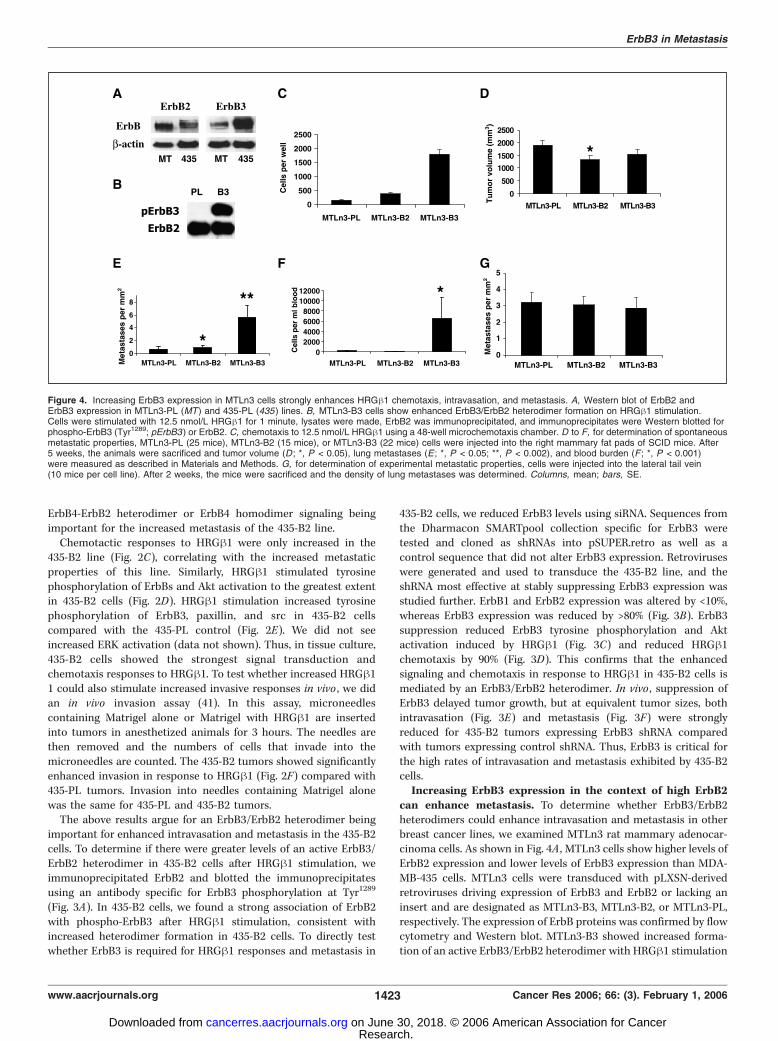
MDA-MB-435 cells with PLXSN, ErbB1, ErbB2, ErbB3, or ErbB4retrovirus will be called 435-PL, 435-B1, 435-B2, 435-B3, or 435-B4,respectively. FACS analysis of all the lines (Table 1) indicated thateach vector increased expression of the targeted ErbB comparedwith the 435-PL cells, and the altered expression remained constantafter passage in vitro and in vivo .We then evaluated metastasis in vivo using the spontaneous
metastasis assay (40). We injected each of the lines into themammary fat pads of SCID mice and measured primary tumorsize, tumor cells in the blood, and lung metastases 15 to 16 weeksafter injection. Tumor growth and final tumor size at analysisshowed no significant differences in primary tumor growthbetween the various transfectants and 435-PL (Fig. 1A), consistentwith similar growth rates observed for the lines in tissue culture(data not shown). The number of lung metastases was significantlyincreased only in animals carrying 435-B2 tumors (Fig. 1B). We havedeveloped previously a method to measure the intravasationefficiency of primary tumors by culturing blood from the rightatrium of the heart and using the number of tumor cell coloniesthat form to determine the number of viable tumor cells (40). Thisintravasation measurement showed a significant increase foranimals carrying 435-B2 tumors (Fig. 1C). To evaluate the relativeabilities of the lines to arrest and proliferate in the lungsindependent of primary tumor formation and intravasation, wedid an experimental metastasis assay. Cells were injected into thelateral tail veins of SCID mice, and 8 weeks later, the mice weresacrificed and the density of lung metastases was determined(Fig. 1D). 435-B2 cells showed a slight increase in experimentalmetastasis, consistent with a previous study (22).These results suggest that for MDA-MB-435 cells increased levels
of ErbB2 have a major effect on intravasation, a minor effect on later
stages of metastasis at the target organ (as assayed by experimentalmetastasis), and little effect on proliferation. The distributionsof lung metastasis sizes for both experimental (Fig. 1E) andspontaneous metastasis (Fig. 1F) assays show the largest increase inthe number of small metastases rather than the number of largemetastases, consistent with ErbB2 enhancing steps early in themetastasis process as opposed to enhancing the growth rate ofestablished metastases.To test whether the increased intravasation might be due to
increased angiogenesis, we evaluated microvessel density asdetected by immunostaining for von Willebrand factor. There wasno significant difference (21F 4 for 435-PL versus 28F 5 for 435-B2;P < 0.22); thus, changes in angiogenesis are unlikely to explain theincreased intravasation observed for 435-B2 tumors. Examination ofoverall tumor structure by H&E staining also did not show dramaticdifferences between tumors from any of the cell lines.Because proliferation and angiogenesis were not enhanced in
435-B2 cells, it is possible that the increased intravasation reflectsincreased motility or chemotaxis. To determine whether chemo-tactic response to a particular heterodimer pair formed by ErbB2was correlated with enhanced intravasation and metastasis, weevaluated responses to EGF, HRGh1, and BTC (17). EGF responsesare mediated by ErbB1, HRGh1 responses are mediated by ErbB3and ErbB4, and BTC responses are mediated by ErbB1 and ErbB4(5). Both 435-B1 and 435-B2 lines showed increased chemotacticresponses to EGF (Fig. 2A), arguing against responses to EGFvia ErbB1 homodimers or ErbB1/ErbB2 heterodimers beingresponsible for the increased metastatic capabilities of the435-B2 line (because 435-ErbB1 tumors do not show increasedmetastasis). Chemotactic responses to BTC were increased in the435-B1 and 435-B4 lines (Fig. 2B) but not in 435-B2, arguing against
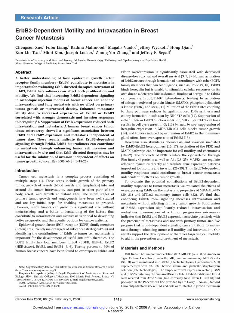
Figure 3. ErbB3/ErbB2 heterodimers mediate HRGh1-induced signaling, migration, and metastasis. A, ErbB3/ErbB2 heterodimers form on HRGh1 stimulation in435-B2 cells. ErbB2 was immunoprecipitated from cell lysates before or 4 minutes after stimulation with 12.5 nmol/L HRGh1 and the immunoprecipitates were blottedfor total ErbB2 and phospho-ErbB3 (Tyr1289). On longer exposure, a low level of immunoprecipitated ErbB2 from 435-PL cells is detectable, consistent with theirlow levels of ErbB2. Total cell lysate h-actin is also shown. B, Western blot analysis of ErbB1, ErbB2, and ErbB3 expression in stable lines transduced by pSUPER.retrocontaining control shRNA (Ctrl ) or ErbB3-targeted shRNA (B3). Scanning and quantitation of the bands indicated changes of <10% for ErbB1 and ErbB2 and >80%for ErbB3. ErbB4 expression was undetectable in either line by Western blot. C, suppression of ErbB3 expression in 435-B2 cells inhibits HRGh1-induced ErbB3phosphorylation and Akt activation. Cells were stimulated with 12.5 nmol/L HRGh1 for 4 minutes and lysed, and Western blots of the lysates were evaluated forphosphorylation of ErbB3 and Akt. D, suppression of ErbB3 expression inhibits chemotaxis to HRGh1 in 435-PL and 435-B2 cells. 435-B2 cells stably transducedby pSUPER.retro driving expression of either control shRNA (black columns ) or B3 shRNA (white columns ) were incubated with buffer or 12.5 nmol/L HRGh1 in thelower chamber. Columns, mean of 11 measurements per condition; bars, SE. E and F, spontaneous lung metastasis analysis. 435-B2 cells transduced withpSUPER.retro driving expression of either control shRNA (11 animals) or ErbB3-targeted shRNA (13 animals) were injected into the mammary fat pads of SCID mice.Primary tumor growth was followed and animals were sacrificed for the measurement of blood burden (E ; P < 0.02) and lung metastasis (F ; P < 0.001) once theaverage tumor size was >2,000 mm3 (127 days for control shRNA tumors and 148 days for B3 shRNA tumors). Columns, mean; bars, SE.
Cancer Research
Cancer Res 2006; 66: (3). February 1, 2006 1422 www.aacrjournals.org
Research. on June 30, 2018. © 2006 American Association for Cancercancerres.aacrjournals.org Downloaded from
ErbB4-ErbB2 heterodimer or ErbB4 homodimer signaling beingimportant for the increased metastasis of the 435-B2 line.Chemotactic responses to HRGh1 were only increased in the
435-B2 line (Fig. 2C), correlating with the increased metastaticproperties of this line. Similarly, HRGh1 stimulated tyrosinephosphorylation of ErbBs and Akt activation to the greatest extentin 435-B2 cells (Fig. 2D). HRGh1 stimulation increased tyrosinephosphorylation of ErbB3, paxillin, and src in 435-B2 cellscompared with the 435-PL control (Fig. 2E). We did not seeincreased ERK activation (data not shown). Thus, in tissue culture,435-B2 cells showed the strongest signal transduction andchemotaxis responses to HRGh1. To test whether increased HRGh11 could also stimulate increased invasive responses in vivo , we didan in vivo invasion assay (41). In this assay, microneedlescontaining Matrigel alone or Matrigel with HRGh1 are insertedinto tumors in anesthetized animals for 3 hours. The needles arethen removed and the numbers of cells that invade into themicroneedles are counted. The 435-B2 tumors showed significantlyenhanced invasion in response to HRGh1 (Fig. 2F) compared with435-PL tumors. Invasion into needles containing Matrigel alonewas the same for 435-PL and 435-B2 tumors.The above results argue for an ErbB3/ErbB2 heterodimer being
important for enhanced intravasation and metastasis in the 435-B2cells. To determine if there were greater levels of an active ErbB3/ErbB2 heterodimer in 435-B2 cells after HRGh1 stimulation, weimmunoprecipitated ErbB2 and blotted the immunoprecipitatesusing an antibody specific for ErbB3 phosphorylation at Tyr1289
(Fig. 3A). In 435-B2 cells, we found a strong association of ErbB2with phospho-ErbB3 after HRGh1 stimulation, consistent withincreased heterodimer formation in 435-B2 cells. To directly testwhether ErbB3 is required for HRGh1 responses and metastasis in
435-B2 cells, we reduced ErbB3 levels using siRNA. Sequences fromthe Dharmacon SMARTpool collection specific for ErbB3 weretested and cloned as shRNAs into pSUPER.retro as well as acontrol sequence that did not alter ErbB3 expression. Retroviruseswere generated and used to transduce the 435-B2 line, and theshRNA most effective at stably suppressing ErbB3 expression wasstudied further. ErbB1 and ErbB2 expression was altered by <10%,whereas ErbB3 expression was reduced by >80% (Fig. 3B). ErbB3suppression reduced ErbB3 tyrosine phosphorylation and Aktactivation induced by HRGh1 (Fig. 3C) and reduced HRGh1chemotaxis by 90% (Fig. 3D). This confirms that the enhancedsignaling and chemotaxis in response to HRGh1 in 435-B2 cells ismediated by an ErbB3/ErbB2 heterodimer. In vivo , suppression ofErbB3 delayed tumor growth, but at equivalent tumor sizes, bothintravasation (Fig. 3E) and metastasis (Fig. 3F) were stronglyreduced for 435-B2 tumors expressing ErbB3 shRNA comparedwith tumors expressing control shRNA. Thus, ErbB3 is critical forthe high rates of intravasation and metastasis exhibited by 435-B2cells.Increasing ErbB3 expression in the context of high ErbB2
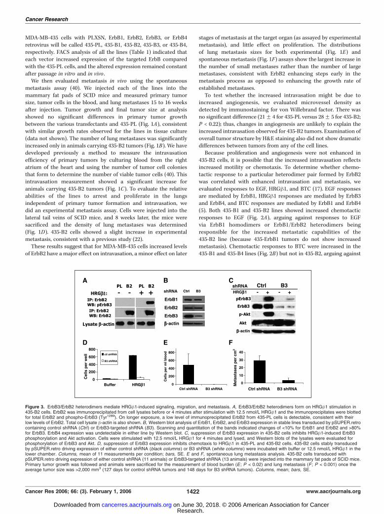
can enhance metastasis. To determine whether ErbB3/ErbB2heterodimers could enhance intravasation and metastasis in otherbreast cancer lines, we examined MTLn3 rat mammary adenocar-cinoma cells. As shown in Fig. 4A , MTLn3 cells show higher levels ofErbB2 expression and lower levels of ErbB3 expression than MDA-MB-435 cells. MTLn3 cells were transduced with pLXSN-derivedretroviruses driving expression of ErbB3 and ErbB2 or lacking aninsert and are designated as MTLn3-B3, MTLn3-B2, or MTLn3-PL,respectively. The expression of ErbB proteins was confirmed by flowcytometry and Western blot. MTLn3-B3 showed increased forma-tion of an active ErbB3/ErbB2 heterodimer with HRGh1 stimulation
Figure 4. Increasing ErbB3 expression in MTLn3 cells strongly enhances HRGh1 chemotaxis, intravasation, and metastasis. A, Western blot of ErbB2 andErbB3 expression in MTLn3-PL (MT) and 435-PL (435 ) lines. B, MTLn3-B3 cells show enhanced ErbB3/ErbB2 heterodimer formation on HRGh1 stimulation.Cells were stimulated with 12.5 nmol/L HRGh1 for 1 minute, lysates were made, ErbB2 was immunoprecipitated, and immunoprecipitates were Western blotted forphospho-ErbB3 (Tyr1289; pErbB3 ) or ErbB2. C, chemotaxis to 12.5 nmol/L HRGh1 using a 48-well microchemotaxis chamber. D to F, for determination of spontaneousmetastatic properties, MTLn3-PL (25 mice), MTLn3-B2 (15 mice), or MTLn3-B3 (22 mice) cells were injected into the right mammary fat pads of SCID mice. After5 weeks, the animals were sacrificed and tumor volume (D ; *, P < 0.05), lung metastases (E ; *, P < 0.05; **, P < 0.002), and blood burden (F ; *, P < 0.001)were measured as described in Materials and Methods. G, for determination of experimental metastatic properties, cells were injected into the lateral tail vein(10 mice per cell line). After 2 weeks, the mice were sacrificed and the density of lung metastases was determined. Columns, mean; bars, SE.
ErbB3 in Metastasis
www.aacrjournals.org 1423 Cancer Res 2006; 66: (3). February 1, 2006
Research. on June 30, 2018. © 2006 American Association for Cancercancerres.aacrjournals.org Downloaded from
as evidenced by blotting ErbB2 immunoprecipitates with anantibody against phospho-ErbB3 (Fig. 4B). Chemotaxis to HRGh1was slightly increased in MTLn3-B2 cells and most stronglyenhanced in MTLn3-B3 cells (Fig. 4C), with no effect on growthrate in vitro .The metastatic properties of these lines were then measured
using the spontaneous and experimental metastasis assays.Primary tumor growth rate was slightly reduced for both MTLn3-B2 and MTLn3-B3 compared with the control MTLn3-PL line(Fig. 4D). On the other hand, lung metastasis was slightly increasedin the MTLn3-B2 line and strongly increased in the MTLn3-B3 line(Fig. 4E). In parallel with the increased lung metastasis, MTLn3-B3generated tumors had significantly increased intravasation (Fig. 4F).Lung colonization efficiency as determined by the experimentalmetastasis assay showed no differences among the MTLn3-B3,MTLn-B2, andMTLn3-PL lines (Fig. 4G). Thus, in the context of highlevels of ErbB2, increased expression of ErbB3 also can enhancechemotaxis, intravasation, and metastasis independent of effects onprimary tumor growth.
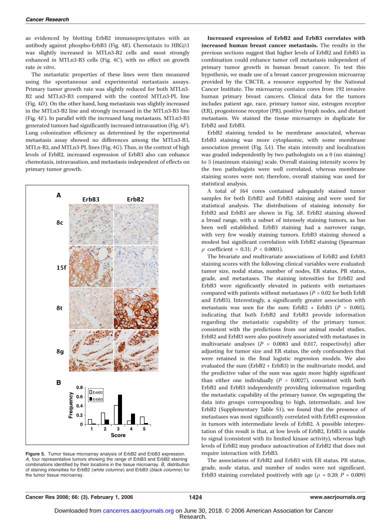
Increased expression of ErbB2 and ErbB3 correlates withincreased human breast cancer metastasis. The results in theprevious sections suggest that higher levels of ErbB2 and ErbB3 incombination could enhance tumor cell metastasis independent ofprimary tumor growth in human breast cancer. To test thishypothesis, we made use of a breast cancer progression microarrayprovided by the CBCTR, a resource supported by the NationalCancer Institute. The microarray contains cores from 192 invasivehuman primary breast cancers. Clinical data for the tumorsincludes patient age, race, primary tumor size, estrogen receptor(ER), progesterone receptor (PR), positive lymph nodes, and distantmetastasis. We stained the tissue microarrays in duplicate forErbB2 and ErbB3.ErbB2 staining tended to be membrane associated, whereas
ErbB3 staining was more cytoplasmic, with some membraneassociation present (Fig. 5A). The stain intensity and localizationwas graded independently by two pathologists on a 0 (no staining)to 5 (maximum staining) scale. Overall staining intensity scores bythe two pathologists were well correlated, whereas membranestaining scores were not; therefore, overall staining was used forstatistical analysis.A total of 164 cores contained adequately stained tumor
samples for both ErbB2 and ErbB3 staining and were used forstatistical analysis. The distributions of staining intensity forErbB2 and ErbB3 are shown in Fig. 5B . ErbB2 staining showeda broad range, with a subset of intensely staining tumors, as hasbeen well established. ErbB3 staining had a narrower range,with very few weakly staining tumors. ErbB3 staining showed amodest but significant correlation with ErbB2 staining (Spearmanq coefficient = 0.31; P < 0.0001).The bivariate and multivariate associations of ErbB2 and ErbB3
staining scores with the following clinical variables were evaluated:tumor size, nodal status, number of nodes, ER status, PR status,grade, and metastases. The staining intensities for ErbB2 andErbB3 were significantly elevated in patients with metastasescompared with patients without metastases (P = 0.02 for both ErbBand ErbB3). Interestingly, a significantly greater association withmetastasis was seen for the sum: ErbB2 + ErbB3 (P = 0.005),indicating that both ErbB2 and ErbB3 provide informationregarding the metastatic capability of the primary tumor,consistent with the predictions from our animal model studies.ErbB2 and ErbB3 were also positively associated with metastases inmultivariate analyses (P = 0.0083 and 0.017, respectively) afteradjusting for tumor size and ER status, the only confounders thatwere retained in the final logistic regression models. We alsoevaluated the sum (ErbB2 + ErbB3) in the multivariate model, andthe predictive value of the sum was again more highly significantthan either one individually (P = 0.0027), consistent with bothErbB2 and ErbB3 independently providing information regardingthe metastatic capability of the primary tumor. On segregating thedata into groups corresponding to high, intermediate, and lowErbB2 (Supplementary Table S1), we found that the presence ofmetastases was most significantly correlated with ErbB3 expressionin tumors with intermediate levels of ErbB2. A possible interpre-tation of this result is that, at low levels of ErbB2, ErbB3 is unableto signal (consistent with its limited kinase activity), whereas highlevels of ErbB2 may produce autoactivation of ErbB2 that does notrequire interaction with ErbB3.The associations of ErbB2 and ErbB3 with ER status, PR status,
grade, node status, and number of nodes were not significant.ErbB3 staining correlated positively with age (q = 0.20; P = 0.009)
Figure 5. Tumor tissue microarray analysis of ErbB2 and ErbB3 expression.A, four representative tumors showing the range of ErbB3 and ErbB2 stainingcombinations identified by their locations in the tissue microarray. B, distributionof staining intensities for ErbB2 (white columns ) and ErbB3 (black columns ) forthe tumor tissue microarray.
Cancer Research
Cancer Res 2006; 66: (3). February 1, 2006 1424 www.aacrjournals.org
Research. on June 30, 2018. © 2006 American Association for Cancercancerres.aacrjournals.org Downloaded from
and negatively with tumor size (q = �0.16; P < 0.041), but theseassociations did not retain significance in multivariate regressionanalyses. ErbB2 was not significantly associated with other clinicalvariables.
Discussion
In the studies reported here, we provide the first direct evidencethat ErbB3-dependent signaling can contribute to metastasis ofbreast cancer through enhancing intravasation. Because tumorformation and angiogenesis were not increased in parallel withintravasation and metastasis, it is unlikely that the proliferativeconsequences of ErbB3 signaling are the basis for the enhancedmetastasis we observed. Our results support a model (13, 22, 42) inwhich both biological effects of ErbB3 signaling (enhancedtumorigenesis and invasion) can contribute to breast cancer. Inthis model, low levels of ErbB3 signaling are sufficient fortumorigenesis, forming an oncogenic unit (13) through generationof signals that suppress apoptosis and enhance cell cycleprogression. Because ErbB3 signaling occurs mainly throughheterodimerization with ErbB2, low levels of signaling could occurin tumors expressing high levels of either ErbB2 or ErbB3 throughspontaneous heterodimer formation (in the absence of heregulin)or in tumors expressing lower levels of ErbB3/ErbB2 that areexposed to heregulin. However, chemotaxis and invasion would berelatively weak, and such tumors would be unable to effectively useErbB3/ErbB2 signaling for invasion. On the other hand, tumorsexpressing moderate to high levels of both ErbB2 and ErbB3(potentially through genomic amplification) could show strongchemotaxis and invasion, resulting in enhanced intravasation andmetastasis due to ErbB3/ErbB2 signaling.In this model, the endogenous ErbB2 and ErbB3 levels in the
MDA-MB-435 and MTLn3 cells are sufficient for full stimulation oftumorigenesis in vivo but are suboptimal for maximal chemotaxisand invasion [low levels of ErbB2 in MDA-MB-435 cells (16, 22, 43)and low levels of ErbB3 in MTLn3 cells]. Overexpressing thelimiting partner (ErbB2 in MDA-MB-435 cells or ErbB3 in MTLn3cells) enables stronger signaling resulting in enhanced chemotaxisand invasion without affecting primary tumor growth. Theenhanced chemotaxis and invasion could reflect both immediateErbB activation (17) and ErbB-induced alterations in geneexpression patterns (44, 45). Suppression of ErbB3 in 435-B2 cellsreduces the signaling efficiency below that which is optimal fortumorigenesis, resulting in slowed tumor growth. However, even ongrowth of the tumors to a large size, intravasation and metastasis
remain reduced as predicted if an ErbB3-dependent intravasationstep is required for efficient metastasis of the 435-B2 cells.The tumor tissue microarray analysis of human breast cancer
expression of ErbB3 and ErbB2 is consistent with this model intwo ways. First, the combination of ErbB2 and ErbB3 was moresignificantly associated with the presence of metastasis than thelevel of ErbB2 or ErbB3 staining alone, consistent with the impor-tance of a complex of ErbB2 and ErbB3 for invasion and metastasis.Second, there was no correlation between tumor size and thecombination of ErbB2 and ErbB3, suggesting that these tumorshave progressed past the early steps in tumorigenesis. In addition,ErbB3 expression levels may be useful in prognosis of metastasisfor tumors that have intermediate levels of ErbB2.This model has implications for therapies that are targeted to
inhibition of ErbB2 or ErbB3 (and by extension other receptorsthat have functions in both growth control and chemotaxis). Itsuggests that, for the subset of tumors in which ErbB3/ErbB2signaling is near the threshold required for tumorigenesis, partialsuppression of either ErbB2 or ErbB3 could directly affect tumorgrowth as is seen for a limited number of tumors. However, for thesubset of tumors that have moderate to high levels of both ErbB2and ErbB3, partial suppression might only affect chemotaxis andinvasion without affecting tumor growth, similar in action tometastasis suppressors (46). Such tumors would be scored asnonresponding in terms of growth but might still be inhibited interms of invasion. Our results support the targeting of ErbB3-dependent signaling for inhibition of invasion in tumors over-expressing ErbB2 and ErbB3. Suppression of invasion wouldcontribute to limiting the further spread of tumor fragments andmetastases that remain after removal of the primary tumor andcould be useful in combination with treatments that directly targetgrowth (47). In addition, patients with tumors having high levels ofErbB3 in the presence of intermediate to high levels of ErbB2 areat higher risk of metastasis and thus might need to be moreaggressively treated.
Acknowledgments
Received 2/17/2005; revised 10/11/2005; accepted 11/18/2005.Grant support: CA77522 and CA100324 (J.E. Segall) and CA69202 (Z-Y. Zhang) for
support.The costs of publication of this article were defrayed in part by the payment of page
charges. This article must therefore be hereby marked advertisement in accordancewith 18 U.S.C. Section 1734 solely to indicate this fact.
We thank the members of the Cox, Segall, and Condeelis laboratories and GlennKroog for comments and suggestions and David Stern, Bristol Myers Squibb, and GaryP. Nolan for generously providing the reagents.
References
1. Chambers AF, Groom AC, MacDonald IC. Metastasis:dissemination and growth of cancer cells in metastaticsites. Nat Rev Cancer 2002;2:563–72.
2. Shawver LK, Slamon D, Ullrich A. Smart drugs:tyrosine kinase inhibitors in cancer therapy. CancerCell 2002;1:117–23.
3. McKeage K, Perry CM. Trastuzumab: a review of itsuse in the treatment of metastatic breast canceroverexpressing HER2. Drugs 2002;62:209–43.
4. Mendelsohn J, Baselga J. Status of epidermal growthfactor receptor antagonists in the biology and treatmentof cancer. J Clin Oncol 2003;21:2787–99.
5. Yarden Y, Sliwkowski MX. Untangling the ErbBsignalling network. Nat Rev Mol Cell Biol 2001;2:127–37.
6. Holbro T, Civenni G, Hynes NE. The ErbB receptors
and their role in cancer progression. Exp Cell Res 2003;284:99–110.
7. Slamon DJ, Clark GM, Wong SG, et al. Human breastcancer: correlation of relapse and survival with amplifica-tion of the HER-2/neu oncogene. Science 1987;235:177–82.
8. Press MF, Pike MC, Chazin VR, et al. Her-2/neuexpression in node-negative breast cancer: direct tissuequantitation by computerized image analysis andassociation of overexpression with increased risk ofrecurrent disease. Cancer Res 1993;53:4960–70.
9. Stern DF. ErbBs in mammary development. Exp CellRes 2003;284:89–98.
10. Citri A, Skaria KB, Yarden Y. The deaf and the dumb:the biology of ErbB-2 and ErbB-3. Exp Cell Res 2003;284:54–65.
11. Alimandi M, Romano A, Curia MC, et al. Cooperativesignaling of ErbB3 and ErbB2 in neoplastic transforma-
tion and human mammary carcinomas. Oncogene 1995;10:1813–21.
12. Vijapurkar U, Kim MS, Koland JG. Roles of mitogen-activated protein kinase and phosphoinositide 3V-kinasein ErbB2/ErbB3 coreceptor-mediated heregulin signal-ing. Exp Cell Res 2003;284:291–302.
13. Holbro T, Beerli RR, Maurer F, et al. The ErbB2/ErbB3heterodimer functions as an oncogenic unit: ErbB2requires ErbB3 to drive breast tumor cell proliferation.Proc Natl Acad Sci U S A 2003;100:8933–8.
14. Tsai MS, Shamon-Taylor LA, Mehmi I, Tang CK, LupuR. Blockage of heregulin expression inhibits tumorige-nicity and metastasis of breast cancer. Oncogene 2003;22:761–8.
15. Siegel PM, Ryan ED, Cardiff RD, Muller WJ. Elevatedexpression of activated forms of Neu/ErbB-2 and ErbB-3are involved in the induction of mammary tumors in
ErbB3 in Metastasis
www.aacrjournals.org 1425 Cancer Res 2006; 66: (3). February 1, 2006
Research. on June 30, 2018. © 2006 American Association for Cancercancerres.aacrjournals.org Downloaded from
Cancer Research
Cancer Res 2006; 66: (3). February 1, 2006 1426 www.aacrjournals.org
transgenic mice: implications for human breast cancer.EMBO J 1999;18:2149–64.
16. Adelsman MA, McCarthy JB, Shimizu Y. Stimulationof h1-integrin function by epidermal growth factor andheregulin-h has distinct requirements for erbB2 but asimilar dependence on phosphoinositide 3-OH kinase.Mol Biol Cell 1999;10:2861–78.
17. Spencer KS, Graus-Porta D, Leng J, Hynes NE, KlemkeRL. ErbB2 is necessary for induction of carcinoma cellinvasion by ErbB family receptor tyrosine kinases. J CellBiol 2000;148:385–97.
18. Hinton DR, He S, Graf K, et al. Mitogen-activatedprotein kinase activation mediates PDGF-directedmigration of RPE cells. Exp Cell Res 1998;239:11–5.
19. Summy M, Gallick GE. Src family kinases in tumorprogression and metastasis. Cancer Metastasis Rev 2003;22:337–58.
20. Chausovsky A, Waterman H, Elbaum M, Yarden Y.Molecular requirements for the effect of neuregulin oncell spreading, motility and colony organization. Onco-gene 2000;19:878–88.
21. Adam L, Vadlamudi R, Kondapaka SB, et al. Here-gulin regulates cytoskeletal reorganization and cellmigration through the p21-activated kinase-1 viaphosphatidylinositol-3 kinase. J Biol Chem 1998;273:28238–46.
22. Tan T, Yao J, Yu D. Overexpression of the c-erbB-2gene enhanced intrinsic metastasis potential in humanbreast cancer cells without increasing their transforma-tion abilities. Cancer Res 1997;57:1199–205.
23. Katso R, Okkenhaug K, Ahmadi K, et al. Cellularfunction of phosphoinositide 3-kinases: implications fordevelopment, homeostasis, and cancer. Annu Rev CellDev Biol 2001;17:615–75.
24. Fukata M, Nakagawa M, Kaibuchi K. Roles of Rho-family GTPases in cell polarisation and directionalmigration. Curr Opin Cell Biol 2003;15:590–7.
25. Merlot S, Firtel RA. Leading the way: directional
sensing through phosphatidylinositol 3-kinase and othersignaling pathways. J Cell Sci 2003;116:3471–8.
26. Reddy KB, Nabha SM, Atanaskova N. Role of MAPkinase in tumor progression and invasion. CancerMetastasis Rev 2003;22:395–403.
27. Xia Y, Karin M. The control of cell motility andepithelial morphogenesis by Jun kinases. Trends CellBiol 2004;14:94–101.
28. Webb DJ, Donais K, Whitmore LA, et al. FAK-Srcsignalling through paxillin, ERK and MLCK regulatesadhesion disassembly. Nat Cell Biol 2004;6:154–61.
29. Ozanne BW, McGarry L, Spence HJ, et al. Transcrip-tional regulation of cell invasion: AP-1 regulation of amultigenic invasion programme. Eur J Cancer 2000;36:1640–8.
30. Price JE. Analyzing the metastatic phenotype. J CellBiochem 1994;56:16–22.
31. Sellappan S, Grijalva R, Zhou X, et al. Lineage infidelityof MDA-MB-435 cells: expression of melanocyte proteinsin a breast cancer cell line. Cancer Res 2004;64:3479–85.
32. Lichtner RB, Nicolson GL. Organization of cytoskeletalstructures in 13762NF rat mammary adenocarcinomacell lines and clones of different metastatic potentials.Invasion Metastasis 1987;7:73–82.
33. Welch DR, Neri A, Nicolson GL. Comparison of‘‘spontaneous’’ and ‘‘experimental’’ metastasis using rat13762 mammary adenocarcinoma metastatic cellclones. Invasion Metastasis 1983;3:65–80.
34. Riese DJ, van Raaij TM, Plowman GD, Andrews GC,Stern DF. The cellular response to neuregulins isgoverned by complex interactions of the erbB receptorfamily. Mol Cell Biol 1995;15:5770–6.
35. Pear WS, Nolan GP, Scott ML, Baltimore D. Produc-tion of high-titer helper-free retroviruses by transienttransfection. Proc Natl Acad Sci U S A 1993;90:8392–6.
36. Wyckoff JB, Insel L, Khazaie K, et al. Suppression ofruffling by EGF in chemotactic cells. Exp Cell Res 1998;242:100–9.
37. Wyckoff J, Wang W, Lin EY, et al. A paracrine loopbetween tumor cells and macrophages is required fortumor cell migration in mammary tumors. Cancer Res2004;64:7022–9.
38. Wang W, Wyckoff JB, Wang Y, et al. Gene expressionanalysis on small numbers of invasive cells collected bychemotaxis from primary mammary tumors of themouse. BMC Biotechnol 2003;3:13.
39. Kempiak SJ, Yip SC, Backer JM, Segall JE. Localsignaling by the EGF receptor. J Cell Biol 2003;162:781–7.
40. Wyckoff JB, Jones JG, Condeelis JS, Segall JE. A criticalstep in metastasis: in vivo analysis of intravasation atthe primary tumor. Cancer Res 2000;60:2504–11.
41. Wyckoff JB, Segall JE, Condeelis JS. The collection ofthe motile population of cells from a living tumor.Cancer Res 2000;60:5401–4.
42. Xu FJ, Stack S, Boyer C, et al. Heregulin and agonisticanti-p185(c-erbB2) antibodies inhibit proliferation butincrease invasiveness of breast cancer cells that over-express p185(c-erbB2): increased invasiveness may con-tribute to poor prognosis. Clin Cancer Res 1997;3:1629–34.
43. Aguilar Z, Akita RW, Finn RS, et al. Biologic effects ofheregulin/neu differentiation factor on normal andmalignant human breast and ovarian epithelial cells.Oncogene 1999;18:6050–62.
44. Yao J, Xiong S, Klos K, et al. Multiple signalingpathways involved in activation of matrix metallopro-teinase-9 (MMP-9) by heregulin-h1 in human breastcancer cells. Oncogene 2001;20:8066–74.
45. Grothey A, Hashizume R, Ji H, et al. C-erbB-2/HER-2upregulates fascin, an actin-bundling protein associatedwith cell motility, in human breast cancer cell lines.Oncogene 2000;19:4864–75.
46. Shevde LA, Welch DR. Metastasis suppressor path-ways—an evolving paradigm. Cancer Lett 2003;30:1–20.
47. Sledge GW, Jr., Miller KD. Exploiting the hallmarks ofcancer: the future conquest of breast cancer. Eur JCancer 2003;39:1668–75.
Research. on June 30, 2018. © 2006 American Association for Cancercancerres.aacrjournals.org Downloaded from

2006;66:1418-1426. Cancer Res Chengsen Xue, Fubo Liang, Radma Mahmood, et al. MetastasisErbB3-Dependent Motility and Intravasation in Breast Cancer
Updated version
http://cancerres.aacrjournals.org/content/66/3/1418
Access the most recent version of this article at:
Material
Supplementary
http://cancerres.aacrjournals.org/content/suppl/2006/02/02/66.3.1418.DC1
Access the most recent supplemental material at:
Cited articles
http://cancerres.aacrjournals.org/content/66/3/1418.full#ref-list-1
This article cites 47 articles, 18 of which you can access for free at:
Citing articles
http://cancerres.aacrjournals.org/content/66/3/1418.full#related-urls
This article has been cited by 13 HighWire-hosted articles. Access the articles at:
E-mail alerts related to this article or journal.Sign up to receive free email-alerts
Subscriptions
Reprints and
To order reprints of this article or to subscribe to the journal, contact the AACR Publications
Permissions
Rightslink site. (CCC)Click on "Request Permissions" which will take you to the Copyright Clearance Center's
.http://cancerres.aacrjournals.org/content/66/3/1418To request permission to re-use all or part of this article, use this link
Research. on June 30, 2018. © 2006 American Association for Cancercancerres.aacrjournals.org Downloaded from