equine metabolic syndromefiles.clinicadegrandes.webnode.com.br/200000077-ab7beac75e... · equine...
TRANSCRIPT

Equine MetabolicSyndrome
Nicholas Frank, DVM, PhDa,b,*
KEYWORDS
� Obesity � Regional adiposity � Hyperinsulinemia� Insulin resistance � Laminitis
Veterinarians have long recognized that obese horses and ponies are prone to lami-nitis, but the concept of an equine metabolic syndrome (EMS) was first proposedby Johnson1 in 2002. This concept has developed over time, and EMS was recentlydescribed in a consensus statement released by the American College of VeterinaryInternal Medicine.2 In human medicine, metabolic syndrome (MetS) refers to a setof risk factors that predict the risk of cardiovascular disease,3 including obesity,glucose intolerance and insulin resistance (IR), dyslipidemia, microalbuminuria, andhypertension. Associated conditions in humans include nonalcoholic fatty liverdisease and polycystic ovary syndrome. EMS shares some of the features of MetS,including obesity, IR, and dyslipidemia, but differs in that laminitis is the primarydisease of interest.
COMPONENTS OF THE SYNDROME
EMS is not a specific disease entity, but rather a clinical syndrome associated withlaminitis.4,5 Increased adiposity, hyperinsulinemia, and IR are the 3 principal compo-nents of this syndrome, and it is difficult to separate these factors from one another.Hyperinsulinemia is detected in most insulin resistant horses and affected animalsare usually obese or exhibit regional adiposity (Fig. 1). One or all of these factorsmay determine laminitis susceptibility, but it is also conceivable that another, as yetunidentified, factor predisposes horses with EMS to laminitis. Other components ofEMS include dyslipidemia,4–6 altered blood adipokine concentrations,5,7,8 systemicinflammation,9 and seasonal arterial hypertension.10 In contrast to MetS in humans,atherosclerosis and coronary heart disease are not detected in horses with EMS,and this may be explained by the herbivorous diet of horses or lipoprotein composition
a Department of Large Animal Clinical Sciences, University of Tennessee College of VeterinaryMedicine, 2407 River Drive, Knoxville, TN 37996, USAb Division of Medicine, School of Veterinary Medicine and Science, University of Nottingham,Sutton Bonington Campus, Leicestershire LE12 5RD, UK* Department of Large Animal Clinical Sciences, University of Tennessee College of VeterinaryMedicine, 2407 River Drive, Knoxville, TN 37996.E-mail address: [email protected]
Vet Clin Equine 27 (2011) 73–92doi:10.1016/j.cveq.2010.12.004 vetequine.theclinics.com0749-0739/11/$ – see front matter � 2011 Elsevier Inc. All rights reserved.

Fig. 1. A 7-year-old Morgan horse mixed breed mare with physical characteristics of equinemetabolic syndrome.
Frank74
of equine blood. Most circulating cholesterol is carried within high-density lipoproteinsin horses, rather than low-density lipoproteins, which are atherogenic.11
BREED PREDISPOSITION
EMS occurs most commonly in pony breeds, Morgan horses, Paso Finos, Arabians,Saddlebreds, Quarter horses, and Tennessee Walking horses. Most horses andponies with EMS are obese, and owners often describe them as “easy keepers.” Envi-ronmental issues such as overfeeding and lack of exercise contribute to obesity, andthese problems are increasing with modern management practices.
CLINICAL PRESENTATIONLaminitis
Horses and ponies with EMS are predisposed to laminitis, so this is the most commonpresenting complaint. Laminitis typically develops after animals have been grazing onpasture, which is referred to as pasture-associated laminitis.12 Episodes of laminitisoften occur after heavy rains and abundant sunlight, when grasses have been growingrapidly and accumulating water-soluble carbohydrates (WSC) through increasedphotosynthesis.13 Grazing on rapidly growing pastures increases total energy intakeand promotes obesity, while also increasing WSC consumption. Resting insulinconcentrations increase as a result, and this alters proxy measures of insulin sensi-tivity and pancreatic output.4,5 However, proxy measures used for blood sampleshave been collected under fed conditions in previous studies, so it is difficult to deter-mine whether insulin sensitivity per se progressively decreases in response to pasturegrazing. This distinction is an important one because laminitis has been experimentallyinduced in ponies and horses by infusing insulin intravenously, suggesting that hyper-insulinemia is the trigger for disease.14,15 One challenge for researchers in the future isto conduct studies in which the effects of obesity, IR, and hyperinsulinemia on laminitisdevelopment are evaluated independently.Pasture grazing also raises the risk of intestinal carbohydrate overload, particularly
when animals are moved onto new pastures without gradual transition.12 In these situ-ations, the amount of WSC entering the intestinal tract exceeds the digestive andabsorptive capacities of the small intestine and increases the amount of substrate

Equine Metabolic Syndrome 75
available for fermentation within the large intestine. Increased fermentation raiseslactic acid concentrations, lowers pH, and increases mucosal permeability.16 Move-ment of gut-derived factors including exotoxins, endotoxins, and vasoactive aminesinto the circulation induces a systemic inflammatory response and activatesplatelets.17 Evidence for an intestinal trigger for pasture-associated laminitis comesfrom studies in which oligofructose has been administered to horses as a model forcarbohydrate overload on pasture. Treated horses exhibit clinical signs consistentwith a systemic inflammatory response, followed by laminitis.18–20
Laminitis is typically thought of as a catastrophic event causing severe lameness,but a milder form of laminitis is often detected in horses and ponies with EMS. Diver-gent growth rings (founder lines) are sometimes recognized in horses that are walkingsoundly, indicating that hoof growth has been disrupted by a previous laminitisepisode (Fig. 2). A growth ring is considered divergent when the distance to the coro-nary band is shorter at the dorsum than the heel. These findings suggest that horsesdevelop laminitis that goes unnoticed by the owner, particularly when animals are kepton pasture. A full diagnostic evaluation should therefore include lateral radiographs ofthe feet and placement of hoof testers. Mild lameness associated with laminitis cansometimes be detected by tightly circling the horse on a hard surface.
Complications of Hospitalization
There are anecdotal reports of horses and ponies with EMS showing greater suscep-tibility to laminitis triggered by grain overload, retained fetal membranes, and colitis. Itis therefore important to recognize the EMS phenotype and alert owners to the poten-tial risk of laminitis. Obese horses are also susceptible to colic caused by lipomas. Apedunculated lipoma can lead to strangulation of the small intestine and moderate tosevere colic, accompanied by intestinal distension and reflux.Problems with IR are sometimes recognized for the first time when hypertriglyceri-
demia develops in hospitalized patients that enter negative energy balance. Insulin-resistant horses mobilize lipids more readily and are more susceptible to equinehyperlipemia.11 Hyperglycemia and glucosuria may also be detected in affectedhorses when dextrose is administered intravenously to provide partial parenteral nutri-tion. Exogenous insulin is often required in these cases to maintain plasma glucoseconcentrations below renal threshold while dextrose is administered. Of interest, there
Fig. 2. Divergent growth rings (founder lines) indicating previous laminitis.
Frank76
have been no reports of laminitis developing as a result of intravenous insulin infusionto manage hyperglycemia.
Obesity and Regional Adiposity
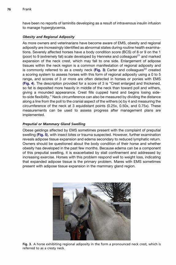
As more owners and veterinarians have become aware of EMS, obesity and regionaladiposity are increasingly identified as abnormal states during routine health examina-tions. Severely affected horses have a body condition score (BCS) of 8 or 9 on the 1(poor) to 9 (extremely fat) scale developed by Henneke and colleagues21 and markedexpansion of the neck crest, which may fall to one side. Enlargement of adiposetissues within the neck region is a common manifestation of regional adiposity andis commonly referred to as a cresty neck (Fig. 3) Carter and colleagues22 createda scoring system to assess horses with this form of regional adiposity using a 0 to 5range, and scores of 3 or more are often detected in horses or ponies with EMS(Fig. 4). The description provided for a score of 3 is “Crest enlarged and thickened,so fat is deposited more heavily in middle of the neck than toward poll and withers,giving a mounded appearance. Crest fills cupped hand and begins losing side-to-side flexibility.” Neck circumference can also be measured by dividing the distancealong a line from the poll to the cranial aspect of the withers (x) by 4 and measuring thecircumference of the neck at 3 equidistant points (0.25x, 0.50x, and 0.75x). Thesemeasurements can be used to assess progress after management plans areimplemented.
Preputial or Mammary Gland Swelling
Obese geldings affected by EMS sometimes present with the complaint of preputialswelling (Fig. 5), with insect bites or trauma suspected. However, further examinationreveals adipose tissue expansion and edema secondary to reduced lymphatic return.Owners should be questioned about the body condition of their horse and whetherobesity has developed in the past few months. Because edema can be a componentof this preputial swelling, it is exacerbated by stall confinement and addressed byincreasing exercise. Horses with this problem respond well to weight loss, indicatingthat expanded adipose tissue is the primary problem. Mares with EMS sometimespresent with adipose tissue expansion in the mammary gland region.
Fig. 3. A horse exhibiting regional adiposity in the form a pronounced neck crest, which isreferred to as a cresty neck.

Fig. 4. Crest neck scoring system. (Reprinted from Carter RA, Geor RJ, Burton SW, et al.Apparent adiposity assessed by standardised scoring systems and morphometric measure-ments in horses and ponies. Vet J 2009;179:204; with permission.)
Equine Metabolic Syndrome 77
PATHOPHYSIOLOGYPredisposition
Some horses and ponies appear to be genetically predisposed to EMS, and this is thefocus of ongoing study at the University of Minnesota (www.cvm.umn.edu/equinegenetics/ems). There are numerous anecdotal reports of EMS in related horsesand ponies, and Treiber and colleagues4 detected a dominant inheritance pattern forlaminitis in ponies with EMS. Many animals with EMS appear to require fewer caloriesto maintain body weight, indicating enhanced metabolic efficiency. Genetic
Fig. 5. Adipose tissue expansion around the prepuce of a 16-year-old Tennessee Walkinghorse gelding.

Frank78
predisposition to obesity may involve specific gene mutations, and the concept of“thrifty genes” warrants consideration.23 This theory has been applied to humans,and focuses on the concept of famine conditions leading to selection of metabolicgenes that improve metabolic efficiency, promote obesity, and increase appetitewhen food is plentiful. One candidate gene is the melanocortin-4 receptor (MC4R),which regulates feed intake, insulin sensitivity, and adiposity, and results of a prelimi-nary study indicate that a single nucleotide polymorphism exists within the codingregion of this gene in horses.24
Fetal programming might also affect metabolic status because fetal birth weight hasbeen inversely associated with the risk of developing type 2 diabetes mellitus inhumans.25 This feature can be described as a “thrifty phenotype” that is determinedby environment rather than genetics.23 For example, the increased risk of type 2 dia-betes mellitus has been attributed to inadequate pancreatic development caused bynutrient deprivation during pregnancy.25 With respect to obesity, some reports indi-cate that in utero nutrient deficiency increases the incidence of this problem, whereasothers demonstrate a positive association between birth weight and body mass indexlater in life.24 Fetal programming has been examined in horses. Ousey andcolleagues26 fed mares to maintain moderate or high BCSs during pregnancy, butall horses inadvertently lost approximately 10% of body mass at mid-gestation asa result of Streptococcus equi infection. When intravenous glucose tests were per-formed in foals at 2 to 4 days of age, insulin concentrations were higher in foalsfrom moderate BCS mares. These results suggest that acute nutrient restriction atmid-gestation affected foals in utero and altered b-cell responsiveness or insulinsensitivity. Feeding mares a high starch diet during pregnancy has also been shownto affect glucose concentrations and insulin sensitivity in preweaned foals.27 A trendtoward lower insulin sensitivity was detected in foals at 160 days of age. Furtherstudies are therefore required to determine whether nutrient deficiency or excessduring gestation contribute to the development of EMS. It is possible that epigeneticsplays a role in EMS if environmental conditions induce heritable changes in geneexpression without altering the DNA sequence.3 Epigenetic effects are mediated byalterations in DNAmethylation or histone configuration, and might explain why in uteroconditions during one pregnancy affect subsequent generations.
Hyperinsulinemia
It is assumed that hyperinsulinemia results from increased pancreatic insulin secretionin response to reduced insulin sensitivity, and this is referred to as compensatedinsulin resistance.28 Values for insulin sensitivity and the acute insulin response toglucose (AIRg) provide evidence of compensated IR in insulin-resistant horses andponies. These values are estimated from minimal model analysis of frequently-sampled intravenous glucose tolerance test (FSIGTT) data, with AIRg representingpancreatic insulin secretion. Treiber and colleagues29 reported higher AIRg values inhorses and ponies with lower insulin sensitivity, and Carter and colleagues30 demon-strated that mean AIRg increased by 408% as insulin sensitivity decreased by 71%when obesity was induced in Arabian geldings. In contrast, uncompensated insulinresistance refers to inadequate insulin secretion in response to IR, with higher glucoseconcentrations detected. This situation has been described in clinically laminiticponies, and should be suspected whenever hyperglycemia is detected in an animalwith the physical characteristics of EMS.4 A dynamic test is recommended in thesecases because hyperinsulinemia may be absent. Diabetes mellitus also occurs inhorses and is characterized by persistent hyperglycemia, with glucosuria detectedin some cases. This condition may be more common than previously thought, and

Equine Metabolic Syndrome 79
has been detected in horses with pituitary pars intermedia dysfunction.31 Inadequatepancreatic insulin secretion results in hyperglycemia with concurrent IR in some, butnot all, cases.Recent evidence suggests that higher blood insulin concentrations also result from
reduced hepatic insulin clearance.32 Pancreatic insulin secretion can be assessed bymeasuring serum connecting peptide (C-peptide) concentrations because this mole-cule is released with insulin as the hormone is secreted. Approximately 70% of insulinsecreted by the pancreas is cleared by the liver, whereas C-peptide remains incirculation.32 The C-peptide to insulin ratio therefore reflects hepatic insulin clearance.Obese horses have high insulin and C-peptide concentrations, yet lower C-peptide toinsulin ratios, indicating both increased insulin secretion and reduced hepaticclearance.32
Hepatic Insulin Resistance
Higher plasma g-glutamyl transferase (GGT) and aspartate aminotransferase (AST)activities are detected in some horses with EMS, and lipid accumulation within hepa-tocytes is a common postmortem finding. This feature suggests that hepatic lipidosisdevelops in some horses with EMS in the same way that nonalcoholic fatty liversyndrome has been associated with MetS in humans.3 Reduced insulin clearanceby the liver in horses with EMS is a manifestation of hepatic IR. This problem reflectsthe impact of obesity on liver function and includes upregulation of inflammatorypathways.33 Results of a preliminary study indicate that Toll-like receptor pathwaysare upregulated in the liver of obese insulin-resistant horses.34 Impaired hepatic func-tion might also increase the risk of laminitis by reducing the clearance of gut-derivedtriggers for laminitis or altering the metabolism of dietary carbohydrates.
Peripheral Insulin Resistance
Insulin resistance is defined as a reduction in the action of insulin on target tissues.35
Normal actions of insulin include inhibition of gluconeogenesis and lipolysis and stim-ulation of glycogen synthesis.33 Mechanisms of IR include defects in the insulinreceptor, insulin signaling pathways, or glucose transporter 4 (GLUT4) synthesis,translocation, or function. One important action of insulin is to stimulate glucose trans-port into cells, and this occurs rapidly as GLUT4 proteins translocate to cellmembranes. Vesicles containing preformed GLUT4 are present within the cytoplasm,and transporters move to the plasma membrane after activation by the insulinsignaling cascade. Results of a recent study indicate that GLUT4 translocation isimpaired in insulin-resistant horses. Waller and colleagues36 demonstrated thatGLUT4 translocation to the cell surface is significantly reduced in skeletal musclefrom insulin-resistant horses, despite normal protein abundance. Results of thispreliminary study provide the first information regarding mechanisms of IR in horses.
Obesity
Obesity develops as animals consume more energy than they expend. Studies havenot been performed to measure metabolic efficiency or compare rates of weightgain among different breeds of horse, but increased awareness of EMS has led clini-cians to recognize that some horses and ponies develop obesity more readily, and thisproblem is difficult to reverse in the same animals. When obesity has been induced inhorses, the breed of horse has affected outcomes. Quinn and colleagues37 failed todetect a decrease in insulin sensitivity associated with weight gain in Thoroughbredgeldings whereas Carter and colleagues30 induced IR in Arabian geldings by providing200% of daily digestible energy requirement for 16 weeks. Body weight increased by

Frank80
20% and insulin sensitivity decreased by 71% in the latter study as obesity wasinduced. These findings suggest that obesity has a greater impact on insulin sensitivityin certain animals, which corresponds with clinical observations that some obesehorses are insulin resistant whereas others have normal insulin sensitivity. There isalso evidence that obesity is more difficult to reverse in individual animals. In a studyof obese Shetland ponies, it was necessary to lower feed amounts to 35% of mainte-nance energy requirement to maintain weight loss equivalent to 1% of ideal bodyweight per week across a 16-week study period.38
Lipotoxicity
Increased adiposity and IR are associated in animals and humans, and several mech-anisms have been proposed to explain this finding, including (1) intracellular lipidaccumulation, (2) inflammatory mediator production by adipose tissues, and (3)altered adipokine secretion by adipose tissues. The first mechanism is referred toas lipotoxicity and involves repartitioning of fatty acids to skeletal muscle and othertissues, including the liver and pancreas. As adipose tissues reach their capacity forlipid storage, fatty acid uptake by other tissues increases. Randle and colleagues39
demonstrated in a series of classic studies that a glucose fatty acid cycle exists inwhich fatty acids compete with glucose for oxidation within muscle. As fatty acid influxincreases, intracellular lipid metabolites such as diacylglycerol, fatty acid coenzyme A,and ceramide accumulate, and this increases phosphorylation of serine/threoninesites on insulin receptor substrates 1 and 2, which reduces phosphatidylinositol3-kinase activity.40 This disruption in the insulin signaling pathway results in IR.
Inflammation
Adipokines are released from adipocytes and include leptin, resistin, adiponectin, vis-fatin, apelin, and macrophage chemoattractant proteins.41 Proinflammatory cytokinessuch as tumor necrosis factor a (TNFa) and interleukins 1 (IL-1b) and 6 (IL-6) are alsoreleased from macrophages residing within adipose tissues. Vick and colleagues9
provided evidence of systemic inflammation in obese horses by detecting increasedexpression of TNFa and IL-1b within the blood. However, no differences in proinflam-matory cytokine expression were detected in adipose tissues when Burns andcolleagues42 compared insulin-resistant horses with control animals. It is interestingthat the same study revealed higher mRNA expression of IL-1b and IL-6 mRNA innuchal ligament adipose tissue when compared with omental, retroperitoneal, meso-colic, and tail head depots, which supports assertions that the cresty neck is an impor-tant phenotypic marker for IR.6,22 More recently, the same research group reportedincreased macrophage chemoattractant protein-2 (MCP-2) mRNA expression withinomental adipose tissue samples.43 Omental adipose tissues had greater MCP-2expression than other adipose tissue depots, and mRNA abundance was significantlyhigher in insulin-resistant horses.Omental adipose tissue depots warrant close examination because visceral
adiposity is an important component of MetS in humans.44 Waist circumference isoften measured to assess adiposity and abdominal obesity is predictive for IR inhumans.45 Potential explanations for this association include (1) increased releaseof fatty acids into the portal circulation leading to hepatic IR, (2) higher adipokineand inflammatory cytokine secretion by visceral adipose tissues, and (3) greaterexpansion of omental adipose tissues compared with subcutaneous tissues inresponse to overall adipose tissue dysfunction.44 The first explanation centers onthe findings that omental tissues have higher rates of lipolysis and that fatty acidsare carried to the liver by the portal circulation. As explained earlier, hepatic IR results

Equine Metabolic Syndrome 81
in decreased insulin clearance and therefore hyperinsulinemia, increased glucoseproduction, and very low-density lipoprotein (VLDL) secretion. Higher VLDL-triglyc-eride concentrations have been detected in obese insulin-resistant horses.6
Abdominal obesity might also be connected with IR through altered cortisol produc-tion within visceral adipose tissues; specifically, increased 11-b hydroxysteroid dehy-drogenase 1 (11bHSD1) activity.1 Preadipocytes within adipose tissue are convertedto adipocytes under the influence of corticosteroids, and these cells produce11bHSD1, which locally amplifies glucocorticoid action. Diet-induced obesity leadsto increased visceral fat preadipocyte differentiation in wild-type but not 11bHSD1(�/�) mice, and this suggests that 11bHSD1 (ketoreductase) activity is augmentedin mouse mesenteric preadipocytes, where it contributes to visceral fat accu-mulation.46 In humans, mesenteric and omental adipose tissues are thought to playa more important role in the development of type 2 diabetes mellitus.47 Only one pub-lished study48 describes the measurement of 11bHSD1 activity in adipose tissuescollected from horses, and omental fat was not examined. The results suggestedthat some horses with EMS had higher 11bHSD1 activity within subcutaneous adiposetissues, but groups did not differ significantly. Further studies are therefore required toexamine 11bHSD1 activity within omental adipose tissues collected from insulin-resistant horses.
Adipokines
Two adipokines have been examined to date in horses: leptin and adiponectin. Leptinis sometimes referred to as the satiety factor because this adipokine is released byadipose tissues when energy supplies are plentiful.49 Receptors on neurons foundwithin the arcuate nucleus of the hypothalamus respond to circulating leptin concen-trations, with both appetite-stimulating (orexigenic) and satiety (anorexigenic) neuronsexpressing leptin receptors. Activation of leptin receptors on orexigenic neuronscauses downregulation and suppressed appetite. Leptin signaling also increasespro-opiomelanocortin synthesis and therefore the production of a melanocyte-stimu-lating hormone, which is an agonist for MC4R. As explained earlier, MC4R is involvedin appetite and body weight regulation, and defects in the gene for this receptor aremonogenic causes of obesity and body fat distribution in humans.50
Higher leptin concentrations are detected in insulin-resistant horses andponies,5,6,8,51 and this might represent a state of leptin resistance. Hyperleptinemiahas been associated with obesity,6,8 but horses with leaner BCSs are also affected.51
Cut-off values for defining hyperleptinemia have differed among studies, dependingon whether concentrations are measured as a diagnostic test or are used to definestudy groups. Carter and colleagues5 determined a cut-off value of 7.3 ng/mL topredict the occurrence of laminitis in ponies using receiver operating characteristicplots, whereas horses were allocated to normoleptinemic (<5 ng/mL) or hyperleptine-mic (>12 ng/mL) groups in another report.52 In the latter study, hyperleptinemic horseswere insulin resistant when compared with horses of the same body condition that hadlow leptin concentrations. These findings suggest that leptin concentrations can bemeasured to detect IR in horses and that hyperleptinemia is a component of EMS.Leptin resistance is a concept that warrants consideration, because owners havesubjectively observed that horses and ponies with EMS show greater appetite andconsume more grass when allowed to graze freely. Leptin resistance might also affectmetabolic efficiency, because concentrations of this hormone increase in the latesummer as horses accumulate body fat mass and then decline again in the winter.51
It is therefore conceivable that horses with EMS maintain a state of leptin resistancethroughout the year and gain weight as a result.

Frank82
Adiponectin is considered an insulin-sensitizing adipokine, and blood concentra-tions are positively correlated with insulin sensitivity in humans and animals.41,53
This protein is secreted as a homotrimer and circulates as trimers, hexamers, andhigh molecular weight (HMW) multimers composed of 4 to 6 noncovalently bondedtrimers. Kearns and colleagues8 used a murine/rat enzyme-linked immunosorbentassay (ELISA) to measure total adiponectin, and found that blood concentrationswere inversely proportional to body fat mass in horses. A validated assay for total adi-ponectin is no longer available for horses, but results of a preliminary study usinga commercially available ELISA for human HMW adiponectin have recently beenreported.54 Lower concentrations were detected in obese horses and those withevidence of systemic inflammation.HMW adiponectin is the metabolically active form of adiponectin, but results of
studies performed in humans have been mixed with respect to the importance ofmeasuring this isoform. One group reported that HMW adiponectin concentrationsare an independent predictor of insulin sensitivity, whereas another concluded thatHMW and total adiponectin were equally useful for diagnosing IR.55,56 Additionalstudies are required to determine whether HMW concentrations can be used to diag-nose IR in horses.
DIAGNOSISScreening Tests
Screening testing for EMS is outlined in Box 1. EMS should be suspected when anobese horse with regional adiposity presents for examination, particularly if laminitisis also detected. Most owners describe their horses as easy keepers when providinga history, and sometimes report that related horses have suffered from obesity and
Box 1
Screening diagnostic testing for EMS
Screening tests
Historical information
� Owner reports that the horse is an easy keeper (high metabolic efficiency)
Physical examination findings
� Obese (BCS �7/9)
� Pronounced neck crest (score �3/5)
� Other evidence of regional adiposity (tail head, prepuce, mammary gland region)
� Divergent growth rings (founder lines) or lameness associated with laminitis
Blood testing (leave only one flake of hay after 10:00 PM; collect blood in the morning)
� Fasting glucose concentration above reference range (>110 mg/dL)
� Fasting insulin concentration >20 mU/mL
� Fasting leptin concentration >7 ng/mL
Data from Henneke DR, Potter GD, Kreider JL, et al. Relationship between condition score,physical measurements and body fat percentage in mares. Equine Vet J 1983;15:371–2; CarterRA, Geor RJ, Burton SW, et al. Apparent adiposity assessed by standardised scoring systems andmorphometric measurements in horses and ponies. Vet J 2009;179:204–10; Coat-A-Countinsulin radioimmunoassay, Siemens Medical Solutions Diagnostics, Los Angeles, CA; Multi-species leptin radioimmunoassay, Millipore Inc, St Charles, MO.

Equine Metabolic Syndrome 83
laminitis. Fasting blood glucose and insulin concentrations should be measured toscreen for hyperglycemia and hyperinsulinemia, which serve as indicators of IR. It isalso advisable to measure plasma adrenocorticotropin hormone in horses olderthan 10 years because pituitary pars intermedia dysfunction (PPID) can develop inEMS horses as they age. Leptin measurements are not currently offered by commer-cial laboratories, but it is expected that testing will be introduced soon. Plasma triglyc-eride concentrations are currently available and can be requested as part of a plasmabiochemistry analysis. Hypertriglyceridemia is more commonly detected in ponieswith EMS4,5 than in horses.6
Resting glucose and insulin concentrations are usually measured in a single bloodsample to screen for hyperglycemia and hyperinsulinemia, but analysis of multiplesamples increases the accuracy of testing. A standardized approach is recommended,which consists of leaving only one flake of hay with the horse after 10:00 PM the nightbefore and then collecting blood the next morning. Blood samples should be kept coolusing ice packs or a refrigerator and then sent to an established laboratory.Blood glucose concentrations are within reference range in most insulin-resistant
horses because euglycemia is maintained through increased pancreatic insulin secre-tion. However, glucose concentrations should always be measured to detect uncom-pensated IR or diabetes mellitus. Some of these patients can only be identified bydetecting hyperglycemia because insulin concentrations have returned to referencerange as a result of pancreatic insufficiency.At present, the most useful screening test for IR is the resting insulin concentration,
which must be performed after a short fast to minimize the impact of feeding. As withmany tests, the result is more likely to be a true positive the further it falls outside ofreference range. A markedly elevated (>100 mU/mL) fasting insulin concentrationtherefore serves as a good indication of IR. However, it is more difficult to interpretresults that are closer to reference range, and breed-specific ranges are needed toimprove accuracy. At present, a cut-off value of 20 mU/mL is recommended for theradioimmunoassay (Coat-A-Count insulin radioimmunoassay, Siemens Medical Solu-tions Diagnostics, Los Angeles, CA, USA) commonly used by commercial laborato-ries, with blood collected under fasting conditions. However, reference ranges forother types of insulin assay should be used where appropriate.The glucose-to-insulin ratio can also be calculated by dividing the glucose concen-
tration in mg/dL by the insulin concentration in mU/mL (or mU/L). Proponents of thistest consider a ratio below 10 to indicate IR and refer to horses with ratios less than4.5 as severely insulin-resistant or decompensated. This test is not recommendedbecause results are confounded by stress-induced hyperglycemia and glucoseconsumption by erythrocytes when samples are collected improperly. Furthermore,the ratio does not take into account differences in insulin assays, whereas hyperinsu-linemia can be defined by the individual laboratory.Proxy measurements have also been used to assess insulin sensitivity and pancre-
atic insulin secretion in horses.29 The 2 proxies used are the reciprocal of the squareroot of insulin (RISQI) and the modified insulin to glucose ratio (MIRG). The RISQIrepresents the degree of insulin sensitivity (a low number indicates IR) and theMIRG represents the ability of the pancreas to secrete insulin. Horses with compen-sated IR have higher MIRG values. The RISQI value is more important, and can beeasily calculated by dividing 1 by the square root of the insulin concentration. A RISQIless than 0.29 indicates IR, which is equivalent to a serum insulin concentration of12 mU/mL. This method is not recommended, because values were established fora specific group of animals and 20 mU/mL is a more appropriate cut-off value forhyperinsulinemia.

Frank84
Dynamic Tests
Dynamic tests for EMS are listed in Box 2. It is necessary to perform a dynamic test(see Box 2) when the animal exhibits physical characteristics of EMS, but screeningtest results are equivocal. Testing is also recommended to assess the degree of IRand monitor progress. The combined glucose-insulin test (CGIT) was established byEiler and colleagues57 and can be performed under field conditions. Euglycemic-hyperinsulinemic clamp and FSIGTT procedures are used in research studies, butthe CGIT is a more practical test that requires fewer samples.58
When the CGIT is performed, insulin sensitivity is assessed by measuring the timefor blood glucose concentrations to return to baseline and the insulin concentrationat 45 minutes. Blood glucose concentrations are measured at each time point witha hand-held glucometer until a concentration below baseline is detected, and thenthis time is recorded for future reference. A blood sample is also collected at 45minutes and is submitted for the measurement of insulin. An alternative approach isto collect 2 blood samples (0 and 45 minutes) and submit them to a commercial labo-ratory for glucose and insulin measurements. When this test is used, IR is diagnosedby detecting a blood glucose concentration higher than baseline at 45 minutes6 or aninsulin concentration greater than 100 mU/mL at the same time point. Hypoglycemia isa rare complication of testing, and can be addressed by injecting 50% dextrose(120 mL) intravenously and feeding the horse.An oral sugar test has recently been developed to assess horses in the field.59 This
oral glucose tolerance test is performed using corn syrup (Karo Light Syrup, Ach Food
Box 2
Dynamic diagnostic testing for EMS
Combined glucose-insulin test
Method
� Perform under fasting conditions (leave one flake of hay after 10:00 PM)
� Obtain a preinfusion blood sample to measure the baseline glucose concentration
� Inject 150 mg/kg body weight 50% dextrose solution intravenously, immediatelyfollowed by 0.10 U/kg body weight regular insulin. For a horse weighing 500 kg, inject150 mL 50% dextrose and 0.50 mL of 100 U/mL insulin
� Collect blood at 1, 5, 15, 25, 35, 45, 60, 75, 90, 105, 120, 135, and 150 minutes
� Measure insulin concentration at 45 minutes
Interpretation
� Insulin resistant if the blood glucose concentration is above baseline or insulinconcentration is greater than100 mU/mL at 45 minutes
Oral sugar test
Method
� Fast horse before testing (leave one flake of hay after 10:00 PM)
� Owner administers Karo Light Corn Syrup orally using two 60-mL catheter-tip syringes ata dosage of 15 mL per 100 kg (75 mL for a 500-kg horse)
� Collect one blood sample 60–90 minutes later
Interpretation
� Insulin concentration greater than 60 mU/mL at either time point indicates insulinresistance
Equine Metabolic Syndrome 85
Companies Inc, Cordova, TN, USA), which can be purchased and administered by theowner. Corn syrup contains glucose, maltose, maltotriose, and other sugars, and 1mLsyrup provides 1 g total glucose-based digestible carbohydrates. A dose of 150mg/kgis used, which is equivalent to 0.15 mL/kg or 15 mL per 100 kg body weight (75 mL fora 500-kg horse). Results of a preliminary study indicate that blood insulin concentra-tions exceed 60 mU/mL at 60, 75, and 90 minutes in insulin-resistant horses. It is there-fore recommended that the veterinarian arrive at the farm in time to collect a bloodsample 60 to 90 minutes after the test dose has been administered by the owner.Corn syrup can be administered with a dose syringe and is very palatable to horses.This test was developed to address concerns that current screening tests fail to detecthorses with more pronounced glucose and insulin responses to feeding.
MANAGEMENT
EMS is a disorder that should be managed with diet, housing, and exercise interven-tions. The 2 principal strategies for addressing IR in horses are to induce weight loss inobese horses and improve insulin sensitivity through dietary management andexercise.
Weight Reduction
Obese horses should be placed on a weight reduction diet consisting of hay plusprotein/vitamin/mineral supplement. Horses should initially receive hay in amountsequivalent to 1.5% of ideal body weight per day (ie, 7.5 kg for a 500-kg horse), andthis amount should be lowered to 1% of initial body weight after 1 month if the horseor pony fails to lose weight. Sweet feed should be eliminated from the diet, and horsescannot be allowed to graze on pasture during the weight loss period. In a recent study,restriction of dry matter intake to 1% of initial body mass for 16 weeks was shown tobe an effective strategy for inducing weight loss in overweight and obese ponies.60
The minimum amount of hay recommended for horses is 1% of body weight perday.61 Increased physical activity promotes weight loss by increasing energy expen-diture, so obese horses that are free of laminitis should be exercised as frequently aspossible. It is recommended that obese horses be exercised under saddle (or ona lounge line) 4 to 7 days a week for a minimum of 30 minutes at a trot or canter,excluding the time required for warm up and cool down.Analysis of hay is recommended to ensure that the nonstructural carbohydrate
(NSC) content of the forage is low. Equi-analytical Laboratories (Ithaca, NY; www.equi-analytical.com) analyzes hay and provides starch, ethanol-soluble carbohydrate(ESC), and WSC content as percentages of dry matter. ESCs include simple sugarssuch as monosaccharides and disaccharides, whereas the WSC measurementincludes the same sugars plus long-chain fructans. The NSC content of the hay iscalculated by taking the sum of WSC and starch values. Some nutritionists considerit more appropriate to exclude long-chain fructans and take the sum of ESC and starchvalues to calculate NSC, because long-chain fructans are primarily digested in thelarge intestine and are not expected to elicit postprandial glucose and insulinresponses.A general recommendation is to select hay with NSC content of less than 10% (dry
matter basis) for insulin-resistant horses and ponies. However, the importance of NSCcontent depends on the severity of IR and hyperinsulinemia in the individual animal.Acquiring hay with less than 10%NSC content is very important for horses and ponieswith marked fasting hyperinsulinemia (>100 mU/mL), but greater flexibility can beshown when managing mildly affected animals. Most horses with EMS suffer from

Frank86
obesity, and the reversal of this condition has the greatest influence on insulin sensi-tivity. Because low-NSC hay usually contains less digestible energy, it can also beselected to promote weight loss.This review has primarily focused on the obese phenotype because this is the most
commonmanifestation of EMS. However, some affected animals exhibit a leaner bodycondition, with expanded adipose tissue deposits in specific regions of the body.Examples include (1) previously obese horses that have lost weight after effectivemanagement and (2) older animals that have developed PPID, yet remain affectedby EMS. In the first situation, IR remains present or will develop again if the patientis allowed to gain weight again. In the case of PPID, anecdotal reports suggest thatthe layering of this endocrinopathy on top of preexisting EMS exacerbates IR andincreases the risk of laminitis. Pergolide treatment is warranted in these cases. Leanerhorses that are insulin resistant or have a history of this problemmust receive sufficientenergy for maintenance without inducing obesity or exacerbating IR; this can beachieved by increasing the amount of hay fed or providing a low-NSC pelleted feeddesigned for insulin-resistant horses. Each individual horse must be fed accordingto its body condition and rate of weight gain. Most low-NSC feeds are palatable,but it may take several weeks for the horse to accept a new feed.
Pasture Access
Obesity often develops in horses that are predisposed to EMS when they are givenfree access to pasture and rarely exercised. Feed intake can be very high when horsesare permitted to graze on large pastures or after grass quality increases as a result ofreseeding and fertilization. This form of overfeeding is difficult to explain to owners,and represents an important interaction between the metabolism of the individualhorse and its diet. Because energy intake cannot be controlled when horses aregrazing freely, access to pasture must be limited while inducing weight loss. Strategiesfor limiting grass consumption on pasture include short (<1 hour) turnout periods twicedaily, confinement in a small paddock, round pen, or area enclosed with electric fence,or use of a grazing muzzle. Horses should be housed in dirt paddocks or small grasslots the rest of the time, and addition of a companion to the enclosure increases exer-cise. Pasture access should also be restricted because affected animals are predis-posed to laminitis.12
Pasture access should be incrementally increased by 1 hour per turnout per weekonce obesity and IR have resolved, but body condition should be monitored closelybecause genetically predisposed animals will return to an obese insulin-resistant stateif managed inappropriately. Even when horses have been returned to full pastureaccess, care should be taken to restrict grazing time when the grass is going throughdynamic phases, such as rapid growth in the spring and late summer or at the onset ofcold weather in the fall. Horses and ponies with EMS that fail to respond to manage-ment or develop laminitis again when permitted to graze must be held off pastureindefinitely.
MEDICAL TREATMENT
Veterinarians have a responsibility to recommend management changes anddiscourage horse owners from administering drugs as a substitute, but there are 2indications for pharmacologic intervention: (1) short-term (3–6 months) treatmentwhile management changes are taking effect, and (2) refractory cases.

Equine Metabolic Syndrome 87
Levothyroxine Sodium
When administered at high dosages levothyroxine induces weight loss in horses, andthis is accompanied by an increase in insulin sensitivity.62–64 In a recent study,pretreatment with levothyroxine for 14 days also prevented healthy horses from devel-oping IR following endotoxin infusion.65 Levothyroxine has been administered at anapproximate dose of 0.1 mg/kg, which is rounded to 48 mg per day for horses weigh-ing 450 to 525 kg. It is assumed that levothyroxine induces weight loss by raisingcirculating thyroxine concentrations and stimulating basal metabolic rate. Weightloss can be enhanced during treatment by restricting caloric intake and increasingexercise. Horses should not be permitted to graze on pasture because levothyroxineis likely to induce hyperphagia, which offsets its effects on body weight. Levothyroxineis primarily administered for the purpose of accelerating weight loss in obese horses,and can be prescribed for 3 to 6 months while other management practices areinstituted.
Metformin Hydrochloride
Metformin is a biguanide drug that is administered to control hyperglycemia andincrease tissue insulin sensitivity in humans with diabetes mellitus. This drugsuppresses hepatic glucose production by activating AMP-activated protein kinase,which inhibits gluconeogenesis and lipogenesis while increasing fatty acid oxidationand lipolysis.66 Two key gluconeogenesis enzymes, phosphoenolpyruvate carboxyki-nase and glucose-6-phosphatase, are inhibited by metformin through this mecha-nism. The insulin-sensitizing effects of metformin may also be mediated by skeletalmuscle adenosine monophosphate kinase (AMPK), causing increased GLUT4 abun-dance within cell membranes and enhanced glucose uptake.67 One study alsodescribes AMPK-independent effects of metformin on cardiac muscle, with resultsindicating that p38 mitogen-activated protein kinase and protein kinase C pathwaysare activated.68
Only a small number of studies have been performed to examine the efficacy of met-formin in horses. Durham and colleagues69 reported that resting insulin concentrationsand proxy measures of insulin sensitivity improved in insulin-resistant horses andponies with metformin treatment (15 mg/kg every 12 hours orally). Administration ofmetformin at this dosage was associated with positive clinical outcomes, but a subse-quent study revealed that the oral bioavailability of this drug is low in a horses.70 Asingle dose of 3 g metformin has oral bioavailability of 7.1% � 1.5% in fasted horsesand 3.9% � 1.0% in fed animals.70 In a recent study, metformin was administeredorally to 6 horses with EMS for 14 days after they were moved from stalls to grasspaddocks, which was expected to exacerbate IR. All horses received metformin(15 mg/kg every 12 hours) for 2 weeks, followed by a washout period, and then treat-ment at a higher dosage (30 mg/kg every 12 hours) for 2 additional weeks.71 Metformintreatment did not affect insulin sensitivity, but resting insulin concentrationsdecreased in response to treatment at the higher dosage. Of note, insulin sensitivityincreased in response to turnout, which supports recommendations to provide horseswith adequate space for exercise. At this point in time metformin is still recommendedfor the management of IR in horses, but further research is required to determine theappropriate dosage for horses.
Other Antidiabetic Drugs
Pioglitazone is the only other insulin-sensitizing drug that has been evaluated to date inhorses. Healthy horses were treated with pioglitazone (1 mg/kg every 24 hours orally)
Frank88
for 14 days and then challenged with lipopolysaccharide.72 Treatment with pioglita-zone did not alter resting insulin sensitivity or prevent endotoxin-induced IR, but insulinreceptor mRNA expression increased in skeletal muscle. Pioglitazone belongs to thethiazolidinedione class of antidiabetic drugs that includes rosiglitazone. These drugsstimulate peroxisome proliferator-activated receptor-g (PPARg), which is a nuclearreceptor that regulates genes involved in glucose and lipid metabolism. Activationof PPARg increases glucose uptake into adipose, muscle, and liver tissues, stimulateslipogenesis, and inhibits hepatic gluconeogenesis and glycogenolysis. Thiazolidine-dione drugs improve glycemic control and increase insulin sensitivity in humans, butfurther research is required in horses.
SUMMARY
EMS is a clinical syndrome associated with laminitis that includes increased adiposity,hyperinsulinemia, and insulin resistance. This syndrome should be suspected inhorses with generalized obesity and/or regional adiposity, and horses can bescreened for insulin resistance by measuring resting glucose and insulin concentra-tions. Hyperinsulinemia is usually detected in insulin-resistant horses, while bloodglucose concentrations are maintained within reference range. A simple oral sugartest can also be performed in the field to test for insulin resistance, and the CGIT isused to confirm the problem. Management focuses on diet and exercise interventionsto address obesity, and most horses respond well to this approach. Horses thatremain insulin resistant after weight loss and those with a leaner body condition aremore challenging to diagnose and manage. Medical treatments are sometimes neces-sary in these cases, and more studies are required to assess insulin-sensitizing drugsin horses.
REFERENCES
1. Johnson PJ. The equine metabolic syndrome peripheral Cushing’s syndrome. VetClin North Am Equine Pract 2002;18(2):271–93.
2. Frank N, Geor RJ, Bailey SR, et al. Equine metabolic syndrome. J Vet Intern Med2010;24(3):467–75.
3. Bruce KD, Hanson MA. The developmental origins, mechanisms, and implica-tions of metabolic syndrome. J Nutr 2010;140(3):648–52.
4. Treiber KH, Kronfeld DS, Hess TM, et al. Evaluation of genetic and metabolicpredispositions and nutritional risk factors for pasture-associated laminitis inponies. J Am Vet Med Assoc 2006;228(10):1538–45.
5. Carter RA, Treiber KH, Geor RJ, et al. Prediction of incipient pasture-associatedlaminitis from hyperinsulinaemia, hyperleptinaemia and generalised and local-ised obesity in a cohort of ponies. Equine Vet J 2009;41(2):171–8.
6. Frank N, Elliott SB, Brandt LE, et al. Physical characteristics, blood hormoneconcentrations, and plasma lipid concentrations in obese horses with insulinresistance. J Am Vet Med Assoc 2006;228(9):1383–90.
7. Cartmill JA, Thompson DL Jr, Storer WA, et al. Endocrine responses in mares andgeldings with high body condition scores grouped by high vs. low resting leptinconcentrations. J Anim Sci 2003;81(9):2311–21.
8. Kearns CF, McKeever KH, Roegner V, et al. Adiponectin and leptin are related tofat mass in horses. Vet J 2006;172(3):460–5.
9. Vick MM, Adams AA, Murphy BA, et al. Relationships among inflammatory cyto-kines, obesity, and insulin sensitivity in the horse. J Anim Sci 2007;85(5):1144–55.

Equine Metabolic Syndrome 89
10. Bailey SR, Habershon-Butcher JL, Ransom KJ, et al. Hypertension and insulinresistance in a mixed-breed population of ponies predisposed to laminitis. AmJ Vet Res 2008;69(1):122–9.
11. Watson TD, Packard CJ, Shepherd J. Plasma lipid transport in the horse (Equuscaballus). Comp Biochem Physiol B 1993;106(1):27–34.
12. Geor RJ. Current concepts on the pathophysiology of pasture-associated lami-nitis. Vet Clin North Am Equine Pract 2010;26(2):265–76.
13. Longland AC, Byrd BM. Pasture nonstructural carbohydrates and equine lami-nitis. J Nutr 2006;136(Suppl 7):2099S–102S.
14. Asplin KE, Sillence MN, Pollitt CC, et al. Induction of laminitis by prolonged hyper-insulinaemia in clinically normal ponies. Vet J 2007;174(3):530–5.
15. de Laat MA, McGowan CM, Sillence MN, et al. Equine laminitis: induced by 48 hhyperinsulinaemia in Standardbred horses. Equine Vet J 2010;42(2):129–35.
16. Elliott J, Bailey SR. Gastrointestinal derived factors are potential triggers for thedevelopment of acute equine laminitis. J Nutr 2006;136(Suppl 7):2103S–7S.
17. Bailey SR, Adair HS, Reinemeyer CR, et al. Plasma concentrations of endotoxinand platelet activation in the developmental stage of oligofructose-induced lami-nitis. Vet Immunol Immunopathol 2009;129(3–4):167–73.
18. van Eps AW, Pollitt CC. Equine laminitis induced with oligofructose. Equine Vet J2006;38(3):203–8.
19. Kalck KA, Frank N, Elliott SB, et al. Effects of low-dose oligofructose treatmentadministered via nasogastric intubation on induction of laminitis and associatedalterations in glucose and insulin dynamics in horses. Am J Vet Res 2009;70(5):624–32.
20. Toth F, Frank N, Chameroy KA, et al. Effects of endotoxaemia and carbohydrateoverload on glucose and insulin dynamics and the development of laminitis inhorses. Equine Vet J 2009;41(9):852–8.
21. Henneke DR, Potter GD, Kreider JL, et al. Relationship between condition score,physical measurements and body fat percentage in mares. Equine Vet J 1983;15(4):371–2.
22. Carter RA, Geor RJ, Burton SW, et al. Apparent adiposity assessed by standar-dised scoring systems and morphometric measurements in horses and ponies.Vet J 2009;179(2):204–10.
23. Prentice AM. Early influences on human energy regulation: thrifty genotypes andthrifty phenotypes. Physiol Behav 2005;86(5):640–5.
24. Armstrong C, Streeter C, Brooks S. Identification of SNPs within MCR4 as a candi-date for obesity in the horse. J Equine Vet Sci 2009;29(5):322–3.
25. Ozanne SE, Hales CN. Early programming of glucose-insulin metabolism. TrendsEndocrinol Metab 2002;13(9):368–73.
26. Ousey JC, Fowden AL, Wilsher S, et al. The effects of maternal health and bodycondition on the endocrine responses of neonatal foals. Equine Vet J 2008;40(7):673–9.
27. George LA, Staniar WB, Treiber KH, et al. Insulin sensitivity and glucosedynamics during pre-weaning foal development and in response to maternaldiet composition. Domest Anim Endocrinol 2009;37(1):23–9.
28. Treiber KH, Kronfeld DS, Geor RJ. Insulin resistance in equids: possible role inlaminitis. J Nutr 2006;136(Suppl 7):2094S–8S.
29. Treiber KH, Kronfeld DS, Hess TM, et al. Use of proxies and reference quintilesobtained from minimal model analysis for determination of insulin sensitivityand pancreatic beta-cell responsiveness in horses. Am J Vet Res 2005;66(12):2114–21.

Frank90
30. Carter RA, McCutcheon LJ, George LA, et al. Effects of diet-induced weight gainon insulin sensitivity and plasma hormone and lipid concentrations in horses. AmJ Vet Res 2009;70(10):1250–8.
31. Durham AE, Hughes KJ, Cottle HJ, et al. Type 2 diabetes mellitus with pancreaticb-cell dysfunction in 3 horses confirmed with minimal model analysis. Equine Vet J2009;41(9):924–9.
32. Toth F, Frank N, Martin-Jimenez T, et al. Measurement of C-peptide concentra-tions and responses to somatostatin, glucose infusion, and insulin resistance inhorses. Equine Vet J 2010;42:149–55.
33. Samuel VT, Petersen KF, Shulman GI. Lipid-induced insulin resistance: unravel-ling the mechanism. Lancet 2010;375(9733):2267–77.
34. Stokes AM, Keowen ML, McGeachy M, et al. Potential role of the Toll-like receptorsignaling pathway in equine laminitis [abstract]. J Equine Vet Sci 2010;30(2):113–4.
35. Kahn CR. Insulin resistance, insulin insensitivity, and insulin unresponsiveness:a necessary distinction. Metabolism 1978;27(12 Suppl 2):1893–902.
36. Waller AP, Kohler K, Burns TA, et al. Regulation of glucose transport: novelinsights into the pathogenesis of insulin resistance in horses. In: ACVIM forumproceedings. Anaheim (CA); 2010. p. 198.
37. Quinn RW, Burk AO, Hartsock TG, et al. Insulin sensitivity in Thoroughbred geld-ings: effect of weight gain, diet, and exercise on insulin sensitivity in Thorough-bred geldings. J Equine Vet Sci 2008;28(12):728–38.
38. Van Weyenberg S, Hesta M, Buyse J, et al. The effect of weight loss by energyrestriction on metabolic profile and glucose tolerance in ponies. J Anim PhysiolAnim Nutr (Berl) 2008;92(5):538–45.
39. Randle PJ, Garland PB, Newsholme EA, et al. The glucose fatty acid cycle inobesity and maturity onset diabetes mellitus. Ann N Y Acad Sci 1965;131(1):324–33.
40. Shulman GI. Cellular mechanisms of insulin resistance. J Clin Invest 2000;106(2):171–6.
41. Radin MJ, Sharkey LC, Holycross BJ. Adipokines: a review of biological andanalytical principles and an update in dogs, cats, and horses. Vet Clin Pathol2009;38(2):136–56.
42. Burns TA, Geor RJ, Mudge MC, et al. Proinflammatory cytokine and chemokinegene expression profiles in subcutaneous and visceral adipose tissue depotsof insulin-resistant and insulin-sensitive light breed horses. J Vet Intern Med2010;24(4):932–9.
43. Burns TA, Geor RJ, Mudge MC, et al. Characterization of adipose tissue macro-phage infiltration in insulin-resistant and insulin-sensitive light breed horses[abstract]. J Vet Intern Med 2010;24(3):782.
44. Despres JP, Lemieux I, Bergeron J, et al. Abdominal obesity and the metabolicsyndrome: contribution to global cardiometabolic risk. Arterioscler ThrombVasc Biol 2008;28(6):1039–49.
45. Grundy SM. Metabolic syndrome pandemic. Arterioscler Thromb Vasc Biol 2008;28(4):629–36.
46. De Sousa Peixoto RA, Turban S, Battle JH, et al. Preadipocyte 11beta-hydroxys-teroid dehydrogenase type 1 is a keto-reductase and contributes to diet-inducedvisceral obesity in vivo. Endocrinology 2008;149(4):1861–8.
47. Santosa S, Jensen MD. Why are we shaped differently, and why does it matter?Am J Physiol Endocrinol Metab 2008;295(3):E531–5.

Equine Metabolic Syndrome 91
48. Schott HC, Graves EA, Refsal KR, et al. Diagnosis and treatment of pituitarypars intermedia dysfunction (classical Cushing’s disease) and metabolicsyndrome (peripheral Cushing’s syndrome) in horses. Adv Vet Dermatol 2005;5:159–69.
49. Houseknecht KL, Spurlock ME. Leptin regulation of lipid homeostasis: dietaryand metabolic implications. Nutr Res Rev 2003;16(1):83–96.
50. Chen D, Garg A. Monogenic disorders of obesity and body fat distribution.J Lipid Res 1999;40(10):1735–46.
51. Gentry LR, Thompson DL Jr, Gentry GT Jr, et al. The relationship between bodycondition, leptin, and reproductive and hormonal characteristics of mares duringthe seasonal anovulatory period. J Anim Sci 2002;80(10):2695–703.
52. Caltabilota TJ, Earl LR, Thompson DL Jr, et al. Hyperleptinemia in mares andgeldings: assessment of insulin sensitivity from glucose responses to insulininjection. J Anim Sci 2010;88(9):2940–9.
53. Wang Y, Zhou M, Lam KS, et al. Protective roles of adiponectin in obesity-relatedfatty liver diseases: mechanisms and therapeutic implications. Arq Bras Endocri-nol Metabol 2009;53(2):201–12.
54. Wooldridge AA, Taylor DR, Zhong Q, et al. High molecular weight adiponectin isreduced in horses with obesity and inflammatory disease [abstract]. J Vet InternMed 2010;24(3):781.
55. Wickham EP 3rd, Cheang KI, Clore JN, et al. Total and high-molecular weight adi-ponectin in women with the polycystic ovary syndrome. Metabolism 2010. [Epubahead of print]. DOI:101016/j.metabol.2010.02.019.
56. Almeda-Valdes P, Cuevas-Ramos D, Mehta R, et al. Total and high molecularweight adiponectin have similar utility for the identification of insulin resistance.Cardiovasc Diabetol 2010;9:26.
57. Eiler H, Frank N, Andrews FM, et al. Physiologic assessment of blood glucosehomeostasis via combined intravenous glucose and insulin testing in horses.Am J Vet Res 2005;66(9):1598–604.
58. Firshman AM, Valberg SJ. Factors affecting clinical assessment of insulin sensi-tivity in horses. Equine Vet J 2007;39(6):567–75.
59. Schuver A, Frank N, Chameroy K, et al. Use of an oral sugar test to assess insulinsensitivity in healthy and insulin-resistant horses [abstract]. J Vet Intern Med2010;24(3):780.
60. Dugdale AH, Curtis GC, Cripps P, et al. Effect of dietary restriction on body condi-tion, composition and welfare of overweight and obese pony mares. Equine Vet J2010;42(7):600–10.
61. Geor RJ, Harris P. Dietary management of obesity and insulin resistance: coun-tering risk for laminitis. Vet Clin North Am Equine Pract 2009;25(1):51–65, vi.
62. Frank N, Buchanan BR, Elliott SB. Effects of long-term oral administration of lev-othyroxine sodium on serum thyroid hormone concentrations, clinicopathologicvariables, and echocardiographic measurements in healthy adult horses. Am JVet Res 2008;69(1):68–75.
63. Frank N, Elliott SB, Boston RC. Effects of long-term oral administration of levothyr-oxine sodium on glucose dynamics in healthy adult horses. Am J Vet Res 2008;69(1):76–81.
64. Sommardahl CS, Frank N, Elliott SB, et al. Effects of oral administration of levo-thyroxine sodium on serum concentrations of thyroid gland hormones andresponses to injections of thyrotropin-releasing hormone in healthy adult mares.Am J Vet Res 2005;66(6):1025–31.

Frank92
65. Toth F, Frank N, Geor RJ, et al. Effects of pretreatment with dexamethasone or lev-othyroxine sodium on endotoxin-induced alterations in glucose and insulindynamics in horses. Am J Vet Res 2010;71(1):60–8.
66. Kim YD, Park KG, Lee YS, et al. Metformin inhibits hepatic gluconeogenesisthrough AMP-activated protein kinase-dependent regulation of the orphannuclear receptor SHP. Diabetes 2008;57(2):306–14.
67. Musi N, Hirshman MF, Nygren J, et al. Metformin increases AMP-activated proteinkinase activity in skeletal muscle of subjects with type 2 diabetes. Diabetes 2002;51(7):2074–81.
68. Saeedi R, Parsons HL, Wambolt RB, et al. Metabolic actions of metformin in theheart can occur by AMPK-independent mechanisms. Am J Physiol Heart CircPhysiol 2008;294(6):H2497–506.
69. Durham AE, Rendle DI, Newton JE. The effect of metformin on measurements ofinsulin sensitivity and beta cell response in 18 horses and ponies with insulinresistance. Equine Vet J 2008;40(5):493–500.
70. Hustace JL, Firshman AM, Mata JE. Pharmacokinetics and bioavailability of met-formin in horses. Am J Vet Res 2009;70(5):665–8.
71. Chameroy K, Frank N, Elliott SB. Effects of metformin hydrochloride on glucosedynamics during transition to grass paddocks in insulin-resistant horses[abstract]. J Vet Intern Med 2010;24(3):690.
72. Wearn JG, Suagee JK, Crisman MV, et al. Effects of the insulin sensitizing drugpioglitazone on indices of insulin homeostasis in horses following endotoxinadministration [abstract]. J Vet Intern Med 2010;24(3):709.