enzymes used in molecular biology: a useful … · practical considerations on the in vitro use of...
TRANSCRIPT

1
ENZYMES USED IN MOLECULAR BIOLOGY: A USEFUL GUIDE
Laure Rittié1, Bernard Perbal2. 1Department of Dermatology, University of Michigan, Ann Arbor, MI, and 2L’Oréal USA Research
and Development, Clark, NJ. USA
2
TABLE OF CONTENT
INTRODUCTION............................................................................................................................. 5
DNA POLYMERASES (DNA-DEPENDENT DNA POLYMERASE, EC 2.7.7.7).................... 5 Prokaryotic DNA Polymerases ..................................................................................................... 6
Pol I And Klenow Fragment ...................................................................................................................... 6 T4 DNA Polymerase .................................................................................................................................. 7 Modified Bacteriophage T7 DNA Polymerase .......................................................................................... 7 Terminal Deoxynucleotidyl Transferase (DNA Nucleotidyl-Exotransferase , EC 2.7.7.31 ) .................... 8
Thermostable DNA Polymerases.................................................................................................. 8 Bst Polymerase........................................................................................................................................... 8 Taq Polymerase.......................................................................................................................................... 9 Tth Polymerase........................................................................................................................................... 9
Thermostable DNA Polymerases With Proofreading Activity ................................................ 10
REVERSE TRANSCRIPTASE (RNA-DEPENDENT DNA POLYMERASE EC 2.7.7.49) ... 11 Natural Occurences of Reverse Transcriptase.......................................................................... 11
Viral Reverse-Transcriptases ................................................................................................................... 11 Non-Viral Reverse Transcriptases............................................................................................................ 12
Reverse Transcriptase For Molecular Biology.......................................................................... 13 AMV/ MAV RT....................................................................................................................................... 13 MuLV RT................................................................................................................................................. 14
Thermostable Reverse Transcriptases....................................................................................... 14
RNA POLYMERASE (DNA DEPENDENT-RNA POLYMERASES) ..................................... 15
LIGASES ......................................................................................................................................... 15 DNA Ligases (EC 6.5.1.1 and 6.5.1.2 for ATP and NAD+ DNA Ligases, Repectively) .......... 16
Non-Thermostable DNA Ligases............................................................................................................. 16 Thermostable DNA Ligases ..................................................................................................................... 17
RNA Ligases (Ec 6.5.1.3) ............................................................................................................. 18 T4 RNA Ligases....................................................................................................................................... 18 Thermostable RNA Ligases ..................................................................................................................... 19
PHOSPHATE TRANSFER AND REMOVAL............................................................................ 19 Alkaline Phosphatase (EC 3.1.3.1) ............................................................................................. 19
3
T4 Polynucleotide Kinase (Polynucleotide 5’-Hydroxy-Kinase, EC 2.7.1.78) ........................ 20
Tobacco Acid Pyrophosphatase.................................................................................................. 21
NUCLEASES................................................................................................................................... 21 Deoxyribonucleases...................................................................................................................... 22
Deoxyribonuclease I (DNase I, EC 3.1.21.1)........................................................................................... 22 Exonuclease III (Exodeoxyribonuclease III, EC 3.1.11.2)....................................................................... 22 Bal 31 Nucleases (EC 3.1.11) .................................................................................................................. 23 Exonuclease VII (EC 3.1.11.6) ................................................................................................................ 24
Ribonucleases ............................................................................................................................... 25 Pancreatic Ribonuclease (RNase A, EC 3.1.27.5).................................................................................... 25 Ribonuclease H (3.1.26.4)........................................................................................................................ 25 Other Ribonucleases................................................................................................................................. 26
DNA/ RNA Nucleases .................................................................................................................. 27 S1 Nuclease (EC 3.1.30.1) ....................................................................................................................... 27 Mung Bean Nuclease ............................................................................................................................... 28
METHYLASES............................................................................................................................... 28 Methylases From Bacterial Restriction-Modification Systems ............................................... 28
Dam And Dcm Methylases.......................................................................................................... 30
Practical Considerations On The In Vitro Use Of Methylases................................................ 30
OTHER ENZYMES ....................................................................................................................... 31 Polyadenylate Polymerase of E. Coli (Polynucleotide Adenylyl-Transferase, EC 2.7.7.19) . 31
Topoisomerase I (EC 5.99.1.2).................................................................................................... 32
Topoisomerase II (E. Coli Dna Gyrase, EC 5.99.1.3) ............................................................... 33
Guanylyl Transferase From Vaccinia Virus ............................................................................. 33
CONCLUSION ............................................................................................................................... 34
REFERENCES................................................................................................................................ 35

4
ABSTRACT
Since molecular cloning has become routine laboratory technique, manufacturers offer
countless sources of enzymes to generate and manipulate nucleic acids. Thus, selecting the
appropriate enzyme for a specific task may seem difficult to the novice. This review aims at
providing the readers with some cues for understanding the function and specificities of the
different sources of polymerases, ligases, nucleases, phosphatases, methylases, and topoisomerases
used for molecular cloning. We provide a description of the most commonly used enzymes of each
group, and explain their properties and mechanism of action. By pointing out key requirements for
each enzymatic activity and clarifying their limitations, we aim at guiding the reader in selecting
appropriate enzymatic source and optimal experimental conditions for molecular cloning
experiments.

5
INTRODUCTION
At a time when molecular cloning has become routine laboratory technique, we thought it
was important to provide readers with some cues for understanding the function and specificities of
the different enzymes used to generate and manipulate nucleic acids. Over the past few years, the
tremendous expansion of cloning techniques and applications has triggered an enormous interest
from laboratory suppliers. As a result, countless sources of enzymes are now available, and
selecting the appropriate enzyme for a specific task may seem difficult to the novice.
Nucleic acids used for molecular cloning can be of natural or synthetic origin, and their
length ranges from a few to several thousands nucleotides. Nucleic acids can be extensively
manipulated, in order to acquire specific characteristics and properties. Such manipulations include
propagation, ligation, digestion, or addition of modifying groups such as phosphate or methyl
groups. These modifications are catalyzed by polymerases, ligases, nucleases, phosphatases, and
methylases, respectively. In this review, we provide a description of the main enzymes of each
group, and explain their properties and mechanism of action. Our goal is to give the reader a better
understanding of the fundamental enzymatic activities that are used in molecular cloning.
DNA POLYMERASES (DNA-DEPENDENT DNA POLYMERASE,
EC 2.7.7.7)
DNA polymerases are enzymes that catalyze the formation of polymers made by the
assembly of multiple structural units or deoxyribo-nucleotides triphosphate (dNTPs). None of the
DNA polymerases that have been characterized thus far can direct de novo synthesis of a
polynucleotidic molecule from individual nucleotides. The DNA polymerases only add nucleotides
to the 3’--OH end of a pre-existing primer containing a 5’-phosphate group. Primers are short
stretches of RNA complementary to about 10 nucleotides of DNA at the 5’ end of the molecule to
replicate. Primers are synthesized by an RNA polymerase called primase.
Most polymerases used in molecular biology originate from bacteria or their infecting
viruses (bacteriophages or phages). We will only discuss prokaryotic polymerases in this review.
The functional and structural properties of eukaryotic DNA polymerases, which are specific for
chromatin-embedded DNA, are reviewed elsewhere (Frouin 2003, Garg 2005).

6
A brief description, requirements, enzymatic activities, and main applications of the DNA
polymerases presented in this section are summarized in Table 1.
PROKARYOTIC DNA POLYMERASES
In bacteria, three DNA polymerases act in concert to achieve DNA replication: Pol I, Pol II,
and Pol III. All three enzymes catalyze 5´→ 3´ elongation of DNA strands in the presence of
primer and dNTPs, but have variable elongation rate. DNA Pol III was discovered in 1970
(Kornberg 1970) and is the main enzymatic complex driving prokaryotic replication. All three
DNA Pol also have a 3´→ 5´ (reverse) exonuclease activity, otherwise known as proofreading
activity as it initiates removal of incorrectly added bases as polymerization progresses. The
proofreading activity increases fidelity but slows down polymerase progression. In addition to
polymerase and proofreading common to all three DNA polymerases, DNA Pol I also has a 5´→ 3´
exonuclease activity, which is used for the removal of RNA primer from the 5´ end of DNA chains,
and for excision-repair (upstream of polymerization). The 5’→ 3’ exonuclease activity of Pol I is
utilized in vitro for nick-translation (i.e. tagging technique in which some of the nucleotides of a
DNA sequence are replaced with their labeled analogues).
POL I AND KLENOW FRAGMENT
The native DNA Pol I has been successfully used to remove 3´ protruding DNA ends (in the
absence of dNTPs), or to fill in cohesive ends (in the presence of dNTPs) before addition of
molecular linkers. However, the 5’→ 3’ exonuclease activity of DNA Pol I makes it unsuitable for
all applications that require polymerization activity alone (e.g. to fill in cohesive ends before
addition of linkers, or to copy single-stranded DNA in the dideoxy method for sequencing).
Fortunately, it was discovered that proteolytic digestion of E. Coli DNA Pol I (109 kDa) generates
two fragments (76 and 36 kDa). The large fragment, also known as DNA Pol IK or Klenow
fragment (named after its inventor, (Klenow 1970)), contains the 5´→ 3´ polymerase and 3´→ 5´
exonuclease (proofreading) activities of DNA Pol I, while the small fragment exhibits the 5´→ 3´
exonuclease activity alone. Since then, recombinant sources of Klenow significantly improved the
functional quality of this fragment by eliminating contaminations due to the presence of residual
native enzyme in proteolytically treated preparations.

7
T4 DNA POLYMERASE
Bacteriophage T4 polymerase requires a template and a primer to exhibits two activities: it
is a 3´→ 5´ (reverse) exonuclease in the absence of dNTPs, and a 5´→ 3´ polymerase in the
presence of dNTPs. Unlike the E. Coli DNA Pol I, T4 DNA Pol does not exhibit a 5´→ 3´
exonuclease activity (Englund 1971). Therefore, T4 DNA Pol can be used instead of Klenow to fill
in 5´-protruding ends of DNA fragments, for nick translation, or for labeling 3´ ends of duplex
(double-stranded) DNA. T4 DNA Pol exonuclease rate is approximately 40 bases per minutes on
double stranded DNA, and about 4,000 bases on single stranded DNA. T4 DNA Pol
polymerization rate reaches 15,000 nucleotides per minutes when assayed under standardized
conditions.
MODIFIED BACTERIOPHAGE T7 DNA POLYMERASE
A chemically modified phage T7 DNA polymerase has been described by Richardson et al.
as an ideal tool for DNA sequencing (Huber 1987, Tabor 1987a, Tabor 1987b). Modified
bacteriophage T7 DNA Pol is a complex of two proteins: the 84 kDa product of the T7 gene 5, and
the 12 kDa E. Coli thioredoxin (Mark 1976, Modrich 1975). The T7 gene 5 protein provides
catalytic properties to the complex, while the thioredoxin protein connects the T7 gene 5 protein to
a primer template, which allows the polymerization of thousands of nucleotides without
dissociation, thereby increasing the efficiency of the T7 polymerase. Hence, modified T7 DNA Pol
has a polymerization rate of more than 300 nucleotides per second, which makes it more than 70
times faster than that of AMV reverse transcriptase. Thus, this enzymatic complex can be used for
preparation of radioactive probes and amplification of large DNA fragments.
Further characterization identified a 28-amino acid region (residues 118-145) as essential to
T7 DNA Pol 3´→ 5´ exonuclease activity (Tabor 1989). In vitro mutagenesis of the corresponding
nucleotides in the T7 gene 5 resulted in complete elimination of the exonuclease activity, thereby
increasing polymerase efficiency (9-fold) and spontaneous mutation rate (14-fold) (Tabor 1989).
The mutant T7 polymerase/ thioredoxin complex, commercially available under the name
sequenase (United States Biochemical Corporation), is used for DNA sequencing because of its
high efficiency and ability to incorporate nucleotide analogs (such as 5´-(α-thio)-dNTPs, dc7-GTP,
or dITP used to improve the resolution of DNA sequencing gels, and to avoid gel compression
resulting from base pairing).
8
TERMINAL DEOXYNUCLEOTIDYL TRANSFERASE (DNA NUCLEOTIDYL-EXOTRANSFERASE ,
EC 2.7.7.31 )
Terminal deoxynucleotidyl transferase (TdT), initially purified from calf thymus (Krakow
1962), is a DNA polymerase that catalyzes the addition of a homopolymer tail to 3’-OH ends of
DNA, in a template-independent manner. TdT is used in molecular biology for labeling DNA
3’ends with modified nucleotides (such as ddNTP, DIG-dUTP, or radiolabeled nucleotides), for
primer extension, or for DNA sequencing. It is also used in TUNEL (TdT dUTP Nick End
Labeling) assay for the demonstration of apoptosis (Gavrieli 1992).
TdT requires a single-stranded DNA primer in the presence of Mg2+ (three nucleotide-long
minimum), but can accept double-stranded DNA as a primer in the presence of cobalt ions
(Roychoudhury 1976). However, addition of Co2+ may result in a relaxation of the helical structure
of the DNA, thereby allowing the tailing of internal nicks. Use of Mg2+ reduces this problem but
results in a significant decrease of the tail length (approximately one-fifth of the length obtained in
the presence of Co2+ at identical enzyme:DNA ratios). Hence, optimization of incubation
conditions is critical to control specificity, reactivity, and activity of TdT.
THERMOSTABLE DNA POLYMERASES
Although thermostable DNA polymerases were purified in the early seventies, their
considerable interest for molecular cloning emerged from the development of polymerase chain
reaction (PCR) and subsequent need for enzymes able to perform DNA synthesis at high
temperature. Since thermostable polymerases are functional at high temperature, they can replicate
DNA regions with high G/C content, similar to those frequently found in thermophilic organisms
(high C/G content sequences form secondary structures that need to be properly denatured in order
to be efficiently copied).
BST POLYMERASE
Bst polymerase is a thermostable DNA Pol I that was isolated in 1968 (Stenesh 1968) from
the thermophilic bacterium Bacillus stearothermophilus (Bst), which proliferates between 39 and
70°C. Bst Pol I is active at an optimal temperature of 65°C, and is inactivated after 15 min
incubation at 75°C. Bst Pol I possesses a 5'→ 3' exonuclease activity and requires high Mg2+
concentration for maximum activity.

9
Protease digestion of Bst DNA Pol I generates two protein fragments. The large protein
fragment of Bst DNA Pol I is thermostable, and thus very useful for sequencing reactions
performed at 65°C to avoid problems due to hairpin formation. Like Klenow, the Large Fragment
of Bst DNA Pol I shows a faster strand displacement than its full length counterpart. Recombinant
Bst DNA Pol I is presently available from Epicentre Biotechnologies, which also commercializes
the Large Fragment of rBst DNA Pol I under the name IsoTherm™.
TAQ POLYMERASE
Taq polymerase was first purified in 1976 (Chien 1976) from a bacterium discovered eight
years earlier in the Great Fountain region of Yellowstone National Park (Brock 1969). This
bacterium, which thrives at 70°C and survives at temperatures as high as 80°C, was named
Thermophilus aquaticus (T. aquaticus or Taq). Taq polymerase has a halflife of 40 minutes at
95°C, and 5 minutes at 100°C, which allows the PCR reaction. For optimal activity, Taq
polymerase requires Mg2+ and a temperature of 80°C.
Taq polymerase lacks 3’→ 5’ exonuclease (proofreading) activity, and is therefore often
described as being a low fidelity enzyme (error rate between 1x10⁻⁴ and 2x10⁻⁵ errors per base pair,
depending on experimental conditions). Yet, one should keep in mind that this corresponds to a
quite good accuracy (inverse of the error rate) since 45,000 nucleotides can be incorporated into
newly synthesized DNA strands before an error occurs.
Like other DNA polymerases lacking 3'→ 5' exonuclease activity, Taq polymerase exhibits
a deoxynucleotidyl transferase activity that is accountable for the addition of a few adenine residues
at the 3’-end of PCR products.
Later on, additional thermostable DNA polymerases have been discovered and
commercialized (see below). These enzymes have better accuracy than the original Taq. However,
the term “Taq” is commonly used in place of “thermostable DNA polymerase”, hence the
erroneous term “high fidelity Taq”, often used in laboratories.
TTH POLYMERASE
Tth polymerase, isolated from Thermus thermophilus HB-8 (Ruttimann 1985), is 94 kDa
thermostable polymerase lacking 3’→5’ exonuclease (proofreading) activity. Tth polymerase
catalyzes the polymerization of nucleotides into duplex DNA from a DNA template (DNA

10
polymerase) in the presence of Mg2+, and from an RNA template (reverse transcriptase) in the
presence of Mn2+.
THERMOSTABLE DNA POLYMERASES WITH PROOFREADING ACTIVITY
Several DNA polymerases isolated from thermophillic organisms exhibit 3’→ 5’
exonuclease (proofreading) activity, which increases fidelity. Among them, DNA polymerase from
the archebacteirum Pyrococcus furiosus (Pfu) has an error rate (1.6x10⁻⁶) 10-fold lower than that of
Taq polymerase. It can be purchased from numerous providers.
Pow polymerase (isolated from Pyrococcus woesei) has a half-life greater than 2 hours at
100°C and a very low error rate of 7.4x10⁻⁷. Pow polymerase provides high PCR yields only for
templates shorter than 3.5 kb. To circumvent PCR product size limitation, a mixture of Taq and
Pow DNA polymerases has been commercialized by Roche Molecular Chemicals (under the name
“Expand high fidelity”). This system allows amplification and cloning of long stretches of DNA
(20-35 kb), and represents a unique tool for analyzing and sequencing large eucaryotic genes, or
introducing large DNA fragments in lambda phages or cosmid vectors.
Vent polymerase (also known as Tli polymerase since isolated from Thermococcus
litoralis) has an error rate comprised between that of Taq and Pfu. Vent polymerase has a half-life
of 7 hours at 95°C. It is marketed by New England Biolabs, together with a modified version that
lacks exonuclease (proofreading) activity.
Two DNA polymerase (Pol I and II) that exhibit a 3’→ 5’ exonuclease proofreading activity
were also isolated from Pyrococcus abyssi (Gueguen 2001). Pol I DNA polymerase from P. abyssi
(Pab), marketed by Qbiogen as “Isis DNA Polymerase”, has an error rate (0.66x10⁻⁶) similar to
that of Pfu. However, Pab is more thermostable than Pfu or Taq (Pab has a half-life of 5 hours at
100ºC), therefore being very useful for conducting PCR reactions that involve high temperature
incubations.
Most of DNA polymerases and reverse transcriptases (described below) are commercially
available in “Hot Start” (or equivalent) version. Hot Start modification is aimed at preventing non-
specific priming events such as template/ primer hybridization or primer dimmer formation, which
occur in low stringency conditions (during PCR preparation). Non-specific priming events
generate secondary products during the preparation and in the first cycle of the PCR, and are

11
further amplified in subsequent cycles. Hot Start technology introduces a “barrier” between
secondary structures and the polymerase DNA-binding site. The “barrier”, which can be an
antibody, an oligonucleotide, or a reversible chemical modification of the polymerase’s amino
acids, is released from the enzyme during the first denaturation cycle of the PCR, thus restoring
enzymatic activity only after denaturation of secondary structures. Hot Start modifications are very
efficient in limiting polymerase activity at room temperature, and thereby facilitate PCR
preparation and while enhancing PCR specificity.
REVERSE TRANSCRIPTASE (RNA-DEPENDENT DNA
POLYMERASE EC 2.7.7.49)
Until the 1960’s, the transfer of genetic information was thought to flow unidirectionally
from DNA to RNA (Crick 1958). Characterization of reverse transcriptase (RT) from Rous
Sarcoma Virus (RSV) (Temin 1970) and Rauscher Leukaemia virus (RLV) (Baltimore 1970)
exemplified that single stranded DNA could be synthesized from an RNA template. The resulting
single-stranded DNA is a “complementary” copy of the RNA template, or cDNA.
The main characteristics of the reverse transcriptases discussed in this section are
summarized in Table 2.
NATURAL OCCURENCES OF REVERSE TRANSCRIPTASE
VIRAL REVERSE-TRANSCRIPTASES
RSV and RLV belong to the group of retroviridae (retroviruses). The life cycle of
retroviruses includes an RT-directed transcription of their RNA genome and formation of a proviral
double stranded molecule of DNA, which is integrated in the host genome. Infectious viral
progeny is produced from transcription of the proviral DNA.
Many viruses contain a reverse transcription stage in their replication cycle:
- Metaviridae are closely related to retroviruses and exist as retrotransposons in the eucaryotic
host genome. Retrotransposons are mobile elements that amplify (multiply) through
intermediate RNA molecules, which are reverse transcribed and integrated at new places in the
host genome. Examples of metaviridae include the Saccharomyces cerevisiae Ty3 virus,
Drosophila melanogaster gypsy virus, and Ascaris lumbricoides Tas virus.

12
- Pseudoviridae (e.g. Saccharomyces cerevisiae Ty1 virus and Drosophila melanogaster copia
virus) have a segmented single stranded RNA genome.
- Hepadnaviridae (like the hepatitis B virus (Seeger 2000)) have a genome made of two uneven
strands of DNA that exist as stretches of both single and double stranded circular DNA. The
viral polymerase, which catalyses RNA- and DNA-dependent DNA synthesis, possesses both
RNase H and protein priming activities. Upon infection, the relaxed circular DNA is converted
into a circular DNA that is transcribed by the host RNA polymerase. This “pre-genomic” RNA
is retrotranscribed in cDNA by the viral RT to give rise to the genomic viral DNA strands.
- Caulimoviridae are unenveloped viruses that infect plants, including cauliflower (cauliflower
mosaic virus) and soybean (soybean chlorotic mottle virus). Upon infection, the
polyadenylated viral RNA is reverse-transcribed by the viral RT to give rise to genomic double-
stranded DNA molecules. Viral RNA is also used for viral protein synthesis in the cytoplasm
of infected cells. Although caulimoviridae’s life cycle resembles that of retrovirus, replication
of caulimoviridae does not involve the integration of proviral DNA in the host genome.
NON-VIRAL REVERSE TRANSCRIPTASES
Transposons: Nearly all organisms contain variable amounts of repetitive mobile DNA
known as transposable elements (TEs) or transposons. TEs constitute more than 80% of the total
genome in plants, while it represents about 45% of the human genome. Finnegan’s classification
(Finnegan 1989) distinguishes RNA transposons (Class I or retrotransposons), which amplify
through a “copy and paste” type of transposition, and DNA transposons (Class II), which use a “cut
and paste” type of transposition. However, the newer Wicker’s classification takes into account the
recent discovery of bacterial and eukaryotic TEs that copy and paste without RNA intermediates,
and of new miniature inverted repeat transposable elements (MITEs) (Wicker 2007).
Retrotransposons, which make up to 42% of human genome, encode an RT used to make a
cDNA copy of an RNA intermediate, which is produced after transcription of the retrotransposon
integrated in the genome.
Telomerase: Telomeres are non-coding, linear sequences that cap the ends of DNA
molecules in eukaryotic chromosomes. Telomeres are made of up to 2,000 repeats of TTAGGG
stretches. In normal cells, the DNA replication machinery is unable to duplicate the complete

13
telomeric DNA. As a result, telomeres are shortened after every cell division. Having reached a
critical length, telomeres arerecognized as double strand break DNA lesions, and cells eventually
enter senescence.
In embryonic and adult stem cells, which have an extended lifespan, telomere length is
maintained through activation of telomerase. Telomerase is a ribonucleoproteic complex that
contains an RNA (TElomere RNA Component, or TERC) and an RT (TElomere Reverse
Transcriptase, or TERT). The RNA provides the AAUCCC template directing the synthesis of
TTAGGG repeats by the RT. Interestingly, telomerase is also activated during carcinogenesis
(Raynaud 2008).
REVERSE TRANSCRIPTASE FOR MOLECULAR BIOLOGY
The use of RT in fundamental and applied molecular biology has been propelled by the
introduction of the Reverse Transcription Polymerase Chain Reaction (RT-PCR). As a result,
commercial sources of RT have flourished over the past two decades.
Because RTs are deprived of 3’→ 5’ exonuclease (proofreading) activity, they have much
lower fidelity than DNA-dependent DNA polymerases. For instance, HIV RT has an error rate of 1
mutation per 1,500 nucleotides, whereas RT of avian and murine origin generate 1 mutation per
17,000 and 30,000 nucleotides, respectively (Arezi 2007, Roberts 1998).
AMV/ MAV RT
The RT the most commonly used in molecular biology is the one that allows the replication
of the Avian Myeoloblastosis Virus (AMV), an alpha retrovirus that induces myeoloblastosis in
chicken (Baluda 1983). Interestingly, cloning of the AMV genome identified the v-myb oncogene
as responsible for intense proliferation of transformed myeloblasts. Insertion of v-myb oncogenic
sequences in the AMV genome interrupts the coding sequence of the RT, thus making AMV RT-
deficient. Like every deficient viruses, AMV depends on a helper virus for its replication. AMV
helper, the Myeoloblastosis Associated Virus (MAV), is in fact the “real” source of RT in the life
cycle of AMV, and for molecular biology (Perbal 2008).
The AMV/MAV RT is composed of two structurally related sub-units designated α and β
(65 kDa and 95 kDa, respectively). The α subunit of the enzyme provides RT and RNase H
activities. The RT activity requires the presence of a primer and a template (Leis 1983, Verma

14
1977). AMV/MAV RT is widely used to copy total messenger RNAs using polydT or random
primers.
RNase H activity is generated by proteolytic cleavage of the α subunit and associated with a
24 kDa fragment. RNase H is a processive exoribonuclease that degrades specifically RNA strands
in RNA— DNA hybrids in either 5´→ 3´ or 3´→ 5´ directions.
The use of reverse transcriptase has found many applications in molecular cloning, two
well-documented examples being the synthesis of cDNA from RNA in the preparation of
expression libraries, as a first step for quantitative PCR, and for nucleotide sequencing. Several
protocols have establish optimal conditions for high yields reactions (Berger 1983), or for the
synthesis of large RNA templates (Retzel 1980).
MULV RT
The pol gene of Moloney murine leukemia virus (M-MuLV) encodes an RT lacking DNA
endonuclease activity, and exhibiting a lower RNAse H activity than AMV/MAV RT (Moelling
1974). M-MuLV RT is 4-times less efficient, and at least 4-times less stable than AMV/MAV RT.
Therefore, comparable yields of cDNA synthesis require six to eight times more M-MuLV RT than
AMV/MAV RT. However, M-MuLV RT is able to generate longer transcripts than does
AMV/MAV RT when used in excess (Houts 1979).
THERMOSTABLE REVERSE TRANSCRIPTASES
Several RTs identified in thermophilic organisms are commercially available and are useful
in certain challenging conditions (e.g. to transcribe templates with high CG contents or abundant
secondary structures).
Thermus thermophilus (Tth) is a DNA polymerase (see above) that efficiently reverse-
transcribes RNA in the presence of MnCl2. Upon chelation of the Mn2+ ions, its DNA polymerase
activity allows for PCR amplification in the presence of MgCl2.
Epicentre's MonsterScript™ RT is an Mg2+-dependent thermostable RT that lacks RNase H
activity, and is fully active at temperatures up to 65°C. According to its manufacturer, this enzyme
can produce cDNA larger than 15 kb.
The Klenow fragment of Carboxydothermus hydrogenoformans (C. therm.) polymerase is a
Mg2+-dependent RT that is active at temperatures up to 72°C. It is marketed by Roche (C. therm.
Mix) for RT PCR uses.

15
Thermo-X™ RT from Invitrogen has a half-life of 120 minutes at 65°C, which is the
highest stability reported so far.
RNA POLYMERASE (DNA DEPENDENT-RNA POLYMERASES)
DNA-dependent RNA polymerases catalyze the 5’→ 3’ elongation of RNA copies from
DNA templates, a process called transcription. Like DNA polymerases that lack proofreading
exonuclease activity, RNA polymerases can add an extra base at the end of a transcript.
The two main RNA polymerases used in molecular biology are SP6- and T7- RNA
polymerases. SP6 RNA polymerase is a 96 kDa polypeptide purified from SP6 bacteriophage-
infected Salmonella typhimurium LT2 (Butler 1982), while T7 RNA polymerase is a 98 kDa
polypeptide (Stahl 1981) produced by the T7 bacteriophage (Chamberlin 1970). RNA polymerases
are used for in vitro synthesis of anti-sense RNA transcripts (Melton 1984), production of labeled
RNA probes, or for RNase protection mapping (Zinn 1983).
SP6 and T7 RNA polymerases are similar enzymes: both require Mg2+ and a double-
stranded DNA template, and are greatly stimulated by spermidine and serum albumine (Butler
1982). In addition, T7 RNA polymerase typically requires the presence of dithiothreitol in reaction
buffer.
SP6 and T7 RNA polymerases are extremely promoter specific and will transcribe any
DNA sequence cloned downstream of their respective promoter (SP6 and T7). Importantly, SP6-
or T7- RNA polymerase transcription proceeds through poly(A) stretches (Melton 1984), and thus
can progress around a circular template multiple times before disassociating. Therefore,
linearization of the template prior to translation (a blunt or 5' overhang is recommended (Schenborn
1985)) will guaranty an efficient termination of transcription. The RNA produced under these
conditions is biologically active (Krieg 1984) and can be properly spliced (Green 1983).
LIGASES
The main characteristics of the ligases discussed in this section are summarized in Table 3.
16
DNA LIGASES (EC 6.5.1.1 AND 6.5.1.2 FOR ATP AND NAD+ DNA LIGASES,
REPECTIVELY)
DNA ligases connect DNA fragments by catalyzing the formation of a phosphodiester bond
between a 3’-OH and a 5’-phosphate group at a single-strand break in double-stranded DNA
(Lehman 1974). In cells, DNA ligases are essential for joining Okazaki fragments during
replication, and in the last step of DNA repair process. DNA ligases are used in molecular biology
to join DNA fragments with blunt or sticky ends such as those generated by restriction enzyme
digestion, add linkers or adaptors to DNA, or repair nicks.
DNA ligases operate in a three-step reaction. The fist step involves the creation of a ligase-
adenylate intermediate, in which a phosphoamide bond is created between a lysine residue and one
AMP molecule of the enzyme cofactor (ATP or NAD+). Second, the AMP is transferred to the
5’-phosphate end of the DNA nick to form a DNA-adenylate (AppDNA). Finally, a nucleophilic
attack from the 3’ end of the DNA nick directed to the AppDNA results in joining of the two
polynucleotides and release of AMP.
Original observations suggested that bacterial DNA ligases use NAD+ as a cofactor whereas
DNA ligases from eukaryotes, viruses and bacteriophages use ATP (Doherty 1999, Timson 1999).
However, it is known now that some ligases can accept either cofactor, even though both cofactors
are not equally efficient (Nakatani 2000, Rolland 2004).
NON-THERMOSTABLE DNA LIGASES
The smallest known DNA ligases are the ATP-dependant DNA ligase from Chlorella virus
and bacteriophage T7 (34 and 41 kDa, respectively). They are much smaller than eukaryotic DNA
ligases, which can reach 100 kDa in size. While T7 and chlorella-encoded ATP-dependent ligases
both contain only a nucleotidyl-transferase domain and an OB-fold domain (Agrawal 2003, Arcus
2002, Theobald 2003), the eukaryotic ligases contain additional domains that include zinc fingers
and BRCT (C-terminal portion of BRCA-1) domains with nuclear or mitochondrial localization
signals (Martin 2002).
The ligase the most frequently used in molecular biology is the bacteriophage T4 DNA
ligase. T4 DNA ligase is a 68 kDa monomer that requires Mg2+ and ATP as cofactors. T4 DNA
ligase can connect blunt and cohesive ends, or repair single stranded nicks in duplex DNA, RNA,
or DNA/RNA hybrids.

17
The E. Coli DNA ligase works preferentially on cohesive double-stranded DNA ends.
However, it is also active on blunt ends DNA in the presence of Ficoll or polyethylene glycol.
Hybrids such as DNA-RNA or RNA-RNA are not efficiently formed by E. Coli DNA ligase. This
can be used as an advantage when double stranded DNA ligation is wanted and blunt end ligation
needs to be avoided.
Because DNA ligases’ activity depend on several factors (such as temperature, fragment
concentration, nature of fragments -- blunt or sticky ends, length of sticky end, stability of
hydrogen bonded structure --), it is difficult to presume the ideal incubation conditions for a
specific ligation. For instance, ligation of fragments generated by Hind III is 10-40 times faster
than the one for fragments generated with Sal I (even though both enzymes generate sticky
fragments). For these reasons, the definition of the ligase unit is very specific: one ligation unit is
defined by most suppliers as the amount of enzyme that catalyzes 50% ligation of Hind III
fragments of lambda DNA in 30 minutes at 16°C under standard conditions.
Most standard ligation protocols recommend incubating the ligation reaction overnight at
16°C. However, ligation protocols should be empirically optimized for every ligation and
according to the amount of DNA present in the reaction (although most suppliers give guidelines
according to reaction volume, not DNA concentration). Typically, successful ligation with T4
DNA ligase have been reported with incubation varying from 10 minutes at room temperature to 24
hours at 4 ºC. In addition, it is good practice to verify the activity of a ligase preparation being kept
at -20°C for a long period of time, performing a periodical ligation test. A typical test for ligation
of sticky ends is performed with Hind III-digested λ DNA. For blunt end ligations, the same
procedure can be used with Hae III-digested DNA.
THERMOSTABLE DNA LIGASES
Thermostable DNA ligases can perform ligation of duplex molecules and repair of single
stranded nicks at temperatures ranging from 45 to 80°C, they provide a higher specificity and are
very well suited for applications that need high stringency ligations. Thermostable DNA ligases
are isolated from diverse sources such as Thermus thermophilus (Takahashi 1984), Bacillus
stearothermophilus (Brannigan 1999), Thermus scotoductus (Jonsson 1994), and Rhodothermus
marinus (Thorbjarnardottir 1995).

18
Thermostable DNA ligases are usually not a substitute for T4 or E. Coli DNA ligases but
are used for very specific techniques such as Ligase Chain Reaction (LCR). LCR is a technique
used to detect single base mutations: a primer is synthesized in two fragments that cover both sides
of a possible mutation. Thermostable ligase will connect the two fragments only if they match
exactly to the template sequence. Subsequent PCR reactions will amplify only if the primer is
ligated.
RNA LIGASES (EC 6.5.1.3)
RNA ligases catalyze the ATP-dependent formation of phosphodiester bonds between
5’-phosphate and 3’-OH termini of single stranded RNA or DNA molecules. In cells, they act
mainly to reseal broken RNAs. Like DNA ligases, RNA ligases operate in a three-step reaction
(Silber 1972, Sugino 1977). First, the RNA ligase reacts with ATP to form a covalent ligase-(lysyl-
N)-AMP intermediate, and pyrophosphate. Then the AMP moiety of the ligase adenylate is
transfered to the 5’-phosphate end of the RNA to form an RNA-adenylate intermediate (AppRNA).
In the third step, a nucleophilic attack of the 3’-OH end of the RNA on the AppRNA creates a
phosphodiester bond, which seals the two RNA ends.
T4 RNA LIGASES
The best characterized RNA ligase is the T4-bacteriophage RNA ligase (gp63), which was
identified in T4-infected E. Coli. T4 RNA ligase is also the most commonly used RNA ligase in
molecular biology. T4 RNA ligase 1 is used to ligate single stranded nucleic acids and
polynucleotides to RNA molecules, usually to label RNA molecules at the 3’-end for RNA
structure analysis, protein binding site mapping, rapid amplification of cDNA ends (RLM-RACE)
(Liu 1993, Maruyama 1994), ligation of oligonucleotide adaptors to cDNA (Tessier 1986, Zhang
1996), oligonucleotide synthesis (Kaluz 1995), 5' nucleotide modifications of nucleic acids
(Kinoshita 1997), and for primer extension for PCR. T4 RNA ligase can also be used to circularize
RNA and DNA molecules. This enzyme is used in Ambion's FirstChoice™ RLM-RACE Kit for
tagging the 5' ends of mRNA with oligonucleotide adaptor.
A second RNA ligase encoded by the bacteriophage T4 has recently been described (Ho
2002). T4 RNA ligase 2, also known as T4 Rnl-2 (gp24.1), catalyzes both intramolecular and
intermolecular RNA strand ligation. Unlike T4 RNA ligase 1, T4 RNA ligase 2 is much more
active joining nicks on double stranded RNA than on joining the ends of single stranded RNA. T4

19
RNA ligase 2 ligates 3’-OH/ 5’-phosphate RNA nicks, and can also ligate 3´-OH of RNA to the 5´-
phosphate of DNA in a double stranded structure (Nandakumar 2004a, Nandakumar 2004b).
A truncated form of T4 RNA ligase 2 (truncated T4 RNL2, also known as RNL2 [1-249]) is
commercialized by New England Biolabs. Truncated T4 RNL2, first 249 amino acids of the full
length T4 RNA Ligase 2 (Ho 2002), is unable to perform the first adenylation step of the ligation
reaction. Thus, the enzyme does not require ATP but does need the pre-adenylated substrate to
specifically ligates pre-adenylated 5´ end of DNA or RNA to 3´ end of RNA molecules (Ho 2002,
Nandakumar 2004a). Truncated T4 RNL2 reduces background ligation because it selects
adenylated primers. This enzyme can be use for optimized linker ligation for the cloning of
microRNAs (Aravin 2005, Pfeffer 2005).
An RNA ligase coding frame has also been identified in the pnk/pnl gene (ORF 86) from
the baculovirus Autographa californica nucleopolyhedrovirus (ACNV) (Durantel 1998, Martins
2004a) and from the radiation-resistant bacterium Deinococcus radiodurans (Dra) (Martins 2004b).
DraRnl ligates 3’-OH/ 5’-phosphate RNA nicks that can occur in either duplex RNA or in
RNA/DNA hybrids. However, it cannot ligate nicks in DNA molecules, as it requires a
ribonucleotidic 3’-OH.
THERMOSTABLE RNA LIGASES
Thermostable RNA ligases have been isolated from bacteriophages rm378 and TS2126 that
infect the eubactrium rhodothermus marinus and bacterium thermus scotoductus respectively.
PHOSPHATE TRANSFER AND REMOVAL
The principal characteristics of the enzymes used for phosphate transfer and removal in
molecular biology discussed in this section are summarized in Table 4.
ALKALINE PHOSPHATASE (EC 3.1.3.1)
Alkaline phosphatase is purified from either E. Coli or higher organisms (e.g. calf intestine).
It is used for removal of 5´-phosphate groups from nucleic acids in order to prevent
recircularization of DNA vectors in cloning experiments. Alkaline phosphatase does not hydrolyze
phosphodiester bonds.
Although E. Coli and calf intestine alkaline phosphatases have different structures (80 and
140 kDa, respectively), both enzymes are zinc and magnesium containing protein (Reid 1971).

20
They are inactivated by chelating agents such as EGTA, and by low concentrations of inorganic
phosphate. Importantly, inorganic phosphates need to be removed after restriction endonuclease
cleavage and prior to incubation with alkaline phosphatase. Inorganic phosphates are certainly
removed if cloning strategy involves gel migration and purification of linearized vector. If no
purification is made, dialysis of endonuclease-digested DNA is required prior to alkaline
phosphatase treatment (ethanol precipitation does not efficiently remove inorganic phosphates).
Similarly, when labeling DNA fragments at the 5´-end (e.g. 32P labeling), incubation with alkaline
phosphatase needs to precede incubation with polynucleotide kinase in the presence of
[32P]deoxynucleoside triphosphate.
T4 POLYNUCLEOTIDE KINASE (POLYNUCLEOTIDE 5’-HYDROXY-KINASE, EC
2.7.1.78)
Polynucleotide kinase catalyzes the transfer of a phosphate group from an ATP molecule to
the 5´-OH terminus of a nucleic acid (DNA or RNA, with no size limitations), the exchange of
5´-phosphate groups (Berkner 1980, Chaconas 1980), or the phosphorylation of 3´-ends of
mononucleotides. Encoded by the pse T gene of bacteriophage T4 (Depew 1975), the T4
polynucleotide kinase is a tetramer of four identical 33-kDa monomers (Lillehaug 1977, Panet
1973). Polynucleotide kinase is commonly used for labeling experiments with radiolabeled ATP
utilized as a phosphate donor.
Phosphate transfer activity is optimum at 37°C, pH 7.6, in the presence of Mg2+ and
reducing reagents (DTT or β2-mercaptoethanol), and with a minimum of 1 mM ATP and a 5:1
ratio of ATP over 5´-OH ends (Lillehaug 1976). When substrate concentration is limited, addition
of 6% polyethylene glycol (PEG 8000) in the reaction mixture enhances radiolabeling of recessed,
protruding, and blunt 5´-termini of DNA (Harrison 1986).
When radiolabeled ATP is used (e.g. γ32P-ATP), polynucleotide kinase generates labeled
DNA or RNA molecules with 32P at their 5´ ends, by catalyzing either direct phosphorylation of
5´-OH groups generated after alkaline phosphatase digestion, or exchange of the 5´-phosphate
groups. This reaction is efficiently used for labeling DNA or RNA strands prior to base specific
sequencing. Polynucleotide kinase is also used for mapping of restriction sites, DNA and RNA
fingerprinting (Galas 1978, Gross 1978, Reddy 1981), and synthesis of substrates for DNA or RNA
ligase (Khorana 1972, Silber 1972).

21
In addition to the kinase activity described above, T4 polynucleotide kinase also exhibits a
3´-phosphatase activity (Cameron 1977). Optimum pH for 3’ phosphatase activity is comprised
between 5 and 6, i.e. more acidic than for phosphate transfer activity. Based on this property,
protocols using T4 polynucleotide kinase as specific 3´-phosphatase have been developed
(Cameron 1978).
A T4 polynucleotide kinase lacking 3´-phosphatase activity has been purified from E. Coli
infected with a mutant T4 phage producing an altered pse T1 gene product (Cameron 1978). Like
the wild-type enzyme, the mutant polynucleotide kinase is made of four subunits of 33 kDa each,
effectively transfers the gamma phosphate of ATP to the 5’-OH terminus of DNA and RNA. In
addition, the mutant and the wild type polynucleotide kinases require similar magnesium ion
concentrations, have the same pH optima and are both inhibited by inorganic phosphate. The
mutant polynucleotide kinase is very useful when the 3´-exonuclease activity must be avoided (e.g.
5´-32P terminal labeling of 3´-CMP in view of 3’ end-labeling of RNA species prior to
fingerprinting or sequencing).
TOBACCO ACID PYROPHOSPHATASE
Tobacco acid pyrophosphatase is used as a first step for in labeling mRNAs at their of 5´
ends, which are usually capped, in order to generate radiolabeled probes of for RNA sequencing: in
vitro, 32P-labeling of mRNA 5´ terminus requires the elimination of the 7-methylguanosine and 5´-
phosphate moieties of the capped end. Tobacco acid pyrophosphatase hydrolyzes the
pyrophosphate bond in the cap’s triphosphate bridge, generating a 5’-phosphate terminus on the
RNA molecule (leading to p7MeG, pp7MeG, and ppN— pN— mRNA). The generated open cap
can then be dephosphorylated by alkaline phosphatase, and labeled with T4 polynucleotide kinase
using γ32P-ATP.
NUCLEASES
Nucleases cleave phosphodiester bonds in the nucleic acids backbone. Based on their mode
of action, two main classes have been defined: exonucleases are active at the end of nucleic acid
molecules, and endonucleases cleave nucleic acids internally. Deoxyribonucleases cleave DNA
and generates nicks (point in a double stranded DNA molecule where there is no phosphodiester
bond between adjacent nucleotides of one strand, typically through damage or enzyme action),

22
whereas ribonucleases cleave RNA. Nucleases are double edge swords for molecular biologists: on
one hand, they are the worst enemy to nucleic acids integrity and, on the other hand, they are very
useful to cut and manipulate nucleic acids for cloning purposes.
The principal characteristics of the nucleases discussed in this section are summarized in
Table 5.
DEOXYRIBONUCLEASES
DEOXYRIBONUCLEASE I (DNASE I, EC 3.1.21.1)
Deoxyribonuclease I (DNase I) is an endonuclease that acts on single- or double-stranded
DNA (either isolated or incorporated in chromatin). DNAse I is used for nick translation of DNA,
for generating random fragments for dideoxy sequencing, to digest DNA in RNA or protein
preparations, and for DNA-protein interactions analysis in DNase footprinting.
DNase I is a 31 kDa glycoprotein, usually purified from bovine pancreas as a mixture of
four isoenzymes (A, B, C, and D). It is important to note that crude preparations of DNAse I are
often contaminated with RNase A. Thus, great attention should be paid to the quality control
provided by the manufacturer to ensure lack of RNase A activity in DNase I preparations. At the
end of the reaction, DNase I can be removed from the preparation by thermal denaturation at 75°C
for 5 minutes in the presence of 5 mM EGTA (Huang 1996).
DNase I-catalyzed cleavage occurs preferentially in 3’ of a pyrimidine (C or T) nucleotide,
and generates polynucleotides with free 3’-OH group and a 5’-phosphate. In the presence of
Mg2+, DNase I hydrolyzes each strand of duplex DNA independently, generating random
cleavages. Maximal activity is obtained in the presence of Ca2+, Mg2+, and Mn2+ ions (Kunitz
1950). The nature of the divalent cations present in the incubation mixture affects both specificity
and mode of action of DNase I (Campbell 1980, Junowicz 1973). For instance, in the presence of
Mn2+, DNase I cleaves both DNA strands at approximately the same site, producing blunt ends or
fragments with 1-2 base overhangs.
EXONUCLEASE III (EXODEOXYRIBONUCLEASE III, EC 3.1.11.2)
Exonuclease III was first isolated from the BE 257 E. Coli strain, which contains a thermo-
inducible overproducing plasmid (pSGR3). Exonuclease III is a 28 kDa monomeric enzyme
(Weiss 1976) with several interesting activities (Mol 1995): i) 3’-exonuclease activity:

23
exonuclease III catalyzes the removal of mononucleotides from 3´-OH ends of double-stranted
DNA; ii) phosphatase activity: exonuclease III dephosphorylates DNA chains that terminate with
a 3´-phosphate group (this type of chain is usually inert as a primer and inhibits DNA polymerase
action); iii) RNAse H activity: exonuclease III degrades RNA strands of DNA/RNA hybrids
(Rogers 1980); iv) endonuclease activity: exonuclease III cleaves apurinic and apyrimidic bases
from the sugar phosphate backbone in DNA (Keller 1972).
Another significant feature of exonuclease III is its relative specificity for double-stranted
DNA. When this enzyme acts as an endonuclease, it generates a gap at nicks in the double-stranted
DNA, whereas when it acts as an exonuclease, it generates a 5´-protruding end (that is resistant to
further digestion since exonuclease III is not active on single-stranted DNA). Exonuclease III is
unique among the exonucleases in its phospho-monoesterase action on a 3´-phosphate terminus
(Demple 1994, Mol 1995).
Exonuclease III is often used in conjunction with the Klenow fragment of E. Coli DNA
polymerase to generate radio-labeled DNA strands. It is also used sequentially with S1 nuclease to
reduce the length of double stranded DNA.
Optimum pH for exonuclease III’s endonucleolytic and exonucleolytic activities is between
7.6 and 8.5, while phosphatase activity is maximal at pH between 6.8 and 7.4. The presence of
Mg2+ or Mn2+ ions is required for optimal activity, while the presence of Zn2+ inhibits enzyme
activity (Richardson 1964a, Richardson 1964b, Rogers 1980).
BAL 31 NUCLEASES (EC 3.1.11)
Two distinct molecular species described as fast (F) and slow (S) Bal 31 nucleases have
been purified from the culture medium of Alteromonas espejiana Bal 31. Both species shorten
duplex DNA (at both 3’ and 5’ ends) without introducing internal nicks, and exhibit a highly
specific single stranded DNA endonuclease activity (cleaves at nicks, gaps and single-stranded
regions of duplex DNA and RNA).
The purified F-Bal 31 nuclease is used for restriction mapping, removing long stretches (up
to thousands of base pairs) or short stretches (tens to hundreds of base pairs) from duplex DNA,
mapping B-Z DNA junctions, cleaving DNA at sites of covalent lesions (such as UV-induced), and
shortening RNA molecules. The S-Bal 31 is a slower acting enzyme used only for restriction
24
mapping and removal of short stretches from DNA duplex. Since Ca2+ is an essential cofactor,
EGTA is required to inactivate both enzymes.
Most Bal 31-generated DNA fragments have fully base paired ends, which may further be
ligated to any blunt end DNA fragments like molecular linkers. A small fraction of Bal 31-
generated DNA fragments is harboring 5´-protruding ends, suggesting that Bal 31 acts sequentially
as a 3’→ 5´ exonuclease, followed by endonucleolytic removal of the protruding ends (Shishido
1982, Talmadge 1980, Wei 1983). Thus, the efficacy of a blunt-end ligation can be increased by
utilizing Klenow fragment of E. Coli DNA polymerase or T4 DNA polymerase to fill the
5’-protuding ends generated by Bal 31.
Although F and S species of Bal 31 have similar activities on single stranded DNA, they act
differently on double stranded DNA: F-Bal 31 can shorten duplex DNA approximately 20 times
faster than S-Bal 31 (Talmadge 1980, Wei 1983), although the reaction rate of digestion is
dependent upon the C/G content of the substrate DNA (Kilpatrick 1983). When tested under
standard conditions, 1 μg/ml of the F- and S-Bal 31 shortens DNA at rates of approximately 130
and 10 base pairs/ terminus/ minute, respectively.
EXONUCLEASE VII (EC 3.1.11.6)
Exonuclease VII has first been isolated from the E. Coli K12 strain as a 88 kDa polypeptide
(Chase 1974b). Exonuclease VII specifically degrades single stranded DNA, with no apparent
activity on RNA or DNA/ RNA hybrids (Chase 1974a). Since it is able to attack either end of the
DNA molecules, it is often utilized to degrade long single protruding strands from duplex DNA
(such as those generated by restriction endonucleases) and generate blunt end DNA.
Exonuclease VII differs from exonucleases I and III since i) it can degrade DNA from either its 5´
or 3´ end, ii) it can generate oligonucleotides, and iii) it is still fully active in 8 mM EDTA.
Exonuclease VII is particularly useful for rapid removal of single-stranded oligonucleotide primers
from a completed PCR reaction, when different primers are required for subsequent PCR reactions
(Li 1991).

25
RIBONUCLEASES
PANCREATIC RIBONUCLEASE (RNASE A, EC 3.1.27.5)
Pancreatic ribonuclease (also called RNase A) is an endonuclease that cleaves single
stranded RNA at nucleoside 3’-phosphates and 3’-phospho-oligonucleotides ending in Cp or Up.
RNase A is used to reduce RNA contamination in plasmid DNA preparations, and for mapping
mutations in DNA or RNA by mismatch cleavage, since it cleaves the RNA in RNA/DNA hybrids
at sites of single nucleotide mismatch.
RNAse A degrades the RNA into 3’-phosphorylated mononucleotides and oligonucleotides,
generating a 2',3’-cyclic phosphate intermediate during the reaction (Anfinsen 1961).
RNase A is active under very different conditions and difficult to inactivate. Hence, great
care needs to be taken to avoid contamination with RNAse A and further degradation of RNA
samples. RNase-inhibitor from human placenta might be used to inhibit RNAse activity.
However, we recommend using diethyl pyrocarbonate (DEPC), guanidinium salts, beta-
mercaptoethanol, heavy metals, or vanadyl-ribonucleoside-complexes for efficient inhibition of
RNAse A.
RNase A is available from many commercial sources. As stated above for other enzymes,
very few suppliers provide useful information regarding origin or purity of their enzyme
preparation.
RIBONUCLEASE H (3.1.26.4)
Ribonuclease H (RNase H) specifically degrades RNA in RNA/ DNA hybrids. It allows
removal of RNA probes from prior hybridizations, or removal of poly-A tails at the 3´ end of
mRNAs.
Optimal activity of RNase H is achieved at a pH comprised between 7.5 and 9.1 (Berkower
1973) in the presence of reducing reagents. RNase H activity is inhibited in the presence of N-
ethylmaleimide (a chemical that react with SH groups), and is not markedly affected by high ionic
strength (50% activity is retained in the presence of 0.3 M NaCl. RNase H requires Mg2+ ions,
which can be replaced partially by Mn2+ ions.

26
OTHER RIBONUCLEASES
Many other ribonucleases have been used for RNA sequencing. Each of them has specific
cleavage requirement and specificity. Below is a list of these ribonucleases.
Ribonuclease Phy I can be used for rapid sequencing of RNA. It is isolated from cultures of
Physarum polycephalum. Ribonuclease Phy I cleaves the RNA molecule at G, A, and U, but not at
C residues. The products are 3´ mononucleotides with 5´ C termini.
RNase CL3 is used for sequencing RNA. It is isolated from chicken liver (Gallus gallus).
RNase CL3 digests RNA adjacent to cytidilic acid in a ratio of 60 C residues digested for every U
residue digested. The enzyme activity is inhibited by poly-A tracts, unless spermidine is added.
Used primarily for RNA sequencing (Lockard 1978), Cereus ribonuclease is an
endoribonuclease that preferentially cleaves RNA at U and C residues.
Ribonuclease Phy M, purified from Physarum polycephalum, has also been used for
sequencing of RNA (Donis-Keller 1980). Ribonuclease Phy M preferentially cleaves RNA at U
and A residues. Occasionally, cleavage at G may occur. This unwanted reaction can be minimized
in the presence of 7 M urea.
RNase T1 is an endoribonuclease that specifically degrades single-stranded RNA at G
residues. It cleaves the phosphodiester bond between 3’-guanylic residues and the 5’-OH residues
of adjacent nucleotides with the formation of corresponding intermediate 2', 3’-cyclic phosphates.
RNase T1 is an 11 kDa protein purified from Aspergillus oryzae.
RNAse T2 is also purified from Aspergillus oryzae. It has a molecular weight of 36,000,
and cleaves all phosphodiester bonds in RNA, with a preference for adenylic bonds.
RNase U2, purified from Ustilago sphaerogena, is utilized as complement of RNase T1 in
RNA sequencing, to discriminate purines residues since it specifically cleaves adenine residues
when incubated at 50°C, pH3.5, and in the presence of 8 M urea. When incubated under standard
conditions (Takahashi 1961), RNase U2 specifically cleaves the 3´-phosphodiester bond adjacent to
purines, therefore generating purines 3´-phosphates or oligonucleotides with purine 3´-phosphate
terminal groups. Cyclic 2´,3´ purine nucleotides are obtained as intermediates and that reversal of
the final reaction step can be used to synthetize ApN and GpN. RNase U2 is thermostable (80°C
for 4 minutes) in aqueous solution at pH 6.9.

27
DNA/ RNA NUCLEASES
S1 NUCLEASE (EC 3.1.30.1)
S1 nuclease is a very useful tool for measuring the extent of hybridization (DNA— DNA or
DNA— RNA), probing duplex DNA regions, and removing single stranded DNA of protruding
ends generated by restriction enzymes. S1 nuclease is purified from Aspergillus oryzae. This
enzyme degrades RNA or single stranded DNA into 5´ mononucleotides, but does not degrade
duplex DNA or RNA— DNA hybrids in native conformation.
S1 nuclease is a 32 kDa metalloprotein (Vogt 1973). It requires Zn2+ for activity. Co2+ and
Hg2+ can replace Zn2+, but are less effective as cofactors. S1 nuclease’s optimal activity is
achieved at pH 4.0– 4.3 (a 50% reduction of activity is observed at pH 4.9, and the enzyme is
inactive at pH > 6.0). S1 nuclease is strongly inhibited by chelating agents such as EDTA and
citrate, or by low concentration of sodium phosphate (as low as 10 mM). Nuclease S1 is resistant
to denaturing agents such as urea, SDS, or formamide (Hofstetter 1976, Vogt 1973) and is
thermostable (Ando 1966).
S1 nuclease hydrolyzes single-stranded DNA five times faster than RNA, and 75,000 times
faster than double-stranded DNA. The low level of strand breaks which can be introduced by S1
nuclease in duplex DNA (Beard 1973, Shenk 1975, Vogt 1973, 1980, Wiegand 1975) can be
further reduced by high salt concentration (i.e. 0.2 M). S1 nuclease is active at S1 sensitive sites
generated by negative supercoiling of the helical DNA structure (Beard 1973, Godson 1973,
Mechali 1973), UV irradiation (Heflich 1979, Hofstetter 1976, Shishido 1974), or depurination
(Shishido 1975).
S1 nuclease is widely used when specific removal of single portions of duplex DNA, RNA/
DNA hybrids, or RNA molecules is required. Among these applications are mapping of spliced
RNA molecules, the isolation of duplex regions in single stranded viral genomes (Shishido 1970,
1971), probing strand breaks in duplex DNA molecules (Germond 1974, Shishido 1975, Vogt
1973), cleavage of regions with lesser helix stability (Lilley 1980, Panayotatos 1981, Shishido
1979), localization of inverted repeated sequences (Lilley 1980, Panayotatos 1981, Shishido 1979),
introduction of deletion mutation at D loop sites in duplex DNA (Green 1980), and mapping of the
genomic regions involved in interactions with DNA binding proteins (Meyer 1980).
28
MUNG BEAN NUCLEASE
Mung bean nuclease can be used in the same way as nuclease S1, to remove protruding
ends in duplex DNA or for transcription promoter mapping.
Mung bean nuclease is a single stranded specific DNA and RNA endonuclease purified
from mung bean sprouts. It yields 5´-phosphate terminated mono- and oligonucleotides (Johnson
1968, 1970, Mikulski 1970, Sung 1962). Complete duplex DNA degradation may occur when high
enzyme concentration and extended incubation time are used. This is due to a two-step process: the
enzyme first introduces single stranded nicks, followed by double stranded scissions and
exonucleolytic digestion of the resulting fragments.(Johnson 1970, Kroeker 1978, Kroeker 1976)
Proper trimming of the 5´-protruding extensions is achieved when the final blunt end
contained a G-C base pair at its terminus (Ghangas 1975). Presence of an A-T base pair at the
position where the fragment would end after trimming seemed to interfere with precise removal of
the protruding end. Nucleotide composition of the overhang does not affect efficiency or quality of
the nucleolytic digestion (Ghangas 1975).
Mung bean nuclease was also proven to be very useful for excising cloned DNA fragments
inserted in vectors following a dA·dT tailing (Wensink 1974). Because poly(dA/ dT)·are not
recognized as typical double stranded structures (Johnson 1970), they are hydrolyzed at half the
rate of single stranded tails, but they are more efficiently cleaved than other duplex regions in
DNA.
Mung bean nuclease requires Zn2+ and a reducing agent such as cysteine for maximum
activity and stability. It is inhibited by high salt concentrations (80 to 90% inhibition in 200–
400 mM NaCl). Triton X-100 (0.001% w/v) can increase mung bean nuclease stability (when used
at low concentrations such as less than 50 U/μl) and to prevent nuclease adhesion to surfaces.
METHYLASES
METHYLASES FROM BACTERIAL RESTRICTION-MODIFICATION SYSTEMS
Bacteria use restriction endonucleases to degrade foreign DNA introduced by infectious
agents such as bacteriophages. In order to prevent destruction of its own DNA by the restriction
enzymes, bacteria mark their own DNA by adding methyl groups to it (methylation). This
modification, which must not interfere with the DNA base-pairing, usually affects only a few

29
specific bases on each strand. Methylases are part of the bacterial restriction- modification (RM)
system. They catalyze the transfer of methyl groups from S-adenosyl-methionine (SAM) to
specific nucleotides of double stranded DNA molecules. Methylases from type II RM systems (the
most common) are encoded by separate proteins and act independently of their respective
restriction endonucleases, whereas methylase and restriction activities of type I and III RM systems
are provided by a unique protein complex (Wilson 1991).
Methylations often inhibit restriction enzymes that recognize the corresponding sequences
(Sistla 2004), although there are exceptions to this rule (Gruenbaum 1981a): i) some restriction
endonucleases cleave DNA at a recognition sequence being modified by the dam or dcm
methylases (see below) (e.g. Apy I, Bam HI, Sau 3AI, Bgl II, Pvu I, or BstNI); ii) some bacteria
have restriction endonucleases that only degrade methylated DNA and not the host unmethylated
DNA (to overcome the camouflage evolution of some bacteriophages that contain methylated
DNA).
Methylases are often used in molecular cloning to protect DNA from digestion, during
DNA cloning or cDNA or genomic library construction. For example, when cloning a DNA
fragment at the BamH I site of a given vector, the BamHI methylase can first be used to protect
potential internal Bam HI sites in the vector, before addition of Bam HI linkers and digestion with
the Bam HI endonuclease.
A methylase can also inhibit a restriction endonuclease of different RM systems (e.g. TaqI
methylase inhibits BamHI restriction enzyme). The extent of overlap between restriction and
methylation sequences determines the extent to which methylation alters endonuclease cleavage
specificity and/or activity. There are two types of overlaps: in the first case, the methylation
sequence completely overlaps with the one of restriction endonuclease, and methylation alters
cleavage specificity. In the second case, the methylation sequence partially overlaps with the one
of restriction endonucleases, and nucleotide methylation modifies the endonuclease recognition
sequence, which becomes resistant to the endonuclease activity (see below).
The first class of overlap occurs when restriction endonucleases have degenerated
recognition sequence, and when methylases are active on only one of the possible sites. For
example, the Hinc II recognition sequence is GTPyPuAC, which means that Hinc II can cleave
duplex DNA at the following four combinations: GTCGAC, GTCAAC, GTTGAC, and GTTAAC.
TaqI methylase (designated M.TaqI) catalyzes the transfer of a methyl group to the adenine residue
30
of the TCGA sequence. Thus, among the four possible Hinc II recognition sites, only those
containing a TCGA sequence are methylated by M.TaqI, and consequently become resistant to
Hinc II digestion. This kind of overlap can be used to create new specificities for restriction
endonucleases in duplex DNA (Nelson 1984).
The second class of overlap occurs at the boundaries of the recognition sequences for a
restriction endonuclease and a methylase. For example, when the GGATCC sequence of a Bam HI
restriction site is followed by GG, the Bam HI site partially overlaps with the CCGG methylation
site of M.MspI. Since M.MspI transfers a methyl group to the 5´ cytosine residue in the CCGG
sequence, it methylates the Bam HI site at its internal cytosine, thereby making this sequence
resistant to the Bam HI endonuclease.
DAM AND DCM METHYLASES
Most B, K, and W strains of E. Coli also contain two site-specific DNA methylases, which
are not part of the RM system and are encoded by the dam and dcm genes (Pirrotta 1976). The dam
methylase transfers a methyl group from SAM to the N6 position of the adenine residue in the
sequence GATC, while the dcm methylase (also known as mec) transfers a methyl group from
SAM to the internal cytosine residues in the sequences CCAGG or CCTGG (Geier 1979, Marinus
1973, May 1975).
Dam methylation regulates post-replication mismatch repairs. All DNA isolated from E.
Coli is not methylated to the same extent. For instance, the pBR322 plasmid DNA purified from E.
Coli after amplification with chloramphenicol is resistant to the digestion by Mbo I, which does not
cut at methylated sites. Furthermore, only 50% of the λ DNA obtained from a dam+ strain of E.
Coli is methylated. The degree of in vivo cytosine methylation in the sequence CpG is related to
the level of gene expression in eukaryotic cells, and quantity of methylated cytosine residues
inversely correlates with gene activity (Ehrlich 1981, Gruenbaum 1981b, Razin 1977, Razin 1980,
Sutter 1980).
PRACTICAL CONSIDERATIONS ON THE IN VITRO USE OF METHYLASES
DNA fragments obtained by restriction endonuclease digestion of in vitro methylated DNA
are in most respects indistinguishable from unmethylated DNA. However, it is important to note
that methylated cytosine does not generate a band in the C channel when sequencing DNA by the
Maxam and Gilbert method. In addition, the presence of methyl-cytosine residues in DNA reduces

31
efficiency of transformation in most common strains of E. Coli. This second problem results from
the existence of restrictions systems in E. Coli K12 responsible for a specific degradation of DNA
containing methylated cytosines. These restriction systems, designated 5-methylcytosine-specific
restriction enzyme (mcr)-A and mcrB, are similar to those degrading hydroxymethylcytosine-
containing DNA (rglA and rglB). The mcrA+ strains cleaves DNA modified by the Hpa II
methylase while mcrB cleaves DNA modified by the Hae III, Alu I, Hha I, and Msp I methylases.
E. Coli strains deficient in the mcrB system are available from New England Biolabs.
DNA methylases from of type II RM systems perform the methylation reaction under
similar conditions as do restriction endonucleases, except that the methylase requires SAM as a
methyl group donor. Therefore, it is generally acceptable to carry out the methylation reaction
using standard restriction endonuclease buffers to which SAM has been added. Importantly,
methylases do not require divalent cations to be active, while most restriction endonucleases do.
OTHER ENZYMES
POLYADENYLATE POLYMERASE OF E. COLI (POLYNUCLEOTIDE ADENYLYL-
TRANSFERASE, EC 2.7.7.19)
Polyadenylate (polyA) polymerase of E. Coli is a 58 kDa polypeptide that polymerizes
adenylate residues at the 3´ end of various polyribonucleotides, in a template-independent manner.
In vitro, it is used for RNA 3’ extension to prepare a priming site for cDNA synthesis using oligo-
dT, or to prepare RNA for poly-dT-based purification. Depending on the experimental conditions,
polyA polymerase can polymerize a polyA stretches from 20- to 2,000-nucleotide long.
E. Coli polyA polymerase catalyzes the addition of adenosine monophosphates (AMPs)
using ATP as a substrate. ADP, dATP, and GTP are not polyA polymerase substrates, but CTP and
UTP are (though at less than 5% of the rate obtained with ATP). PolyA polymerase is inhibited by
phosphate and pyrophosphate ions, and aurin tricarboxylic acid, an inhibitor of both RNA
polymerase and of the binding of mRNA to ribosomes. .PolyA polymerase is a rather unusual
enzyme in that its optimal activity requires the presence of high concentrations of monovalent
cations (e.g. 400 mM NaCl). It is stimulated by Mn2+ and is insensitive to antibiotics such as
rifampicin and streptolygdin (transcriptional inhibitors of initiation and elongation, respectively).

32
In contrast to polynucleotide phosphorylase, another template-independent polynucleotide
synthesizing enzyme, polyA polymerase does not degrade its own polyA product.
PolyA polymerase uses a wide variety of single stranded RNA species as primers. Double
stranded RNA and some polynucleotides (such as polyUG, polyC, or di- and trinucleotides) are
very poor primers. DNA does not function as a primer. This latter property is common to virtually
all other polyA polymerases, except for the enzyme isolated from plants such as maize that shows
considerable activity with natural and synthetic oligo- and poly-dNTPs.
TOPOISOMERASE I (EC 5.99.1.2)
Toppoisomerase I is a 105 kDa protein (Liu 1981) that catalyzes the relaxation of
supercoiled DNA by transiently cleaving one strain of duplex DNA in the sugar— phosphate
backbone, and further ligating the two generated ends (Champoux 1978, Gellert 1981).
Topoisomerase I is an ubiquitous nuclear protein that is used by cells during processes such as
replication, recombinaison, or DNA- protein interactions that involve unwinding of the DNA, and
thereby create excessive supercoiling upstream of the unwinding point (Champoux 2001).
In vitro, topoisomerase I was initially used for circular DNA preparation (Martin 1983), and
for studying nucleosome assembly (Germond 1974, Laskey 1977) or DNA tertiary structures (Peck
1981, Wang 1979). Later, it was shown that topoisomerase I also catalyzes the covalent transfer of
a single stranded DNA (donor) to an heterologous DNA (acceptor) (Been 1981, Halligan 1982).
This reaction involves the initial binding of the enzyme to single stranded DNA and formation of a
covalent DNA/ enzyme complex, which is then able to ligate the single stranded fragment to the
5’-OH end of the acceptor DNA (Halligan 1982, Prell 1980). This ability of topoisomerase I to
function as both a nuclease and a ligase has been utilized to develop new cloning vectors
(Invitrogen TOPO® Cloning Vectors). The TOPO® cloning method uses topoisomerase I of
vaccinia virus that i) specifically cuts double stranded DNA at the end of a (C/T)CCTT sequence,
and ii) binds covalently to the 3’-phosphate of the thymidine of the generated DNA. The ligase
activity of topoisomerase I can then join an acceptor DNA with compatible ends. TOPO® vectors
form Invitrogen are provided as linear duplex DNA containing a topoisomerase I covalently linked
to the 3’-phosphate on one strand, and a GTGG overhang on the other strand. Cloning is achieved
using PCR products that have been amplified using a forward primer that contains four extra bases
(CACC) at the 5’ end. The overhang in the cloning vector (GTGG) hybridizes the 5′ end of the

33
PCR product, anneals to the added bases, and topoisomerase I ligates the PCR product in the
correct orientation. Topoisomerase I also ligates the PCR product at the blunt 3’ end.
Topoisomerase I is active at pH 7.5 in the presence of 50 to 200 mM NaCl, and is
inactivated by 0.2% SDS. Covalent binding to DNA is stimulated by 5-10 mM Mg2+.
TOPOISOMERASE II (E. COLI DNA GYRASE, EC 5.99.1.3)
Topoisomerase II, which is purified from M. Luteus (Klevan 1980), catalyzes the breakage
and resealing of both strands of duplex DNA, thereby changing the linking number of discrete
supercoiled forms of DNA by two (Brown 1979, Liu 1981). Topoisomerase II is also capable of
reversibly knotting intact circular DNA (Brown 1979, Liu 1981). Type II topoisomerases are
multimeric proteins that require ATP to be fully active. Type II topoisomerase is inhibited by
etoposide, a chemotherapeutic agent used to reduce growth of rapidly dividing cancer cells
(Baldwin 2005).
GUANYLYL TRANSFERASE FROM VACCINIA VIRUS
Guanylyl transferase is a capping enzyme complex isolated from Vaccinia virus. Guanylyl
transferase is used to label either 5´ di- and triphosphate ends of RNA molecules, or capped 5´ ends
of RNA after chemical removal of the terminal 7-methyl-guanosine (m7G) residue.
This complex has three enzymatic activities: 1) it acts as an RNA triphosphatase by
catalizing the cleavage of a pyrophosphate from a trisphosphate end of an RNA molecule, releasing
a bisphosphate RNA end; 2) it is a guanylyl transferase, able to transfer one molecule of GTP to
RNA, releasing pyrophosphate (because the 5' end of the RNA molecule ends in a phosphate group,
the bond formed between the RNA and the GTP molecule is an unusual 5’-5' triphosphate linkage,
instead of the 3’-5' bounds that exist between the other nucleotides forming an RNA strand); and 3)
it is an RNA (guanine-7)-methyl transferase that transfers a methyl group from SAM to the 5’
guanidine residue of an RNA molecule.
Guanylyl transferase complex does not accept monophosphate RNA as a substrate.
Consequently, degraded or nicked RNA will not be labeled except at the 5´-cap end. The optimal
amount of enzyme to be used must be determined empirically.
During labeling experiments, it is recommended to verify that all labeling is incorporated in
an authentic cap structure. To this end, tobacco acid pyrophosphatase can be used on an aliquot of
the labeled samples: all labeling should be released in the form of 5’ GMP after digestion.
34
CONCLUSION
Countless sources of enzymes are now available to scientist for propagating, cutting, or
modifying nucleic acids. A good understanding of the activity of each enzymes utilized in a
specific protocol will certainly help selecting the most appropriate source of enzymatic activity
needed for a specific purpose, and defining the optimal reaction conditions.
Most commercial preparations of enzymes are reliable and can be used with great
confidence. However, it is not always possible to obtain information regarding the biological
origin, extent of quality controls, and unit definition for various enzymes offered by different
suppliers. Nevertheless, such information is crucial to optimally define cloning reaction conditions
(unit per reaction, optimal temperature, buffer composition, incubation time, denaturation
conditions, enzymatic contamination, etc). Carefully chosen enzymatic activities and quality
reagents are keys to successful cloning experiments.
35
REFERENCES
Agrawal V, Kishan KV (2003). OB-fold: growing bigger with functional consistency. Curr Protein Pept Sci 4:195-206
Ando T (1966). A nuclease specific for heat-denatured DNA in isolated from a product of Aspergillus oryzae. Biochim Biophys Acta 114:158-168
Anfinsen CB, White FHJ, The ribonucleases, occurrence, structure and properties. In: Boyer PD, Lardy H, & Myrback K (1961). The Enzymes, 2nd Ed. Academic Press, New-York, NY, pp.95-122
Aravin A, Tuschl T (2005). Identification and characterization of small RNAs involved in RNA silencing. FEBS Lett 579:5830-5840
Arcus V (2002). OB-fold domains: a snapshot of the evolution of sequence, structure and function. Curr Opin Struct Biol 12:794-801
Arezi B, Hogrefe HH (2007). Escherichia coli DNA polymerase III epsilon subunit increases Moloney murine leukemia virus reverse transcriptase fidelity and accuracy of RT-PCR procedures. Anal Biochem 360:84-91
Baldwin EL, Osheroff N (2005). Etoposide, topoisomerase II and cancer. Curr Med Chem Anticancer Agents 5:363-372
Baltimore D (1970). RNA-dependent DNA polymerase in virions of RNA tumour viruses. Nature 226:1209-1211
Baluda MA, Perbal B, Rushlow KE, Papas TS (1983). Avian myeloblastosis virus: a model for the generation of viral oncogenes from potentially oncogenic cellular genetic elements. Folia Biol (Praha) 29:18-34
Beard P, Morrow JF, Berg P (1973). Cleavage of circular, superhelical simian virus 40 DNA to a linear duplex by S1 nuclease. J Virol 12:1303-1313
Been MD, Champoux JJ (1981). DNA breakage and closure by rat liver type 1 topoisomerase: separation of the half-reactions by using a single-stranded DNA substrate. Proc Natl Acad Sci U S A 78:2883-2887
Berger SL, Wallace DM, Puskas RS, Eschenfeldt WH (1983). Reverse transcriptase and its associated ribonuclease H: interplay of two enzyme activities controls the yield of single-stranded complementary deoxyribonucleic acid. Biochemistry 22:2365-2372
Berkner KL, Folk WR (1980). Polynucleotide kinase exchange as an assay for class II restriction endonucleases. Methods Enzymol 65:28-36
Berkower I, Leis J, Hurwitz J (1973). Isolation and characterization of an endonuclease from Escherichia coli specific for ribonucleic acid in ribonucleic acid-deoxyribonucleic acid hybrid structures. J Biol Chem 248:5914-5921
Brannigan JA, Ashford SR, Doherty AJ, Timson DJ, Wigley DB (1999). Nucleotide sequence, heterologous expression and novel purification of DNA ligase from Bacillus stearothermophilus(1). Biochim Biophys Acta 1432:413-418
Brock TD, Freeze H (1969). Thermus aquaticus gen. n. and sp. n., a nonsporulating extreme thermophile. J Bacteriol 98:289-297
Brown PO, Peebles CL, Cozzarelli NR (1979). A topoisomerase from Escherichia coli related to DNA gyrase. Proc Natl Acad Sci U S A 76:6110-6114
Butler ET, Chamberlin MJ (1982). Bacteriophage SP6-specific RNA polymerase. I. Isolation and characterization of the enzyme. J Biol Chem 257:5772-5778

36
Cameron V, Uhlenbeck OC (1977). 3'-Phosphatase activity in T4 polynucleotide kinase. Biochemistry 16:5120-5126
Cameron V, Soltis D, Uhlenbeck OC (1978). Polynucleotide kinase from a T4 mutant which lacks the 3' phosphatase activity. Nucleic Acids Res 5:825-833
Campbell VW, Jackson DA (1980). The effect of divalent cations on the mode of action of DNase I. The initial reaction products produced from covalently closed circular DNA. J Biol Chem 255:3726-3735
Chaconas G, van de Sande JH (1980). 5'-32P labeling of RNA and DNA restriction fragments. Methods Enzymol 65:75-85
Chamberlin M, McGrath J, Waskell L (1970). New RNA polymerase from Escherichia coli infected with bacteriophage T7. Nature 228:227-231
Champoux JJ (1978). Proteins that affect DNA conformation. Annu Rev Biochem 47:449-479 Champoux JJ (2001). DNA topoisomerases: structure, function, and mechanism. Annu Rev
Biochem 70:369-413 Chase JW, Richardson CC (1974a). Exonuclease VII of Escherichia coli. Mechanism of action. J
Biol Chem 249:4553-4561 Chase JW, Richardson CC (1974b). Exonuclease VII of Escherichia coli. Purification and
properties. J Biol Chem 249:4545-4552 Chien A, Edgar DB, Trela JM (1976). Deoxyribonucleic acid polymerase from the extreme
thermophile Thermus aquaticus. J Bacteriol 127:1550-1557 Crick FH (1958). On protein synthesis. Symp Soc Exp Biol 12:138-163 Demple B, Harrison L (1994). Repair of oxidative damage to DNA: enzymology and biology.
Annu Rev Biochem 63:915-948 Depew RE, Snopek TJ, Cozzarelli NR (1975). Characterization of a new class of deletions of the D
region of the bacteriophage T4 genome. Virology 64:144-145 Doherty AJ, Wigley DB (1999). Functional domains of an ATP-dependent DNA ligase. J Mol Biol
285:63-71 Donis-Keller H (1980). Phy M: an RNase activity specific for U and A residues useful in RNA
sequence analysis. Nucleic Acids Res 8:3133-3142 Durantel D, Croizier L, Ayres MD, Croizier G, Possee RD, Lopez-Ferber M (1998). The pnk/pnl
gene (ORF 86) of Autographa californica nucleopolyhedrovirus is a non-essential, immediate early gene. J Gen Virol 79 ( Pt 3):629-637
Ehrlich M, Wang RY (1981). 5-Methylcytosine in eukaryotic DNA. Science 212:1350-1357 Englund PT (1971). Analysis of nucleotide sequences at 3' termini of duplex deoxyribonucleic acid
with the use of the T4 deoxyribonucleic acid polymerase. J Biol Chem 246:3269-3276 Finnegan DJ (1989). Eukaryotic transposable elements and genome evolution. Trends Genet 5:103-
107 Frouin I, Montecucco A, Spadari S, Maga G (2003). DNA replication: a complex matter. EMBO
Rep 4:666-670 Galas DJ, Schmitz A (1978). DNAse footprinting: a simple method for the detection of protein-
DNA binding specificity. Nucleic Acids Res 5:3157-3170 Garg P, Burgers PM (2005). DNA polymerases that propagate the eukaryotic DNA replication fork.
Crit Rev Biochem Mol Biol 40:115-128 Gavrieli Y, Sherman Y, Ben-Sasson SA (1992). Identification of programmed cell death in situ via
specific labeling of nuclear DNA fragmentation. J Cell Biol 119:493-501

37
Geier GE, Modrich P (1979). Recognition sequence of the dam methylase of Escherichia coli K12 and mode of cleavage of Dpn I endonuclease. J Biol Chem 254:1408-1413
Gellert M (1981). DNA topoisomerases. Annu Rev Biochem 50:879-910 Germond JE, Vogt VM, Hirt B (1974). Characterization of the single-strand-specific nuclease S1
activity on double-stranded supercoiled polyoma DNA. Eur J Biochem 43:591-600 Ghangas GS, Wu R (1975). Specific hydrolysis of the cohesive ends of bacteriophage lambda DNA
by three single strand-specific nucleases. J Biol Chem 250:4601-4606 Godson GN (1973). Action of the single-stranded DNA specific nuclease S1 on double-stranded
DNA. Biochim Biophys Acta 308:59-67 Green C, Tibbetts C (1980). Targeted deletions of sequences from closed circular DNA. Proc Natl
Acad Sci U S A 77:2455-2459 Green MR, Maniatis T, Melton DA (1983). Human beta-globin pre-mRNA synthesized in vitro is
accurately spliced in Xenopus oocyte nuclei. Cell 32:681-694 Gross HJ, Domdey H, Lossow C, Jank P, Raba M, Alberty H, Sanger HL (1978). Nucleotide
sequence and secondary structure of potato spindle tuber viroid. Nature 273:203-208 Gruenbaum Y, Cedar H, Razin A (1981a). Restriction enzyme digestion of hemimethylated DNA.
Nucleic Acids Res 9:2509-2515 Gruenbaum Y, Stein R, Cedar H, Razin A (1981b). Methylation of CpG sequences in eukaryotic
DNA. FEBS Lett 124:67-71 Gueguen Y, Rolland JL, Lecompte O, Azam P, Le Romancer G, Flament D, Raffin JP, Dietrich J
(2001). Characterization of two DNA polymerases from the hyperthermophilic euryarchaeon Pyrococcus abyssi. Eur J Biochem 268:5961-5969
Halligan BD, Davis JL, Edwards KA, Liu LF (1982). Intra- and intermolecular strand transfer by HeLa DNA topoisomerase I. J Biol Chem 257:3995-4000
Harrison B, Zimmerman SB (1986). T4 polynucleotide kinase: macromolecular crowding increases the efficiency of reaction at DNA termini. Anal Biochem 158:307-315
Heflich RH, Mahoney-Leo E, Maher VM, McCormick JJ (1979). Removal of thymine-containing pyrimidine dimers from UV light-irradiated DNA by S1 endonuclease. Photochem Photobiol 30:247-250
Ho CK, Shuman S (2002). Bacteriophage T4 RNA ligase 2 (gp24.1) exemplifies a family of RNA ligases found in all phylogenetic domains. Proc Natl Acad Sci U S A 99:12709-12714
Hofstetter H, Schambock A, Van Den Berg J, Weissmann C (1976). Specific excision of the inserted DNA segment from hybrid plasmids constructed by the poly(dA). poly (dT) method. Biochim Biophys Acta 454:587-591
Houts GE, Miyagi M, Ellis C, Beard D, Beard JW (1979). Reverse transcriptase from avian myeloblastosis virus. J Virol 29:517-522
Huang Z, Fasco MJ, Kaminsky LS (1996). Optimization of Dnase I removal of contaminating DNA from RNA for use in quantitative RNA-PCR. Biotechniques 20:1012-1014, 1016, 1018-1020
Huber HE, Tabor S, Richardson CC (1987). Escherichia coli thioredoxin stabilizes complexes of bacteriophage T7 DNA polymerase and primed templates. J Biol Chem 262:16224-16232
Johnson PH, Laskowski M, Sr. (1968). Sugar-unspecific mung bean nuclease I. J Biol Chem 243:3421-3424
Johnson PH, Laskowski M, Sr. (1970). Mung bean nuclease I. II. Resistance of double stranded deoxyribonucleic acid and susceptibility of regions rich in adenosine and thymidine to enzymatic hydrolysis. J Biol Chem 245:891-898

38
Jonsson ZO, Thorbjarnardottir SH, Eggertsson G, Palsdottir A (1994). Sequence of the DNA ligase-encoding gene from Thermus scotoductus and conserved motifs in DNA ligases. Gene 151:177-180
Junowicz E, Spencer JH (1973). Studies on bovine pancreatic deoxyribonuclease A. II. The effect of different bivalent metals on the specificity of degradation of DNA. Biochim Biophys Acta 312:85-102
Kaluz S, Kaluzova M, Flint AP (1995). Enzymatically produced composite primers: an application of T4 RNA ligase-coupled primers to PCR. Biotechniques 19:182-184, 186
Keller W, Crouch R (1972). Degradation of DNA RNA hybrids by ribonuclease H and DNA polymerases of cellular and viral origin. Proc Natl Acad Sci U S A 69:3360-3364
Khorana HG, Agarwal KL, Buchi H, Caruthers MH, Gupta NK, Kleppe K, Kumar A, Otsuka E, RajBhandary UL, Van de Sande JH, Sgaramella V, Terao T, Weber H, Yamada T (1972). Studies on polynucleotides. 103. Total synthesis of the structural gene for an alanine transfer ribonucleic acid from yeast. J Mol Biol 72:209-217
Kilpatrick MW, Wei CF, Gray HB, Jr., Wells RD (1983). BAL 31 nuclease as a probe in concentrated salt for the B-Z DNA junction. Nucleic Acids Res 11:3811-3822
Kinoshita Y, Nishigaki K, Husimi Y (1997). Fluorescence-, isotope- or biotin-labeling of the 5 '-end of single-stranded DNA/RNA using T4 RNA ligase. Nucleic Acids Res 25:3747-3748
Klenow H, Henningsen I (1970). Selective elimination of the exonuclease activity of the deoxyribonucleic acid polymerase from Escherichia coli B by limited proteolysis. Proc Natl Acad Sci U S A 65:168-175
Klevan L, Wang JC (1980). Deoxyribonucleic acid gyrase-deoxyribonucleic acid complex containing 140 base pairs of deoxyribonucleic acid and an alpha 2 beta 2 protein core. Biochemistry 19:5229-5234
Kornberg T, Gefter ML (1970). DNA synthesis in cell-free extracts of a DNA polymerase-defective mutant. Biochem Biophys Res Commun 40:1348-1355
Krakow JS, Coutsogeorgopoulos C, Canellakis ES (1962). Studies on the incorporation of deoxyribonucleic acid. Biochim Biophys Acta 55:639-650
Krieg PA, Melton DA (1984). Functional messenger RNAs are produced by SP6 in vitro transcription of cloned cDNAs. Nucleic Acids Res 12:7057-7070
Kroeker WD, Kowalski D, Laskowski M, Sr. (1976). Mung bean nuclease I. Terminally directed hydrolysis of native DNA. Biochemistry 15:4463-4467
Kroeker WD, Kowalski D (1978). Gene-sized pieces produced by digestion of linear duplex DNA with mung bean nuclease. Biochemistry 17:3236-3243
Kunitz M (1950). Crystalline desoxyribonuclease; isolation and general properties; spectrophotometric method for the measurement of desoxyribonuclease activity. J Gen Physiol 33:349-362
Laskey RA, Mills AD, Morris NR (1977). Assembly of SV40 chromatin in a cell-free system from Xenopus eggs. Cell 10:237-243
Lehman IR (1974). DNA ligase: structure, mechanism, and function. Science 186:790-797 Leis J, Duyk G, Johnson S, Longiaru M, Skalka A (1983). Mechanism of action of the
endonuclease associated with the alpha beta and beta beta forms of avian RNA tumor virus reverse transcriptase. J Virol 45:727-739
Li HH, Cui XF, Arnheim N (1991). Eliminating primers from completed polymerase chain reactions with exonuclease VII. Nucleic Acids Res 19:3139-3141

39
Lillehaug JR, Kleppe RK, Kleppe K (1976). Phosphorylation of double-stranded DNAs by T4 polynucleotide kinase. Biochemistry 15:1858-1865
Lillehaug JR (1977). Physicochemical properties of T4 polynucleotide kinase. Eur J Biochem 73:499-506
Lilley DM (1980). The inverted repeat as a recognizable structural feature in supercoiled DNA molecules. Proc Natl Acad Sci U S A 77:6468-6472
Liu LF, Miller KG (1981). Eukaryotic DNA topoisomerases: two forms of type I DNA topoisomerases from HeLa cell nuclei. Proc Natl Acad Sci U S A 78:3487-3491
Liu X, Gorovsky MA (1993). Mapping the 5' and 3' ends of Tetrahymena thermophila mRNAs using RNA ligase mediated amplification of cDNA ends (RLM-RACE). Nucleic Acids Res 21:4954-4960
Lockard RE, Alzner-Deweerd B, Heckman JE, MacGee J, Tabor MW, RajBhandary UL (1978). Sequence analysis of 5'[32P] labeled mRNA and tRNA using polyacrylamide gel electrophoresis. Nucleic Acids Res 5:37-56
Marinus MG, Morris NR (1973). Isolation of deoxyribonucleic acid methylase mutants of Escherichia coli K-12. J Bacteriol 114:1143-1150
Mark DF, Richardson CC (1976). Escherichia coli thioredoxin: a subunit of bacteriophage T7 DNA polymerase. Proc Natl Acad Sci U S A 73:780-784
Martin IV, MacNeill SA (2002). ATP-dependent DNA ligases. Genome Biol 3:REVIEWS3005 Martin SR, McCoubrey WK, Jr., McConaughy BL, Young LS, Been MD, Brewer BJ, Champoux
JJ (1983). Multiple forms of rat liver type I topoisomerase. Methods Enzymol 100:137-144 Martins A, Shuman S (2004a). Characterization of a baculovirus enzyme with RNA ligase,
polynucleotide 5'-kinase, and polynucleotide 3'-phosphatase activities. J Biol Chem 279:18220-18231
Martins A, Shuman S (2004b). An RNA ligase from Deinococcus radiodurans. J Biol Chem 279:50654-50661
Maruyama K, Sugano S (1994). Oligo-capping: a simple method to replace the cap structure of eukaryotic mRNAs with oligoribonucleotides. Gene 138:171-174
May MS, Hattman S (1975). Analysis of bacteriophage deoxyribonucleic acid sequences methylated by host- and R-factor-controlled enzymes. J Bacteriol 123:768-770
Mechali M, de Recondo AM, Girard M (1973). Action of the S1 endonuclease from Aspergillus oryzae on simian virus 40 supercoiled component I DNA. Biochem Biophys Res Commun 54:1306-1320
Melton DA, Krieg PA, Rebagliati MR, Maniatis T, Zinn K, Green MR (1984). Efficient in vitro synthesis of biologically active RNA and RNA hybridization probes from plasmids containing a bacteriophage SP6 promoter. Nucleic Acids Res 12:7035-7056
Meyer RR, Glassberg J, Scott JV, Kornberg A (1980). A temperature-sensitive single-stranded DNA-binding protein from Escherichia coli. J Biol Chem 255:2897-2901
Mikulski AJ, Laskowski M, Sr. (1970). Mung bean nuclease I. 3. Purification procedure and (3') omega monophosphatase activity. J Biol Chem 245:5026-5031
Modrich P, Richardson CC (1975). Bacteriophage T7 deoxyribonucleic acid replication invitro. Bacteriophage T7 DNA polymerase: an an emzyme composed of phage- and host-specific subunits. J Biol Chem 250:5515-5522
Moelling K (1974). Characterization of reverse transcriptase and RNase H from friend-murine leukemia virus. Virology 62:46-59

40
Mol CD, Kuo CF, Thayer MM, Cunningham RP, Tainer JA (1995). Structure and function of the multifunctional DNA-repair enzyme exonuclease III. Nature 374:381-386
Nakatani M, Ezaki S, Atomi H, Imanaka T (2000). A DNA ligase from a hyperthermophilic archaeon with unique cofactor specificity. J Bacteriol 182:6424-6433
Nandakumar J, Ho CK, Lima CD, Shuman S (2004a). RNA substrate specificity and structure-guided mutational analysis of bacteriophage T4 RNA ligase 2. J Biol Chem 279:31337-31347
Nandakumar J, Shuman S (2004b). How an RNA ligase discriminates RNA versus DNA damage. Mol Cell 16:211-221
Nelson M, Christ C, Schildkraut I (1984). Alteration of apparent restriction endonuclease recognition specificities by DNA methylases. Nucleic Acids Res 12:5165-5173
Panayotatos N, Wells RD (1981). Cruciform structures in supercoiled DNA. Nature 289:466-470 Panet A, van de Sande JH, Loewen PC, Khorana HG, Raae AJ, Lillehaug JR, Kleppe K (1973).
Physical characterization and simultaneous purification of bacteriophage T4 induced polynucleotide kinase, polynucleotide ligase, and deoxyribonucleic acid polymerase. Biochemistry 12:5045-5050
Peck LJ, Wang JC (1981). Sequence dependence of the helical repeat of DNA in solution. Nature 292:375-378
Perbal B (2008). Avian Myeoloblastosis Virus (AMV) : only one side of the coin. Retrovirology 5:49
Pfeffer S, Sewer A, Lagos-Quintana M, Sheridan R, Sander C, Grasser FA, van Dyk LF, Ho CK, Shuman S, Chien M, Russo JJ, Ju J, Randall G, Lindenbach BD, Rice CM, Simon V, Ho DD, Zavolan M, Tuschl T (2005). Identification of microRNAs of the herpesvirus family. Nat Methods 2:269-276
Pirrotta V (1976). Two restriction endonucleases from Bacillus globiggi. Nucleic Acids Res 3:1747-1760
Prell B, Vosberg HP (1980). Analysis of covalent complexes formed between calf thymus DNA topoisomerase and single-stranded DNA. Eur J Biochem 108:389-398
Raynaud CM, Sabatier L, Philipot O, Olaussen KA, Soria JC (2008). Telomere length, telomeric proteins and genomic instability during the multistep carcinogenic process. Crit Rev Oncol Hematol 66:99-117
Razin A, Cedar H (1977). Distribution of 5-methylcytosine in chromatin. Proc Natl Acad Sci U S A 74:2725-2728
Razin A, Riggs AD (1980). DNA methylation and gene function. Science 210:604-610 Reddy R, Henning D, Epstein P, Busch H (1981). Primary and secondary structure of U2 snRNA.
Nucleic Acids Res 9:5645-5658 Reid TW, Wilson IB (1971). Conformational isomers of alkaline phosphatase in the mechanism of
hydrolysis. Biochemistry 10:380-387 Retzel EF, Collett MS, Faras AJ (1980). Enzymatic synthesis of deoxyribonucleic acid by the avian
retrovirus reverse transcriptase in vitro: optimum conditions required for transcription of large ribonucleic acid templates. Biochemistry 19:513-518
Richardson CC, Kornberg A (1964a). A Deoxyribonucleic Acid Phosphatase-Exonuclease from Escherichia Coli. I. Purification of the Enzyme and Characterization of the Phosphatase Activity. J Biol Chem 239:242-250

41
Richardson CC, Lehman IR, Kornberg A (1964b). A Deoxyribonucleic Acid Phosphatase-Exonuclease from Escherichia Coli. Ii. Characterization of the Exonuclease Activity. J Biol Chem 239:251-258
Roberts NA, Craig JC, Sheldon J (1998). Resistance and cross-resistance with saquinavir and other HIV protease inhibitors: theory and practice. Aids 12:453-460
Rogers SG, Weiss B (1980). Cloning of the exonuclease III gene of Escherichia coli. Gene 11:187-195
Rolland JL, Gueguen Y, Persillon C, Masson JM, Dietrich J (2004). Characterization of a thermophilic DNA ligase from the archaeon Thermococcus fumicolans. FEMS Microbiol Lett 236:267-273
Roychoudhury R, Jay E, Wu R (1976). Terminal labeling and addition of homopolymer tracts to duplex DNA fragments by terminal deoxynucleotidyl transferase. Nucleic Acids Res 3:863-877
Ruttimann C, Cotoras M, Zaldivar J, Vicuna R (1985). DNA polymerases from the extremely thermophilic bacterium Thermus thermophilus HB-8. Eur J Biochem 149:41-46
Schenborn ET, Mierendorf RC, Jr. (1985). A novel transcription property of SP6 and T7 RNA polymerases: dependence on template structure. Nucleic Acids Res 13:6223-6236
Seeger C, Mason WS (2000). Hepatitis B virus biology. Microbiol Mol Biol Rev 64:51-68 Shenk TE, Rhodes C, Rigby PW, Berg P (1975). Mapping of mutational alterations in DNA with
S1 nuclease: the location of deletions, insertions and temperature-sensitive mutations in SV40. Cold Spring Harb Symp Quant Biol 39 Pt 1:61-67
Shishido K, Ikeda Y (1970). Some properties of the polynucleotide segments isolated from heat-denatured DNA by digestion with a nuclease specific for single stranded DNA. J Biochem 67:759-765
Shishido K, Ikeda Y (1971). Isolation of double-helical regions rich in adenine-thymine base pairing from bacteriophage f1 DNA. J Mol Biol 55:287-291
Shishido K, Ando T (1974). Cleavage of ultraviolet light-irradiated DNA by single strand-specific S1 endonuclease. Biochem Biophys Res Commun 59:1380-1388
Shishido K, Ando T (1975). Site-specific fragmentation of bacteriophage T5 DNA by single-strand-specific S1 endonuclease. Biochim Biophys Acta 390:125-132
Shishido K (1979). Location of S1 nuclease-cleavage sites on circular, superhelical DNAs between polyoma virus and simian virus 40. Agric Biol Chem 43:1093-1102
Shishido K, Ando T, Single-strand-specific nucleases. In: Linn S & Roberts R (1982). Nucleases. Cold Spring Harbor Laboratory Press, Cold Spring Harbor, pp.155-185
Silber R, Malathi VG, Hurwitz J (1972). Purification and properties of bacteriophage T4-induced RNA ligase. Proc Natl Acad Sci U S A 69:3009-3013
Sistla S, Rao DN (2004). S-Adenosyl-L-methionine-dependent restriction enzymes. Crit Rev Biochem Mol Biol 39:1-19
Stahl SJ, Zinn K (1981). Nucleotide sequence of the cloned gene for bacteriophage T7 RNA polymerase. J Mol Biol 148:481-485
Stenesh J, Roe BA, Snyder TL (1968). Studies of the deoxyribonucleic acid from mesophilic and thermophilic bacteria. Biochim Biophys Acta 161:442-454
Sugino A, Snoper TJ, Cozzarelli NR (1977). Bacteriophage T4 RNA ligase. Reaction intermediates and interaction of substrates. J Biol Chem 252:1732-1738
Sung SC, Laskowski M, Sr. (1962). A nuclease from mung bean sprouts. J Biol Chem 237:506-511

42
Sutter D, Doerfler W (1980). Methylation of integrated adenovirus type 12 DNA sequences in transformed cells is inversely correlated with viral gene expression. Proc Natl Acad Sci U S A 77:253-256
Tabor S, Huber HE, Richardson CC (1987a). Escherichia coli thioredoxin confers processivity on the DNA polymerase activity of the gene 5 protein of bacteriophage T7. J Biol Chem 262:16212-16223
Tabor S, Richardson CC (1987b). DNA sequence analysis with a modified bacteriophage T7 DNA polymerase. Proc Natl Acad Sci U S A 84:4767-4771
Tabor S, Richardson CC (1989). Selective inactivation of the exonuclease activity of bacteriophage T7 DNA polymerase by in vitro mutagenesis. J Biol Chem 264:6447-6458
Takahashi K (1961). The Structure and Function of Ribonuclease T1: I. Chromatographic Purification and Properties of Ribonuclease T1. J Biochem 49:1-10
Takahashi M, Yamaguchi E, Uchida T (1984). Thermophilic DNA ligase. Purification and properties of the enzyme from Thermus thermophilus HB8. J Biol Chem 259:10041-10047
Talmadge K, Stahl S, Gilbert W (1980). Eukaryotic signal sequence transports insulin antigen in Escherichia coli. Proc Natl Acad Sci U S A 77:3369-3373
Temin HM, Mizutani S (1970). RNA-dependent DNA polymerase in virions of Rous sarcoma virus. Nature 226:1211-1213
Tessier DC, Brousseau R, Vernet T (1986). Ligation of single-stranded oligodeoxyribonucleotides by T4 RNA ligase. Anal Biochem 158:171-178
Theobald DL, Mitton-Fry RM, Wuttke DS (2003). Nucleic acid recognition by OB-fold proteins. Annu Rev Biophys Biomol Struct 32:115-133
Thorbjarnardottir SH, Jonsson ZO, Andresson OS, Kristjansson JK, Eggertsson G, Palsdottir A (1995). Cloning and sequence analysis of the DNA ligase-encoding gene of Rhodothermus marinus, and overproduction, purification and characterization of two thermophilic DNA ligases. Gene 161:1-6
Timson DJ, Wigley DB (1999). Functional domains of an NAD+-dependent DNA ligase. J Mol Biol 285:73-83
Verma IM (1977). The reverse transcriptase. Biochim Biophys Acta 473:1-38 Vogt VM (1973). Purification and further properties of single-strand-specific nuclease from
Aspergillus oryzae. Eur J Biochem 33:192-200 Vogt VM (1980). Purification and properties of S1 nuclease from Aspergillus. Methods Enzymol
65:248-255 Wang JC (1979). Helical repeat of DNA in solution. Proc Natl Acad Sci U S A 76:200-203 Wei CF, Alianell GA, Bencen GH, Gray HB, Jr. (1983). Isolation and comparison of two molecular
species of the BAL 31 nuclease from Alteromonas espejiana with distinct kinetic properties. J Biol Chem 258:13506-13512
Weiss B (1976). Endonuclease II of Escherichia coli is exonuclease III. J Biol Chem 251:1896-1901
Wensink PC, Finnegan DJ, Donelson JE, Hogness DS (1974). A system for mapping DNA sequences in the chromosomes of Drosophila melanogaster. Cell 3:315-325
Wicker T, Sabot F, Hua-Van A, Bennetzen JL, Capy P, Chalhoub B, Flavell A, Leroy P, Morgante M, Panaud O, Paux E, SanMiguel P, Schulman AH (2007). A unified classification system for eukaryotic transposable elements. Nat Rev Genet 8:973-982
Wiegand RC, Godson GN, Radding CM (1975). Specificity of the S1 nuclease from Aspergillus oryzae. J Biol Chem 250:8848-8855

43
Wilson GG (1991). Organization of restriction-modification systems. Nucleic Acids Res 19:2539-2566
Zhang XH, Chiang VL (1996). Single-stranded DNA ligation by T4 RNA ligase for PCR cloning of 5'-noncoding fragments and coding sequence of a specific gene. Nucleic Acids Res 24:990-991
Zinn K, DiMaio D, Maniatis T (1983). Identification of two distinct regulatory regions adjacent to the human beta-interferon gene. Cell 34:865-879

Table 1: Main DNA polymerases used in molecular biology
ENZYME REQUIREMENTS ACTIVITY MAIN APPLICATIONS / NOTES PROKARYOTIC DNA POLYMERASES Pol I Template
Primer dNTPs
5’→ 3’ polymerase 3’→ 5’ exonuclease (proofreading) 5’→ 3’ exonuclease
DNA replication Nick translation Removal of 3´ protruding DNA ends (without dNTPs) Primer removal
Pol II Template Primer dNTPs
5’→ 3’ polymerase 3’→ 5’ exonuclease
DNA replication
Pol III Template Primer dNTPs
5’→ 3’ polymerase 3’→ 5’ exonuclease
DNA replication
Klenow (large fragment of Pol I)
Template Primer dNTPs
5’→ 3’ polymerase 3’→ 5’ exonuclease
DNA replication when exonuclease activity in 3’ needs to be avoided (fill in large gaps)
T4 DNA Pol Template Primer
5’→ 3’ polymerase (with dNTPs) 3’→ 5’ exonuclease (without dNTPs)
DNA replication when exonuclease activity in 3’ needs to be avoided (fill in large gaps)
Modified T7 DNA Pol
Template Primer dNTPs
5’→ 3’ polymerase 3’→ 5’ exonuclease Thioredoxin connects Pol to template to limit dissociation
Amplification of large DNA fragments. Preparation of radioactive probes
Mutated modified T7 DNA Pol
Template Primer dNTPs
5’→ 3’ polymerase Thioredoxin connects Pol to template to limit dissociation
Sequencing
Terminal deoxynucleotidyl transferase (TdT)
DNA primer Mg2+ or Co2+ No template
Addition of a homopolymer tail to 3’-OH ends of DNA using single-stranded DNA primer (with Mg2+) or double-stranted DNA primer with (Co2+)
Labeling DNA 3’ends with modified nucleotides Addition of a homopolymer tail to 3’-OH ends of DNA
THERMOSTABLE DNA POLYMERASES WITHOUT PROOFREADING ACTIVITY Bst polymerase Template, MgCl2
Optimal Temp of 65°C
5’→ 3’ polymerase 5’ → 3’ exonuclease
Taq polymerase Template, MgCl2 Optimal Temp of 80°C
5’→ 3’ polymerase 5’ → 3’ exonuclease
PCR from DNA template Error rates: Taq (8.0x10⁻⁴) < Tth (7.7x10⁻⁵) < Bst (1.5x10⁻⁵) Caution: for PCR reaction, annealing temperature is = or > to the lower Tm of primers; primers Tm should be compatible with enzyme stability.
Tth polymerase Template (DNA or RNA, with Mg2+ or
DNA Pol in the presence of Mg2+ RT in the presence of Mn2+
PCR or RT-PCR, depending on buffer composition

Mn2+) Optimal temp of 74°C
THERMOSTABLE DNA POLYMERASES WITH PROOFREADING ACTIVITY Pfu
Pow Vent Pab
Template dNTPs MgCl2
5’→ 3’ polymerase 5’ → 3’ exonuclease 3’ → 5’ exonuclease (proofreading)
High-fidelity PCR from DNA template. Error rates: Pfu (1.3x10⁻⁶) < Deep Vent (2.7x10⁻⁶) < Vent (2.8x10⁻⁶) < Pow (7.4x10⁻⁷)

Table 2: Main reverse transctiptases used in molecular biology
ENZYME REQUIREMENTS ACTIVITY MAIN APPLICATIONS / NOTES REVERSE TRANSCRIPTASES AMV/MAV RT RNA Template
Primer RT RNase H
Synthesis of cDNA from RNA (RT-PCR)
MuLV RT RNA Template Primer
RT RNase H
RT-PCR for long transcripts
THERMOSTABLE REVERSE TRANSCRIPTASES Tth Template
Primer Mn2+
RT in the presence of Mn2+ DNA Pol in the presence of Mg2+
PCR or RT-PCR, depending on buffer composition
MonsterScript™ RT Template Primer
RT, no RNase H activity RT-PCR for long transcripts
Klenow fragment of C. therm
Template Primer Mg2+
RT RT-PCR
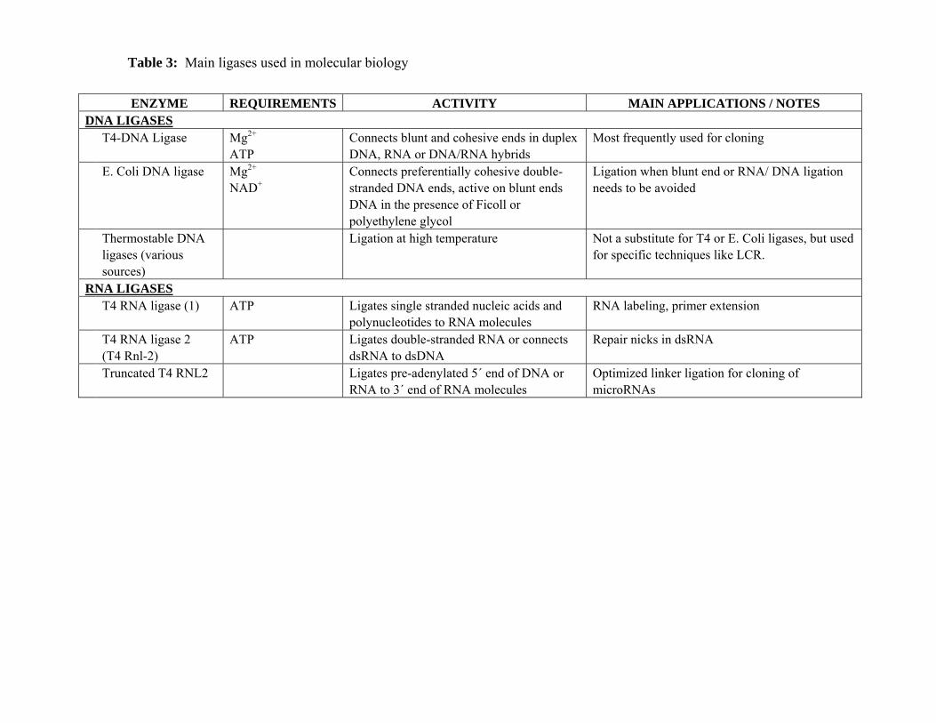
Table 3: Main ligases used in molecular biology
ENZYME REQUIREMENTS ACTIVITY MAIN APPLICATIONS / NOTES DNA LIGASES T4-DNA Ligase Mg2+
ATP Connects blunt and cohesive ends in duplex DNA, RNA or DNA/RNA hybrids
Most frequently used for cloning
E. Coli DNA ligase Mg2+ NAD+
Connects preferentially cohesive double-stranded DNA ends, active on blunt ends DNA in the presence of Ficoll or polyethylene glycol
Ligation when blunt end or RNA/ DNA ligation needs to be avoided
Thermostable DNA ligases (various sources)
Ligation at high temperature Not a substitute for T4 or E. Coli ligases, but used for specific techniques like LCR.
RNA LIGASES T4 RNA ligase (1) ATP Ligates single stranded nucleic acids and
polynucleotides to RNA molecules RNA labeling, primer extension
T4 RNA ligase 2 (T4 Rnl-2)
ATP Ligates double-stranded RNA or connects dsRNA to dsDNA
Repair nicks in dsRNA
Truncated T4 RNL2 Ligates pre-adenylated 5´ end of DNA or RNA to 3´ end of RNA molecules
Optimized linker ligation for cloning of microRNAs
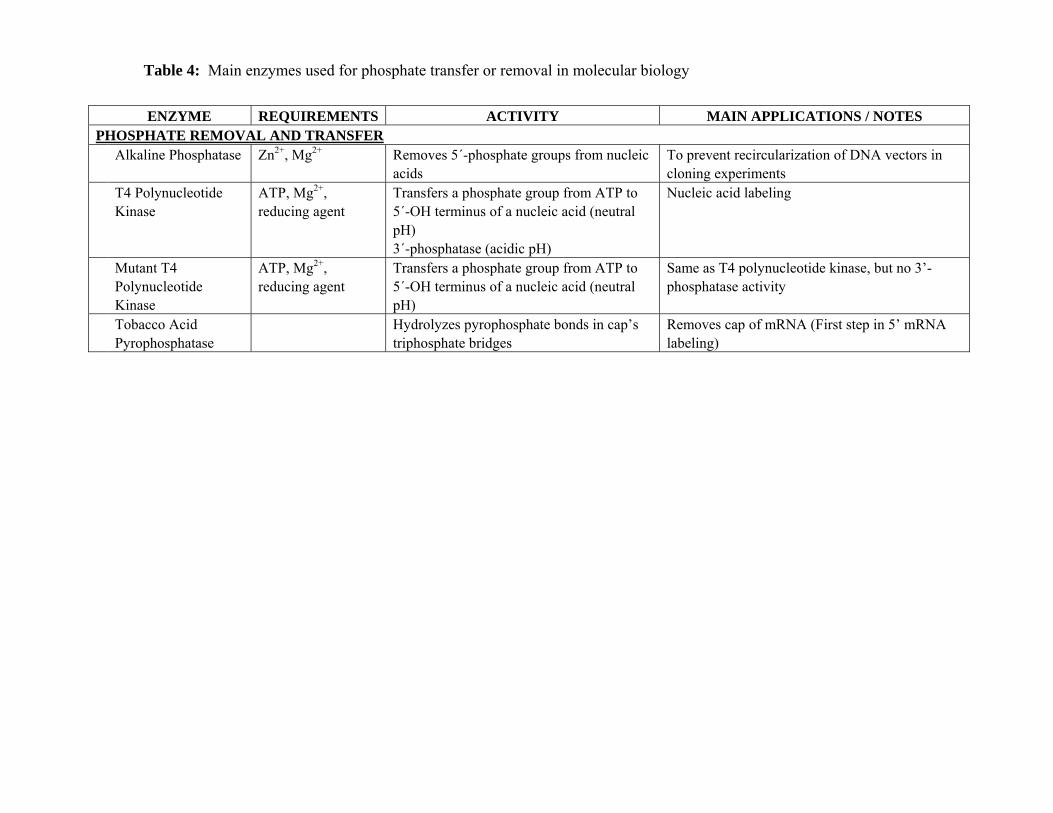
Table 4: Main enzymes used for phosphate transfer or removal in molecular biology
ENZYME REQUIREMENTS ACTIVITY MAIN APPLICATIONS / NOTES PHOSPHATE REMOVAL AND TRANSFER Alkaline Phosphatase Zn2+, Mg2+ Removes 5´-phosphate groups from nucleic
acids To prevent recircularization of DNA vectors in cloning experiments
T4 Polynucleotide Kinase
ATP, Mg2+, reducing agent
Transfers a phosphate group from ATP to 5´-OH terminus of a nucleic acid (neutral pH) 3´-phosphatase (acidic pH)
Nucleic acid labeling
Mutant T4 Polynucleotide Kinase
ATP, Mg2+, reducing agent
Transfers a phosphate group from ATP to 5´-OH terminus of a nucleic acid (neutral pH)
Same as T4 polynucleotide kinase, but no 3’-phosphatase activity
Tobacco Acid Pyrophosphatase
Hydrolyzes pyrophosphate bonds in cap’s triphosphate bridges
Removes cap of mRNA (First step in 5’ mRNA labeling)

Table 5: Main nucleases used in molecular biology
ENZYME REQUIREMENTS ACTIVITY MAIN APPLICATIONS / NOTES DEOXYRIBONUCLEASES DNase I Divalent cation
(nature affects specificity)
Single or double-stranded DNA, isolated or incorporated in chromatin
Nick translation of DNA, Dideoxy sequencing, elimination of DNA in RNA or protein preparations, DNase footprinting
Exonuclease III Mg2+ or Mn2+ 3’ exonuclease on double stranded DNA RNase H Phosphatase (pH< 7.4) Endonuclease (pH> 7.6)
DNA labeling (used with Klenow) or DNA length reduction (with nuclease S1)
Bal31 Ca2+, Mg2+ Shorten duplex DNA (both ends) Endonuclease on single stranded DNA
Restriction mapping and shortening of DNA or RNA.
Exonuclease VII Degrades single-stranded DNA (both ends) No activity on RNA or DNA/ RNA hybrids
Removal of protruding ends from DNA Removal of primers from a completed PCR reaction
RIBONUCLEASES Pancreatic
ribonuclease (RNase A)
Degrades RNA in 3’ Cleaves RNA in DNA/ RNA hybrids at site of single mismatch
RNA elimination from DNA preparation Mutation mapping
Ribonuclease H (RNase H)
Reducing agents 7.5<pH<9.1 Mg2+
Degrades RNA in DNA/ RNA hybrids Removal of RNA probes Removal of polyA tails of mRNA
Phy I Cleaves RNA at G, A, and U, but not at C residues.
RNA sequencing
CL3 Cleaves RNA adjacent to cytidilic acid RNA sequencing Cereus Cleaves RNA at U and C residues RNA sequencing Phy M cleaves RNA at U and A residues RNA sequencing RNase T1 Cleaves single-stranded RNA at G residues RNA sequencing RNAse T2 cleaves all phosphodiester bonds in RNA 3′-terminal analysis of RNA and for RNase
protection assays RNase U2 Cleaves the 3´-phosphodiester bond
adjacent to purines (standard conditions) Cleaves adenine residues (50°C)
RNA sequencing, in complement with RNase T1
DNA/ RNA NUCLEASES

Nuclease S1 Zn2+ 4.0<pH<4.3
Degrades RNA or single stranded DNA into 5´ mononucleotides
Removes single-stranded protruding ends of DNA
Mung Bean Nuclease Zn2+ Reducing agent
Degrades single stranded DNA and RNA Removes single-stranded protruding ends of DNA