enzyme-immunoassay of thromboxane b2 at the picogram level
TRANSCRIPT

PROSTAGLANDINS
ENZYME-IMMUNOASSAY OF THROMBOXANE BZ AT THE PICOGRAM LEVEL
M. Sawada, T. Inagawa* and J. C. Frijlich
Institute of Clinical Pharmacology Hannover Medical School
Hannover / Germany (FRC)
*On0 Pharmaceutical Co., Ltd. Research Institute
Shimamoto, Mishima Osaka 618, Japan
Abstract
A highly sensitive and reproducible enzyme-immunoassay for the measurement of thromboxane B2 was developed. Thromboxane I3 (TxB ) was coupled with R-D- galactosidase by mixed anhydride reaction. Throm a 2B oxane -antiserum ‘was genera- ted in rabbits and used at a final dilution of 1:480,000. ThJ separation of immuno- complex from the free form of TxB2 was accomplished by the double antibody method. The second antibody was sheep anti rabbit IgC. The precipitated enzyme activity was measured fluorometrically with 4-methyl-umbelliferyI-R_D-galactoside as substrate.
This method allowed to measure TxB2 in the range of 0.002 - 5 picomole per tube. The cross-reactivity of the anti-thromboxane B2-antiserum with 2,3-dinor throm- boxane B2 was about 20%, but it was less than 0.2% for the other prostanoids tested.
TxB2 extracted from human urine was measured by enzyme-immunoassay (y) and radioimmunoassay (x) which has been found closely correlated to values obtained by gas chromatography-mass spectrometry. Regression analysis of the data comparing enzyme-immunoassay and radioimmunoassay gave the equation y = 0.996 x + 0.470, correlation coefficient r = 0.9947. Inter-assay coefficient of variation was 3.1%.
The assay was further simplified by coating the second antibody on lass beads. The regression equation between this solid-phase enzyme immunoassay $ y) and radioim- munoassay (x) was y = 0.9860 x 1.927, r = 0.9895, and enzyme immunoassay (y) was y = 0.9749 x - 0.94808, r q 0.9887. Thus, the enzyme-immunoassay shows specificity and sensititity comparable to radioimmunoassay making use of radioactive tracer unnecessary.
Imroduction
The quantitative determination of arachidonate metabolites has been performed by various methods. Radioimmunoassay (RIA) (1) and gaschromatography-mass spec- trometry (CC-MS (2) as reference method have been widely employed and accepted as sensitive and specific methods. However, RIA poses practical problems, inclu- ding the requirement for tracer with high specific activity, radioactive contamina- tion and waste disposal and radiolysis of the tracer making frequent checks on its purity mandatory.
The use of an enzyme or chemiluminesence as a label in place of radioactivity has been applied to overcome the disadvantages of RIA. Recently, enryme-immunoas- say (EIA) (3) and chemiluminescence immunoassay (CIA) (4) have been introduced for prostaglandin analysis.
JUNE 1985 VOL. 29 NO. 6 1039

PROS’TAGLANDINS
In this report we describe the development of an EIA including a solidphase system, for the determination of TxB2, the hydrolysis product of TxA.2, which is a potent endogenous mediator of platelet aggregation and vasoconstriction.
Materials and Methods
Materials
Authentic TxB2 and anti TxB2 antiserum were provided by Ono Pharmaceutical Co., Ltd. (Osaka/Japan). Tritiated TxB2 (specific activity 140 Ci/mmole) was ob- tained from New England Nuclear (Dreieichl; Isobutylamine, tri-n-butylamine, glu- taraldehyde, 4-methyl-umbellifetyl-R-D-galactoside (Sigma), R-D-galactosidase (242 U/mg lyophilisate; for EIA) (Boehringer), and LH-2a normal rabbit serum and sheep anti-rabbit antiserum (Miles Laboratories) were purchased.
Three buffers were used: Buffer A consisted of 0.01 M phosphate buffer (pH 7.21, I mM of magnesium chloride, 0.1 M sodium chloride and 0.1% sodium azide. Buffer B was prepared by adding to Buffer A 0.1% (w/v) of bovine serum albumine. Buffer C consisted of 0.1 M glycine-sodium hydroxide (pH 10.3) and was used as enzyme reaction stoppet.
Preparation of TxB2enzyme conjugate
The carboxy group of TxB2 was conjugated with an amino group of the enzyme B- D-galactosidase by the mixed anhydride reaction according to the methods of Er- Ianger and co-workers (5). The reaction mixture was fractionated by Sephadex G-25 (1.5 x 70 cm) column. The column was packed with water pH 6.2 and eluted with Buffer A. The retention volume of TxB2 coupled to the enzyme was 50 to 65 ml.
Enzyme-immunoassay procedure
The enzyme-immunoassay procedure is summarized in Scheme 1. Enzyme conjuga- ted TxB and substrate solution were dissolved ion Buffer B. Anti TxB2 antiserum, standar b or sample, normal rabbit serum and second antibody were dissolved in Buffer A. The first immunoreaction, e&zyme labeled TxB performed by incubation for 1 hr at 37 C. At the end of t
.+, antigen and TxB2, Was e first immunoreaction,
the second antibody was added with normal rabbit serum as carrier and incubated overnight at 4 C. After the second immunoreaction, the immunocomplex was pre- cipitated by centrifugation, suspended in 0.2 ml of the substrate solution (0.1 mM of 4-methyl-umbelliferyl-B-D-galactoside) and reacted for 30 min at 37’C. Then the enzyme reaction was stopped by addition of 3 ml of Buffer C. The fluorescence was measured at an excitation wavelength of 360 nm, and at emission wavelength of 450 nm, using spectrofluorometer model JY3D (Jobin-Yvon Co, Ltdl.
1040 JUNE 1985 VOL. 29 NO. 6

PROSTAGLANDINS
scheme 1.
&mph or 8tmrtdxrd _ _ _ _ _ _ _ 0.1 ml
Ensymelrbelsd Awn _ _ _ _ _ - _ 0.1 ml
1x62 Antisam _______ O.lml
I 3f CforGOmin
Normal nbblt serum ______- O.lml
7
mti-rabbii IgG _____-- 0.1 ml
4’ C for ovamight
Genlrilugs
Wash
ro! ______- 0.2ml
SoDdpA __-___- 3.0 ml
I GemrMV
*pmont
flu8omstry (XEr260 nm, XEm-450 nmI
Radioimmunoassay procedure
RIA was carrid out according to Inagawa et al. (6). Free and bound 3H-TxB separated by the dextran-coated charcoal (6% charcoal and 0.6% dextran, w/8 method. Radioactivity of the bound form was measured by liquid scintillation coun- ting.
Solid-phase EIA procedure
Sheep antirabbit IgG was diluted 100 times with Buffer A. About 500 glass beads were treated with glutaraldehyde (1%). After glutaraldehyde treatment, diluted second antibody was added and reacted overnight at 4’C. Subsequently, the glass beads were washed with Buffer A and placed in Buffer B for 18 hrs.
The solid-phase EIA was carrid out by addition of a glass bead with second antibo- dy. After the first immunoreaction, a second antibody covered glass bead was added into each tube. The tube with the bead was shaken for 1 hr at room tempera- ture and subsequently left at 4’C overnight. Following the solid-phase reaction, the bead was washed with 0.5 ml of Buffer A and transferred to another tube contai- ning 200 ul of substrate solution. The enzyme activity was measured fluorometri- tally.
Extraction of TxB z
from human urine
One ml of human urine was adjusted to pH 3 with I N-HCI, and then extracted with 3 volumes of ethyl acetate (EtoAc) two times. The combined EtoAc phases were diluted with 4 ml of cyclohexane. The organic extracts were applied to a Na2S0, (Merck) column (3 /5mm x 10 cm). The column was washed with 5 ml of EtoAc/cy clohexane (6 : 4 v v) 7 and TxB2 was applied to a Si02-column (Sep-Pak, !XJa:-\;:sj
JUNE 1985 VOL. 29 NO. 6 1041

PROSTAGLANDINS
and eluted with 5 ml of EtoAc/cyclohexane/methanol (6 : 4 : 2 v/v). The NaSO4- column was packed in a Pasteur-pipette and its tip directly connected to the SiO - column. The eluate was evaporated under vacuum, the residue dissolved in a sma I 3 volume of 15% methanol in EtoAc, applied to thin layer plate (Silica gel 60.F 254; Merck) and developed using the solvent system EtoAc-chloroform:ethanol:acetic acid (200:200:40:10; by volume). After TLC separation, the TxB2 fraction was loca- lized by locating reference TxB
z with phosphomolybdic acid spray on a separate
strip of the TLC plate removed rom the piate by cutting the plate longitudinally, scraped off and eluted with pure methanol. The methanol was evaporated and the residue extracted with EtoAc to remove the binder eluted from TLC. The residues were analysed by three different methods: RIA, EIA and solid phase EIA. The reco- very of tritiated TxB2 added to human urine was 85 + 7% (mean + SD, n = 9).
CC/MS analysis
For CC/MS analysis, 10 ng of w-bishomo-PCF2 (One Pharmaceuticals) was added as internal standard and derivatized to the mahylester-tris-trimethyl-silyl-ether (2). A 2 m glass column, 2 mm diameter, packed with 1% SE 30 on Gaschrom Q (100-120 mesh) was eluted with helium at temperature of 23O’C. The injector tern erature of the Jeol mass spectrometer model JMS D 100 was 270°C, separator 260
8 C, ion source 250°C, ionizing voltage 20 eV and accelerating voltage 3 kV. The
ion common to the TxB2-derivative and its internal standard m/e 256 was monito- red. Accuracy was determined using X/MS analysis of human serum TxB2. Blood was drawn, placed into glass tubes and left at room temperature for 45 min. Two ml of serum were extracted with Folch solution and purified by Sephadex G-25 column before analysis (6).
Results
Characteristic properties of enzyme conjugated TxB2
According to the binding of 3H-TxB about 100 moles of TxB were conjugated with I mole of R-D-galactosidase whzh has about 120 free amin8 groups (7). Rela- tion between enzyme activity and incubation time was checked with four different enzyme concentrations using 4-methyl-umbelliferyI_B-D-galactoside as the substra- te. One ng of enzyme incubated with 0.1 mM substrate for 30 min at 37’C was regarded as the most suitable concentration of enzyme with respect to light inten- sity and lineartiy. These relationships are shown in Fig. 1.
1042 JUNE 1985 VOL. 29 NO. 6

PROSTAGLANDINS
Fig. 1 The enzymic kinetics of TxB -R-galactosidase conjugate for &methyl- umbefliferil-galactoside t-O-; d.4 pmol as TxB and 2.0 ng as enzyme, -o-; 0.2 pm01 as TxB and 1.0 ng as enzyme, - n-; 6.1 pmol as TxB2 and 0.5 ng as enzyme, -e-; a.05 pmol as TxB2 and 0.25 ng as enzyme, respectrvely)
The properties of the enzyme conjugated with antigen were compared with those of the original enzyme. Km values were about 0.1 mM with the original enzyme and 0.11 mM with the TxB -linked enzyme. units/mg protein, respec&vely.
Maximum velocities were 321 and 287
The most suitable concentration of enzyme conjugated antigen was incubated with the first antibody at various dilutions. At 160,000-fold dilution of the first antibo- dy, about 50% of the enzyme-labeled antigen formed immunocomplexes which were pricipitated with the second antibody.
The effect of various incubation times of the first immunoreaction was studied. When the first immunoreaction was carried out at 37’C for I hr maximal light intensity, i.e. completeness of reaction was achieved.
Sensitivity
The detection limit of TxB2 by EIA that could be measured as different from blank values was defined from triplrcate values (mean + SD). A significant difference was accepted if there was no overlap in the standard deviations of neighbouring values. Under these criteria, it was possible to distinguish about 2 femtomole TxB zero. A typical standard curve is shown in Fig. 2. Reproducibility is given in Ta ?I
from le I.
JUNE 1985 VOL. 29 NO. 6 1043

PROSTAGLANDINS
I I. I I I I I I I I I I I 0 0.9 3.9 15.6 62.5 250 1000
0.5 1.9 7.6 31.3 125 500 2000
Thromboxane B2 (pgitube)
Fig. 2 Standard curve for TxB2 measured by EIA (o-----o) and RIA (o-----o)
Table 1
% bound
m/tube Exp.1 Exp.2 Exp.3 mean + SD CV(%)
1000 8.5 7.3 8.4 8.1 t 0.7 8.5
500 11.9 12.5 13.8 12.7 : 1.0 7.9
250 17.0 20.4 22.9 20.1 + 3.0 14.9
125 33.2 31.5 34.6 33.1 + 1.6 4.7
62.5 45.2 45.3 48.6 46.4 + 1.9 4.2
31.3 56.0 59.6 62.9 59.5 t 3.5 5.8
15.6 69.0 73.0 75.4 72.5 t 3.2 4.5
7.8 79.2 82.6 83.6 81.8 i 2.3 2.8
3.9 88.4 89.6 90.6 89.5 + 1.0 1.2
2 93.8 93.8 94.5 93.8 t 0.8 0.8
1 96.0 96.5 96.8 96.4 + 0.4 0.4
0.5 98.8 97.8 98.0 98.2 + 0.5 0.5
1044 JUNE 1985 VOL. 29 NO. 6

PROSTAGLANDINS
Cross reactivity
The cross-reactivity of the anti TxB antiserum with the other prostanoids was lower than 0.2% and only 2,3-dinor TX d showed about 20% cross-reactivity at 50% binding (Table 2). The cross-reactivity? measured by RIA was almost identical to that of EIA (Table 2).
Table 2
The cross-reaction of selected compounds
cross-reactivity ( % )
RIA EIA
TxB2
2,3-dinor TxB2
PGE2
pGF1a
6- keto- PGF, a
pG A2
PG D2
100 100
34.9 21.8
0.05 0.02
0.2 0.16
0.04 O-02
0.9
Accuracy
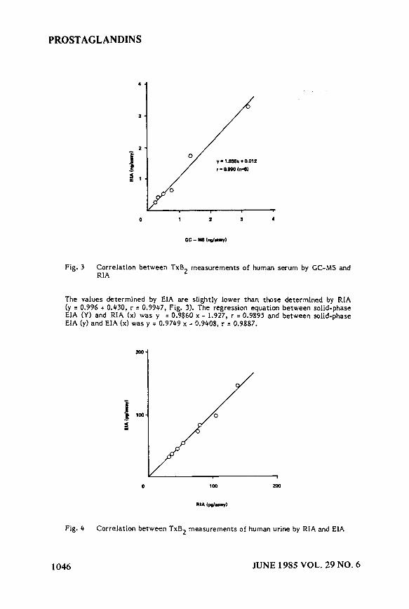
Figure 3 presents a correlation between the values of human serum TxB2 measured by RIA and CC-MS. For K-MS analysis purification was performed using the in- ternal standard and procedure described above. Because of the relatively tight correlation between RIA and CC-MS shown in Fig. 3 the RIA was used as a basis of comparison for the EIA and solidphase EIA. Samples of purified human urinary TxB2 were subjected to EIA and solid phase EIA. Regression analysis of the data gave a significant positive correlation.
JUNE 1985 VOL. 29 NO. 6 1045
PROSTAGLANDINS
0 1 2 a 4
Fig. 3 Correlation between TxB2 measurements of human serum by CC-MS and RIA
The values determined by EIA are slightly lower than those determined by RIA (y = 0.996 + 0.430, r F 0.9947, Fig. 3). The regression equation between solid-phase EIA (Y) and RIA (xl was y = 0.9860 x - 1.927, r = 0.9895 and between solid-phase EIA (y) and EIA (xl was y = 0.9749 x - 0.9408, r = 0.9887.
0 100 200
Fig. 4 Correlation between TxB2 measurements of human urine by RIA and EIA
1046 JUNE 1985 VOL. 29 NO. 6

PROSTAGLANDINS
Precision
Precision was determined by measuring TxB2 in human urine. Four different samp- les were determined on 5 different occasions. The means of coefficient of variation was 3.13%.
Diiion
We selected B-D-galactosidase as the enzyme for labeling TxB2 for the following reasons: The enzyme has a high turnover rate, active free amino groups for conju- gating reaction with hapten and it can be stored at 4’C for a long time witho%t loss of activity. The Txf+labeled enzyme was found to be stable for 1 year at -20 C.
The conjugating ratio of i3-D-galactosidase and TxB2 was determined from radio- activity. It was estimated that 100 moles of TxB2 were conjugated with 1 mole of R-D-galactosidas (molecular weight 540.000 and about 120 amino groups are avai- lable per mole R-D-galactosidase (7)).
The enzyme conjugated TxB2 was easily reacted with anti TxB antiserum (1:480.000 at final dilution), and the first immunoreaction require2 only 1 hour incubation at 37’C.
The sensitivity of EIA is dependent on the saturation level between enzyme and hapten. In this respect the relatively high yield of the reaction of TxB2 with the enzyme obtained in the present assay is a critical determinant of the assay sensiti- vity. Furthermore, the enzyme properties were changed very little by coupling it to TxB2. Thus, the enzyme labeled TxB2 used in our assay corresponded to a radioac- tive tracer of high specific activity.
Methods for the determination of TxB by EIA (3) and CIA (4) have been reported, and the detection limit was found to b e 0.1 pmole by EIA and 6.25 pg by CIA, respectively. Our results show that the detection limit for TxB can be signi- ficantly reduced for EIA. This is most likely due to an improved bin mg of TxB2 to . %. the enzyme and an antibody to TxB2 with a high titre.
Purification of the samples is required for 2 reasons: Firstly endogenous substances such as lactate may function as substrate for the enzyme and confuse the results. Secondly, the specificity of the antibody is limited. The 20% cross reaction with 2,3 dinor-TxB2 which is present in urine in much higher concentrations than TxB2 would result in erroneous results if urine were measured directly.
The present EIA for TxB gave a significant correlation with RIA which was fur- ther validated by CC-MS.%hus, acuracy, assay precision and sensitivity make this a potentially useful assay with the advantages of EIA.
Referenozs
GranstrGm, E. and Kindahl, H. In: Methods in Prostaglandin Research. (J.C. Friilich, ed.), Raven Press, New York, 1978, p. 119
Green, K., Hamberg, M., Samuelsson, B., Smigel, M. and FrGlich, 3.C. In: Methods in Prostaglandin Research. U.C. Friilich, ed.), Raven Press, New York, 1978.
Hayashi, Y., Veda, N., Yokata, K., Kawamura, S., Ogushi, F., Miyazaki, H., Kato, K. and Terao, S., Biochim. Biophys. Acta 750: 322, 1983.
JUNE 1985 VOL. 29 NO. 6 1047

PROSTAGLANDINS
4. Weerasekera, P.A., Koullapis, E.N., Kim, J.B., Barnard, C.3. and Collins, W.P. V. International Conference on Prostaglandins, Florence, May 1982, Abstract p. 600.
5. Erlanger, B.F., Borek, F., Beister, S.M. and Liebermann, S.J., Biol. Chem. 234: 1090, 1959.
6. Inagawa, T., Ohki, S., Sawada, M. and hirata, F., Yakugaku Zasshi. 92: 1187, 1971.
7. Comoglio, S. and Celada, F., J. Immunol. Methods IO:.161, 1976.
Editor: E. Granstrom Received: 5-31-84 Accepted: 3-15-85
1048 JUNE 1985 VOL. 29 NO. 6