enzyme assays in liver disease - jcp.bmj.com · confined to the cell sap. ... in hepatitis the...
TRANSCRIPT

J. clin. Path., 24, suppl. (Ass. Clin. Path.), 4, 51-59
Enzyme assays in liver diseaseR. J. WIEME AND L. DEMEULENAERE
From the Laboratory for Protein Chemistry, Medical Department, Academic Hospital, University of Ghent.Belgium
It is in the field of hepatology that enzyme assays areperformed most frequently for diagnostic purposes.Their interpretation requires knowledge of manyfactors that influence the serum level of the particularenzymes assayed. Therefore, these factors arediscussed briefly in relation to liver disease beforeconsidering important developments such as thevalue of estimating D-glutamyltransferase (EC2.3.2.1)' and the isoenzymes of alkaline phosphatase(EC 3.1.3.1). Finally, the application of these criteriato specific diagnostic problems is considered.
General Considerations
The concentrations in normal human liver of themost important enzymes for the diagnosis of liverdisease are given in Fig. 1; they are based onpublished data with the exception of those for D-glutamyltransferase which are the results of just twoanalyses which we performed to complete thissurvey. It is clear that the diagnostic value of an'Also known as y-glutamyltranspeptidase.
IU/g1501
100
50
LD
MD
enzyme cannot be ascertained from its concentrationin the liver.
In pathological conditions these concentrationsmay be very different. For example, the liverisoenzyme of lactate dehydrogenase (LD5, EC1.1.1.27) decreases rapidly after a single hepatotoxicdose of carbon tetrachloride, so that a second dosegiven 24 hours later has no marked effect on theserum level (Wieme and van Maercke, 1961).Conversely, the liver content of alkaline phosphatasewas found to increase sevenfold within 12 hours ofligating the common bile duct in the rat, whereasaspartate aminotransferase (EC 2.6.1.1) decreased by25% (Kaplan and Righetti, 1969). The increase inalkaline phosphatase was inhibited by cyclohexi-mide, suggesting that for at least some liver enzymesincreased synthesis is a factor in the response tobiliary obstruction, as emphasized previously byPolin, Spellberg, Teitelman, and Okumura (1962).Induced synthesis of D-glutamyltransferase alsoseems to occur easily.Another important factor influencing serum levels
in disease is the localization of an enzyme within theliver cell (Fig. 2). Some enzymes are found in morethan one site, each site containing different iso-
Fig. 1 Concentration of important enzymes in liver,expressed in international units per gram fresh liver tissue.LD = lactate dehydrogenase, MD = malatedehydrogenase, As-AT = aspartate aminotransferase,Al-AT = alanine aminiotransferase, AP = alkalinephosphatase, GMD = glutamate dehydrogenase,SD = sorbitol dehydrogenase, ALD =ketose-phosphate aldolase, GMT = D-glutamyltrans-ferase, and OCT = ornithine carbamoyltransferase.
51
Fig. 2 Schematic representation of the intracellularlocalization of important liver enzymes. Glutamatedehydrogenase and ornithine carbamoyltransferase arefound in mitochondria, alkaline phosphatase and D-glutamyltransferase in microsoines (abbreviations as inFigure 1).
LDAl - AT
AP SD MDGMT ALD As-AT GMD
| \ OCT
ks-AT
Al - ATAP GMD
I I I~~ SD IALD IGMT IOCTII I I I I I
copyright. on 13 June 2019 by guest. P
rotected byhttp://jcp.bm
j.com/
J Clin P
athol: first published as 10.1136/jcp.s1-4.1.51 on 1 January 1970. Dow
nloaded from
R. J. Wieme and L. Demeulenaere
enzymes. Aspartate aminotransferase, for example,is found both in the cell sap and in the mitochondria,whereas alanine aminotransferase (EC 2.6.1.2.) isconfined to the cell sap. Slight cell damage tends torelease only the enzymes in the soluble fraction ofthe cell whilst necrotic lesions, which also affect themitochondria, release both types. It has beensuggested that the ratio of serum aspartate to alanineaminotransferase can be used as an index of theseverity of liver cell damage (de Ritis, Coltorti, andGuisti, 1955), values less than 1 indicatingreversible cell damage not involving the mito-chondria, as in early infective hepatitis, and valuesgreater than 1 indicating more extensive celldamage as in the acute exacerbations in the course ofliver cirrhosis. According to Schmidt and Schmidt(1962) and Laudahn (1963), a better index of cellnecrosis is given by the ratio of the sum of the serumlevels of these transferases to the level of the purelymitochondrial enzyme, glutamate dehydrogenase(EC 1.4.1.3). In acute hepatitis, for example, the veryhigh levels of the aminotransferases produce a veryhigh ratio despite the increase in glutamate dehydro-genase levels. However, the marked increase ofserum glutamate dehydrogenase, sometimes foundin severe cases of myocardial infarction inwhich liver cell necrosis is absent, suggests that themitochondria are very sensitive to hypoxia. Theusefulness of this method of recognizing necrosis istherefore limited.
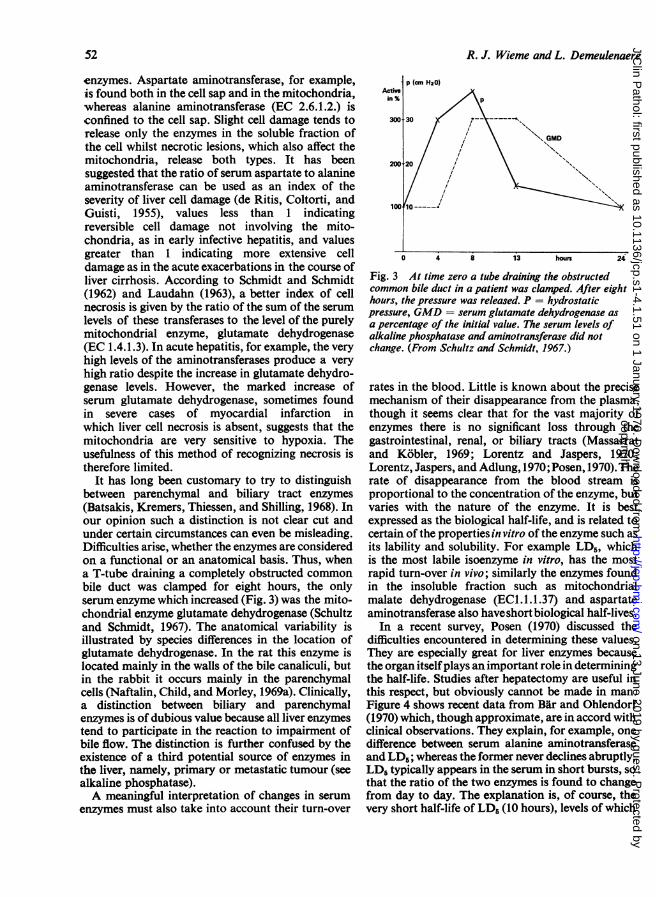
It has long been customary to try to distinguishbetween parenchymal and biliary tract enzymes(Batsakis, Kremers, Thiessen, and Shilling, 1968). Inour opinion such a distinction is not clear cut andunder certain circumstances can even be misleading.Difficulties arise, whether the enzymes are consideredon a functional or an anatomical basis. Thus, whena T-tube draining a completely obstructed commonbile duct was clamped for eight hours, the onlyserum enzyme which increased (Fig. 3) was the mito-chondrial enzyme glutamate dehydrogenase (Schultzand Schmidt, 1967). The anatomical variability isillustrated by species differences in the location ofglutamate dehydrogenase. In the rat this enzyme islocated mainly in the walls of the bile canaliculi, butin the rabbit it occurs mainly in the parenchymalcells (Naftalin, Child, and Morley, 1969a). Clinically,a distinction between biliary and parenchymalenzymes is of dubious value because all liver enzymestend to participate in the reaction to impairment ofbile flow. The distinction is further confused by theexistence of a third potential source of enzymes inthe liver, namely, primary or metastatic tumour (seealkaline phosphatase).A meaningful interpretation of changes in serum
enzymes must also take into account their turn-over
Fig. 3 At time zero a tube draining the obstructedcommon bile duct in a patient was clamped. After eighthours, the pressure was released. P = hydrostaticpressure, GMD = serum glutamate dehydrogenase asa percentage of the initial value. The serum levels ofalkaline phosphatase and aminotransferase did notchange. (From Schultz and Schmidt, 1967.)
rates in the blood. Little is known about the precisemechanism of their disappearance from the plasma,though it seems clear that for the vast majority ofenzymes there is no significant loss through thegastrointestinal, renal, or biliary tracts (Massarratand Kobler, 1969; Lorentz and Jaspers, 1970;Lorentz, Jaspers, and Adlung, 1970; Posen, 1970). Therate of disappearance from the blood stream isproportional to the concentration of the enzyme, butvaries with the nature of the enzyme. It is bestexpressed as the biological half-life, and is related tocertain of the properties in vitro of the enzyme such asits lability and solubility. For example LD5, whichis the most labile isoenzyme in vitro, has the mostrapid turn-over in vivo; similarly the enzymes foundin the insoluble fraction such as mitochondrialmalate dehydrogenase (EC1.1.1.37) and aspartateaminotransferase also haveshort biological half-lives.
In a recent survey, Posen (1970) discussed thedifficulties encountered in determining these values.They are especially great for liver enzymes becausethe organ itselfplays an important role in determiningthe half-life. Studies after hepatectomy are useful inthis respect, but obviously cannot be made in man.Figure 4 shows recent data from Bar and Ohlendorf(1970) which, though approximate, are in accord withclinical observations. They explain, for example, onedifference between serum alanine aminotransferaseand LD6; whereas the former never declines abruptly,LD typically appears in the serum in short bursts, sothat the ratio of the two enzymes is found to changefrom day to day. The explanation is, of course, thevery short half-life of LD5 (10 hours), levels of which
52
copyright. on 13 June 2019 by guest. P
rotected byhttp://jcp.bm
j.com/
J Clin P
athol: first published as 10.1136/jcp.s1-4.1.51 on 1 January 1970. Dow
nloaded from

Enzyme assays in liver disease
Half-life LD(hours) L
100+
50+
Fig. 4 Biological half-lifevalues of important liverenzymes in the plasma. LD1and LD5 are at theextremes of the scale.CK = creatine kinase(other abbreviations as inF-igure 1).
Al - AT
ALDE VPe
therefore reflect more closely what is going on in thecell at the time the blood sample is taken than dolevels of the aminotransferase (half-life, about 50hours) which may relate to an event that occurredsome time before. The biological half-life also hasan important effect on the magnitude of the rise inthe serum, since the longer the half-life the greaterthe accumulation of the enzyme in the serum.
D-Glutamyltransferase
Although the clinical usefulness of assays of thisenzyme was described 10 years ago (Szewczuk andOrlowski, 1960; Szczeklik, Orlowski, and Szewczuk,1961), interest in it was limited until the developmentof a stable, soluble and safe substrate, y-glutamyl-p-nitroanilide (Goldbarg, Pineda, Smith, Friedman,and Rutenberg, 1963; Orlowski and Meister, 1963).A number of laboratories have since reported theirexperience, the reports being generally favourable(Rutenburg, Goldbarg, and Pineda, 1963; Kam-meraat, 1970; Villa, Dioguardi, Agostoni, Ideo, andStabilini, 1966; Lukasik, Richterich, and Colombo,1968; Szasz, Rosenthal, and Fritzsche, 1969a and1969b; Zein and Discombe, 1970). Our own ex-perience extends to more than 600 determinations.Its behaviour resembles both alkaline phosphataseand the aminotransferases, which is apt to beconfusing. We have often experienced difficulty in
finding an explanation for pathological values. Thecorrelation with alkaline phosphatase has not beenas close as that reported by Ideo andDioguardi(1970)and the correlation with leucine aminopeptidase(EC 3.4.1.1) is likewise very loose.
In hepatitis the serum D-glutamyltransferaserises at most to about four times normal at the timewhen aminotransferases reach their peak, but tendsto increase further when these begin to return tonormal. It also increases in the recovery phase ofacute myocardial infarction (Agostini, Ideo, andStabilini, 1965), a phenomenon difficult to explainbecause the enzyme is absent from the heart muscle(Naftalin etal, 1969a). It is found in kidney, pancreas,liver, and spleen, and-according to our observations-also in placenta, in the ratio 10-0: 8-3: 3 9: 15: 1 0.The enzyme is thus far from being liver specific, andincreased serum levels are found, as expected, inacute pancreatitis (Naftalin, 1970).A number of properties render it eminently
attractive in the clinical laboratory. These include itsstability in serum samples, its absence from red cells,the large range of variation combined with the easeof estimation by spectrophotometry (Szasz, 1969) orcolorimetry (Naftalin, Sexton, Whitaker, and Tracey,1969b). It seems likely to become important inbiochemical screening.
Lactate Dehydrogenase (LD) Isoenzymes
Isoenzyme studies, although regarded as being ofgreat diagnostic value (Wilkinson, 1970a and b) arenot widely used in clinical chemistry because theirdetermination by electrophoresis is relatively difficultand cumbersome. In fact there are now techniqueswhich are quite simple, which are based on thedifferences in isoenzymes revealed by differentconditions of assay, and which are readily adapted tothe average laboratory ('class II' methods, Wieme,1966). If a mixture contains two isoenzymes, atleast two sets of conditions are needed. If the totalactivity of such a mixture under standard and underselective conditions of assay respectively is repre-sented by Y and Y', and the contribution of eachisoenzyme by A and A' and B and B' respectively,then the degree of activation or inhibition of the twoisoenzymes under the selective conditions may beexpressed by their respective inhibition or activationconstants:
A' B'a =x- and b =- (1)
The contribution of the isoenzymes for any mixtureof unknown composition can be calculated from theexpression:
53copyright.
on 13 June 2019 by guest. Protected by
http://jcp.bmj.com
/J C
lin Pathol: first published as 10.1136/jcp.s1-4.1.51 on 1 January 1970. D
ownloaded from
R. J. Wieme and L. Demeulenaere
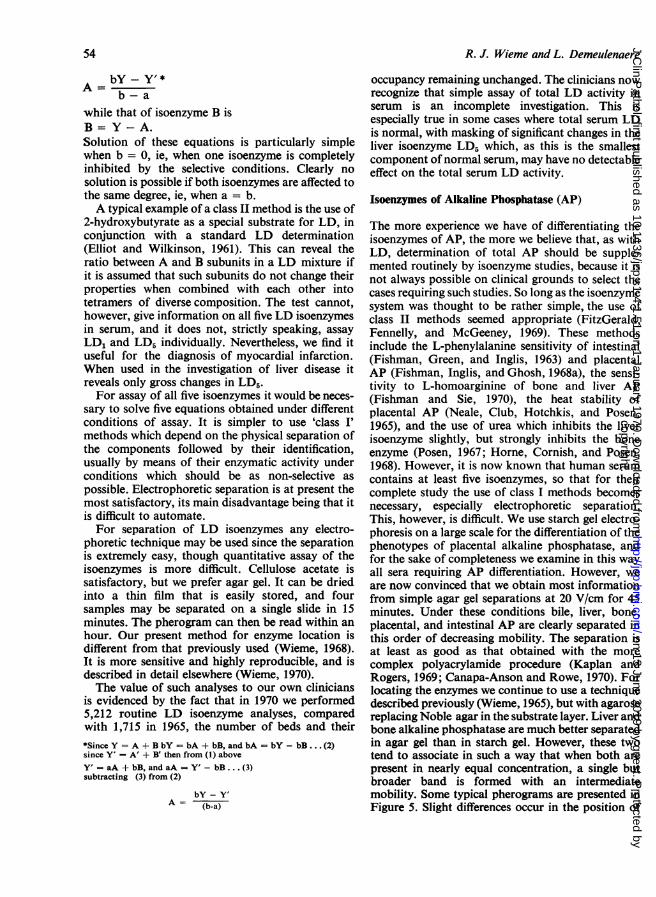
bY - Y'*b-a
while that of isoenzyme B isB= Y-A.Solution of these equations is particularly simplewhen b = 0, ie, when one isoenzyme is completelyinhibited by the selective conditions. Clearly nosolution is possible if both isoenzymes are affected tothe same degree, ie, when a = b.A typical example of a class II method is the use of
2-hydroxybutyrate as a special substrate for LD, inconjunction with a standard LD determination(Elliot and Wilkinson, 1961). This can reveal theratio between A and B subunits in a LD mixture ifit is assumed that such subunits do not change theirproperties when combined with each other intotetramers of diverse composition. The test cannot,however, give information on all five LD isoenzymesin serum, and it does not, strictly speaking, assayLD1 and LD5 individually. Nevertheless, we find ituseful for the diagnosis of myocardial infarction.When used in the investigation of liver disease itreveals only gross changes in LD5.For assay of all five isoenzymes it would be neces-
sary to solve five equations obtained under differentconditions of assay. It is simpler to use 'class I'methods which depend on the physical separation ofthe components followed by their identification,usually by means of their enzymatic activity underconditions which should be as non-selective aspossible. Electrophoretic separation is at present themost satisfactory, its main disadvantage being that itis difficult to automate.For separation of LD isoenzymes any electro-
phoretic technique may be used since the separationis extremely easy, though quantitative assay of theisoenzymes is more difficult. Cellulose acetate issatisfactory, but we prefer agar gel. It can be driedinto a thin film that is easily stored, and foursamples may be separated on a single slide in 15minutes. The pherogram can then be read within anhour. Our present method for enzyme location isdifferent from that previously used (Wieme, 1968).It is more sensitive and highly reproducible, and isdescribed in detail elsewhere (Wieme, 1970).The value of such analyses to our own clinicians
is evidenced by the fact that in 1970 we performed5,212 routine LD isoenzyme analyses, comparedwith 1,715 in 1965, the number of beds and their*Since Y =A + B bY = bA + bB, and bA = bY - bB ... .(2)since Y' = A' + B' then from (1) aboveY' = aA + bB, and aA = Y'-bB ... (3)subtracting (3) from (2)
bY -Y'A (b-a)
occupancy remaining unchanged. The clinicians nowrecognize that simple assay of total LD activity inserum is an incomplete investigation. This isespecially true in some cases where total serum LDis normal, with masking of significant changes in theliver isoenzyme LD5 which, as this is the smallestcomponent of normal serum, may have no detectableeffect on the total serum LD activity.
Isoenzymes of Alkaline Phosphatase (AP)
The more experience we have of differentiating theisoenzymes of AP, the more we believe that, as withLD, determination of total AP should be supple-mented routinely by isoenzyme studies, because it isnot always possible on clinical grounds to select thecases requiring such studies. So long as the isoenzymesystem was thought to be rather simple, the use ofclass II methods seemed appropriate (FitzGerald,Fennelly, and McGeeney, 1969). These methodsinclude the L-phenylalanine sensitivity of intestinal(Fishman, Green, and Inglis, 1963) and placentalAP (Fishman, Inglis, and Ghosh, 1968a), the sensi-tivity to L-homoarginine of bone and liver AP(Fishman and Sie, 1970), the heat stability ofplacental AP (Neale, Club, Hotchkis, and Posen,1965), and the use of urea which inhibits the liverisoenzyme slightly, but strongly inhibits the boneenzyme (Posen, 1967; Horne, Cornish, and Posen,1968). However, it is now known that human serumcontains at least five isoenzymes, so that for theircomplete study the use of class I methods becomesnecessary, especially electrophoretic separation.This, however, is difficult. We use starch gel electro-phoresis on a large scale for the differentiation of thephenotypes of placental alkaline phosphatase, andfor the sake of completeness we examine in this wayall sera requiring AP differentiation. However, weare now convinced that we obtain most informationfrom simple agar gel separations at 20 V/cm for 45minutes. Under these conditions bile, liver, bone,placental, and intestinal AP are clearly separated inthis order of decreasing mobility. The separation isat least as good as that obtained with the morecomplex polyacrylamide procedure (Kaplan andRogers, 1969; Canapa-Anson and Rowe, 1970). Forlocating the enzymes we continue to use a techniquedescribed previously (Wieme, 1965), but with agarosereplacing Noble agar in the substrate layer. Liver andbone alkaline phosphatase are much better separatedin agar gel than in starch gel. However, these twotend to associate in such a way that when both arepresent in nearly equal concentration, a single butbroader band is formed with an intermediatemobility. Some typical pherograms are presented inFigure 5. Slight differences occur in the position of
54
copyright. on 13 June 2019 by guest. P
rotected byhttp://jcp.bm
j.com/
J Clin P
athol: first published as 10.1136/jcp.s1-4.1.51 on 1 January 1970. Dow
nloaded from

Enzyme assays in liver disease
a
1 bc
a
11b
a
b
a
b
.. .. ............
I I I I
........-
Fig. 5 Separation ofserum alkaline phosphatases in a
few typical cases. Agar gel electrophoresis, barbitonebuffer, pH 8-4, u 005, 20 V/cm for 4S min at 12°C.Numbers indicate respectively the bile (1), liver (2),
bone (3), placental (4), and intestinal (5) isoenzymes.Ia = increased bone fraction (Paget's disease); Ib =rormal; Ic = normal; IIa = increased liver fraction(hepatitis); IIb = combined increase of bone and liverfractions; lIIa and b = placental alkaline phosphatasefrom pregnant women (serum heated at 65° for 5 minso that other enzymes are destroyed); IVa and b =bile, liver and intestinal fractions (liver malignancy oncirrhotic base).
placental AP, corresponding to the various pheno-types which are better revealed by starch gel electro-phoresis.
It is excessively rare to find all five fractionstogether in the same sample of serum. Placental APis present only in pregnant women. The presence ofintestinal AP seems related to liver cirrhosis and alsoto the ABO secretor status, higher levels being foundin individuals of blood groups 0 and B (Beckman,Bj6rling, and Heiken, 1966; Stolbach, Krant, Inglis,and Fishman, 1967) though the concentration in theintestinal tissue is not related to blood groups(Schreffler, 1966).
Electrophoresis is of value in differentiating theliver and the bone enzyme, and even more so indemonstrating the presence of the bile enzyme (deJong and Haije, 1970). In this case agar gel is superiorto both starch gel and polyacrylamide gel, since thebile enzyme does not penetrate into the latter mediaowing to its high molecular weight. The presence ofboth liver and bile isoenzymes in an anicteric patient
points to an impairment of the bile flow which wecall 'focal block'. The macromolecular bile com-ponents are refluxed back into the blood which theycannot leave again. The low molecular weightcomponents such as bilirubin are, however, againextracted from the blood and secreted in zones wherethe bile flow is not blocked (Hill and Sammons, 1967)The most frequent cause of this type of obstruction isthe presence of malignant primary or secondarytumour in the liver.There is another interesting change in serum
alkaline phosphatase in malignancy, namely, theappearance in the serum of an isoenzyme that cannotbe distinguished from placental AP, called Reganisoenzyme after the patient in whom it was firstdemonstrated. In this patient with a bronchialcarcinoma the isoenzyme was found both in theserum and in the tumour (Fishman, Inglis, Green,Anstiss, Gosh, Reif, Rustigan, Krant, and Stolbach,1968b). On starch gel electrophoresis Reganisoenzyme moves as a rapid band, but its maincharacteristic is its resistance to heating at 65°C for5 minutes. It was found in the serum of 27 out of 500patients with various malignant diseases (Stolbach,Krant, and Fishman, 1969), though the serum of 10had to be concentrated threefold for demonstrationby electrophoresis (Fishman, Inglis, Stolbach, andKrant, 1968c). Warnock and Reisman (1969) founda similar enzyme in extracts of eight out of 10hepatomas and also in the serum of some of thesepatients; however the serum enzyme was also foundin two controls out of 60, namely, cases of chronichepatitis and fatty liver respectively. The enzymediffered slightly from the Regan isoenzyme,being less thermostable. Another alkaline phos-phatase which was heat-labile and insensitive toL-phenylalanine was extracted from a bronchialcarcinoma (Timperley, 1968); appropriate studies onserum were not performed in this case though totalserum alkaline phosphatase was increased.
Other Isoenzyme Systems
Other systems exist, including those of ketose 1-phosphate aldolase (EC 4.1.2.7),1 aspartate amino-transferase, glutamate dehydrogenase, and malatedehydrogenase. The latter is probably the mostuseful clinically.
Malate dehydrogenase occurs in the form ofcytoplasmic and mitochondrial isoenzymes. Reportson their electrophoretic differentiation must beviewed with caution because of two possible arte-facts. First, oxaloacetate, used as substrate, islabile and breaks down to pyruvate; thus LD bandsmay also be revealed. Secondly, the migration of the'Also known as aldolase or fructose 1-phosphate aldolase.
55
...
...........
copyright. on 13 June 2019 by guest. P
rotected byhttp://jcp.bm
j.com/
J Clin P
athol: first published as 10.1136/jcp.s1-4.1.51 on 1 January 1970. Dow
nloaded from
R. J. Wieme and L. Demeulenaere
M :
+- 'E.,
50-
40-
i 30-,l 20-
ZI0
..*............
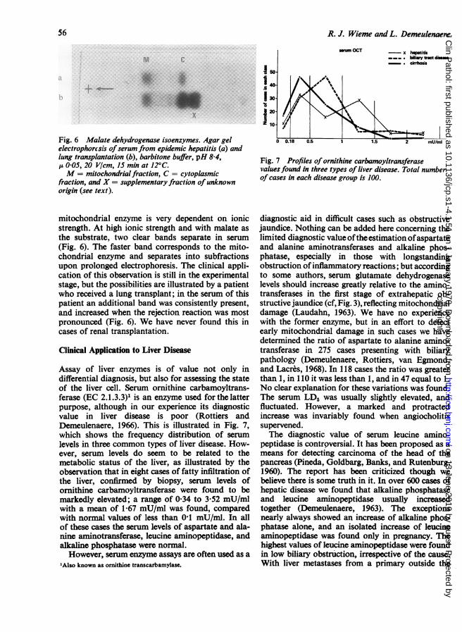
Fig. 6 Malate dehydrogenase isoenzymes. Agar gelelectrophorcsis ofserum from epidemic hepatitis (a) andlung transplantation (b), barbitone buffer, pH 8-4,p 005, 20 V/cm, 15 min at 12°C.M = mitochondrialfraction, C = cytoplasmic
fraction, and X = supplementary fraction ofunknownorigin (see text).
mitochondrial enzyme is very dependent on ionicstrength. At high ionic strength and with malate asthe substrate, two clear bands separate in serum(Fig. 6). The faster band corresponds to the mito-chondrial enzyme and separates into subfractionsupon prolonged electrophoresis. The clinical appli-cation of this observation is still in the experimentalstage, but the possibilities are illustrated by a patientwho received a lung transplant; in the serum of thispatient an additional band was consistently present,and increased when the rejection reaction was mostpronounced (Fig. 6). We have never found this incases of renal transplantation.
Clinical Application to Liver Disease
Assay of liver enzymes is of value not only indifferential diagnosis, but also for assessing the stateof the liver cell. Serum ornithine carbamoyltrans-ferase (EC 2.1.3.3)1 is an enzyme used for the latterpurpose, although in our experience its diagnosticvalue in liver disease is poor (Rottiers andDemeulenaere, 1966). This is illustrated in Fig. 7,which shows the frequency distribution of serumlevels in three common types of liver disease. How-ever, serum levels do seem to be related to themetabolic status of the liver, as illustrated by theobservation that in eight cases of fatty infiltration ofthe liver, confirmed by biopsy, serum levels ofornithine carbamoyltransferase were found to bemarkedly elevated; a range of 034 to 3-52 mU/mlwith a mean of 1-67 mU/ml was found, comparedwith normal values of less than 01 mU/ml. In allof these cases the serum levels of aspartate and ala-nine aminotransferase, leucine aminopeptidase, andalkaline phosphatase were normal.However, serum enzyme assays are often used as a
'Also known as ornithine transcarbamylase.
emm OCT
/.1
C'4o4. '4I0
0 0.18 0.5
- x heptitis___ . b;ilry trwt dise_ sis
1_'D-S~ ~~~~I--
1.5 2 mU/ml
Fig. 7 Profiles of ornithine carbamoyltransferasevalues found in three types of liver disease. Total numberof cases in each disease group is 100.
diagnostic aid in difficult cases such as obstructivejaundice. Nothing can be added here concerning thelimited diagnostic value ofthe estimation ofaspartateand alanine aminotransferases and alkaline phos-phatase, especially in those with longstandingobstruction ofinflammatory reactions; but accordingto some authors, serum glutamate dehydrogenaselevels should increase greatly relative to the amino-transferases in the first stage of extrahepatic ob-structive jaundice (cf, Fig. 3), reflecting mitochondrialdamage (Laudahn, 1963). We have no experiencewith the former enzyme, but in an effort to detectearly mitochondrial damage in such cases we havedetermined the ratio of aspartate to alanine amino-transferase in 275 cases presenting with biliarypathology (Demeulenaere, Rottiers, van Egnond,and Lacres, 1968). In 118 cases the ratio was greaterthan 1, in 110 it was less than 1, and in 47 equal to 1.No clear explanation for these variations was found.The serum LD5 was usually slightly elevated, andfluctuated. However, a marked and protractedincrease was invariably found when angiocholitissupervened.The diagnostic value of serum leucine amino-
peptidase is controversial. It has been proposed as ameans for detecting carcinoma of the head of thepancreas (Pineda, Goldbarg, Banks, and Rutenburg,1960). The report has been criticized though webelieve there is some truth in it. In over 600 cases ofhepatic disease we found that alkaline phosphataseand leucine aminopeptidase usually increasedtogether (Demeulenaere, 1963). The exceptionsnearly always showed an increase of alkaline phos-phatase alone, and an isolated increase of leucineaminopeptidase was found only in pregnancy. Thehighest values of leucine aminopeptidase were foundin low biliary obstruction, irrespective of the cause.With liver metastases from a primary outside the
56copyright.
on 13 June 2019 by guest. Protected by
http://jcp.bmj.com
/J C
lin Pathol: first published as 10.1136/jcp.s1-4.1.51 on 1 January 1970. D
ownloaded from
Enzyme assays in liver disease
pancreas and biliary tract there is no great increase ofleucine aminopeptidase. Here our findings differfrom those of Rutenburg's group (Pineda et al, 1960).Serum alkaline phosphatase, on the other hand, canbe extremely high in all cases of obstruction irre-spective of the site of obstruction. Our experiencewith D-glutamyltransferase is limited but we find it auseful addition to the conventional group of liverfunction tests because any increase tends to be verylarge.
In patients with liver metastases most help isprobably obtained from alkaline phosphataseisoenzymes. Not only is the serum level regularlyincreased early in these cases, but electrophoresisreveals characteristic elevation of both the liver andthe so-called bile fractions (Fig. 5). In an anictericpatient this change makes it essential to search for aprimary cancer.
Inflammatory liver disease is fairly easily ex-plored by enzyme analysis. The value of theaminotransferases in the diagnosis of viral hepatitisis now well established. Serum lactate dehydrogenaseis useful in following the course of the disease as it isthe first enzyme to return to normal during recovery.This should be progressive and smooth, and repeatedfluctuations occurring after the third week suggestsome complication, especially if unrelated to physicalexercise. Repeated determinations of serum LD5 areespecially valuable for follow up in chronic activehepatitis. The fluctuations are of brief durationcompared with those of the aminotransferases. This,together with the appearance of slow y-globulins, isvery typical for the disease.
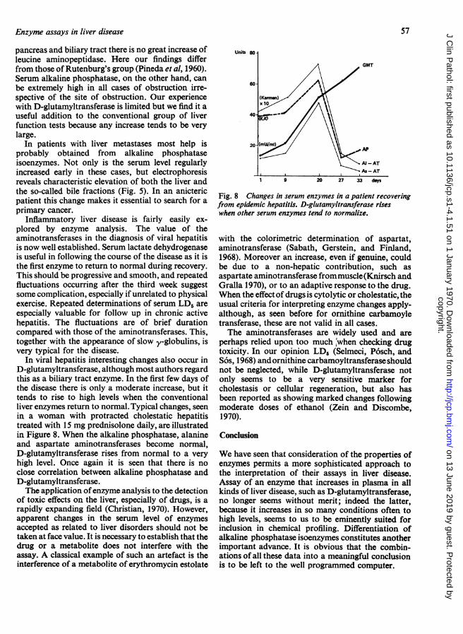
In viral hepatitis interesting changes also occur inD-glutamyltransferase, although most authors regardthis as a biliary tract enzyme. In the first few days ofthe disease there is only a moderate increase, but ittends to rise to high levels when the conventionalliver enzymes return to normal. Typical changes, seenin a woman with protracted cholestatic hepatitistreated with 15 mg prednisolone daily, are illustratedin Figure 8. When the alkaline phosphatase, alanineand aspartate aminotransferases become normal,D-glutamyltransferase rises from normal to a veryhigh level. Once again it is seen that there is noclose correlation between alkaline phosphatase andD-glutamyltransferase.The application ofenzyme analysis to the detection
of toxic effects on the liver, especially of drugs, is arapidly expanding field (Christian, 1970). However,apparent changes in the serum level of enzymesaccepted as related to liver disorders should not betaken at face value. It is necessary to establish that thedrug or a metabolite does not interfere with theassay. A classical example of such an artefact is theinterference of a metabolite of erythromycin estolate
57
Unit 80 1
Fig. 8 Changes in serum enzymes in a patient recoveringfrom epidemic hepatitis. D-glutamyltransferase riseswhen other serum enzymes tend to normalize.
with the colorimetric determination of aspartat,aminotransferase (Sabath, Gerstein, and Finland,1968). Moreover an increase, even if genuine, couldbe due to a non-hepatic contribution, such asaspartate aminotransferase frommuscle(Knirsch andGralla 1970), or to an adaptive response to the drug.When the effect of drugs is cytolytic or cholestatic, theusual criteria for interpreting enzyme changes apply-although, as seen before for ornithine carbamoyletransferase, these are not valid in all cases.The aminotransferases are widely used and are
perhaps relied upon too much 'when checking drugtoxicity. In our opinion LD6 (Selmeci, P6sch, andSos, 1968) and ornithine carbamoyltransferase shouldnot be neglected, while D-glutamyltransferase notonly seems to be a very sensitive marker forcholestasis or cellular regeneration, but also hasbeen reported as showing marked changes followingmoderate doses of ethanol (Zein and Discombe,1970).
Conclusion
We have seen that consideration of the properties ofenzymes permits a more sophisticated approach tothe interpretation of their assays in liver disease.Assay of an enzyme that increases in plasma in allkinds of liver disease, such as D-glutamyltransferase,no longer seems without merit; indeed the latter,because it increases in so many conditions often tohigh levels, seems to us to be eminently suited forinclusion in chemical profiling. Differentiation ofalkaline phosphatase isoenzymes constitutes anotherimportant advance. It is obvious that the combin-ations of all these data into a meaningful conclusionis to be left to the well programmed computer.
copyright. on 13 June 2019 by guest. P
rotected byhttp://jcp.bm
j.com/
J Clin P
athol: first published as 10.1136/jcp.s1-4.1.51 on 1 January 1970. Dow
nloaded from

58
Our investigations of AP isoenzymes were supportedby grant no. 1095 from the Foundation for MedicalResearch in Belgium.References
Agostini, A., Ideo, G., and Stabilini, R. (1965). Serum y-glutamyltranspeptidase activity in myocardial infarction. Brit. Heart J.,27, 688-690.
Bar, U., und Ohlendorf, S. (1970). Studien zur Enzymelimination. I.Halbwertszeiten einiger Zellenzyme beim Menschen. Klin.Wschr., 48, 776-780.
Batsakis, J. G., Kremers, B. J., Thiessen, M. M., and Shilling, J. M.(1968). Biliary tract enzymology: a clinical comparison ofserum alkaline phosphatase, leucine aminopeptidase, and 5'-nucleotidase. Amer. J. clin. Path., 50, 485-490.
Beckman, L., Bjorling, G., and Heiken, A. (1966). Human alkalinephosphatases and the factors controlling their appearance inserum. Acta genet. (Basel), 16, 305-312.
Canapa-Anson, R., and Rowe, D. J. F. (1970). Electrophoreticseparation of tissue-specific serum alkaline phosphatases. J.clin. Path., 23, 499-508.
Christian, D. G. (1970). Drug interference with laboratory bloodchemistry determinations. Amer. J. clin. Path., 54, 118-142.
Demeulenaere, L. (1963). Fermentdiagnostiek in de hepatologie. IV.Transaminasen GO en GP, Alkalische Fosfatase en Leucine-Aminopeptidase in het serum bij angiocholitis. T. Gastro-ent.,6, 179-191.
Demeulenaere, L., Rottiers, R., van Egmond, J., and Lacres, R. (1968).Aspects particuliers du diagnostic enzymologique en hepato-logie. Acta gastroent. belg., 31, 747-757.
Elliott, B. A., and Wilkinson, J. H. (1961). Serum 'a-hydroxybutyricdehydrogenase' in myocardial infarction in liver disease.Lancet, 1, 698-699.
Fishman, W. H., Green, S., and Inglis, N. R. (1963). L-phenylalanine:an organ specific, stereo-specific inhibitor of human intestinalalkaline phosphatase. Nature (Lond.), 198, 685-686.
Fishman, W. H., Inglis, N. R., and Ghosh, N. K. (1968a). Distinctionsbetween intestinal and placental isoenzymes of alkalinephosphatase. Clin. chinm. Acta, 19, 71-79.
Fishman, W. H., Inglis, N., R., Green, S., Anstiss, C. L., Gosh, N.K., Reif, A. E., Rustigian, R., Krant, M. J., and Stolbach,L. L. (1968b). Immunology and biochemistry of Regan iso-enzyme of alkaline phosphatase in human cancer. Nature(Lond.), 219, 697-699.
Fishman, W. H., Inglis, N. R, Stolbach, L. L., and Krant, M. J.(1968c). A serum alkaline phosphatase isoenzyme of humanneoplastic cell origin. Cancer Res., 28, 150-154.
Fishman, W. H., and Sie, H. G. (1970). L-Homoarginine; an inhibitorof serum 'bone and liver' alkaline phosphatase. Clin. chi,n.Acta., 29, 339-341.
FitzGerald, M. X. M., Fennelly, J. J., and McGeeney, K. (1969). Thevalue of differential alkaline phosphatase thermostability inclinical diagnosis. Amer. J. c/in. Path., 51, 194-201.
Goldbarg, J. A., Pineda, E. P., Smith, E. E., Friedman, 0. M., andRutenburg, A. M. (1963). A method for the colorimetricdetermination of y-glutamyl transpeptidase in human serum;enzymatic activity in health and disease. Gastroenterology, 44,127-133.
Hill, P. G., and Sammons, H. G. (1967). An interpretation of theelevation ofserum alkaline phosphatase in disease. J. clin. Path.,20, 654-659.
Horne, M., Cornish, C. J., and Posen. S. (1968). Use of urea denatura-tion in the identification of human alkaline phosphatases. J.Lab. clin. Med., 72, 905-915.
Id6o, G., and Dioguardi, N. (1970). Gammaglutamyl transpeptidaseas a diagnostic aid. Lancet, 2, 1036-1037.
de Jong, M., and Haije, W. G. (1970). De betekenis van de bepalingvan de alkalische-fosfatase-activiteit in serum. Ned. T. Geneesk.,114, 759-762.
Kammeraat, C. (1970). Serum gamma-glutamyl transpeptidase: eengevoelige indicator van hepatobiliaire aandoeningen. Ned. T.Geneesk., 114, 1814-1816.
Kaplan, M. M., and Righetti, A. (1969). Induction of liver alkalinephosphatase by bile duct ligation. Biochim. biophys. Acta(Amst.), 184, 667-669.
Kaplan, M. M., and Rogers, L. (1969). Separation of human serum-alkaline-phosphatase isoenzymes by polyacrylamide gelelectrophoresis. Lancet, 2, 1029-1031.
R. J. Wieme and L. Demeulenaere
Knirsch, A. K., and Gralla, E. J. (1970), Abnormal serum trans-aminase levels after parenteral ampicillin and carbenicillinadministration. New. Engl. J. Med., 282, 1081-1082.
Laudahn, G. (1963). Bestimmung der Glutaminsaure-Dehydrogenase-Aktivitat im Serum bei verschiedenen inneren Krankheiten.Klin. Wschr., 41, 618-619.
Lorentz, K., and Jaspers, G. (1970). Untersuchungen zur biliarenElimination von Enzymen. I. Verhalten von Coeruloplasmin,Acyicholinhydrolase, Benzoylcholinhydrolase, alkalischerPhosphatase, Ornithin-Carbamyl-Transferase, Glutamatde-hydrogenase und Glucose-6-Phosphatdehydrogenase. Klin.Wschr., 48, 215-218.
Lorentz, K., Jaspers, G., and Adlung, J. (1970). Untersuchungen zurbiliaren Elimination von Enzymen. 11. Verhalten von Lactat-dehydrogenase, Hydroxybutyratdehydrogenase, Creatin-Phos-phokinase, Glutamat-Oxalacetat-Transaminase und Glutamat-Pyruvat-Transaminase. Klin. Wschr., 48, 218-220.
Lukasik, S., Richterich, R., and Colombo, J. P. (1968). Der diag-nostische Wert der alkalischen Phosphatase, der Leucinamino-peptidase und der Gamma-Glutamyl-Transpeptidase beiErkrankungen der Gallenwege. Schiweiz. med. Wschr., 98,81-83.
Massarrat, S., and Kobler, H. (1969). Uber die Elimination dercytoplasmatischen Glutamat-Oxalacetat-Transaminase (C-GOT) in Intestinaltrakt, Galle und Urin. Arc/i. klin. Med.,216, 285-302.
Naftalin, L. (1970). Serum gamma-glutamyl transpeptidase as adiagnostic aid. Lancet, 2, 829.
Naftalin, L., Child, V. J., and Morley, D. A. (1969a). Observations onthe site of origin of serum gamma-glutamyl-transpeptidase.C/in. chi,n. Acta, 26, 297-300.
Naftalin, L., Sexton, M., Whitaker, J. F., and Tracey, D. (1969b). Aroutine procedure for estimating serum gamma-glutamyl-transpeptidase activity. Clin. chi/rn. Acta, 26, 293-296.
Neale, F. C., Clubb, J. S., Hotchkis, D., and Posen, S. (1965). Heatstability of human placental alkaline phosphatase. J. c/in. Path.18, 359-363.
Orlowski, M., and Meister, A. (1963). y-Glutamyl-p-nitroanilide:a new convenient substrate for determination and studyof L-and D-y-glutamyltranspeptidase activities. Biochim.biopliys. Acta (Amst.), 73, 679-681.
Pineda, E. P., Goldbarg, J. A., Banks, B. M., and Rutenburg, A. M.(1960). Serum leucine aminopeptidase in pancreatic andhepatobiliary diseases. Gastroenterology, 38, 698-714.
Polin, S. G., Spellbarg, M. A., Teitelman, L., and Okumura, M. (1962).The origin of elevation of seruns alkaline phosphatase inhepatic disease. Gastroenterology, 42, 431-438.
Posen, S. (1967). Alkaline Phosphatase. Ann. intern. Med., 67, 183-203.Posen, S. (1970). Turnover of circulating enzymes. Clin. Chenii., 16,
71-84.de Ritis, F., Coltorti, M., and Giusti, G. (1955). Attivita transaminasic-
del siero umano nell 'epatite virale. Minerva Med., 1, 1207a1209.
Rottiers, R., and Demeulenaere, L. (1966). Valeur de la determinatonde l'ornithine-carbamyl-transferase (CCT) dans les affectionshepatiques. Acta gastro-ent. belg., 29, 821-830.
Rutenburg, A. M., Goldbarg, J. A., and Pineda, E. P. (1963). Serumgammaglutamyl transpeptidase activity in hepatobilarypancreatic disease. Gastroentrology, 45, 43-48.
Sabath, L. D., Gerstein, D. A., and Finland, M. (1968). Serumglutamic oxalacetic transaminase. False elevations duringadministration of erythromycin. New. Engl. J. Med., 279,1137-1139.
Schmidt, E., and Schmidt, F. W. (1962). Methode und Wert derBestimmung der Glutaminsaure-Dehydrogenase-Aktivitat imSerum. Ein Beitrage zur Bedeutung der Untersuchung vonEnzym-Relationen im Serum. Klin. Wschr., 40, 962-969.
Schreffler, D. C. (1966). Relationship of alkaline phosphatase levelsin intestinal mucosa to ABO and secretor blood groups. Proc.Soc. exp. Biol. (N. Y.), 123, 423-427.
Schultz, C., and Schmidt, E. (1967). Gallengangsverschluss undEnzymaktivitaten im Serum. Klin. Wschr., 4-5, 162-163.
Selmeci, L., P6sch, E., and S6s, J. (1968). Drug-induced changes ofserum lactic dehydrogenase (LDH) activity and isoenzymepattern in rats. Life Sci., 7, 951-955.
Stolbach, L. L., Krant, M. J., Inglis, N. I., and Fishman, W. H. (1967).Correlation of serum L-phenylalanine-sensitive alkalinephosphatase, derived from intestine, with the ABO blood groupof cirrhotics. Gastroenterology, 52, 819-827.
Stolbach, L. L., Krant, M. J., and Fishman, W. H. (1969). Ectopic
copyright. on 13 June 2019 by guest. P
rotected byhttp://jcp.bm
j.com/
J Clin P
athol: first published as 10.1136/jcp.s1-4.1.51 on 1 January 1970. Dow
nloaded from
Enzyme assays in liver disease
production of an alkaline phosphatase isoenzyme in patientswith cancer. New Engl. J. Med., 281, 757-762.
Szasz, G. (1969). A kinetic photometric method for serum gamma-glutamyl transpeptidase. Clin. Chem., 15, 124-136.
Szasz, G., Rosenthal, P., and Fritzsche, W. (1969a). Die Gamma-Glutamyl-Transpeptidase-Aktivitat im Serum bei hepato-biliaren Erkrankungen. Dtsch. med. Wschr., 94, 1911-1917.
Szasz, G., Rosenthal, P., and Fritzsche, W. (1969b). Gamma-Glut-amyl-Transpeptidase, Leuzinaminopeptidase und alkalischePhosphatase. Schweiz. med. Wschr., 99, 606-608.
Szczeklik, E., Orlowski, M., and Szewczuk, A. (1961). Serum gamma-glutamyl transpeptidase activity in liver disease. Gastroenter-ology, 41, 353-359.
Szewczuk, A., and Orlowski, M. (1960). The use of a-(N-gamma-DL-glutamyl)-aminonitriles for the colorimetric determination of aspecific peptidase in blood serum. C in. chim. Acta, 5, 680-688.
Timperley, W. R. (1968). Alkaline-phosphatase-secreting tumour oflung. Lancet, 2, 356.
Villa, L., Dioguardi, N., Agostoni, A., Ideo, G., and Stabilini, R.(1966). Prognostic value of serum v-glutamyl transpep-tidase activity in liver disease. Enzymol. biol. clin., 7, 109-114.
Warnock, M. L., and Reisman, R. (1969). Variant alkaline phos-
59
phatase in human hepatocellular cancers. Clin. chim. Acta, 24,5-11.
Wieme, R. J. (1965). Agar Gel Electrophoresis, p. 163. Elsevier,Amsterdam and London.
Wieme, R. J. (1966). Improved agar support for electrophoresis ofLDH isoenzymes. Clin. chim. Acta, 13, 138-140.
Wieme, R. J. (1968). Advances in diagnostic isoenzymology. InProceedings of the 6th International Congress on ClinicalChemistry, Munich, 1966, vol. 2: Clinical Enzymology, pp. 21-46. Karger, Basel and New York.
Wieme, R. J. (1970). LDH isoenzymes. In Methoden der enzymatischeAnalyse, edited by H. Bergmeyer, Verleg Chemie, Weinheim.pp. 557-560.
Wieme, R. J., and van Maercke, Y. (1961). On the fifth (electro-phoretically slowest) serum lactic dehydrogenase as an index ofliver injury. Ann. N. Y. Acad. Sci., 94, 898-911.
Wilkinson, J. H. (1970a). Clinical applications of isoenzymes. Clin.Chem., 16, 733-739.
Wilkinson, J. H. (1970b). Clinical applications of isoenzymes. InTopics in Medicinal Chemistry, vol. 3, pp. 25-55. Wiley,New York.
Zein, M., and Discombe, G. (1970). Serum gamma-glutamyl trans-peptidase as a diagnostic aid. Lancet, 2, 748-750.
copyright. on 13 June 2019 by guest. P
rotected byhttp://jcp.bm
j.com/
J Clin P
athol: first published as 10.1136/jcp.s1-4.1.51 on 1 January 1970. Dow
nloaded from