origins of - jcp.bmj.com · cook nies successfullymarketedmuchoftheworld's histological dyes....
TRANSCRIPT
7 Clin Pathol 1997;50:716-720
Origins of . .
Tinctorial methods in histology
H C Cook
Tinctorial methods, by definition, are thosethat employ dyes to demonstrate tissues andtheir constituents. The following account willnot be concerned with techniques where a col-our compound is formed in situ. This meansthat silver techniques, and what is traditionallytermed histochemical techniques, will not beconsidered. Equally, there will be no attempt toconsider each and every method in what is ahighly impressive repertoire. The choice ofsubject will be restricted to methods that wereeither influential in their day, or have stood thetest of time and remain a feature of the currenthistological scene.
Department ofPathology, WestMiddlesex UniversityHospital, Isleworth,Middlesex TW7 6AF,UK
Accepted for publication10 December 1996
The early dyesOver the years a wide range of dyes has beenused for histological staining methods. Most ofthese have been adapted from those in use inthe textile dyeing industry, and it is only inrecent decades that a clearer appreciation hasbeen reached of the underlying mechanismsthat result in the binding of dyes to tissue.The dye with probably the greatest claim to
antiquity is indigo. It is extracted from theindigofera plant, and there is evidence of its useto dye cloth by the ancient Egyptians some
3000 years ago. Coincidentally, the Arabic forindigo is annil from which is derived the wordaniline, the substance extracted from indigo inthe early 19th century.' The aniline group ofdyes would become the dominant force in thegrowth of the textile dyeing industry and histo-logical stain technology. Examples of otherearly dyes are woad from the plant Isatis tincto-ria much favoured by the ancient Britons, and,moving into the 13th century, the use by RobinHood et al of Lincoln Green, which comes
from the plant Genista tinctoria.Turning to rather more contemporary times,
the pioneer microscopists employed a variety ofnaturally occurring dyes to colour their some-
what rudimentary specimens. These includedTyrian purple from a type of Mediterraneanshell fish, alizarin from the madder plant,saffron from the stamens of the crocus, andcarmine, much favoured by the early micro-scopists and first used by Leeuwenhoek in the18th century.2 This last dye is extracted fromcochineal, which is obtained from the femalebeetle of that name. Iodine was also quitepopular for colouring specimens, particularlyamong French workers. Raspail used iodine in1825 to demonstrate starch in plant cells, andClaude Bernard, the famous French physiolo-
gist, used it to stain a substance in liver3 that hewould later (1849) identify and call glycogen.
Early staining proceduresIt should be borne in mind that earlymicroscopy was performed either on plantmaterial or simple tissue preparations, and itwas only when fixation and sectioning tech-niques improved in the latter half of the 19thcentury that histological staining, as we know ittoday, became a reality.As indicated previously, cochineal was one of
the earlier, commonly used dyes often in simplealcoholic solution. The dominant figures of17th century microscopy Leeuwenhoek andHooke used cochineal4 as did Sir John Hill, anoted English microscopist who in 1770stained wood fibres by this means.5 In lateryears a number of eminent German micro-scopists would advance the knowledge of tissuestructure, again using carmine staining. Theyincluded Ehrenberg in 1838, Goppert andCohn in 1849, Corti in 1851,6 and von Gerlachin 1858.7
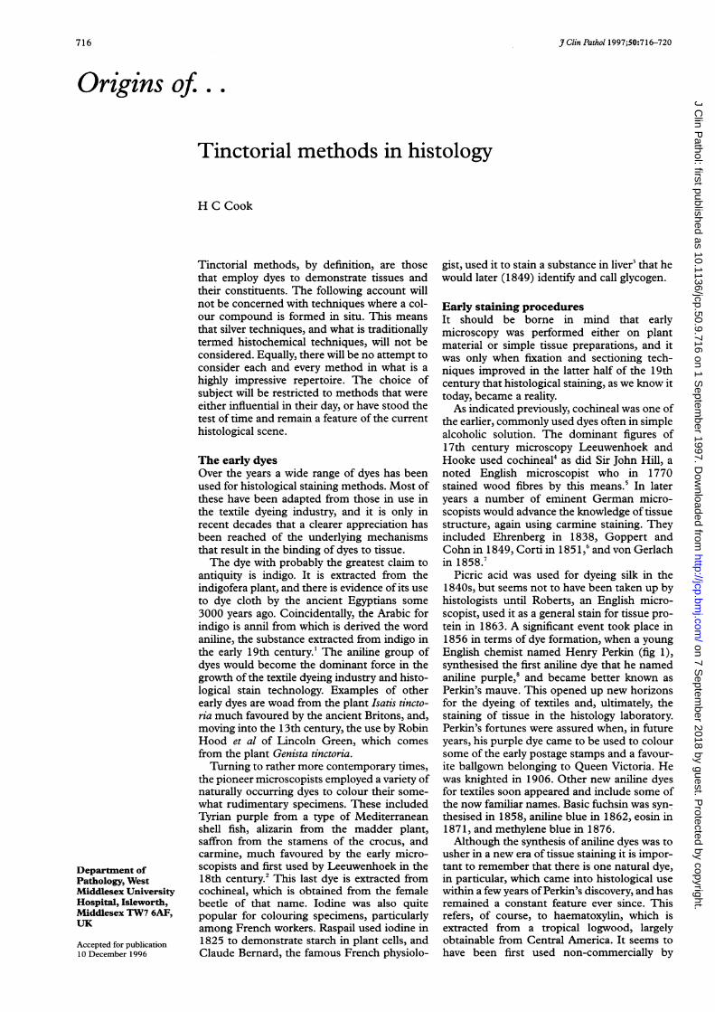
Picric acid was used for dyeing silk in the1 840s, but seems not to have been taken up byhistologists until Roberts, an English micro-scopist, used it as a general stain for tissue pro-tein in 1863. A significant event took place in1856 in terms of dye formation, when a youngEnglish chemist named Henry Perkin (fig 1),synthesised the first aniline dye that he namedaniline purple,8 and became better known asPerkin's mauve. This opened up new horizonsfor the dyeing of textiles and, ultimately, thestaining of tissue in the histology laboratory.Perkin's fortunes were assured when, in futureyears, his purple dye came to be used to coloursome of the early postage stamps and a favour-ite ballgown belonging to Queen Victoria. Hewas knighted in 1906. Other new aniline dyesfor textiles soon appeared and include some ofthe now familiar names. Basic fuchsin was syn-thesised in 1858, aniline blue in 1862, eosin in1871, and methylene blue in 1876.Although the synthesis of aniline dyes was to
usher in a new era of tissue staining it is impor-tant to remember that there is one natural dye,in particular, which came into histological usewithin a few years ofPerkin's discovery, and hasremained a constant feature ever since. Thisrefers, of course, to haematoxylin, which isextracted from a tropical logwood, largelyobtainable from Central America. It seems tohave been first used non-commercially by
716
on 7 Septem
ber 2018 by guest. Protected by copyright.
http://jcp.bmj.com
/J C
lin Pathol: first published as 10.1136/jcp.50.9.716 on 1 S
eptember 1997. D
ownloaded from

Origins of . Tinctorial methods in histology
eV;
Figure 1 A young Henry Perkin.
Reichel in the 18th century6 and in a commer-cial sense in the 1840s.9 The active stain ingre-dient is created by oxidation, and it was Mayerin 1891 who demonstrated that the compoundso formed was haematein.'° Waldeyer iscredited with the introduction of haematoxylinin 1863 as a tissue stain," although this is notwithout dispute.Bohmer in 1865 combined alum with
haematoxylin to form a distinctive albeitimprecise stain for cell nuclei.'2 More selectivenuclear staining was achieved when in 1886Ehrlich (fig 2) added acetic acid to thesolution.'3 A number of the earlier alumhaematoxylin solutions are still very much inuse today and include those of Ehrlich of 1886,Harris 1900, and Mayer's haemalum 1903.Harris was an American born in 1875 aboutwhom little is known. Paul Ehrlich was one ofthe giants of German medical science with sig-nificant contributions to bacteriology, haema-tology, and immunology as well as histology.He was cousin to Carl Weigert (fig 3), anotherfamous figure. Much of Ehrlich's creative workwas carried out in Berlin and Frankfurt, wherehe was to die in 1915 at the age of 61. PaulMayer, who died in 1923 aged 78, was ofGerman-Italian extraction and spent much ofhis working life as a histologist at the Zoologi-cal Station in Naples, Italy. As well as his hae-matoxylin solution he is also remembered forthe mucicarmine and mucihaematein tech-niques for mucin, both published in 1896. Theuse of haematoxylin is not, of course, restrictedto the demonstration of cell nuclei. Other useshave appeared and include staining for myelinin 1889, muscle in 1900, elastin in 1908, and,in more modern times for neuroendocrine cells(1969), etc.
Figure 2 A young Paul Ehrlich.
Figure 3 Carl Weigert.
Before concluding this section on thehistorical development of histological dyesmention must be made of their commercialprocurement. As has already been indicated,commercial dye production was primarilyaimed at the textile industry rather than thelaboratory and, initially, little or no attempt wasmade to classify and standardise dyes in ascientific sense. An important step forward waswhen a Dr Grubler of Leipzig formed in 1880a company for the express purpose of market-ing dyes for microscopy. Later in 1897 anothersimilar company was formed by a Dr Hollbornand for many years these two German compa-
717
on 7 Septem
ber 2018 by guest. Protected by copyright.
http://jcp.bmj.com
/J C
lin Pathol: first published as 10.1136/jcp.50.9.716 on 1 S
eptember 1997. D
ownloaded from

Cook
nies successfully marketed much of the world'shistological dyes.
The development ofhistologicaltechniquesSynthetic dyes began to be more widely used inthe early 1860s but the actual methods wererelatively crude. They invariably consisted ofone-step staining, the excess stain beingwashed off in water or alcohol before the slidewas mounted. In 1867 Schwartz introducedtwo-dye sequential staining interspersed with asimple washing stage.'4 The technique was fur-ther refined when in 1869 Bottcher incorpo-rated an alcohol differentiation step'5; however,some simple staining regimens remained popu-lar. An example is the metachromatic stainingof cartilage which dates from 1875 and isattributed to the celebrated French histologistLouis Ranvier who used the dye cyanine.'6
It was at this time that the haematoxylin andeosin method was born. The H&E has survivedthe passage of time to become the standardmorphological staining method for just aboutevery histological laboratory in the world. Aswe have seen, alum haematoxylin had emergedas a nuclear stain and one of the early counter-stains was aniline blue-hardly an ideal choice.Eosin had been reported as a general stain fortissues by workers such as Dreschfeld andFischer in the 1870s. The actual combinationof the two dyes to form a single method hasbeen variously attributed to Wissowzky in1875, Reynaud in 1876, and Busch in1876-78.'7 Eosin is also a fluorochrome and, acentury later, this property would be utilised bythe increased fluorescence shown by eosinstained myocardial fibres when undergoingearly necrotic change.'8The 1 880s saw the inception of three tincto-
rial techniques that are in popular use today.What is popularly known as the ZN techniqueseems to have had a particularly complex gen-esis. The original stain was devised by RobertKoch in 1882 who used an alkaline methyleneblue solution to stain the tubercle bacilli, coun-terstaining with Bismarck brown. In the sameyear Ehrlich modified this by changing the pri-mary stain to a mixture of aniline and methylviolet, and the counterstain to methylene blue.Later that year Friedrich Ziehl replaced theaniline with phenol-that is, the primary stainis now a carbol (from carbolic acid) methylviolet. Finally, Friedrich Neelsen in 1883replaced the methyl violet with basic fuchsin.Thus was realised the now-familiar carbolfuchsin stain."' Interestingly, given the contextof this staining method, both Ehrlich andNeelsen contracted tuberculosis during theirlifetime.8 The former survived his infection,but Neelsen succumbed to his at the compara-tively early age of 40. The second of the threetechniques is the Gram stain for organisms.Hans Gram, who died at the age of 85 in 1938,was a Professor of Medicine in Copenhagenalthough his famous technique was devised in1884 while working in Berlin. The method wasbased on an earlier one devised by the ubiqui-tous Paul Ehrlich, and a key feature was theLugol's iodine step following the initial staining
by gentian violet. The iodine was used almostby accident as Gram had intended it as a coun-terstain rather than as a dye trapping agent forwhat came to be known as the Gram positivebacteria.20 It is of interest that Jean Lugol was aParis physician who in 1830 developed thesolution that carries his name as a treatment forscrofula (cervical gland tuberculosis). Thethird of these three techniques is the vanGieson for collagen. Ira van Gieson was a NewYork neuropathologist of Dutch-Jewish parent-age who died in 1913, having published histechnique in 1889.21The closing years of the 19th century
brought a number of significant tinctorialmethods to the fore, one of which, althoughprimarily a haematological stain, has enjoyedconsiderable use by histologists (and cytolo-gists). This is the stain formulated by DimitriRomanowsky a Russian protozoologist work-ing in St Petersburg, where he died aged 60 in1921. The dye compound is actually based onan earlier one devised by Ehrlich who is alsocredited with having coined the term neutraldye.2' Romanowsky described his now famousstaining technique in the course of his 1892MD thesis on malaria infected blood cells. Anumber of important modifications to hismethod have appeared over the years, andinclude those ofLeishman in 1901 and Giemsain 1902. William Leishman, who died in 1926aged 61, was of English stock and served in theRoyal Army Medical Corps where he rose tobecome Lieutenant General. Gustav Giemsa,on the other hand, was a German bacteriologistand chemist at Hamburg University. He diedaged 81 in 1948.The demonstration of lipids in tissue re-
ceived a boost in 1896 when an Italian namedDaddi-about whom little is known-introduced the Sudan dyes for that purpose.'3Another important landmark in the history ofhistological staining methodology was theadvent of the so called trichrome techniques in1900. These, together with the phosphotung-stic acid-haematoxylin method of 1897, aredesigned to show in contrasting colours a widerange of tissue constituents-particularly mus-cle and the connective tissues. The earlyhistopathologist, whose name is most oftenassociated with this group of tinctorial meth-ods, is Frank Mallory who was Professor ofPathology at the Boston City Hospital.8 Hedied in 1941 having only three years earlierpublished an authoritative text on histologicaltechniques when aged 76. Another classictrichrome method later appeared in 1929 andis that of Pierre Masson (fig 4) a Frenchpathologist who settled in Montreal, Canadawhere he was to die aged 79 in 1959.At the end of the 19th century a number of
now familiar methods for elastic fibres madetheir appearance. The first of significance wasUnna's orcein technique of 1890, Paul Unnabeing a Professor ofDermatology in Hamburg,Germany. As a stain for elastin orcein hasdiminished in popularity over the years but, asmodified by Shikata in 1974, has taken on anew lease of life in the demonstration of viralhepatitis antigen. Unna is also credited with
718
on 7 Septem
ber 2018 by guest. Protected by copyright.
http://jcp.bmj.com
/J C
lin Pathol: first published as 10.1136/jcp.50.9.716 on 1 S
eptember 1997. D
ownloaded from

Origins of .. Tinctorial methods in histology
fi .:.
I
._
Figure 4 Pierre Masson.
having discovered the existence of plasmacells,8 and his name is linked with the originalhistological technique for these cells namelythe Unna-Pappenheim. This was published in1899 by Pappenheim (a student ofUnna's) andwas based on an original technique by theubiquitous Paul Ehrlich. Unna's modificationofthe solution followed in 1902. The outstand-ing contribution to elastic fibre staining,however, was undoubtedly that of Carl Weigertworking in Breslau and Leipzig, who gave ushis classic elastin stain in 1898. The ironhaematoxylin solution for cell nuclei followedin 1904, the year of his death at the relativelyearly age of 59. Another still popular elastinstain is the 1908 method of the Americanpathologist and ophthalmologist FrederickVerhoeff, who was based in Massachusetts.
Staining techniques: the later yearsIt was inevitable that, as time passed, themomentum of tinctorial method innovationwould slacken as other forms of histologicaldemonstration technique took shape; immuno-cytochemistry is the most obvious example.Indeed, from the 1920s onwards comparativelyfew significant staining techniques emerged.Congo red is a cotton dye introduced to the
textile industry in 1884, and takes its namefrom the Congo Free State which was formedin that year.'4 Griesbach seems to have usedCongo red in 1886 as a stain for axons but itwas another German worker, Bennhold, whodescribed in 1922 its all important use foridentifying amyloid in tissue sections.'5 Thediagnostic value of this was considerablyheightened when in 1927 two French workers,Divry and Florkin, demonstrated the greenbirefringence effect of amyloid deposits stainedwith Congo red.'6 In the same year Southgate,an English pathologist at the Croydon GeneralHospital, published his variant of Mayer's
mucicarmine solution. This remained for manyyears the standard method in Britain formucins. Curiously, Southgate's mucicarminehas waned in popularity in this country, whilein America Mayer's format continues to flour-ish.The year 1950 saw the appearance of several
tinctorial methods ofnote. Harold Steedman, aresearch scientist at Glasgow University, pub-lished the alcian blue technique for acidmucins," and George Gomori the aldehydefuchsin technique for elastic fibres and mastcells.28 The latter worker also published, in thatyear, his one-step trichrome technique forwhich there are adherents today. Gomori, whodied at the relatively young age of 53 in 1957,was an American pathologist of Hungarianextraction, at the University of Chicago.29 Aremarkable researcher, he is chiefly remem-bered for his original contributions to the fieldsof silver demonstration techniques and enzymehistochemistry. Steedman, who was also wellknown for his publications on ester wax andmicrotomy, died in 1991 at the age of 84.
In this period a new staining technique madeits appearance for the demonstration ofnormalmyelin; the luxol fast blue of Kluver andBarrera in 1953.30 This dye is closely related toalcian blue, being of the copper phthalocyaningroup. Its affinity for myelin, however, remainspoorly understood.
Tinctorial methods and the modernlaboratoryThe more traditional staining methods willprobably continue to play a part in histologyeither, as in the case of the H&E, to afford adiagnosis per se, or as special stains to assist inarriving at a diagnosis. As special stains theycan be used alone, or in a complementary roleto the more sophisticated technology. Theadvent, and now wide employment, of immu-nocytochemistry plus the growing involvementof molecular biology in diagnosis has, inevita-bly, signalled a decreasing role for the tinctorialmethod. However, certain of these stains arestill very much in evidence-for example, in thedemonstration of lipids and carbohydrates. Inthe field of infective agents, particularly thoseassociated with the immunocompromised dis-orders, there has been something of a resur-gence in the use of tinctorial methods such asthe ZN. Other, newly identified, pathogenshave created a demand for the more traditionalstaining methods, a good example being gastri-tis and the need to demonstrate the associatedhelicobacter organisms. As discussed, the mainchallenge to the use of tinctorial methods inhistology has come from immunocytochemis-try as a routine tool. Consequently, the moresubjective, colourful staining techniques suchas those for the constituent cells of the anteriorpituitary gland and the pancreas are rarelycalled for. It is probably also true to say that thetrichrome methods are less often used. How-ever, an important role for staining techniquesis that they continue to complement the infor-mation furnished by an antigen-antibody reac-tion. To give but one example: by means ofimmunocytochemistry it is now possible to
719
r
on 7 Septem
ber 2018 by guest. Protected by copyright.
http://jcp.bmj.com
/J C
lin Pathol: first published as 10.1136/jcp.50.9.716 on 1 S
eptember 1997. D
ownloaded from

Cook
characterise the precise type of abnormalprotein present in certain amyloid deposits.This might well be the A4 protein that is asso-ciated with Alzheimer's disease and found inblood vessel walls and plaques of affected braintissue. Lacking the availability of a suitablepan-amyloid antiserum in the immunocyto-chemical armamentarium, however, there isstill a well defined need for an initial indicatorfor the presence of amyloid per se. Until such ageneral amyloid antiserum becomes available itis likely that the Congo red staining methodwill continue to flourish. It would seem, there-fore, that the tinctorial method still has animportant part to play in today's histologicalenvironment. 31
1 Dorland's Illustrated Medical Dictionary. 20th edn. Philadel-phia: WB Saunders Company, 1985.
2 Racegirdle B. A history of microtechnique. Ithaca: CornellUniversity Press, 1978.
3 Pearse AGE. Histochemistry theoretical and applied. London:J&A Churchill Ltd, 1953.
4 Hooke R. Micrographia. London: Royal Society, 1665.5 Hill J. The construction of timber from its early growth;
explained by the microscope etc. 1770. Cited from SmithGM. The development of botanical microtechnic. TransAm Mic Soc 1915;34:71 -129.
6 Lillie RD. H _7 Conn's biological stains. 8th edn. Baltimore:Williams and Wilkins Company, 1969.
7 von Gerlach J. Mikro Stud aus dem Gebiet der mensch Morph.Enke: Erlangen, 1858.
8 Clark G, Kasten FH. History of staining. 3rd edn. Baltimore:Williams and Wilkins Company, 1983.
9 Quekett J. Practical treatise on the use of the microscope.London: Bailliere, 1848.
10 Mayer P. Ueber das Farben mit Hamatoxylin. Mitt Zool StatNeapel 1891;10:170-86.
11 Waldeyer W Untersuchungen uber den Usprung und denVerlauf des Axencylinders bei Wirbellosen und Wirbelth-ieren. Henle Pfeifer Z Rat Med 1863;20:193-256.
12 Bohmer F. Zur pathologischen Anatomie der Meningitiscerebromedullaris epidemica. Aerztl Intelligenzbl (Munich)1865;12:539-50.
13 Ehrlich P. Fragkasten Zeits fur wiss mikr und fur mikros tech1886;3: 150.
14 Schwartz E. Uber eme meth doppel Farbung mikroObjekte. Sitz Akad Wiss Wien Math Naturwiss Classe551867;Abtl 1:671-91.
15 Bottcher A. Ueber entwicklung und bau des Gehorlaby-rinths nach Untersuchungen an Saugethieren. I Theil. VerKais Leop Deutsch Akad Naturforsch 1869;35:203.
16 Ranvier L. Traite technique d'histologie. Paris: Savy, 1875.17 Busch H. Uber die Dappelfarbung des Ossificationsrandes
mit Eosin und Haematoxylin. Arch Physiol 1878:594-5.18 Siegel RG, Fishbein MC. Evaluation of fluorescence micro-
scopy for the identification of necrotic myocardium. HumPathol 1982;13:1091 -4.
19 Brock TD. Robert Koch: a life in medicine and bacteriology.Madison: Science Tech Publishers, 1988.
20 Baker J. Biological microtechnique. London: Methuen andCompany, 1958.
21 van Gieson I. Laboratory notes of technical methods for thenervous system. N YMedJ 1889;50:57.
22 Ehrlich P. Uber die specifischen Granulationen des Blutes.Arch Anat Phys; Phys Abt 1879:571-9.
23 Daddi L. Nouvelle methode pour colorer la graisse dans lestissues. Arch Ital Biol 1896;26:143.
24 Lubs HA. The chemistry of synthetic dyes and pignments. NewYork: Reinhold, 1955.
25 Bennhold H. Eine specifische amyloidfarbung mit kangorot.Munch Medi Wochen 1922;33:1537.
26 Divry P, Florkin M. Sur les proprietes de l'amyloide.Comptes Rendus de Seances de la Societe de Biologie et ses Fili-ales 1927;97:1808.
27 Steedman HF. Alcian blue 8G5:a new stain for mucin. Q 7Microbiol Sci 1950;91:477-9.
28 Gomori G. Aldehyde-fuchsin: a new stain for elastic tissuc.Am 7 Cli71 Pathol 1950;20:665-6.
29 Carpenter A-M, ed. Proceedings of the 5th Congress of theInternational Federation of Societies for Histochemistryand Cytochemistry 1976:1001 -2.
30 Kluver H, Barrera A. A method for the combined staining ofcells and fibres of the nervous system. I Neuropath ExpNeurol 1953;12:400.
31 Cook HC. Is there still a place for conventional stains in his-topathology. .T Histotech 199 1; 14: 147-8.
720
on 7 Septem
ber 2018 by guest. Protected by copyright.
http://jcp.bmj.com
/J C
lin Pathol: first published as 10.1136/jcp.50.9.716 on 1 S
eptember 1997. D
ownloaded from