endogenous endophthalmitis: diagnosis, management, and prognosis … · endogenous endophthalmitis:...
TRANSCRIPT
REVIEW Open Access
Endogenous endophthalmitis: diagnosis,management, and prognosisMohammad Ali Sadiq1, Muhammad Hassan1, Aniruddha Agarwal1, Salman Sarwar1, Shafak Toufeeq1,Mohamed K. Soliman1,2, Mostafa Hanout1, Yasir Jamal Sepah1, Diana V. Do1 and Quan Dong Nguyen1*
Abstract
Endogenous endophthalmitis is an ophthalmic emergency that can have severe sight-threatening complications. Itis often a diagnostic challenge because it can manifest at any age and is associated with a number of underlyingpredisposing factors. Microorganisms associated with this condition vary along a broad spectrum. Depending uponthe severity of the disease, both medical and surgical interventions may be employed. Due to rarity of the disease,there are no guidelines in literature for optimal management of these patients. In this review, treatment guidelinesbased on clinical data and microorganism profile have been proposed.
Keywords: Endophthalmitis, Endogenous, Bacterial, Fungal, Review, Metastatic
ReviewIntroductionIntraocular infection affecting the inner coats of the eyeassociated with significant, progressive vitreous inflam-mation is termed as endophthalmitis [1–4]. Endophthal-mitis is an ophthalmic emergency that can result indevastating ocular and systemic complications. The mostcommon route of entry of infective organisms is throughan external wound of entry, such as trauma, surgery, orinfected cornea. These cases of endophthalmitis aretermed as exogenous endophthalmitis. Endogenous en-dophthalmitis (EE), on the other hand, results from thehematogenous spread of microorganisms from distantfoci [5–7].EE accounts for approximately 2–8 % of all cases of
endophthalmitis [2, 8–11]. Due to paucity of the disease,literature on EE mostly comprises of case series or singlecase reports. Unlike exogenous endophthalmitis, demo-graphics, treatment options, and outcome measures inpatients with EE have not been studied in large-scalestudies.
The first case of bacterial EE has been published in1856 [12]. Subsequently, a major review including ap-proximately 335 cases of bacterial EE was published in2003 [11], and the authors have recently updated theirinitial data by accommodating further reports [13].However, there have been no major reviews encompass-ing all the infective etiologies, including both bacterialand fungal, in literature. With changing patterns of micro-bial disease epidemiology, re-emergence of certain infec-tious diseases, antibiotic susceptibility, and developmentof superbugs, a systematic reappraisal of EE is necessary.
Causative organismsThe etiology of EE is multifactorial, and the list of causa-tive organisms is extensive, with significant geographicvariation. Both bacterial and fungal agents are noted inthe literature as potential agents of EE in the developedworld. However, fungal organisms account for the major-ity of the cases [9, 10]. The organisms responsible for bac-terial EE differ depending on the geographic location. Inthe developed world, gram-positive organisms (Strepto-cocci and Staphylococci) dominate the infection, whereasgram-negative organisms are more common in the Asianpopulation [9, 14]. Asian studies have reported fungi asthe causative organisms in approximately 11.1 to 17.54 %of total cases of EE, with the rest being attributed to bac-terial causes [14, 15].
* Correspondence: [email protected] Imaging Research and Reading Center, Stanley M. Truhlsen EyeInstitute, University of Nebraska Medical Center, 3902 Leavenworth Street,Omaha, NE 68105, USAFull list of author information is available at the end of the article
© 2015 Sadiq et al. Open Access This article is distributed under the terms of the Creative Commons Attribution 4.0International License (http://creativecommons.org/licenses/by/4.0/), which permits unrestricted use, distribution, andreproduction in any medium, provided you give appropriate credit to the original author(s) and the source, provide a link tothe Creative Commons license, and indicate if changes were made.
Sadiq et al. Journal of Ophthalmic Inflammation and Infection (2015) 5:32 DOI 10.1186/s12348-015-0063-y
Risk factorsEE is frequently associated with many underlying systemicrisk factors [3, 5, 10, 16–23]. The most common riskfactors include recent hospitalization, diabetes mellitus,urinary tract infection, immunosuppression (especiallyassociated with underlying malignancy, neutropenia, andHIV (human immunodeficiency virus)), intravenous drugabuse (IVDA), and indwelling catheters [10].Liver abscesses have been noted to be associated with
EE, especially those caused by gram-negative rods suchas Klebsiella pneumonia [24]. In most of these caseswith Klebsiella, diabetes is the major underlying sys-temic risk factor [25, 26]. This finding is most promin-ently noted in the Asian population where bacterialendophthalmitis is more common [15]. Infective endo-carditis (IE) is another important risk factor commonlyassociated with EE in the western countries [27, 28].Various causes of transient bacteremia such as routinecolonoscopy can also lead to EE [29].According to a study assessing differences between the
risk factors for mold and yeast infections, patients withmold infections were more likely to be associated withthe use of chemotherapy as well as organ transplantationespecially cardiac and liver transplants [16]. Similar re-sults have been reported with molds as a common causeof EE in patients on immunosuppressive therapy forhematopoietic stem cell transplantation (HSCT) or forany hematological malignancy [30]. Patients with lunginvolvement by Aspergillus are at a specially increasedrisk for developing EE [4, 30, 31].Neonatal endogenous endophthalmitis deserves a special
mention. Unlike endophthalmitis in adults, neonatal casesare overwhelmingly as a result of an endogenous source ofinfection. Neonates with candidemia, bacteremia, and ret-inopathy of prematurity and low birth weight are at signifi-cant risk for developing EE [32–34]. According to a largecohort study, the odds of neonates with bacteremia, candi-demia, and retinopathy of prematurity to develop EE are21.11, 2.36, and 2.05, respectively (p < 0.0001) [32]. Thecausative organisms are often bacteria from Streptococcispecies, especially S. agalactiae, gram-negative rods likeKlebsiella or Pseudomonas and fungi including Candidaspecies. A recent report suggested decreasing incidence ofneonatal EE in the developed world [32].It is important to note that EE has also been reported
in immunocompetent patients without underlying pre-disposing conditions. EE may be the first manifestationof an underlying occult systemic focus of infection, whilethe systemic cultures for infective organisms are stillnegative [35–39].
PathophysiologyEndogenous endophthalmitis results from metastaticspread of the organism from a primary site of infection
in the setting of bacteremia or fungemia [40]. Mostfrequently, the organism reaches the eye through theposterior segment vasculature. The right eye is morecommonly involved probably due to the more directroute through the right carotid artery [40]. Direct spreadfrom contagious sites can also occur in cases of centralnervous system infection via the optic nerve [41]. Unlikepostoperative and posttraumatic endophthalmitis wheretissue damage results primarily from toxins produced bythe organism, it is postulated that in endogenous en-dophthalmitis, damage is most probably due to a septicembolus that enters the posterior segment vasculatureand acts as a nidus for dissemination of the organisminto the surrounding tissues after crossing the blood-ocular barrier to cause microbial proliferation and in-flammatory reactions within these tissues. Infection thenextends from the retina and the choroid into the vitre-ous cavity and thereafter to the anterior chamber of theeye [42].
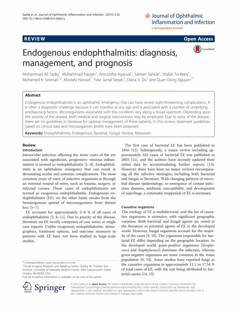
Clinical featuresThe diagnosis of EE may be difficult because of the vari-ability in the clinical signs and symptoms. The organismscausing EE gain access to the internal ocular tissuesthrough the blood-ocular barrier [43]. Due to progressiveinflammation, the patients may experience decreasedvision, which is the most common reason for visiting adoctor [5, 18, 37]. The other classic features include eyelidedema, conjunctival injection, circumcorneal congestion,pain, photophobia, and the presence of floaters [5]. Anter-ior chamber inflammation with hypopyon, absent redreflex, vitreous cells, and haze may also occur [21, 28].These findings of anterior chamber involvement are morecommon in bacterial causes of EE [6]. There may be apoor view of the fundus due to the presence of exudatesand vitreous haze. Other findings include corneal edema,presence of iris nodules, and pupillary distortion sec-ondary to synechiae formation [44, 45]. Bilateral in-volvement can also occur. Causative organisms suchas Mycobacterium tuberculosis can present with bilateralendogenous endophthalmitis and scleral inflammation(Fig. 1).The hallmark of EE is significant involvement of the
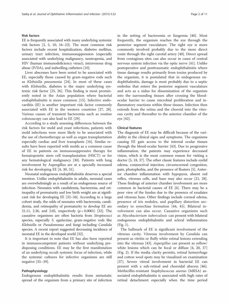
vitreous cavity. Vitreous involvement by Candida canpresent as vitritis or fluffy white retinal lesions extendinginto the vitreous [43]. Aspergillus can present as yellow/white lesions which can be focal or diffuse [4, 20, 37](Fig. 2). If the media clarity permits, retinal hemorrhageand cotton wool spots may be visualized on examination[37]. Severe vitreal involvement in bacterial EE canpresent with a sub-retinal and choroidal abscess [46].Methicillin-resistant Staphylococcus aureus (MRSA) as-sociated endophthalmitis is associated with high rates ofretinal detachment especially when the time period
Sadiq et al. Journal of Ophthalmic Inflammation and Infection (2015) 5:32 Page 2 of 11
Fig. 1 A case of bilateral tubercular endogenous endophthalmitis with scleritis. a Slit lamp biomicroscopy of the left eye with diffuse andcircumcorneal congestion and scleral involvement. There is corneal edema and opacification superiorly. The pupil has broad-based synechiae,and the view of the posterior segment was hazy. b The right eye with severe congestion and ciliary injection. There was a yellow glow present(visible near the inferior pupillary border). c A wide-angled fundus photograph of the left eye with vitreous haze secondary to vitritis along withfocal sheathing of superior vessels. The fluorescein angiography (d) shows presence of superior perivascular hyperfluorescence and leakage ofdye in the superotemporal periphery
Fig. 2 Fundus photograph of a 78-year-old male (a) with a yellow white mass in the temporal paramacular region with some superficialhemorrhages suggestive of a choroidal abscess. The patient was diagnosed with Nocardia endophthalmitis based on retinal aspirates (d, e).b Fundus photograph taken at 3 weeks following intravenous trimethoprim-sulfamethoxazole therapy. There was a marked resolution of thelesion and improvement in media clarity at month 3 (c). d Hematoxylin-eosin staining (×20) of the retinal aspirate. e Gram-positive branching rodsof Nocardia species (×40)
Sadiq et al. Journal of Ophthalmic Inflammation and Infection (2015) 5:32 Page 3 of 11
between onset of symptoms and presentation is delayedby more than 2 weeks [3]. Other non-specific findingscan include flame-shaped hemorrhages, Roth spots andcotton wool spots [6, 45].Clinical findings in EE can be subdivided into three
categories to aid the ophthalmologist to rule in the diag-nosis. Positive signs are strongly suggestive of endogenousendophthalmitis, whereas probable signs are non-specificbut could be present in a case of EE. Table 1 provides a listof clinical signs associated with EE.Visual acuity, as explained above, can be variably af-
fected at the time of presentation, but is generally used asan outcome measure along with a dilated funduscopicexamination to follow up the patient after starting treat-ment. A relative afferent pupillary defect (RAPD) can alsobe present and can guide the need for a vitrectomy [26].A large study was conducted to assess the involvement
of eyes in patients with candidemia. A total of 370patients were enrolled; among them, 60 (16.2 %) patientswere found to have ocular manifestations on fundoscopicexamination. Among these 60 subjects with ocularinvolvement, 6 patients were diagnosed with EE [43]. Inapproximately 18 % of the patients, new lesions were seenafter an initial negative funduscopic examination. This ledto a hypothesis that there is a significant time delaybetween seeding and development of visible retinal le-sions; therefore, patients may have a normal retinal examinitially.In order to classify the severity of ocular involvement
in EE, numerous attempts have been made to classifythe disease. However, there is no unifying broadly
accepted classification for EE available till date. Ishibashiet al. and Petit et al. have previously proposed clinicalclassifications of fungal EE [47, 48].
DiagnosisThe diagnosis of EE requires a high index of suspicionwith presence of one of the above mentioned systemicrisk factors and/or presence of characteristic ocular find-ings on detailed ophthalmoscopic examination (Table 1)[49]. However a clinical diagnosis of EE is always diffi-cult as it has a high false negative rate for EE [5, 49].Multiple clinic visits may be required to confirm thediagnosis. It is also important to note that the presenceof EE is generally not among the major concerns in pa-tients with life-threatening invasive fungal diseases orsepsis secondary to a bacterial etiology [50], and hencethe diagnosis of EE may be delayed with other morbid-ities being managed acutely.To confirm the presence of a specific etiology, vitreous
aspiration and diagnostic vitrectomy followed by a cul-ture and histological examination are commonly used[16, 43, 51]. The need for a diagnostic vitrectomy isdependent on the clinician’s judgment. Vitrectomy has ahigher diagnostic yield for culture (92 %) compared to avitreous aspirate (44 %) as shown by Lingappan et al. [5].Similar results were obtained in another study with nee-dle biopsy negative cases growing organisms on culturefollowing vitrectomy [52]. The study showed that vitre-ous samples during vitrectomy were taken near the ret-inal surface, which can potentially explain the loweryield of needle biopsy as early or localized infectionlocated near the retinal surface might be missed by aneedle biopsy [16].Another emerging technique is the use of real-time
polymerase chain reaction (RT-PCR) of aqueous and vit-reous samples for detection of the etiology of EE. Sugitaet al. reported excellent sensitivity as well as specificityof RT-PCR for detection of fungi [53]. In the same study,PCR was able to detect causative fungi in 5 culture nega-tive specimens. This technique has the advantage ofrapid diagnosis (within 90 min), better detection thancultures as well as no fear of contamination of culturesamples yielding false positive results [10, 54, 55]. Somyaet al. in their study demonstrated increased sensitivity ofPCR over culture [56]. PCR-Based techniques can beused to rule out the presence of pathogens with confi-dence, which is a unique advantage of this methodology.This diagnostic tool promises to be useful in the man-agement of patients with endophthalmitis, especially insamples that are culture negative [57]. However, a po-tential disadvantage of this diagnostic technique is theinability to determine antibiotic susceptibility [21].The most reliable way of diagnosing systemic infec-
tion is blood culture. Blood must be drawn on three
Table 1 Ocular signs suggestive of endogenous endophthalmitis[13, 42]
Positive Possible Probable
Uveal tissue abscesses Hypopyon≤ 1.5 mm Conjunctival injection/chemosis
Hypopyon≥ 1.5 mm Vitreous haze butno visible exudates
Anterior chamberinflammation butno hypopyon
Vitreous exudates Non-necrotizing, focal,discrete chorioretinallesions
Absence of vitreoushaze
Visible arteriolarseptic emboli
Optic neuritis Lid edema
Necrotizing retinitis Intra-retinalhemorrhages
Fever
Perivascularhemorrhages withinflammatory infiltrate
Neonate withwhite reflexa
Panophthalmitis Scleritis
Corneal infiltrates orulcer
Varying combination of symptoms may be presentaIn a neonate presenting with white reflex, endogenous endophthalmitis canbe considered in the differential diagnosis
Sadiq et al. Journal of Ophthalmic Inflammation and Infection (2015) 5:32 Page 4 of 11
consecutive days using sterile precautions. Previouslarge series have shown higher rates of positivity fol-lowing blood culture as compared to vitreous aspiratepossibly due to larger volume sampled. It is also import-ant to culture other extra ocular sites to identify the pos-sible nidus of infection and guide systemic therapyaccordingly, for example, urine cultures. Confirmatoryidentification of extra ocular sources of infection are re-ported in 21–100 % of cases in the literature [5, 18, 21].Identification of these infectious foci is particularly im-portant in cases where vitreous cultures are negative [15].Imaging of ocular tissues is an important means to
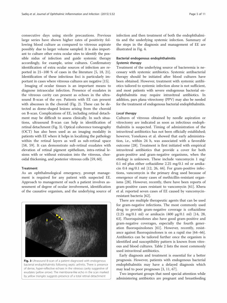
diagnose intraocular infection. Presence of exudates inthe vitreous cavity can present as echoes in the ultra-sound B-scan of the eye. Patients with EE can presentwith abscesses in the choroid (Fig. 2). These can be de-tected as dome-shaped lesions arising from the choroidon B-scan. Complications of EE, including retinal detach-ment may be difficult to assess clinically. In such situa-tions, ultrasound B-scan can help in identification ofretinal detachment (Fig. 3). Optical coherence tomography(OCT) has also been used as an imaging modality inpatients with EE where it helps in localizing the pathologywithin the retinal layers as well as sub-retinal space[58, 59]. It can demonstrate sub-retinal exudates withelevation of retinal pigment epithelium, intra-retinal le-sions with or without extrusion into the vitreous, chor-oidal thickening, and posterior vitreous cells [59, 60].
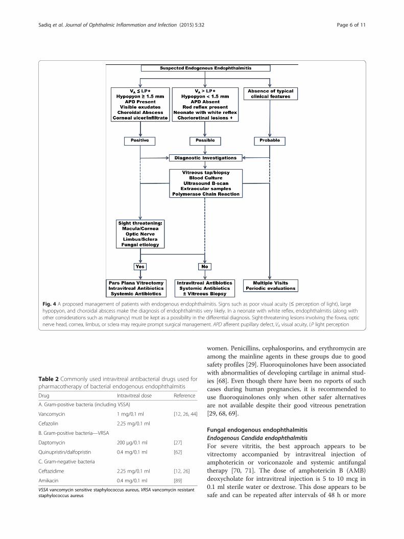
TreatmentAs an ophthalmological emergency, prompt manage-ment is required for any patient with suspected EE.Approach to management of such a patient involves as-sessment of degree of ocular involvement, identificationof the causative organism, and the underlying source of
infection and then treatment of both the endophthalmi-tis and the underlying systemic infection. Summary ofthe steps in the diagnosis and management of EE areillustrated in Fig. 4.
Bacterial endogenous endophthalmitisSystemic therapyTreatment of the underlying source of bacteremia is ne-cessary with systemic antibiotics. Systemic antibacterialtherapy should be initiated after blood cultures havebeen obtained. However, treatment with systemic antibi-otics tailored to systemic infection alone is not sufficient,and most patients with severe endogenous bacterial en-dophthalmitis may require intravitreal antibiotics. Inaddition, pars plana vitrectomy (PPV) may also be neededfor the treatment of endogenous bacterial endophthalmitis.
Local therapyCultures of vitreous obtained by needle aspiration orvitrectomy are indicated as soon as infectious endoph-thalmitis is suspected. Timing of administration of theintravitreal antibiotics has not been officially established;however, Yonekawa et al. showed that early administra-tion, i.e., within 24 h, was associated with a favorableoutcome [28]. Treatment is first initiated with empiricalintravitreal antibiotics that provide a cover for bothgram-positive and gram-negative organisms, when theetiology is unknown. These include vancomycin 1 mg/0.1 ml plus either ceftazidime 2.25 mg/0.1 ml or amika-cin 0.4 mg/0.1 ml [12, 26, 44]. For gram-positive infec-tions, vancomycin is the primary drug used because ofemergence of many cases of methicillin-resistant organ-isms [28]. However, recently, there have been reports ofgram-positive cases resistant to vancomycin [61]. Kheraet al. reported seven cases of EE caused by vancomycin-resistant bacteria [62].There are multiple therapeutic agents that can be used
for gram-negative infections. The most commonly useddrug to provide gram-negative coverage is ceftazidime(2.25 mg/0.1 ml) or amikacin (400 μg/0.1 ml) [14, 28,63]. Fluoroquinolones also have good gram-positive andgram-negative coverages, especially the fourth gener-ation fluoroquinolones [61]. However, recently, resist-ance against fluoroquinolones is on a rapid rise [64–66].Antibiotics can be tailored further once the organism isidentified and susceptibility pattern is known from vitre-ous and blood cultures. Table 2 lists the most commonlyused intravitreal antibiotics.Early diagnosis and treatment is essential for a better
prognosis. However, patients with endogenous bacterialendophthalmitis may have a delayed diagnosis whichmay lead to poor prognoses [3, 11, 67].Two important groups that need special attention while
administering antibiotics are pregnant and breastfeeding
Fig. 3 Ultrasound B-scan of a patient diagnosed with endogenousbacterial endophthalmitis following septic arthritis. There is presenceof dense, hyper-reflective echoes in the vitreous cavity suggestive ofexudates (yellow arrow). The membrane-like echo in the scan markedby yellow triangles suggests presence of a total retinal detachment
Sadiq et al. Journal of Ophthalmic Inflammation and Infection (2015) 5:32 Page 5 of 11
women. Penicillins, cephalosporins, and erythromycin areamong the mainline agents in these groups due to goodsafety profiles [29]. Fluoroquinolones have been associatedwith abnormalities of developing cartilage in animal stud-ies [68]. Even though there have been no reports of suchcases during human pregnancies, it is recommended touse fluoroquinolones only when other safer alternativesare not available despite their good vitreous penetration[29, 68, 69].
Fungal endogenous endophthalmitisEndogenous Candida endophthalmitisFor severe vitritis, the best approach appears to bevitrectomy accompanied by intravitreal injection ofamphotericin or voriconazole and systemic antifungaltherapy [70, 71]. The dose of amphotericin B (AMB)deoxycholate for intravitreal injection is 5 to 10 mcg in0.1 ml sterile water or dextrose. This dose appears to besafe and can be repeated after intervals of 48 h or more
Fig. 4 A proposed management of patients with endogenous endophthalmitis. Signs such as poor visual acuity (≤ perception of light), largehypopyon, and choroidal abscess make the diagnosis of endophthalmitis very likely. In a neonate with white reflex, endophthalmitis (along withother considerations such as malignancy) must be kept as a possibility in the differential diagnosis. Sight-threatening lesions involving the fovea, opticnerve head, cornea, limbus, or sclera may require prompt surgical management. APD afferent pupillary defect, VA visual acuity, LP light perception
Table 2 Commonly used intravitreal antibacterial drugs used forpharmacotherapy of bacterial endogenous endophthalmitis
Drug Intravitreal dose Reference
A. Gram-positive bacteria (including VSSA)
Vancomycin 1 mg/0.1 ml [12, 26, 44]
Cefazolin 2.25 mg/0.1 ml
B. Gram-positive bacteria—VRSA
Daptomycin 200 μg/0.1 ml [27]
Quinupristin/dalfopristin 0.4 mg/0.1 ml [62]
C. Gram-negative bacteria
Ceftazidime 2.25 mg/0.1 ml [12, 26]
Amikacin 0.4 mg/0.1 ml [89]
VSSA vancomycin sensitive staphylococcus aureus, VRSA vancomycin resistantstaphylococcus aureus
Sadiq et al. Journal of Ophthalmic Inflammation and Infection (2015) 5:32 Page 6 of 11

if there is evidence of persistent intraocular infection.Systemic administration of AMB is associated with dose-limiting nephrotoxicity, hypotension, arrhythmias, andinfusion-related fever and chills (“shake and bake”) [8].Voriconazole is a newer agent in the armory of drugsused to treat ocular fungal infections. It achieves an ex-cellent intravitreal concentration after oral or intraven-ous administration [36]. The usual dose of voriconazoleis 100–200 mcg in 0.1 ml sterile water. This doseachieves a final concentration of about 25–50 mcg/ml inthe vitreous [72].Among the azoles, the recommended dose of fluco-
nazole according to the Infectious Disease Society ofAmerica (IDSA) guidelines for Candida endophthalmitisis 400–800 mg daily [73]. Fluconazole is also a broadspectrum agent with a better side effect profile thanAMB. Therefore, it has been used in place of AMB asthe first-line agent against endogenous fungal endoph-thalmitis (EFE) as shown by Hamada et al. [74]. IDSArecommended the use of amphotericin B along withflucytosine for Candida endophthalmitis. Alternatively,fluconazole can be used as well. For severe cases ofendophthalmitis or vitritis, the adjunctive use of vitrec-tomy is recommended [73].The duration of systemic antifungal therapy is a mini-
mum of 6 weeks but the length of therapy depends uponthe resolution of ocular lesions. With severe involvement,usually a longer duration of therapy may be required.
Other endogenous fungal endophthalmitisTreatment in immunocompromised patients includessystemic antifungal therapy (e.g., amphotericin or vori-conazole). If the patient is able to tolerate surgery, vi-trectomy and removal of intraocular lens should beperformed followed by intravitreal antifungal therapy
using amphotericin or voriconazole. However, if the pa-tient cannot tolerate surgery, intravitreal injection withamphotericin or voriconazole should be administeredinitially and repeated as needed. Voriconazole has beenused to treat fungal infections resistant to fluconazoleand amphotericin B [75]. In an in vitro study, voricona-zole showed 100 % activity against Aspergillus species,Paecilomyces species, and Fusarium species [76].Other reports also stated successful treatment of Fu-sarium and Aspergillus endophthalmitis using vorico-nazole [77, 78].IDSA guidelines for the treatment of Aspergillus
endophthalmitis recommend the use of IV amphoter-icin B with addition of intravitreal amphotericin Band pars plana vitrectomy for sight-threatening cases[79]. The recommended alternate therapy is systemicor intravitreal voriconazole. Table 3 summarizes therole of antifungal agents along with their sensitivityprofiles.
Pars plana vitrectomyPPV is a commonly used modality in the treatment ofEE. It is recommended for severe and sight-threateningCandida, Aspergillus, or bacterial endophthalmitis [5,73, 79]. It serves as a diagnostic as well as therapeuticpurpose. It may remove a large number of organismsseeding the vitreous cavity thus lowering the diseaseburden [2, 5, 80]. An intravitreal injection of drugsmay also be given while performing the surgery. Thedecision regarding vitrectomy is usually based on theclinician’s judgment. However, almost all reportedcases where a therapeutic vitrectomy was performedare of patients presenting with either sight-threateningdisease or of those that were irresponsive to systemic ther-apy [15, 17, 49, 52, 67, 80].
Table 3 Commonly used intravitreal antifungal drugs employed for pharmacotherapy of fungal endogenous endophthalmitis alongwith their sensitivity
Drug Intravitreal dose Systemic dose Candida Aspergillus Others
A. Polyene
Amphotericin B 5 μg/0.1 ml 0.5–0.7 mg/kg (IV) ++ +
B. Imidazoles
Miconazole 25–50 μg/0.1 ml – + +
Itraconazole 5 μg/0.05 ml 200–400 mg/day (oral) + +
200 mg/day (IV)
Voriconazole 50–200 μg/0.1 ml 200 mg twice daily (oral) +++ ++ Fusarium +
3–6 mg/kg (IV) twice daily
C. Pyrimidine
5-Flucytosine 2.25 mg/0.1 ml 25–37.5 mg/kg/day − +
D. Echinocandins
Caspofungin – 50 mg/day + +
IV intravenous
Sadiq et al. Journal of Ophthalmic Inflammation and Infection (2015) 5:32 Page 7 of 11
Zhang et al. has reported better visual outcomes incases that underwent early vitrectomy [52]. Decision ofearly vitrectomy has also been associated with a decreasein incidence of retinal detachment and evisceration orenucleation [15, 81]. Sato et al. recommended the use ofvitrectomy for Candida EE before stage IV according toIshibashi’s classification [47]. In cases of bacterial EE,vitrectomy is generally performed when there is no re-sponse to intravitreal antibiotics within 48 h or when theeye condition continues to decline or with a worse gradeof RAPD [26]. Yoon et al. and Ishii et al. suggestedaggressive treatment including early vitrectomy forKlebsiella endophthalmitis might lead to better final out-comes [82, 83]. On the other hand, Sheu et al. found noassociation between the timing of vitrectomy and visualoutcome in Klebsiella endophthalmitis [25]. However,they still suggested the use of surgical intervention, espe-cially in patients with anterior chamber inflammationthat did not respond well to intravitreal antibiotics.
Role of corticosteroidsCurrently, no clear guidelines exist regarding the use ofcorticosteroids in endophthalmitis. Inflammation, al-though essential in combating invading organisms, mayend up damaging retinal structures [84]. Steroids havemultiple anti-inflammatory effects which include but arenot limited to decrease in leucocyte recruitment, attenu-ating production of various inflammatory cytokines andstabilizing membrane barriers including blood-retinalbarrier [85].Clinical studies have reported controversial results on
the use of intravitreal as well as systemic steroids forendophthalmitis [2]. In two case series by Jackson et al.,better visual outcomes were reported in the patientswho received additional treatment with intraocular ste-roids [11, 13]. An interim safety analysis of a prospectivemulticenter randomized placebo-controlled trial of IVTdexamethasone as an adjuvant therapy for endophthal-mitis did not report any safety risks associated with theuse of steroids [85]. On the other hand, Shuwan lee etal. reported no significant association of the use of sys-temic steroids with better visual outcomes [86]. Shah etal. reported a significantly reduced likelihood of obtain-ing a three-line improvement in visual outcomes follow-ing the use of intravitreal steroids in patients withpostoperative endophthalmitis [87].In summary, data on the use of steroids in endophthal-
mitis is limited, and the results of studies are conflicting.Therefore, judicious use of steroids is recommended.
PrognosisIn general, EE does not have a favorable prognosis andresults in complete vision loss, especially if the diagnosisis missed early on and therefore treatment is delayed
[21]. Zenith et al. reported that the eyes with bacterialEE had a worse outcome with more patients requiringenucleation or evisceration compared to patients withfungal EE [21]. The major risk following vitreous aspir-ate in patients with EE is high incidence of retinal de-tachment. Surgery for retinal detachment in these casesis difficult, and there is a need for long-term tamponadein such patients post vitrectomy [88].A clinician has to maintain a very high level of suspi-
cion when a patient with a possible risk factor presentsin association with decreased vision and vitreoretinalchanges on examination. Early diagnosis and treatmenthas been associated with 64 % of patients having visualacuity of counting fingers (CF) or better in one study forbacterial EE [28]. This is well above the percentage ofpatients reported with similar improvement before thisstudy [11]. Itoh et al. also reported that early aggres-sive treatment can lead to good visual outcomes [89].Early vitrectomy within 2 weeks of presentation, espe-cially in severe cases or when suspecting a highlyvirulent organism, can lead to a good overall outcome[79, 82, 83, 86, 90].Virulence of the organism plays an important role in
the visual outcome [15]. Aspergillus and other moldscause more aggressive disease compared to yeasts andtherefore carries a worse prognosis [16, 18, 30, 43, 91].Similarly MRSA endophthalmitis has been reported tobe associated with significant mortality [28]. The associ-ation of MRSA endophthalmitis with visual outcome hasbeen variable, with some studies reporting no associ-ation while others associating it with worse visualoutcome [28, 92, 93]. Connell et al. found that all thepatients in their study needing enucleation were infectedby Klebsiella [10].In a study conducted to determine factors resulting in
poor visual outcome, worse initial visual acuity and cen-trally located lesions were found to be associated withpoor visual outcomes [81]. The same study showed thatearly vitrectomy prevented the development of retinaldetachment. The results of another study in patientswith fungal EE showed that early stages were associatedwith better prognosis. This underscores the importanceof detecting and promptly treating the disease at earlystages to preserve visual acuity [80]. According to Anget al., the main prognostic factor in Klebsiella EE is thepresence of hypopyon [26]. Other prognostic factorsfound in the same study include rapid onset of ocularsymptoms, unilateral involvement, and panophthalmitis.Another study found no association between final visualacuity (log MAR values) and diabetes, causative organ-ism, source of infection, and performance of vitrectomy[15]. However, the study did report better final visualoutcomes in patients with initial visual acuity better thancounting fingers.
Sadiq et al. Journal of Ophthalmic Inflammation and Infection (2015) 5:32 Page 8 of 11
ConclusionsEE is an ophthalmological emergency that requiresprompt diagnosis and management. Figure 4 depicts asimplified flow chart for the diagnosis and managementof EE. The main challenges in the management of EEare early identification and delivering an adequate con-centration of the drug in the vitreous cavity. It may bepossible to overcome this challenge with direct intravit-real administration of the antibiotic.Systemic therapy is used to treat the focus of infection
causing the metastatic spread of the organism to theocular cavity. In mild cases of EE, systemic therapy isthe mainstay of treatment. However, in severe cases, sys-temic therapy is adjuvant to the more aggressive intravit-real administration of drugs.PPV has a diagnostic as well as therapeutic role in the
management of EE. Vitrectomy may be strongly consid-ered as a treatment option if there is no response to sys-temic or local therapy within 24–48 h of presentation orif the patient has possible worsening. Visual acuity, sys-temic debility, etiology of infection, and ocular examin-ation must guide the decision to intervene in such cases.
AbbreviationsAMB: Amphotericin B; CF: Counting fingers; EE: Endogenous endophthalmitis;EFE: Endogenous fungal endophthalmitis; HIV: Human immunodeficiencyvirus; HSCT: Hematopoietic stem cell transplantation; IDSA: Infectious DiseaseSociety of America; IE: Infective endocarditis; IVDA: Intravenous drug abuse;MRSA: Methicillin-resistant Staphylococcus aureus; OCT: Optical coherencetomography; PCR: Polymerase chain reaction; PPV: Pars plana vitrectomy;RAPD: Relative afferent pupillary defect; RT-PCR: Real-time polymerase chainreaction.
Competing interestsThe authors declare that they have no competing interests.
Authors’ contributionsMAS participated in the design and coordination and helped to draft themanuscript. MdH participated in the design of the study and helped to draftthe manuscript. AA participated in the analysis and revision of themanuscript. SS helped analyze the data and revised the manuscript.ST drafted and revised the manuscript. MKS participated in drafting themanuscript. MH participated in the literature search and analysis. YJS wasinvolved in designing the manuscript and revision of the draft. DDparticipated in revising the manuscript. QDN supervised everything andrevised the manuscript. All authors read and approved the final manuscript.
Author details1Ocular Imaging Research and Reading Center, Stanley M. Truhlsen EyeInstitute, University of Nebraska Medical Center, 3902 Leavenworth Street,Omaha, NE 68105, USA. 2Department of Ophthalmology, Assiut UniversityHospital, Assiut University, Assiut, Egypt.
Received: 7 March 2015 Accepted: 28 October 2015
References1. Khan FA, Slain D, Khakoo RA (2007) Candida endophthalmitis: focus on
current and future antifungal treatment options. Pharmacotherapy27(12):1711–1721. doi:10.1592/phco.27.12.1711
2. Novosad BD, Callegan MC (2010) Severe bacterial endophthalmitis: towardsimproving clinical outcomes. Expert Rev Ophthalmol 5(5):689–698.doi:10.1586/eop.10.52
3. Ho V, Ho LY, Ranchod TM, Drenser KA, Williams GA, Garretson BR (2011)Endogenous methicillin-resistant Staphylococcus aureus endophthalmitis.Retina 31(3):596–601. doi:10.1097/IAE.0b013e3181ecccf0
4. Vilela RC, Vilela L, Vilela P, Vilela R, Motta R, Possa AP, de Almeida C,Mendoza L (2013) Etiological agents of fungal endophthalmitis: diagnosisand management. Int Ophthalmol. doi:10.1007/s10792-013-9854-z
5. Lingappan A, Wykoff CC, Albini TA, Miller D, Pathengay A, Davis JL, FlynnHW Jr (2012) Endogenous fungal endophthalmitis: causative organisms,management strategies, and visual acuity outcomes. Am J Ophthalmol153(1):162–166. doi:10.1016/j.ajo.2011.06.020, e161
6. Grixti A, Sadri M, Datta AV (2012) Uncommon ophthalmologicdisorders in intensive care unit patients. Journal of critical care 27(6):746.doi:10.1016/j.jcrc.2012.07.013, e749-722
7. Lynn WA, Lightman S (2004) The eye in systemic infection. Lancet364(9443):1439–1450. doi:10.1016/S0140-6736(04)17228-0
8. Chhablani J (2011) Fungal endophthalmitis. Expert Rev Anti Infect Ther9(12):1191–1201. doi:10.1586/eri.11.139
9. Schiedler V, Scott IU, Flynn HW Jr, Davis JL, Benz MS, Miller D (2004)Culture-proven endogenous endophthalmitis: clinical featuresand visual acuity outcomes. Am J Ophthalmol 137(4):725–731.doi:10.1016/j.ajo.2003.11.013
10. Connell PP, O’Neill EC, Fabinyi D, Islam FM, Buttery R, McCombe M,Essex RW, Roufail E, Clark B, Chiu D, Campbell W, Allen P (2011)Endogenous endophthalmitis: 10-year experience at a tertiary referralcentre. Eye (Lond) 25(1):66–72. doi:10.1038/eye.2010.145
11. Jackson TL, Eykyn SJ, Graham EM, Stanford MR (2003) Endogenous bacterialendophthalmitis: a 17-year prospective series and review of 267 reportedcases. Surv Ophthalmol 48(4):403–423
12. Tsai AS, Lee SY, Jap AH (2010) An unusual case of recurrent endogenousKlebsiella endophthalmitis. Eye (London, England) 24(10):1630–1631.doi:10.1038/eye.2010.95
13. Jackson TL, Paraskevopoulos T, Georgalas I (2014) Systematic review of 342cases of endogenous bacterial endophthalmitis. Surv Ophthalmol.doi:10.1016/j.survophthal.2014.06.002
14. Sharma S, Padhi TR, Basu S, Kar S, Roy A, Das T (2014) Endophthalmitispatients seen in a tertiary eye care centre in Odisha: a clinico-microbiological analysis. Indian J Med Res 139(1):91–98
15. Lim HW, Shin JW, Cho HY, Kim HK, Kang SW, Song SJ, Yu HG, Oh JR, Kim JS,Moon SW, Chae JB, Park TK, Song Y (2014) Endogenous endophthalmitis inthe Korean population: a six-year retrospective study. Retina 34(3):592–602.doi:10.1097/IAE.0b013e3182a2e705
16. Sridhar J, Flynn HW Jr, Kuriyan AE, Miller D, Albini T (2013) Endogenousfungal endophthalmitis: risk factors, clinical features, and treatmentoutcomes in mold and yeast infections. J Ophthalmic Inflamm Infect 3(1):60.doi:10.1186/1869-5760-3-60
17. Durand ML (2013) Endophthalmitis. Clin Microbiol Infect 19(3):227–234.doi:10.1111/1469-0691.12118
18. Lamaris GA, Esmaeli B, Chamilos G, Desai A, Chemaly RF, Raad II, Safdar A,Lewis RE, Kontoyiannis DP (2008) Fungal endophthalmitis in a tertiary carecancer center: a review of 23 cases. Eur J Clin Microbiol Infect Dis 27(5):343–347.doi:10.1007/s10096-007-0443-9
19. Keyashian K, Malani PN (2007) Endophthalmitis associated with intravenousdrug use. South Med J 100(12):1219–1220. doi:10.1097/SMJ.0b013e3181581191
20. Cheng HH, Ding Y, Wu M, Tang CC, Zhang RJ, Lin XF, Xu JT (2011)Endogenous aspergillus endophthalmitis after kidney transplantation.Int J Ophthalmol 4(5):567–571. doi:10.3980/j.issn.2222-3959.2011.05.20
21. Wu ZH, Chan RP, Luk FO, Liu DT, Chan CK, Lam DS, Lai TY (2012) Review ofclinical features, microbiological spectrum, and treatment outcomes ofendogenous endophthalmitis over an 8-year period. J Ophthalmol2012:265078. doi:10.1155/2012/265078
22. de Lima LM, Cecchetti SA, Cecchetti DF, Arroyo D, Romao EA, Dantas M,Neto MM (2012) Endophthalmitis: a rare but devastating metastatic bacterialcomplication of hemodialysis catheter-related sepsis. Ren Fail 34(1):119–122.doi:10.3109/0886022X.2011.623557
23. Reedy JS, Wood KE (2000) Endogenous Pseudomonas aeruginosaendophthalmitis: a case report and literature review. Intensive Care Med26(9):1386–1389
24. Hu CC, Ho JD, Lou HY, Keller JJ, Lin HC (2012) A one-year follow-up studyon the incidence and risk of endophthalmitis after pyogenic liver abscess.Ophthalmology 119(11):2358–2363. doi:10.1016/j.ophtha.2012.05.022
Sadiq et al. Journal of Ophthalmic Inflammation and Infection (2015) 5:32 Page 9 of 11
25. Sheu SJ, Kung YH, Wu TT, Chang FP, Horng YH (2011) Risk factors forendogenous endophthalmitis secondary to klebsiella pneumoniae liverabscess: 20-year experience in Southern Taiwan. Retina 31(10):2026–2031.doi:10.1097/IAE.0b013e31820d3f9e
26. Ang M, Jap A, Chee SP (2011) Prognostic factors and outcomes inendogenous Klebsiella pneumoniae endophthalmitis. Am J Ophthalmol151(2):338–344. doi:10.1016/j.ajo.2010.08.036, e332
27. Buzzacco DM, Carroll CP (2012) Intravitreal daptomycin in a case of bilateralendogenous endophthalmitis. Arch Ophthalmol 130(7):940–941.doi:10.1001/archophthalmol.2011.2527
28. Yonekawa Y, Chan RV, Reddy AK, Pieroni CG, Lee TC, Lee S (2011) Earlyintravitreal treatment of endogenous bacterial endophthalmitis. ClinExperiment Ophthalmol 39(8):771–778. doi:10.1111/j.1442-9071.2011.02545.x
29. Wu AY, Oestreicher JH (2011) Endogenous bacterial endophthalmitis afterroutine colonoscopy. Journal canadien d’ophtalmologie 46(6):556–557.doi:10.1016/j.jcjo.2011.10.002
30. Vergoulidou M, Krause L, Foerster MH, Thiel E, Schwartz S (2011)Endogenous filamentous fungal endophthalmitis—single-centresurvey in patients with acute leukaemia or postallogeneic stem celltransplantation and review of the literature. Mycoses 54(6):e704–e711.doi:10.1111/j.1439-0507.2010.02004.x
31. Sahu C, Kumar K, Sinha MK, Venkata A, Majji AB, Jalali S (2013) Review ofendogenous endophthalmitis during pregnancy including case series. IntOphthalmol 33(5):611–618. doi:10.1007/s10792-012-9697-z
32. Moshfeghi AA, Charalel RA, Hernandez-Boussard T, Morton JM, MoshfeghiDM (2011) Declining incidence of neonatal endophthalmitis in the UnitedStates. Am J Ophthalmol 151(1):59–65. doi:10.1016/j.ajo.2010.07.008, e51
33. Noyola DE, Bohra L, Paysse EA, Fernandez M, Coats DK (2002) Association ofcandidemia and retinopathy of prematurity in very low birthweight infants.Ophthalmology 109(1):80–84
34. Basu S, Kumar A, Kapoor K, Bagri NK, Chandra A (2013) Neonatalendogenous endophthalmitis: a report of six cases. Pediatrics131(4):e1292–e1297. doi:10.1542/peds.2011-3391
35. Mamandhar A, Bajracharya L (2012) Aspergillus endophthalmitisin a healthy individual. Nepal J Ophthalmol 4(1):179–183.doi:10.3126/nepjoph.v4i1.5873
36. Logan S, Rajan M, Graham E, Johnson E, Klein J (2010) A caseof aspergillus endophthalmitis in an immuncompetent woman:intra-ocular penetration of oral voriconazole: a case report. Cases J 3:31.doi:10.1186/1757-1626-3-31
37. Agarwal M, Biswas J, Mathur U, Sijwali MS, Singh AK (2007) Aspergillus irisgranuloma in a young male: a case report with review of literature.Indian J Ophthalmol 55(1):73–74
38. Lee JH, Kim JS, Park YH (2012) Diagnosis and treatment of postpartumCandida endophthalmitis. The J Obstet Gynaecol Res 38(9):1220–1222.doi:10.1111/j.1447-0756.2012.01854.x
39. Shankar K, Gyanendra L, Hari S, Narayan SD (2009) Culture provenendogenous bacterial endophthalmitis in apparently healthy individuals.Ocul Immunol Inflamm 17(6):396–399. doi:10.3109/09273940903216891
40. Greenwald MJ, Wohl LG, Sell CH (1986) Metastatic bacterial endophthalmitis:a contemporary reappraisal. Surv Ophthalmol 31(2):81–101
41. Samiy N, D’Amico DJ (1996) Endogenous fungal endophthalmitis.Int Ophthalmol Clin 36(3):147–162
42. Chee SP, Jap A (2001) Endogenous endophthalmitis. Curr Opin Ophthalmol12(6):464–470
43. Oude Lashof AM, Rothova A, Sobel JD, Ruhnke M, Pappas PG, Viscoli C,Schlamm HT, Oborska IT, Rex JH, Kullberg BJ (2011) Ocular manifestations ofcandidemia. Clin Infect Dis : an official publication of the Infectious DiseasesSociety of America 53(3):262–268. doi:10.1093/cid/cir355
44. Dua S, Chalermskulrat W, Miller MB, Landers M, Aris RM (2006) Bilateralhematogenous Pseudomonas aeruginosa endophthalmitis after lungtransplantation. Am J Transplant 6(1):219–224. doi:10.1111/j.1600-6143.2005.01133.x
45. Khan A, Okhravi N, Lightman S (2002) The eye in systemic sepsis. Clin Med2(5):444–448
46. Chen KJ, Chao AN, Hwang YS, Chen YP, Wang NK (2011) Prognostic factorsand outcomes in endogenous Klebsiella endophthalmitis. Am J Ophthalmol151(6):1105–1106. doi:10.1016/j.ajo.2011.03.015, author reply 1106–1107
47. Sato Y, Miyasaka S, Shimada H (2001) Prognosis of endogenous fungalendophthalmitis and utility of Ishibashi’s classification. Jpn J Ophthalmol45(2):181–186
48. Tanaka M, Kobayashi Y, Takebayashi H, Kiyokawa M, Qiu H (2001)Analysis of predisposing clinical and laboratory findings for thedevelopment of endogenous fungal endophthalmitis. A retrospective12-year study of79 eyes of 46 patients. Retina (Philadelphia, Pa) 21(3):203–209
49. Shen X, Xu G (2009) Vitrectomy for endogenous fungal endophthalmitis.Ocul Immunol Inflamm 17(3):148–152. doi:10.1080/09273940802689396
50. Binder MI, Chua J, Kaiser PK, Procop GW, Isada CM (2003) Endogenousendophthalmitis: an 18-year review of culture-positive cases at a tertiarycare center. Medicine 82(2):97–105
51. Palexas GN, Green WR, Goldberg MF, Ding Y (1995) Diagnostic pars planavitrectomy report of a 21-year retrospective study. Trans Am OphthalmolSoc 93:281–308, discussion 308–214
52. Zhang YQ, Wang WJ (2005) Treatment outcomes after pars planavitrectomy for endogenous endophthalmitis. Retina 25(6):746–750
53. Sugita S, Kamoi K, Ogawa M, Watanabe K, Shimizu N, Mochizuki M (2012)Detection of Candida and Aspergillus species DNA using broad-rangereal-time PCR for fungal endophthalmitis. Graefes Arch Clin Exp Ophthalmol250(3):391–398. doi:10.1007/s00417-011-1819-1
54. Jaeger EE, Carroll NM, Choudhury S, Dunlop AA, Towler HM, Matheson MM,Adamson P, Okhravi N, Lightman S (2000) Rapid detection andidentification of Candida, Aspergillus, and Fusarium species in ocularsamples using nested PCR. J Clin Microbiol 38(8):2902–2908
55. Okhravi N, Adamson P, Lightman S (2000) Use of PCR in endophthalmitis.Ocul Immunol Inflamm 8(3):189–200
56. Sowmya P, Madhavan HN (2009) Diagnostic utility of polymerase chainreaction on intraocular specimens to establish the etiology of infectiousendophthalmitis. Eur J Ophthalmol 19(5):812–817
57. Therese KL, Anand AR, Madhavan HN. Polymerase chain reaction in thediagnosis of bacterial endophthalmitis. Br J Ophthalmol. 1998;82(9):1078–82.
58. Rodrigues IA, Jackson TL (2014) A high-definition view ofendogenous fungal endophthalmitis. The Lancet Infect Dis 14(4):358.doi:10.1016/S1473-3099(13)70216-0
59. Adam CR, Sigler EJ (2014) Multimodal imaging findings inendogenous Aspergillus endophthalmitis. Retina 34(9):1914–1915.doi:10.1097/IAE.0000000000000135
60. Cho M, Khanifar AA, Chan RV (2011) Spectral-domain optical coherencetomography of endogenous fungal endophthalmitis. Retin Cases Brief Rep5(2):136–140. doi:10.1097/ICB.0b013e3181cc2146
61. Ramakrishnan R, Bharathi MJ, Shivkumar C, Mittal S, Meenakshi R, KhadeerMA, Avasthi A (2009) Microbiological profile of culture-proven cases ofexogenous and endogenous endophthalmitis: a 10-year retrospective study.Eye (Lond) 23(4):945–956. doi:10.1038/eye.2008.197
62. Khera M, Pathengay A, Jindal A, Jalali S, Mathai A, Pappuru RR, Relhan N,Das T, Sharma S, Flynn HW (2013) Vancomycin-resistant Gram-positivebacterial endophthalmitis: epidemiology, treatment options, and outcomes.J Ophthalmic Inflamm Infect 3(1):46. doi:10.1186/1869-5760-3-46
63. Espinel-Ingroff A, Boyle K, Sheehan DJ (2001) In vitro antifungal activities ofvoriconazole and reference agents as determined by NCCLS methods:review of the literature. Mycopathologia 150(3):101–115
64. Miller D, Flynn PM, Scott IU, Alfonso EC, Flynn HW Jr (2006) In vitrofluoroquinolone resistance in staphylococcal endophthalmitis isolates.Arch Ophthalmol 124(4):479–483. doi:10.1001/archopht.124.4.479
65. Benz MS, Scott IU, Flynn HW Jr, Unonius N, Miller D (2004) Endophthalmitisisolates and antibiotic sensitivities: a 6-year review of culture-proven cases.Am J Ophthalmol 137(1):38–42
66. Bertino JS Jr (2009) Impact of antibiotic resistance in the management ofocular infections: the role of current and future antibiotics. Clin Ophthalmol3:507–521
67. Smith SR, Kroll AJ, Lou PL, Ryan EA (2007) Endogenous bacterial and fungalendophthalmitis. endophthalmitis. Int Ophthalmol Clin 47(2):173–183.doi:10.1097/IIO.0b013e31803778f7
68. Bar-Oz B, Moretti ME, Boskovic R, O’Brien L, Koren G (2009) The safety ofquinolones—a meta-analysis of pregnancy outcomes. Eur J Obstet GynecolReprod Biol 143(2):75–78. doi:10.1016/j.ejogrb.2008.12.007
69. Padberg S, Wacker E, Meister R, Panse M, Weber-Schoendorfer C,Oppermann M, Schaefer C (2014) Observational cohort study of pregnancyoutcome after first-trimester exposure to fluoroquinolones. AntimicrobAgents Chemother 58(8):4392–4398. doi:10.1128/AAC.02413-14
70. Barza M (1998) Treatment options for candidal endophthalmitis [editorial;comment]. Clin Infect Dis 27(5):1134–1136
Sadiq et al. Journal of Ophthalmic Inflammation and Infection (2015) 5:32 Page 10 of 11
71. Martinez-Vazquez C, Fernandez-Ulloa J, Bordon J, Sopena B, de la Fuente J,Ocampo A, Rubianes M (1998) Candida albicans endophthalmitis in brownheroin addicts: response to early vitrectomy preceded and followed byantifungal therapy. Clin Infect Dis 27(5):1130–1133
72. Breit SM, Hariprasad SM, Mieler WF, Shah GK, Mills MD, Grand MG (2005)Management of endogenous fungal endophthalmitis with voriconazole andcaspofungin. Am J Ophthalmol 139(1):135–140. doi:10.1016/j.ajo.2004.08.077
73. Pappas PG, Kauffman CA, Andes D, Benjamin DK Jr, Calandra TF, Edwards JEJr, Filler SG, Fisher JF, Kullberg BJ, Ostrosky-Zeichner L, Reboli AC, Rex JH,Walsh TJ, Sobel JD, Infectious Diseases Society of A (2009) Clinical practiceguidelines for the management of candidiasis: 2009 update by theInfectious Diseases Society of America. Clin Infect Dis 48(5):503–535.doi:10.1086/596757
74. Hamada Y, Okuma R, Katori Y, Takahashi S, Hirayama T, Ichibe Y, KuroyamaM (2013) Bibliographical investigation (domestic and overseas) on thetreatment of endogenous Candida endophthalmitis over an 11-year period.Med Mycol J 54(1):53–67
75. Sen P, Gopal L, Sen PR (2006) Intravitreal voriconazole for drug-resistantfungal endophthalmitis: case series. Retina 26(8):935–939.doi:10.1097/01.iae.0000250011.68532.a2
76. Marangon FB, Miller D, Giaconi JA, Alfonso EC (2004) In vitro investigationof voriconazole susceptibility for keratitis and endophthalmitis fungalpathogens. Am J Ophthalmol 137(5):820–825. doi:10.1016/j.ajo.2003.11.078
77. Jorgensen JS, Prause JU, Kiilgaard JF (2014) Bilateral endogenous Fusariumsolani endophthalmitis in a liver-transplanted patient: a case report. J MedCase Rep 8(1):101. doi:10.1186/1752-1947-8-101
78. Durand ML, Kim IK, D’Amico DJ, Loewenstein JI, Tobin EH, Kieval SJ, MartinSS, Azar DT, Miller FS 3rd, Lujan BJ, Miller JW (2005) Successful treatment ofFusarium endophthalmitis with voriconazole and Aspergillusendophthalmitis with voriconazole plus caspofungin. Am J Ophthalmol140(3):552–554. doi:10.1016/j.ajo.2005.03.030
79. Walsh TJ, Anaissie EJ, Denning DW, Herbrecht R, Kontoyiannis DP, Marr KA,Morrison VA, Segal BH, Steinbach WJ, Stevens DA, van Burik JA, Wingard JR,Patterson TF, Infectious Diseases Society of A (2008) Treatment ofaspergillosis: clinical practice guidelines of the Infectious Diseases Society ofAmerica. Clin Infect Dis 46(3):327–360. doi:10.1086/525258
80. Takebayashi H, Mizota A, Tanaka M (2006) Relation between stage ofendogenous fungal endophthalmitis and prognosis. Graefes Arch Clin ExpOphthalmol 244(7):816–820. doi:10.1007/s00417-005-0182-5
81. Sallam A, Taylor SR, Khan A, McCluskey P, Lynn WA, Manku K,Pacheco PA, Lightman S (2012) Factors determining visual outcome inendogenous Candida endophthalmitis. Retina 32(6):1129–1134.doi:10.1097/IAE.0b013e31822d3a34
82. Yoon YH, Lee SU, Sohn JH, Lee SE (2003) Result of early vitrectomyfor endogenous Klebsiella pneumoniae endophthalmitis. Retina23(3):366–370
83. Ishii K, Hiraoka T, Kaji Y, Sakata N, Motoyama Y, Oshika T (2011) Successfultreatment of endogenous Klebsiella pneumoniae endophthalmitis: a casereport. Int Ophthalmol 31(1):29–31. doi:10.1007/s10792-010-9387-7
84. Callegan MC, Engelbert M, Parke DW 2nd, Jett BD, Gilmore MS (2002)Bacterial endophthalmitis: epidemiology, therapeutics, and bacterium-hostinteractions. Clin Microbiol Rev 15(1):111–124
85. Lindstedt EW, Bennebroek CA, van der Werf DJ, Veckeneer M, Norel AO,Nielsen CC, Wubbels RJ, van Dissel JT, van Meurs JC (2014) A prospectivemulticenter randomized placebo-controlled trial of dexamethasone as anadjuvant in the treatment of postoperative bacterial endophthalmitis:interim safety analysis of the study drug and analysis of overalltreatment results. Graefes Arch Clin Exp Ophthalmol 252(10):1631–1637.doi:10.1007/s00417-014-2770-8
86. Lee S, Um T, Joe SG, Hwang JU, Kim JG, Yoon YH, Lee JY (2012) Changes inthe clinical features and prognostic factors of endogenous endophthalmitis:fifteen years of clinical experience in Korea. Retina 32(5):977–984.doi:10.1097/IAE.0b013e318228e312
87. Shah GK, Stein JD, Sharma S, Sivalingam A, Benson WE, Regillo CD,Brown GC, Tasman W (2000) Visual outcomes following the use ofintravitreal steroids in the treatment of postoperative endophthalmitis.Ophthalmology 107(3):486–489
88. Kitiratschky VB, Deuter C, Beck R, Schulte B, Muller H, Blumenstock G,Szurman P (2014) Relationship between suspected reasons of intraocularinflammation and the results of diagnostic vitrectomy: an observationalstudy. Ocul Immunol Inflamm. doi:10.3109/09273948.2013.870212
89. Itoh M, Ikewaki J, Kimoto K, Itoh Y, Shinoda K, Nakatsuka K (2010)Two cases of endogenous endophthalmitis caused by gram-positivebacteria with good visual outcome. Case Rep Ophthalmol 1(2):56–62.doi:10.1159/000320601
90. Jalali S, Pehere N, Rani PK, Bobbili RB, Nalamada S, Motukupally SR,Sharma S (2014) Treatment outcomes and clinicomicrobiologicalcharacteristics of a protocol-based approach for neonatal endogenousendophthalmitis. Eur J Ophthalmol 24(3):424–436. doi:10.5301/ejo.5000395
91. Zhang H, Liu Z (2010) Endogenous endophthalmitis: a 10-year review ofculture-positive cases in northern China. Ocul Immunol Inflamm18(2):133–138. doi:10.3109/09273940903494717
92. Major JC Jr, Engelbert M, Flynn HW Jr, Miller D, Smiddy WE, Davis JL (2010)Staphylococcus aureus endophthalmitis: antibiotic susceptibilities,methicillin resistance, and clinical outcomes. Am J Ophthalmol149(2):278–283. doi:10.1016/j.ajo.2009.08.023, e271
93. Ness T, Schneider C (2009) Endogenous endophthalmitis caused bymethicillin-resistant Staphylococcus aureus (MRSA). Retina 29(6):831–834.doi:10.1097/IAE.0b013e3181a3b7a1
Submit your manuscript to a journal and benefi t from:
7 Convenient online submission
7 Rigorous peer review
7 Immediate publication on acceptance
7 Open access: articles freely available online
7 High visibility within the fi eld
7 Retaining the copyright to your article
Submit your next manuscript at 7 springeropen.com
Sadiq et al. Journal of Ophthalmic Inflammation and Infection (2015) 5:32 Page 11 of 11