encapsulation systems for delivery of flavonoids: a review
TRANSCRIPT

https://biointerfaceresearch.com/ 13934
Review
Volume 11, Issue 6, 2021, 13934 - 13951
https://doi.org/10.33263/BRIAC116.1393413951
Encapsulation Systems for Delivery of Flavonoids: A
Review
Mojtaba Yousefi 1 , Mahdi Shadnoush 2 , Sara Sohrabvandi 3 , Nasim Khorshidian 1,* , Amir M.
Mortazavian 4,*
1 Food Safety Research Center (Salt), Semnan University of Medical Sciences, Semnan, Iran; [email protected]
(M.Y.); [email protected] (N.K.); 2 Department of Clinical Nutrition, Faculty of Nutrition Sciences and Food Technology, National Nutrition and Food
Technology Research Institute, Shahid Beheshti University of Medical Sciences, Tehran, Iran; [email protected]
(M.S.); 3 Department of Food Technology Research, Faculty of Nutrition Sciences and Food Technology, National Nutrition and
Food Technology Research Institute, Shahid Beheshti University of Medical Sciences, Tehran, Iran; [email protected]
(S.S.); 4 Food Safety Research Center, Shahid Beheshti University of Medical Sciences, Tehran, Iran; [email protected] (A.M.M.);
* Correspondence: [email protected] (N.K.); [email protected] (A.M.M.);
Scopus Author ID 57192107570 (N.K.); 12792054500 (A.M.M)
Received: 21.01.2021; Revised: 22.02.2021; Accepted: 24.02.2021; Published: 2.03.2021
Abstract: Encapsulation of bioactive compounds s been considered a promising tool for preserving
these compounds. Several studies on dietary sources and health benefits of flavonoids, their chemical
and stability properties, and encapsulation methods used for delivery of flavonoids were reviewed.
Flavonoids comprise the main group of polyphenols widely found in fruits and vegetables responsible
for numerous biological activities. They have a flavan nucleus with 15 carbon atoms organized in three
rings and are categorized into six subgroups. The main dietary sources of flavonoids are fruits,
vegetables, cereals, tea, and some herbs such as Viola odorata Linn. These compounds can prevent
diseases such as cardiovascular, cancers, neurodegenerative, diabetes, and inflammatory bowel disease.
Despite these beneficial biological activities, flavonoids are not stable against environmental
conditions, have low water solubility and low bioavailability after oral administration, which restricts
their application. Accordingly, encapsulation has been utilized in order to improve the stability and
solubility of flavonoids. Various approaches such as spray drying, molecular complexes, liposomes,
nanoparticles, emulsification, and multilamellar vesicles have been applied in the entrapment of
flavonoids. Encapsulation can improve the stability of flavonoids as well as solubility, controlled
release, and bioavailability.
Keywords: flavonoid; encapsulation, stability; Viola odorata L.; health benefits.
© 2021 by the authors. This article is an open-access article distributed under the terms and conditions of the Creative
Commons Attribution (CC BY) license (https://creativecommons.org/licenses/by/4.0/).
1. Introduction
In recent years, the role of diet-derived polyphenolic compounds in preventing diseases
has been realized [1] that has motivated the consumption of plant-based foods and the
development of functional food products enriched with polyphenols. They include a wide
variety of diverse structures, which belong to two main classes: non‐flavonoids (especially
phenolic acids, stilbenes, and lignans) and flavonoids which are characterized by the basic C6‐
C3‐C6 skeleton [2]. Flavonoids are a group of polyphenolic compounds derived from benzo-
pyrone and broadly distributed in fruits and vegetables [3]. They are synthesized as secondary
metabolites in plants through the shikimic pathway by acetic acid/ /phenylalanine [4]. Besides
https://doi.org/10.33263/BRIAC116.1393413951
https://biointerfaceresearch.com/ 13935
their functionality as natural pigments, it has been reported that they possess antioxidant [5, 6],
anticancer [7], anti-inflammatory [8, 9], antimicrobial and antiviral [10, 11] characteristics.
Despite these health-promoting activities, flavonoids have poor stability against environmental
conditions (heat, light and oxidation, pH, etc.), low water solubility, and low bioavailability
after oral administration, which restrict their applications and health benefits [3, 12, 13]. In this
regard, attempts have been conducted to encapsulate flavonoids to preserve their stability and
pharmacological activities and masking unpleasant flavor at high concentrations [14].
Encapsulation is a process for entrapment of active ingredients (solid, liquid, or gas) in
a wall material to fabricate capsules with a micrometer to millimeter size [15-17]. To prepare
capsules with desired properties, the selection of encapsulating agents and method of
encapsulation are of great importance. Encapsulating materials must be noticed as “generally
recognized as safe” GRAS, inexpensive without reactivity with the core material [18].
Furthermore, functionality, capsule level, target release, and stability should be considered in
the coating agent's designation [19]. The main materials used for encapsulation are based on
carbohydrates, proteins, and lipids. Microencapsulation techniques are subdivided into three
groups; physical methods (spray drying, lyophilization, supercritical fluid precipitation, and
solvent evaporation); physicochemical methods (coacervation, liposomes, and ionic gelation),
and chemical methods (interfacial polymerization and molecular inclusion complexation) [15,
16, 20]. Therefore, using suitable wall material and encapsulation methods leads to capsules'
production with favorable physicochemical properties and acceptable release in the
gastrointestinal tract. Overall, in the present review, information on chemistry and dietary
sources of flavonoids, their stability, and various encapsulation and delivery systems used for
flavonoids are covered to give a better perspective for potential applications in food and future
researches.
2. Flavonoids in Viola odorata L. and Health Benefits
Flavonoids are the most common phenolic compounds present in all plant parts such as
Viola odorata L. and an integral part of human and animal diets. Till now, more than 9000
flavonoids have been reported that and their daily intake is in the range of 20-500 mg,
principally from dietary sources including tea, red wine, apples, berries, onions, and tomatoes
[4]. The presence of flavonoids in vegetables and fruits depends on the type of crop, climate,
plant species, type of processing, and storage [21]. The highest level of flavonoids in the human
diet consisted of soy isoflavones (genistein, daidzein, biochanin A), flavonols (quercetin,
myricetin, kaempferol), and flavones (luteolin and apigenin) [22]. Figure 1 shows flavonoids
subgroups, and Table 1 summarizes sources of flavonoids and their level in some foods. More
information on the flavonoid content of various foods is supplied by [23].
Emerging evidence from studies has demonstrated the protective effects of food sources
rich in flavonoids against different diseases. Altogether, flavonoids represent a broad spectrum
of pharmacological properties, including antioxidative, antiallergic, anti-inflammatory,
antidiabetic, hepato- and gastro-protective, antiviral, antibacterial, and anticancer activities
[29]. Flavonoids are capable of scavenging free radicals and active oxygen species due to their
conjugated ring structures and hydroxyl groups. It has been reported that increasing the number
of hydroxyl groups and a decrease in glycosylation increased the antioxidant activity of
flavonoids [22]. Various flavonoid classes such as flavonol, flavone, and flavanone or
isoflavone are potent inhibitors of cycloxygenase-2 (COX-2) and inflammation [30].
Flavonoids exhibit antidiabetic activity by translating glucose transporter type 4 (GLUT4)

https://doi.org/10.33263/BRIAC116.1393413951
https://biointerfaceresearch.com/ 13936
vesicles to the cell membrane, increasing the number of pancreatic β cells and insulin secretion,
reducing insulin resistance and oxidative stress [31, 32].
Antibacterial activity of flavonoids arises from inactivation of microbial adhesins,
inhibition of enzymes and cytoplasmic membrane function, alteration of the membrane
permeability, and weakening of the pathogenicity [33, 34]. It has been reported that there is a
relation between the structure and inactivation of enzymes associated with the life cycle of the
viruses [33].
Flavonoids are effective in different stages of carcinogenesis, including initiation,
promotion, and progression. The mechanisms of action consist of inactivation of carcinogens,
cell proliferation inhibition, enhancement of DNA repair processes, and reduction of oxidative
stress at the initiation stage.
Figure 1. Different subgroups of flavonoids.
Table 1. Sources of flavonoids present in food.
Food sample Type of flavonoid Concentration (mg/100 g or
100 mL sample)
Reference
Grapefruit juice
Lemon juice
Orange juice
Naringenin 43.5
0.38
2.13
[24]
Grapes
Red sorghum
Green olive
Luteolin <0.1–2.6
<0.2–18.2
0.2–1.2
[25]
Lettuce
Chinese cabbage
Apigenin
<0.7-2.7
<0.1-4.5
[25]
Basil
Bay
Quercetin 41-86.5
71-250
[26]
Buckwheat leaves Rutin 3417 [27]
Raw cocoa beans Epicatechin 270 - 1235 [28]
Oregano
Peppermint
Eriodictyol 85.33
30.92
[23]
Carob kibbles Myricetin 11.67 [23]
Raw capers
Raw chives
Fresh tarragon
kaempferol 259.19
17.11
11
[23]
In the progression phase, flavonoids may induce apoptosis, inhibit angiogenesis, exhibit
antioxidant activity, and induce cytotoxic or cytostatic action against cancer cells [35]. More
findings and related studies regarding the exact mechanisms of action and biological activities
of flavonoids can be found in the authors' review articles [30, 33, 36-39]. Some biological
activities of flavonoids are summarized in Table 2.

https://doi.org/10.33263/BRIAC116.1393413951
https://biointerfaceresearch.com/ 13937
Table 2. Some biological activities of flavonoids
Biological activity Study model and
evaluation method
Concentration
of flavonoids
Results Reference
Antioxidant DPPH, ABTS scavenging
activity, and ferric reducing
assay
25-1000 μM Quercetin 7-rhamnoside from Hypericum japonicum
showed antioxidant activity.
[40]
Anti-inflammatory Release of β-glucuronidase
in rat polymorphonuclear
leukocytes induced by
platelet activating-factor
10 μM Four flavonoid alkaloids showed anti-inflammatory
activities, with IC50 values against the release of β-
glucuronidase from polymorphonuclear leukocytes
of rats being in the range 5.16-5.85 μМ.
[41]
murine macrophage RAW
264.7 cells stimulated by
LPS and acute lung injury
induced by LPS in mice
were adopted as in vitro and
in vivo models
12.5-100 μg/mL Production of NO, PGE2, TNF-α, IL-6, MCP-1 and
reactive oxygen species (ROS) was significantly
reduced by flavonoids extracted from Artemisia
scoparia Waldst. et kit. Moreover, alveolar
hemorrhage and neutrophil infiltration, as well as
pulmonary histopathologic changes, were
substantially suppressed in lung tissues.
[42]
Antidiabetic Biochemical and
histopathological studies
carried out in type 2 diabetic
Wistar albino rats
30 and 60 mg/kg Daily oral administration of flavonoid-rich extract of
Synsepalum dulcificum leaf for 21 days improved
biochemical markers and pathological changes in
diabetic rats.
[43]
α-glycosidase inhibiting
activity was evaluated in
vitro, and antidiabetic
activity was tested in
alloxan-induced mice for 14
days
10, 50, and 100
mg/kg body
weight
Ethyl acetate extract of Binahong Leaves showed α-
glycosidase inhibition of 81.23 μg/mL. The
compound 8-Glucopyranosylapigenin isolated from
Binahong Leaves had enzyme inhibiting activity
with IC50 value of 20.23 μg/mL and decreased blood
glucose.
[44]
Anticancer Cellular proliferation and
migration were investigated
in human neuroblastoma
(SH-SY5Y) cells incubated
with isoliquiritigenin
20-100 μM The results showed that the flavonoid had anti-
proliferative and cytotoxic activity on SH-SY5Y
cells via the ATP loss, induction of cell cycle arrest,
and cell death largely through a necroptotic without
apoptotic activity
[45]
Anticancer Effect of xanthohumol on
gastric cancer cells
proliferation, apoptosis and
metastasis was investigated
1-100 μM Xanthohumol reduced viability of gastric cancer
cells. Also, it prevented proliferation, apoptosis, and
metastasis in AGC cells.
[46]
Antimicrobial Disc diffusion and broth
dilution assays were used to
investigate the antimicrobial
activity of flavonoids from
Trianthema decandra
Not mentioned Diameter of the inhibition zone for microorganisms
was in the range of 20-23 mm, and minimal
inhibitory concentration was in the range of 39.05-
312.5 μg/mL.
[47]
Antiviral Human rhabdomyosarcoma
cells infected with human
enterovirus A71 (HEVA71)
0.005-100 μM Flavonoids showed antiviral activity at the level of
50 μM and prevented replication of HEVA71.
[48]
Cardioprotective Cardiotoxicity was
evaluated in rats by using
serum biomarkers, lipid
profile, tissue antioxidants,
and histopathological
examinations
100 mg/kg Pretreatment of rats with flavonoids alleviated the
levels of pathological biochemical markers and
increased the levels of endogenous protective
antioxidant proteins in rats
[49]
3. Chemical Properties and Stability of Viola odorata L. flavonoids
, The chemical structure of flavonoids consists of a fifteen-carbon skeleton involving
two benzene rings (A and B) linked via a heterocyclic pyrane ring (C) (Figure 2) [33]. They
are classified into different groups, including flavanol (e.g., epicatechin, catechin, epicatechin
gallate), flavanone (e.g., naringenin, naringin, hesperetin), flavonol (e.g., kaempferol,
quercetin, fisetin), flavone (e.g., luteolin, apigenin, chrysin), isoflavone (e.g., daidzein,
genistein, daidzin) and anthocyanins [39]. These classes differ in degree of oxidation and
substituents of the C ring, whereas the difference within a class is related to the substitution of
the A and B rings [50].
Flavonoids are based on 2-phenylchromans (flavonols, flavones, flavanones, flavan-3-
ols, anthocyanidins, and condensed tannins) and 3-phenylchromans (iso-flavonoids) [37, 51].

https://doi.org/10.33263/BRIAC116.1393413951
https://biointerfaceresearch.com/ 13938
They can be present as aglycones, glycosides, and methylated derivatives [33]. Flavonoids
exhibit various biological activities such as antioxidant and radical scavenging activity, which
is originated from a double bond situated between carbons two and three, a hydroxyl group in
carbon three, poly-hydroxylation of the aromatic rings A and B, and a carbonyl group located
in the carbon four [52]. Flavonoids are crystalline substances with different molecular weights
and melting points depending on their structure. Catechins, leucoanthocyanidins, flavanes,
isoflavanes, flavanones, flavanonoles are colorless, whereas flavones, flavonoles, chalcones,
and aurones are yellow. Flavonoids in glycoside form are soluble in diluted alcohols and hot
water and in aglycon form, are soluble in apolar organic solvents, and insoluble in water
[37, 53].
Preparation and food processing can decrease the level of flavonoids depending on the
method used [22]. The effect of food processing and formulation on flavonoid behavior has
been reviewed [54]. It has been pointed out that different thermal processes degrade flavonoids
depending on the time duration and temperature of the process and flavonoid structure, food
matrix, and presence of oxygen [55-57]. On the other hand, more innovative processes such as
microwave, infra-red, high-pressure processing have slightly degraded flavonoids [58, 59].
Moreover, mechanical processes such as peeling, trimming, chopping, slicing, crushing and
pressing can decrease the level of flavonoids [60, 61]. Flavonoids are also damaged by common
domestic processes, including boiling, frying, baking, steam-cooking, and microwaving [62-
64]. Studies regarding the effect of light on flavonoids revealed that these compounds might
either increase depending on the type of food (fresh or processed). In fresh fruit and vegetables,
light induces stress signals and increases flavonoid synthesis [65-67]. The light wavelength,
pH, concentration, and structure of flavonoids affect flavonoids' degradation by light [54].
Figure 2. Basic chemical structure of flavonoids.
4. Encapsulation Systems for Flavonoids
Although flavonoids possess potential health benefits, their weak stability and
insolubility are obstacles to their incorporation into foods. Also, flavonoids degrade in the
extreme acidic pH of gastric juice resulting in low bioavailability and absorption [3]. Therefore,
it seems that encapsulation can be an effective approach for the protection of these compounds.
Numerous methods and coating substances are used for the encapsulation of bioactive
components. The methods are classified into physical (spray drying, fluid bed coating,
centrifugal extrusion, processes using the supercritical fluids), chemical (interfacial
polycondensation, in situ polymerization, interfacial polymerization, interfacial cross-linking),
and physicochemical (spray cooling, hot melt coating, ionic gelation, solvent evaporation
extraction, simple or complex coacervation) [68, 69]. Delivery systems used for flavonoids are
presented in the following sections. Table 3 represents some encapsulation approaches applied
for flavonoids.

https://doi.org/10.33263/BRIAC116.1393413951
https://biointerfaceresearch.com/ 13939
4.1. Spray drying.
Spray drying is a technique in which a fluid feed is atomized, evaporated, and converted
to powder. It is widely used in the food industry because of its low production cost. The spray-
dried powders are stable and resistant to microbiological and oxidative degradation and have
low water activity due to low moisture and water activity and higher solubility and quality
characteristics (color, flavor, nutrients) [84, 85]. However, the high temperature of the process
(150-220ºC of inlet air and 50-80ºC of outlet air) has a detrimental effect on sensitive
compounds such as vitamins, colors, lycopene, β-carotene, and polyphenols [84, 85]. Various
coating materials, including maltodextrin, gum arabic, chitosan, and soy protein isolate, have
been applied in microencapsulation of anthocyanins, quercetin, and catechins [86-88].
Hu et al. (2018) [12] encapsulated extracted flavonoids from citrus peels and
encapsulated them in gum arabic and whey protein concentrate using spray drying. It was
shown that microcapsules prepared with a mixture of gum arabic and whey protein concentrate
had higher retention efficiency, encapsulation efficiency, and powder yield, and more stability.
Interaction of polyphenols and protein was expressed as the probable reason for higher
retention of flavonoids encapsulated in gum arabic and whey protein concentration. Naringenin
and quercetin were encapsulated by spray drying (inlet temperature of 125ºC and outlet
temperature of 78-80ºC) using cellulose acetate phthalate (CAP) and some surfactants
(carboxymethylcellulose, sodium dodecylbenzensulfonate, and Tween 85) to enhance
dissolution rate. The results indicated that microcapsules prepared by CAP and Tween had high
encapsulation efficiency, homogenous dimensional distribution, and spherical shape [3].
In a study performed by Wu et al. (2014) [89], microcapsules from flavonoids extract
of Rhodomyrtus tomentosa (Ait.) Hassk. were prepared by spray-drying (inlet temperature of
150ºC and outlet temperature of 100ºC) and response surface methodology with variables
(maltodextrin to gum arabic ratio, solid content, glycerol monostearate, and flavonoids extract
to coating material ratio. The highest encapsulation efficiency (91.75%) was obtained in
conditions with maltodextrin to gum arabic ratio of 1:1.3 (w/w), solid content of 27.4%,
glycerol monostearate of content at 0.25%, and core: coating ration of 3: 7. maltodextrin to
gum arabic ratio and core to coating ratio was mentioned as the most substantial factors
affecting encapsulating efficiency. It was noted that a large number of hydroxyl groups present
in flavonoid extract could form hydrogen bonds with gum arabic retaining flavonoids
throughout the spray drying. Furthermore, microcapsules' antioxidant activity remained
unchanged and higher than citric acid and rutin at the same concentration.
Palma et al. (2012) [90] investigated the release kinetics of flavonoids (quercetin,
naringenin, and epicatechin) microparticles prepared with inulin (lipid-insoluble) and Capsul®
(lipid-soluble and as channelizing agent) in methyl linoleate. Higher encapsulation efficiency
was reported for quercetin and epicatechin (> 60%) than naringenin (~ 40%) in microcapsules
with and without channelizing agent that was ascribed to the higher number of hydroxyl groups
in flavonoids structure.
Table 3. Summary of encapsulation methods for flavonoids.
Type of flavonoid Method of encapsulation Results Reference
Quercetin Molecular complex with b-
cyclodextrin (β-CD), hydroxypropyl-
β-cyclodextrin (HP-βCD) and
sulfobutyl ether β-
cyclodextrin (SBE-βCD)
Higher scavenging capability than that of
quercetin in water and quercetin-SBE-bCD
complex was the most reactive form
[70]
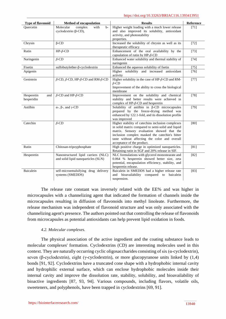
https://doi.org/10.33263/BRIAC116.1393413951
https://biointerfaceresearch.com/ 13940
Type of flavonoid Method of encapsulation Results Reference
Quercetin Molecular complex with b-
cyclodextrin (β-CD),
Higher weight loading with a much lower release
and also improved its solubility, antioxidant
activity, and photostability
properties.
[71]
Chrysin β-CD Increased the solubility of chrysin as well as its
therapeutic efficacy
[72]
Rutin HP-β-CD Enhancement of the oral availability by the
capsulation of rutin by HP-β-CD
[73]
Naringenin β-CD Enhanced water solubility and thermal stability of
naringenin
[74]
Fisetin sulfobutylether-β-cyclodextrin Enhanced the aqueous solubility of fsetin [75]
Apigenin Higher solubility and increased antioxidant
activity
[76]
Genistein β-CD, β-CD, HP-β-CD and RM-β-CD Higher solubility in the case of HP-β-CD and RM-
β-CD
Improvement of the ability to cross the biological
membrane
[77]
Hesperetin and
hesperidin
β-CD and HP-β-CD Improvement on the solubility and chemical
stability and better results were achieved in
complex of HP-β-CD and hesperetin
[78]
Astilbin α-, β-, and γ-CD Solubility of astilbin in β-CD microcapsules
prepared by the freeze-drying method was
enhanced by 122.1-fold, and its dissolution profile
was improved
[79]
Catechin β-CD Higher stability of catechins inclusion complexes
in solid matrix compared to semi-solid and liquid
matrix. Sensory evaluation showed that the
inclusion complex masked the catechin's bitter
taste without affecting the color and overall
acceptance of the product.
[80]
Rutin Chitosan-tripoyphosphate High positive charge in optimized nanoparticles.
Retaining rutin in SGF and 20% release in SIF.
[81]
Hesperetin Nanostructured lipid carriers (NLC)
and solid lipid nanoparticles (SLN)
NLC formulations with glycerol monostearate and
0.064 % hesperetin showed better size, zeta
potential, encapsulation efficiency, stability, and
hesperetin release.
[82]
Baicalein self-microemulsifying drug delivery
systems (SMEDDS)
Baicalein in SMEDDS had a higher release rate
and bioavailability compared to baicalein
suspension.
[83]
The release rate constant was inversely related with the EE% and was higher in
microcapsules with a channelizing agent that indicated the formation of channels inside the
microcapsules resulting in diffusion of flavonoids into methyl linoleate. Furthermore, the
release mechanism was independent of flavonoid structure and was only associated with the
channelizing agent's presence. The authors pointed out that controlling the release of flavonoids
from microcapsules as potential antioxidants can help prevent lipid oxidation in foods.
4.2. Molecular complexes.
The physical association of the active ingredient and the coating substance leads to
molecular complexes' formation. Cyclodextrins (CD) are interesting molecules used in this
context. They are naturally occurring cyclic oligosaccharides consisting of six (α-cyclodextrin),
seven (β-cyclodextrin), eight (γ-cyclodextrin), or more glucopyranose units linked by (1,4)
bonds [91, 92]. Cyclodextrins have a truncated cone shape with a hydrophobic internal cavity
and hydrophilic external surface, which can enclose hydrophobic molecules inside their
internal cavity and improve the dissolution rate, stability, solubility, and bioavailability of
bioactive ingredients [87, 93, 94]. Various compounds, including flavors, volatile oils,
sweeteners, and polyphenols, have been trapped in cyclodextrins [69, 91].

https://doi.org/10.33263/BRIAC116.1393413951
https://biointerfaceresearch.com/ 13941
The majority of studies concerning to encapsulation of flavonoids by cyclodextrins
have used β-cyclodextrin and its derivatives such as hydroxyproyl-β-cyclodextrin (HP-β-CD),
methyl-β-cyclodextrin (M-β-CD), dimethyl-β-cyclodextrin (DM-β-CD), trimethyl-β-
cyclodextrin (TM-β-CD), solphobutylether-β-cyclodextrin (SBE-β-CD), and glucosyl-β-
cyclodextrin (G2-β-CD) [94, 95]. The cyclodextrins can interact with flavonoids conducting
the B-ring toward the CD's secondary rim or heading the A-ring toward the CD's secondary
rim [96].
Yang et al. (2019) [97] investigated the complexation of three flavonoids (taxifolin,
quercetin, and morin hydrate) with propanediamine-β-cyclodextrin (DP-β-CD). It was stated
that the water solubility of taxifolin, quercetin, and morin hydrate was enhanced 70-102 times
after resulting in an inclusion complex with DP-β-CD. Also, the antioxidant activity of DP-β-
CD/ taxifolin complex was better than that of taxifolin. It was declared that hydroxyl groups in
taxifolin are close enough to secondary hydroxyl groups of DP-β-CD to form intramolecular
hydrogen bonds, resulting in an increase of antioxidant activity.
Morin/hydroxypropy-β-cyclodextrin (1:1 molar ratio) inclusion complex was prepared,
and it was demonstrated that dissolution rate, solubility, oral bioavailability, the
antihyperalgesic and anti-inflammatory activity of morin was increased [98]. A flavonoid-rich,
Allium cepa L. var agrogatum Don extract was encapsulated in β-CD (1:3 molar ratio), and the
results showed that aqueous solubility and the bioavailability of the extract was improved and,
according to in vitro skin permeation study, had the potential to be applied for transdermal
delivery [99]. In a similar study, methanolic extract of Hypericum perforatum (St John’s wort)
was encapsulated using β-CD (1:4 mass ratio). Encapsulation efficiency was reported as 27.5,
30, and 35% for catechin, epicatechin, and quercetin, respectively. Moreover, the differential
scanning calorimetry test showed the preventive activity of β-CD against thermal oxidation in
the encapsulated extract at temperatures as high as 300ºC [100].
4.3. Liposome entrapment.
Liposomes are non-toxic lipid vesicles composed of phospholipid bilayers organized in
water to form an aqueous core surrounded by a lipidic bilayer [87, 101]. This structure can
entrap water-soluble, lipid-soluble, and amphiphilic substances [19, 87]. Liposome properties
are influenced by lipid composition, surface charge, size, and method of preparation. Therefore,
the lipid components' choice defines the rigidity, fluidity, and charge of the lipid bilayer and,
consequently, the properties of the liposome. Although liposomes have been studied
extensively as suitable carriers for antioxidants, antimicrobials, therapeutic agents, and
bioactive compounds [101], high-cost production, low physicochemical stability, drug leakage,
and fast release of core substance in the gastrointestinal tract are the major disadvantages [19].
Some approaches have recently been explored to prevail these defects, such as protein coating,
chitosan coating, encapsulation using ultrasound, and coating of micronized sucrose and pro-
liposome hydration method [102-105].
In a study by Tao et al. (2014) [106], propolis flavonoids were encapsulated in a
liposome that resulted in an increase of immunological activity due to enhancement of the
phagocytic function of macrophages and the release of IL-1β, IL6, and interferon γ and in vivo
by activation the cellular and humoral immune response, including inducing higher level
concentrations of immunoglobulin (IgG), IL-4, and interferon γ.
According to the report of Mignet et al. (2012) [107], liposomal encapsulation of fisetin
by Phospholipon® 90G and dioctadecyldimethylammonium chloride-glycin- poly(ethylene

https://doi.org/10.33263/BRIAC116.1393413951
https://biointerfaceresearch.com/ 13942
glycol) 45, yielded liposomes with nanometer scale, high homogeneity, encapsulation
efficiency, and stability as well as maintaining cytotoxic and morphological activities on
endothelial cells. Similarly, fisetin's liposomal formulation was prepared using 1, 2- dioleoyl-
sn-glycero-3-phosphocholine and dioctadecyldimethylammonium chloride-
polyethyleneglycol-2000 with high homogeneity and high encapsulation efficiency, and it was
demonstrated that the bioavailability was 47-fold higher in liposomal form compared to free
fisetin. Furthermore, liposomal fisetin prevented the growth of Lewis lung carcinoma tumors
for a longer period than free fisetin at the same level [108]. This is in consistent with the results
obtained by Goniotaki et al. [109], who reported an increase in growth inhibitory activity of
flavonoids entrapped in liposomes.
Quercetin was encapsulated in nanoliposomes using rice bran phospholipids and the
thin film sonication method. The quercetin-loaded nanoparticles were spherical with a 157 nm
mean diameter and encapsulation efficiency of 84.92%. Nanoparticles were stable regarding
quercetin retention and antioxidant activity stored at 4 and 27ºC during six months. Release of
quercetin from nanoliposomes was limited in SGF (20% after 4 h), while in SIF, an initial
release of 60% after 2 h and sustained release of 70% until 24 h was an indication of an efficient
delivery and absorption of quercetin in the intestine [110]. Likewise, quercetin was
encapsulated in chitosan-coated nanoliposomes by the electrostatic deposition technique.
Encapsulation yielded spherical nanoparticles with 71.14% encapsulation efficiency and
enhanced DPPH antioxidant capacity, hydroxyl radical and superoxide anion radical
scavenging capacity, and ferric reducing capacity [103].
4.4. Polymer nanoparticles.
Polymer nanoparticles can be manufactured using a variety of ingredients such as
natural polymers (proteins and polysaccharides), synthetic polymers (polylactide (PLA), poly
lactide-co-glycolide (PLGA), poly glutamic acid, poly (vinyl alcohol)), and inorganic materials
[87, 111]. Encapsulation of quercetin via BSA (bovine serum albumin), zein, chitosan-alginate,
PLGA nanoparticles, PLA, Poly (caprolactone, PCL), and glycerol diglycidyl ether (GDE)
have been reviewed by [87].
Pool et al. [112] utilized PLGA nanoparticles and the solvent displacement method to
encapsulate catechin and quercetin. Polymeric nanoparticles with a mean diameter of 400 nm
and encapsulation efficiency of 79% were produced. In vitro release study revealed that
flavonoids release was pH-dependent, and higher liberation was observed at acidic pH as a
consequence of PLGA degradation. Quercetin showed a slower release compared to catechin,
which can be attributed to the carboxyl-carbonyl interactions of the polymer and quercetin.
Also, an increase in antiradical and chelating properties of flavonoids was reported by
incorporating them into nanoparticles. Pectin nanoparticles containing citrus peel extract were
prepared by the ionic gelation method. The obtained nanoparticles had an average size of 271.5
nm, and the release profile in simulated gastric fluid demonstrated 73% and 28.78% release
from the free extract and encapsulated extract, respectively, after 2 h. The releasing rate of
flavonoids reached 91.47% from nanoparticles after 24 h in the simulated intestinal fluid (SIF).
Furthermore, the encapsulated extract showed higher antioxidant activity than free extract
according to DPPH and ABTS assays [113]. In another study by [114], whey protein
concentrate was utilized to encapsulate mandarin peel extracts through ionic cross-linking. It
was declared that extracts were entrapped in nanoparticles via hydrophobic interactions or
hydrogen bonds. Encapsulation of extracts decreased the release rate of flavonoids in

https://doi.org/10.33263/BRIAC116.1393413951
https://biointerfaceresearch.com/ 13943
gastrointestinal fluids and improved antioxidant activity in SIF. Fisetin was nano encapsulated
using PCL and poly (D, L-lactic-co-glycolic acid)-block-poly (ethylene glycol)-carboxylic acid
(PLGA-PEG-COOH). Nanoparticles had a mean diameter of 140-200 nm and EE% of 70-82%.
Higher content of PCL in particle formulation yielded higher encapsulation efficiency due to
the hydrophobic nature of both fisetin and PCL. Results of in vitro release indicated ˂ 15% in
SGF during 2 h in all formulations, while in SIF, formulation with a higher content of PLGA-
PEG-COOH showed 70% release after 7 h and complete release after 24 h. In nanoparticles
prepared by the only PCL, 54% and 70% release after 7 h and 24 h was observed, respectively
[115].
4.5. Other types of delivery systems.
Fisetin was encapsulated by osmoporation using Saccharomyces cerevisiae cells, and
the effects of concentration, osmotic pressure, and temperature on the encapsulation and
internalized fisetin content were studied. The results illustrated that osmoporation significantly
increased EE% and entrapped fisetin [116].
Akhtar et al. (2014) [117] produced microcapsules containing rutin and Hibiscus
anthocyanins in multiple emulsions using a spinning disk reactor (SDR). Using this technology,
an emulsion premix was transmitted through a rotating disk at a regulated flow‐rate and disk
rotation speed which provided controllable and low shear conditions for the preparation of
secondary emulsions. It was stated that using 2 wt.% emulsifier polyoxyethylene (20) stearyl
ether produced W/O/W emulsion with fine droplets (13-15 nm) and EE of > 80%. The
utilization of SDR efficiently encapsulated rutin and anthocyanins within multiple emulsions
with a high retention and protection degree.
Onion-type multilamellar vesicles (MLVs) were applied in the entrapment of rutin and
naringenin. It was announced that the encapsulation efficiency of naringin was low (< 10%)
and was greatly adsorbed on MLV surface (> 60%), while rutin showed higher efficiency (>
60%). No rutin release was observed in the concentrated MLVs phase during 30 days, and 16%
release was detected in MLVs dispersion after 31 days [13].
Quercetin from dry onion peel crude extract was encapsulated in reassembled casein
particles, effects of pH, casein levels, and additives such as salts and Cetyl trimethylammonium
bromide (CTAB) on EE% were studied. The highest EE% (97%) was achieved by 0.5% (w/v)
sodium caseinate, 0.1 M of calcium chloride, 0.5 M of dipotassium hydrogen phosphate, 0.1
mM CTAB and 1 M of sodium citrate at a pH of 7 [118]. In another study by Horincar et al.
(2019) [119], flavonoids were extracted from yellow onion skins and microencapsulated in
whey protein isolate and combinations of chitosan, maltodextrin, and pectin. It was observed
that a combination of whey protein isolate, maltodextrin, and pectin resulted in higher EE%
(71%) compared to whey protein isolate and chitosan combination (59%). Also, it was
emphasized that flavonoids interacted with whey protein isolate through van der Waals and
hydrogen bonding. Ban et al. [120] used nanoparticles made up of physiological lipids to
protect flavonoids (naringenin, quercetin and hesperidin) from the digestive system's harsh
conditions until their absorption into enterocytes, thereby improving their bioaccessibility.

https://doi.org/10.33263/BRIAC116.1393413951
https://biointerfaceresearch.com/ 13944
5. Considerable Aspects of Flavonoid Encapsulation
5.1. Biological fate.
One of the key characteristics of an encapsulation system is not having an adverse effect
on flavonoids' bioavailability. Various factors, including size, morphology, composition, and
interfacial properties, can influence flavonoids' bioavailability [121]. These characteristics are
changed during transit of particles from mouth to colon due to the presence of mucin, salts,
digestive enzymes, buffers, and acids that affect flavonoids release rate and region [122].
Flavonoids are absorbed in the small intestine in the form of aglycons or are metabolized by
intestinal microbiota and absorbed in the colon [33]; hence designing a delivery system that
allows the release of flavonoid glycosides in the large intestine is indispensable. Starch-based
wall materials used for entrapment of flavonoids are degraded in the mouth by the activity of
α-amylase. Protein-based delivery systems are hydrolyzed in the stomach exposed to acidic pH
and the enzyme pepsin. Lipid-based particles usually release the core substances in the small
intestine [123]. Therefore, the physicochemical features of the substances used to develop
flavonoid encapsulates and their digestibility in the gastrointestinal tract and the bioactive
ingredient's action site are considerable issues that should be notified.
5.2. Incorporation into food matrices.
Although numerous studies have been conducted regarding the encapsulation of
flavonoids, few of them reported the stability and effect of their addiction to food matrices. By
incorporating microcapsules, appearance, rheology, texture, and sensory properties of food
may be altered. Some flavonoids are colorless, and some are yellow, and it is important to
consider the point that if the emergence of color in the product is required or not. The
incorporation of particles with different sizes, refractive index, and concentration causes
changes in foods and beverages' appearance. In transparent food products, delivery systems
generating small particles (d˂50 nm) should be applied while particles with diameters ˃ 50 can
be used in products whose clarity is not required [122]. In addition, the microcapsules produced
using gelling materials such as gellan, xanthan, or alginate are usually greater than 100 μm and
do not provide acceptable mouthfeel and texture in the food formulation [124]. Particles
obtained by spray drying are below 40 μm and generate a desirable mouthfeel in food products
[125]. Furthermore, food products possess various pH and ionic strength and may undergo
harsh processing conditions (freezing, heating, mixing, shearing, and dehydration), leading to
the disintegration of microcapsules that should be considered in the design and fabrication of
delivery systems [122].
5.3. Safety.
The safety of materials utilized in the encapsulation process is of paramount
importance. In this respect, a very limited number of coatings and excipient materials have
been approved for food use. In some encapsulation methods, the residue of non-food grade
solvents and detergents may pose health problems [126] that should be considered in selecting
the technique for flavonoid encapsulation. Another challenge is the effect of nanoparticles on
the body, dependent on factors including size, composition, surface properties, and their ability
to cross the biological membranes [127, 128]. It has been speculated that reduced-size particles
have a detrimental impact on the biological system. Nanoparticles might increase the
https://doi.org/10.33263/BRIAC116.1393413951
https://biointerfaceresearch.com/ 13945
bioavailability of bioactive substances and induce health risks [121]. There are still unanswered
questions regarding the interaction of nanoparticles and the biological system and their
potential toxicity. It is not possible to elucidate the safety of nano-scale encapsulation.
6. Conclusion and Future Perspective
Flavonoids are important constituents of many fruits and vegetables, which provide
various health benefits that can be extracted from these sources and be utilized to develop
functional foods. However, they are sensitive to light, heat, and oxygen. Their poor
bioavailability after oral administration and degradation under acidic pH of stomach limit their
successful application. In this respect, different encapsulation systems were introduced in this
review that can resolve the mentioned shortcomings. Encapsulation can improve the stability
of flavonoids as well as solubility, controlled release, and bioavailability. However, each
encapsulation method has its advantages and disadvantages that should be considered in
selecting the specific system for the special application field. Based on the collected data, the
future development of delivery systems for flavonoids should be centralized on applying food-
grade ingredients as coating materials, other encapsulation methods with higher efficiency, and
investigation of the application of microcapsules and their effects in food products.
Funding
This research received no external funding.
Acknowledgments
The support for this study provided by Shahid Beheshti University of Medical Sciences is
gratefully acknowledged.
Conflicts of Interest
The authors declare no conflict of interest.
References
1. Croft, K.D. Dietary polyphenols: Antioxidants or not? Arch Biochem Biophys 2016, 595, 120-4,
https://doi.org/10.1016/j.abb.2015.11.014.
2. Amiot, M.; Riva, C.; Vinet, A. Effects of dietary polyphenols on metabolic syndrome features in humans: a
systematic review. Obes Rev 2016, 17, 573-86, https://doi.org/10.1111/obr.12409.
3. Sansone, F.; Picerno, P.; Mencherini, T.; Villecco, F.; D’ursi, A.; Aquino, R.; Lauro, M. Flavonoid
microparticles by spray-drying: Influence of enhancers of the dissolution rate on properties and stability.
J Food Eng 2011, 103, 188-96, https://doi.org/10.1016/j.jfoodeng.2010.10.015.
4. Wang, T.-Y.; Li, Q.; Bi, K.-S. Bioactive flavonoids in medicinal plants: Structure, activity and biological
fate. Asian J Pharm Sci 2018, 13, 12-23, https://doi.org/10.1016/j.ajps.2017.08.004.
5. Chlopicka, J.; Pasko, P.; Gorinstein, S.; Jedryas, A.; Zagrodzki, P. Total phenolic and total flavonoid content,
antioxidant activity and sensory evaluation of pseudocereal breads. LWT- Food Sci Thecnol 2012, 46, 548-
55, https://doi.org/10.1016/j.lwt.2011.11.009.
6. Ghasemzadeh, A.; Ghasemzadeh, N. Flavonoids and phenolic acids: Role and biochemical activity in plants
and human. J Med Plant Res 2011, 5, 6697-703.
7. Madunić, J.; Madunić, I.V.; Gajski, G.; Popić, J.; Garaj-Vrhovac, V. Apigenin: A dietary flavonoid with
diverse anticancer properties. Cancer Lett 2018, 413, 11-22, https://doi.org/10.1016/j.canlet.2017.10.041.
8. Lesjak, M.; Beara, I.; Simin, N.; Pintać, D.; Majkić, T.; Bekvalac, K.; Orčić, D.; Mimica-Dukić, N.
Antioxidant and anti-inflammatory activities of quercetin and its derivatives. J Funct Foods 2018, 40, 68-
75, https://doi.org/10.1016/j.jff.2017.10.047.
9. Spagnuolo, C.; Moccia, S.; Russo, G.L. Anti-inflammatory effects of flavonoids in neurodegenerative
disorders. Eur J Med Chem 2018, 153, 105-15, https://doi.org/10.1016/j.ejmech.2017.09.001.

https://doi.org/10.33263/BRIAC116.1393413951
https://biointerfaceresearch.com/ 13946
10. Orhan, D.D.; Özçelik, B.; Özgen, S.; Ergun, F. Antibacterial, antifungal, and antiviral activities of some
flavonoids. Microbiol Res 2010, 165, 496-504, https://doi.org/10.1016/j.micres.2009.09.002.
11. Seleem, D.; Pardi, V.; Murata, R.M. Review of flavonoids: A diverse group of natural compounds with anti-
Candida albicans activity in vitro. Arch Oral Biol 2017, 76, 76-83,
https://doi.org/10.1016/j.archoralbio.2016.08.030.
12. Hu, Y.; Li, Y.; Zhang, W.; Kou, G.; Zhou, Z. Physical stability and antioxidant activity of citrus flavonoids
in arabic gum-stabilized microcapsules: Modulation of whey protein concentrate. Food Hydrocolloids 2018,
77, 588-97, https://doi.org/10.1016/j.foodhyd.2017.10.037.
13. Kerdudo, A.; Dingas, A.; Fernandez, X.; Faure, C. Encapsulation of rutin and naringenin in multilamellar
vesicles for optimum antioxidant activity. Food Chem 2014, 159, 12-9,
https://doi.org/10.1016/j.foodchem.2014.03.005.
14. McClements, D.J. Design of nano‐laminated coatings to control bioavailability of lipophilic food
components. J Food Sci 2010, 75, R30-R42, https://doi.org/10.1111/j.1750-3841.2009.01452.x.
15. Ozkan, G.; Franco, P.; De Marco, I.; Xiao, J.; Capanoglu, E. A review of microencapsulation methods for
food antioxidants: Principles, advantages, drawbacks and applications. Food Chem 2019, 272, 494-506,
https://doi.org/10.1016/j.foodchem.2018.07.205.
16. Ye, Q.; Georges, N.; Selomulya, C. Microencapsulation of active ingredients in functional foods: From
research stage to commercial food products. Trends Food Sci Technol 2018, 78, 167-179,
https://doi.org/10.1016/j.tifs.2018.05.025.
17. Yousefi, M.; Khorshidian, N.; Mortazavian, A.M.; Khosravi-Darani, K. Preparation optimization and
characterization of chitosan-tripolyphosphate microcapsules for the encapsulation of herbal galactagogue
extract. Int J Biol Macromol 2019, 140, 920-8, https://doi.org/10.1016/j.ijbiomac.2019.08.122.
18. Khorshidian, N.; Mahboubi, A.; Kalantari, N.; Hosseini, H.; Yousefi, M.; Arab, M.; da Cruz, A.G.;
Mortazavian, A.M.; Mahdavi, F.S. Chitosan-coated alginate microcapsules loaded with herbal galactagogue
extract: Formulation optimization and characterization. Iranian Journal of Pharmaceutical Research 2019,
18, 1180-95, https://doi.org/10.22037/ijpr.2019.1100776.
19. Shishir, M.R.I.; Xie, L.; Sun, C.; Zheng, X.; Chen, W. Advances in micro and nano-encapsulation of
bioactive compounds using biopolymer and lipid-based transporters. Trends Food Sci Technol 2018, 78, 34-
60, https://doi.org/10.1016/j.tifs.2018.05.018.
20. Arab, M.; Hosseini, S.M.; Nayebzadeh, K.; Khorshidian, N.; Yousefi, M.; Razavi, S.H.; Mortazavian, A.M.
Microencapsulation of microbial canthaxanthin with alginate and high methoxyl pectin and evaluation the
release properties in neutral and acidic condition. Int J Biol Macromol 2019, 121, 691-8,
https://doi.org/10.1016/j.ijbiomac.2018.10.114.
21. Georgiev, V.; Ananga, A.; Tsolova, V. Recent advances and uses of grape flavonoids as nutraceuticals.
Nutrients 2014, 6, 391-415, https://doi.org/10.3390/nu6010391.
22. Yao, L.H.; Jiang, Y.; Shi, J.; Tomas-Barberan, F.; Datta, N.; Singanusong, R.; Chen, S. Flavonoids in food
and their health benefits. Plant Foods Hum Nutr 2004, 59, 113-22, https://doi.org/10.1007/s11130-004-
0049-7.
23. Bhagwat, S.; Haytowitz, D.B.; Holden, J.M. USDA database for the flavonoid content of selected foods,
Release 3.1. US Department of Agriculture: Beltsville, MD, USA 2014.
24. Den Hartogh, D.J.; Tsiani, E. Antidiabetic properties of naringenin: a citrus fruit polyphenol. Biomolecules
2019, 9, https://doi.org/10.3390/biom9030099.
25. Hostetler, G.L.; Ralston, R.A.; Schwartz, S.J. Flavones: Food sources, bioavailability, metabolism, and
bioactivity. Adv Nutr 2017, 8, 423-35, https://doi.org/10.3945/an.116.012948.
26. Alezandro, M.R.; Lui, M.C.Y.; Lajolo, F.M.; Genovese, M.I. Commercial spices and industrial ingredients:
evaluation of antioxidant capacity and flavonoids content for functional foods development. Food Sci
Technol 2011, 31, 527-33, http://dx.doi.org/10.1590/S0101-20612011000200038.
27. Habtemariam, S. Antioxidant and Rutin Content Analysis of Leaves of the Common Buckwheat (Fagopyrum
esculentum Moench) Grown in the United Kingdom: A Case Study. Antioxidants 2019, 8, 160,
http://doi.org/10.3390/antiox8060160.
28. Othman, A.; Jalil, A.M.M.; Weng, K.K.; Ismail, A.; Ghani, N.A.; Adenan, I. Epicatechin content and
antioxidant capacity of cocoa beans from four different countries. Afr J Biotechnol 2010, 9, 1052-9,
https://doi.org/10.5897/AJB09.1219.
29. González-Paramás, A.M.; Ayuda-Durán, B.; Martínez, S.; González-Manzano, S.; Santos-Buelga, C. The
mechanisms behind the biological activity of flavonoids. Curr Med Chem 2019, 26,
http://doi.org/10.2174/0929867325666180706104829.
30. Panche, A.; Diwan, A.; Chandra, S. Flavonoids: an overview. J Nutr Sci 2016, 5,
http://doi.org/10.1017/jns.2016.41.
31. Babu, P.V.A.; Liu, D.; Gilbert, E.R. Recent advances in understanding the antidiabetic actions of dietary
flavonoids. J Nutr Biochem 2013, 24, 1777-89, http://doi.org/10.1016/j.jnutbio.2013.06.003.
32. Vinayagam, R.; Xu, B. Antidiabetic properties of dietary flavonoids: a cellular mechanism review. Nutr
Metab 2015, 12, https://doi.org/10.1186/s12986-015-0057-7.

https://doi.org/10.33263/BRIAC116.1393413951
https://biointerfaceresearch.com/ 13947
33. Kumar, S.; Pandey, A.K. Chemistry and biological activities of flavonoids: an overview. The Scientific
World Journal 2013, 2013, https://doi.org/10.1155/2013/162750.
34. Xie, Y.; Yang, W.; Tang, F.; Chen, X.; Ren, L. Antibacterial activities of flavonoids: structure-activity
relationship and mechanism. Curr Med Chem 2015, 22, 132-49,
http://doi.org/10.2174/0929867321666140916113443.
35. Vladu, A.; Marin, S.T,.; Neacsu, I.; Truşcă, R.; Kaya, M.; Kaya, D.; Popa, A.M.; Poiană, C.Ă.; Cristescu,
I.O.; Orlov, C. Spongious fillers based on collagen-hydroxyapatite-eugenol acetate with therapeutic potential
in bone cancer. Farmacia 2020, 68, 313-21. https://doi.org/10.31925/farmacia.2020.2.17. 36. Ahmad, A.; Kaleem, M.; Ahmed, Z.; Shafiq, H. Therapeutic potential of flavonoids and their mechanism of
action against microbial and viral infections—A review. Food Res Int 2015, 77, 221-35,
https://doi.org/10.1016/j.foodres.2015.06.021.
37. Faggio, C.; Sureda, A.; Morabito, S.; Sanches-Silva, A.; Mocan, A.; Nabavi, S.F.; Nabavi, S.M. Flavonoids
and platelet aggregation: a brief review. Eur J Pharmacol 2017, 807, 91-101,
https://doi.org/10.1016/j.ejphar.2017.04.009.
38. Tanaka, T.; Takahashi, R. Flavonoids and asthma. Nutrients 2013, 5, 2128-43,
https://doi.org/10.3390/nu5062128.
39. Terahara, N. Flavonoids in foods: a review. Natural product communications. Natural Product
Communications 2015, 10, https://doi.org/10.1177/1934578X1501000334.
40. Huang, Z.Q.; Chen, P.; Su, W.W.; Wang, Y.G.; Wu, H.; Peng, W.; Li, P.-B. Antioxidant activity and
hepatoprotective potential of quercetin 7-rhamnoside in vitro and in vivo. Molecules 2018, 23,
https://doi.org/10.3390/molecules23051188.
41. Han, Q.-T.; Ren, Y.; Li, G.-S.; Xiang, K.-L.; Dai, S.-J. Flavonoid alkaloids from Scutellaria moniliorrhiza
with anti-inflammatory activities and inhibitory activities against aldose reductase. Phytochemistry 2018,
152, 91-6, https://doi.org/10.1016/j.phytochem.2018.05.001.
42. Wang, X.; Huang, H.; Ma, X.; Wang, L.; Liu, C.; Hou, B.; Yang, S.; Zhang, L.; Du, G. Anti-inflammatory
effects and mechanism of the total flavonoids from Artemisia scoparia Waldst. et kit. in vitro and in vivo.
Biomed Pharmacother 2018, 104, 390-403, https://doi.org/10.1016/j.biopha.2018.05.054.
43. Obafemi, T.; Akinmoladun, A.; Olaleye, M.; Agboade, S.O.; Onasanya, A.A. Antidiabetic potential of
methanolic and flavonoid-rich leaf extracts of Synsepalum dulcificum in type 2 diabetic rats. J Ayurveda
Integr Med 2017, 8, 238-46, https://doi.org/10.1016/j.jaim.2017.01.008.
44. Djamil, R.; Winarti, W.; Zaidan, S.; Abdillah, S. Antidiabetic Activity of Flavonoid from Binahong Leaves
(Anredera cordifolia) Extract in Alloxan Induced Mice. J Pharmacogn Nat Prod 2017, 3, 1-4,
https://doi.org/10.4172/2472-0992.1000139.
45. Escobar, S.J.d.M.; Fong, G.M.; Winnischofer, S.M.; Simone, M.; Munoz, L.; Dennis, J.M.; Rocha, M.E.M.;
Witting, P.K. Anti-proliferative and cytotoxic activities of the flavonoid isoliquiritigenin in the human
neuroblastoma cell line SH-SY5Y. Chem Biol Interact 2019, 299, 77-87,
https://doi.org/10.1016/j.cbi.2018.11.022.
46. Wei, S.; Sun, T.; Du, J.; Zhang, B.; Xiang, D.; Li, W. Xanthohumol, a prenylated flavonoid from Hops,
exerts anticancer effects against gastric cancer in vitro. Oncol Rep 2018, 40, 3213-22,
https://doi.org/10.3892/or.2018.6723.
47. Geethalakshmi, R.; Sarada, V.D. In vitro and in silico antimicrobial activity of sterol and flavonoid isolated
from Trianthema decandra L. Microb Pathog 2018, 121, 77-86,
https://doi.org/10.1016/j.micpath.2018.05.018.
48. Min, N.; Leong, P.T.; Lee, R.C.H.; Khuan, J.S.E.; Chu, J.J.H. A flavonoid compound library screen revealed
potent antiviral activity of plant-derived flavonoids on human enterovirus A71 replication. Antiviral Res
2018, 150, 60-8, https://doi.org/10.1016/j.antiviral.2017.12.003.
49. Pradeepkumar, B.; Sudheer, A.; Reddy, T.S.; Reddy, K.S.; Narayana, G.; Veerabhadrappa, K.
Cardioprotective Activity of Flavonoid Fraction of Gymnema Sylvestre Leaves on Doxorubicin Induced
Cardiac Damage. J Young Pharm 2018, 10, 422, https://doi.org/10.5530/jyp.2018.10.93.
50. Farhadi, F.; Khameneh, B.; Iranshahi, M.; Iranshahy, M. Antibacterial activity of flavonoids and their
structure–activity relationship: An update review. Phytotherapy Res 2019, 33, 13-40,
https://doi.org/10.1002/ptr.6208.
51. Nabavi, S.F.; Russo, G.L.; Daglia, M.; Nabavi, S.M. Role of quercetin as an alternative for obesity treatment:
you are what you eat! Food Chem 2015, 179, 305-10, https://doi.org/10.1016/j.foodchem.2015.02.006.
52. Procházková, D.; Boušová, I.; Wilhelmová, N. Antioxidant and prooxidant properties of flavonoids.
Fitoterapia 2011, 82, 513-23, https://doi.org/10.1016/j.fitote.2011.01.018.
53. Chebil, L.; Humeau, C.; Anthoni, J.; Dehez, F.; Engasser, J.M.; Ghoul, M. Solubility of flavonoids in organic
solvents. J Chem Eng Data 2007, 52,1552-1556. https://doi.org/10.1021/je7001094. 54. Ioannou, I.; Hafsa, I.; Hamdi, S.; Charbonnel, C.; Ghoul, M. Review of the effects of food processing and
formulation on flavonol and anthocyanin behaviour. J Food Eng 2012, 111, 208-17,
https://doi.org/10.1016/j.jfoodeng.2012.02.006.
55. Ahmed, M.; Eun, J.-B. Flavonoids in fruits and vegetables after thermal and nonthermal processing: a
review. Crit Rev Food Sci Nutr 2018, 58, 3159-88, https://doi.org/10.1080/10408398.2017.1353480.
https://doi.org/10.33263/BRIAC116.1393413951
https://biointerfaceresearch.com/ 13948
56. Ananingsih, V.K.; Sharma, A.; Zhou, W. Green tea catechins during food processing and storage: a review
on stability and detection. Food Res Int 2013, 50, 469-79, https://doi.org/10.1016/j.foodres.2011.03.004.
57. Patras, A.; Brunton, N.P.; O'Donnell, C.; Tiwari, B. Effect of thermal processing on anthocyanin stability in
foods; mechanisms and kinetics of degradation. Trends Food Sci Technol 2010, 21, 3-11,
https://doi.org/10.1016/j.tifs.2009.07.004.
58. Abert-Vian, M.; Elmaataoui, M.; Chemat, F. A novel idea in food extraction field: study of vacuum
microwave hydrodiffusion technique for by-products extraction. J Food Eng 2011, 105, 351-60,
https://doi.org/10.1016/j.jfoodeng.2011.02.045.
59. Srinivas, K.; King, J.; Monrad, J.; Howard, L.; Zhang, D. Pressurized solvent extraction of flavonoids from
grape pomace utilizing organic acid additives. Ital J Food Sci 2011, 23, 90,
60. Al-juhaimi, F.; Ghafoor, K.; Özcan, M.M.; Jahurul, M.; Babiker, E.E.; Jinap, S.; Sahena, F.; Sharifudin, M.;
Zaidul, I. Effect of various food processing and handling methods on preservation of natural antioxidants in
fruits and vegetables. J Food Sci Technol 2018, 55, 3872-80, https://doi.org/10.1007/s13197-018-3370-0.
61. Pap, N.; Pongrácz, E.; Jaakkola, M.; Tolonen, T.; Virtanen, V.; Turkki, A.; Horváth-Hovorka, Z.; Vatai, G.;
Keiski, R.L. The effect of pre-treatment on the anthocyanin and flavonol content of black currant juice (Ribes
nigrum L.) in concentration by reverse osmosis. J Food Eng 2010, 98, 429-36,
https://doi.org/10.1016/j.jfoodeng.2010.01.024.
62. Islek, M.; Nilufer‐Erdil, D.; Knuthsen, P. Changes in Flavonoids of Sliced and Fried Yellow Onions (A llium
cepa L. var. zittauer) During Storage at Different Atmospheric, Temperature and Light Conditions. J Food
Process Pres 2015, 39, 357-68, https://doi.org/10.1111/jfpp.12240.
63. Miao, J.; Wei, K.; Li, X.; Zhao, C.; Chen, X.; Mao, X.; Huang, H.; Gao, W. Effect of boiling and drying
process on chemical composition and antioxidant activity of Chaenomeles speciosa. J Food Sci Technol
2017, 54, 2758-68, https://doi.org/10.1007/s13197-017-2712-7.
64. Yang, N.; Qiu, R.; Yang, S.; Zhou, K.; Wang, C.; Ou, S.; Zheng, J. Influences of stir-frying and baking on
flavonoid profile, antioxidant property, and hydroxymethylfurfural formation during preparation of
blueberry-filled pastries. Food Chem 2019, 287, 167-75, https://doi.org/10.1016/j.foodchem.2019.02.053.
65. Lee, S.U.; Lee, J.H.; Choi, S.H.; Lee, J.S.; Ohnisi-Kameyama, M.; Kozukue, N.; Levin, C.E.; Friedman, M.
Flavonoid content in fresh, home-processed, and light-exposed onions and in dehydrated commercial onion
products. Journal of Agricultural and Food Chem 2008, 56, 8541-8, https://doi.org/10.1021/jf801009p.
66. Pérez-Gregorio, M.; García-Falcón, M.; Simal-Gándara, J. Flavonoids changes in fresh-cut onions during
storage in different packaging systems. Food Chem 2011, 124, 652-8,
https://doi.org/10.1016/j.foodchem.2010.06.090.
67. Wang, C.Y.; Chen, C.-T.; Wang, S.Y. Changes of flavonoid content and antioxidant capacity in blueberries
after illumination with UV-C. Food Chem 2009, 117, 426-31,
https://doi.org/10.1016/j.foodchem.2009.04.037.
68. Fang, Z.; Bhandari, B. Encapsulation of polyphenols–a review. Trends Food Sci Technol 2010, 21, 510-23,
https://doi.org/10.1016/j.tifs.2010.08.003.
69. Munin, A.; Edwards-Lévy, F. Encapsulation of natural polyphenolic compounds; a review. Pharmaceutics
2011, 3, 793-829, https://doi.org/10.3390/pharmaceutics3040793.
70. Jullian, C.; Moyano, L.; Yanez, C.; Olea-Azar, C. Complexation of quercetin with three kinds of
cyclodextrins: an antioxidant study. Spectrochim Acta A Mol Biomol Spectrosc 2007, 67, 230-4,
https://doi.org/10.1016/j.saa.2006.07.006.
71. Aytac, Z.; Kusku, S.I.; Durgun, E.; Uyar, T. Quercetin/β-cyclodextrin inclusion complex embedded
nanofibres: Slow release and high solubility. Food Chem 2016, 197, 864-71,
https://doi.org/10.1016/j.foodchem.2015.11.051.
72. Zhu, Z.-Y.; Luo, Y.; Liu, Y.; Wang, X.-T.; Liu, F.; Guo, M.-Z.; Wang, Z.; Liu, A.-J.; Zhang, Y.-M. Inclusion
of chrysin in β-cyclodextrin and its biological activities. J Drug Deliv Sci Technol 2016, 31, 176-86,
https://doi.org/10.1016/j.jddst.2016.01.002.
73. Miyake, K.; Arima, H.; Hirayama, F.; Yamamoto, M.; Horikawa, T.; Sumiyoshi, H.; Noda, S.; Uekama, K.
Improvement of solubility and oral bioavailability of rutin by complexation with 2-hydroxypropyl-β-
cyclodextrin. Pharm Dev Technol 2000, 5, 399-407, https://doi.org/10.1081/PDT-100100556.
74. Yang, L. J.; Ma, S. X.; Zhou, S.Y.; Chen, W.; Yuan, M.W.; Yin, Y.Q.; Yang, X.-D. Preparation and
characterization of inclusion complexes of naringenin with β-cyclodextrin or its derivative. Carbohydr
Polym 2013, 98, 861-9, https://doi.org/10.1016/j.carbpol.2013.07.010.
75. Mohtar, N.; Taylor, K.M.; Sheikh, K.; Somavarapu, S. Design and development of dry powder
sulfobutylether-β-cyclodextrin complex for pulmonary delivery of fisetin. Eur J Pharm Biopharm 2017, 113,
1-10, https://doi.org/10.1016/j.ejpb.2016.11.036.
76. Pápay, Z.E.; Sebestyén, Z.; Ludányi, K.; Kállai, N.; Balogh, E.; Kósa, A.; Somavarapu, S.; Böddi, B.; Antal,
I. Comparative evaluation of the effect of cyclodextrins and pH on aqueous solubility of apigenin. J Pharm
Biomed Anal 2016, 117, 210-6, https://doi.org/10.1016/j.jpba.2015.08.019.
77. Daruházi, Á.E.; Kiss, T.; Vecsernyés, M.; Szente, L.; Szőke, É.; Lemberkovics, É. Investigation of transport
of genistein, daidzein and their inclusion complexes prepared with different cyclodextrins on Caco-2 cell
line. J Pharm Biomed Anal 2013, 84, 112-6, https://doi.org/10.1016/j.jpba.2013.05.012.

https://doi.org/10.33263/BRIAC116.1393413951
https://biointerfaceresearch.com/ 13949
78. Tommasini, S.; Calabro, M.; Stancanelli, R.; Donato, P.; Costa, C.; Catania, S.; Villari, V.; Ficarra, P.;
Ficarra, R. The inclusion complexes of hesperetin and its 7-rhamnoglucoside with (2-hydroxypropyl)-β-
cyclodextrin. J Pharm Biomed Anal 2005, 39, 572-80, https://doi.org/10.1016/j.jpba.2005.05.009.
79. Zhang, Q.F.; Nie, H.C.; Shangguang, X.C.; Yin, Z.P.; Zheng, G.D.; Chen, J.G. Aqueous solubility and
stability enhancement of astilbin through complexation with cyclodextrins. J Agric Food Chem 2012, 61,
151-6, https://doi.org/10.1021/jf304398v.
80. Ho, S.; Thoo, Y.Y.; Young, D.J.; Siow, L.F. Stability and recovery of cyclodextrin encapsulated catechin in
various food matrices. Food Chem 2019, 275, 594-9, https://doi.org/10.1016/j.foodchem.2018.09.117.
81. Konecsni, K.; Low, N.; Nickerson, M. Chitosan–tripolyphosphate submicron particles as the carrier of
entrapped rutin. Food Chem 2012, 134, 1775-9, https://doi.org/10.1016/j.foodchem.2012.03.070.
82. Fathi, M.; Varshosaz, J.; Mohebbi, M.; Shahidi, F. Hesperetin-loaded solid lipid nanoparticles and
nanostructure lipid carriers for food fortification: preparation, characterization, and modeling. Food Bioproc
Tech 2013, 6, 1464-75, https://doi.org/10.1007/s11947-012-0845-2.
83. Liu, W.; Tian, R.; Hu, W.; Jia, Y.; Jiang, H.; Zhang, J.; Zhang, L. Preparation and evaluation of self-
microemulsifying drug delivery system of baicalein. Fitoterapia 2012, 83, 1532-9,
https://doi.org/10.1016/j.fitote.2012.08.021.
84. Bora, A.F.M.; Ma, S.; Li, X.; Liu, L. Application of microencapsulation for the safe delivery of green tea
polyphenols in food systems: Review and recent advances. Food Res Int 2018, 105, 241-9,
https://doi.org/10.1016/j.foodres.2017.11.047.
85. Shishir, M.R.I.; Chen, W. Trends of spray drying: A critical review on drying of fruit and vegetable juices.
Trends Food Sci Technol 2017, 65, 49-67, https://doi.org/10.1016/j.tifs.2017.05.006.
86. Gadkari, P.V.; Balaraman, M. Catechins: Sources, extraction and encapsulation: A review. Food Bioprod
Process 2015, 93, 122-38, https://doi.org/10.1016/j.fbp.2013.12.004.
87. Wang, W.; Sun, C.; Mao, L.; Ma, P.; Liu, F.; Yang, J.; Gao, Y. The biological activities, chemical stability,
metabolism and delivery systems of quercetin: A review. Trends Food Sci Technol 2016, 56, 21-38,
https://doi.org/10.1016/j.tifs.2016.07.004.
88. Yousuf, B.; Gul, K.; Wani, A.A.; Singh, P. Health benefits of anthocyanins and their encapsulation for
potential use in food systems: a review. Crit Rev Food Sci Nutr 2016, 56, 2223-30,
https://doi.org/10.1080/10408398.2013.805316.
89. Wu, P.; Deng, Q.; Ma, G.; Li, N.; Yin, Y.; Zhu, B.; Chen, M.; Huang, R. Spray drying of Rhodomyrtus
tomentosa (Ait.) Hassk. flavonoids extract: optimization and physicochemical, morphological, and
antioxidant properties. Int J Food Sci 2014, 2014, https://doi.org/10.1155/2014/420908.
90. Palma, M.; García, P.; Márquez-Ruiz, G.; Vergara, C.; Robert, P. Release kinetics of flavonoids in methyl
linoleate from microparticles designed with inulin and channelizing agent. Food Res Int 2014, 64, 99-105,
https://doi.org/10.1016/j.foodres.2014.05.064.
91. Del Valle, E.M. Cyclodextrins and their uses: a review. Process Biochem 2004, 39, 1033-46,
https://doi.org/10.1016/S0032-9592(03)00258-9.
92. Marques, H.M.C. A review on cyclodextrin encapsulation of essential oils and volatiles. Flavour Fragr J
2010, 25, 313-26, https://doi.org/10.1002/ffj.2019.
93. Arora, D.; Saneja, A.; Jaglan, S. Cyclodextrin-based delivery systems for dietary pharmaceuticals. Environ
Chem Lett 2019, 17, 1263-1270, https://doi.org/10.1007/s10311-019-00878-w.
94. Bilia, A.R.; Isacchi, B.; Righeschi, C.; Guccione, C.; Bergonzi, M.C. Flavonoids loaded in nanocarriers: an
opportunity to increase oral bioavailability and bioefficacy. Food and Nutrition Sciences 2014, 5,
https://doi.org/10.4236/fns.2014.513132.
95. Pinho, E.; Grootveld, M.; Soares, G.; Henriques, M. Cyclodextrins as encapsulation agents for plant
bioactive compounds. Carbohydr Polym 2014, 101, 121-35, https://doi.org/10.1016/j.carbpol.2013.08.078.
96. Kim, H.; Choi, J.; Jung, S. Inclusion complexes of modified cyclodextrins with some flavonols. J Incl
Phenom Macrocycl Chem 2009, 64, https://doi.org/10.1007/s10847-009-9534-9.
97. Yang, S.-L.; Zhao, L.-J.; Chi, S.-M.; Du, J.-J.; Ruan, Q.; Xiao, P.-L.; Zhao, Y. Inclusion complexes of
flavonoids with propylenediamine modified β-cyclodextrin: Preparation, characterization and antioxidant.
J Mol Struct 2019, 1183, 118-25, https://doi.org/10.1016/j.molstruc.2019.01.046.
98. dos Santos Lima, B.; de Alcântara Campos, C.; da Silva Santos, A.C.R.; Santos, V.C.N.; Trindade, G.d.G.G.;
Shanmugam, S.; Pereira, E.W.M.; Marreto, R.N.; Duarte, M.C.; da Silva Almeida, J.R.G. Development of
morin/hydroxypropyl-β-cyclodextrin inclusion complex: Enhancement of bioavailability, antihyperalgesic
and anti-inflammatory effects. Food Chemical Toxicol 2019, 126, 15-24,
https://doi.org/10.1016/j.fct.2019.01.038.
99. Ding, Z.; Wu, M.; Guo, Q.; Yang, X.; Zhang, B. Encapsulation of a Flavonoid-rich Allium cepa L. var.
agrogatum Don Extract in β-Cyclodextrin for Transdermal Drug Delivery. J Agric Food Chem 2013, 61,
4914-20, https://doi.org/10.1021/jf302325d.
100. Kalogeropoulos, N.; Yannakopoulou, K.; Gioxari, A.; Chiou, A.; Makris, D.P. Polyphenol characterization
and encapsulation in β-cyclodextrin of a flavonoid-rich Hypericum perforatum (St John's wort) extract. LWT-
Food Sci Technol 2010, 43, 882-9, https://doi.org/10.1016/j.lwt.2010.01.016.

https://doi.org/10.33263/BRIAC116.1393413951
https://biointerfaceresearch.com/ 13950
101. Simão, A.M.S.; Bolean, M.; Cury, T.A.C.; Stabeli, R.G.; Itri, R.; Ciancaglini, P. Liposomal systems as
carriers for bioactive compounds. Biophys Rev 2015, 7, 391-7, https://doi.org/10.1007/s12551-015-0180-8.
102. Gómez-Mascaraque, L.G.; Sipoli, C.C.; de La Torre, L.G.; López-Rubio, A. Microencapsulation structures
based on protein-coated liposomes obtained through electrospraying for the stabilization and improved
bioaccessibility of curcumin. Food Chem 2017, 233, 343-50,
https://doi.org/10.1016/j.foodchem.2017.04.133.
103. Hao, J.; Guo, B.; Yu, S.; Zhang, W.; Zhang, D.; Wang, J.; Wang, Y. Encapsulation of the flavonoid quercetin
with chitosan-coated nano-liposomes. LWT-Food Sci Technol 2017, 85, 37-44,
https://doi.org/10.1016/j.lwt.2017.06.048.
104. Huang, S.-L.; Hor, T.A.; Jin, G.-X. Photodriven single-crystal-to-single-crystal transformation. Coord Chem
Rev 2017, 346, 112-22, https://doi.org/10.1016/j.ccr.2016.06.009.
105. Silva, G.S.; Jange, C.G.; Rocha, J.S.; Chaves, M.A.; Pinho, S.C. Characterisation of curcumin‐loaded
proliposomes produced by coating of micronised sucrose and hydration of phospholipid powders to obtain
multilamellar liposomes. Int J Food Sci Technol 2017, 52, 772-80, https://doi.org/10.1111/ijfs.13334.
106. Tao, Y.; Wang, D.; Hu, Y.; Huang, Y.; Yu, Y.; Wang, D. The immunological enhancement activity of
propolis flavonoids liposome in vitro and in vivo. Evid Based Complement Alternat Med 2014, 2014,
https://doi.org/10.1155/2014/483513.
107. Mignet, N.; Seguin, J.; Romano, M.R.; Brullé, L.; Touil, Y.S.; Scherman, D.; Bessodes, M.; Chabot, G.G.
Development of a liposomal formulation of the natural flavonoid fisetin. Int J Pharm 2012, 423, 69-76,
https://doi.org/10.1016/j.ijpharm.2011.04.066.
108. Seguin, J.; Brullé, L.; Boyer, R.; Lu, Y.M.; Romano, M.R.; Touil, Y.S.; Scherman, D.; Bessodes, M.; Mignet,
N.; Chabot, G.G. Liposomal encapsulation of the natural flavonoid fisetin improves bioavailability and
antitumor efficacy. Int J Pharm 2013, 444, 146-54, https://doi.org/10.1016/j.ijpharm.2013.01.050.
109. Goniotaki, M.; Hatziantoniou, S.; Dimas, K.; Wagner, M.; Demetzos, C. Encapsulation of naturally
occurring flavonoids into liposomes: physicochemical properties and biological activity against human
cancer cell lines. J Pharm Pharmacol 2004, 56, 1217-24, https://doi.org/10.1211/0022357044382.
110. Rodriguez, E.B.; Almeda, R.A.; Vidallon, M.L.P.; Reyes, C.T. Enhanced bioactivity and efficient delivery
of quercetin through nanoliposomal encapsulation using rice bran phospholipids. J Sci Food Agric 2019, 99,
1980-9, https://doi.org/10.1002/jsfa.9396.
111. Amoabediny, G.; Haghiralsadat, F.; Naderinezhad, S.; Helder, M.N.; Akhoundi Kharanaghi, E.;
Mohammadnejad Arough, J.; Zandieh-Doulabi, B. Overview of preparation methods of polymeric and lipid-
based (niosome, solid lipid, liposome) nanoparticles: A comprehensive review. Int J Polym Mater 2018, 67,
383-400, https://doi.org/10.1080/00914037.2017.1332623.
112. Pool, H.; Quintanar, D.; de Dios Figueroa, J.; Mano, C.M.; Bechara, J.E.H.; Godínez, L.A.; Mendoza, S.
Antioxidant effects of quercetin and catechin encapsulated into PLGA nanoparticles. J Nanomater 2012,
2012, 86, https://doi.org/10.1155/2012/145380.
113. Hu, Y.; Zhang, W.; Ke, Z.; Li, Y.; Zhou, Z. In vitro release and antioxidant activity of Satsuma mandarin
(Citrus reticulata Blanco cv. unshiu) peel flavonoids encapsulated by pectin nanoparticles. Int J Food Sci
Technol 2017, 52, 2362-73, https://doi.org/10.1111/ijfs.13520.
114. Hu, Y.; Kou, G.; Chen, Q.; Li, Y.; Zhou, Z. Protection and delivery of mandarin (Citrus reticulata Blanco)
peel extracts by encapsulation of whey protein concentrate nanoparticles. LWT-Food Sci Technol 2019, 99,
24-33, https://doi.org/10.1016/j.lwt.2018.09.044.
115. Sechi, M.; Syed, D.N.; Pala, N.; Mariani, A.; Marceddu, S.; Brunetti, A.; Mukhtar, H.; Sanna, V.
Nanoencapsulation of dietary flavonoid fisetin: Formulation and in vitro antioxidant and α-glucosidase
inhibition activities. Mater Sci Eng C 2016, 68, 594-602, https://doi.org/10.1016/j.msec.2016.06.042.
116. de Câmara, A.A., Jr.; Dupont, S.; Beney, L.; Gervais, P.; Rosenthal, A.; Correia, R.T.P.; Pedrini, M.R.S.
Fisetin yeast-based bio-capsules via osmoporation: effects of process variables on the encapsulation
efficiency and internalized fisetin content. Appl Microbiol Biotechnol 2016, 100, 5547-58,
https://doi.org/10.1007/s00253-016-7425-8.
117. Akhtar, M.; Murray, B.S.; Afeisume, E.I.; Khew, S.H. Encapsulation of flavonoid in multiple emulsion using
spinning disc reactor technology. Food Hydrocolloids 2014, 34, 62-7,
https://doi.org/10.1016/j.foodhyd.2012.12.025.
118. Ghatak, D.; Iyyaswami, R. Selective encapsulation of quercetin from dry onion peel crude extract in
reassembled casein particles. Food Bioprod Process 2019, 115, 100-9,
https://doi.org/10.1016/j.fbp.2019.03.003.
119. Horincar, G.; Aprodu, I.; Barbu, V.; Râpeanu, G.; Bahrim, G.E.; Stănciuc, N. Interactions of flavonoids from
yellow onion skins with whey proteins: Mechanisms of binding and microencapsulation with different
combinations of polymers. Spectrochim Acta A Mol Biomol Spectrosc 2019, 215, 158-67,
https://doi.org/10.1016/j.saa.2019.02.100.
120. Ban, C.; Park, S.J.; Lim, S.; Choi, S.J.; Choi, Y.J. Improving flavonoid bioaccessibility using an edible oil-
based lipid nanoparticle for oral delivery. J Agric Food Chem 2015, 63, 5266-72,
https://doi.org/10.1021/acs.jafc.5b01495.

https://doi.org/10.33263/BRIAC116.1393413951
https://biointerfaceresearch.com/ 13951
121. Steiner, B.M.; McClements, D.J.; Davidov-Pardo, G. Encapsulation systems for lutein: A review. Trends
Food Sci Technol 2018, https://doi.org/10.1016/j.tifs.2018.10.003.
122. Davidov-Pardo, G.; McClements, D.J. Resveratrol encapsulation: designing delivery systems to overcome
solubility, stability and bioavailability issues. Trends Food Sci Technol 2014, 38, 88-103,
https://doi.org/10.1016/j.tifs.2014.05.003.
123. Martínez-Ballesta, M.; Gil-Izquierdo, Á.; García-Viguera, C.; Domínguez-Perles, R. Nanoparticles and
controlled delivery for bioactive compounds: Outlining challenges for new “Smart-foods” for health. Foods
2018, 7, https://doi.org/10.3390/foods7050072.
124. Crittenden, R.; Weerakkody, R.; Sanguansri, L.; Augustin, M. Synbiotic microcapsules that enhance
microbial viability during nonrefrigerated storage and gastrointestinal transit. Appl Environ Microbiol 2006,
72, 2280-2, https://doi.org/10.1128/AEM.72.3.2280-2282.2006.
125. Duongthingoc, D.; George, P.; Katopo, L.; Gorczyca, E.; Kasapis, S. Effect of whey protein agglomeration
on spray dried microcapsules containing Saccharomyces boulardii. Food Chem 2013, 141, 1782-8,
https://doi.org/10.1016/j.foodchem.2013.04.093.
126. Bouwmeester, H.; Dekkers, S.; Noordam, M.Y.; Hagens, W.I.; Bulder, A.S.; De Heer, C.; Ten Voorde, S.E.;
Wijnhoven, S.W.; Marvin, H.J.; Sips, A.J. Review of health safety aspects of nanotechnologies in food
production. Regul Toxicol Pharmacol 2009, 53, 52-62, https://doi.org/10.1016/j.yrtph.2008.10.008.
127. Ezhilarasi, P.; Karthik, P.; Chhanwal, N.; Anandharamakrishnan, C. Nanoencapsulation techniques for food
bioactive components: a review. Food Biopross Tech 2013, 6, 628-47, https://doi.org/10.1007/s11947-012-
0944-0.
128. Vega-Villa, K.R.; Takemoto, J.K.; Yáñez, J.A.; Remsberg, C.M.; Forrest, M.L.; Davies, N.M. Clinical
toxicities of nanocarrier systems. Adv Drug Deliv Rev 2008, 60, 929-38,
https://doi.org/10.1016/j.addr.2007.11.007.