emergency.vascular.surgery.1st.ed.2007.springer.3 haxap
TRANSCRIPT


Emergency Vascular SurgeryA Practical Guide

Eric Wahlberg Pär Olofsson Jerry Goldstone
Emergency Vascular SurgeryA Practical Guide
With 68 Figures and 39 Tables
Eric Wahlberg, MD, PhDAssociate ProfessorKarolinska Institute and University HospitalDepartment of Vascular Surgery17176 StockholmSweden
Pär Olofsson, MD, PhDAssociate ProfessorKarolinska Institute and University HospitalDepartment of Vascular Surgery17176 StockholmSweden
Professor Jerry Goldstone, MDUniversity Hospital of ClevelandCase Western ReserveDivision of Vascular SurgeryCleveland, Ohio 44106 USA
ISBN 978-3-540-44393-3 Springer Berlin Heidelberg New York
Library of Congress Control Number: 2006936731
This work is subject to copyright. All rights are reserved, whether the whole or part of the material is concerned, specifically the rights of translation, reprinting, reuse of illustrations, recitation, broadcasting, reproduction on microfilm or in any other way, and storage in data banks. Duplication of this publication or parts thereof is permitted only under the provisions of the German Copyright Law of September 9, 1965, in its current version, and permis-sions for use must always be obtained from Springer. Violations are liable for prosecution under the German Copyright Law.
Springer is a part of Springer Science + Business Mediaspringer.com
© Springer-Verlag Berlin Heidelberg 2007
The use of general descriptive names, registered names, trademarks, etc. in this publica-tion does not imply, even in the absence of a specific statement, that such names are exempt from the relevant protective laws and regulations and therefore free for general use.
Product liability: The publishers cannot guarantee the accuracy of any information about dosage and application contained in this book. In every individual case the user must check such information by consulting the relevant literature.
Editor: Gabriele M. Schröder, Heidelberg, GermanyDesk Editor: Stephanie Benko, Heidelberg, GermanyProduction: LE-TeX Jelonek, Schmidt & Vöckler GbR, Leipzig, GermanyDrawings: Medical Art, Gudrun and Adrian Cornford, Reinheim, GermanyTypesetting: am-productions GmbH, Wiesloch, GermanyCover design: Frido Steinen-Broo, eStudio Calamar, Spain
Printed on acid-free paper 24/3180 YL – 5 4 3 2 1 0

Preface
Emergency Vascular Surgery – A Practical Guide provides a concise guide to managing patients with all kinds of emergent vascular problems. It is not in-tended to be a “classic” textbook, so the background information given is very concise. The focus is instead on management and treatment, especially open surgical strategies.
The text is written for newcomers to the vascular surgical field, for surgical trainees, and for all doctors who treat emergent vascular surgical patients in the emergency department. We believe the hands-on approach and the practi-cal tips will be appreciated by these readers in the clinical situation, and we hope the book may also serve as a quick review before a physician takes care of a case. Medical students during surgical clerkships might find parts of the book valuable, as will experienced vascular surgeons.
The book is organized into two sections: specific body areas and general concepts. The former covers the body from head to toe and includes separate chapters for injuries and for nontraumatic disease. In the second part, some general principles related to emergency vascular surgical practice are dis-cussed. Management of complications is difficult to cover in a book of this scope, so the principles given in the chapter pertaining to this area are not very detailed. The final chapter gives general vascular surgical guidelines for the inexperienced surgeon.
All chapters are organized the same way. They start with a brief background that aims to motivate the reader and give an idea of incidence and pathophysi-ology. The rest of the chapter follows the patient’s course through the hospital: The clinical presentation is followed by suggestions for work-up and diagnosis, and the next part concerns management and treatment – emergency treat-ment, selection of patients for emergency surgery, and ways to perform com-mon vascular surgical procedures. Technical tips on complicated procedures are also provided. Finally, the chapters end with a brief summary on manage-ment after treatment, with some examples of outcome and results. Most chap-ters also contain illustrations to facilitate the technical description of the surgi-cal procedures and notes to highlight particularly important aspects. A few references for further reading are also suggested; they have been selected to give a better understanding of the issues and do not necessarily refer to infor-mation given in the text.
The authors would like to acknowledge and thank a number of people in-volved in the writing of this book. First, we are grateful to the initial authors of the Swedish book from 1998 that gave us the idea to expand its contents and write an English version of it. Thank you all for letting us continue in this direction. We are also very grateful to all the residents, vascular trainees, and vascular surgeons who have read chapters and given us valuable suggestions on

how to improve the content and structure. Last but not least, we acknowledge the secretaries Annika Johansson and Synnove Nordstrom for all of their help with this project.
Eric WahlbergPär Olofsson
Jerry GoldstoneStockholm and Cleveland 2007

Contents
Part A Specific Body Areas
Chapter 1 Vascular Injuries to the Neck 1Chapter 2 Vascular Injuries to the Thoracic Outlet Area 15Chapter 3 Vascular Injuries in the Arm 31Chapter 4 Acute Upper Extremity Ischemia 41Chapter 5 Abdominal Vascular Injuries 45Chapter 6 Acute Intestinal Ischemia 65Chapter 7 Abdominal Aortic Aneurysms 75Chapter 8 Aortic Dissection 91Chapter 9 Vascular Injuries in the Leg 101Chapter 10 Acute Leg Ischemia 119
Part B General Concepts
Chapter 11 Vascular Access in Trauma 135Chapter 12 Complications in Vascular Surgery 141Chapter 13 Acute Venous Problems 159Chapter 14 Acute Problems with Vascular Dialysis Access 167Chapter 15 General Principles of Vascular Surgical Technique 179
Subject Index 191

Specific Areas APART

Vascular Injuries to the Neck 1
CONTENTS
1.1 Summary . . . . . . . . . . . . . . . . . . . . . . . . . . . . 3
1.2 Background . . . . . . . . . . . . . . . . . . . . . . . . . 31.2.1 Causes and Mechanism . . . . . . . . . . . . . 41.2.1.1 Penetrating Trauma . . . . . . . . . . . . . . . . . 41.2.1.2 Blunt Trauma . . . . . . . . . . . . . . . . . . . . . . . . 4
1.3 Clinical Presentation . . . . . . . . . . . . . . . 41.3.1 Medical History . . . . . . . . . . . . . . . . . . . . . . 51.3.2 Clinical Signs . . . . . . . . . . . . . . . . . . . . . . . . 6
1.4 Diagnostics . . . . . . . . . . . . . . . . . . . . . . . . . 61.4.1 Penetrating Trauma . . . . . . . . . . . . . . . . . 614.4.2 Blunt Trauma . . . . . . . . . . . . . . . . . . . . . . . . 7
1.5 Management and Treatment . . . . . . 81.5.1 Management Before Treatment . . . . . 81.5.1.1 Management in the Emergency
Department . . . . . . . . . . . . . . . . . . . . . . . . . 81.5.1.2 Airway Obstruction . . . . . . . . . . . . . . . . . 81.5.1.3 Immediate Operation
or Further Diagnostic Work-up? . . . . . 91.5.1.4 Which Patients Can Be
Safely Transported? . . . . . . . . . . . . . . . . . 91.5.2 Operation . . . . . . . . . . . . . . . . . . . . . . . . . . . 101.5.2.1 Preoperative Preparation
and Proximal Control . . . . . . . . . . . . . . . . 101.5.2.2 Exposure and Repair . . . . . . . . . . . . . . . . 111.5.2.3 Exploration of Minor Injuries
and Hematomas . . . . . . . . . . . . . . . . . . . . . 121.5.2.4 Injuries to the Vertebral Artery . . . . . . 121.5.2.5 Venous Injury . . . . . . . . . . . . . . . . . . . . . . . . 121.5.2.6 Endovascular Treatment . . . . . . . . . . . . 121.5.3 Management After Treatment . . . . . . 121.5.4 Results and Outcome . . . . . . . . . . . . . . . 13
Further Reading . . . . . . . . . . . . . . . . . . . . . 13
1.1 Summary
Severe vascular injury after blunt neck trauma can be present even in the absence of clinical signs.Be liberal with duplex or angiography after blunt trauma when cervical vessel injuries cannot be ruled out.Associated injuries on the cervical spine, airway, and digestive tract must always be considered.Always stabilize the neck of patients in all types of severe cervical trauma until the entire spectrum of injuries is known.If the patient is stable angiography should always be performed in penetrating inju-ries to zones I and III.If available, duplex or angiography is rec-ommended in zone II injuries in order to select between conservative and surgical management.
1.2 Background
Traumatic injuries to the cervical vessels are rela-tively uncommon and constitute only about 5–10% of all vascular injuries. In about 25% of patients with blunt head and neck trauma, the cervical ves-sels are involved. The most common mechanism is penetrating injuries, but the incidence of blunt vascular trauma is probably underestimated be-cause related symptoms are often vague and not recognized. The patients are mostly young, and despite the low incidence, mortality and morbidity are very high. Mortality is, in most series, between 5% and 40%, and persistent neurological conse-quences are reported in up to 80% of patients. Mortality is not only caused by massive bleeding

Chapter 1 Vascular Injuries to the Neck4
and cerebral ischemia due to embolization or thrombotic occlusion associated with the vascular injury, but also secondary damage to the aerodi-gestive tract (e.g., airway compression from a large expanding hematoma).
The anatomical location and the often complex associated injuries make traumatic cervical vascu-lar injuries extremely challenging.
1.2.1 Causes and Mechanism
1.2.1.1 Penetrating Trauma The most common mechanism for cervical vascu-lar injuries is penetrating trauma. As shown in Table 1.1, the common carotid is the most fre-quently injured major artery. The type of penetrat-ing trauma is most often stab wounds by knives, but other mechanisms are high- or low-velocity projectile and gunshot wounds, and bone frag-ments from fractures. High-velocity penetrating trauma can also cause secondary “blunt” injuries by a shock wave.
1.2.1.2 Blunt Trauma Blunt trauma to the cervical vessels is thought to be less than 0.5% of all blunt traumas to the body, but recent reports indicate that many blunt vascu-lar injuries go undetected.
The internal carotid artery is involved in more than 90% of these injuries, most commonly its distal parts. Three to ten percent of all carotid injuries are caused by blunt trauma. The true inci-dence is unknown, but a few reports cite figures in the range of 0.1–1.1% of all blunt head and neck injuries. The variation is related to the type of study performed and methodology used; some studies are retrospective, while others use screen-ing with angiography or computed tomography (CT). Blunt carotid injuries occur in motor vehi-cle, industrial accidents or after assaults. Injuries to the vertebral artery are less common because they are well protected by osseous structures. In-juries to the vertebral arteries are most commonly caused by intraoral trauma dislocated fractures or penetrating trauma. The mechanisms are the same as in the internal carotid artery.
The mechanism of injury is either a direct blow or hyperextension and rotation of the neck. In the latter type, the internal carotid artery is stretched over the body of the C2 vertebra and the transverse process of C3, which causes an inti-mal flap with subsequent risk for embolism or dis-section and thrombotic occlusion. Other conse-quences are development of a pseudoaneurysm or, in rare cases, even complete disruption of the internal carotid at the base of the skull. In some reports, up to 50% of patients are reported to have bilateral vascular injuries after blunt trauma to the neck. Carotid dissection is also reported to occur after minor head and neck trauma, and to be associated with activities such as unaccustomed physical exercise; “heading” a soccer ball and childbirth.
1.3 Clinical Presentation
Common to all neck trauma is that many patients with severe vascular or other injuries present with a clinical picture deceptively lacking obvious symptoms and signs of their injuries. Further-more, significant associated intracranial lesions, multiple organ injuries, and alcohol or drug in-toxication often confuse the clinical picture. The history and clinical examination must be per-formed with a high level of suspicion in order to achieve a good platform for the diagnostic evalua-tion and management.
Table 1.1. Frequency of vessel and associated organ injuries in penetrating injuries to the neck
Site of injury
Major vesselsArteries Common carotid artery 73%(10–15%) Internal carotid artery 22%
External carotid artery 5%Veins External jugular 50%(15–25%) Internal jugular 50%
Other organsDigestive tract 5–15%Airway 4–12%Major nerves 3–8%No involvement of important structures
40%
5
1.3.1 Medical History
Knowing the mechanism of injury can provide important clues to the type of and potential vascu-lar injury. Information about the type and extent of trauma should be obtained from the patient, paramedics, or relatives. In penetrating injuries, information about external bleeding is important: the magnitude and volume (brisk and pulsating or oozing), the color (dark venous or bright arterial), and the duration (initial but stopped or ongoing). In cases of brisk bleeding, injuries to the carotid artery or larger veins are likely. Symptoms of hy-povolemia or shock during the course from inci-dent to admission indicate significant blood loss. Respiratory problems indicates the presence of a large hematoma compressing the airway, which could require immediate attention and manage-ment. A history of a symptom-free interval of
hours or days from the injury to the appearance of neurological symptoms is common after blunt carotid trauma. A frequent type of symptom is a typical transient ischemic attack but complete stroke or amaurosis fugax also occurs.
Because the carotid is the most common in-jured artery, it is essential to assess the patient’s mental status, including possible alterations dur-ing transport as well as transient, progressive, or permanent focal neurological changes. It is also important to inquire about symptoms related to associated cranial nerve injuries (see Table 1.2).
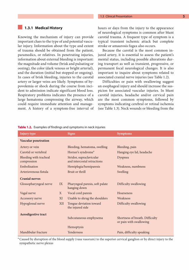
Difficulties or pain with swallowing suggest an esophageal injury and should increase the sus-picion for associated vascular injuries. In blunt carotid injuries, headache and/or cervical pain are the most common symptoms, followed by symptoms indicating cerebral or retinal ischemia (see Table 1.3). Neck wounds or bleeding from the
Table 1.2. Examples of findings and symptoms in neck injuries
Injury type Signs Symptoms
Vascular penetration
Artery or vein Bleeding, hematoma, swelling Bleeding, painCarotid or vertebral Horner’s syndromea Hanging eye lid, headacheBleeding with tracheal compression
Stridor, supraclavicular and intercostal retractions
Dyspnea
Embolization Hemiplegia/hemiparesis Weakness, numbnessArteriovenous fistula Bruit or thrill Swelling
Cranial nervesGlossopharyngeal nerve IX Pharyngeal paresis, soft palate
hanging downDifficulty swallowing
Vagal nerve X Vocal cord paresis HoarsenessAccessory nerve XI Unable to shrug the shoulders WeaknessHypoglossal nerve XII Tongue deviation toward
the injured sideDifficulty swallowing
Aerodigestive tractSubcutaneous emphysema Shortness of breath. Difficulty
or pain with swallowingHemoptysis
Mandibular fracture Tenderness Pain, difficulty speakinga Caused by disruption of the blood supply (vasa vasorum) to the superior cervical ganglion or by direct injury to the
sympathetic nerve plexus
1.3 Clinical Presentation

Chapter 1 Vascular Injuries to the Neck6
mouth, nose, or ears after severe blunt cervical trauma may be associated with injuries to the ver-tebral artery.
1.3.2 Clinical Signs
A penetrating injury is usually obvious at inspec-tion of an open wound with signs of recent or on-going bleeding. A “sucking wound” suggesting a connection with the aerodigestive tract indicates an increased risk for “proximity” injuries to the major cervical arteries (i.e., the vertebral arteries). Even minor external signs of penetrating trauma can be associated with a severe underlying vascu-lar injury. One example is the expanding hemato-ma. The reverse however, is also possible – a large hematoma compressing adjacent structures har-bored by the stiff fascial layers of the neck but undetectable at inspection. Sometimes signs of airway obstruction reveal such injuries. Signs and symptoms of penetrating cervical vascular trauma are summarized in Table 1.2.
Half of the patients with significant blunt vas-cular injuries to the neck lack symptoms at admis-sion but develop symptoms and signs within 24 h. In blunt trauma, it is therefore important to per-form a careful neurological examination at admis-sion to obtain a baseline for later comparisons at the mandatory repeated examinations. The neu-rological evaluation should seek signs of central as well as peripheral nerve injuries – alertness, motor and sensory function, reflexes in the extremities – as well as signs of cranial nerve dysfunction (Ta-ble 1.2). It is important to thoroughly inspect for signs of contusion, asymmetry, or deformity that indicate underlying hematomas and to note the hematoma size for later estimation of possible expansion. Other physical findings indicating a
vascular injury are tenderness over the carotid artery and in the scalp. The most common associ-ated injury is fracture of the mandible.
NOTEThe physical examination can be negative despite severe vascular injury after blunt cervical trauma.
1.4 Diagnostics
1.4.1 Penetrating Trauma
The location of penetrating cervical injuries are generally divided into three different zones that are helpful for planning the diagnostic work-up and management (Fig. 1.1). It is therefore impor-tant to classify the localization of the injury into zones according to this subdivision. The rationale for this lies in difficulties achieving proximal con-trol in zone I injuries and distal control in zone III. Exploration and the possibility of obtaining con-trol are much easier in zone II injuries.
Patients with “hard signs” of major vessel inju-ry – shock, active brisk bleeding, rapidly expand-ing hematoma (for discussion about the definition of “expanding hematoma,” see Chapter 12, p. 149) – and those with neurological deficit or severe airway obstruction should be transported to the operating room for immediate exploration and treatment.
Patients with “soft signs” of major vessel injury – history of bleeding, stable hematoma, and/or cranial nerve injury – usually need further work-up. This is also true for patients who don’t have signs, but who have an injury in proximity to ma-jor vessels. This group constitutes the majority of penetrating neck injuries. The following recom-
Table 1.3. Consequences of blunt injuries to the carotid artery
Type of injury Mechanism Consequences Symptoms/signs
Direct blow Rupture Hematoma Swelling and respiratory problemsPseudoaneurysms Bruit, swelling
Intimal tear Thrombosis Stroke, focal neurology
Rotation-extension Intimal tear Dissection Stroke, focal neurologyThrombosis Stroke, focal neurology

7
mendations have been generally accepted in these cases:
Zones I and III: With the exception of unstable patients, angiography is always indicated. The goals are detecting injury, planning an operation possibly requiring special exposures (e.g., intra-thoracic clamping), and excluding indications for operation.
Zone II: Injuries penetrating the platysma in this zone require surgical exploration to identify and treat vascular injuries as well as injuries to the aerodigestive tract unless the patient is asymp-tomatic and angiography, duplex ultrasound, and CT have ruled out such injuries.
Duplex ultrasound in the hands of an experi-enced examiner has been shown to be consistent with angiography findings in more than 90% of cases. It can reveal dissections, thrombotic occlu-sion, intimal flaps, pseudoaneurysms, and hema-
tomas. Altered flow patterns indicating high resis-tance or abnormal turbulence can be associated with a lesion distally in the internal carotid artery. The developing experience of the duplex technique has prompted a policy change in some hospitals, and duplex ultrasound is used as the primary diagnostic tool for all injuries in stable patients. This examination is performed prior to the deci-sion about angiography or surgical exploration and irrespective of anatomic zone. In some cen-ters, therapeutic decisions are based on thin-slice high-speed CT scans instead.
Gunshot wounds deserve special comment. All patients with such injuries should be screened with angiography or duplex ultrasound. Because associated injuries are common, CT scanning of the head, surgical spine, and aerodigestive tract should also be included in the diagnostic work-up.
NOTEInjuries not penetrating the platysma need no further vascular evaluation.
1.4.2 Blunt Trauma
The diagnosis of vascular injuries after blunt trau-ma is much more challenging. As previously not-ed, clinical signs and symptoms are frequently subtle or absent, and initial transient or late neu-rological deficits are common. The most com-monly injured vascular segment is the distal inter-nal carotid artery. Dissection, with varying de-grees of luminal narrowing, is the most common injury. Other types are pseudoaneurysm or even total transection of the artery with free extravasa-tion. If an expert ultrasonographer is available, duplex ultrasound can be considered as a primary screening method, but a negative study cannot be relied upon to exclude the presence of a clinically significant injury. For all other patients, the rec-ommendation is angiography as the first option, as it is still the gold standard.
In all patients with basilar skull fractures, un-stable cervical spine fractures, Horner’s syndrome, or LeFort-II or-III facial fractures, recent studies advocate a more aggressive attitude with angio-graphic screening for blunt carotid injuries. Extra-cranial carotid injuries are also reported to be
Fig. 1.1. Division of the neck into three zones aids in the management of penetrating cervical vascular inju-ries (Monson DO, Saletta JD, Freeark RJ. Carotid-verte-bral trauma. J Trauma 1969; 9:987–999). Zone I extends inferiorly from 1 cm above the manubrium to include the thoracic outlet; zone II extends from the upper limit of zone I to the angle of the mandible; and zone III is between the angle of the mandible and the base of the skull
1.4 Diagnostics

Chapter 1 Vascular Injuries to the Neck8
more common in patients with a Glasgow coma scale of 8 or less and thoracic injury. A typical finding at angiography in caroid dissection is a stenosis, irregular and often tapered, beginning 2 or 3-cm distal to the bifurcation and often ex-tending up to the base of the skull, above which it is abruptly reconstituted with a normal lumen. Occasionally a typical “string sign” can be seen in the stenosed segment (Fig. 1.2).
CT is not reliable for diagnosing blunt cervical vascular injuries. CT angiography is under devel-opment and is likely to play a greater role in the future. It has the advantage of short examination times and concurrent diagnosis of other injuries, such as brain injuries and skull or facial fractures. Magnetic resonance imaging (MRI) has a high sensitivity and specificity in relation to angiogra-phy (95% and 99%, respectively) for detecting blunt carotid injuries, as does MR angiography (84% and 99%), but both are time-consuming and complicate monitoring and resuscitation of the critically injured patient.
NOTEA patient with neck trauma and a possible vascular injury who is stable, has stabi-lized after resuscitation, or has a trans-cervical gunshot injury should undergo a selective aortic arch angiography.
1.5 Management and Treatment
1.5.1 Management Before Treatment
1.5.1.1 Management in the Emergency Department
As with other major trauma, the Advanced Trau-ma Life Support guidelines should be followed for severe cervical vascular injuries. Consequently, airway and respiration have first priority, followed by control of bleeding. Control of bleeding is best achieved by external finger or manual compres-sion applied directly to the bleeding site. Blindly applied clamps should not be attempted because of the risk of iatrogenic injuries to blood vessels as well as to other organs.
Resuscitation to hemodynamic stability is im-portant. Hypertension may increase bleeding and also induce progress of a dissection, while hypo-tension will increase the risk for thrombosis and cerebral malperfusion.
1.5.1.2 Airway Obstruction Patients with neck trauma and airway obstruction require meticulous management and close co-operation with the anesthesiologist. Intubation should be performed in an anesthetized patient to avoid gagging, which might discharge clots and thus cause profuse bleeding or embolization. Cau-tion should also be taken when the patient’s neck is flexed at the intubation because of the risk of associated cervical vertebral fractures. There is an obvious risk for dislocation and spinal injury. The intubation might also be technically challenging because a large hematoma might cause total com-pression of the trachea. Emergency tracheotomy or coniotomy is then the only alternative, but it may also be complicated by the deranged anatomy and risk of bleeding. The risk of profuse uncon-trolled bleeding is greatest if the hematoma is located on the anterior aspect of the neck because the tamponade will be immediately lost when the
Fig. 1.2. Angiography showing a dissection of the internal carotid artery resulting in a narrowing of the lumen with the typical “string sign” appearance (ar-rows)

9
pretracheal fascia is incised. By using liberal intu-bation early on and under controlled conditions, such situations could be avoided.
1.5.1.3 Immediate Operation or Further Diagnostic Work-up?
The surgeon has to decide in the emergency de-partment whether the patient requires immediate operation, further diagnostic examination, or continued observation.
It is important to emphasize that although standard teaching has been that exploration is re-quired for all zone-II injuries that penetrate the platysma, as well as for gunshot wounds that cross the midline, the availability of better diagnostic modalities has permitted the use of selective ex-ploration protocols.
The following recommendations are given for this initial selection:1. Immediate operation is indicated for unstable
patients with active bleeding not responsive to vigorous resuscitation or with rapidly expand-ing hematoma or airway obstruction, irrespec-tive of anatomical zone.
2. Injuries in zone II not penetrating the platysma need no further examination.
3. All others require further diagnostic evalua-tion with angiography, duplex ultrasound, and CT to determine whether critical structures have been injured. If angiography or high-qual-ity duplex ultrasound is not available, injuries in zone II need to be surgically explored.
Depending on the results of these diagnostic stud-ies, the following general recommendations can be given regarding management of vascular injuries.
Repair is recommended in all patients with penetrating carotid injuries when there is still evi-dence of prograde flow and the patient has no ma-jor neurological symptoms. For minor injuries to the carotid artery, including those with small but adherent intimal flaps, defects, or pseudoaneu-rysms <5 mm in size, repair is recommended in symptomatic patients. If the patient is asymptom-atic and there is no ongoing active bleeding, a con-servative approach has proven to be a safe alter-native. The patient needs to be followed on an inpatient basis for a couple of days to monitor for the appearance of neurological symptoms. Liberal indications for repeated duplex examination are
employed. Anticoagulation therapy, antiplatelet therapy, or both should also be initiated.
Management of significant carotid artery in-jury existing with major neurological deficit and coma is controversial. Some surgeons suggest only observation and palliation, especially in patients with CT-verified cerebral infarction, because of the poor prognosis. Others advocate repair. This standpoint is based on the difficulties of deciding whether the vascular trauma, cerebral contusion, or drugs caused the coma. Some reports, however, indicate a possibility of better outcome after ex-ploration and repair.
When the carotid artery is occluded and there are no neurological symptoms, observation and anticoagulation with heparin followed by 3 to 6-months of coumadin is a general treatment irrespective of the type of trauma. In patients with major neurological symptoms and a verified infarction on CT, anticoagulation is associated with a considerable risk for bleeding. The patients prognosis is then poor, and palliation is recom-mended.
In blunt trauma, indications for exploration and open repair are rarer due to the mostly distal location of injuries to the internal carotid artery. For asymptomatic injuries, including dissection, anticoagulation is usually the only treatment need-ed and is indicated to prevent thrombosis of the injured segment and/or embolization from it. Only injuries that after angiography appear to be easily accessible during surgery and are symptomatic should be considered for repair. This is particu-larly important when there appears to be a high risk for embolization. In patients with decreased consciousness having an occluded internal carotid artery after blunt trauma and with no, or with se-vere, neurological symptoms, most reports advo-cate only observation without surgical exploration. As already mentioned, however, some do recom-mend a more aggressive approach. This strategy requires extensive experience in carotid surgery.
1.5.1.4 Which Patients Can Be Safely Transported?
Because cervical vascular injuries often require an experienced vascular surgeon, a thoracic surgeon, an endovascular specialist, and frequently also a specialist in head and neck surgery, some patients would benefit from being transported to a hospital
1.5 Management and Treatment

Chapter 1 Vascular Injuries to the Neck10
where such expertise is available. A stable patient can be transported to another hospital after intu-bation, or with readiness for emergency intubation in the ambulance, with no major risks. An unsta-ble patient should, of course, remain where he or she is.
1.5.2 Operation
1.5.2.1 Preoperative Preparation and Proximal Control
The management of patients with major external bleeding is mostly related to penetrating injuries to the carotid artery. Surgical exposure of this artery is described in the Technical Tips box. It is important to prepare and drape the patient for possible median sternotomy, which might be nec-essary to obtain proximal control. Bleeding can usually be controlled by external compression by a gloved finger during the preparation and until the complete surgical team is at hand. In the rare and challenging circumstance in which this is impos-sible, a nonspecialist surgeon might be forced to attempt exposure and control of the artery proxi-mal to the injury through a separate incision with continued external finger compression over the lesion by an assistant. During control by direct finger compression of the artery, care must be taken to minimize manipulation because of the risk of thrombus fragmentation and embolization. Compression also needs to be balanced with the desire to maintain flow in the artery. Clamping the common or internal carotid artery for control might be necessary but should be avoided unless the patient’s condition is life-threatening. The rea-son for this is the risk of embolization and cerebral ischemia due to interrupted flow. Inserting a shunt may be the only way to avoid a major stroke; the risk of major stroke in this patient category after carotid clamping may be as high as 50%. Shunting usually requires proximal and distal control, arteriotomy, and possibly also extraction of the thrombus before inserting the shunt. Extracting a thrombus requires a delicate technique and spe-cial consideration because it can easily be frag-mented and dislodged as embolic masses. The safest way is to use traction with forceps and/or suction. If the extraction is followed by brisk back-flow, the result is satisfactory. The use of throm-
bectomy catheters is not recommended because of the risks of mechanical disruption of the throm-bus and bringing fragments up into the circle of Willis. Clamping of the external carotid artery is, on the other hand, almost always safe and can be used more liberally.
TECHNICAL TIPSSurgical Exposure of the Carotid Arteries
An incision anterior and parallel to the anterior border of the sternocleidomastoid muscle is recommended (Fig. 1.3). The incision can be ex-tended dorsal to the ear and down to the sternal notch. As previously noted, preparations need to have been made to allow elongation of the incision into a median sternotomy in order to obtain proximal control of, for instance, the bra-chiocephalic trunk on the right side or the com-mon carotid on the left. After the skin has been incised, subcutaneous fat is divided and, if need-ed, the external jugular vein ligated and divided. The sternocleidomastoid muscle is retracted posteriorly, and a dissection plane anterior to the muscle is identified. The next structure to identify is usually the facial vein and its conflu-ence to the internal jugular vein. The former is suture-ligated and divided and is usually a very good landmark because it is located just above the carotid bifurcation. Dividing this vein allows posterior retraction of the internal jugular vein and exposure of the common carotid and its bifurcation. When preparing the carotid arteries, extreme care must be taken to avoid squeezing of the vessel, and other types of operative ma-nipulation or trauma because of the risk of em-bolization to the brain. Vessel loops are applied, and the most cranial clamp is applied first to avoid embolization when the more proximal parts are clamped. Important structures to pro-tect are the hypoglossal and vagus nerves. The former usually crosses over the internal carotid artery 2–3 cm cranial to the bifurcation and is best exposed after cranial retraction of the di-gastric muscle. The latter runs parallel and dorsal to the common carotid artery. The cervical ansa, often located obliquely over the carotid bifur-cation, can be divided to facilitate exposure.

11
1.5.2.2 Exposure and RepairFor larger arterial injuries, proximal and distal control is mandatory. This always requires an in-direct assessment of the cerebral perfusion by checking the backflow from the internal carotid artery. Measuring the stump pressure in the inter-nal carotid artery is recommended if there is sufficient time. This is obtained by first clamping the common and external carotid arteries and then puncturing the internal carotid artery with a small-caliber injection needle connected to a catheter filled with saline and a pressure trans-ducer. If the backflow is poor or the mean stump pressure is <50 mmHg, shunting is probably nec-essary.
Different repair techniques include the follow-ing:
Simple suture: Simple sutures are mostly suffi-cient in minor penetrating injuries or small pseudoaneurysms.Patch: A patch can be used for a minor wall de-fect or to compensate for diameter loss after arteriotomy and intimal repair.
Resection with end-to-end anastomosis: This is possible in limited injuries requiring minor re-sections, allowing an anastomosis without any tension. Resection with an interposition graft: When larger segments must be excised because of the injury, continuity might be restored with an au-tologous vein graft harvested from the greater saphenous vein or with prosthetic grafts for un-contaminated wounds. Transposition of external to internal carotid artery: This is a good alternative in special cas-es in which vein grafts are unavailable. Ligation or balloon occlusion: Ligation should be reserved for inaccessible injuries that are im-possible to repair. In cases with very distal inju-ries to the internal carotid artery, even ligation might be difficult. An occluding balloon cath-eter can then be inserted into the artery at the base of the skull and insufflated until bleeding stops. The balloon catheter can be left in place for 1 to 2 days or more and then be deflated and removed.
Fig. 1.3 a Recommended incision for exposure of the carotid arteries, anterior and parallel to the anterior border of the sternocleidomastoid muscle. The incision can be extended into a median sternotomy or cranially
dorsal to the ear. b The normal anatomy and relations between major arteries, veins, and important nerve structures at exposure of the carotid and jugular ves-sels
1.5 Management and Treatment

Chapter 1 Vascular Injuries to the Neck12
1.5.2.3 Exploration of Minor Injuries and Hematomas
A patient with an injury penetrating the platysma in anatomical zone II without a history of bleeding may be explored by extending the traumatic skin wound to allow inspection of the injured area, including vessels, trachea, and esophagus. If, how-ever, duplex ultrasound or angiography excludes a major vascular injury, there is no vascular indi-cation for exploration. Patients with a history of significant bleeding as well as those with large hematomas should be treated according to what has been described above for proximal control, before exploring the wound and evacuating the hematoma.
1.5.2.4 Injuries to the Vertebral Artery Due to its relatively inaccessible location, injuries to the vertebral artery in the cervical region are usually best treated by modern endovascular techniques. The options are intraluminal covered stents or endovascular embolization with coil. Be-sides bleeding and hematomas, pseudoaneurysms and arteriovenous fistulas can occur after verte-bral artery trauma and also be successfully treated by transcatheter embolization. In unstable pa-tients with life-threatening bleeding, immediate intervention with proximal and distal ligation might be necessary.
1.5.2.5 Venous Injury Major venous injuries in the neck are almost ex-clusively seen after penetrating trauma. The most commonly injured veins are the external and in-ternal jugular. Due to the low pressure in veins, many such injuries are never recognized. Isolated venous injuries consequently rarely need explora-tion or repair. Venous injuries encountered during exploration of a neck injury can be treated either by repair using simple or running sutures or by ligation. In bilateral injuries to the internal jugular veins, however, reconstruction of one of the sides is indicated to avoid severe venous hypertension. As in all other types of venous surgery, gentle and meticulous technique must be applied and caution taken not to extend the injuries to the veins by a traumatic technique. (See Chapter 15 on vascular surgical technique.)
1.5.2.6 Endovascular Treatment As already described in sections 1.5.2.2 and 1.5.2.4, endovascular technique is an alternative to con-sider in some cervical vascular injuries. Endovas-cular treatment is the first option in injuries to the vertebral artery due to its location and surgical in-accessibility. The application of detachable bal-loons, coils, stents, or hemostatic agents is usually successful for managing bleeding, aneurysms, and fistulas. Endovascular techniques have also been reported to be successful for managing some ca-rotid artery injuries (such as traumatic lacera-tions), pseudoaneurysms, and injuries in some noncritical and small terminal branches. If endo-vascular occlusion of an internal carotid artery is considered in severe traumatic injuries, the neuro-logic effect of a temporary occlusion should be evaluated prior to occlusion. It is reasonable to be-lieve that the endovascular treatment option will become even more useful in the near future.
1.5.3 Management After Treatment
As in elective carotid surgery, monitoring and cor-recting blood pressure is important to minimize risks for bleeding and cerebral complications. Most common are thrombosis and reperfusion problems. Acceptable postoperative systolic blood pressure limits are 100–180 mmHg.
The patient should be checked for clinical signs and symptoms of embolization, and if emboliza-tion is suspected, the operated artery should be checked with ultrasound for complications that might need urgent repair.
Antiplatelet and/or anticoagulation therapy should be considered to maintain patency in re-constructed arteries and to avoid thrombosis in bluntly injured but still patent vessels.
Traumatic and surgical wounds should be checked for signs of a developing hematoma and infection. The latter is particularly important in contaminated wounds and when repair has in-cluded the use of prosthetic material. Antibiotic treatment should be given liberally to avoid the development of severe deep infections in penetrat-ing cervical injuries. For the same reason, meticu-lous wound care is important.

13
1.5.4 Results and Outcome
Several authors report better results after repair of penetrating injuries to the carotid artery com-pared with ligation, with regard to neurological outcome (44% vs. 16% neurologically intact) and mortality (5% vs. 11%).
The results of conservative treatment of carotid dissection, the most common type of injury after blunt trauma, are generally good with duplex-veri-fied resorption of intramural hematoma within 3 months of anticoagulation. The key to this is early detection, preferably before the onset of symptoms, and institution of a heparin infu-sion followed by oral anticoagulation for at least 3 months. Table 1.4 above summarizes the experi-ence of Biffl and colleagues at Brown Medical School (Rhode Island Hospital, Providence, RI, USA) from 109-patients with carotid injuries.
Recent series report good results in managing cervical vascular injuries, irrespective of zone, with only careful physical examination, but the general impression is that it is too early to elimi-nate angiography and duplex ultrasound from the work-up.
As seen, there is a correlation between the inci-dence of stroke and mortality in carotid injuries that is not found in injuries to the vertebral artery and the posterior circulation. Overall mortality after vertebral artery injuries seems to be around 20%, but this is usually in patients with a combi-
Table 1.4. Stroke and mortality rates after different grades of blunt carotid and vertebral artery injuries. (From Biffl et al. Ann Surg 2002; 235: 699–707)
Injury grade/description Carotid artery injury Vertebral artery injury
Stroke Death Stroke Death
I Luminal irregularity or dissection, <25% narrowing
3% 11% 19% 31%
II Dissection or intramuralhematoma, >25%narrowing or intraluminalthrombus or raised intimal flap
11% 11% 40% 0
III Pseudoaneurysm 33% 11% 13% 13%IV Occlusion 44% 22% 33% 11%V Transection and free extravasation 100% 100% – –
nation of other major injuries. Mortality directly related to vertebral artery injuries has been report-ed to be as low as 4%.
Further Reading
Baumgartner RW, Arnold M, Baumgartner I, et al. Ca-rotid dissection with and without ischemic events; local symptoms and cerebral artery findings. Neu-rology 2001; 57:827–832
Biffl WL, Ray CE, Moore EE, et al. Treatment related outcomes from blunt cerebrovascular injuries. Ann Surg 2002; 235:699–707
Demetriades D, Theodorou D, Ascentio J. Management options in vertebral artery injuries. Br J Surg 1996; 83:83–86
Demetriades D, Theodoru D, Cornwell E, et al. Evalua-tion of penetrating injuries to the neck: prospective study of 223 patients. World J Surg 1997; 21:41–48
Gomez CR, May K, Terry J B, et al. Endovascular thera-py of traumatic injuries of the extracranial cerebral arteries. Crit Care Clin 1999; 15:789–809
Kreker C, Mosso M, Georgiadis D, et al. Carotid dissec-tion with permanent and transient occlusion or se-vere stenosis: long term outcome. Neurology 2003; 28:271–275
Mwipatayi BP, Jeffery P, Beningfield SJ, et al. Manage-ment of extra-cranial vertebral artery injuries. Eur J Vasc Endovasc Surg 2004; 27:157–162
Navsaria P, Omoshoro-Jones J. An analysis of 32 surgi-cally managed penetrating carotid artery injuries. Eur J Vasc Endovasc Surg 2002; 24:349–355
Reid J, Weigfelt J. Forty-three cases of vertebral artery trauma. J Trauma 1988; 28:1007–1012
1.5 Management and Treatment

Chapter 1 Vascular Injuries to the Neck14
Romily RC, Newell DW, Grady MS, et al. Gunshot wounds of the internal carotid artery at the skull base: management with vein bypass grafts and a review of the literature. J Vasc Surg 2001; 33:1001–1007
Sekharan J, Dennis JW, Veldenz H, et al. Continued ex-perience with physical examination alone for evalu-ation of penetrating zone 2 neck injuries; result of 145 cases. J Vasc Surg 2000; 32:483
Singh RR, Barry MC, Ireland A, et al. Current diagnosis and management of blunt internal carotid artery in-jury. Eur J Vasc Endovasc Surg 2004; 27:577–584
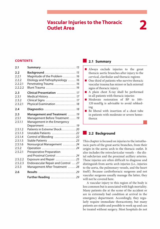
Vascular Injuries to the Thoracic Outlet Area 2
CONTENTS
2.1 Summary . . . . . . . . . . . . . . . . . . . . . . . . . . . . 15
2.2 Background . . . . . . . . . . . . . . . . . . . . . . . . . 152.2.1 Magnitude of the Problem . . . . . . . . . . 162.2.2 Etiology and Pathophysiology . . . . . . 162.2.2.1 Penetrating Trauma . . . . . . . . . . . . . . . . . 162.2.2.2 Blunt Trauma . . . . . . . . . . . . . . . . . . . . . . . . 16
2.3 Clinical Presentation . . . . . . . . . . . . . . . 172.3.1 Medical History . . . . . . . . . . . . . . . . . . . . . . 172.3.2 Clinical Signs . . . . . . . . . . . . . . . . . . . . . . . . 172.3.2.1 Physical Examination . . . . . . . . . . . . . . . . 18
2.4 Diagnostics . . . . . . . . . . . . . . . . . . . . . . . . . 18
2.5 Management and Treatment . . . . . . 192.5.1 Management Before Treatment . . . . . 192.5.1.1 Management in the Emergency
Department . . . . . . . . . . . . . . . . . . . . . . . . . 192.5.1.2 Patients in Extreme Shock . . . . . . . . . . . 202.5.1.3 Unstable Patients . . . . . . . . . . . . . . . . . . . 222.5.1.4 Control of Bleeding . . . . . . . . . . . . . . . . . 222.5.1.5 Stable Patients . . . . . . . . . . . . . . . . . . . . . . 232.5.1.6 Nonsurgical Management . . . . . . . . . . 242.5.2 Operation . . . . . . . . . . . . . . . . . . . . . . . . . . . 242.5.2.1 Preoperative Preparation
and Proximal Control . . . . . . . . . . . . . . . . 242.5.2.2 Exposure and Repair . . . . . . . . . . . . . . . . 252.5.2.3 Endovascular Repair and Control . . . 272.5.3 Management After Treatment . . . . . . 28
2.6 Results . . . . . . . . . . . . . . . . . . . . . . . . . . . . . . 29
Further Reading . . . . . . . . . . . . . . . . . . . . 29
2.1 Summary
Always exclude injuries to the great thoracic aortic branches after injury to the cervical, clavikular and thoracic regionsOne third of patients who survive thoracic vascular trauma has minor or lack external signs of thoracic injury.A plain chest X-ray shall be performed in all patients with thoracic injuriesModerate restoration of BP to 100–120 mmHg is advisable to avoid rebleed-ingBe liberal with insertion of a chest tube in patients with moderate or severe hemo-thorax
2.2 Background
This chapter is focused on injuries to the intratho-racic parts of the great aortic branches, from their origin in the aortic arch to the thoracic outlet. It also includes the retroclavicular vessels – the dis-tal subclavian and the proximal axillary arteries. These injuries are often difficult to diagnose and distinguish from aortic arch injuries (i.e., injuries to the aorta, the pulmonary vessels, and the heart itself). Because cardiothoracic surgeons and not vascular surgeons usually manage the latter, they will not be covered here.
A vascular injury to this region of the body is less common but is associated with high mortality. Many patients die at the scene of the accident or are in extremely bad condition at arrival in the emergency department. Accordingly, they regu-larly require immediate thoracotomy, but many patients are stable and possible to work up and can be treated without surgery. Most hospitals do not
Chapter 2 Vascular Injuries to the Thoracic Outlet Area16
have a thoracic surgeon on call; therefore, these patients are often initially managed by general surgeons with limited experience in thoracic or vascular surgical procedures. Basic information about exposure and access routes and ways to achieve proximal and distal control of intratho-racic great vessels is important not only in this situation but also to obtain proximal control of bleeding vessels in cervical and proximal upper extremity vascular injuries (these areas are dis-cussed in Chapters 1 and 3). Good anatomical knowledge, including that of common variations, is critical, especially for the difficult exposures of the subclavian and axillary vessels, such as when the right subclavian artery originates directly from the aortic arch or has a common trunk with the right carotid artery.
NOTEAnatomical aortic arch and branch variations can be expected in 25–35% of cases.
2.2.1 Magnitude of the Problem
The number of thoracic injuries (all types includ-ed) is steadily increasing in the United States and is estimated to be 12 per million inhabitants per year. In penetrating neck and chest injuries, 3% are associated with injuries to the subclavian and axillary arteries, and in 20% of those injuries, veins are also injured. In a meta-analysis of 2,642 civilian cases of penetrating thoracic trauma, the incidence of great vessel injuries was 1% innomi-nate artery, 5% subclavian, and 6% axillary artery injuries. But because many patients die at the scene, particularly after penetrating trauma, these numbers are uncertain. Irrespective of the type of injuries, trauma to the thoracic great vessels is associated with a high mortality: 80–90% die at the scene. The mortality among patients who survive transport to the hospital is also high.
Patients with injuries in the distal parts of the intrathoracic arteries have a better chance of sur-vival because these vessels are covered with soft tissue, providing better prerequisites for sponta-neous tamponade.
More proximal injuries increase the risk for ex-sanguination into the pleural cavities. Venous in-
juries often remain unrecognized. Arteriograms in patients with a widened mediastinum on plain x-ray after thoracic trauma have been found to be negative for arterial injuries in 85%; this suggests that the mediastinal enlargement was caused mainly by venous injury.
NOTEInjuries to subclavian and axillary arteries are most common after penetrating trauma.
2.2.2 Etiology and Pathophysiology
2.2.2.1 Penetrating TraumaKnife stabbings or missiles from firearms cause a majority of injuries to the great vessels. In this type of penetrating trauma, all intrathoracic ves-sels are at risk of being injured. The extent of inju-ries is related to aspects of the weapon, such as the length of a knife or the velocity (high vs. low) and caliber (small vs. large) of a gun. The innominate artery is injured mostly by bullets from firearms. Stab wounds by knives directed inferiorly into the right clavicular region may also damage the in-nominate artery. The same mechanisms are com-mon for injuries to the subclavian and proximal axillary arteries. Stab wounds are associated with a better chance of survival than are injuries from firearms, particularly shotguns. Blood loss after a knife injury is often limited by a sealing mecha-nism in the wound channel. Furthermore, if the vascular injury is small, the adventitia also limits the bleeding.
The development of hypotension is another factor contributing to limited blood loss. Injuries to the major blood vessels in the thoracic outlet are always challenging because they are rare and tech-nically difficult to expose and control. This is re-flected in the high mortality reported in the litera-ture.
2.2.2.2 Blunt TraumaBlunt trauma to the intrathoracic vessels occurs in motor vehicle and industrial accidents and in falls from heights. If it leads to total disruption of the vessel, the patient will exsanguinate at the scene. When the adventitia remains intact, the possibility

17
of survival is better. The mechanism is shear caused by acceleration/deceleration or compres-sion forces. Deceleration forces are associated with injuries to the aorta but may also cause injuries to the innominate artery. The innominate and com-mon carotid artery might be exposed to shear forces at their origin from compression of the an-terior chest wall. The subclavian and axillary ar-teries can also be injured by blunt trauma, and then mostly in association with clavicle or 1st-rib fractures. Other possible mechanisms are hyper-extension combined with neck rotation, causing tension and stretching of the contralateral subcla-vian vessels. Alternative mechanisms include stretching over the clavicle. Blunt injuries to the subclavian artery after deceleration trauma are rare. There are, however, some controversies re-garding the association between 1st-rib fractures and injuries to the subclavian vessels. Two series of 49 and 55 patients, respectively, reported an inci-dence of 14% and 5% of vascular injuries in asso-ciation with rib fractures. On the other hand, in a large cohort of 466 patients only 0.4% was found.
NOTEInjuries to large veins in the thoracic outlet region are associated with a risk of air embolism and if this occurs, it significantly increases mortality.
2.3 Clinical Presentation
2.3.1 Medical History
The diagnosis is obvious in most cases of penetrat-ing vascular trauma, but the following informa-tion is important for management. In injuries caused by a firearm, the type of weapon used (shotgun, hand weapon, high or low velocity, small or large caliber) and the distance from where it was fired are relevant. For knife stabbings, the blade length and size are important, as well as the angle and direction in which it struck the body. Stabbings directed inferiorly in the clavicular re-gion or at the base of the neck are associated with an increased risk for injuries to the innominate or subclavian arteries.
In blunt trauma, information about the direc-tion and localization of force, the velocity of the
motor vehicle, use of a safety belt, or the height of a fall can indicate the risk for intrathoracic vascu-lar injuries.
When deciding whether immediate thoracoto-my is needed, the course of transport and time elapsed from injury to admission is always of potential importance.
2.3.2 Clinical Signs
As in other vascular injuries, the following “hard signs” strongly indicate severe vascular injury:
Severe bleedingShock or severe anemia Expanding hematomaAbsent or weak peripheral pulses Bruits
“Soft signs” that also indicate vascular injuries in-clude the following:
Local and stable hematomaMinor continuous bleedingMild hypotension Proximity to large vessels Any periclavicular trauma
Injuries to the large vessels in the thorax are frequently associated with injuries to the aero-digestive tract. The following signs and symptoms should alert the responsible surgeon to exclude underlying severe vascular injuries:
Air bubbles in the woundRespiratory distressSubcutaneous emphysemaHoarsenessHemoptysis Hematemesis
NOTEPatients with periclavicular trauma should always be suspected to have intrathoracic great vessel injuries.
Intrathoracic injuries to the subclavian and axil-lary arteries are associated with high mortality. Like injuries to the thoracic aorta, the presenta-tion varies widely, from a fairly stable to a more extreme situation with massive bleeding and ex-sanguination and death at the scene or during
2.3 Clinical Presentation

Chapter 2 Vascular Injuries to the Thoracic Outlet Area18
transport. The latter is more common after blunt trauma that causes avulsion of great vessels and penetrating trauma to the subclavian artery or vein. The consequence of subclavian vessel injury is bleeding into the pleural cavity with or without air embolization. At arrival in the emergency de-partment, a patient with a penetrating intratho-racic vascular injury is typically hemodynamically unstable, whereas a blunt vessel injury is not always immediately apparent.
Blunt injuries to the innominate artery are rela-tively rare, and 75% are combined with other inju-ries such as rib fractures, flail chest, hemothorax or pneumothorax, extremity or facial fractures, or head or abdominal injuries in multitrauma cases. Because there are no typical clinical signs or symp-toms, diagnosis is difficult. The only frequent clin-ical finding is that 50–70% of such patients have a weak radial or brachial pulse. Distal extremity ischemia is uncommon, however, due to good col-lateral circulation in the shoulder region. This ex-plains the possibility of having a palpable distal pulse despite a severe proximal arterial injury.
The subclavian artery is usually injured by direct trauma associated with first-rib or clavicu-lar fractures that cause occlusion of the artery. About half of the patients have a combined injury to the brachial plexus. Accordingly, clinical signs and symptoms indicating such neurological inju-ries (see Chapter 3, p. 33) should increase the sus-picion of injuries associated with the subclavian artery.
2.3.2.1 Physical Examination The entire thorax should be inspected for stab wounds. It is important not to forget skin folds, the axilla, or areas with thick hair. A penetrating trauma to this region is always obvious at arrival in the emergency department. It is also important to remember that one-third of patients who sur-vive blunt trauma and are taken to the emergency department have minor or even no external signs of thoracic injury.
A pulsatile mass or hematoma at the base of the neck, with or without a bruit, indicates an injury to the subclavian artery with leakage through the vessel wall.
At physical examination, auscultation can re-veal signs of hemothorax or pneumothorax. The entire chest and back should be auscultated for
bruits. A systolic bruit over the back and upper chest usually indicates a false aneurysm in any of the great intrathoracic vessels. A continuous bruit indicates the presence of an arteriovenous fistula.
Peripheral pulses, including axillary, brachial, and radial, should always be examined. They are normal in about half of cases with significant vessel injury. Absence of a radial pulse indicates a injury to the axillary, subclavian, or innominate arteries, causing occlusion, dissection, or emboli-zation. The latter is occasionally caused by an em-bolizing bullet.
A thorough neurological evaluation is also rel-evant when considering the possibility of com-bined brachial plexus and vascular injuries. The absence of a radial pulse in combination with Horner’s syndrome is suspicious for injury to the subclavian artery.
Coma or major neurological deficits can also occur as a consequence of injuries to the innomi-nate and common carotid arteries leading to occlusion or embolization and different levels of cerebral ischemia. Therefore, it is important to evaluate the patient’s mental status upon admis-sion. The result influences the decision about if and when to perform emergency surgical repair. This evaluation may also be important during the course of management as a baseline for later re-evaluations.
The management and diagnostic work-up in the emergency department are strongly related to the condition in which the patient arrives. In these types of injuries, the patient is often in an extreme condition, requiring immediate transfer to the operating room for an emergency thoracotomy or other surgical repair. Thoracotomy may even be indicated in the emergency department for a dying patient.
NOTEOne-third of patients who survive blunt thoracic vascular trauma have minor or no external signs of thoracic injury.
2.4 Diagnostics
At arrival, most patients are in a condition that necessitates immediate transfer to the operating room for surgical exploration and treatment. In

19
the remaining patients, the diagnostic work-up depends on the type of trauma and the patient’s condition. In a stable patient, such examinations can provide information of great importance for the management strategy. A good rule is not to start time-consuming examinations while the patient is still hemodynamically unstable.
In a stable patient, plain neck and chest x-rays should always be done to see whether he or she has any of the following:
Hemothorax or pneumothoraxWidened mediastinumIrregular outline of the descending aorta Tracheal dislocationBlurring of the aortic knobDilatation of the aortic bulbPresence of bullets or fracture fragmentsFractures in cervical vertebrae, clavicles, or ribs
Duplex examination has its limitations for detect-ing injuries to the innominate and subclavian ar-teries because of their deep intrathoracic location, particularly in obese patients. It is also examiner-dependent, but nowadays a first choice in many centers. Transesophageal echocardiography may be valuable for diagnosing aortic injuries, but less so in injuries to the aortic branches.
Spiral computed tomography (CT) with intra-venous contrast is mostly used to obtain informa-tion about a missile’s direction and trajectory through the body. The trajectory’s vicinity to great vessels is important when selecting patients for angiography. The modern multislice CT angio-graphy has the potential to become an important diagnostic tool for providing more detailed de-scription of thoracic vascular injuries.
Angiography can be diagnostic as well as thera-peutic. It reveals the presence and localization of occlusions, bleeding, leakage, or pseudoaneu-rysms as well as intimal tears. To detect potential tears and other injuries in the innominate artery, aortography should be performed with posterior oblique projections. A bulbous dilatation at or just distal to its origin and the visualization of an inti-mal flap in the lumen indicate a tear injury to the artery.
In subclavian injuries, a pseudoaneurysm or occlusion can be found. It is important to remem-ber that 10% of patients with innominate or sub-clavian injuries also have other injuries to great
intrathoracic vessels, why it is important that the angiography visualizes the entire thoracic aorta and its branches. The endovascular treatment of these injuries is discussed later in this chapter.
Chest tube placement should have liberal indi-cations for diagnostic as well as therapeutic pur-poses, as a chest tube can reveal the presence of hemothorax or pneumothorax. The technique is described in detail in the section on management below.
NOTEA plain chest x-ray should be performed in all patients with thoracic trauma.
2.5 Management and Treatment
2.5.1 Management Before Treatment
2.5.1.1 Management in the Emergency Department
Management of these often severely injured pa-tients in shock follows the usual Advanced Trau-ma Life Support principles of trauma resuscita-tion. The first priority is always airway control and resuscitation for hypovolemia. Injuries to the great vessels in the thoracic outlet frequently re-sult in expanding mediastinal hematoma, causing tracheal compression and requiring emergency endotracheal intubation.1. Clear and maintain the airway.2. Secure ventilation by endotracheal intubation
and 100% oxygen.3. Consider chest tube insertion. 4. Place two or three intravenous lines, preferably
in the legs and/or the opposite arm.5. Support adequate circulation by rapid volume
replacement with 2.000–3.000 ml of a warm balanced electrolyte solution and blood prod-ucts.
6. Control bleeding. (See below.)7. Consider putting the patient in Trendelenburg
position to avoid air embolism when major venous injuries cannot be excluded.
8. Insert a Foley catheter.
As in patients with a ruptured abdominal aortic aneurysm, resuscitation aims at keeping blood pressure around 100–120-mmHg because of the
2.5 Management and Treatment

Chapter 2 Vascular Injuries to the Thoracic Outlet Area20
risk of sudden massive rebleeding if the blood pressure gets too high. Another event posing risk for new bleeding during resuscitation is gagging during endotracheal intubation or the insertion of an esophageal tube.
If possible, obtain written consent from the pa-tient or his or her family in case emergency sur-gery is necessary. The surgical procedure that may be required often includes clamping of central ar-teries, the aorta, or the common carotids, with a great risk for severe cerebral and spinal complica-tions. Therefore, it is advisable to alert an experi-enced thoracic and/or vascular surgeon for early help with management.
NOTEModerate restoration of blood pressure to 100–120 mmHg is advisable to avoid rebleeding.
2.5.1.2 Patients in Extreme Shock In this category are patients who, most commonly after penetrating thoracic trauma, have lost con-sciousness and present with no vital signs despite resuscitation during the transport but who still show activity on electrocardiography. Other pa-tients in this category are those with acute thera-py-resistant deterioration, those with severe and persistent shock despite very rapid and aggressive volume resuscitation (2.000–3.000 ml of fluids
within minutes) and systolic blood pressure <50 mmHg, and those who experience cardiac ar-rest in the emergency department. These patients are candidates for thoracotomy in the emergency department, aiming at controlling bleeding by manual compression, tamponade, or clamping. This allows more effective resuscitation and is a last lifesaving effort to improve these patients’ vital functions enough to allow transfer to the operating room for immediate surgery.
In such an extreme situation, surgeons with no or only limited experience in thoracotomy can be forced to choose between the two ultimate alterna-tives: to open the patient’s chest or to let him or her die. The prognosis for such a patient is, irrespec-tive of who is performing the thoracotomy, poor, and the survival rate is only around 5%. This should be weighed against the alternative, which is 100% mortality. More than 20% of patients with injuries to subclavian and axillary vessels are in an extreme condition with no vital signs or with im-minent cardiac rest upon arrival to the emergency department. These patients have a very poor prog-nosis.
NOTEDo not hesitate to perform a thoracotomy in the emergency department on a patient with persistent electrocardiographic activity but with no detectable vital signs.

21
Fig. 2.1. Steps for chest tube insertion
TECHNICAL TIPSChest Tube Insertion
Start by determining the desired site of insertion. The recommended site is the 4th or 5th intercostal space, landmark the nipple level just anterior to the midaxillary line, which is good for draining air as well as blood. Scrub and drape the predeter-mined area. Anesthetize the skin, intercostal mus-cles, pleura, and rib periosteum locally (Fig. 2.1 a).
Make a 3 to 4 cm long skin incision over the intercostal space, parallel to the ribs (Fig. 2.1 b).Bluntly dissect the subcutaneous tissue over the cranial aspect of the rib to avoid the intercostal vessels. Continue dissection down to the pleura, preferably with a curved clamp or a finger. Then puncture the parietal pleura with the tip of a
clamp and then expand it with a gloved finger. This is to take precautions against iatrogenic injury to the lung (Fig. 2.1 c, d).
Insert a catheter (32-French or 36-French) with the curved clamp and guide it with a finger. To drain blood, it is best to direct it posterolaterally, and to remove air, an apical position is preferred.
Correct intrapleural position is indicated by “fogging” in the catheter during respiration and when the first side hole is 1 to 2 cm inside the chest wall. Connect the tube to a water-suction device. Secure the tube with a separate suture, and suture the skin.
2.5 Management and Treatment

Chapter 2 Vascular Injuries to the Thoracic Outlet Area22
TECHNICAL TIPSEmergency Anterolateral 5th-Interspace Thoracotomy for Control of the Aorta
The patient must be intubated and ventilated. Incise the skin from the sternum to the axillary line along the upper border of the 5th rib on the left side. In women, the submamillary groove is a landmark. Continue cutting the muscles with scissors or a scalpel all the way down to the pleu-ra. Open the pleural sheath with a pair of scis-sors. The opening should be as large as the hand. One or two costal cartilages can be cut to obtain better access through the thoracotomy. Follow the aortic arch, pass the left subclavian artery and pulmonary artery, and mobilize the heart slightly to the right. Press the descending aorta manually or with an aortic occluder against the spine and try to achieve the best possible occlu-sion. This occlusion is maintained under contin-uous fluid resuscitation and while the patient is transferred to the operating room. Alternatively, place a Satinsky clamp just distal to the origin of the left subclavian artery. The proximal blood pressure must be kept <180 mmHg after clamp placement, and it should be removed as soon as possible.
The left subclavian artery is, in contrast to the right, an intrapleural structure and can in most cases be visualized relatively easy and directly compressed with a finger, clamped, or packed. A left-sided thoracotomy can be extended over to the right, aiming at a higher interstitium. If, how-ever, it is obvious that the injury is on the right side, the thoracotomy should be performed on that side. Severe right-sided intrathoracic bleed-ing is best controlled by finger compression and packing a tamponade in the apex of the right pleural cavity, combined with heavy manual compression in the right supraclavicular fossa.
If resuscitation fails despite adequate fluid substitution and successful control of bleeding, air embolism should be suspected if there are injuries to large veins. Puncture and aspiration in the right ventricle is diagnostic as well as thera-peutic.
2.5.1.3 Unstable Patients Patients with blood pressure <50 mmHg and in severe shock are candidates for immediate sur-gery. A rapid infusion of 2–3 l of a balanced elec-trolyte solution over 10–15 min should be given, aiming to keep blood pressure between 70 and 90 mmHg. It is probably important to keep this level of blood pressure to avoid the risk of in-creased bleeding associated with a higher blood pressure. If the patient does not respond to this volume replacement, he or she should be taken to the operating room for immediate surgery.
Antibiotics covering staphylococci and strepto-cocci should be administered according to the local protocols. One suggestion is cephalosporins. Analgesics, morphine 10 mg intravenously, and, in penetrating injuries, prophylaxis against teta-nus should also be administered.
2.5.1.4 Control of Bleeding In penetrating injuries with continued external bleeding, control is achieved by finger compres-sion over the wound. A gloved finger can also be inserted into the wound to compress the bleeding and stop the outflow of blood. Another recom-mended method is to insert a 24-French Foley catheter into the wound tract and fill the balloon with water or saline (Fig. 2.3). The catheter is clamped after insufflation of the balloon, and if
Fig. 2.2. Incision for emergency anterolateral thora-cotomy

23
the wound penetrates into the pleural cavity, it is gently pulled so the balloon tamponades the pleu-ral entrance. If external bleeding persists after this maneuver, a second balloon can be inserted into the wound and insufflated to stop external bleed-ing from the wound tract. By applying some trac-tion to the catheters, the balloon can also compress injured vessels against the clavicle or the ribs.
If there are clinical indications or radiological signs of moderate or large hemothorax, a chest tube should be inserted for its evacuation. The ra-tionale is that a hemothorax can contribute to con-tinued intrathoracic bleeding and restrict ventila-tion and venous return. Depending on the results when the pleural cavity is drained, different ac-tions can be taken. In an unstable patient, the fol-lowing are considered strong indicators for emer-gency thoracotomy:
1.500 ml of blood drained directly after inser-tion of the tube
>300 ml blood drained through the tube within an hourDeterioration of vital signs when the drain is opened
Even in initially unstable patients, this strategy with evacuation of hemothorax and volume re-placement is often successful. It may allow enough time to let the patient undergo emergency work-up under close surveillance. Information obtained from CT scanning and/or angiography facilitates decisions regarding optimal positioning and routes for exposure of the injury at final surgical treatment (see section 2.5.2, p. 25–27). As de-scribed below, in many situations this type of management stabilizes the patient enough to allow continued nonsurgical management.
NOTEBe liberal with chest tube insertion in patients with moderate or severe hemothorax.
2.5.1.5 Stable Patients Initial management is the same as described above for unstable patients or patients in extreme shock, as summarized in Table 2.1.
Diagnostic examinations in stable patients in-clude repeat plain chest x-ray, angiography or du-plex ultrasound under close surveillance. Also in stable patients chest tubes should be placed on lib-eral indications for evacuation and monitoring of bleeding. The following indicate continued bleed-ing and the possible need for surgical treatment:
Deterioration of vital signs (i.e., hypotensive reaction) when the drain is started1.500–2.000 ml of blood within the first 4–8 hDrainage of blood exceeding 300 ml/h for more than 4 hMore than half of pleural cavity filled with blood on x-ray despite a well functioning chest tube
All of these factors may indicate thoracotomy and should alert the surgeon to consider operation and contact with a cardiothoracic surgeon when needed.
Fig. 2.3. Temporary balloon tamponade of bleeding after penetrating injury to a major subclavian vessel. A Foley catheter is gently inserted to the bottom of the wound tract. After the balloon is filled with saline, gentle traction is applied to the catheter, causing com-pression of the vessels against the clavicle
2.5 Management and Treatment

Chapter 2 Vascular Injuries to the Thoracic Outlet Area24
2.5.1.6 Nonsurgical Management An initially unstable patient who responds well to resuscitation and becomes stable, as well as stable patients with a continued stable course, and with no major vascular injury necessitating surgery revealed at the work-up can often be managed by blood transfusions, fluid replacement, and a chest tube to drain a hemothorax.
The management of patients with major neuro-logical deficits or coma is a matter of debate. Many physicians argue that these patients are never candidates for surgical intervention due to their severe brain injury and poor prognosis. Others ar-gue that vascular injuries should be repaired in all of these cases because it is impossible to exclude that the unconsciousness is related to some injury other than a vascular one.
2.5.2 Operation
2.5.2.1 Preoperative Preparation and Proximal Control
The patient is scrubbed and draped to allow inci-sions from the neck down to at least the knee. In an emergency situation without knowledge about the exact injury site, the patient is best positioned supine with the arms abducted 30°.
The aim of emergency thoracotomy in an un-stable patient is primarily to control bleeding. This can be achieved by surgeons without experience in cardiothoracic surgery. Once control is accom-plished, the repair can wait to allow time for fur-ther resuscitation and for experienced assistance to arrive. Most experienced trauma surgeons to-day recommend a median sternotomy because it is considered the most versatile approach. Such an incision can easily be extended up along the ster-nocleidomastoid muscle on either side or laterally over the clavicle as needed. This approach is there-fore recommended when localization of the injury is uncertain (Fig. 2.4).
Table 2.1. Initial work-up and treatment of patients with thoracic outlet vascular injuries of different severity (US ultrasound, CT computed tomography, ED emergency department, OR operating room)
Patient’s condition Responds to resuscitation
US CT Angiography Treatment
Extreme shock No No No No Emergency thoracotomy in the ED
Unstable No
Yes
No
Maybe
No
Maybe
No
Yes
Emergency thoracotomy in the OR or EDAs above or continued non-op management if only moderate injuries
Stable Yes
Deteriorates after opening chest drain
Maybe
No
Maybe
No
Yes
Maybe
Operative or nonoperative management depending on findingsEmergency operation in the OR

25
2.5.2.2 Exposure and RepairFor innominate artery injuries, the median ster-notomy is extended along the anterior border of the sternocleidomastoid muscle to the right. The overlying innominate vein, which is often also in-jured, has to be divided to achieve exposure. If the injury is located at the base of the artery, which is common in blunt trauma, a reconstruction with an 8–10-mm prosthetic graft end-to-side from the ascending aorta to the divided innominate is frequently employed. This can be performed with
only a partially occluding clamp on the aorta. A total exposure of the proximal aorta and the innominate bifurcation is, however, frequently needed for proximal and distal control before opening the hematoma. In minor penetrating in-juries, simple sutures may occasionally be suffi-cient (Fig. 2.5).
The need for shunting to prevent cerebral isch-emia during innominate artery clamping is con-troversial. Some argue that it is not needed if (1) clamping is caudal to the origin of the common carotid artery, (2) blood pressure and cardiac out-put are normal, and (3) the contralateral carotid artery is open. Others believe it is preferable to always measure stump pressure in the common carotid artery distal to the innominate clamp. Shunting is recommended if the mean pressure is <50 mmHg or not measurable.
Injuries to the common carotid arteries are ex-posed and managed according to the same guide-lines as for innominate injuries. Proximal injuries can be repaired by a prosthetic bypass or simple suture. In cases with occlusion also involving the internal carotid artery, ligation is the safest meth-od. This applies for neurologically intact patients as well as those who are in deep coma.
The subclavian arteries, in particular the retro-clavicular portion of the left artery, are difficult to expose and manage. Before approaching the in-jured area it is extremely important to have a strat-egy for how to obtain proximal control in these injuries; otherwise, severe uncontrollable bleeding may occur. The subclavian arteries can be exposed
Fig. 2.4. Median sternotomy with possible extensions for proximal and distal control in vascular injuries in the thoracic outlet region
Fig. 2.5. Repair of a penetrating injury to the innominate artery. a Clamp occlusion at the origin from the aorta. b Final repair with suture
2.5 Management and Treatment
a b

Chapter 2 Vascular Injuries to the Thoracic Outlet Area26
On the left side, an alternative for proximal control is a high thoracotomy in the 3rd intercos-tal space combined with a supraclavicular incision for distal control (Fig. 2.7).
A thoracotomy is not necessary in injuries to the distal subclavian and proximal axillary artery. The recommended approach for control in these cases is a clavicular incision, starting at the sterno-clavicular joint, extending over the medial portion of the clavicle to curve down into the deltopectoral groove. The exposure of the retroclavicular por-tion of the subclavian artery sometimes requires division of the major pectoral and subclavian muscles, the sternoclavicular joint, and the clavi-cle. After these procedures, the medial portion of the clavicle can be resected or rotated laterally. Clavicular resection in combination can also be employed with a laterally extended median
Fig. 2.7. In unstable patients and when rapid bleeding control is needed, some prefer an anterior 3rd-inter-space thoracotomy in combination with a supraclavic-ular incision for distal control of the subclavian artery
sternotomy. By placing a longitudinal pillow be-tween the shoulders of the patient, the shoulder can be pushed dorsally to further facilitate expo-sure (Fig. 2.8).
Principles of repair of the subclavian and axil-lary arteries usually involve resection and inter-position grafting. Autogenous vein graft is the first choice, but polytetrafluoroethylene (PTFE) or polyester grafts are alternatives. Resection and end-to-end anastomosis are rarely possible, and these vessels are particularly fragile, thus increas-ing the risk for tension in the anastomosis.
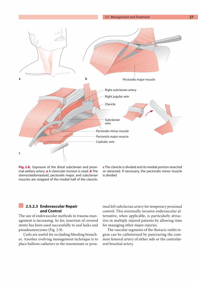
27
2.5.2.3 Endovascular Repairand Control
The use of endovascular methods in trauma man-agement is increasing. So far, insertion of covered stents has been used successfully to seal leaks and pseudoaneurysms (Fig. 2.9).
Coils are useful for occluding bleeding branch-es. Another evolving management technique is to place balloon catheters in the innominate or prox-
imal left subclavian artery for temporary proximal control. This minimally invasive endovascular al-ternative, when applicable, is particularly attrac-tive in multiply injured patients by allowing time for managing other major injuries.
The vascular segments of the thoracic outlet re-gion can be catheterized by puncturing the com-mon femoral artery of either side or the contralat-eral brachial artery.
Fig. 2.8. Exposure of the distal subclavian and proxi-mal axillary artery. a A clavicular incision is used. b The sternocleidomastoid, pectoralis major, and subclavian muscles are stripped of the medial half of the clavicle.
c The clavicle is divided and its medial portion resected or retracted. If necessary, the pectoralis minor muscle is divided
2.5 Management and Treatment
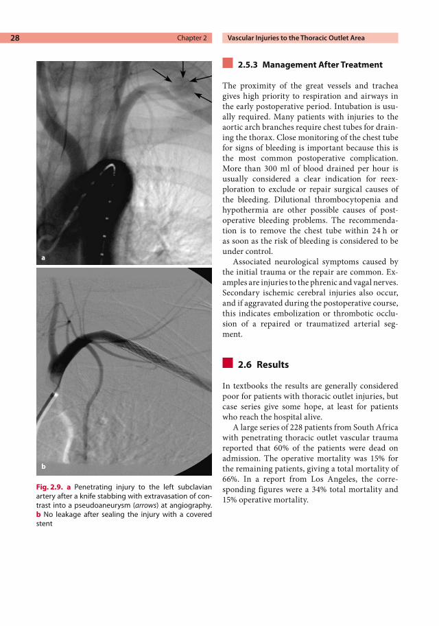
Chapter 2 Vascular Injuries to the Thoracic Outlet Area28
2.5.3 Management After Treatment
The proximity of the great vessels and trachea gives high priority to respiration and airways in the early postoperative period. Intubation is usu-ally required. Many patients with injuries to the aortic arch branches require chest tubes for drain-ing the thorax. Close monitoring of the chest tube for signs of bleeding is important because this is the most common postoperative complication. More than 300 ml of blood drained per hour is usually considered a clear indication for reex-ploration to exclude or repair surgical causes of the bleeding. Dilutional thrombocytopenia and hypothermia are other possible causes of post-operative bleeding problems. The recommenda-tion is to remove the chest tube within 24 h or as soon as the risk of bleeding is considered to be under control.
Associated neurological symptoms caused by the initial trauma or the repair are common. Ex-amples are injuries to the phrenic and vagal nerves. Secondary ischemic cerebral injuries also occur, and if aggravated during the postoperative course, this indicates embolization or thrombotic occlu-sion of a repaired or traumatized arterial seg-ment.
2.6 Results
In textbooks the results are generally considered poor for patients with thoracic outlet injuries, but case series give some hope, at least for patients who reach the hospital alive.
A large series of 228 patients from South Africa with penetrating thoracic outlet vascular trauma reported that 60% of the patients were dead on admission. The operative mortality was 15% for the remaining patients, giving a total mortality of 66%. In a report from Los Angeles, the corre-sponding figures were a 34% total mortality and 15% operative mortality.
Fig. 2.9. a Penetrating injury to the left subclavian artery after a knife stabbing with extravasation of con-trast into a pseudoaneurysm (arrows) at angiography. b No leakage after sealing the injury with a covered stent
a
b

29
The results, however, vary greatly between different studies. In a study from 1989 30 patients with blunt thoracic vascular trauma, substantially better results were reported; the operative mortal-ity was 6.7%, and overall graft patency was 90% after 5 years of follow-up. Another report from the same year had a similar mortality rate of 6.5%. It included 46 patients with 51 intrathoracic arte-rial injuries, 42 of which were penetrating injuries. The incidence of neurological consequences in the different series was relatively low, 5–29%. Combined arterial and venous injuries or venous injuries are associated with a significantly higher mortality, up to 50% and higher. This is blamed the risk for air embolization and more severe blood loss due to less contractility of the veins.
In summary, it seems that most patients die at the scene or during transport, but if the patient arrives alive in the emergency department, the results seem to be fair.
Further Reading
Abouljoud MS, Obeid FN, Horst HM. Arterial injury to the thoracic outlet – a ten year experience. Am Surg 1993; 59:590–595
Axisa BM, Loftus IM, Fishwick G, et al. Endovascular repair of an innominate artery false aneurysm fol-lowing blunt trauma. J Endovasc Ther 2000; 7:245–250
Cox CS, Allen GS, Fischer RP, et al. Blunt versus pen-etrating subclavian artery injury: Presentation, in-jury pattern, and outcome. J Trauma 1999; 46:445–449
Demetriades D, Chahwan S, Gomez H, et al. Penetrat-ing injury to the subclavian and axillary vessels. J Am Coll Surg 1999; 188:290–295
Hajarizadeh H, Rohrer MJ, Cutler BS. Surgical exposure of the left subclavian artery by median sternotomy and left supraclavicular extension. J Trauma 1996; 41:136–139
Hoff SJ, Reilly MK, Merrill WH, et al. Analysis of blunt and penetrating injury of the innominate and sub-clavian arteries. Am Surg 1994; 60:151–154
Pate JW, Cole FH, Walker WA, et al. Penetrating injury of the aortic arch and its branches. Am Thor Surg 1993; 55:586–589
Miles EJ, Buche A, Thompson W, et al. Endovascular re-pair of acute innominate artery injury due to blunt trauma. Am Surg 2003; 69(2):155–159
Weiman DS, McCoy DW, Haan CK, et al. Blunt in-jury of the brachiocephalic artery. Am Surg 1998; 64(5):383–387
2.6 Results

Vascular Injuries in the Arm 3
CONTENTS
3.1 Summary . . . . . . . . . . . . . . . . . . . . . . . . . . . . 31
3.2 Background . . . . . . . . . . . . . . . . . . . . . . . . . 313.2.1 Background . . . . . . . . . . . . . . . . . . . . . . . . . . 313.2.2 Etiology and Pathophysiology . . . . . . 32
3.3 Clinical Presentation . . . . . . . . . . . . . . . 333.3.1 Medical History . . . . . . . . . . . . . . . . . . . . . . 333.3.2 Clinical Signs and Symptoms . . . . . . . . 33
3.4 Diagnostics . . . . . . . . . . . . . . . . . . . . . . . . . 33
3.5 Management and Treatment . . . . . . 343.5.1 Management Before Treatment . . . . . 343.5.1.1 Severely Injured
and Unstable Patients . . . . . . . . . . . . . . . 343.5.1.2 Less Severe Injuries . . . . . . . . . . . . . . . . . . 343.5.1.3 Amputation . . . . . . . . . . . . . . . . . . . . . . . . . 353.5.2 Operation . . . . . . . . . . . . . . . . . . . . . . . . . . . 363.5.2.1 Preoperative Preparation . . . . . . . . . . . 363.5.2.2 Proximal Control . . . . . . . . . . . . . . . . . . . . 363.5.2.3 Exploration and Repair . . . . . . . . . . . . . . 363.5.2.4 Finishing the Operation . . . . . . . . . . . . . 383.5.2.5 Endovascular Treatment . . . . . . . . . . . . 393.5.3 Management After Treatment . . . . . . 39
3.6 Results and Outcome . . . . . . . . . . . . . . . 39
3.7 Iatrogenic Vascular Injuries . . . . . . . . 39
Further Reading . . . . . . . . . . . . . . . . . . . . 40
3.1 Summary
Suspect vascular injuries in patients with shoulder or elbow dislocation.When blood pressures in the arms differ, exclude vascular injuries in proximal ar-teries.It is the nerve injury that determines the functional outcome of arm injuries.Evaluate the brachial plexus and the me-dian nerve function before and during vascular exploration.Repair of vascular injuries in the upper limb is wise even when ischemia appears to be limited.
3.2 Background
3.2.1 Background
Arteries and veins in the arms are the second most common location for vessel injuries in the body and constitute almost half of all peripheral vascu-lar injuries. Much more often than in the legs they occur together with neurological and skeletal injuries. Although vascular injuries in the arms rarely lead to fatal or serious bleeding, ischemic consequences are common. The extensive collat-eral network around the elbow makes clinical signs variable and often minute. On the other hand, if the brachial artery is obstructed proximal to the origin of the deep brachial artery, the risk for amputation is substantial: up to 50% of such patients lose the arm if the vessel is not repaired. While the vascular injury per se often can be man-aged easily, it is the damaged nerves that cause the main functional disturbances in the long run.

Chapter 3 Vascular Injuries in the Arm32
Because arm vessel trauma is common and sometimes appear without signs and symptoms, missed injuries cause considerable morbidity in trauma patients. Awareness and optimal manage-ment may reduce this morbidity.
The arteries supplying blood to the arms – the subclavian and axillary arteries – are located in the thorax or thoracic outlet, and if these vessels are traumatized, the consequences are often more serious. This “intrathoracic part of the arm” is covered in Chapter 2 on vascular injuries in the thoracic outlet area.
3.2.2 Etiology and Pathophysiology
Injury mechanisms are the same in the arms and legs, and the brachial, radial, and ulnar arteries can be damaged by both penetrating and blunt trauma. Knives and gunshots usually cause pene-trating injuries (most often to the brachial artery), but lacerations secondary to fractures occur regu-larly. Sharp fragments commonly penetrate vessel walls (Fig. 3.1). Blunt injuries occur in road traffic accidents because of fractures and joint dis-locations. The most frequent orthopedic arm inju-ries associated with vessel damage are listed in Table 3.1.
There are also types of trauma specific for arm vessel injuries. A large number of upper limb vas-cular trauma are caused by industrial and domes-tic accidents. Splintered glass as well as self-in-flicted wounds regularly damage vessels below the elbow. The popularity of using the brachial artery as a site for vascular access for endovascular pro-cedures has caused an increase in iatrogenic cath-eter-related injuries to the brachial artery in prox-imity to the elbow. Pseudoaneurysms are often caused by radial artery punctures for arterial blood samples.
As in all traumatized vessels, transection or laceration may cause bleeding, thrombosis, or both. Transections, intimal tears, and contusions are more frequent after blunt trauma. The mecha-nisms are described in more detail in Chapter 9 (p. 102). Tissue in the distal parts of the arm is as susceptible to ischemia as in the legs, and the time limit of 6–8 h before irreversible damage occurs is also valid for arm injuries. Concomitant nerve injuries, as mentioned, are the main cause of mor-
bidity long term. Such injuries are equally com-mon after penetrating and blunt trauma. In the literature, 35–60% of arterial injuries in the upper arm are associated with nerve injuries, and over 75% are associated with nerve, bone, or venous damage.
Fig. 3.1. Angiography showing an occluded brachial artery severed by the sharp ends of a shaft fracture of the humerus
Table 3.1. Most common sites for combined orthope-dic and vascular injury
Orthopedic injury Vascular injury
Fractured clavicle Subclavian arteryShoulder dislocation Axillary arterySupracondylar fracture of the humerus
Brachial artery
Elbow dislocation Brachial artery

33
3.3 Clinical Presentation
3.3.1 Medical History
Patients with vascular injuries in the arms arrive at the emergency department after accidents, knife or shooting assaults, or car crashes causing multi-ple injuries. As with all injuries, it is important to interview the rescue personnel and accompanying persons about the type of injury and the type of bleeding. The exact time of the injury should be established to facilitate planning of repair. Because orthopedic injuries are associated with arterial damage, it is also essential to ask whether joint dislocations or fractures were noted or reduced. Complaints of pain from areas around a joint indi-cate a possible luxation. Even more important is to ask for symptoms of nerve damage, including per-manent or transient numbness and impaired mo-tor function in any part of the arm.
NOTEIt is essential to evaluate nerve function before taking the patient to the operating room.
3.3.2 Clinical Signs and Symptoms
Both the “hard” and the “soft” signs of vascular trauma occur after upper extremity vascular inju-ries. Examples, in descending frequency of occur-rence, include diminished or absent radial pulse, motor deficit, sensory loss, hemorrhage, and ex-
panding hematoma. It is common for a dimin-ished radial pulse or an abnormal brachial blood pressure to be the only sign of vascular obstruc-tion. Because pulse wave propagation through a thrombus is possible, a palpable radial pulse does not completely exclude arterial obstruction; there-fore, a high suspicion of arterial injury is neces-sary even when a palpable pulse is found. As a guideline, a difference of more than 20 mmHg in blood pressure between the arms should make the examiner suspect a vascular injury. Inability to move the fingers, hands, and arms as well as dis-turbances in sensation are frequently associated with vascular injury. The sensory and motor func-tions must therefore be carefully examined and evaluated to disclose any nerve damage that should be repaired. A list of what this examination should cover is given in Table 3.2. In unconscious pa-tients, this examination is the only way to reveal indications of arterial damage.
3.4 Diagnostics
Investigations beyond the physical examination of the patient should be done only in stable patients. Accordingly, most patients with distal arm inju-ries can undergo angiography or duplex scanning provided that these investigations do not delay treatment. Arteriography is indicated when arte-rial involvement not is obvious. For example, pa-tients with trauma in the elbow region – blunt or penetrating – with clear ischemia and no radial pulse do not need arteriography before surgery.
Table 3.2. Evaluation of nerve function in the arm
Injured nerve (s) Symptom Findings
Brachial plexus Inability to move the arm; limb hangs with extended elbow and forearm pronated
Unable to discriminate sensation on the neck
Axillary nerve Inability to abduct the arm Unable to discriminate sensation on the dorsal side of the shoulder
Ulnar nerve Numbness and inability to move the 5th finger
Unable to discriminate sensation in the pulp of the 5th finger
Median nerve Inability to flex the hand and numbness in the three middle fingers
Unable to discriminate sensation in the pulp of the index finger
Radial nerve Numbness and inability to move the thumb
Unable to discriminate sensation in the web between the thumb and index finger
3.4 Diagnostics

Chapter 3 Vascular Injuries in the Arm34
If the patient has multiple injuries, shotgun in-juries, or suspected proximal arterial involvement, arteriography is recommended to determine the exact site of injury. Arteriography should also be done when there are indistinct signs of ischemia and arterial injury is only suspected. Included in this indication for arteriography is the so-called proximity injury, referring to injury in patients without signs of distal ischemia but with trauma in close proximity to a major artery. Of patients undergoing arteriography for this indication, 10–20% are reported to have arterial lesions. If the arteriogram reveals injury to the subclavian or axillary artery, endovascular treatment can pro-ceed right away.
Duplex scanning has replaced arteriography in some hospitals and is probably just as accurate in experienced hands. Intimal flaps and small areas of vascular wall thrombosis may be difficult to identify with duplex scanning under some cir-cumstances, but such small lesions in the arm can, on the other hand, usually be treated without exploration.
Computed tomography (CT) angiography is an important modality for diagnosing proximal arte-rial injuries in particular. It is reported to be at least as accurate as arteriography in this area. The use of CT angiography is likely to increase in the near future because it is quicker than angiography, and most trauma centers have rapid access to good-quality CT.
3.5 Management and Treatment
3.5.1 Management Before Treatment
3.5.1.1 Severely Injured and Unstable Patients
Patients arriving to the emergency department with active serious bleeding after a single injury to an arm are rare. When this does occur, manual di-rect pressure over the wound can control the bleeding while general resuscitation measures ac-cording to Advanced Trauma Life Support princi-ples are undertaken: oxygen, monitoring of vital signs, placement of intravenous (IV) lines, and in-fusion of fluids (see also Chapter 9, p. 105, for sug-gestions). It is important not to forget to adminis-
ter analgesics (5–10 mg of an opiate IV) and, when indicated, antibiotics and tetanus prophylaxis.
Multiply injured patients with signs of arm ischemia should be treated according to the hospi-tal’s general trauma management protocol, and the vascular injury is usually evaluated during the second survey. Serious ongoing bleeding has high priority, but arm ischemia should be managed after resuscitation and treatment of life-threaten-ing injuries but before orthopedic repair in most circumstances. When the patient has stabilized, arteriography can be performed if indicated (see above).
3.5.1.2 Less Severe Injuries Most patients with arm injuries arrive in the emer-gency department in a stable condition without ongoing bleeding but with signs of hand ischemia. For these patients, careful examination of the arm including assessment of nerve function, is essen-tial. Dislocated fractures or luxations should be reduced under proper analgesia. After reduction, examination of vascular function should be re-peated. If the radial pulse and distal perfusion return, the position should be stabilized and fixed. Repeated examinations during the following 4 hare mandatory to ensure that the returned perfu-sion is persistent.
If the vessel injury is definite – an absent radial pulse and reduced hand perfusion – and the site of vascular injury is apparent, the patient can be transferred to the operating room without further diagnostic measures. Patients with findings indi-cating vascular injury at examination, and those with obvious arterial disruption but with arms so traumatized that the site of arterial injury cannot be determined, should undergo arteriography or duplex scanning. Expediency of repair is required for all locations of arterial injuries in the arm. The proposed time limits indicating a low risk for per-manent tissue damage range from 4 h for brachial artery injuries and up to 12 h for forearm injuries. The risk limit for irreversible ischemia following forearm injuries is valid for patients with an in-complete palmar arch. The frequency of this ana-tomical variation is 20% in most Western popula-tions.
Suspected injuries to the radial and ulnar ar-tery should be treated according to the general principles discussed above. Even cases with nor-

35
mal perfusion in the hand but without a radial pulse should be explored and repaired if reason-ably simple. When forearm arterial injury is un-clear, the Allen test (Table 3.3) can be added to the examination procedure. A positive Allen test to-gether with a history of trauma to an area in close proximity indicate that the radial or ulnar artery is indeed affected. The wound should then be ex-plored and the traumatized artery inspected and mended. Patients with multiple severe injuries and high-risk patients should not be explored if perfu-sion to the hand is rendered sufficient. For those circumstances, repeat examinations every hour are mandatory to make sure that perfusion is ade-quate and stable.
3.5.1.3 Amputation Some arms with vascular injuries are so extensive-ly damaged that amputation is a treatment option. The decision of when to perform a primary ampu-tation versus trying to repair vessels, nerves, ten-
dons, and muscles is difficult. As a general princi-ple, arms with multiple fractures, nerve disrup-tion, ischemia-time longer than 6 h, and extensive crush injuries involving muscle and skin will nev-er regain function and should be amputated. An-other principle is that when four out of the five components of the arm are injured – skin, bone, muscles, and vessels – but there is only minor nerve injury, an attempt to save the arm is reason-able. One must keep in mind, however, that the arm needs at least some protective sensation in or-der to be functional. Children have a greater chance of regaining a functional arm than adults do, and a generous attitude to surgical repair in children is recommended.
NOTEThe surgeon should not try to save an arm when it only has a small chance of being functional and when repair can be accomplished only at considerable risk.
The mangled extremity severity score (MESS) is a grading system designed to aid the decision pro-cess for managing massive upper and lower ex-tremity trauma. A score of 7 or more has been pro-posed as a cut-off value for indicating when ampu-tation cannot be avoided and should be performed as the primary procedure. In some studies, a score 7 predicted an eventual amputation with 100% ac-curacy. The basis of the MESS scoring system is given in Table 3.4.
As shown in Table 3.4, a crush injury is regard-ed as particularly unfavorable. The duration of ischemia is also a significant factor taken into account in the MESS system.
Table 3.3. Allen test
1. Elevate the arm over the head2. Occlude the radial and ulnar arteries at the wrist4. Lower the arm5. Release blood flow through the ulnar artery6. Inspect and time the return of perfusion
7. Repeat, and release blood flow through the radial artery instead.
8. A return of perfusion >5 s is considered a positive Allen test, and the artery is suspected to be inadequate
3.5 Management and Treatment

Chapter 3 Vascular Injuries in the Arm36
3.5.2 Operation
3.5.2.1 Preoperative PreparationHemodynamically stable patients are placed on their back with the arm abducted 90º on an arm surgery table. The forearm and hand should be in supination. Peripheral or central IV lines should not be inserted on the injured side. Any continu-ing bleeding is controlled manually directly over the wound. If the site of injury is the brachial ar-tery or distal to it, a tourniquet can be used to achieve proximal control. It is then placed before draping and should be padded to avoid direct skin contact with the cuff. This minimizes the risk for skin problems during inflation. The arm is washed so the skin over the appropriate artery can be in-cised without difficulty. The draping should allow palpation of the radial pulse and inspection of fin-ger pulp perfusion. One leg is also prepared in case vein harvest is needed.
The position of the arm is the same for more proximal injuries. Proximal control of high bra-chial and axillary artery trauma may involve ex-posure and skin incisions in the vicinity of the clavicle and the neck, so for proximal injuries the draping must also allow incisions at this level.
3.5.2.2 Proximal Control For distal vessel injury, proximal control can be achieved by inflating the previously placed tourni-quet to a pressure around 50 mmHg above systolic pressure. The cuff should be inflated with the arm elevated to minimize bleeding by venous conges-tion. After inflation, the wound is explored direct-ly at the site of injury.
For more proximal injuries, control is achieved by exposing a normal vessel segment above the wounded area. The most common sites for proxi-mal control in the arm are the axillary artery be-low the clavicle, and the brachial artery (which is what the artery is called distal to the teres major muscle) somewhere in the upper arm. Some com-mon exposures are described in the Technical Tips box.
3.5.2.3 Exploration and RepairDistal control is achieved by exploring the wound. Sometimes this requires additional skin incisions. The most common site for vascular damage in the arm is the brachial artery at the elbow level. These injuries occurs, for example, because of supracon-dylar fractures in children and adults. In such cases, exposure and repair of the brachial artery through an incision in the elbow crease is appro-priate. The anatomy is shown in Fig. 3.1, and a brief description of the technique is given in the Technical Tips box. Hematomas should be evacu-ated to allow inspection of nerves and tendons.
Table 3.4. MESS: Mangled Extremity Severity Score (BP blood pressure)
Types Injury characteristics Points
Low energyMedium energyHigh energyMassive crush
Stab wounds, simple closed fractures, small-caliber gunshot woundsOpen fractures, multiple fractures, dislocations, small crush injuriesShotgun blasts, high-velocity gunshot woundsLogging, railroad accidents
1234
No shock (BP normal)Transient hypotensionProlonged hypotension
BP stable at the site and at the hospitalBP unstable at the site but normalizes after fluid substitutionBP <90 mmHg
123
No distal ischemiaMild ischemiaModerate ischemiaSevere ischemia
Distal pulses, no signs of ischemiaAbsent or diminished pulses, no signs of ischemiaNo signals by continuous-wave Doppler, signs of distal ischemiaNo pulse; cool, paralyzed limb; no capillary refill
12a
3a
4a
<30 years old patient>30 years old patient>50 years old patient
123
a Points are doubled if ischemia lasts longer than 6 h.

37
Fig. 3.2. The most proximal part of the axillary ar-tery can be exposed through an incision parallel to and just below the clavicle. Exposure of the brachial artery is through an incision in the medial aspect of the upper arm. This incision can be elongated and con-nected with the clavicular incision to allow exposure and repair of the entire axillary and brachial artery seg-ments
TECHNICAL TIPSExposure for Proximal Control of Arteries in the Arm
Axillary Artery Below the ClavicleAn 8-cm horizontal incision is made 3 cm below the clavicle (Fig. 3.2). The pectoralis major muscle fibers are split parallel to the skin incision. The pectoralis minor muscle is divided close to its insertion. The nerve crossing the pectoralis minor muscle can also be divided without subsequent morbidity. The axillary artery lies immediately below the fascia together with the vein inferiorly, and the lateral cord of the brachial plexus is located above the artery.
Brachial Artery in the Upper ArmThe incision is made along the posterior border of the biceps muscle; a length of 6–8 cm is usually enough (Fig. 3.3). The muscles are retracted medi-ally and laterally, and the artery lies in the neuro-vascular bundle immediately below the muscles. The sheath is incised and the artery freed from the
median nerve and the medial cutaneous nerve that surrounds it.
Brachial Artery at the ElbowThe incision is placed 2 cm below the elbow crease and should continue up on the medial side along the artery. If possible, veins transversing the wound should be preserved, but they can be di-vided if necessary for exposure. The medial inser-tion of the biceps tendon is divided entirely, and the artery lies immediately beneath it. By follow-ing the wound proximally, more of the artery can be exposed (Fig. 3.3). If the origins of the radial and ulnar artery need to be assessed, the wound can be elongated distally on the ulnar side of the volar aspect of the arm. The median nerve lies close to the brachial artery, and it is important to avoid injuring it.
For supracondylar fractures, the brachial artery, the median nerve, and the musculocutaneous nerves must sometimes be pulled out of the frac-ture site. Before the artery is clamped, the patient is given 50 units of heparin/kg body weight IV. Re-pair should also be preceeded by testing inflow and backflow from the distal vascular bed by tem-porary tourniquet or clamp release. It is often also wise to pass a #2 Fogarty catheter distally to ensure that no clots have formed. Occasionally, inflow is questionable, and proximal obstruction must be ruled out. This can be done intraoperatively by re-trograde arteriography as described in Chapter 4 (p. 44) or by duplex scanning.
As a general principle, all vascular injuries in the arms should be repaired, except when revascu-larization may jeopardize the patient’s life. Arte-rial ligation should be performed only when am-putation is planned. Postoperative arm amputa-tion rates are reported to be 43% if the axillary artery is ligated and 30% at the brachial artery level. Another exception is forearm injuries. When perfusion to the hand is rendered adequate – as assessed by pulse palpation and the Allen test – one of these two arteries can be ligated without
3.5 Management and Treatment

Chapter 3 Vascular Injuries in the Arm38
morbidity. In a substantial number of patients with differing vessel anatomy, however, ligation of either the ulnar or radial artery may lead to hand amputation. If both arteries are damaged, the ul-nar artery should be prioritized because it is usu-ally responsible for the main part of the perfusion to the hand.
For most arterial injuries, vein interposition is necessary for repair. Veins are harvested from the same arm, from parts of the cephalic or basilic vein if the trauma is limited, or from the leg. The saphenous vein in the thigh is suitable for axillary and brachial artery repair, while distal ankle vein pieces can be used for interposition grafts to the
radial and ulnar arteries. Before suturing the anas-tomoses, all damaged parts of the artery must be excised to reduce the risk of postoperative throm-bosis. Rarely, primary suture with and without patching can be used to repair minor lacerations.
Shunting of an arterial injury to permit osteo-synthesis is rarely needed in the arm. Vascular in-terposition grafting can usually be done with an appropriate graft length before final orthopedic repair. Also, extremity shortening due to fractures is less of a problem in the arms (in contrast to the legs), and orthopedic treatment without osteo-synthesis is common especially in older patients. Nevertheless, for some arm injuries shunting is a practical technique that allows time for fracture fixation, thus avoiding the risks of redisplacement and repeated vessel injury. One example is injuries to the axillary or brachial artery caused by a proxi-mal humeral fracture, where the fragment needs to be fixed in order to prevent such injuries. Another example is humeral shaft fracture, which needs to be rigidly fixed to abolish the instability that may otherwise endanger the vascular graft. For more details about shunting, see Chapter 9 (p. 111).
Veins should also be repaired if reasonably sim-ple. If the vein injury is caused by a single wound with limited tissue damage, concomitant veins to the distal brachial artery can be ligated. For more extensive injuries where the superficial large veins are likely to be ruined, it is wise to try to repair the deep veins. For very proximal injuries in the shoul-der region, vein repair is important to avoid long-term problems with arm swelling. It is also impor-tant to cover the mended vessel segment with soft tissue to minimize the risk for infection that may involve the arteries.
3.5.2.4 Finishing the Operation When the repaired artery or graft’s function is doubtful and when the surgeon suspects distal clotting, intraoperative arteriography should be performed. The technique is described in Chap-ter 10 (p. 128). After completion, all devitalized tissue should be excised and the wound cleaned. For penetrating wounds, damaged tendons and transected nerves should also be sutured. This is not worthwhile for most blunt injuries. Fascioto-my should also be considered before finishing the operation. As in the leg, long ischemia times and successful repair increase the risk of reperfusion
Fig. 3.3. Transverse incision in the elbow for exposing the brachial artery and with possible elongations (dot-ted lines) when access to the ulnar and radial branches as well as to more proximal parts of the brachial artery is needed
39
and compartment syndrome, but the overall risk for compartment syndrome is reported to be less in the arm than in the leg. For a description of arm fasciotomy techniques, we recommend con-sulting orthopedic textbooks. After the wounds are dressed, a fractured arm is put into a plaster splint for stabilization.
3.5.2.5 Endovascular Treatment In contrast to proximal arm vessel trauma, there are few instances in distal injuries when endovas-cular treatment is a feasible treatment option. Be-cause the brachial artery and the forearm vessels are easy to expose with little morbidity, open re-pair during exploration of the wound is usually the best option. Possible exceptions to this are treatment of the late consequences of vascular trauma, such as arteriovenous fistulas and pseu-doaneurysms.
Especially in the shoulder region, including the axilla, primary endovascular treatment is often the best treatment option. Another circumstance when endovascular treatment is favorable is bleed-ing from axillary artery branches – such as the circumflex humeral artery – due to penetrating trauma. Active bleeding from branches, but not from the main trunk, observed during arteriogra-phy is preferably treated by coiling. The bleeding branches are then selectively cannulated with a guidewire and coiled, using spring coils or injec-tions of thrombin to occlude the bleeding artery.
3.5.3 Management After Treatment
Postoperative monitoring of hand perfusion and radial pulse is recommended at least every 30 min for the first 6 h. When deteriorated function of the repaired artery is suspected, duplex scanning can verify or exclude postoperative problems. Appar-ent occlusions should be treated by reoperation as soon as possible. Compartment syndrome in the lower arm may also evolve over time, and swelling, muscle tenderness, and rigidity must also be mon-itored during the initial days. For most patients, treatment with low molecular weight heparin is continued postoperatively. A common dose is 5,000 units subcutaneously twice daily.
Keeping the hand elevated as much as possible may reduce swelling of the hand and arm as well as
problems with hematoma formation around the wound. Early mobilization of the fingers facilitate blood flow to the arm and should be encouraged.
3.6 Results and Outcome
The patency of arterial repair in the arm is often excellent, but unfortunately, this appears to have little impact on the eventual arm function. For most patients in whom vessel trauma is associated with nerve and soft tissue injury, it is the nerve function that determines the outcome. Outcome data after arterial repair in upper extremity inju-ries have been reported in observational studies and case series. One example is a review from the United States of 101 patients with penetrating trauma, including 13 axillary or subclavian cases. Half of the patients had nerve injuries as well. At follow-up the limb salvage rate was 99%, and all patients who needed only vascular repair had ex-cellent functional outcomes. Among arms that re-quired nerve repair, 64% had severe impairment of arm function. The corresponding figure for mus-culoskeletal repair only was 25%.
A report from the United Kingdom included 28 cases of brachial artery injuries, of which six were blunt. In this study, half of the patients had con-comitant nerve injury and underwent immediate nerve repair. All vascular repairs were successful, but the majority of patients undergoing nerve re-pair appear to have had some functional deficit at follow-up.
Fortunately, it seems that function improves over time in many patients. The risk factors for poor outcome are similar to the ones used for the MESS score – severity of the fracture and soft tis-sue damage, length of the ischemic period, severity of neurological involvement, and presence of associated injuries.
3.7 Iatrogenic Vascular Injuries
The brachial artery is increasingly being used for cannulation, both for vascular access and for en-dovascular procedures. The latter requires large introducer sheaths, and it is likely that we will ex-perience an increase in the number of problems related to this. Associated injuries are bleeding
3.7 Iatrogenic Vascular Injuries

Chapter 3 Vascular Injuries in the Arm40
and thrombosis. (Both of these issues are dis-cussed in Chapter 12.) Management of bleeding is fairly straightforward. Bleeding is usually easy to control by manual compression; exposure is sim-ple; and repair is often accomplished by a few simple sutures. Thrombosis is much less common but is more complicated to handle. Management should follow the guidelines given in Chapter 4.
Another problem that may be encountered is related to arterial blood sampling from the radial artery. Occasionally, thrombosis of this artery will cause severe arm ischemia. This should then be resolved by embolectomy and patch closure of the injured vessel segment. Sporadically, vein graft in-terposition is needed. Bleeding or an expanding hematoma due to arterial puncture rarely occurs, but pseudoaneurysm formation is not so infre-quent. Such problems should be handled by sur-gery, including proximal control and patch closure of the injured vessel.
The radial artery is sometimes used as a graft for coronary bypass procedures. This appears to work extremely well, with little late morbidity in the arm where the artery was harvested. We have encountered occasional patients with mild hand ischemia immediately after surgery, but only a few cases who eventually needed revascularization. For these rare patients, a vein bypass from the bra-chial artery to the site where the ligature was placed at harvest is the recommended treatment.
Further Reading
Fields CE, Latifi R, Ivatury RR. Brachial and fore-arm vessel injuries. Surg Clin North Am 2002; 82(1):105–114
McCready RA. Upper-extremity vascular injuries. Surg Clin North Am 1988; 68(4):725–740
Myers SI, Harward TR, Maher DP, et al. Complex upper extremity vascular trauma in an urban population. J Vasc Surg 1990; 12(3):305–309
Nichols JS, Lillehei KO. Nerve injury associated with acute vascular trauma. Surg Clin North Am 1988; 68(4):837–852
Ohki T, Veith FJ, Kraas C, et al. Endovascular ther-apy for upper extremity injury. Semin Vasc Surg 1998;11(2):106–115
Pillai L, Luchette FA, Romano KS, et al. Upper-extrem-ity arterial injury. Am Surg 1997; 63(3):224–227
Shaw AD, Milne AA, Christie J, et al. Vascular trauma of the upper limb and associated nerve injuries. In-jury 1995; 26(8):515–518
Stein JS, Strauss E. Gunshot wounds to the upper ex-tremity. Evaluation and management of vascular injuries. Orthop Clin North Am 1995; 26(1):29–35
Thompson PN, Chang BB, Shah DM, et al. Outcome fol-lowing blunt vascular trauma of the upper extrem-ity. Cardiovasc Surg 1993; 1(3):248–250
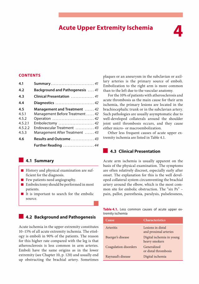
Acute Upper Extremity Ischemia 4
CONTENTS
4.1 Summary . . . . . . . . . . . . . . . . . . . . . . . . . . . . 41
4.2 Background and Pathogenesis . . . . 41
4.3 Clinical Presentation . . . . . . . . . . . . . . . 41
4.4 Diagnostics . . . . . . . . . . . . . . . . . . . . . . . . . 42
4.5 Management and Treatment . . . . . . 424.5.1 Management Before Treatment . . . . . 424.5.2 Operation . . . . . . . . . . . . . . . . . . . . . . . . . . . 424.5.2.1 Embolectomy . . . . . . . . . . . . . . . . . . . . . . . 424.5.2.2 Endovascular Treatment . . . . . . . . . . . . 434.5.3 Management After Treatment . . . . . . 43
4.6 Results and Outcome . . . . . . . . . . . . . . . 43
Further Reading . . . . . . . . . . . . . . . . . . . . 44
4.1 Summary
History and physical examination are suf-ficient for the diagnosis.Few patients need angiography.Embolectomy should be performed in most patients.It is important to search for the embolic source.
4.2 Background and Pathogenesis
Acute ischemia in the upper extremity constitutes 10–15% of all acute extremity ischemia. The etiol-ogy is emboli in 90% of the patients. The reason for this higher rate compared with the leg is that atherosclerosis is less common in arm arteries. Emboli have the same origins as in the lower extremity (see Chapter 10, p. 120) and usually end up obstructing the brachial artery. Sometimes
plaques or an aneurysm in the subclavian or axil-lary arteries is the primary source of emboli. Embolization to the right arm is more common than to the left due to the vascular anatomy.
For the 10% of patients with atherosclerosis and acute thrombosis as the main cause for their arm ischemia, the primary lesions are located in the brachiocephalic trunk or in the subclavian artery. Such pathologies are usually asymptomatic due to well-developed collaterals around the shoulder joint until thrombosis occurs, and they cause either micro- or macroembolization.
Other less frequent causes of acute upper ex-tremity ischemia are listed in Table 4.1.
4.3 Clinical Presentation
Acute arm ischemia is usually apparent on the basis of the physical examination. The symptoms are often relatively discreet, especially early after onset. The explanation for this is the well devel-oped collateral system circumventing the brachial artery around the elbow, which is the most com-mon site for embolic obstruction. The “six Ps” – pain, pallor, paresthesia, paralysis, pulselessness,
Table 4.1. Less common causes of acute upper ex-tremity ischemia
Cause Characteristics
Arteritis Lesions in distal and proximal arteries
Buerger’s disease Digital ischemia in young heavy smokers
Coagulation disorders Generalized or distal thrombosis
Raynaud’s disease Digital ischemia
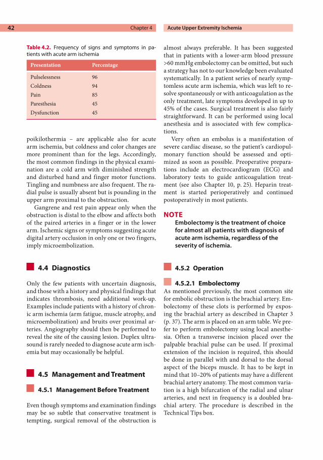
Chapter 4 Acute Upper Extremity Ischemia42
poikilothermia – are applicable also for acute arm ischemia, but coldness and color changes are more prominent than for the legs. Accordingly, the most common findings in the physical exami-nation are a cold arm with diminished strength and disturbed hand and finger motor functions. Tingling and numbness are also frequent. The ra-dial pulse is usually absent but is pounding in the upper arm proximal to the obstruction.
Gangrene and rest pain appear only when the obstruction is distal to the elbow and affects both of the paired arteries in a finger or in the lower arm. Ischemic signs or symptoms suggesting acute digital artery occlusion in only one or two fingers, imply microembolization.
4.4 Diagnostics
Only the few patients with uncertain diagnosis, and those with a history and physical findings that indicates thrombosis, need additional work-up. Examples include patients with a history of chron-ic arm ischemia (arm fatigue, muscle atrophy, and microembolization) and bruits over proximal ar-teries. Angiography should then be performed to reveal the site of the causing lesion. Duplex ultra-sound is rarely needed to diagnose acute arm isch-emia but may occasionally be helpful.
4.5 Management and Treatment
4.5.1 Management Before Treatment
Even though symptoms and examination findings may be so subtle that conservative treatment is tempting, surgical removal of the obstruction is
almost always preferable. It has been suggested that in patients with a lower-arm blood pressure >60 mmHg embolectomy can be omitted, but such a strategy has not to our knowledge been evaluated systematically. In a patient series of nearly symp-tomless acute arm ischemia, which was left to re-solve spontaneously or with anticoagulation as the only treatment, late symptoms developed in up to 45% of the cases. Surgical treatment is also fairly straightforward. It can be performed using local anesthesia and is associated with few complica-tions.
Very often an embolus is a manifestation of severe cardiac disease, so the patient’s cardiopul-monary function should be assessed and opti-mized as soon as possible. Preoperative prepara-tions include an electrocardiogram (ECG) and laboratory tests to guide anticoagulation treat-ment (see also Chapter 10, p. 25). Heparin treat-ment is started perioperatively and continued postoperatively in most patients.
NOTEEmbolectomy is the treatment of choice for almost all patients with diagnosis of acute arm ischemia, regardless of the severity of ischemia.
4.5.2 Operation
4.5.2.1 Embolectomy As mentioned previously, the most common site for embolic obstruction is the brachial artery. Em-bolectomy of these clots is performed by expos-ing the brachial artery as described in Chapter 3 (p. 37). The arm is placed on an arm table. We pre-fer to perform embolectomy using local anesthe-sia. Often a transverse incision placed over the palpable brachial pulse can be used. If proximal extension of the incision is required, this should be done in parallel with and dorsal to the dorsal aspect of the biceps muscle. It has to be kept in mind that 10–20% of patients may have a different brachial artery anatomy. The most common varia-tion is a high bifurcation of the radial and ulnar arteries, and next in frequency is a doubled bra-chial artery. The procedure is described in the Technical Tips box.
Table 4.2. Frequency of signs and symptoms in pa-tients with acute arm ischemia
Presentation Percentage
Pulselessness 96Coldness 94Pain 85Paresthesia 45Dysfunction 45

43
An alternative location for embolectomy in the arm is to expose the brachial artery in the bicipi-tal groove. A longitudinal incision starting 10 cm above the elbow that is extended proximally is then used.
TECHNICAL TIPSEmbolectomy via the Brachial Artery
Exposure of this vessel is described in Chapter 3. A transverse arteriotomy in the brachial artery is made as close as possible to the bifurcation of the ulnar and radial arteries. The embolecto-my is performed in proximal and distal direc-tions with #2 and #3 Fogarty catheters. Separate embolectomy in each branch should be done if technically simple. The Fogarty catheter other-wise slips down into the larger and straighter ulnar artery. The route of the catheter can be checked by palpation at the wrist level when the inflated balloon passes. On the other hand, restored flow in one of the arteries is usually enough for a result that is sufficient for adequate hand perfusion. The arteriotomy is closed with interrupted 6-0 sutures, and distal pulses and the perfusion in the hand are evaluated. If the result is inadequate – poor backflow after embolectomy, absence of pulse, a weak continu-ous-wave Doppler signal, and questionable hand perfusion – the arteriotomy should be reopened and intraoperative angiography per-formed (Table 4.3 and Chapter 10, p. 128).
If it is hard to achieve a good inflow, a proximal lesion may cause the embolization or thrombosis. More complicated vascular procedures are then required to reestablish flow. The embolectomy attempt is then discontinued and the patient taken to the angiography suite for a complete examina-tion. If practically feasible, an alternative is to obtain the angiogram in the operating room. Fre-quently, however, the preferred treatment is endo-vascular, and this is better done in the angiography suite. Occasionally the films will reveal a proximal obstruction that needs open repair. Examples of such are carotid-subclavian, subclavian-axillary, and axillary-brachial bypasses.
4.5.2.2 Endovascular Treatment Thrombolysis is as feasible for acute upper extrem-ity ischemia as it is in the leg. The limited ischemia that often occurs after most embolic events be-cause of the collateral network around the elbow also allows the time needed for planning and mov-ing the patient to the angiosuite. The technique involves cannulation in the groin with a 7-French sheath. Long guide wires and catheters are re-quired to reach the occluded site and makes iden-tification of proximal lesions possible. A new arte-rial puncture in the brachial artery may be neces-sary for thrombolysis of distal occlusions.
It can be argued that thrombolysis in spite of acceptable results, rarely is needed for treating this disease because open embolectomy can be per-formed under local anesthesia with good results and little surgical morbidity. The advantages with endovascular treatment are indeed limited. For patients in whom suspicion of thrombosis is strong or when proximal lesions are likely, it should be attempted first. However, case series indicates that results of thrombolysis are inferior for forearm occlusions. In summary, thrombolysis is an alter-native but has little to offer in reducing risk or improving outcome compared with embolectomy for most patients.
4.5.3 Management After Treatment
Patients usually regain full function of their hands immediately after the procedure, and postopera-tive regimens consist of anticoagulation and a search for the embolic source. Heparin or low molecular weight heparin is administered as de-scribed in Chapter 10 (p. 129), usually followed by coumadin. The search for cardiac sources may advocate repeated ECGs, echocardiography, and duplex ultrasound of proximal arteries.
4.6 Results and Outcome
The number of salvaged arms after surgical inter-vention is very high, 90–95%, and arm function is usually fully recovered. The remaining 5–10% represents patients with extensive thrombosis involving many vascular segments and most branches of the distal arteries. The postoperative
4.6 Results and Outcome

Chapter 4 Acute Upper Extremity Ischemia44
mortality is around 10–40% in most patient series, reflecting that embolization often is a consequence of severe cardiac disease. Postoperative mortality is similar for thrombolysis to treat acute arm isch-emia, while early technical success is slightly lower or similar. Less favorable results with thromboly-sis are achieved when the distal arteries also are obstructed.
Further Reading
Baguneid M, Dodd D, Fulford P, et al. Management of acute nontraumatic upper limb ischemia. Angiol-ogy 1999; 50(9):715–720
Eyers P, Earnshaw JJ. Acute non-traumatic arm isch-aemia. Br J Surg 1998; 85(10):1340–1346
Pentti J, Salenius JP, Kuukasjarvi P, et al. Outcome of surgical treatment in acute upper limb ischaemia. Ann Chir Gynaecol 1995; 84(1):25–28
Ricotta JJ, Scudder PA, McAndrew JA, et al. Manage-ment of acute ischemia of the upper extremity. Am J Surg 1983; 145(5):661–666
Whelan TJ Jr. Management of vascular disease of the upper extremity. Surg Clin North Am 1982; 62(3):373–389
Table 4.3. Technique for retrograde intraoperative an-giography
1. Control proximal to arteriotomy is achieved by finger compression and/or vessel loop
2. Insert an angiography catheter or a small caliber baby feeding tube through the arteriotomy in retrograde direction
3. Place the tip of the catheter proximal to the suspected obstructing lesion
4. Inject contrast under simultaneous fluoroscopy in lateral projection with a C-arm
Abdominal Vascular Injuries 5
CONTENTS
5.1 Summary . . . . . . . . . . . . . . . . . . . . . . . . . . . . 45
5.2 Background . . . . . . . . . . . . . . . . . . . . . . . . . 465.2.1 Background . . . . . . . . . . . . . . . . . . . . . . . . . . 465.2.2 Magnitude of the Problem . . . . . . . . . . 465.2.3 Etiology and Pathophysiology . . . . . . 465.2.3.1 Penetrating Injury . . . . . . . . . . . . . . . . . . . 465.2.3.2 Blunt Injury . . . . . . . . . . . . . . . . . . . . . . . . . . 465.2.3.3 Pathophysiology . . . . . . . . . . . . . . . . . . . . 465.2.3.4 Associated Injuries . . . . . . . . . . . . . . . . . . 47
5.3 Clinical Presentation . . . . . . . . . . . . . . . 475.3.1 Medical History . . . . . . . . . . . . . . . . . . . . . . 475.3.2 Clinical Signs and Symptoms . . . . . . . . 48
5.4 Diagnostics . . . . . . . . . . . . . . . . . . . . . . . . . 48
5.5 Management and Treatment . . . . . . 505.5.1 Management Before Treatment . . . . . 505.5.1.1 Treatment and Management
in the Emergency Department . . . . . . 505.5.1.2 Unstable Patients . . . . . . . . . . . . . . . . . . . 505.5.1.3 Stable Patients . . . . . . . . . . . . . . . . . . . . . . 515.5.1.4 Laparotomy or Not?. . . . . . . . . . . . . . . . . . 515.5.1.5 Renal Artery Injuries . . . . . . . . . . . . . . . . . 515.5.2 Operation . . . . . . . . . . . . . . . . . . . . . . . . . . . 525.5.2.1 Preoperative Preparation . . . . . . . . . . . . 525.5.2.2 Exploration . . . . . . . . . . . . . . . . . . . . . . . . . . 525.5.2.4 Vessel Repair . . . . . . . . . . . . . . . . . . . . . . . . 575.5.2.5 Finishing the Operation . . . . . . . . . . . . . 605.5.3 Endovascular Treatment . . . . . . . . . . . . 605.5.4 Management After Treatment . . . . . . 60
5.6 Results and Outcome . . . . . . . . . . . . . . . 61
5.7 Iatrogenic Vascular Injuries in the Abdomen . . . . . . . . . . . . . . . . . . . . 61
5.7.1 Laparoscopic Injuries . . . . . . . . . . . . . . . . 615.7.2 Iliac Arteries and Veins
During Surgery for Malignancies in the Pelvis . . . . . . . . . . . . . . . . . . . . . . . . . 62
5.7.3 Iliac Artery Injuries During Endovascular Procedures . . . 62
5.7.4 Iatrogenic Injuries During Orthopedic Procedures . . . . . 62
Further Reading . . . . . . . . . . . . . . . . . . . . 63
5.1 Summary
Up to 25% of patients with abdominal trauma may have major vascular injury.Shock out of proportion to the extent of ex-ternal injury suggests abdominal vascular injury.Isolated abdominal injury in patients with shock suggests major vascular injury that requires emergency laparotomy for con-trol.After the abdomen is entered, immediate control of the supraceliac aorta should be considered before continuing the opera-tion.Retroperitoneal hematomas should not be explored right away unless they are actively bleeding.Stopping the procedure after the initial ex-ploration for damage control to allow time for resuscitation in the intensive care unit is often a reasonable initial treatment.If the patient’s condition allows and if en-dovascular methods are available, consider placing an aortic balloon from the left bra-chial artery for temporary occlusion.

Chapter 5 Abdominal Vascular Injuries46
5.2 Background
5.2.1 Background
Abdominal vascular trauma is fairly common in modern civilian life and is a highly lethal injury, with overall mortality around 40% in some reported series. The main cause for this high mortality relates to problems transporting injured patients to the hospital fast enough to prevent ex-sanguination. Furthermore, abdominal vascular injuries are rarely isolated, and other organs are often severely damaged as well. These factors make it essential to resuscitate promptly and es-tablish a rapid diagnosis.
The surgeon managing patients with major ab-dominal injuries must be experienced with vascu-lar surgical techniques and be able to expose the aorta and its main branches, as well as the vena cava. Dissection and the extensive organ mobili-zation required for control and repair are often difficult. It is therefore important to develop a routine that can be employed during exploration and control.
5.2.2 Magnitude of the Problem
Major abdominal vascular injury is seen in up to 25% of patients admitted with vascular trauma. Blunt trauma is more common than penetrating trauma in most European countries, while the op-posite is reported in areas where gunshot wounds are more frequent. Abdominal injury represents 10–20% of all traumas to the body caused by road traffic accidents. Major vascular injury is estimat-ed to occur in about 10% of cases of penetrating stab wounds in the abdomen and in about 25% of gunshot wounds. Blunt abdominal trauma affects major vessels less frequently, estimates of below 5% is common in the literature.
NOTEMajor vascular injury is rather common after abdominal trauma.
5.2.3 Etiology and Pathophysiology
5.2.3.1 Penetrating InjuryPenetrating injury creates the types of damage that are common for most arteries – transection, laceration, intimal dissection, and thrombosis, as well as false aneurysms and arteriovenous fistula formation. The first two are more common after stab wounds. Gunshot wounds inflict more wide-spread damage to the vessel wall, depending on the bullet’s velocity. For example, high-velocity missiles at speeds >700 m/sec cause up to 20 times more damage than low-velocity projectiles. An artery located within 10–15 cm of the trajectory regularly thromboses after a high-velocity gun-shot injury.
5.2.3.2 Blunt Injury Typically, blunt injury to abdominal vessels occurs after road traffic accidents or falls from heights. Most commonly damaged are upper abdominal arteries and veins such as the infrarenal aorta. The mechanism is compression of the aorta against the lumbar spine by the steering wheel, especially when seat belts are not used. This causes intimal tears and thrombosis of the aorta. Full-blown rup-ture has also been reported. Vessel injuries are much less frequent when seat belts are used. Avul-sion of branches is also common and there is a high incidence of associated injury to the small arteries. Veins are usually not affected by blunt trauma, except for the left renal vein. Major ab-dominal injuries may cause avulsion of arteries; in descending order of occurrence the vessels injured are the left renal vein, the renal arteries, the supe-rior mesenteric artery (SMA), and the abdominal aorta just distal to the renal arteries.
5.2.3.3 Pathophysiology When an artery is perforated, blood extravasates into surrounding tissues, causing a hematoma that counteracts the blood pressure and facilitates spontaneous closure of the hole in the vessel. When a vein is damaged, tamponade of the bleed-ing often occurs, especially if retroperitoneal, un-less the peritoneum is torn or is entered during laparotomy. If vein damage is caused by a pelvic fracture a cavity is created around the fragments, preventing effective tamponade, and the bleeding continues. Venous and arterial bleeding within

47
the mesentery is also enhanced by the same mech-anism. The high blood flow through major arter-ies in the abdomen makes spontaneous cessation of bleeding less likely. Even the aorta, however, has been reported to seal spontaneously after pene-trating trauma when it is completely transected. If an artery is partially lacerated, the severed ends cannot contract; the hole is held open, and blood flows more easily into the abdominal cavity. Pa-tients rarely survive for long in this circumstance.
There are two principle mechanisms of vascu-lar injury in blunt abdominal trauma: compres-sion and deceleration forces. The former may cause crush injuries and intramural hematoma or lacer-ations. The latter cause stretching that creates ten-sion between fixed and movable organs, leading to avulsion or intimal disruption and thrombosis.
5.2.3.4 Associated Injuries Any and all organs within the abdomen may be injured in association with a major vessel injury. A general rule is that for every major vascular injury, three to four other organs are damaged as well. The rate depends on the etiology of the trauma, the location on the abdominal wall where the im-pact or wound is located, and the direction of the traumatic force. Table 5.1 gives an estimation of the likelihood of injury to individual organs in association with major vascular injury.
In general, blunt injury is more commonly as-sociated with injury to many other organs, while this is slightly less likely for penetrating trauma. The small bowel is often injured by blunt trauma, and the kidneys and spleen are frequently dam-aged in both trauma types.
5.3 Clinical Presentation
5.3.1 Medical History
In patients who arrive to the emergency depart-ment in shock with signs of penetrating or blunt abdominal injury, the medical history does not add much to the management, although informa-tion about the mechanism of trauma is useful when estimating the risk of associated injuries (Table 5.1). Knowing exactly when the injury oc-curred and when the patient became unconscious may assist in predicting outcome.
Stable patients allows more time to gather in-formation, and it is possible to ask direct questions about the injury. This may provide important clues about the possibility for major vascular injury. For example, patients with contained he-matomas are either stable or have a history of a transient hypotensive period. This information is easy to get from Emergency medical personnel. Patients complaining of increasing abdominal pain after either penetrating or blunt trauma should be suspected of bleeding intraabdominally, especially if the blood pressure is decreasing. Shoulder pain and pain when breathing indicate referred pain from blood irritating the diaphragm. Patients should be asked about leg pain as an indi-cation of arterial occlusion or embolization; this is particularly important after blunt trauma. A history of hematuria indicates renal or bladder trauma.
Table 5.1. Probability of organ injury together with major arterial injury in the abdomen (compiled from seven case series)
Stabbing Gunshot Blunt trauma
Liver + ++ +++Pancreas ++ + ++Stomach + ++ ++Kidney +++ ++ ++Spleen ++ + +++Duodenum + + –Small bowel + ++ ++Colon ++ ++ +
5.3 Clinical Presentation

Chapter 5 Abdominal Vascular Injuries48
5.3.2 Clinical Signs and Symptoms
The patient who presents with shock a short time after injury to the abdomen should be presumed to have a major vascular injury, with bleeding directly into the peritoneal cavity. Increasing ab-dominal distension or persistent hypotension de-spite aggressive resuscitation are other signs sug-gestive of continuing bleeding from an injured vessel, liver, or spleen. Shock out of proportion to the extent of external injuries, including fractures, suggests abdominal vascular injury as the cause of the bleeding. The finding of a mass during palpa-tion, which is sometimes enlarging and pulsating, strongly suggests major vessel damage. The ana-tomical location gives some hint about the specific vessel injured.
NOTEAbdominal distension and shock out of proportion to the extent of external injuries indicate major vascular trauma.
In stable patients, assessment should include the location of the wounds to assess the likelihood for intraabdominal injury. As a general rule, all pen-etrating wounds between the nipple line and the groin should be presumed to have penetrated the abdominal wall. Penetrating wounds in the mid-line carry a substantial risk for aortic and vena ca-val injury, but lateral wounds can also cause injury to these structures. Wounds around the umbilicus indicates that the bifurcation of these vessels is likely to be affected. Entrance wounds located be-low the umbilicus suggest iliac vessel injury. A tra-jectory of a gunshot wound that passes the midline also indicated major vascular trauma. It has to be remembered, however, that it is notoriously diffi-cult to assess trajectories, and bone and even the muscle fascia may deflect bullets. The victim’s body position at the time of injury can also influ-ence which structures are damaged. Intraabdomi-nal injuries may also be a result of wounds to the back and buttocks.
Large hematomas tend to cause abdominal dis-tension and tenderness in conscious patients. Ten-derness may also be a result of peritonitis due to contamination by perforated bowel or bowel isch-emia. Blood in the urine, rectum, vagina, or a na-sogastric tube also indicates intraperitoneal pene-
tration. Signs of a pelvic fracture should lead to a high suspicion for iliac vessel damage.
Distal ischemia should also be excluded, and palpation of pulses in the groins and distally is obligatory after any major trauma. Particularly after blunt trauma, distal ischemia may be the only sign suggesting vessel damage. Unfortunate-ly, 25% of patients who experience blunt trauma causing some degree of arterial obstruction have normal femoral pulses. Physical examination should also include an assessment of the “six Ps” (see Chapter 10, p. 121). In hemodynamically sta-ble patients with abnormal pulse examination, the ankle–brachial index (ABI) should be measured to aid in assessing limb ischemia. An ABI <0.9 – especially unilaterally – implies some degree of vessel obstruction. In general, trauma patients tend to be young and therefore do not have sig-nificant atherosclerosis, so an asymmetrical ABI could be the only clue to an occult vascular injury. Penetrating injury together with absent pulses strongly indicates trauma to a major axial artery.
5.4 Diagnostics
In some circumstances, patients are so unstable that they must be taken to the operating room for laparotomy without diagnostic procedures. In stable multitrauma patients in whom laparotomy is not indicated for other reasons, additional diagnostic measures may identify major vascular injury, determine the extent of damage to other organs, and facilitate treatment planning.
Ultrasonography can and should be performed in most patients with abdominal trauma, regard-less of their condition. The abdomen can be scanned in the emergency department without moving the patient and often takes less than 10 min to perform. Its main objective is to detect hemo-peritoneum as a possible source of hypotension. It may also detect large hematomas and pseudoaneu-rysms, but often misses retroperitoneal hemor-rhage. Ultrasound also has low sensitivity for de-tecting and excluding injuries to other organs such as the intestines, liver, spleen, and kidneys.
Computed tomography (CT) has become a valuable and widely used tool for evaluating most stable patients with abdominal trauma. The CT scan provides detailed information about the ret-

49
roperitoneal space, presence of hemoperitoneum, active bleeding, false aneurysms, and damage to other organs. The main limitation is its inability to identify intestinal perforation, diaphragmatic in-juries, and mesenteric tears. For blunt trauma it gives information about the extent of damage to the liver and spleen and thereby often identifies patients who do not need laparotomy and those who should undergo arteriography. Because CT is unreliable in diagnosing intestinal perforation, it has not been as valuable after penetrating trauma. The use of contemporary spiral CT with intrave-nous contrast has made it possible to detect active bleeding, missile paths, and visceral perforation in both blunt and penetrating trauma. For most ab-dominal vascular injuries in stable patients, it is an excellent screening tool, and when enhanced by contrast, bleeding and vessel thrombosis can also be diagnosed. Examples include detection of renal and visceral artery injuries, as seen in Fig. 5.1.
Some authors have recently suggested that CT should be performed even in unstable patients in order to reduce the number of unnecessary lapa-rotomies. This concept depends in part on the availability of CT, its location in the hospital and on a strict management protocol.
Angiography is rarely used today to diagnose arterial injury after abdominal trauma. Excep-tions are stable patients with no signs of peritonitis for whom a CT scan has given some indirect evi-
dence of arterial damage. The arteriogram is then the initial step in an endovascular procedure for definite treatment. Examples are arteriovenous fistulas, pseudoaneurysm, active bleeding from branch vessels, liver and spleen injuries, and pelvic fractures. Other indications for angiography are to diagnose suspected minor arterial lesions after blunt trauma and to assess patients with signs of organ or distal ischemia. Examples include aortic and renal artery intimal tears and thrombosis.
A plain x-ray may be indicated in patients with gunshot wounds in order to locate the bullet, to facilitate estimation of the trajectory after apply-ing markers at the entry and exit sites. If the bullet is suspected to have passed through regions where major vessels are located, angiography may be indicated. Plain x-ray can also identify gas in the abdominal cavity. Most of this information can also be gained from CT.
Diagnostic peritoneal lavage (DPL) was the standard way to diagnose intraabdominal bleed-ing before the CT era. Because of its invasiveness and the very high sensitivity in detecting even minute intraabdominal bleedings that often does not need surgical repair, it is much less often per-formed today. Furthermore, it may not detect even significant retroperitoneal bleeding. DPL is indi-cated in unstable patients when it is vital to deter-mine the source of bleeding and when ultrasound is inconclusive and CT not possible to perform. It may also be considered in stable patients when CT and ultrasound are not available or in multitrau-ma patients who require neurological or orthope-dic operations and therefore will be inaccessible for evaluation for long time periods. Technical details about DPL are beyond the scope of this text, so for descriptions on how to carry out DPL, we recommend textbooks on abdominal trauma.
Intravenous pyelography (IVP) is a tool for di-agnosing renal vascular injury that largely has been replaced by CT. It may still have a place in the operating room because it can be used during sur-gery. The sign of renal vascular injury is lack of the appearance of contrast in one of the kidneys. Its main limitation is low sensitivity, and up to a third of patients with vascular injury have normal IVPs
Laparoscopy has yet to find its place for evalu-ating patients with suspected abdominal vascular injury. It requires an operating suite and general anesthesia, it cannot easily evaluate the retroperi-
Fig. 5.1. An example of computed tomography show-ing retroperitoneal bleeding caused by blunt abdomi-nal trauma
5.4 Diagnostics

Chapter 5 Abdominal Vascular Injuries50
toneal space, and even small amounts of intraab-dominal bleeding may disturb visualization. Its main advantage is reported to be diagnosis of dia-phragmatic injuries in stable patients.
5.5 Management and Treatment
5.5.1 Management Before Treatment
5.5.1.1 Treatment and Management in the Emergency Department
The early management should follow the ABCs of trauma resuscitation. Patients in shock should be intubated and ventilated with 100% oxygen, and at least two large intravenous (IV) lines should be inserted, preferably in the upper extremity. Fluid replacement through vascular access in the lower extremities may extravasate and not reach the heart if pelvic veins or the vena cava are injured. The strategy and technique for obtaining rapid ve-nous access in trauma are described in Chapter 11, p. 137. When the lines are in place blood should be drawn for routine analysis and blood typing and cross-matching. Laboratory studies follow stan-dard trauma management and should also include acid-base balance, serum amylase, and urineanal-ysis. Fluid resuscitation with warm lactated Ring-er’s solution is continued or started. If the patient has obvious severe blood loss, blood and plasma are added as soon as possible. Platelet substitution should also be considered. A Foley catheter and a nasogastric tube should be inserted in all patients with abdominal trauma. Hypothermia must be prevented by all means.
Physical examination should be done during the second survey, and the findings lead the man-agement. For some patients, further diagnostic procedures will determine whether they require surgery or nonsurgical treatment. Ultrasound, for instance, performed in the emergency department can rule out or verify intraabdominal bleeding. Patients with associated thoracic injury should undergo chest x-ray to detect hemothorax and other thoracic injuries as possible sources of bleeding. However, as outlined below, CT is now the most important diagnostic modality for stable patients.
5.5.1.2 Unstable Patients The management of unstable patients is summa-rized in Table 5.2. Patients in shock with isolated abdominal injury should undergo emergency lap-arotomy. An abdominal ultrasound scan is needed in multiply injured patients with injuries in the thorax, head, or extremities. Unstable patients with multiple injuries and a negative ultrasound scan are a specific diagnostic problem. It may be worthwhile to pursue the evaluation to rule out abdominal bleeding as a possible cause of hypo-tension in these patients. If they are in severe shock, DPL may be indicated to rule out intra-abdominal origin of the bleeding. CT may also be an option in “less” unstable patients, especially if there is improvement with resuscitation and CT is readily available.
NOTEUnstable patients with a negative ultra-sound scan pose a particular diagnostic problem when trying to exclude a major vascular abdominal injury.
If DPL is negative, the cause of bleeding is likely to be outside the abdomen, but false negatives can occur. DPL may miss a serious retroperitoneal bleeding. The ultimate management will then be a matter of clinical judgment regarding whether the patient will tolerate a CT scan or must be moved to the operating room for emergency laparotomy. The boxes in Table 5.2 indicating “maybe” repre-sent circumstances in which clinical judgment is especially important for the management.
If the patient is severely unstable and probably not tolerates examination with CT, DPL could possibly rule out intraperitoneal hemorrhage. A positive DPL is an indication for surgery, while a negative DPL points to the need for continued evaluation as discussed above. More resuscitation, for example, may be attempted followed by a CT scan under close surveillance.
Emergency Thoracotomy Patients with penetrating abdominal injury who are unconscious and have prolonged severe hypo-tension (<70 mmHg) but no other apparent inju-ries causing the shock may occasionally be saved by immediate proximal control of the aorta in the emergency/operating room. Cross-clamping

51
of the descending thoracic aorta through a thora-cotomy in the 4th or 5th interspace may then be attempted if the patient is believed to have a realis-tic chance of survival (e.g., became moribund in the emergency department or lost measurable blood pressure during the last part of the trans-port to the hospital). The technique is briefly sum-marized in Chapter 2 (p. 22). Aortic clamping be-fore laparotomy can facilitate perfusion through the coronary and carotid arteries and prevent fur-ther bleeding during laparotomy. Deterioration is common in these severely ill patients when the ab-dominal wall is incised and the tamponade it maintains is released. It is disappointing, however, how seldom this maneuver leads to the patient’s survival.
5.5.1.3 Stable Patients Stable patients with clinical signs of peritonitis af-ter penetrating trauma should undergo laparoto-my without delay for diagnostic procedures. All others – with either blunt or penetrating trauma – should be evaluated with CT to reveal the extent of injury (Table 5.2). If ultrasonography in the emergency department is performed routinely in all trauma patients, it can be added to the diagnos-tic process, but most stable patients admitted after blunt trauma will need CT scanning regardless of ultrasound findings. For example, surgery is indi-cated for patients with ongoing active bleeding, aortic thrombosis, or large hematomas caused by organ injury. For other injuries, such as branch vessel bleeding, renal or SMA thrombosis, and pelvic arterial injuries, angiography followed by endovascular treatment is often the best option.
Stable patients undergoing CT must be super-vised at all times because they may become un-stable quickly. Personnel must therefore be skilled in assessing vital signs and the abdomen through-out the examination.
5.5.1.4 Laparotomy or Not?Unnecessary laparotomy is performed in up to 25% of patients with abdominal trauma and is as-sociated with considerable morbidity and cost. Nonoperative treatment has therefore grown in popularity but has to be balanced against the price of missed injuries. This approach has increased the need for additional diagnostic procedures to aid the decision process. Table 5.2 summarizes these diagnostic modalities and how they can be used for managing the patients.
Nonoperative treatment is particularly appeal-ing in stable and multitrauma patients. Examples of injuries that may be treated nonoperatively are some liver, spleen, and renal injuries. For detailed discussion on this subject, we recommend text-books on trauma. Vascular injuries may be treated without open surgery using endovascular meth-ods. One example is embolization of bleeding pel-vic vessels caused by pelvic fractures; another is renal artery injuries.
5.5.1.5 Renal Artery Injuries The most common type of renal vascular injury after blunt trauma is thrombosis. This is usually diagnosed by CT. For most blunt injuries, non-operative treatment is appropriate if there are no other indications for operative intervention, such as when the diagnosis is made more than 12 h after
Table 5.2. Management of abdominal injuries when vascular damage is suspected
Patient’s condition
Other injuries
Ultrasonography Computed tomography
Diagnostic peritoneal lavage
Surgery
Finding Finding FindingUnstable No No
(Yesa)No No Yes
Yes Yes Positive No No YesNegative Maybe Maybe Positive Yes
Negative MaybeStable Yes/no No Yes Positive No Maybe
Negative No ObservationaAfter blunt trauma, all patients should undergo ultrasonography.
5.5 Management and Treatment

Chapter 5 Abdominal Vascular Injuries52
the trauma occurred. Reconstruction attempts after renal ischemia of over 10–12 h are usually futile, and the kidney will not regain its function if this time limit is passed; however, successful revascularization has been performed after 24 h of ischemia. Exceptions when salvage may be tried after longer ischemia times, are bilateral renal ischemia, and patients with retrograde blood flow as observed on an arteriogram indicating some collateral supply.
Renal artery thrombosis following trauma can usually be treated by angioplasty and stenting, provided that rapid access to the angiosuite is pos-sible. Also, minor lesions such as intimal flaps in the renal arteries do not always need surgical treatment. Such minor lesions should be treated by observation. This includes patients with segmen-tal parenchymal ischemia. Accordingly, surgical reconstruction is saved for patients with active bleeding and for situations when the diagnosis is made during laparotomy.
5.5.2 Operation
5.5.2.1 Preoperative PreparationThe following section describes the recommended procedure for a patient with active intraabdomi-nal bleeding. It is also applicable for stable patients in whom more time is initially available. Regard-less, the patient should be prepped from the chin to the knees so thoracic and groin vessel access is possible if required. The saphenous vein must also be accessible for harvest. After the patient is prepped and draped and the surgeon is dressed and ready, the patient is quickly anesthetized fol-lowed by the start of the operation.
5.5.2.2 Exploration
ExplorationA midline incision from the xiphoid process to the pubic bone is best for most situations. It is impor-tant to divide fat and fascia for the entire length of the wound before the peritoneum is incised. The peritoneum is then opened rapidly – particularly if the blood pressure drops after the abdomen is entered – and the lesser omentum is opened and widened using fingers. The aorta is palpated with the index finger and can be occluded manually or
by compressing it against the spine with an aortic occluder. To perform this maneuver it is some-times necessary to mobilize the left lobe of the liver to the right, as described in Chapter 7 (pp. 83, 84). If the hematoma is located above the trans-verse mesocolon and aortic compression does not rise the blood pressure, supraceliac or juxtaceliac bleeding should be suspected. Extension of the in-cision into the thoracic area to obtain occlusion of the descending thoracic aorta is then recommend-ed. This can be accomplished by dividing the diaphragmatic crura and rarely requires median sternotomy.
The aortic compression or occlusion is main-tained while evacuating blood and blood clots. Re-member that blood clots tend to accumulate close to the bleeding site. Next, all sites where active bleeding is noticed or suspected are packed with laparotomy pads. Such pads usually stop even quite substantial bleeding from the liver and spleen as well as all venous bleedings, including bleeding from the vena cava. Bleeding from the aorta, iliac, celiac axis, SMA, and renal arteries, on the other hand, will usually continue despite pack-ing if the aortic occlusion is released and the pa-tient not is hypotensive. Visual large arterial hem-orrage may be handled without further dissection, by ligature but one must be careful not to interrupt the proximal SMA, aorta, or the renal arteries. Temporary shunting may be a solution for these vessels.
So far the whole procedure should take less than 10–20 min.
NOTEFor patients in shock, the peritoneum is left intact until the fascia is opened in its entire length to preserve the peritoneal tamponade as long as possible.
At this point, bleeding sources and their serious-ness are assessed. If the patient is hypothermic and has coagulopathy, the best decision could be to stop the procedure, to temporarily close the ab-domen and continue resuscitation in the intensive care unit. This option, or “damage control” break, may be considered even with some continuing ac-tive bleeding. Damaged bowel segments are ligated and injured ureters externalized before temporary closure of the skin. The other option is to continue

53
the operation by focusing on definite control of the most severe bleeding sites.
NOTEAortic compression at the supraceliac level is often a good way to achieve temporary proximal control while assessing the damage.
Exposure and Control Before the operation for definitive control contin-ues, packing is reinforced by adding more pads. Those packs should reapproximate disrupted tis-sue planes if possible. Minor bleeding sites should be left for later, unless they disturb the surgical field. If the main bleeding appears to come from the aorta at the suprarenal or juxtarenal level, the manual supraceliac aortic occlusion is changed to a clamp. During the time needed for the initial ex-ploration described before, resuscitation can often improve the patient’s condition enough to allow temporary release of the aortic occlusion. If not, it is often wise to wait a while before trying to clamp the supraceliac aorta more permanently. The aorta is freed by finger dissection, sporadically aided by cutting the muscle fibers from the diaphragmatic crus with a long-bladed pair of scissors. This exposure is necessary for clamp placement. The
technique is further described in Chapter 7 (p. 84). While the exposure also gives satisfactory proxi-mal control to repair infrarenal aortic injuries, the clamp should be moved to an infrarenal site as soon as possible. If the patient’s condition allows and endovascular methods are available, the place-ment of an aortic balloon through a left brachial or femoral artery access, is of great potential value. This is best performed in the operating room. It makes temporary occlusion of the aorta at differ-ent levels possible in case of uncontrollable bleed-ing during dissection. The technique is also de-scribed in Chapter 7 (p. 83).
The technique for vascular exposure and final control of other bleeding sites is described in the Technical Tips box. Active bleeding from arteries and veins around the liver hilus can be controlled by using the “Pringle maneuver” – digital occlu-sion of the hepatoduodenal ligament – followed by careful dissection of the separate vessels.
NOTEStopping the procedure after the initial exploration of damage control to allow time for resuscitation in the intensive care unit is often a reasonable initial treatment.
TECHNICAL TIPSExposure of Different Intraabdominal Vascular Segments
Suprarenal Aorta and its BranchesThe best way to expose the suprarenal aorta, the origin of the SMA, the celiac axis, and the left renal artery is to perform a “left medial visceral rota-tion.” Divide the peritoneal reflection of the de-scending colon, release the splenic flexure, and cut the attachments between the spleen and the diaphragm. Rotate the table slightly to the right and move all viscera, including the colon, small bowel, spleen, and the gastric fundus, to the right side of the abdomen and cover all organs in large, moist lap pads. This maneuver can be employed either in a plane dorsal to the left kidney – which will include the kidney with the viscera rotated to the right – or ventral to the kidney. It is slightly more difficult to find the appropriate dissection plane for the latter approach, but this is more
practical for repairing most injuries. On the other hand, including the left kidney with the rotated viscera gives access to the posterior wall of the aorta. When performed, little additional dissec-tion enables proximal control using a Satinsky clamp. The clamp is placed as distal as possible on the aorta but sufficiently above the wounded area to permit repair. Distal control is achieved by clamps, balloons, or a Foley catheter.
Injuries to the portal vein are exposed and con-trolled by dividing the head of the pancreas be-tween clamps or staplers to control the superior mesenteric and splenic veins. Sometimes the gas-troduodenal artery must be divided to facilitate exposure. (See Fig. 5.2.)
5.5 Management and Treatment

Chapter 5 Abdominal Vascular Injuries54
TECHNICAL TIPSExposure of Different Intraabdominal Vascular Segments (continued)
Vena Cava and Right Renal Vein and Artery
Exposure of the infrahepatic vena cava and the right side of aorta, including the portal vein and the distal part of the SMA, is initiated by dividing the attachments of the ascending colon, including the hepatic flexure. Mobilize the colon, duode-num, and the head of pancreas medially – perform a full “Kocher maneuver” by dividing the lateral, superior, and inferior attachments of the duode-num – and cover the organs in lap pads and place them under retractors. When the dissection is con-tinued through the hematoma, the renal vein is encountered first. To enable mobilization upward and downward, it is banded and freed from sur-rounding tissue. The renal artery is usually located below or somewhat cranially to the vein. Proximal control of the renal artery is accomplished either on the left side of the vena cava or below the renal vein. A DeBakey clamp is used unless the injury is located close to the arterial origin; then partial aortic occlusion with a larger clamp is necessary. As shown in Fig. 5.2, venous control is accom-plished by digital or sponge-stick compression of the vena cava distal and proximal to the wounded area. Another option for control during vena cava repair is to insert a Foley catheter into the injured vessel and inflate the balloon in the hole. Dorsal cava injuries at the level of the renal vein some-times necessitate mobilization and medial rota-tion of the right kidney to expose the wounded area. This approach is also the best way to expose the portal vein within the head of the pancreas. The reason why it works is that it is the most dorsal structure in the portal triad (Fig. 5.3).
Retrohepatic Vena CavaFirst, the inflow to the liver – the hepatic artery and portal vein – is clamped together with the bile duct. Use a small angled vascular clamp. Sec-ond, the infrahepatic vena cava is freed as de-scribed above and carefully cross-clamped proxi-mal to the renal veins. Third, the proximal aorta is exposed to be ready for cross-clamping if the patient becomes hypotensive due to the cava disruption. This exploration is performed through
the omentum minus as described in the main text. The recommendation is to clamp the aorta if the blood pressure falls to 60 mmHg or less. If possi-ble, infrarenal cross-clamping is employed, espe-cially if clamping is required for a long time to achieve vascular repair. The fourth step is to mobi-lize the liver by dividing all hepatic ligaments – the falciform, teres, and right and left triangular liga-ments – and clamp the suprarenal cava. This must be done with care so the bleeding does not in-crease. Control of the proximal vena cava can be achieved below the diaphragm by continuing the blunt and sharp dissection through the falciform ligament. At this level the vena cava is freed cir-cumferentially to permit clamping well above the hepatic veins. Sometimes supradiaphragmatic ex-posure is necessary. A right anterolateral incision is then made in the diaphragm, and the dissection is continued by opening the dorsal pericardial fold until the suprahepatic vena cava is reached. Finally, the liver is mobilized upward from the right and left to expose as much as possible of the retrohepatic vena cava.
Infrarenal Aorta The aorta is exposed as for elective aortic proce-dures. Wrap the small bowel in moist lap pads and move it to the right side of the abdomen. Incise the peritoneal reflection over the distal portions of the duodenum and mobilize it to the right and cephalad. Open the peritoneum directly over the infrarenal aorta and free it from surrounding tis-sue so that a clamp can be placed proximal to the injury. Another clamp distally or a Foley catheter inserted in the hole is used for distal control. If the injury is in one of the common iliac arteries or is close to the bifurcation, the iliac arteries must be controlled distally. Dissection and clamping of particularly the right common iliac artery must be done with care so the iliac vein located under-neath not is damaged. If the iliac veins are injured, exposure sometimes necessitates temporary divi-sion of the iliac artery to reach the injured vein. Control is obtained by manual compression.
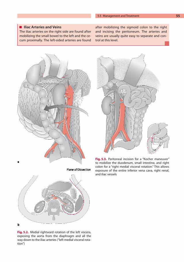
55
Fig. 5.2. Medial rightward rotation of the left viscera, exposing the aorta from the diaphragm and all the way down to the iliac arteries (“left medial visceral rota-tion”)
Fig. 5.3. Peritoneal incision for a “Kocher maneuver” to mobilize the duodenum, small intestine, and right colon for a “right medial visceral rotation.” This allows exposure of the entire inferior vena cava, right renal, and iliac vessels
Iliac Arteries and VeinsThe iliac arteries on the right side are found after mobilizing the small bowel to the left and the ce-cum proximally. The left-sided arteries are found
after mobilizing the sigmoid colon to the right and incising the peritoneum. The arteries and veins are usually quite easy to separate and con-trol at this level.
5.5 Management and Treatment

Chapter 5 Abdominal Vascular Injuries56
It is necessary to have previous experience in liver surgery to successfully accomplish “total” control of liver injuries, and the medial visceral rotation for suprarenal aortic and cava exposure may also be very difficult without experience.
Retrohepatic Injuries. Particularly cumbersome is control of injuries to the retrohepatic vena cava. This type of exposure is difficult because the liver covers the entire anterior surface of the vena cava. The low number of patients surviving long enough to arrive at the hospital with this type of injury also makes it hard for most surgeons to gather experience with it. The special problems encoun-tered concern the difficult access (because, as stated, the liver covers the vena cava) and the re-duced blood volume returning to the heart when the vena cava is clamped.
A number of methods have been suggested for control. One example is atriocaval shunting by in-serting a large tube into the vena cava through a hole in right atrium’s appendage. In the Technical Tips box, the technique for total clamping and control directly without adjunctive measures is described because we feel this may occasionally be a practical approach for controlling unmanageable bleeding from this area. For immediate control during the exploratory procedure for total control (clamping the aorta, the infrarenal vena cava, and the suprahepatic vena cava and doing the Pringle maneuver), the liver is compressed dorsally against the spine manually and by using lap pads. Control of bleeding by direct pressure is facilitated by di-viding the falciform ligament and tilting the liver downward. However, it is reasonable to refrain from attempting to repair injuries to the retrohe-patic vena cava and instead, as the only measure taken, pack the liver to reduce the bleeding.
NOTEIt is rarely sensible to try to repair retro-hepatic vena cava injuries in unstable patients.
Superior Mesenteric Artery Injuries. SMA in-juries can also be quite difficult to expose and control. The importance of the SMA for perfusing the intestine makes SMA injuries particularly cumbersome to manage. Delaying restoration of flow more than 4–6 h inevitably leads to bowel
necrosis and possibly death. “Medial visceral rota-tion” or “high” infrarenal aortic exposure provides access to the first 3–4 cm of the SMA, but the next part of the vessel is incorporated in the pancreas. Surgical hematomas in this area make the dissec-tion even more difficult. Therefore, it has been suggested that the pancreas shall be divided to expose SMA injuries. Another option is to leave the injured area and perform a bypass from the aorta to a distal part of the SMA and ligate it at its origin. When a large hematoma around the head of the pancreas is encountered and the bowel is ischemic, the middle part of the SMA is probably injured, and such a bypass can be attempted for maintaining bowel perfusion.
NOTEThe aorta, the renal arteries, and the proximal part of the SMA should not be ligated for control during damage control surgery.
Retroperitoneal Hematomas Particularly after blunt trauma, intact retroperito-neal hematomas are a common finding during laparotomy. If such hematomas are not bleeding actively or expanding, they should not be explored right away. Other injuries can be treated first if needed and if sufficient time is available, addition-al diagnostic work-up pursued. Hematomas with signs of active bleeding and those that appear to be expanding rapidly should be left intact until prox-imal and distal control is achieved.
Even small hematomas can harbor significant vessel injuries.
When the surgeon is selecting the approach for vascular exposure and control, the location of the hematoma should be considered. A midline hematoma superior to the transverse mesocolon indicates injury to the suprarenal aorta or its branches. If combined with ischemic bowel signs, injury to the SMA should be suspected. Blood in the area of the portal triad suggests hepatic artery or portal vein injury. A midline infrarenal aortic or vena cava injury is suspected when the hemato-ma is located below the mesocolon. Lateral perito-neal hematomas occur after renal vessel and pa-renchymal injuries. A pelvic hematoma indicate iliac vessel damage.
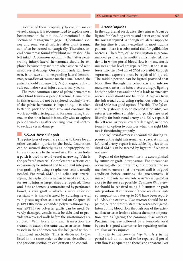
57
Because of their propensity to contain major vessel damage, it is recommended to explore most hematomas in the midline. As mentioned in the section on management (page 51), contained kid-ney and renal vessel injuries after blunt trauma can often be treated nonsurgically. Therefore, lat-eral hematomas found after blunt injury should be left intact. A common opinion is that, after pene-trating injury, lateral hematomas should be ex-plored because they are more often associated with major vessel damage. Our recommendation, how-ever, is to leave all nonexpanding lateral hemato-mas, regardless of trauma mechanism. Instead, the patient should undergo CT, IVP, or angiography to rule out major vessel injury and urinary leaks.
The most common cause of pelvic hematomas after blunt trauma is pelvic fracture. Hematomas in this area should not be explored routinely. Even if the pelvic hematoma is expanding, it is often better to pack the pelvic area and continue the work-up with arteriography. For penetrating trau-ma, on the other hand, it is usually wise to explore pelvic hematomas after securing proximal control to exclude vessel damage.
5.5.2.4 Vessel RepairThe principles of repair are similar to those for all other vascular injuries in the body. Lacerations can be sutured directly, using polypropylene su-ture appropriate to the vessel size. For larger holes a patch is used to avoid vessel narrowing. Vein is the preferred material. Complete transections can occasionally be sutured end to end, but interposi-tion grafting by using a saphenous vein is usually needed. For renal, SMA, and celiac axis arterial repair, the saphenous vein can be used as it is, but for aortic injuries larger sizes are required. Then, and if the abdomen is contaminated by perforated bowel, a vein graft – which is more infection resistant – is manufactured by suturing several vein pieces together as described on Chapter 15, p. 189. Otherwise, expanded polytetrafluoroethyl-ene (ePTFE) or polyester grafts can be used. Se-verely damaged vessels must be debrided to pro-vide intact vessel walls before the anastomoses are sutured. Vein lacerations and transection are treated in exactly the same way as arteries. Some vessels in the abdomen can also be ligated without significant morbidity. This is discussed below, listed in the same order as the areas described in the previous section on exploration and control.
Arterial Injuries In the suprarenal aortic area, the celiac axis can be ligated for bleeding control and better exposure of the aorta if injured. Although collateral supply to the intestine is usually excellent in most trauma patients, there is a substantial risk for gallbladder necrosis. Therefore, celiac axis ligation is recom-mended primarily in multitrauma high-risk pa-tients in whom portal blood flow is intact. Aortic injuries at this level are repaired by 3-0 or 4-0 su-tures. The first 3–4 cm of SMA accessible through suprarenal exposure must be repaired if injured. The middle portion can be ligated provided that blood flow through the celiac axis and inferior mesenteric artery is intact. Accordingly, ligating both the celiac axis and the SMA leads to extensive necrosis and should not be done. A bypass from the infrarenal aorta using saphenous vein to the distal SMA is a good option if feasible. The left re-nal artery should also be mended if possible; 5-0 sutures are often suitable, and patches are used liberally for both renal artery and SMA repair. If the left renal artery is severely damaged, nephrec-tomy is an option to consider when the right kid-ney is functioning properly.
The right renal artery is encountered during ex-posure of the right infrarenal vena cava. As for the left renal artery, repair is advisable. Injuries to the distal SMA can be treated by ligature if repair is not easy.
Repair of the infrarenal aorta is accomplished by suture or graft interposition. For thrombosis occurring after blunt trauma, it is important to re-member to ensure that the vessel wall is in good condition before suturing the anastomosis. If injured, the inferior mesenteric artery is ligated as close to the aorta as possible. Common iliac arter-ies should be repaired using 5-0 sutures or graft interposition. If either one of these vessels is ligat-ed, amputation rates up to 50% have been report-ed. Also, the external iliac arteries should be re-paired, but the internal iliac arteries can be ligated. Interrupting blood flow through one of the exter-nal iliac arteries leads to almost the same amputa-tion rate as ligating the common iliac arteries. Proximal ligature followed by a femorofemoral bypass is a good alternative for repairing unilat-eral iliac artery injuries.
Injuries to the common hepatic artery in the portal triad do not need to be repaired if portal vein flow is adequate and there is no apparent liver
5.5 Management and Treatment

Chapter 5 Abdominal Vascular Injuries58
damage. If the proper hepatic artery is ligated, the gallbladder may become gangrenous and should be excised liberally. If possible, lacerations in the proper hepatic artery should be sutured, but the artery must be separated from the portal vein and the common bile duct to avoid injuries to these structures. Splenic and gastric arteries can be ligated without morbidity.
Venous Injuries In general, venous injuries are more difficult to manage than arterial ones. There are several rea-sons for this. It is more difficult to expose and re-pair vein injuries due to their thin and fragile walls. Distal control is also more difficult to achieve. While arterial backbleeding often is sparse when the patient is in shock, distal bleeding from injured veins increases after proximal con-trol. For surgeons without experience in venous surgery, the consequence is that it is difficult to re-pair major venous injuries. Fortunately, many veins can be ligated in difficult situations.
The left renal vein encountered during suprare-nal aortic exposure can be ligated, preferably as close to vena cava as possible to allow alternative outflow through collaterals. Injured veins around the celiac axis can also be ligated. If possible, the proximal superior mesenteric vein should be re-paired. This vein lies in close connection to the SMA. Control is achieved by manual or rubber-band occlusion while suturing the defect. If repair is not possible, ligation leads to venous congestion of the intestine. In general, this is quite well toler-ated, and the patient usually survives. However, if the patient becomes hypotensive in the postopera-tive period, it may be fatal.
Infrahepatic vena cava injuries should be re-paired if possible. Interrupted 4-0 sutures can be used for most lacerations. For stab wounds pene-trating both the ventral and dorsal part of the vein, access for repair includes extending the anterior opening to be able to close the hole on the dorsal side from the inside. Alternatively, the vena cava is dissected free and the lumbar branches secured and rolled over to expose the wound for suturing. (See Fig. 5.4.)
Small dorsal vena cava injuries not actively bleeding can be observed. In multiply injured pa-tients in bad condition, ligation rather than repair may be preferable. This leads to leg swelling in the
postoperative period but is usually well tolerated. No effort should be spared to repair the right renal vein if injured because, in contrast to the left side, collateral venous outflow is essentially lacking. If the vein must be ligated in difficult situations, right-sided nephrectomy is warranted. Also, the distal parts of the superficial mesenteric vein should be repaired if straightforward. Portal vein injuries are taken care of by venoraphy or graft interposition using 5-0 sutures if reasonably easy. Portacaval shunts have also been constructed to repair injuries to the portal vein. It the patient is hypotensive and hypothermic with extensive injuries, it is wise to ligate the portal vein. In most patient series, this maneuver is reported to be associated with survival and low postoperative portal hypertension rates.
NOTERepair of the right renal vein is important to save renal function on this side.
Suspected injuries to the retrohepatic vena cava area should be packed, and this is often sufficient for permanent bleeding control. Repair of injuries to the vena cava behind the liver and the few cen-timeters of the right and left hepatic veins outside it requires total vascular control as described pre-viously. A few successful cases have been reported in the literature. To facilitate repair, one branch from the hepatic vein can be ligated without mor-bidity. If the total venous outflow is compromised by interruption of the entire hepatic vein, lobec-tomy may be necessary. Clips can control caudate veins behind the liver. Anecdotally, retrohepatic caval injuries have been repaired through a liver injury separating the lobes. Final access to the cava may then be achieved by separating parts of any remaining liver tissue using the “finger frac-ture” technique.
Damaged common iliac veins and the first parts of the vena cava are difficult to expose for repair. The aortic bifurcation and the common iliac ar-teries must be freed entirely to allow mobilization and control of the veins. This includes division of lumbar arteries and the sacral artery. As men-tioned, temporary division of the left iliac artery is often required to provide exposure of the left iliac vein. Polypropylene suture, 5-0, is appropriate for repair. A good option for multiply injured patients

59
in shock is ligation of the distal vena cava or the common iliac vein.
Distal iliac vein injuries should be repaired. Li-gation of the internal iliac vein often facilitates re-lease of the external iliac vein and provides better exposure of the injured site. In high-risk patients if repair is not feasible, a good option is ligation. Un-fortunately, distal control of internal iliac veins is difficult. Often the best way is to use compression with a sponge-stick for distal control while sutur-ing the lacerations. It is important to reduce bleed-ing by closing the hole even if narrowing or ob-struction of the vein is the final result.
Final Vascular Repair After “Damage Control”
With any luck the patient will have improved he-modynamically after a period of resuscitation in the intensive care unit and does not have hypo-
thermia, coagulopathy, or acidosis and is more stable. He or she is then returned to the operating room for final repair of vascular and other inju-ries. When arterial injury is suspected at the pri-mary operation, angiography should be performed first to identify and provide information before repair. This can take place any time between a few hours to 10 days after the primary operation. The second operation consists of meticulous explora-tion of injured areas still bleeding, including he-matomas and cavities. Any recurrent bleeding is controlled and repaired as outlined previously. Shunted vessel segments must also be controlled and repaired. It is difficult to give well-founded advice regarding final repair of previously ligated vessels. A suggestion is to consider the hepatic ar-tery and the SMA for secondary repair. It is usu-ally not worthwhile to try to mend ligated veins. After final repair of organ and intestinal injuries,
Fig. 5.4. a Manual control of bleeding from an injury in the ventral wall of vena cava. b Repair of the dor-sal injury of the vena cava through an anterior injury after stabbing through both walls. Note that no vascu-
lar clamps are used for bleeding control. c Repair of a dorsal injury after separation and rotation of the vena cava
5.5 Management and Treatment

Chapter 5 Abdominal Vascular Injuries60
the packs are removed and the abdomen closed. It is not uncommon that renewed hemorrhage ne-cessitates repacking and a second period in the intensive care unit. It the literature this is reported to happen in up to 10% of patients.
5.5.2.5 Finishing the Operation After vascular repair, other injuries are taken care of. For a detailed description, we recommend trauma textbooks. If the peritoneal cavity is con-taminated, careful cleansing using warmed fluids is recommended. If possible, vascular anastomo-ses should be covered with tissue. If the SMA and proximal aorta are injured, it is important to as-sess the viability of the intestine before closing the abdomen. Sites of vessel repair should also be checked one more time. Minor – and even quite substantial – bleeding from such areas can be managed by hemostatic adjuvant therapy, such as local application of fibrin glue or gel (page 189).
5.5.3 Endovascular Treatment
Endoluminal aortic stent-graft repair has become a possible option for blunt aortic injuries missed during initial exploration, especially in the tho-racic part of the aorta. In some of cases reported in the literature, the injured aortic site causing dis-section was treated by fenestration and stent place-ment. Other patients had stable hematomas that were examined with CT and found to involve par-tial aortic occlusion. Also, injuries in the common iliac artery caused by pelvic fracture have been treated by stent-grafts. In one series, a few patients had iliac artery occlusions that were passed with a guide wire and then successfully treated with a covered stent. This approach may be particularly tempting when conventional repair is not possible due to associated injuries and pelvic hematoma. Angiography and subsequent embolization of branches from the internal iliac artery for bleed-ing due to pelvic fracture is successful in many instances. One should remember that in up to 5% of patients, gluteal muscle necrosis occurs after such branch embolization.
Blunt and penetrating renal trauma can also be managed by endovascular methods. Selective em-bolization of bleeding renal artery branches is of-ten successful. Isolated dissection and subsequent
thrombosis of a renal artery after blunt trauma di-agnosed during early management is preferably treated by angioplasty and stenting, providing that angiography facilities are available and that such management does not delay final treatment.
Blunt abdominal trauma causing splenic injury can also be treated by endovascular embolization. In most published patient series, CT has been in-sufficient for selecting patients for endovascular therapy, and diagnostic angiography is recom-mended to rule out this possibility. High-quality CT angiography, however, readily identifies such lesions. Observed patients who continue to require fluids and blood because of the organ injury should undergo arteriography to rule out treatable injuries. Examples are intraperitoneal or intrapa-renchymal contrast extravasation and vessel trun-cation, which are all amenable to embolization. Treatment then consists of selective catheteriza-tion and injection of microcoils.
The late consequences of abdominal vascular injuries – pseudoaneurysm and arteriovenous fis-tula – can also be treated by endovascular meth-ods in most locations. To our knowledge, there are no reports of successful endovascular treatment of venous injuries in the abdomen.
5.5.4 Management After Treatment
It is obvious that patients with abdominal vascular injuries have a high risk for developing serious complications in the postoperative period. Hypo-tension due to continued blood loss is common, and reoperation should be employed liberally. Vis-ceral and leg ischemia may also occur due to li-gated or thrombosed repaired vessel segments. The abdominal appearance and leg perfusion must therefore be monitored meticulously in the post-operative period. Examination should, besides ab-dominal palpation, consist of a rectal examination and inspection of the nasogastric tube to check for blood. Renal artery thrombosis may manifest as flank pain and a temporary rise in serum creati-nine. Occasionally, emergency nephrectomy is necessary in the postoperative period due to pain or a very high blood pressure.
As mentioned before, it is extremely important to keep the blood pressure at adequate levels if the intestinal blood supply is compromised by a delib-

61
erate ligation during exploration. Extra careful cardiac monitoring, fluid resuscitation, and phar-macological blood pressure adjustment are war-ranted. If intestinal ischemia is suspected, imme-diate relaparotomy is indicated.
Swelling after vein ligation or thrombosis of a repaired major vein segment is also a common problem. The measures recommended to mini-mize this problem are supplying the patient with compression stockings and infusing dextran to optimize the rheology of the blood. Furthermore, as soon as the patient is hemodynamically stable, standardized heparinization should be initiated. Patients with repaired injuries in the portal vein and the superior mesenteric vein may also develop portal hypertension and hepatic failure.
Antibiotics should be continued postopera-tively. Patients arriving in shock are prone to infection, especially if intestinal perforation is part of the trauma spectrum. Careful monitoring of infection signs is necessary, and CT examina-tion is indicated if intraabdominal infection is suspected.
5.6 Results and Outcome
Outcome after abdominal vascular trauma is strongly related to whether shock is present at ar-rival. The time elapsing from the trauma to the patient’s arrival at the hospital is important. For example, few patients survived penetrating ab-dominal vascular trauma during World War II, whereas 42% did during the Vietnam War. In se-ries from civilian life looking at survival of pa-tients with aortic or vena cava injuries arriving alive to the hospital, around half have been report-ed to survive. Besides shock, free bleeding in the peritoneal cavity and suprarenal location of the injury are risk factors for poor outcome. Survival rates after blunt trauma are around 75% in the literature. Observational studies including 200 pa-tients or more list suprarenal or juxtarenal aortic injuries, retrohepatic and hepatic vein injuries, and portal vein injuries as associated with the highest mortality.
It is more difficult to find data on survival rates for isolated injuries to a specific vessel. One report of isolated arterial injuries or those combined with other arterial injuries in the abdomen found mor-
tality to range from 30% for hepatic artery to 80% for aortic injuries. The mortality for renal, iliac, and SMA injuries was around 50–60%.
Abdominal venous trauma is also associated with high mortality due to exsanguination. Over-all, mortality ranges from 30–70%. The worst re-sults come from patient series of retrohepatic vena cava injuries, reporting a mortality of over 90%. Also, portal vein and superior mesenteric vein in-juries lead to substantial mortality. In one study, 30% died after lateral repair of the portal vein and 78% after ligation of this vessel. The latter proce-dure, however, was performed in more severely injured patients with more associated injuries. Another study reported only 20% mortality after portal vein ligation. In patients with only venous injuries or in combination with other venous trau-ma, the mortality rates were 75% for inferior vena cava injury, 72% for portal vein injury, 56% for renal vein injury, and 44% for iliac vein injury.
5.7 Iatrogenic Vascular Injuries in the Abdomen
It is not uncommon that vessels are injured during abdominal surgery for malignancy or other proce-dures. Some procedures are particularly prone to cause injury to abdominal vessels. A discussion on some of these follows below. The principles of repair are essentially the same as for traumatic injury caused by accidents or violence.
5.7.1 Laparoscopic Injuries
Trocars used for laparoscopic access frequently cause injury to major blood vessels in the abdo-men. When the aorta or vena cava is injured, out-come may even be fatal. The insufflation needle may also cause severe injuries. Injury is more com-mon in thin patients who have previously under-gone abdominal operations and in patients in whom a blind technique for inserting the trocar is used. When blood returns through the trocar or needle, a severe injury should be suspected. An-other situation indicating vascular injury occurs when the patient becomes hypotensive or when the abdomen swells rapidly before the gas is insuf-flated. If the aorta or iliac arteries are injured con-
5.7 Iatrogenic Vascular Injuries in the Abdomen
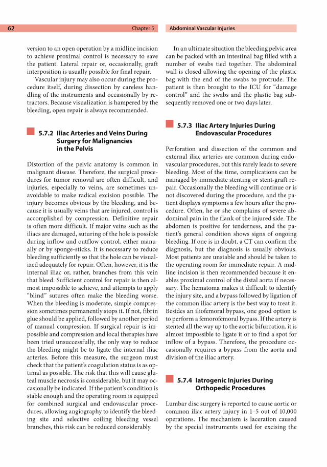
Chapter 5 Abdominal Vascular Injuries62
version to an open operation by a midline incision to achieve proximal control is necessary to save the patient. Lateral repair or, occasionally, graft interposition is usually possible for final repair.
Vascular injury may also occur during the pro-cedure itself, during dissection by careless han-dling of the instruments and occasionally by re-tractors. Because visualization is hampered by the bleeding, open repair is always recommended.
5.7.2 Iliac Arteries and Veins During Surgery for Malignancies in the Pelvis
Distortion of the pelvic anatomy is common in malignant disease. Therefore, the surgical proce-dures for tumor removal are often difficult, and injuries, especially to veins, are sometimes un-avoidable to make radical excision possible. The injury becomes obvious by the bleeding, and be-cause it is usually veins that are injured, control is accomplished by compression. Definitive repair is often more difficult. If major veins such as the iliacs are damaged, suturing of the hole is possible during inflow and outflow control, either manu-ally or by sponge-sticks. It is necessary to reduce bleeding sufficiently so that the hole can be visual-ized adequately for repair. Often, however, it is the internal iliac or, rather, branches from this vein that bleed. Sufficient control for repair is then al-most impossible to achieve, and attempts to apply “blind” sutures often make the bleeding worse. When the bleeding is moderate, simple compres-sion sometimes permanently stops it. If not, fibrin glue should be applied, followed by another period of manual compression. If surgical repair is im-possible and compression and local therapies have been tried unsuccessfully, the only way to reduce the bleeding might be to ligate the internal iliac arteries. Before this measure, the surgeon must check that the patient’s coagulation status is as op-timal as possible. The risk that this will cause glu-teal muscle necrosis is considerable, but it may oc-casionally be indicated. If the patient’s condition is stable enough and the operating room is equipped for combined surgical and endovascular proce-dures, allowing angiography to identify the bleed-ing site and selective coiling bleeding vessel branches, this risk can be reduced considerably.
In an ultimate situation the bleeding pelvic area can be packed with an intestinal bag filled with a number of swabs tied together. The abdominal wall is closed allowing the opening of the plastic bag with the end of the swabs to protrude. The patient is then brought to the ICU for “damage control” and the swabs and the plastic bag sub-sequently removed one or two days later.
5.7.3 Iliac Artery Injuries During Endovascular Procedures
Perforation and dissection of the common and external iliac arteries are common during endo-vascular procedures, but this rarely leads to severe bleeding. Most of the time, complications can be managed by immediate stenting or stent-graft re-pair. Occasionally the bleeding will continue or is not discovered during the procedure, and the pa-tient displays symptoms a few hours after the pro-cedure. Often, he or she complains of severe ab-dominal pain in the flank of the injured side. The abdomen is positive for tenderness, and the pa-tient’s general condition shows signs of ongoing bleeding. If one is in doubt, a CT can confirm the diagnosis, but the diagnosis is usually obvious. Most patients are unstable and should be taken to the operating room for immediate repair. A mid-line incision is then recommended because it en-ables proximal control of the distal aorta if neces-sary. The hematoma makes it difficult to identify the injury site, and a bypass followed by ligation of the common iliac artery is the best way to treat it. Besides an iliofemoral bypass, one good option is to perform a femorofemoral bypass. If the artery is stented all the way up to the aortic bifurcation, it is almost impossible to ligate it or to find a spot for inflow of a bypass. Therefore, the procedure oc-casionally requires a bypass from the aorta and division of the iliac artery.
5.7.4 Iatrogenic Injuries During Orthopedic Procedures
Lumbar disc surgery is reported to cause aortic or common iliac artery injury in 1–5 out of 10,000 operations. The mechanism is laceration caused by the special instruments used for excising the

63
herniated disc. This injury generally presents as a substantial bleeding in the wound, with an associ-ated systemic hypotension. Occasionally, the diag-nosis becomes apparent after the procedure when signs of shock develop during the first postopera-tive hours. Even more common is that an arterio-venous fistula or pseudoaneurysm is found, which is diagnosed any time from a few hours after the procedure to several years postoperatively. Find-ings suggesting such injuries are, in descending order of frequency, bruits, heart failure, abdomi-nal pain, and hypotension. The disc level where the surgery is performed determines which vessel becomes injured. At the L4–L5 and L5–S1 levels, the common iliac artery and vein are injured. Higher up, the aorta and vena cava are at risk.
For emergency repair, a midline incision for ex-posure is needed, and the same principles are ap-plicable as for other types of trauma: lateral repair, patching, or graft insertion. Arteriovenous fistu-las and pseudoaneurysms may also be treated using endovascular methods.
During hip arthroplasty, the external iliac ves-sels or the common femoral artery may be injured. While uncommon at primary procedures, it hap-pens more often during revisions because of the need to remove previous prosthetic material and the anatomical alterations caused by previous surgery. The left side is more often injured. The mechanism is sometimes direct lacerations by ac-etabular screws, dissection, or traction injury, but more common is cement destruction of the ves-sels. Arterial repair is performed after obtaining proximal control of the common iliac artery. Usually, a “hockey-stick” incision is sufficient to obtain exposure. Destroyed vessel segments by cement need graft interposition or a bypass.
Further Reading
Baker WE, Wassermann J. Unsuspected vascular trau-ma: blunt arterial injuries. Emerg Med Clin North Am 2004; 22(4):1081–1098
Brown CV, Velmahos GC, Neville AL, et al. Hemody-namically “stable” patients with peritonitis after penetrating abdominal trauma: identifying those who are bleeding. Arch Surg. 2005; 140(8):767–772
Fuller J, Ashar BS, Carey-Corrado J. Trocar-associ-ated injuries and fatalities: an analysis of 1399 re-ports to the FDA. J Minim Invasive Gynecol 2005; 12(4):302–307
Gupta N, Solomon H, Fairchild R, et al. Manage-ment and outcome of patients with combined bile duct and hepatic artery injuries. Arch Surg 1998; 133(2):176–181
Lee JT, Bongard FS. Iliac vessel injuries. Surg Clin North Am 2002; 82(1):21–48
Malhotra AK, Latifi R, Fabian TC, et al. Multiplicity of solid organ injury: influence on management and outcomes after blunt abdominal trauma. J Trauma 2003; 54(5):925–929
Nicholas JM, Rix EP, Easley KA, et al. Changing pat-terns in the management of penetrating abdominal trauma: the more things change, the more they stay the same. J Trauma 2003; 55(6):1095–1108; discus-sion 1108–110
Parks RW, Chrysos E, Diamond T. Management of liver trauma. Br J Surg 1999; 86(9):1121–1135
Smith SR. Traumatic retroperitoneal venous haemor-rhage. Br J Surg 1988; 75(7):632–636
Sugrue M, D’Amours SK, Joshipura M. Damage control surgery and the abdomen. Injury 2004; 35(7):642–648
Weber S, Murphy MM, Pitzer ME, et al. Management of retrohepatic venous injuries with atrial caval shunts. AORN J 199664(3):376–377, 380–382
Further Reading

Acute Intestinal Ischemia 6
CONTENTS
6.1 Summary . . . . . . . . . . . . . . . . . . . . . . . . . . . . 65
6.2 Background . . . . . . . . . . . . . . . . . . . . . . . . . 656.2.1 Magnitude of the Problem
and Patient Characteristics . . . . . . . . . . 66
6.3 Pathophysiology . . . . . . . . . . . . . . . . . . . 66
6.4 Clinical Presentation . . . . . . . . . . . . . . . 676.4.1 Medical History . . . . . . . . . . . . . . . . . . . . . . 676.4.1.1 Embolism . . . . . . . . . . . . . . . . . . . . . . . . . . . . 676.4.1.2 Thrombosis . . . . . . . . . . . . . . . . . . . . . . . . . . 676.4.2 Physical Examination . . . . . . . . . . . . . . . . 68
6.5 Diagnostics . . . . . . . . . . . . . . . . . . . . . . . . . 686.5.1 Laboratory Tests . . . . . . . . . . . . . . . . . . . . 686.5.2 Angiography . . . . . . . . . . . . . . . . . . . . . . . . 696.5.3 Other Options . . . . . . . . . . . . . . . . . . . . . . . 706.5.4 Diagnostic Pitfalls . . . . . . . . . . . . . . . . . . . 70
6.6 Management and Treatment . . . . . . 706.6.1 Management Before Treatment . . . . . 706.6.1.1 In the Emergency Department . . . . . . 706.6.2 Operation . . . . . . . . . . . . . . . . . . . . . . . . . . . 716.6.2.1 Embolic Occlusion . . . . . . . . . . . . . . . . . . 716.6.2.2 Arterial Thrombosis . . . . . . . . . . . . . . . . . 716.6.2.3 Venous Thrombosis and NOMI . . . . . . 726.6.2.4 Endovascular Treatment . . . . . . . . . . . . 736.6.3 Management After Treatment . . . . . . 73
6.7 Results and Outcome . . . . . . . . . . . . . . . 73
Further Reading . . . . . . . . . . . . . . . . . . . . 74
6.1 Summary
Triad of symptoms 1. History of embolization2. Pain out of proportion3. Intestinal emptyingUrgent management is essential: rehydra-tion, angiography and laparotomyIf arterial obstruction – aggressive surgical treatmentIf venous obstruction – restrictive with surgical treatmentEmbolectomy if jejunum is normal
6.2 Background
Acute intestinal ischemia is often a fatal disease, and many patients with this disorder will die re-gardless of treatment. Increased awareness and rapid management can improve this pessimistic course. Using wide definition acute intestinal ischemia is hypoxia of the small intestinal wall due to a sudden decrease of perfusion caused by emboli or arterial or venous thrombosis. The symptoms are not specific, and the diagnosis is regularly established at laparotomy late in the course when peritonitis has developed. With rapid and efficient management, including an aggres-sive diagnostic work-up, the number of successful embolectomies can increase and the need for ex-tensive intestinal resections can be diminished. The diagnosis must be established early in the course of the disease. A high level of clinical suspi-cion when evaluating acute abdominal pain, prompt management in the emergency depart-ment, and early angiography or laparotomy is required to achieve this.

Chapter 6 Acute Intestinal Ischemia66
6.2.1 Magnitude of the Problem and Patient Characteristics
Even if patients with acute intestinal ischemia are usually admitted and treated by general surgeons, cooperation with a vascular surgeon may be a possible way to improve treatment results. Vascu-lar surgeons contribute with their experience of angiography as well as with operations in the area around the superior mesenteric artery (SMA).
The disease is relatively uncommon. Among all patients arriving in the emergency department be-cause of abdominal pain, 0.5 % have acute intesti-nal ischemia. The true incidence is probably high-er because patients can be suspected to die from intestinal ischemia without an established diagno-sis. The relatively low incidence in combination with the imprecise symptoms and moderate find-ings at physical examination early in the course of the disease contribute to the bad prognosis. In ob-servational studies the 30-day mortality is 60–85% for patients who are not treated surgically with the diagnosis established by angiography or physical examination. One more factor contributing to the poor prognosis is that this category of patients consists of elderly who have complicating diseases such as chronic obstructive pulmonary disease and generalized arteriosclerosis, including coro-nary disease. In most studies, the mean patient age is around 70 years. Two-thirds of the patients are female.
Intestinal ischemia secondary to mesenteric ve-nous thrombosis is associated with another group of patients and has a significantly better progno-sis. The 30-day mortality is around 30%. Five to 15% of all cases presenting with intestinal isch-emia are caused by venous thrombosis.
6.3 Pathophysiology
The main blood supply to the small intestine comes from the SMA, which also perfuses the first half of the colon. The inferior mesenteric artery and branches from the internal iliac arteries sup-ply the distal part of colon and rectum. This dou-ble blood supply and an extensive collateral net-work explain why occlusion of the inferior mesen-teric artery seldom causes severe ischemia in the distal colon. Primary ischemia of the colon is
unusual and is further discussed in Chapter 12 on complications in vascular surgery. The rest of this chapter will deal with acute ischemia of the small intestine.
NOTEOcclusion of the SMA has devastat-ing effects on the perfusion of the intestine.
Because almost the entire small intestine gets its blood supply from one single artery, a sudden oc-clusion of this vessel has major consequences. The initial response is spasm and vigorous contrac-tion. Because of its high metabolic activity 80% of the blood supply to the intestine is consumed by the mucosa. This explains why the mucosa is dam-aged before the rest of the intestinal wall is. The cells at the tip of the villi are most sensitive and die first. Under the microscope, ischemic changes can be seen in the mucosa within 30 min after occlu-sion. Patients with SMA occlusion will, very early after onset, vomit and have diarrhea and abdomi-nal pain. Occasionally they have blood in their stools. Granulocytes are also activated early, and oxidants and proteolytic enzymes affect the intes-tine. Hypotension develops as the next step in the course of the disease and contributes to further ischemic damage of the intestinal wall. This is followed by diffuse necrosis in the mucosa that spreads to the submucosal layer and finally ex-tends through the entire intestinal wall. The result is transmural infarction and local peritoni-tis. The intestine then may perforate, and the patient develops general peritonitis. Metabolic acidosis, dehydration, anuria, and multiple organ failure could be the end result.
The main etiology of acute intestinal ischemia is embolization or thrombosis of the SMA, both being equally common. In general, an embolus oc-cludes a relatively healthy artery with immediate dramatic consequences as described above, where-as a thrombotic occlusion is preceded by a steno-sis, allowing collaterals to develop. The artery may then occlude without causing symptoms or isch-emic damage to the intestine.
A less common cause is venous thrombosis. This frequently affects younger patients and typi-cally is secondary to trauma, inflammation, and other diseases in which hypercoagulation is com-

67
mon. It may also be a consequence of congenital coagulation disorders.
Other more unusual causes for acute intestinal ischemia, which are not within the scope of this book, are embolic or thrombotic occlusion of the celiac trunk and the low-flow state nonocclu-sive intestinal ischemia (NOMI), a result of severe cardiac dysfunction.
6.4 Clinical Presentation
6.4.1 Medical History
In many patients the initial clinical presentation of SMA obstruction is vague, making diagnosis difficult. A triad of symptoms in the patients’ medical history should make the surgeon suspi-cious for acute intestinal ischemia caused by occlusion of the SMA:1. Severe periumbilical pain (“pain out of propor-
tion”)2. Vomiting and/or diarrhea (“gut-emptying”)3. Possible source of an embolus, or a previous
embolization in the medical history.
NOTEIt is important to remember the triad of symptoms associated with occlusion of SMA.
6.4.1.1 Embolism For a typical patient with embolic occlusion of the SMA, the symptoms include all three elements of the triad. These are then sufficient for deter-mining the diagnosis, as well as for differentiating it from other causes of acute abdominal pain and thrombosis of the same artery. The pain, which often precedes vomiting or diarrhea, is the key symptom. It has a dramatic precipitous onset and is localized in the paraumbilical region. The pain is usually severe and colicky. The expression “pain out of proportion” indicates that there is a discrepancy between the findings in the physical examination of the abdomen and the pain inten-sity. The pain disappears when the intestine becomes necrotic, which may create a pain-free interval that frequently is misinterpreted as if the patient has improved. The pain returns when the intestine perforates. Ninety-five percent of
these patients have a history of previous cardiac disease, and 30% have had earlier episodes of em-bolization to other vascular systems (Table 6.1). Embolization is common after acute myocardial infarction, debut of arterial fibrillation, and as a complication of angiography and endovascular treatment.
6.4.1.2 Thrombosis Thrombosis of the SMA occurs in patients with general arteriosclerosis and a history remarkable for previous manifestations of cardiac and periph-eral vascular disease. Sometimes symptoms of chronic intestinal ischemia also are present. The onset of symptoms after acute thrombotic occlu-sion is more insidious than for embolic disease. The pain is usually constant and progressive over several hours but is otherwise similar to what has been described for embolism. (See Table 6.2.)
For thrombosis of the mesenteric vein, the du-ration of symptoms is commonly several days and the symptoms are even more imprecise than for arterial occlusion. The pain is less pronounced but is present to some degree in 90% of patients. Fever is also a common sign. Eighty-five percent of patients have a history of hypercoagulation disor-ders such as deep venous thrombosis or have had other diseases or risk factors predisposing them to thrombosis. Examples include pregnancy, oral contraceptive use, malignancy, inflammatory dis-eases, portal hypertension, and trauma.
6.4 Clinical Presentation
Table 6.1. Percentage of patients with symptoms and laboratory findings at the time of admission to the hos-pital, where the diagnosis acute intestinal ischemia due to arterial occlusion was established later
Symptoms/finding Frequency
Abdominal pain 100%Diarrhea or vomiting 84%Previous embolization/source of emboli
33%
Blood in stools 25%Elevated lactate in plasma 90%Leukocytosis 65%Metabolic acidosis 60%

Chapter 6 Acute Intestinal Ischemia68
6.4.2 Physical Examination
Findings at physical examination in acute intesti-nal ischemia can be vague and difficult to inter-pret. It is still, however, very important to care-fully examine the patient. The examination reveals signs of arteriosclerosis – carotid bruits, heart murmur, and so on – as well as sources of embolus. Abdominal examination findings are the basis for emergency management. For instance, without signs of peritonitis, a patient should not undergo laparotomy when venous thrombosis is the sus-pected diagnosis. A patient with arterial occlusion, however, needs surgery before peritonitis evolves.
The abdominal findings vary with the time point during the course of the illness when the pa-tient is examined. Anything from normal findings to general peritonitis may be found. In early stag-es, a slight tenderness and amplified bowel sounds are common findings, but when peritonitis is es-tablished, tenderness with muscular guarding and a lack of bowel sounds due to paralysis are found. Abdominal distension is a very late sign in the course of the disease. The examination should also assess the patient’s general condition, includ-ing possible dehydration.
6.5 Diagnostics
For the majority of patients with the triad of symp-toms described the need for further diagnostic work-up is limited and immediate laparotomy is indicated. Laboratory tests can support the diag-nosis but should not delay management and treat-
ment. The only radiologic examination that is warranted, besides computed tomography (CT) for diagnosing suspected venous thrombosis, is angiography and perhaps plain x-ray. The resourc-es and expertise available in the hospital should also influence the decision of whether any further investigations or tests are performed.
6.5.1 Laboratory Tests
The leukocyte count is elevated early in the dis-ease course. Together with the clinical triad, a leu-kocyte count higher than 15 109/l is pathogno-monic for acute intestinal ischemia. Values above normal for serum lactate and D-dimer have also been suggested as prognostic markers for patients who need surgery. A lactate concentration exceed-ing 2.6 mmol/l is considered to have a high sensi-tivity (90–100%) for acute mesenteric ischemia, meaning that only one patient in 10 with intestinal ischemia has a value <2.6 mmol/l and is at risk to be missed by this test. The specificity with this cut-off value, however, is rather low (around 40%). Overall, provided that shock, diabetes, severe re-nal insufficiency, and pancreatitis have been ruled out, an elevated plasma lactate indicates that the patient has a disease very likely to be acute intesti-nal ischemia, and that the patient definitely has a disease that needs surgery. More pronounced leu-kocytosis and elevated hemoglobin and hemato-crit values are secondary to plasma losses in the injured intestine. Later, when the intestinal wall becomes necrotic and blood leaks into the intesti-nal lumen, hemoglobin and hematocrit decrease.
Table 6.2. Differentiation between causes of intestinal ischemia (DVT deep vein thrombosis)
Arterial embolism Arterial thrombosis Venous thrombosis
Older + + –Younger – – +Previous symptoms of chronic intestinal ischemia
– + –
Previous DVT – – +Possible source emboli + – –Sudden onset + – –Insidious onset – + +

69
Metabolic acidosis also occurs late in the course of the disease, and as a diagnostic test it has no value. The acid-base balance, however, needs to be moni-tored and corrected continuously during the course of treatment as a general measure.
6.5.2 Angiography
In hospitals where angiography is available 24 h a day and can be performed rapidly, it is recom-mended before laparotomy for most patients sus-pected of having this disease. Exceptions are pa-tients with peritonitis. Angiography can possibly be preceded by a plain x-ray to exclude free gas in the abdomen. Besides establishing the diagnosis, angiography is also helpful for separating the dif-ferent etiologies for acute intestinal ischemia:1. Embolization to the SMA: Typically, this ap-
pears as a “meniscus” occlusion located 5–7 cm out in the SMA, which has its first branches open and filled with contrast. Such emboli are usually possible to extract by simple embolec-tomy.
2. Arterial thrombosis in previously atheroscle-rotic arteries: On the films an occlusion of the SMA is found approximately 1–2 cm from its origin and no distal branches are filled with contrast. Sometimes the patient can then be reconstructed with a bypass from the aorta. In many circumstances, however, it is wise to avoid laparotomy if the contrast does not reach any part of the SMA. Total SMA thrombosis is rarely curable by reconstructive vascular sur-gery. Thrombolysis may then be an option if a guide wire can be inserted into the artery.
3. Venous thrombosis or NOMI: If the branches from the SMA can be followed some distance out in the mesentery – more than 10 cm from the origin – and the contrast is moving slowly, the finding indicates a state of threatening in-farction without arterial occlusion. This can be due to either venous thrombosis or NOMI. Laparotomy is not indicated in such patients.
NOTEEmergency angiography is often helpful for diagnosing and managing acute intestinal ischemia.
The surgeon is responsible for making sure that angiography will not cause an unacceptable delay in the management process. It should be per-formed with close observation of the patient, including continuous monitoring of vital signs and abdominal status. In hospitals without avail-able angiography, management has to be based on clinical findings only. If this investigation resource is available, however, and the department has experience with emergency angiography, it is rec-ommended.
TECHNICAL TIPSThe technique for angiography in acute mesenteric ischemia
1. Scrub and dress for groin puncture.2. Shoot one plain frontal and one lateral x-ray
(to exclude free gas).3. Puncture the femoral artery. Insert a guide
wire and any angiography catheter. Place the tip at the level of the first lumbar vertebra.
4. Withdraw the guide wire and rapidly inject by hand 10 ml of x-ray contrast. Images are first obtained in the frontal plane to visualize em-bolization to the SMA.
5. Repeat with the lateral projection (this is al-ways necessary to diagnose thrombosis when the SMA is occluded at the origin).
6. Consider injecting papaverine (1–2 ml of 40 mg/ml) through the catheter, preferably after its tip has been placed selectively into the SMA.
7. Pull the catheter and control the puncture site by digital compression.
6.5.3 Other Options
Ultrasound, including determination of flow ve-locity and color coding (duplex), is often techni-cally difficult to perform because of obscuring in-testinal gas and is not recommended for diagnos-ing acute intestinal ischemia. There are occasional reports in the literature, however, about successful visualization of an occluded SMA that has been helpful for diagnosis. Another possible benefit of an ultrasound examination is to exclude other
6.5 Diagnostics

Chapter 6 Acute Intestinal Ischemia70
causes of the patient’s symptoms such as renal or gall bladder diseases.
When there is a strong suspicion of venous thrombosis – for example in young patients with a history of hypercoagulation who have mild pro-longed symptoms without peritonitis on physical examination – a CT scan with contrast should be performed to establish the diagnosis. The findings on the CT scan that indicate thrombosis are thrombus in the superior mesenteric vein and occasionally in the portal and splenic veins to-gether with splenomegaly. Gas bubbles in these veins may also be found.
6.5.4 Diagnostic Pitfalls
The three main difficulties in diagnosing and managing patients with acute mesenteric ischemia are (1) to suspect the diagnosis, (2) to make the diagnosis fast enough, and (3) to differentiate between thrombotic and embolic etiologies.
Patients with ruptured abdominal aortic aneu-rysms, a ruptured urinary bladder, hemorrhagic pancreatitis, or a perforated ulcer may also have “pain out of proportion.” But their medical history and physical findings are usually sufficient to differentiate between these alternative diagnoses and acute intestinal ischemia. Moreover, for all patients with these diseases, except for pancreati-tis, emergency laparotomy is indicated, and a wrong preoperative diagnosis is not so harmful. If not earlier the correct diagnosis can then be estab-lished during surgery. Overall, a high level of suspicion and early laparotomy will probably save lives.
While an early diagnosis is essential, the delay caused by performing angiography is often worth the time. Besides establishing the diagnosis and avoiding unnecessary laparotomies, it will also support management decisions during surgery. The relatively low complication rate of angiogra-phy also motivates liberal use. It will not negative-ly affect the management of the few patients suspected to have acute intestinal ischemia who later turn out to have other diseases. Thrombolysis may also be a reasonable treatment option for acute mesenteric ischemia, further supporting an aggressive preoperative diagnostic work-up that includes angiography.
6.6 Management and Treatment
6.6.1 Management Before Treatment
6.6.1.1 In the Emergency Department Most patients admitted to the emergency depart-ment with the described typical combination of physical findings and medical history should un-dergo immediate angiography and laparotomy. Although patients with only segmental and not transmural ischemia may improve spontaneously because of sufficient collateral blood flow, this is difficult to identify preoperatively. As soon as the operating room has been notified, the following measures can be taken:1. Place at least one large-bore intravenous (IV)
line.2. Start infusion of fluids. Ringer’s acetate is the
first option, but dextran is an alternative, espe-cially if venous thrombosis is suspected.
3. Obtain an electrocardiogram.4. Draw blood for hemoglobin and hematocrit,
prothrombin time, partial thromboplastin time, complete blood count, creatinine, sodi-um, and potassium as well as a sample for blood type and cross-match. Consider ordering a D-dimer as well.
5. Draw arterial blood for acid-base balance, including lactate.
6. Obtain informed consent.7. Consider administering analgesics (5–10 mg
opiate IV).
Early involvement of the anesthesiologist to dis-cuss the patient’s condition and optimization of organ function is wise. If time allows, this work-up can be done in the intensive care unit. Any acidosis should be corrected, and blood and plasma infusions are often required. Administra-tion of drugs that reduce blood flow to the in-testine should be stopped as soon as possible. Such drugs include digitalis, calcium channel blockers, diuretics, and nonsteroidal anti-inflam-matory drugs. When the decision to operate is made the patient should receive analgesics; a suggestion is an opiate 5–20 mg IV. Antibiotics directed against intestinal bacteria, such as a cephalosporin and metronidazole, should also be given preoperatively.
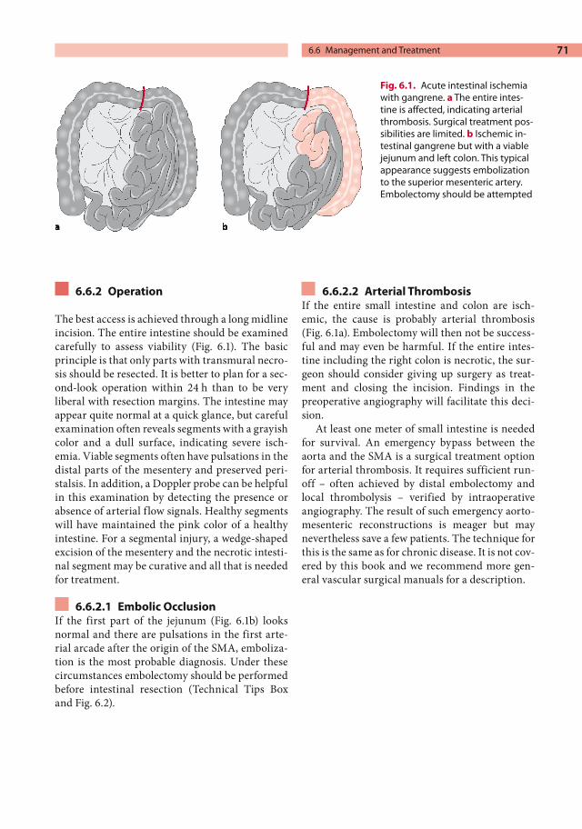
71
6.6.2 Operation
The best access is achieved through a long midline incision. The entire intestine should be examined carefully to assess viability (Fig. 6.1). The basic principle is that only parts with transmural necro-sis should be resected. It is better to plan for a sec-ond-look operation within 24 h than to be very liberal with resection margins. The intestine may appear quite normal at a quick glance, but careful examination often reveals segments with a grayish color and a dull surface, indicating severe isch-emia. Viable segments often have pulsations in the distal parts of the mesentery and preserved peri-stalsis. In addition, a Doppler probe can be helpful in this examination by detecting the presence or absence of arterial flow signals. Healthy segments will have maintained the pink color of a healthy intestine. For a segmental injury, a wedge-shaped excision of the mesentery and the necrotic intesti-nal segment may be curative and all that is needed for treatment.
6.6.2.1 Embolic Occlusion If the first part of the jejunum (Fig. 6.1b) looks normal and there are pulsations in the first arte-rial arcade after the origin of the SMA, emboliza-tion is the most probable diagnosis. Under these circumstances embolectomy should be performed before intestinal resection (Technical Tips Box and Fig. 6.2).
6.6.2.2 Arterial Thrombosis If the entire small intestine and colon are isch-emic, the cause is probably arterial thrombosis (Fig. 6.1a). Embolectomy will then not be success-ful and may even be harmful. If the entire intes-tine including the right colon is necrotic, the sur-geon should consider giving up surgery as treat-ment and closing the incision. Findings in the preoperative angiography will facilitate this deci-sion.
At least one meter of small intestine is needed for survival. An emergency bypass between the aorta and the SMA is a surgical treatment option for arterial thrombosis. It requires sufficient run-off – often achieved by distal embolectomy and local thrombolysis – verified by intraoperative angiography. The result of such emergency aorto-mesenteric reconstructions is meager but may nevertheless save a few patients. The technique for this is the same as for chronic disease. It is not cov-ered by this book and we recommend more gen-eral vascular surgical manuals for a description.
Fig. 6.1. Acute intestinal ischemia with gangrene. a The entire intes-tine is affected, indicating arterial thrombosis. Surgical treatment pos-sibilities are limited. b Ischemic in-testinal gangrene but with a viable jejunum and left colon. This typical appearance suggests embolization to the superior mesenteric artery. Embolectomy should be attempted
6.6 Management and Treatment

Chapter 6 Acute Intestinal Ischemia72
Fig. 6.2. Exposure of the superior mesenteric artery for embolectomy through an incision in the posterior peritoneum
TECHNICAL TIPSEmbolectomy of the Superior Mesenteric Artery
Move the transverse colon cranially and identify the SMA by using the fingers to palpate the area ventral to the pancreas behind the superior mes-enteric vein. This is facilitated by holding the mes-enteric root between the thumb and the fingers. Expose the artery by incising the dorsal peritone-um longitudinally just over the area where the pulse is lost (Fig. 6.2).
This sometimes requires partial division of the ligament of Treitz as well as inferior mobilization of the 4th portion of the duodenum. Apply vessel loops above and below the site of the intended arteriotomy. At least 4–5 cm needs to be exposed. Administer 5,000 units of heparin IV, clamp the ar-tery as close to the aorta as possible, and make a transverse arteriotomy distal to the clamp. Perform embolectomy with a #4 Fogarty cathe-ter. Start proximally while controlling bleeding through the arteriotomy using the vessel loop and a finger. Inflow is usually quite vigorous, and
it is important to not cause unnecessary bleeding. Continue distally towards the intestine with same catheter. A #3 catheter is occasionally needed to reach all the way out to the periphery. If the diag-nosis is correct, an embolus with a secondary thrombus is extracted, and the backflow is brisk. If not, try a second time with the catheter directed manually into the branches. Inject 2–4 ml of pa-paverine through a catheter into the SMA. If the backbleeding is inadequate, try to instill the same amount of rtPA into the distal branches. Close the artery with interrupted 6-0 prolene sutures. Place the intestine in its normal position and check the final result by palpating distal pulses and inspect-ing the intestine. If the viability of the intestine is uncertain, wait 20–30 min before deciding on what parts to remove. Finish the operation by re-secting nonviable parts as needed and close the abdomen.
If the intestine not is totally necrotic, it is sensible to wait for 30 min to see whether some segments of the intestine improve so that only a limited resection can be performed.
6.6.2.3 Venous Thrombosis and NOMI The entire intestine can also be affected in venous thrombosis and NOMI. At laparotomy (which should be avoided if possible), the intestine affect-ed by venous thrombosis will look hyperemic and swollen and may have petechial bleedings in the serosa. If these are found, the operation should be stopped, the abdominal wall closed, and a second-look operation planned. Systemic anticoagulation is the best treatment. At the second-look opera-tion, segments with petechial bleeding might be hard to differentiate from gangrene. Such seg-ments need to be carefully examined to avoid un-necessary resections. Devitalized intestine in seg-mental venous thromboses, however, should be resected with safe margins. This is different from what is recommended for intestinal ischemia with arterial causes. Venous thrombectomy and throm-bolysis have anecdotally been reported, but there is not much evidence that this is beneficial for the patient.

73
Patients with NOMI may also not be discovered before laparotomy. The intestine then displays the same appearance as for embolic and especially thrombotic obstruction of the main artery. Intra-operative diagnosis relies on pulse palpation and insonation of the distal vascular bed with continu-ous-wave Doppler. Patients with NOMI have pre-served pulses and flow signals – monophasic and throbbing – quite far out distally in the mesentery. These findings together with a typical medical history should be enough to stop the attempt to revascularize the intestine surgically, and the pa-tient needs optimization of cardiac function. The distinction from other arterial causes is difficult, however, and many patients with NOMI are likely to undergo embolectomy by mistake.
6.6.2.4 Endovascular Treatment At present only limited experience of thrombo-lytic therapy for acute mesenteric ischemia is available. Until 2003 about 50 cases had been re-ported in the literature. But the results are promis-ing and the technique may evolve as a primary choice in the future because it fits well with the aggressive work-up required.
The technique for thrombolysis involves groin access and diagnostic angiography as described, preferably with selective catheterization of the SMA and introduction of a guide wire into the thrombus. An end-hole or side-hole catheter is then advanced into the clot and the infusion start-ed by a bolus injection. The catheter is then pulled back somewhat and the continued infusion initi-ated. The preferable agent is rtPA.
6.6.3 Management After Treatment
While waiting for the second-look operation, the patient should, if possible, be monitored in the in-tensive care unit. Besides continued fluid losses from the injured intestine, toxic metabolites and proteolytic enzymes are released, which negatively influence heart and lung function. There is also a risk for septicemia because of bacterial transloca-tion. Therefore, rehydration, administration of plasma and blood, and antibiotic treatment are recommended in the early postoperative period. Anticoagulation with heparin should be contin-ued or begun in order to prevent further emboli-
zation and as general prophylaxis against throm-bosis, but it probably does not diminish further development of a thrombus in the intestine itself. Because the damaged intestine is prone to bleed, anticoagulation treatment also needs to be moni-tored carefully.
Reperfusion after a successful embolectomy contributes, as in acute leg ischemia, to morbidity and mortality. The primary damage caused by hypoxia of the intestinal wall is followed by a sec-ondary reperfusion injury. Therefore, patients with acute intestinal ischemia who have been re-vascularized are possible candidates for adjuvant pharmaceutical treatment to diminish the nega-tive effects on the central organs. The possible substances are often called “scavengers,” and ex-amples include superoxide-dismutase, allopuri-nol, and mannitol. Although all three of these do decrease mortality in animal models, there are presently no clinical trials to support their use in patients.
6.7 Results and Outcome
As mentioned earlier, the mortality associated with acute intestinal ischemia is reported to be very high. Intestinal resection as the only treat-ment will result in a 30-day mortality of 85–100%. If combined with embolectomy or vascular recon-struction, mortality can be reduced to 55%. In two studies with very positive results, it was proposed that mortality could be reduced to less than 45% if patients are managed very aggressively, with angi-ography in all patients (except for those with gen-eral peritonitis), followed by immediate laparoto-my. Ninety percent of the patients who survived in these two studies lost no or less than 30 cm of intestine. The most successful results were ob-served if the patient reached the operating room within 12 h after the onset of symptoms. Throm-bolysis in case series is reported to have excellent results, although the case mix was not comparable to open surgery series, and only a few patients had embolus as the etiology. It is likely that throm-bolysis should be attempted as the first option for thrombosis and that this will improve patient survival considerably. Accordingly, the results from most studies favor management and treat-ment as outlined in this chapter: aggressive diag-
6.7 Results and Outcome

Chapter 6 Acute Intestinal Ischemia74
nostic work-up, liberal indications for laparatomy, embolectomy, and reconstructive vascular sur-gery.
Further Reading
Angelelli G, Scardapane A, Memeo M, et al. Acute bowel ischemia: CT findings. Eur J Radiol 2004; 50(1):37–47
Burns BJ, Brandt LJ. Intestinal ischemia. Gastroenterol Clin North Am 2003; 32(4):1127–1143
Oldenburg WA, Lau LL, Rodenberg TJ, et al. Acute mes-enteric ischemia: a clinical review. Arch Intern Med 2004; 164(10):1054–1062
Schoots IG, Levi MM, Reekers JA, et al. Thrombolytic therapy for acute superior mesenteric artery occlu-sion. J Vasc Interv Radiol 2005; 16(3):317–329
Williams LF Jr. Mesenteric ischemia. Surg Clin North Am 1988; 68(2):331–353
Ward D, Vernava AM, Kaminski DL, et al. Improved outcome by identification of high-risk nonocclusive mesenteric ischemia, aggressive reexploration, and delayed anastomosis. Am J Surg 1995; 170(6):577–580

Abdominal Aortic Aneurysms 7
CONTENTS
7.1 Summary . . . . . . . . . . . . . . . . . . . . . . . . . . . . 75
7.2 Background . . . . . . . . . . . . . . . . . . . . . . . . . 757.2.1 Magnitude of the Problem . . . . . . . . . . . 757.2.2 Pathogenesis . . . . . . . . . . . . . . . . . . . . . . . . 76
7.3 Clinical Presentation . . . . . . . . . . . . . . . 767.3.1 Medical History . . . . . . . . . . . . . . . . . . . . . . 767.3.2 Examination . . . . . . . . . . . . . . . . . . . . . . . . . 767.3.3 Differential Diagnosis . . . . . . . . . . . . . . . 777.3.4 Clinical Diagnosis . . . . . . . . . . . . . . . . . . . 77
7.4 Diagnostics . . . . . . . . . . . . . . . . . . . . . . . . . 77
7.5 Management and Treatment . . . . . . 787.5.1 Management Before Treatment . . . . . 787.5.1.1 Ruptured AAA . . . . . . . . . . . . . . . . . . . . . . . 787.5.1.2 Suspected Rupture . . . . . . . . . . . . . . . . . . 797.5.1.3 Possible Rupture . . . . . . . . . . . . . . . . . . . . 797.5.1.4 Rupture Unlikely . . . . . . . . . . . . . . . . . . . . 807.5.2 Operation . . . . . . . . . . . . . . . . . . . . . . . . . . . 807.5.2.1 Starting the Operation . . . . . . . . . . . . . . . 807.5.2.2 Exposure and Proximal Control . . . . . 807.5.2.3 Other Options
for Proximal Control . . . . . . . . . . . . . . . . . 817.5.2.4 Continuing the Operation . . . . . . . . . . . 837.5.2.5 What to do While Waiting for Help . . 867.5.2.6 Endovascular Treatment . . . . . . . . . . . . 867.5.3 Management After Treatment . . . . . . 87
7.6 Results and Outcome . . . . . . . . . . . . . . . 87
7.7 Unusual Types of Aortic Aneurysms . . . . . . . . . . . . . . . . 87
7.7.1 Inflammatory Aneurysm . . . . . . . . . . . . . 877.7.2 Aortocaval Fistula . . . . . . . . . . . . . . . . . . . 887.7.3 Thoracoabdominal Aneurysm . . . . . . 887.7.4 Mycotic Aneurysm . . . . . . . . . . . . . . . . . . 89
7.8 Ethical Considerations . . . . . . . . . . . . . 89
Further Reading . . . . . . . . . . . . . . . . . . . . 90
7.1 Summary
Abdominal aortic aneurysm rupture should always be suspected in men older than 60 years with acute abdominal pain.Patients who present with the triad of cir-culatory shock, abdominal or back pain, and a positive examination for a pulsating mass in the abdomen should immediately be transferred to the operating room for emergency laparotomy.Urgent surgery should not be delayed by unnecessary computed tomography or ultrasound scans.
7.2 Background
7.2.1 Magnitude of the Problem
Abdominal aortic aneurysm (AAA) is common. In men older than 60 years the prevalence is 5–10%, which is four times the prevalence in women (Table 7.1). Not more than 1% of men over 60 years of age, however, have an AAA with a diameter around 5 cm, which is the limit at which the risk of rupture is considered motivation for elective op-eration. The rupture incidence is reported to be 3–15% per 100,000 individuals per year. This means that every surgeon on call as well as emer-gency department physicians will most likely manage several patients with a ruptured AAA each year. It is possible that the number of patients with ruptured AAAs will decrease in the future because of screening programs. Presently, how-ever, it is still a very common patient type in many countries.

Chapter 7 Abdominal Aortic Aneurysms76
7.2.2 Pathogenesis
AAA is a dilatation of the aorta caused by degen-eration of the elastic components of the arterial wall. The risk for developing AAA is related to atherosclerosis, hypertension, and a genetic pre-disposition, but its etiology and the pathologic process leading to AAA are unclear. Aneurysms usually originate below the renal arteries and ex-tend down to the aortic bifurcation. The natural course is a gradually increasing dilatation leading to a progressively thinner wall that might end with rupture. The risk of rupture starts to increase ex-ponentially when the aneurysm diameter exceeds 5 cm, but aneurysms of smaller sizes can also rup-ture. The mortality from a ruptured AAA left un-treated is close to 100%, but the length of the pro-cess that leads to exsanguination and death varies from minutes to several days. The longer time pe-riod involves circumstances when the bleeding is contained within the retroperitoneal space.
7.3 Clinical Presentation
When patients seek medical attention for abdomi-nal or back pain, it is extremely important to always keep the diagnosis of a ruptured AAA in mind.
NOTEAn early correct diagnosis is crucial because the prognosis for patients who are not yet in shock is much better than for those in whom shock has already developed.
7.3.1 Medical History
The classic case of a ruptured AAA is brought to the emergency department by ambulance. Often the patient is a man who experienced immediate onset of severe pain in the upper abdomen with radiation to the back and flanks a few hours earli-er. The patient often describes an episode of un-consciousness, dizziness, or sweating when the pain started. Sometimes the family knows that the patient has been previously diagnosed to have an asymptomatic AAA.
7.3.2 Examination
The patient may be circulatory-stable but with positive signs of impending hypovolemic shock: affected consciousness, tachycardia, sweating, and hypotension. A pulsating tender mass is usually found in the epigastrium above the umbilicus. Be-cause the aorta is a dorsal structure in the abdo-men, a mass is easy to miss in obese patients. It is also difficult to palpate a pulsating mass when the blood pressure is low because of shock. Accord-ingly, a pale patient with an increased heart rate and blood pressure <90 mmHg but negative for a pulsating mass may have a ruptured AAA. A dis-tinct local tenderness over the aneurysm is also a common finding. The pain is caused by the retro-peritoneal bleeding surrounding the aneurysm. While almost all incipient and already ruptured AAAs are tender, the specificity of this sign is low.
Table 7.1. Prevalence of asymptomatic abdominal aortic aneurysms (>3 cm) in different populations, as deter-mined by ultrasound
Country Year N Population Prevalence
United Kingdom 1993 Men, 65–75 years 8.4%United States 1997 73,451 50–79 years 4.7% (men)
1.3% (women)Netherlands 1998 2,419 Men, 60–80 years 8.1%Sweden 2001 505 65–75 years 16.9% (men)
3.5% (women)

77
7.3.3 Differential Diagnosis
Patients with a ruptured AAA who are not in shock present with signs that are similar to a vari-ety of other acute diseases in the abdomen or back. To avoid misdiagnosis with conditions that do not require emergency laparotomy, careful examina-tion of the abdominal aorta is important.
Ruptured AAA, or symptomatic aneurysms with incipient rupture, should be included in the discussion about differential diagnosis in all ab-dominal emergencies, particular in elderly men. Kidney stones located in the ureter, diverticulitis, constipation, intestinal obstruction, pancreatitis, gastric or intestinal perforation, intestinal isch-emia, vertebral body compression, and even acute myocardial infarction are all primary diagnoses that can be mixed up with a ruptured AAA. Of course, there is a potential risk of sending a patient home believing that, for example, a ureteral stone has caused the trouble when AAA rupture is the true diagnosis. A significant risk is also related to performing a major operation because of a sus-pected ruptured AAA in a patient who actually is suffering from an acute myocardial infarction. The only way to avoid this is to keep the AAA di-agnosis in mind and to carefully examine the patient.
Another important differential diagnosis is aortic dissection. It is common that a patient will initially have been treated at a smaller healthcare unit or in the emergency department where an ultrasound was performed and misinterpreted as “dissection in an aortic aneurysm.” This misun-derstanding is caused by the thrombus within the AAA, which can be interpreted as a doubled aortic lumen. There is, however, a clear distinction be-tween rupture and dissection. Rupture is a true burst of the aortic wall with bleeding out from the vessel. Dissection starts with a tear in the inner layer of the vascular wall through which the blood passes and cause a longitudinal separation of the layers, causing a double lumen. Rupture is com-mon in AAA, but dissection is rare (see the infor-mation on aortic dissection in Chapter 8).
7.3.4 Clinical Diagnosis
A summary of different clinical presentations of AAA is presented in Table 7.2. These different sce-narios can be used in determining the risk for the presence of a ruptured AAA.
NOTEThe presentation of a patient with a ruptured AAA varies, but in most cases a classic triad is found:– Abdominal pain– Circulatory instability– Tender pulsating massThis combination of symptoms and clinical findings should always be regard-ed as a ruptured AAA until the opposite is proven.
The purpose of Table 7.2 is to facilitate patient management, and the remaining part of this chap-ter is largely based on this table. It should be re-membered, however, that patients might present with a clinical picture that lies in between the cat-egories.
7.4 Diagnostics
When an aid in detecting AAA is needed, a com-puted tomography (CT) scan is the first choice for all categories used in Table 7.2. When the suspi-cion is strong and the risk for sudden deterioration is considered high, the scan should be performed quickly. The responsible surgeon should supervise the procedure so that it can be stopped if neces-sary and the patient transferred to the operating room immediately. The CT scan should be per-formed with contrast. The primary questions the scan should answer are as follows: Is there an AAA? Are there signs of rupture? What size is the AAA, and how far proximally and distally does it extend?
NOTEIn the classic case of a ruptured AAA, no diagnostic tools except the physical examination are needed.
7.4 Diagnostics

Chapter 7 Abdominal Aortic Aneurysms78
To look for anything other than what is mentioned above is unnecessary in an emergency work-up of a patient with a suspected ruptured AAA. The di-agnosis made by CT is easy, and typical findings are demonstrated in Fig. 7.1.
Signs of rupture on the scan include a hemato-ma and contrast that is visible outside the aortic wall retroperitoneally. An early sign of rupture is the presence of contrast in the thrombus and a very thin aortic wall overlying it. The location of the aneurysm in relation to the renal arteries is important for planning an operation but rarely
influences the indication for surgery. It is impor-tant to remember that a patient with a diagnosed AAA and pain but with a CT scan showing no signs of rupture needs to be managed as if the pa-tient has impending rupture. Pain may precede rupture, and the scan only answers the question of whether a rupture is already present at the exami-nation. Unfortunately, no signs can predict wheth-er an AAA is going to rupture soon.
There is rarely a place for ultrasound when try-ing to diagnose a ruptured AAA. Performed in the operating room, it might occasionally be helpful to exclude or verify the presence of an AAA.
When the patient is hemodynamically stable or when the suspicion of rupture is low, the use of ad-ditional diagnostic tests to exclude other illnesses is encouraged. Examples of such diseases are pan-creatitis and myocardial infarction. These can be verified by electrocardiogram (ECG), a plain ab-dominal x-ray, a CT scan, ultrasound, or urogra-phy as well as by blood tests.
7.5 Management and Treatment
7.5.1 Management Before Treatment
7.5.1.1 Ruptured AAA If the triad is present the patient needs to be oper-ated without delay caused by preoperative exami-nations or tests. The time available for making the
Table 7.2. Clinical findings and management of ruptured aortic aneurysms (AAA abdominal aortic aneurysm, OR operating room, CT computed tomography)
Pain Hemodynamic instability
Pulsating mass
Clinical diagnosis Measures
Yes Yes Yes Ruptured AAA(classic triad)
Immediate transfer to OR
Yes Yes No Rupture suspected(lack of mass may be due to obesity or low blood pressure)
If history of AAA or signs peritonitis, transfer to OR;Perform ultrasound scan in the OR or CT scan with the surgeon present
Yes No Yes Rupture possible(may have an incipient rupture or an inflammatory aneurysm)
Perform CT scan and consider urgent surgery if diagnosis of AAA is made
Yes No No Rupture unlikely(may have a contained rupture if the patient obese or difficult to palpate)
Perform CT or ultrasound scan
Fig. 7.1. Typical appearance on computed tomog-raphy of a ruptured abdominal aortic aneurysm with contrast in lumen, thrombus, calcifications in the wall, and a large retroperitoneal hematoma

79
correct decision regarding patient management is usually limited. The following measures should rapidly be done in the emergency department:
1. Obtain vital signs, medical history, and physi-cal examination.
2. Administer oxygen.3. Monitor vital signs (heart rate, blood pressure,
respiration, SPO2).4. Obtain informed consent.5. Place two large-bore intravenous (IV) lines.
Insertion of central lines is time-consuming, and to avoid delays it is better done in the operating room after surgery has started.
6. Start infusion of fluids. 7. Obtain blood for hemoglobin, hematocrit,
prothrombin time, partial thromboplastin time, complete blood count, creatinine, blood urea nitrogen, sodium, and potassium, as well as a sample for blood type and cross-match.
8. Catheterize the urinary bladder (this often has to be done in the operating room to gain time) and start recording urine output.
9. Administer analgesics, such as 2–3 mg mor-phine sulphate IV up to 15 mg, depending on the patient’s vital signs, severity of pain, and body weight.
10. Order eight units of packed red blood cells and four of plasma.
The list suggested above may vary among different hospitals. Remember to include pulses, including femoral, popliteal, and pedal, in the physical ex-amination. This is important as a baseline test in case of thromboembolic complications to the legs during surgery. It is also important to be cautious about rehydration and administration of inotropic drugs. The latter should be used only when the pa-tient is in shock and when the low blood pressure threatens to affect cardiac or renal function. The aim should not be to restore the patient’s normal blood pressure; a pressure of around 100 mmHg is satisfactory if the patient’s vital functions are in-tact. Hypotension may be an important factor minimizing the bleeding and keeping it contained within the retroperitoneal space. Too intense vol-ume replacement and increased blood pressure may initiate rebleeding.
As soon as possible, the patient should be taken to the operating room and a vascular surgeon con-tacted. If no surgeon with experience performing
AAA procedures is available, consider contacting another hospital and presenting the case to the vascular surgeon there. The patient may then be referred to that hospital or the vascular surgeon could come and perform the procedure if the pa-tient’s condition does not allow transport. Even stable patients might start to rebleed at any mo-ment and should therefore not be transported too liberally. If the patient is hemodynamically stable, the start of operation should be delayed until an experienced surgeon is available. However, if there are signs of hemodynamic instability or manifest shock despite treatment, the operation should be initiated. The aim then is to achieve control of the bleeding.
7.5.1.2 Suspected Rupture The checklist described before is, by and large, also valid when rupture is only suspected.
This category of patients is the most challeng-ing, and generally applicable advice is difficult to give. This category includes patients with a rup-tured aneurysm but without a palpable pulsating mass due to obesity and severe hypotension. There are also many other life-threatening conditions that should not be treated with surgery in this group. One such condition is acute myocardial in-farction, which also may start with thoracic and abdominal pain and hypotension. Therefore, the surgeon must rapidly decide whether to perform an emergency operation or order diagnostic ex-aminations to verify the diagnosis. In the case of an actual rupture, it is evident that examinations that delay the start of the operation are associated with severe risk. Therefore, every such step should be performed simultaneously with other preoper-ative measures if possible. For example, ECG is helpful in the diagnosis of myocardial infarction, and ultrasound can verify or exclude the presence of an AAA.
7.5.1.3 Possible Rupture A tender pulsating mass supports the suspicion of rupture. In a circulatory-stable patient with pos-sible rupture, the following is done in the emer-gency department: 1. Place an IV line and start a slow infusion of
Ringer’s acetate.2. Order an emergency CT scan, with the patient
monitored by a nurse.
7.5 Management and Treatment

Chapter 7 Abdominal Aortic Aneurysms80
If the CT scan shows an AAA >5 cm in diameter without signs of rupture and the patient has not displayed hemodynamic instability, the diagnosis impending rupture should be considered. The patient then needs surgery within 24 h. The timing of the operation is based on the patient’s condition and the hospital’s available resources. While awaiting surgery, patients who need medi-cal treatment to improve cardiac or pulmonary function should receive it. In this category they are also possible candidates for transfer to other hos-pitals if necessary.
If the patient already has a known aneurysm at admission, the management is also as described above. However, if this known aneurysm has a di-ameter <4 cm, rupture is unlikely. In such patients the sign of a pulsating mass is also probably lack-ing. A patient with a known small aneurysm who is in shock should be resuscitated followed by a CT scan. The possibility of cardiogenic shock due to an acute myocardial infarction is a possibility that has to be considered. If cardiac causes have been excluded and the shock is refractory to treatment, laparotomy is advised.
7.5.1.4 Rupture Unlikely This category of patients should be evaluated with regard to all possible differential diagnoses and managed as any case of “acute abdomen.” To rule out or verify AAA a CT scan or ultrasound is per-formed. The risk for rupture is substantially less for an AAA <5 cm in diameter than for larger aneurysms. The patient should be admitted for observation and worked up considering any other causes of pain, such as kidney stone, pancreatitis, gallstone, perforated duodenal ulcer, perforated intestine, acute myocardial infarction, or vertebral body compression. If the patient does not improve and no other reasonable cause for the pain can be identified, operation of the aneurysm should be considered if it is large.
7.5.2 Operation
7.5.2.1 Starting the OperationElevated blood pressure in association with anes-thesia induction can accentuate the retroperito-neal bleeding. The patient should therefore be scrubbed and draped and the surgeon ready to
start the operation before the patient is anesthe-tized and intubated. The procedure starts with a long midline incision from the xiphoid process to the pubis. This allows fast and good access to the abdomen. Proximal control of the aorta above the aneurysm is of highest priority. The rest of the op-eration includes reconstructing the aorta with a straight aortic tube graft or an aortoiliac or aorto-femoral bypass graft. The use of autotransfusion of blood, a “cell saver,” is recommended. Resusci-tation and anesthesia must be monitored closely. The goal is to achieve optimal hemodynamics, with a balance between infused volume and actu-al, as well as expected, bleeding. The surgeon must realize that it is sometimes necessary to stop the procedure and maintain temporary bleeding con-trol by tamponade or manual compression in or-der to allow time for the anesthesiologist to com-pensate for blood and fluid losses. Close contact with the anesthesiologist is important during the entire operation.
7.5.2.2 Exposure and Proximal Control The conventional technique for exposure and proximal control with a long midline incision and incision of the dorsal peritoneum is recommend-ed. The exposure must sometimes be modified because of bleeding or presence of a hematoma. Infiltration of blood in the tissue surrounding the aneurysm makes it difficult to identify structures such as the mesenteric, renal, and lumbar veins. On the other hand, it often facilitates dissection of the proximal neck by loosening the fibrous tissue adjacent to the aorta.
In a hemodynamically stable patient it is rec-ommended to apply a self-retaining retractor after entering the abdomen. Preferably, a type that is fixed to the table (such as the OmniTractm) is used. This facilitates dissection by reducing protruding organs. After incision of the dorsal peritoneum and mobilization of the duodenum to the right, sharp and blunt dissection is used to carefully ap-proach the anterior aspect of the aneurysmal neck (Fig. 7.2).
The correct plane of dissection is reached when the white and smooth surface of the aorta is visu-alized. An important guide during the dissection through the hematoma is the aortic pulse. Accord-ingly, a weak pulse due to hypotension makes the dissection more difficult. Exposure of the aneu-

81
rysmal neck is usually facilitated by the dissection of tissue around the anterior aorta caused by the hematoma. Blunt dissection with a finger behind the aorta in the “friendly triangle” can therefore often be the easiest way to achieve control of the aorta (Fig. 7.3).
When a finger can be pushed behind the aorta, application of the aortic clamp is possible. In this situation an angled Satinsky clamp is suitable. When it is difficult to circumferentially free the aorta, a straight clamp can be applied in an an-teroposterior position just inferior to the renal ar-teries, leaving the aorta adherent dorsally. This often works well, but suturing the anastomosis can be more difficult. The dissection behind the aorta should be performed with great care to avoid
damage to the left renal vein, its gonadal branches, and the lumbar veins. Bleeding during this part of the dissection usually emanates from any of these veins and is controlled by ligature, suture, or a local tamponade. Another common source for venous bleeding is the inferior mesenteric vein. It can also be ligated. If profuse bleeding from the ruptured aorta occurs during dissection control can be obtained by several different strategies.
7.5.2.3 Other Options for Proximal Control
There are ways to achieve proximal control of the aorta that fit most situations. The recommenda-tions listed below are ordered according to the probability that they might be needed.
Fig. 7.2. Incision in the posterior peritoneum for exposure of the infrarenal aorta and the neck of an abdominal aortic aneurysm. The incision is placed in the angle between the duodenum and the inferior mesenteric vein, which occasionally has to be divided for good access. A 1–2-cm edge of the peritoneum is left on the duode-num to facilitate restoration of the anatomy at closure
7.5 Management and Treatment

Chapter 7 Abdominal Aortic Aneurysms82
1. Manual local compression or “a thumb in the hole”Apply local compression over the rupture with one or several swabs, or try to seal it by putting a finger or thumb into the hole in the aneu-rysm. This method is convenient when the an-eurysm ruptures suddenly during dissection of the neck. It can often be followed by option number two below.
2. Occlusion with balloon catheter A Foley catheter, size 24-French or larger, is inserted through the hole and the tip is placed proximal to the aneurysmal neck. The balloon is filled with saline until the bleeding dimin-ishes; usually 15–20 ml is sufficient. The re-maining bleeding is caused by backbleeding from the distal vascular bed. If it is significant, it has to be controlled before proceeding with dissection of the aneurysmal neck. With this technique the aorta is usually occluded at a su-prarenal level and occasionally even higher. When this method is used, the operation should be continued as quickly as possible with expo-sure of the neck of the aneurysm to allow an aortic clamp to be applied in an infrarenal posi-tion. The balloon should then be removed im-mediately before the clamp is applied. Specially designed balloon catheters for aortic occlusion are also available to facilitate this method of control.
3. Straight aortic clamp on the neck of the anu-erysm – anterior approachIf the patient is in severe shock and rapid aortic control is necessary, there is little time for circumferential dissection and exposure. A straight clamp can then be applied as soon as the dorsal peritoneum is divided and the duo-denum retracted to the right. It is placed from the ventral portion at the level of the neck. The clamp is positioned by blunt dissection and guided in place by the fingers. The surgeon must be aware of the risk of damaging the vena cava and should also check that the clamp bite includes the entire aortic wall.
4. Manual compression of the subdiaphragmat-ic aortaIf the rupture is located on the anterior aspect of the aneurysm and there is ongoing signifi-cant bleeding within the peritoneal sac, an as-sistant can achieve temporary proximal control by manual compression of the subdiaphrag-matic aorta. This is performed by simply plac-ing the fist against the lesser omentum high up under the xiphoid process and pushing down-ward and cranially, thereby compressing the aorta against the vertebral column. This gives the surgeon an opportunity to visualize and find the hole, followed by insertion of an oc-clusive balloon as previously described.
Fig. 7.3. When an abdominal aortic aneurysm is pres-ent the anatomy is often changed. The first centimeters of the infrarenal aorta (the neck of the aneurysm) are usually angulated ventrally. The triangular space be-tween the spine, the aneurysm, and its neck is called the “friendly triangle” because its tissue usually allows blunt dissection easily

83
5. Straight clamp on subdiaphragmatic aorta through the lesser omentumBetter control can be achieved by placing an aortic clamp in the subdiaphragmatic position (Fig. 7.4 a–d). The technique is not so easy but is useful when there is a very large hematoma sur-rounding the neck of the aneurysm, indicating that the rupture is located in that area. In such a case there is considerable risk for uncontrol-lable bleeding through the rupture when the dorsal peritoneum is opened to expose the aneurysmal neck. To achieve subdiaphragmatic control, the lesser omentum is incised, the aor-tic hiatus at the diaphragmatic crus is exposed, and the aorta is clamped. The triangular liga-ment must be divided to allow retraction of the left liver lobe to the right. To avoid damage to the ventricle and esophagus, these organs need to be retracted to the left. Thereafter the muscle fibers in the diaphragmatic crus are divided to allow the straight clamp to be applied in an an-teroposterior position. A straight clamp, how-ever, has a tendency to slip off the aorta and cause rebleeding, and repositioning of it is of-ten necessary. This risk is increased if the mus-cle fibers in the diaphragmatic crus are not cut sufficiently. Great care must be taken to avoid damaging the esophagus and vena cava. As soon as possible, any supraceliac aortic occlu-sion is replaced by one in an infrarenal posi-tion.
6. Clamping of the thoracic aortaTransthoracic control of the aorta can be used in extreme situations. It is performed through a low left-sided thoracotomy in the 5th–6th in-tercostal space. The incision starts in the mid-clavicular line and is extended dorsally as far as possible. After the pleura is incised, the lung is retracted anteriorly and caudally, after which exposure of the thoracic aorta is relatively easy. There are few disturbing surrounding struc-tures. This technique, however, is associated with increased postoperative morbidity and is rarely necessary in the management of rup-tured abdominal aortic aneurysms.
7. Proximal endovascular aortic controlIn potentially technically challenging and se-vere cases of ruptured aortic or iliac aneurysms in obese patients or in those with a “hostile” ab-domen or traumatic injuries to large intraab-
dominal, retroperitoneal, or pelvic vessels, it can be advantageous to start the procedure by percutaneously inserting an intraluminal bal-loon for proximal aortic control (Fig. 7.5). De-pending on the location of the injury, this can be done from the groin through the femoral ar-tery or from the arm through the brachial ar-tery. In the former situation, a supporting long introducer left in place is often needed to pre-vent dislocation by the bloodstream. This pro-cedure requires the surgeon to have experience in endovascular methods or an interventional radiologist to be available for assistance. Briefly the technique is as follows. The brachial artery is punctured with a 12-French introducer. A guide wire is inserted under fluoroscopy with its tip then in the proximal aorta. A 100-cm long catheter with a 46-mm compliant balloon is inserted over the guide wire and connected to a syringe with saline for insufflation. If the pa-tient is in shock the balloon is immediately insufflated by the surgeon for resuscitation. Once positioned such an intraaortic balloon can be temporarily insufflated when needed. This might be a salvaging procedure in many cases of extensive vascular injuries because it controls hemorrhage while allowing dissection of the injured segment. Subsequent application of ordinary vascular clamps can then provide better control. Aortic balloon occlusion can also be valuable in extensive venous injuries in the abdomen or pelvic area because the stopped aortic inflow secondarily leads to diminished venous bleeding.
7.5.2.4 Continuing the Operation Proximal aortic control usually stabilizes the pa-tient and the operation can proceed as in elective operations for AAA. The iliac arteries are exposed. The aorta and the iliac arteries are clamped, the aneurysm incised, and the thrombus extracted. If there are firm adhesions between the iliac artery and the vein, dissection may be dangerous, poten-tially causing severe bleeding by injuries to the iliac vein. This can be avoided by using balloon occlusion of the iliac arteries from inside the an-eurysm once it has been opened. If there is back-bleeding from lumbar arteries, the inferior mesen-teric artery, or the median sacral artery, their ori-gins are controlled with 2-0 suture from the inside
7.5 Management and Treatment

Chapter 7 Abdominal Aortic Aneurysms84
Fig. 7.4. a The left triangular ligament is divided to facilitate exposure of aorta at its diaphragmatic hilus. b The gastrohepatic omentum is divided longitudi-nally, the lesser omental sac entered, and the aorta digitally mobilized at the diaphragmatic crus. c After proximal subdiaphragmatic control is achieved by a
straight clamp, the posterior peritoneum is divided and the neck of the aneurysm is palpated and digitally dissected, as previously described, through the hema-toma. d A second clamp is then placed on the neck of the aneurysm and the subdiaphragmatic clamp slowly released
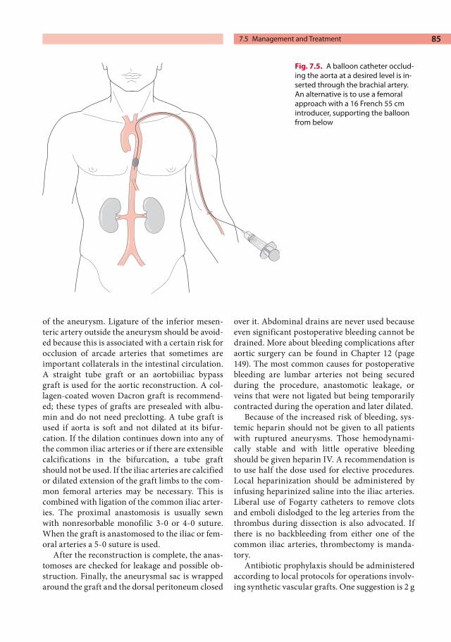
85
of the aneurysm. Ligature of the inferior mesen-teric artery outside the aneurysm should be avoid-ed because this is associated with a certain risk for occlusion of arcade arteries that sometimes are important collaterals in the intestinal circulation. A straight tube graft or an aortobiiliac bypass graft is used for the aortic reconstruction. A col-lagen-coated woven Dacron graft is recommend-ed; these types of grafts are presealed with albu-min and do not need preclotting. A tube graft is used if aorta is soft and not dilated at its bifur-cation. If the dilation continues down into any of the common iliac arteries or if there are extensible calcifications in the bifurcation, a tube graft should not be used. If the iliac arteries are calcified or dilated extension of the graft limbs to the com-mon femoral arteries may be necessary. This is combined with ligation of the common iliac arter-ies. The proximal anastomosis is usually sewn with nonresorbable monofilic 3-0 or 4-0 suture. When the graft is anastomosed to the iliac or fem-oral arteries a 5-0 suture is used.
After the reconstruction is complete, the anas-tomoses are checked for leakage and possible ob-struction. Finally, the aneurysmal sac is wrapped around the graft and the dorsal peritoneum closed
over it. Abdominal drains are never used because even significant postoperative bleeding cannot be drained. More about bleeding complications after aortic surgery can be found in Chapter 12 (page 149). The most common causes for postoperative bleeding are lumbar arteries not being secured during the procedure, anastomotic leakage, or veins that were not ligated but being temporarily contracted during the operation and later dilated.
Because of the increased risk of bleeding, sys-temic heparin should not be given to all patients with ruptured aneurysms. Those hemodynami-cally stable and with little operative bleeding should be given heparin IV. A recommendation is to use half the dose used for elective procedures. Local heparinization should be administered by infusing heparinized saline into the iliac arteries. Liberal use of Fogarty catheters to remove clots and emboli dislodged to the leg arteries from the thrombus during dissection is also advocated. If there is no backbleeding from either one of the common iliac arteries, thrombectomy is manda-tory.
Antibiotic prophylaxis should be administered according to local protocols for operations involv-ing synthetic vascular grafts. One suggestion is 2 g
Fig. 7.5. A balloon catheter occlud-ing the aorta at a desired level is in-serted through the brachial artery. An alternative is to use a femoral approach with a 16 French 55 cmintroducer, supporting the balloon from below
7.5 Management and Treatment

Chapter 7 Abdominal Aortic Aneurysms86
cloxacillin given at the start of the operation, with the dose repeated after 4 h in prolonged proce-dures. Besides general perioperative IV fluids, mannitol is recommended to maintain urinary output.
7.5.2.5 What to do While Waiting for Help
For surgeons without experience in AAA surgery it is generally a good idea to wait for a more expe-rienced colleague if the patient is reasonably stable. While the surgeon is waiting for help the patient should be prepared up to the point of anesthesia induction. The surgeon scrubs and the patient is also scrubbed and draped while the an-esthesiologist closely monitors the patient’s vital functions and hemodynamics. If the patient’s blood pressure drops and cannot be maintained at an acceptable level, the patient is anesthetized and laparotomy is initiated without experienced help. The goal is then to achieve control of the bleeding. Besides the previously described techniques to gain proximal control of the aorta, tamponade with lots of swabs and compression with the fist over the bleeding area is usually enough in this situation. These simple measures combined with IV fluids and inotropic drugs is often sufficient to stabilize the patient until help arrives.
7.5.2.6 Endovascular Treatment In recent years more than 300 patients with rup-tured AAA or incipient rupture have been treated with endovascular techniques. The results pre-sented are observational studies and show that endovascular repair of rupture is feasible. A large percentage of the patients in these early series were not in severe shock and the mortality rate aver-aged around 10%. Furthermore, reduced post-operative morbidity rates compared with conven-tional open repair have been suggested.
One major benefit of endovascular treatment is the possibility of obtaining rapid proximal control by inserting an inflatable balloon from the groin or through the brachial artery that occludes aorta. This technique makes it possible to delay final treatment until the patient is stabilized. Another potential advantage may be that high-risk patients can also be treated. Particularly favorable is the possibility of using only local anesthesia and seda-tion for repair.
The problems related to endovascular repair include the availability and storage of suitable grafts as well as logistical problems getting the pa-tients worked up rapidly. Pretreatment evaluation with CT angiography or digital subtraction arteri-ography is necessary to evaluate the possibility for endovascular repair and to plan the procedure. The number of different grafts needed to meet in-dividual requirements is minimized if a unilateral aortoiliac tube graft is used in combination with an occluder of the contralateral iliac system and a femorofemoral crossover, as shown in Fig. 7.6.
Fig. 7.6. One alternative way to treat a ruptured AAA with endovascular technique. A unilateral aortoiliac en-dovascular graft decompresses the aortic aneurysm. A coil in the right internal iliac artery and an occluder in the left common iliac artery eliminate pressure caused by backflow, the latter deployed to allow retrograde flow to the internal iliac artery from the groin. A femo-rofemoral bypass restores perfusion of the left leg

87
The technique involves the following steps: The patient is prepped and draped as for an elective AAA procedure. The common femoral arteries are surgically exposed if a bifurcated graft is inserted or if unilateral aortoiliac tube grafts in combination with a femorofemoral crossover bypass are used. For tube grafts access of only one common femoral artery is enough. One of the femoral arteries is punctured and an introducer is put in place, often a size 7 to 9-French. A guide wire is inserted, an aortogram obtained, and land-marks, either radiolucent (placed preoperatively) or external (such as clamps), are used to assess the length of the AAA. After systemic heparinization, the sheath with the graft is introduced over the guide wire to a level just below the renal arteries. The sheath is then withdrawn somewhat to allow proximal release of the graft. After final adjust-ment of the proximal fixation level the system is secured by angioplasty. The distal end of the endo-luminal graft is deployed in the common, external iliac, or common femoral artery with angioplasty of stents. Depending on the conditions a hand-sewn anastomosis is another option. An occluder of the common iliac is inserted from the contralat-eral femoral artery. If a bifurcated endoluminal graft is used, the contralateral graft limb is insert-ed through the same route. Finally, a completion angiogram is performed after withdrawal of the entire sheath.
A bifurcated aortobiiliac endoluminal prosthe-sis as a primary alternative in rupture is also grow-ing in popularity. The procedure requires a com-pliant large-diameter balloon for aortic occlusion, 5 and 12-French introducers, Amplatz guide wires, high-resolution fluoroscopy, and an assortment of endoluminal aortic stent grafts with a body diam-eters ranging from 22 to 34 mm and limb diame-ters of 12 to 24 mm.
7.5.3 Management After Treatment
The patient is treated in the intensive care unit un-til circulatory, respiratory, and renal functions are stable. This usually takes at least a couple of days. The most common early postoperative complica-tions are congestive heart failure, renal failure, and ischemic colitis. The patient, often with con-comitant coronary heart disease, is exposed to se-
vere stress during preoperative shock and aortic clamping and declamping. Deterioration of cardi-ac function with secondary hypotension that re-quires inotropic treatment is common. Renal function is also often impaired and occasionally the patient requires dialysis. Almost all patients have increased creatinine and blood urea nitrogen elevations after operation for ruptured AAA. These increases are also due to preoperative hypo-tension and the stress of the operation. If the pa-tient develops renal insufficiency with low urinary output, dialysis should be considered at an early stage.
The greatest risk for developing ischemic colitis is in patients with a ruptured aneurysm and shock. The severity of ischemic colitis varies from only discharge of the mucosa to transmural necrosis. Registration of pH at the wall of the sigmoid with a tonometer can be used to determine the risk for developing ischemic colitis. This condition is fur-ther discussed in Chapter 12 on complications in vascular surgery (page 145).
7.6 Results and Outcome
The 30-day mortality after surgery for ruptured AAA averages from 30% to 50%, the variability depending on whether the patient developed shock and whether concomitant diseases were present. For patients without shock, it is 20–25%, which can be compared to 60–70% for those without. The long-term results and prognosis for patients who survive the initial postoperative period is good. Outcome is even better than for patients who have undergone elective aneurysm repair. The reason for this is probably selection – the sick-est patients die of rupture, and the survivors who reach the hospital have fewer risk factors.
7.7 Unusual Types of Aortic Aneurysms
7.7.1 Inflammatory Aneurysm
An AAA can be symptomatic and cause pain without actual or imminent rupture. The most common cause for this pain is an inflammatory reaction in and around the wall – an inflammato-
7.7 Unusual Types of Aortic Aneurysms

Chapter 7 Abdominal Aortic Aneurysms88
ry AAA. CT, which then shows a thickened aneu-rysm wall, verifies the presence of such a condi-tion (Fig. 7.7). It could be presumed that the thick wall prevents rupture, but rupture of inflamma-tory AAAs is not uncommon. Because inflamma-tory AAAs often are painful, separating them from ruptured AAAs is a real diagnostic problem. Elevated erythrocyte sedimentation rate (ESR) or C-reactive protein (CRP) supports the diagnosis, but CT is the only way to exclude it.
7.7.2 Aortocaval Fistula
A special form of AAA rupture occurs when the bloodstream penetrates into the vena cava causing an aortocaval fistula (Fig. 7.8). The patient typi-cally develops sudden cardiac failure and cyanosis of the lower extremities. The cardiac failure is due to the large shunt and the discoloration of the legs occurs because of venous stasis in combination with the heart failure. At physical examination the patient is positive for a bruit and a palpable aneu-rysm in the abdomen. Treatment for an aortocaval fistula is an emergency operation, but in most cas-es some time for preoperative preparation is avail-able. The operation follows the strategy for other aneurysms as outlined previously and the fistula is usually closed by suture from inside the aneurysm while vena cava is controlled by manual compres-sion proximally and distally.
7.7.3 Thoracoabdominal Aneurysm
A small number of aortic aneurysms engage the suprarenal or thoracoabdominal parts of the aorta including the orifices of the renal arteries, the superior mesenteric artery, and the celiac trunk. They originate in the thoracic part of the aorta or anywhere below the level of the diaphragm. Man-agement of rupture in such aneurysms is challeng-
Fig. 7.7. Typical appearance on computed tomogra-phy of an inflammatory abdominal aortic aneurysm with its thick wall
Fig. 7.8. Computed tomography of a patient with an abdominal aortic aneurysm and a fistula into the inferior vena cava

89
ing, and if its extension is known prior to the operation someone with experience should be contacted before surgery begins. If such an aneu-rysm is achieved during surgery for rupture, prox-imal control is sought by one of the techniques described previously. Endovascular repair is also an option that needs to be considered.
7.7.4 Mycotic Aneurysm
Another special type of AAA is caused by a local infection in an atherosclerotic and degenerated aortic wall, known as mycotic or septic aneurysm. Different from ordinary AAAs that are fusiform, mycotic aneurysms are usually saccular (Fig. 7.9). Patients with this type of AAA frequently have a medical history that includes fever and malaise. Elevated ESR, CRP, and other inflammatory pa-rameters are also common. The most common bacteria found in mycotic aneurysms and in the patient’s blood are of the Salmonella species. It is the infectious process in the wall that causes ero-
sion and subsequently rupture. Treatment is the same as for other AAAs with the addition of long-term antibiotics.
7.8 Ethical Considerations
Difficult and delicate ethical considerations often arise when managing patients with ruptured AAA. Accordingly, it has to be emphasized that the ad-vice given above often needs to be modified in very old patients, patients with dementia, and patients with other serious medical conditions implying only a short expected survival time. On the other hand, patients who previously were de-termined not suitable for elective repair because of high risk should sometimes be considered for repair of a ruptured AAA. When rupture has already occurred the risk/benefit situation is com-pletely different. The patient has little to lose by undergoing an emergency operation.
Rupture of an AAA often occurs in elderly patients and a complete medical history and infor-mation about their present quality of life is fre-quently missing when they are admitted. Because nonsurgical management is associated with 100% mortality, a policy of accepting every patient for surgical treatment is advocated in many hospitals. A certain selectivity, however, is often wise. It is obvious that a patient who had cardiac arrest in the ambulance and remains unconscious at ad-mission, is anuric, and has ECG signs of myocar-dial ischemia is extremely unlikely to survive sur-gery. If the patient is 80 years old and also is known to have dementia, difficulties ambulating, and need for geriatric care, it is reasonable to avoid surgery and instead give the patient terminal care of high quality. Unfortunately, there are no reli-able prognostic factors for treatment outcome for the individual patient, but many studies report relationships between presence of different risk factors and survival.
A common conclusion in the literature, how-ever, is that age should never be considered as a contraindication to surgery. It is always the sur-geon, the patient and relatives and their individual judgment that finally decide whether to operate or not. If one is in doubt, a good general rule is to be liberal with repair attempts. It is always possible, but difficult, to change such a strategy later during
Fig. 7.9. Angiographic appearance of a typical mycotic aneurysm, with its saccular shape caused by local ero-sion of the aortic wall and subsequent leakage of blood into an aneurysmal sac consisting of a fibrous capsule (not a true vascular wall)
7.8 Ethical Considerations

Chapter 7 Abdominal Aortic Aneurysms90
the course when more information is available. Accordingly, stopping the support of vital func-tions and taking the patient to the floor for pallia-tion is a viable option. Some factors in the postop-erative course – large bleedings and cardiac, renal, respiratory, and infectious complications – are considered to be associated with a worse prognosis and thus might indicate a suitable point at which to make such a decision.
Further Reading
Bengtsson H, Bergquist D. Ruptured abdominal aortic aneurysm: a population-based study. J Vasc Surg 1993; 18:74–80
Harris LM, Faggioli GL, Fiedler R et al. Ruptured ab-dominal aortic aneurysms: factors affecting mortal-ity rates. J Vasc Surg 1991; 14:812–820
Johansson G, Swedenborg J. Ruptured abdominal aortic aneurysms: a study of incidence and mortality. Br J Surg 1986; 73:101–103
Johnston KW. Ruptured abdominal aortic aneurysms: six-year follow-up of a multicenter prospective study. J Vasc Surg 1994; 19:888–900
Ouriel K, Geary K, Green RM, et al. Factors determin-ing survival after ruptured aortic aneurysm: the hospital, the surgeon, and the patient. J Vasc Surg 1990; 12:28–33
Ohki T, Veith FJ. Endovascular grafts and other im-age-guided catheter-based adjuncts to improve the treatment of ruptured aortoiliac aneurysms. Ann Surg 2000; 232(4):466–479

Aortic Dissection 8
CONTENTS
8.1 Summary . . . . . . . . . . . . . . . . . . . . . . . . . . . . 91
8.2 Background . . . . . . . . . . . . . . . . . . . . . . . . . 918.2.1 Magnitude of the Problem . . . . . . . . . . 918.2.2 Classification and Definition . . . . . . . . 928.2.3 Etiology . . . . . . . . . . . . . . . . . . . . . . . . . . . . . 928.2.4 Pathophysiology . . . . . . . . . . . . . . . . . . . . 93
8.3 Clinical Presentation . . . . . . . . . . . . . . . 948.3.1 Signs and Symptoms . . . . . . . . . . . . . . . . 948.3.2 Medical History . . . . . . . . . . . . . . . . . . . . . . 948.3.3 Physical Examination . . . . . . . . . . . . . . . . 96
8.4 Diagnostics . . . . . . . . . . . . . . . . . . . . . . . . . 96
8.5 Management . . . . . . . . . . . . . . . . . . . . . . . 978.5.1 Treatment in the Emergency
Department. . . . . . . . . . . . . . . . . . . . . . . . . . 978.5.2 Emergency Surgery . . . . . . . . . . . . . . . . . 988.5.3 Type B dissection . . . . . . . . . . . . . . . . . . . . 988.5.4 Endovascular Treatment . . . . . . . . . . . . 98
8.6 Results and Outcome . . . . . . . . . . . . . . . 99
Further Reading . . . . . . . . . . . . . . . . . . . 100
8.1 Summary
Aortic dissection is one of the “great mas-queraders,” so always suspect this diagno-sis in any acute painful illness with a pulse deficit.A practical classification in the emergency situation is type A, involving the ascend-ing thoracic aorta and the arch and type B involving the aorta distal to the left subcla-vian artery.Treatment of type A dissection is always surgical.Treatment of type B dissection is medical in most cases and surgical if there is com-plicating organ ischemia or bleeding.Alert the attending thoracic or vascular surgeon on call early during management, especially in type A dissections.
8.2 Background
Dissection of the thoracic aorta represents a major clinical problem that is extremely demanding to manage even for experienced surgeons. Once it is diagnosed this condition is usually managed by an experienced specialist in thoracic or vascular surgery. The responsibility for the diagnosis and its primary management, however, mostly belongs to the surgical or medical emergency physicians.
8.2.1 Magnitude of the Problem
The true prevalence and incidence of aortic dis-section are unknown, but it has been reported to have an annual occurrence of 5–10 cases per

Chapter 8 Aortic Dissection92
million and to affect between 10,000 and 25,000 patients annually in the United States. Autopsy studies in the United States and Denmark report dissections in 0.2–0.8% and 0.2% of cases, respec-tively. An age-adjusted mortality rate from aortic dissections of 0.5–2.7% per 100,000 inhabitants was calculated from 1950 to 1981. The overall inci-dence of aortic dissections consequently is in the same range or possibly up to two to three times greater than that for ruptured abdominal aortic aneurysm. It is two to five times more common in men than in women, and maximum occurrence is in the 5th decade of life. Still, in our era of modern diagnostic methods, a majority of patients proba-bly die with this disease undetected. Aortic dissec-tion is a dramatic and dangerous condition with a very high mortality: 20–50% of patients die within the first 24–48 h, and up to 75% within the first 2 weeks. It is considered as one of the “great mas-queraders,” with a wide range of presenting symp-toms. Because the diagnosis is difficult, awareness of aortic dissection in the differential diagnosis is essential, as is rapid and correct management.
8.2.2 Classification and Definition
Aortic dissection is characterized by two or more communicating flow channels originating from a proximal intimal tear, with propagation of the bloodstream within the medial layer. It should be distinguished from an intramural hematoma, which is a hemorrhage into the medial layer of the aortic wall without an intimal tear. Intramural he-matomas have a natural history similar to aortic dissection and are treated similarly.
The most useful classification of aortic dis-sections in the acute situation is the one proposed by Daily (Stanford classification), as shown in Fig. 8.1.
Other classic classifications are by DeBakey and Crawford (thoracoabdominal aneurysms and chronic dissections). The information in this chap-ter is based on the Stanford classification because it simplifies the acute management. A type-A dissec-tion always involves the ascending aorta, regard-less of the distal extension. A type-B dissection does not involve the ascending aorta. There is con-sensus in the literature that an aortic dissection is considered acute if the onset of symptoms occurred
within 14 days of presentation, and chronic if more than 14 days have elapsed.
NOTEThe most practical classification in the emergency situation is– Type A: involving the ascending
thoracic aorta and the arch– Type B: involving the aorta distal
to the left subclavian artery
8.2.3 Etiology
Aortic dissection is usually related to some kind of degenerative changes in the aortic wall, in partic-ular the media. Even if a dissection primarily starts with a tear in the intima, its propagation within the media varies considerably from almost none to rapid progression along the entire length of the aorta. The variation is related to the condi-tion of the medial layer. Some congenital connec-tive tissue defects are known to cause such degen-eration, including Marfan’s syndrome, Turner syndrome, and Ehlers–Danlos’ syndrome. Cystic media necrosis is another predisposing condition. The role of arteriosclerosis is often discussed. It is present in older patients with hypertension pre-
Fig. 8.1. Classification of aortic dissection in types A and B, according to Daily (Stanford)

93
senting with dissection and might constitute a rare mechanism by penetration of atherosclerotic ulcers extending through the intima into the me-dia. This is, however, mostly considered coinci-dental rather than causative, and some authors even argue that atherosclerotic changes within the aortic wall might be a barrier to the extension of a dissection.
Arterial hypertension is the most important predisposing factor. It is noteworthy that the sud-den extreme hypertension associated with severe physical exercise may cause aortic dissection in younger persons. Also, pregnancy with its hyper-circulation and hormonal changes affecting con-nective tissue, is a certain risk factor particularly during the last trimester and during labor. Fortu-nately, aortic dissections in women are rare, but 50% of dissections occurring in women younger than 40 years old do occur during pregnancy.
Iatrogenic injuries during coronary diagnostic and therapeutic procedures with catheter manip-ulations can also cause aortic dissection. Blunt chest trauma in otherwise healthy persons may cause aortic dissection, but such dissections are usually very limited due to the minimal degenera-tion in these structurally normal aortas.
NOTEA degenerative process causing weaken-ing of the aortic wall in combination with hypertension is the most important etiologic factor.
8.2.4 Pathophysiology
Type A dissection, constituting 60–70% of all aor-tic dissections, is mostly seen in younger patients with some elastic and connective tissue abnormal-ity. It characteristically starts with a primary inti-mal tear just distal to the sinotubular ridge in the ascending aorta. This location is in the vicinity of the cephalad extension of the aortic valve com-missures. The tear is commonly transverse and has a length corresponding to 50–60% of the aortic circumference. The dissection process starts in the intimal tear and its extension and direction vary, as does the speed with which it propagates.
Typically, a type A dissection affects the right lateral wall of the greater curvature of the ascend-
ing aorta. The dissection is usually directed ante-gradely, but retrograde extension is also relatively common. A primary entry in the ascending aorta is associated with a great risk for bleeding into the pericardium, causing cardiac tamponade.
Type B dissection usually starts with a primary intimal tear in the descending thoracic aorta just distal to the origin of left subclavian artery. This type constitutes approximately 25% of all aortic dissections. A patient with a type-B dissection is typically older, in the 6th–7th decade of life, and has thoracic aortic degeneration and hyperten-sion.
Other possible but less common sites of the pri-mary tear are the aortic arch, occurring in approx-imately 10% of cases, and the abdominal aorta, occurring in only 2%. As already mentioned, the dissection in the aortic media can travel in a retro-grade as well as an antegrade direction, causing two flow channels with a false and a true lumen. Secondary tears and reentries usually occur dis-tally, allowing flow from the false into the true lumen.
Rupture is the most common cause of death in patients with aortic dissection and is mostly lo-cated near the site of the primary intimal tear. Consequently, a type-A dissection usually rup-tures into the pericardial sac, causing cardiac tamponade, or an aortic arch rupture that bleeds into the mediastinum. In addition, the close rela-tion to the aortic valve commisures can result in acute valve regurgitation due to prolapse of the commissural attachments. Dissection into the aortic root may also involve the coronary arteries, leading to myocardial ischemia or infarction. A type-B descending aortic dissection typically rup-tures into the left pleural cavity, and less frequent-ly into the right.
As the dissection extends along the aorta it will subsequently engage major important cere-bral and visceral branches, possibly resulting in threatening end-organ ischemia. The mechanisms behind this are compression of the true lumen by the false lumen or shearing of the branch by the dissection process. A third possibility is disrup-tion of an important dissection membrane, caus-ing an intimal flap covering the orifice of a branch. Such peripheral vascular complications occur in 25–30% of patients with aortic dissection and can critically affect cerebral, renal, visceral, and lower
8.2 Background

Chapter 8 Aortic Dissection94
extremity perfusion. Because the dissection in the descending aorta mainly engages its left perimeter, the left renal and left iliac arteries are at higher risk than the right ones.
NOTEPeripheral vascular complications occur in 25–30% of cases of dissection of the aorta.
8.3 Clinical Presentation
8.3.1 Signs and Symptoms
Acute aortic dissection patients can display a variety of symptoms, and affected individuals can develop symptoms mimicking those of almost any other acute medical or surgical condition. Aortic dissection must be considered in patients
presenting with symptoms indicating acute arte-rial occlusion and an acute illness that seems to involve unrelated organ systems.
8.3.2 Medical History
The most dominant symptom is severe pain, which is migrating or nonmigrating and experienced by more than 90% of patients. When analyzing the pain its typical characteristics are evaluated; if it is: sudden, severe, new, ripping or tearing, and constant. The pain is typically related to the loca-tion of the dissection and its propagation distally into different aortic segments. In proximal dissec-tion, the most common pain location is the ante-rior chest. The pain frequently radiates into the neck and jaws and can be associated with swallow-ing difficulties. As the dissection propagates dis-tally, the pain migrates to an interscapular loca-tion followed by pain in the midback, lumbar, and groin regions (Fig. 8.3).
Abdominal pain might be severe in patients suffering from visceral or renal ischemia. As pre-viously mentioned, the left renal artery is more likely to be compromised, which may explain why severe left flank pain mimicking ureteral colic is often included in the reported history. One should always include questions about hypertension, car-diac disease, peripheral vascular disease, connec-tive tissue abnormalities (such as Marfan’s, Turner, and Ehlers–Danlos’ syndromes), cystic media ne-
Table 8.1. Differential diagnoses in aortic dissection
Possible differential diagnoses
Coronary ischemiaMyocardial infarctionAortic regurgitation without dissectionAortic aneurysm with dissectionMusculoskeletal painMediastinal tumors or cystsPericarditisGall bladder diseasePulmonary embolismStroke Visceral or lower extremity ischemia without dissection
Fig. 8.2. Mechanisms of branch occlusion and organ malperfusion in aortic dissection. a False lumen expan-sion causes compression of a side branch. b The orifice of a side branch is disrupted by dissection, and its inner layers are impacted distally

95
crosis, diagnostic or therapeutic catheter manipu-lations, or intense exercise.
Secondary effects of aortic dissection with or-gan malperfusion necessitate a thorough and com-plete history to include previous, present, and un-dulating symptoms of the following:
Cerebral ischemia: stroke, loss of conscious-ness, focal neurological symptomsSpinal ischemia: paraplegia or paraparesesRenal ischemia: flank pain, hematuria, dimin-ished urinary output
Visceral ischemia: severe abdominal pain (for further details of typical symptoms, see Chapter 6, p. 67)Lower-extremity ischemia: loss of pulse, loss of sensory or/and motor function, severe pain and coldness (see Chapter 10, p. 122)Cardiac malperfusion: angina pectoris or symptoms of acute congestive heart failure
Possible differential diagnoses are listed in Table 8.1.
Fig. 8.3. a Radiation of pain in type A dissection. The pain is usually referred to the neck, anterior chest, and interscapular area. b Radiation of pain in type B dissection. Pain is primarily interscapular, but with distal progression of dissection, pain is often referred to the lower back and groin
8.3 Clinical Presentation

Chapter 8 Aortic Dissection96
NOTEBe aware of aortic dissection as an important differential diagnosis in any acute case presenting with sudden painful illness, in particular if it is associated with symptoms or signs of organ ischemia.
8.3.3 Physical Examination
Complete and repeated physical examinations are of paramount importance in diagnosing and man-aging patients with suspected or verified acute aortic dissection since this condition can affect so many different organ systems and has a dynamic course.
The typical patient presents with paradoxical physical findings. He or she is frequently pale, restless, and in preshock or shock, and has an ap-pearance indicating poor peripheral perfusion but with a paradoxically high blood pressure. Eighty percent of the patients have arterial hypertension at admission. The high blood pressure is second-ary to underlying essential hypertension, elevated catecholamine levels due to severe pain, or occlu-sion of the renal arteries or even the thoracic or abdominal aorta.
Twenty percent of the patients have a low blood pressure instead. This is usually secondary to car-diac tamponade or rupture, or to acute congestive heart failure secondary to acute aortic insuffi-ciency. Another possible explanation is pseudo-hypotension secondary to mechanical obstruction from the dissection of one or both subclavian arteries.
Auscultation of the chest is of vital importance. A cardiac murmur indicates aortic regurgitation. The first heart sound is diminished or absent due to elevated end diastolic ventricular pressure. There might be an S3 gallop rhythm. A continu-ous murmur usually indicates rupture into the right atrium. A pericardial friction rub indicates leakage into the pericardial sac. Auscultation of the lungs might reveal signs of pulmonary edema. Loss of alveolar breath sounds can be found after leakage or rupture into one or both of the pleural cavities. Jugular venous distension is also a com-mon finding.
A complete and repeated neurological exami-nation is mandatory. Horner’s syndrome, loss of consciousness, loss of sensory or motor function, paraparesis, paralysis, or paraplegia might be pres-ent. Acute cerebral vascular occlusion is for obvi-ous anatomic reasons, more common in proximal dissection, but fortunately neurological deficits occur in only about 20% of those patients.
Lower extremity paralysis in the examina-tion is caused by shearing off or compression of major arteries feeding the spinal cord (intercos-tal-T8–L1). Another possible explanation is occlu-sion of the thoracic or abdominal aorta, causing ischemia of the lower body including peripheral nerves. The clinical distinction is important be-cause spinal cord ischemia has a poor prognosis, while a peripheral nerve ischemia has a better prognosis if treated. This distinction can be made by examining peripheral pulses. The latter condi-tion is usually combined with loss of pulses in the groins and distally in the affected lower extremi-ties.
Repeated examination of peripheral pulses as well as blood pressures in the arms and ankles are important indicators of the extension of a dissec-tion and its consequences of organ malperfusion. Repeated examinations are important in order to follow the development. A peripheral pulse may disappear, or a pulse deficit may be dynamic and resolve spontaneously, which is reported to occur in one-third of the patients. Such a dynamic course is probably related to redirection of flow from the false into the true lumen after spontaneous fenes-tration of the aortic septum known as the reentry phenomenon.
A new pulse deficit is found in approximately 60% of the patients.
8.4 Diagnostics
An electrocardiogram (ECG) should be obtained in the emergency department. Low voltage might indicate pericardial tamponade, and ST–T wave changes could indicate myocardial ischemia.
The following blood tests should be ordered: complete blood cell count, arterial blood gases, protrombin and thromboplastin times, serum electrolytes, creatinine, blood urea nitrogen, liver enzymes and lactate.

97
As with the physical examination, repeated blood tests according to the patient’s clinical course might be of great diagnostic value during the acute stage of the disease. Mild anemia is com-mon, while severe anemia indicates rupture and bleeding. Hemolysis with elevated bilirubin or lac-tic acid concentrations can also be found. A leuko-cytosis with a count of 10,000–15,000 is common. Blood gases might reveal a metabolic acidosis due to anaerobic metabolism in ischemic tissue. Urinary tests showing hematuria indicates renal involvement.
A plain chest x-ray in standard anteroposterior and lateral projections is rarely diagnostic, but the following findings indicates the presence of aortic dissection:
Abnormal shadow adjacent to the descending thoracic aortaDeformity of the aortic knobDensity adjacent to the brachiocephalic trunkEnlarged cardiac shadowDisplaced esophagus, trachea, or bronchusAbnormal mediastinumIrregular aortic contourLoss of sharpness of the aortic shadowPleural effusionExpanded aortic diameter
Helical CT is accurate for determining the pres-ence of an aortic dissection and provides informa-tion for classification. The identification of an intimal tear is, however, difficult and motion artifacts of the ascending aorta are sometimes misinterpreted as dissection. MRI is highly accu-rate and gives valuable information about the pathoanatomy. Unfortunately it cannot be per-formed in hemodynamically unstable patients who are on ventilator support.
TEE (Transesophageal echocardiography) is often considered as one of the most valuable diag-nostic tools, making it possible to determine the type and extent of the aortic dissection, especially distally. It has limitations in visualization of the distal ascending aorta and the arch. TTE (Trans-thoracic echocardiography) is, on the other hand, superior for evaluating involvement of the proxi-mal part of the descending aorta in the dissection. Together, TEE and TTE yield a sensitivity and specificity approaching 100% for diagnosing dis-section and are thus probably the best – but unfor-
tunately often not available – diagnostic modali-ties.
Aortography is the old gold standard and is highly accurate in diagnosing aortic dissection, but it can fail to recognize a thrombosed false lu-men. It also provides better information than CT or MRI about the condition and involvement of the aortic branches. Furthermore, aortography can be combined with therapeutic endovascular management. However, the modern CT scanners with up to 64 detectors can produce extremely detailed images and, when available, should be the first imaging study after the chest x-ray.
8.5 Management
8.5.1 Treatment in the Emergency Department
As soon as aortic dissection is clinically suspected, aggressive medical treatment must be started im-mediately. The goals are to (1) stabilize dissection, (2) prevent rupture, and (3) prevent organ isch-emia.
These goals can be achieved by diminishing the stress on the aortic wall. Consequently, the thera-peutic cornerstone is to reduce blood pressure in order to minimize the force of the left ventricular ejection (dP/dT). The reduction in blood pressure must, however, be balanced against what is needed for adequate cerebral, coronary, renal, and visceral perfusion. A useful guideline is that the systolic arterial blood pressure should be kept around 100–110 mmHg and mean arterial pressure be-tween 60 and 75 mmHg, provided that urinary output and neurology are unaffected.
In the emergency department the following measures can be employed:1. Insert one or two large-bore intravenous (IV)
lines for administering antihypertensive drugs and fluids.
2. Obtain an ECG.3. Order blood tests as stated above.4. Obtain a plain chest x-ray.5. Administer oxygen by mask6. Consider injection of a strong analgesic IV,
such as morphine 5–10-mg.7. Insert an arterial catheter for blood pressure
monitoring.
8.5 Management

Chapter 8 Aortic Dissection98
8. Start administration of a beta-blocker as de-scribed below.
The recommended agents for medical manage-ment of acute aortic dissection are direct vaso-dilators, beta-blockers, nitroglycerin and calcium channel blockers if beta blockers cannot be used.
Beta-blockers orally are recommended for all patients. Contraindications for beta-blockers are heart failure, bradyarrhythmias, atrioventricular blocks, and bronchospastic disease.
Suggested emergency medical treatment (local variations in drug choices are of course common) is as follows:
Start propranolol treatment, 1 mg IV, every 3–5 min until achieving a systolic blood pressure around 100 mmHg and a heart rate of 60–80 beats/min (maximum dose, up to 0.15 mg/kg). Continue thereafter with 2–6 mg IV every 4–6 h. In patients with severe hypertension an IV infusion of nitro-glycerin is started and the dose titrated after blood pressure and heart rate.
NOTEThe main objective of the medical treatment is to lower the blood pressure to a level of 100–110 mmHg. It is manda-tory to check the patient for the develop-ment of new complications of the dissec-tion during medical treatment.
8.5.2 Emergency Surgery
Emergency surgery should be considered in type Adissections involving the intrapericardial ascend-ing aorta and the aortic arch. A distal type B dis-section with retrograde dissection involving the aortic arch is also a case for acute operation. A double aortic lumen in the pericardial portion of the ascending aorta is an absolute indication for emergency operation. Depending on the patient’s general condition prior to the dissection there are, as usual, exceptions from these basic rules. Con-traindications include very advanced age and se-vere debilitating or terminal illnesses.
Surgical repair of the condition requires tho-racic surgical expertise and includes replacing the ascending aorta and resecting the primary intimal tear. The operation involves cardiopulmonary by
pass. In type A dissection with persistent organ ischemia despite open surgical repair and replace-ment of the ascending aorta, endovascular treat-ment of the rest of the dissection is often a success-ful complement.
8.5.3 Type B dissection
The management of acute distal aortic dissection is initially always medical because this results in lower morbidity and mortality than emergent sur-gical repair. Consequently, the continued regimen for these patients follows the previously given rec-ommendations regarding beta blockade and vaso-dilators started in the emergency department.
The medical treatment must be combined with careful observation for complications. Surgical or endovascular intervention should be considered for the following situations:
Aortic ruptureIncreasing periaortic or intrapleural fluid (sug-gesting aneurysmal leakage)Rapidly expanding aortic diameterUncontrolled hypertensionPersistent pain despite adequate medical the-rapyOrgan malperfusion – ischemia of brain, spinal cord, abdominal viscera, or limbs
The goal of surgical repair in a type B dissection is, as with all other treatment options, to prevent rup-ture and restore visceral and limb perfusion. Be-cause a common site of rupture is associated with the site of primary dissection, at least the upper half of the descending thoracic aorta needs to be replaced in most cases. Graft replacement in the acute setting should be limited and replacement of the entire thoracic aorta avoided if possible. An abdominal fenestration procedure is sometimes necessary to restore flow to the lower extremities. Extraanatomical by pass is another possible way to reestablish flow to the legs.
8.5.4 Endovascular Treatment
In patients with peripheral vascular complications due to extension of the dissection into a branch, causing compression and obstruction of its true

99
lumen, as well as in patients with central aortic true lumen collapse, the endovascular option should be considered. Provided, of course, that the institution has technically skilled physicians, the necessary equipment and back-up support. It is possible to create a fenestration through the inti-mal flap from the false into the true lumen with endovascular techniques. As shown in Fig. 8.2 a, stenting of the entry site to occlude flow into the false lumen will probably be successful in restor-ing flow into a branch with its orifice obstructed by the false lumen and the dissection membrane. If there is an avulsion of the intima of that branch as in Fig. 8.2 b, this is not an option.
Endovascular management is developing as an attractive alternative to surgical repair. Patients with an acute type B dissection who are not good candidates for surgery can be considered for endovascular management. Stenting has also been reported to give successful results in aortic collapse with severe ischemia of the lower part of the body.
An endovascular approach can also be used as the initial treatment by performing aortic fenes-tration and stenting. Most centers prefer to delay either surgical or endovascular repair until after the patient has recovered from the acute phase of malperfusion, whereas others advocate early pro-phylactic stenting and coverage of the intimal tear to occlude the false lumen and prevent further dis-section (Fig. 8.4).
8.6 Results and Outcome
A recent article from 12 international centers covering 464 patients with aortic dissection re-ported, in-hospital mortality rates for type-A dis-sections treated surgically of 28%, and medically of 58%. The corresponding figures for type B were 31% and 10%, respectively.
Successful closure of the intimal tear with en-dovascular stent grafts and subsequent thrombo-sis of the false lumen is reported in up to 75% of patients. Branch occlusions with ischemic symp-toms were relieved in 75–95% of the cases. Sur-vival after 30 days was 75–85 %, and long-term results are good, with <1% related deaths and veri-fied thrombosis of the false lumen in 100% of the survivors. No thromboembolic complications oc-
Fig. 8.4. a Computed tomography showing a type B dissection and its entry in the first part of the descend-ing aorta in a patient with a previous reconstruction of the arch and the brachiocephalic trunk after a type A dissection. The true anterior aortic lumen is severely compressed causing obstruction of the main visceral branches and leading to visceral ischemia. b Flow into the true aortic lumen and all branches is restored after deploying a covered stent over the entry site in the de-scending aorta
b
a
8.6 Results and Outcome
Chapter 8 Aortic Dissection100
cured. In general, survival is lower for patients with paraplegia or visceral or renal ischemia. In cases with type B dissections and indications for surgical intervention, the results of endovascular intervention seem more favorable compared with conventional surgical repair, but the number of reported cases from any single center is still low. In all cases, long-term follow-up regarding devel-opment of aneurysms and continued antihyper-tensive medication is essential.
Further Reading
Cambria RP, Brewster DC, Gertler, et al. Vascular com-plications associated with spontaneous aortic dis-section. J Vasc Surg 1988; 7:199–209
Cigarroa JE, Isselbacher EM, DeSanctis RW, et al. Diag-nostic imaging in the evaluation of suspected aor-tic dissections: Old standards and new directions. N Engl J Med 1993; 328:35–43
Daily PO, Trueblood HW, Stinson EB, et al. Manage-ment of acute aortic dissections. Ann Thorac Surg 1970; 10:237
De Bakey ME, McCollum CH, Crawford ES, et al. Dis-section and dissecting aneurysms of the aorta: twenty year follow up of five hundred twenty-seven patients treated surgically. Surgery 1982; 92:1118
Hagan PG, Nienaber CA, Isselbacher EM, et al. The International Registry of Acute Aortic Dissection – new insights into an old disease. JAMA 2000; 283(7):897903
Lilienfeld DE, Gundersson PD, Sprafka JM, et al. Epi-demiology of aortic aneurysms. Mortality trends in the United States, 1951–1981. Arteriosclerosis 1987; 7:637
Slonim SM, Miller DC, Mitchell RS, et al. Percutaneous Balloon fenestration and stenting for life threaten-ing ischemic complications in patients with acute aortic dissection. J Thorac Cardiovasc Surg 1999; 117(6):1118–1126
Williams DM, Lee DY, Hamilton BH, et al. The dis-sected aorta: Percutaneous treatment of ischemic complications principles and results. J Vasc Interv Radiol 1997; 8:605-625

Vascular Injuries in the Leg 9
CONTENTS
9.1 Summary . . . . . . . . . . . . . . . . . . . . . . . . . . . 101
9.2 Background . . . . . . . . . . . . . . . . . . . . . . . . 1019.2.1 Background . . . . . . . . . . . . . . . . . . . . . . . . . 1019.2.2 Magnitude of the Problem . . . . . . . . . 1029.2.3 Etiology and Pathophysiology . . . . . 1029.2.3.1 Penetrating Injury . . . . . . . . . . . . . . . . . . 1029.2.3.2 Blunt Injury . . . . . . . . . . . . . . . . . . . . . . . . . 1029.2.3.3 Pathophysiology . . . . . . . . . . . . . . . . . . . 102
9.3 Clinical Presentation . . . . . . . . . . . . . . 1039.3.1 Medical History . . . . . . . . . . . . . . . . . . . . . 1039.3.2 Clinical Signs and Symptoms . . . . . . . 103
9.4 Diagnostics . . . . . . . . . . . . . . . . . . . . . . . . 1049.4.1 Angiography . . . . . . . . . . . . . . . . . . . . . . . 1049.4.2 Duplex Ultrasound . . . . . . . . . . . . . . . . . 104
9.5 Management and Treatment . . . . . . 1059.5.1 Management Before Treatment . . . . 1059.5.1.2 Severe Vessel Injury . . . . . . . . . . . . . . . . . 1059.5.1.2 Less Severe Injuries . . . . . . . . . . . . . . . . . 1059.5.1.3 Angiography Findings . . . . . . . . . . . . . 1059.5.1.4 Primary Amputation . . . . . . . . . . . . . . . 1069.5.2 Operation . . . . . . . . . . . . . . . . . . . . . . . . . . 1069.5.2.1 Preoperative Preparation . . . . . . . . . . 1069.5.2.2 Proximal Control . . . . . . . . . . . . . . . . . . . 1069.5.2.3 Distal Control and Exploration . . . . . 1099.5.2.4 Shunting . . . . . . . . . . . . . . . . . . . . . . . . . . . 1109.5.2.5 Vessel Repair . . . . . . . . . . . . . . . . . . . . . . . 1129.5.2.6 Finishing the Operation . . . . . . . . . . . . 1139.5.3 Endovascular Treatment . . . . . . . . . . . 1139.5.4 Management After Treatment . . . . . 114
9.6 Results and Outcome . . . . . . . . . . . . . . 114
9.7 Fasciotomy . . . . . . . . . . . . . . . . . . . . . . . . . 115
9.8 Iatrogenic Vascular Injuries to the Legs . . . . . . . . . . . . . . . . . . . . . . . . . 117
Further Reading . . . . . . . . . . . . . . . . . . . 117
9.1 Summary
Major bleeding is controlled by manual compression.Vascular injuries should always be sus-pected in extremities with fractures.Most vascular injuries are revealed by care-ful and repeated clinical examination.Obtain proximal control before exploring a wound in a patient with a history of sub-stantial bleeding.
9.2 Background
9.2.1 Background
Vascular trauma to extremity vessels is caused by violent behavior or accidents. Because of the rise in the number of endovascular procedures, iatro-genic injuries have also become an increasing part of vascular trauma. Vascular injuries may cause life-threatening major bleeding, but distal ischemia is more common. Ischemia occurs after both blunt and penetrating trauma. The vascular injury is often one of many injuries in multiply traumatized patients that make the recognition of signs of vascular injury – which can be blurred by more apparent problems – and the diagnosis difficult. Table 9.1 lists common locations of com-bined orthopedic and vascular injury. Multiple injuries also bring problems regarding priority.

Chapter 9 Vascular Injuries in the Leg102
9.2.2 Magnitude of the Problem
Data on the true incidence of vascular injuries to the legs is hard to gather. The incidence of vascu-lar trauma varies among countries and also be-tween rural and urban areas. It is usually higher where gunshot wounds are common. There is an equal share of blunt and penetrating injury in most studies from Europe, whereas penetrating injury is slightly more common in the United States. Approximately 75% of all vascular injuries are localized to the extremities and more than 50% to the legs. The true incidence of iatrogenic trauma is unknown.
9.2.3 Etiology and Pathophysiology
9.2.3.1 Penetrating Injury Penetrating vascular injury is caused by stab and cutting injuries, gunshots, and fractures, the latter when sharp bone fragments penetrate the vascular wall. Gunshots cause major bleeding by direct ar-tery trauma, while high-velocity bullets create a cavitation effect with massive soft tissue destruc-tion and secondary arterial damage. In fact, after all types of penetrating trauma both bleeding and indirect blunt arterial injury with ischemia may occur. Bleeding is more often exsanguinating after sharp injury and partial vessel transection. Com-plete avulsion, especially when caused by blunt trauma, makes the vessel more prone to retrac-tion, spasm, and thrombosis. This diminishes the risk for major bleeding. Iatrogenic injuries can be caused by catheterization and during surgical dissection.
NOTEPenetrating injuries can cause both major bleeding and ischemia.
9.2.3.2 Blunt Injury Blunt vascular injuries are usually caused by mo-tor vehicle and other accidents. The consequences are thrombosis and ischemia distal to the injured vessel. The media and the intimal layers of the ves-sel wall are easily separated, and subsequent dis-section by the bloodstream between the layers may lead to lumen obstruction. Blunt injuries also in-duce thrombosis. This type of vessel injury is par-ticularly common when the artery is hyperextend-ed as in knee joint luxations and upper arm frac-tures. Contusion of the vessel may also cause bleeding in the vessel wall. Thrombosis and isch-emia by this mechanism can occur several hours after the traumatic situation. Narrowing of the ar-terial lumen following blunt trauma is rarely caused by spasm and it can be disregarded as etiology.
9.2.3.3 Pathophysiology The main pathophysiological issue after vascular injuries to the extremities is ischemia. The process is identical to what happens during acute leg isch-emia due to embolization (see Chapter 10, p. 120). Irreversible damage to the distal parts of the legs is not infrequent and the diagnosis is more difficult to determine than for other types of leg ischemia. The reason is the multiple manifestations of the trauma. It must be kept in mind that the time lim-it for acute leg ischemia – 4–6 h before permanent changes occur – is also valid for trauma.
NOTEIrreversible tissue damage may occur if more than 6 h passes before blood flow to the leg is restored.
A vascular injury missed during the initial exami-nation may develop into a pseudoaneurysm or an arteriovenous fistula. A pseudoaneurysm is a he-matoma with persistent blood flow within it that may enlarge over time and cause local symptoms and sometimes even rupture. When both an ar-tery and an adjacent vein are injured simultane-ously an arteriovenous fistula may develop. These can become quite large with time and even cause cardiac failure due to increased cardiac output.
Table 9.1. Most common locations for combined or-thopedic and vascular injury
Orthopedic injury Vascular injury
Femoral shaft fracture Superficial femoral arteryKnee dislocation Popliteal arteryFractured clavicle Subclavian arteryShoulder dislocation Axillary arterySupracondylar fracture of the humerusElbow dislocation
Brachial artery
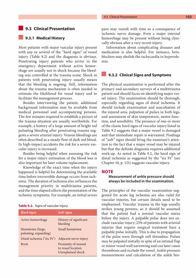
103
9.3 Clinical Presentation
9.3.1 Medical History
Most patients with major vascular injury present with any or several of the “hard signs” of vessel injury (Table 9.2) and the diagnosis is obvious. Penetrating injury patients who arrive in the emergency department without active hemor-rhage are usually not in shock because the bleed-ing was controlled at the trauma scene. Shock in patients with penetrating injury usually means that the bleeding is ongoing. Still, information about the trauma mechanism is often needed to estimate the likelihood for vessel injury and to facilitate the management process.
Besides interviewing the patient, additional background information may be available from medical personnel and accompanying persons. The few minutes required to establish a picture of the trauma situation are usually worthwhile. For example, a history of a large amount of bright red pulsating bleeding after penetrating trauma sug-gests a severe arterial injury. Venous bleedings are often described as a steady flow of dark red blood. In high-impact accidents the risk for a severe vas-cular injury is increased.
Besides being helpful when assessing the risk for a major injury estimation of the blood loss is also important for later volume replacement.
Knowledge of the exact time when the injury happened is helpful for determining the available time before irreversible damage occurs from isch-emia. The duration of ischemia also influences the management priority in multitrauma patients, and the time elapsed affects the presentation of the ischemic symptoms. For example, an initial severe
pain may vanish with time as a consequence of ischemic nerve damage. Even a major internal hemorrhage may be present without being clini-cally obvious after a very recent injury.
Information about complicating diseases and medication is also helpful. For instance, beta-blockers may abolish the tachycardia in hypovole-mia.
9.3.2 Clinical Signs and Symptoms
The physical examination is performed after the primary and secondary surveys of a multitrauma patient and should focus on identifying major ves-sel injury. The examination should be thorough, especially regarding signs of distal ischemia. It should include examination and auscultation of the injured area, palpation of pulses in both legs, and assessment of skin temperature, motor func-tion, and sensibility. The presence of one or more of the classic hard signs of vascular injury listed in Table 9.2 suggests that a major vessel is damaged and that immediate repair is warranted. Findings of “soft” signs should bring the examiner’s atten-tion to the fact that a major vessel may be injured but that the definite diagnosis requires additional work-up. As noted in Table 9.2, the hard sign of distal ischemia as suggested by the “six Ps” (see Chapter 10, p. 121) suggests vascular injury.
NOTEMeasurement of ankle pressure should always be included in the examination.
The principles of the vascular examination sug-gested for acute leg ischemia are also valid for vascular injuries, but certain details need to be emphasized. Vascular trauma in the legs usually strikes young persons, so it should be assumed that the patient had a normal vascular status before the injury. A palpable pulse does not ex-clude vascular injury; 25% of patients with arterial injuries that require surgical treatment have a palpable pulse initially. This is due to propagation of the pulse wave through soft thrombus. Pulses may be palpated initially in spite of an intimal flap or minor vessel wall narrowing and can later cause thrombosis and occlude the vessel. Ankle pressure measurements and calculation of the ankle bra-
Table 9.2. Signs of vascular injury
Hard signs Soft signs
Active hemorrhage History of significant bleeding
Hematoma (large, pulsating, expanding)
Small hematoma
Distal ischemia (“six Ps”) Adjacent nerve injuryBruit Proximity of wound
to vessel locationUnexplained shock
9.3 Clinical Presentation

Chapter 9 Vascular Injuries in the Leg104
chial index (ABI) should therefore supplement palpation of pulses. If the ABI is ≤0.9, arterial inju-ries should be suspected.
Findings in the physical examination of a pa-tient in shock are particularly difficult to inter-pret. In several aspects findings of distal ischemia caused by vascular injury are similar to vasocon-striction of the skin vessels in the foot. Differences in pallor, the presence of pulses, and skin tempera-ture between the injured and uninjured leg there-fore should be interpreted as the possible presence of vascular injury. Ankle pressure measurements are also valuable during such circumstances.
It is important to remember to listen for bruits and thrills over the wounded area to reveal a pos-sible arteriovenous fistula.
9.4 Diagnostics
Recommendations for management of suspected vascular injuries in the leg have evolved from mandatory exploration of all suspected injuries (a common practice during past wars), to routine angiography for most patients, to a more selective approach today. Regarding exploration and subse-quent angiography, it was found that negative explorations and arteriograms were obtained in over 80% of the patients. The associated risk for complications and morbidity after these invasive procedures is the rationale for a more selective approach. Rapid transportation, clinical exa-mination, ankle pressure measurements, careful monitoring, and duplex examination leave angi-ography for some of the patients and urgent explo-ration for a few.
9.4.1 Angiography
Angiography is unnecessary when a vascular in-jury is obvious after the examination. The two most common indications for excluding vascular injury are (1) when there are no hard signs at the examination, and (2) when clinical findings are imprecise but the ABI is <0.9.
Angiography is more often indicated after blunt trauma than penetrating. The reason for this is the more difficult clinical examination because of the more extensive soft tissue and nerve damage after
blunt trauma. It may occasionally be helpful to perform angiography even when injury is evident in order to exactly locate the injured vessel. An option is to perform it intraoperatively. The tech-nique is described in Chapter 10, p. 128. Contra-lateral puncture is important when the injury is close to the groin.
The purpose of the arteriography is to identify and locate lesions such as occlusions, narrowing, and intimal flaps. Contrast leakage outside the vessel can be visualized, and it also serves to pro-vide a road map before surgery. It has, however, been argued that it is unnecessary to search for minimal lesions; some studies have shown that it is safe and effective to manage such lesions nonop-eratively. On the other hand, angiography may be the first step in the final treatment of such small lesions by stenting.
When the injury is caused by a shotgun blast, angiography should always be performed because multiple vascular injuries are common. It is then indicated regardless of the clinical signs and symp-toms. The risk for complications after angiogra-phy is very low, but the risk of complications is higher when the punctured artery is small. Chil-dren therefore have a rather high rate of complica-tions. A contributing factor is that their very vaso-active arteries are prone to temporary spasm. Overall, as described above, the risk for complica-tions after angiography does not warrant avoiding it when indicated.
Occasionally it is worthwhile to order veno-graphy. It may be indicated in patients not sub-jected to exploration because arterial injury was ruled out but in whom a major venous injury is suspected. As an example, 5–10% of all popliteal venous injuries are reported to occur without arte-rial damage.
9.4.2 Duplex Ultrasound
Despite the usefulness of duplex scanning in gen-eral for vascular diagnosis, it has not been univer-sally accepted for diagnosis of vascular trauma despite the fact that it is noninvasive. It is operator dependent and vessels may be difficult to assess in multiply injured patients, legs with skeletal de-formities, large hematomas, and through splints and dressings. In some hospitals with expertise in
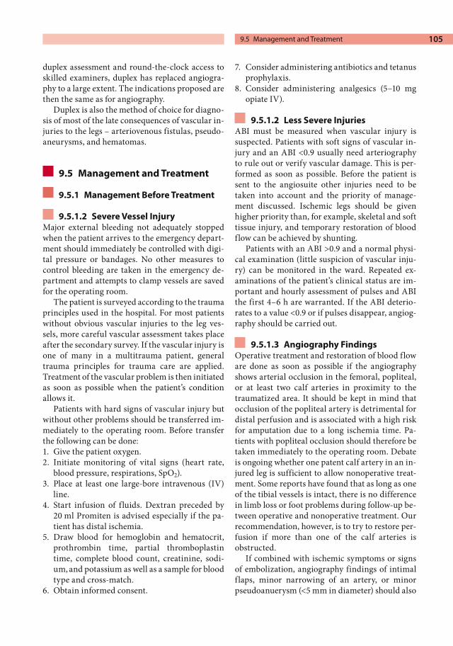
105
duplex assessment and round-the-clock access to skilled examiners, duplex has replaced angiogra-phy to a large extent. The indications proposed are then the same as for angiography.
Duplex is also the method of choice for diagno-sis of most of the late consequences of vascular in-juries to the legs – arteriovenous fistulas, pseudo-aneurysms, and hematomas.
9.5 Management and Treatment
9.5.1 Management Before Treatment
9.5.1.2 Severe Vessel InjuryMajor external bleeding not adequately stopped when the patient arrives to the emergency depart-ment should immediately be controlled with digi-tal pressure or bandages. No other measures to control bleeding are taken in the emergency de-partment and attempts to clamp vessels are saved for the operating room.
The patient is surveyed according to the trauma principles used in the hospital. For most patients without obvious vascular injuries to the leg ves-sels, more careful vascular assessment takes place after the secondary survey. If the vascular injury is one of many in a multitrauma patient, general trauma principles for trauma care are applied. Treatment of the vascular problem is then initiated as soon as possible when the patient’s condition allows it.
Patients with hard signs of vascular injury but without other problems should be transferred im-mediately to the operating room. Before transfer the following can be done:1. Give the patient oxygen.2. Initiate monitoring of vital signs (heart rate,
blood pressure, respirations, SpO2).3. Place at least one large-bore intravenous (IV)
line.4. Start infusion of fluids. Dextran preceded by
20 ml Promiten is advised especially if the pa-tient has distal ischemia.
5. Draw blood for hemoglobin and hematocrit, prothrombin time, partial thromboplastin time, complete blood count, creatinine, sodi-um, and potassium as well as a sample for blood type and cross-match.
6. Obtain informed consent.
7. Consider administering antibiotics and tetanus prophylaxis.
8. Consider administering analgesics (5–10 mg opiate IV).
9.5.1.2 Less Severe Injuries ABI must be measured when vascular injury is suspected. Patients with soft signs of vascular in-jury and an ABI <0.9 usually need arteriography to rule out or verify vascular damage. This is per-formed as soon as possible. Before the patient is sent to the angiosuite other injuries need to be taken into account and the priority of manage-ment discussed. Ischemic legs should be given higher priority than, for example, skeletal and soft tissue injury, and temporary restoration of blood flow can be achieved by shunting.
Patients with an ABI >0.9 and a normal physi-cal examination (little suspicion of vascular inju-ry) can be monitored in the ward. Repeated ex-aminations of the patient’s clinical status are im-portant and hourly assessment of pulses and ABI the first 4–6 h are warranted. If the ABI deterio-rates to a value <0.9 or if pulses disappear, angiog-raphy should be carried out.
9.5.1.3 Angiography Findings Operative treatment and restoration of blood flow are done as soon as possible if the angiography shows arterial occlusion in the femoral, popliteal, or at least two calf arteries in proximity to the traumatized area. It should be kept in mind that occlusion of the popliteal artery is detrimental for distal perfusion and is associated with a high risk for amputation due to a long ischemia time. Pa-tients with popliteal occlusion should therefore be taken immediately to the operating room. Debate is ongoing whether one patent calf artery in an in-jured leg is sufficient to allow nonoperative treat-ment. Some reports have found that as long as one of the tibial vessels is intact, there is no difference in limb loss or foot problems during follow-up be-tween operative and nonoperative treatment. Our recommendation, however, is to try to restore per-fusion if more than one of the calf arteries is obstructed.
If combined with ischemic symptoms or signs of embolization, angiography findings of intimal flaps, minor narrowing of an artery, or minor pseudoanuerysm (<5 mm in diameter) should also
9.5 Management and Treatment

Chapter 9 Vascular Injuries in the Leg106
be treated. Endovascular stenting is then a good alternative to operative treatment. Expectancy could be favorable for asymptomatic patients with normal ABI. Such occult arterial injuries appear to have an uneventful course and late occlusion is extremely rare. The occasional pseudoaneurysm that will enlarge with time appears to benefit from later repair.
9.5.1.4 Primary Amputation In most circumstances, but not always, it is rea-sonable to repair injured vessels. For a few patients, however, primary amputation is a better option. This is often a difficult decision. Primary amputa-tion is favorable for the patient if the leg is massa-cred or if the duration of ischemia is very long (>12 h) and appears to be irreversible in the clini-cal examination (Chapter 10, p. 123). Primary amputation may also be considered for certain pa-tients: multitrauma patients, patients with severe comorbid disease, and those in whom the leg was already paralyzed at the time of injury. Extensive nerve damage, lack of soft tissue to cover the wound, and duration of ischemia >6 h support primary amputation for these patient groups.
There are scoring systems, such as the Mangled Extremity Severity Score (MESS), to aid in making the decision to amputate a leg or an arm. For ex-ample, a patient over 50 years old with persistent hypotension and a cool paralyzed distal leg after high-energy trauma should have the leg amputat-ed according to MESS. It must be stressed, how-ever, that repair of both venous and arterial inju-ries is superior for most patients. The MESS score is described in Chapter 3 (p. 36).
9.5.2 Operation
Surgical treatment of vascular injuries in the leg usually proceeds in a particular order common for most patients. First the patient is scrubbed, anes-thetized, and prepared for surgery. The next step is to achieve proximal control. Occasionally, con-trol of the bleeding by manual compression with a gloved hand needs to be maintained throughout these first two steps. Proximal control is followed by measures to achieve distal control, often ac-complished during exploration of the wound. Fi-nally, the vessels are repaired and the wound cov-
ered with soft tissue. When the patient has other injuries that motivate urgent treatment, or has fractures in the leg that need to be surgically re-paired, this last step can be delayed while perfu-sion to the distal leg is maintained by a shunt tem-porarily bypassing the injured area.
9.5.2.1 Preoperative Preparation The patient is placed on a surgical table that allows x-ray penetration. If not administered previously, infection prophylaxis treatment is started. The en-tire injured extremity is scrubbed with the foot draped in a transparent plastic bag. A very good marginal of the sterile field is essential because in-cisions need to be placed much more proximal than the wound to achieve proximal control. The contralateral leg should also be scrubbed and draped to allow harvest of veins for grafts. The ve-nous system in the injured leg should be kept in-tact if possible. If a patient is in shock and the bleeding is difficult to control, it is recommended to delay inducing the anesthesia until just before the operation begins in order to avoid increased bleeding and an accentuated drop in systemic blood pressure due to loss of adrenergic activity.
NOTEIt is usually wise to achieve proximal control through a separate incision before exploring the wounded area.
9.5.2.2 Proximal Control In patients with injuries proximal to the femoral vessels, control is achieved through an incision in the abdominal fossa. The common or external iliac artery can then be exposed retroperitoneally and secured. Proximal control for injuries in the thigh, proximal to the poplital fossa, is usually obtained by exposing the common femoral artery and its branches in the groin. Popliteal vessel trau-ma can be controlled by exposing the distal super-ficial femoral artery or the proximal popliteal ar-tery through a medial incision above the knee. This is not too difficult, and the principles follow the outline given in the Technical Tips box. Inflow control for calf vessel injuries is reached by expos-ing the popliteal artery below the knee.
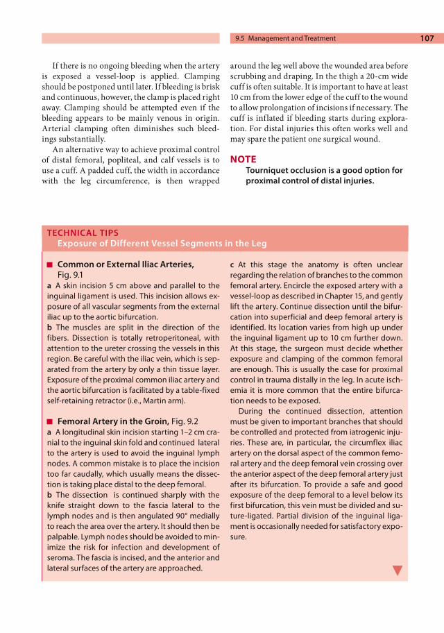
107
TECHNICAL TIPSExposure of Different Vessel Segments in the Leg
Common or External Iliac Arteries, Fig. 9.1
a A skin incision 5 cm above and parallel to the inguinal ligament is used. This incision allows ex-posure of all vascular segments from the external iliac up to the aortic bifurcation.b The muscles are split in the direction of the fibers. Dissection is totally retroperitoneal, with attention to the ureter crossing the vessels in this region. Be careful with the iliac vein, which is sep-arated from the artery by only a thin tissue layer. Exposure of the proximal common iliac artery and the aortic bifurcation is facilitated by a table-fixed self-retaining retractor (i.e., Martin arm).
Femoral Artery in the Groin, Fig. 9.2a A longitudinal skin incision starting 1–2 cm cra-nial to the inguinal skin fold and continued lateral to the artery is used to avoid the inguinal lymph nodes. A common mistake is to place the incision too far caudally, which usually means the dissec-tion is taking place distal to the deep femoral.b The dissection is continued sharply with the knife straight down to the fascia lateral to the lymph nodes and is then angulated 90° medially to reach the area over the artery. It should then be palpable. Lymph nodes should be avoided to min-imize the risk for infection and development of seroma. The fascia is incised, and the anterior and lateral surfaces of the artery are approached.
c At this stage the anatomy is often unclear regarding the relation of branches to the common femoral artery. Encircle the exposed artery with a vessel-loop as described in Chapter 15, and gently lift the artery. Continue dissection until the bifur-cation into superficial and deep femoral artery is identified. Its location varies from high up under the inguinal ligament up to 10 cm further down. At this stage, the surgeon must decide whether exposure and clamping of the common femoral are enough. This is usually the case for proximal control in trauma distally in the leg. In acute isch-emia it is more common that the entire bifurca-tion needs to be exposed.
During the continued dissection, attention must be given to important branches that should be controlled and protected from iatrogenic inju-ries. These are, in particular, the circumflex iliac artery on the dorsal aspect of the common femo-ral artery and the deep femoral vein crossing over the anterior aspect of the deep femoral artery just after its bifurcation. To provide a safe and good exposure of the deep femoral to a level below its first bifurcation, this vein must be divided and su-ture-ligated. Partial division of the inguinal liga-ment is occasionally needed for satisfactory expo-sure.
9.5 Management and Treatment
If there is no ongoing bleeding when the artery is exposed a vessel-loop is applied. Clamping should be postponed until later. If bleeding is brisk and continuous, however, the clamp is placed right away. Clamping should be attempted even if the bleeding appears to be mainly venous in origin. Arterial clamping often diminishes such bleed-ings substantially.
An alternative way to achieve proximal control of distal femoral, popliteal, and calf vessels is to use a cuff. A padded cuff, the width in accordance with the leg circumference, is then wrapped
around the leg well above the wounded area before scrubbing and draping. In the thigh a 20-cm wide cuff is often suitable. It is important to have at least 10 cm from the lower edge of the cuff to the wound to allow prolongation of incisions if necessary. The cuff is inflated if bleeding starts during explora-tion. For distal injuries this often works well and may spare the patient one surgical wound.
NOTETourniquet occlusion is a good option for proximal control of distal injuries.

Chapter 9 Vascular Injuries in the Leg108
TECHNICAL TIPSExposure of Different Vessel Segments in the Leg (continued)
Superficial Femoral Artery, Fig. 9.3A skin incision is made along the dorsal aspect of the sartorius muscle at a midthigh level. It is im-portant to avoid injuries to the greater saphenous vein, which usually is located in the posterior flap of the incision. The incision can be elongated as needed. After the deep fascia is opened and the sartorius muscle is retracted anteriorly, the femo-ral artery is found and can be mobilized. Division of the adductor tendon is sometimes required for exposure.
Popliteal Artery Above the Knee, Fig. 9.4
a The knee is supported on a sterile, draped pillow. The skin incision is started at the medial aspect of the femoral condyle and follows the anterior border of the sartorius muscle 10–15 cm in a proximal direction. Protect the greater sa-phenous vein and the saphenous nerve during dissection down to the fascia. After dividing the fascia longitudinally, continue the dissection in the groove between the sartorius and gracilis muscles, which leads to the fat in the popliteal fossa.
a The popliteal artery and adjacent veins and nerve are then, without further division of mus-cles, easily found and separated in the anterior aspect of the fossa.
Popliteal Artery Below the Knee, Fig. 9.5
a A sterile pillow or pad is placed under the distal femur. The incision is placed 1 or 2 cm posterior to the medial border of the tibia, starting at the tibial tuberosity and extending 10–12 cm distally. Sub-cutaneous fat and fascia are sharply divided, with caution to the greater saphenous vein.b The popliteal fossa is reached by retracting the gastrocnemius muscle dorsally. The deep fas-cia is divided and the artery usually easier to iden-tifiy. Occasionally, pes anserinus must be divided for adequate exposure. The popliteal artery is often located just anterior to the nerve and in close contact with the popliteal vein and crossing branches from concomitant veins. If it is necessary to expose the more distal parts of the popliteal artery, the soleus muscle has to be divided and partly separated from the posterior border of the tibia.
Fig. 9.1. Exposure of common or external iliac arteries. Note the ureter crossing the arteries anteriorly

1099.5 Management and Treatment
Fig. 9.2. Exposure of femoral artery in the groin
Fig. 9.3. Incision for exposure of the superficial femo-ral artery
9.5.2.3 Distal Control and Exploration Distal control is achieved by distal elongation of the incision used to explore the site of injury. Through this incision careful dissection in intact tissue distal to the injured area usually reveals the injured artery. When it is identified and found to be not completely transected, a vessel-loop is posi-tioned around it. If the artery is cut, a vascular clamp is applied on the stump. It is also possible to gain distal control during the exploration of the injured area, but dissection may be tricky because of hematoma, edema, and distorted anatomy. Usu-ally the backbleeding from the distal artery is minimal and does not disturb visualization dur-ing dissection. Simultaneous venous bleeding that emerges from major veins must also be controlled. This can be done by balloon occlusion or clamps. The latter should be used with caution and closed as little as possible.

Chapter 9 Vascular Injuries in the Leg110
When control is obtained the wound is explored and the site of vessel injury identified. The best way is to follow the artery proximally from where the artery was exposed for distal control. Of course, any foreign materials encountered need to be removed. The injured artery should be explored in both directions until a normal arterial wall is reached. Several centimeters of free vessels are needed. Side branches are controlled using a double loop of suture with a hanging mosquito or by small vascular clamps (see Chapter 15, p. 181). Thrombosed arteries usually have a hema-toma in the vessel wall giving it a dark blue color. Such parts need to be cut out and the vessel edges trimmed before shunting or repair. For penetrat-ing injuries all parts of the vascular wall that are lacerated must be excised to ensure that the intima will be enclosed in the suture line during repair. After this procedure the vessel can be shunted or repaired.
Finally, other parts of the wound are explored and all devitalized skin and muscle tissue is ex-cised. Injured small adjacent veins should be li-gated and all larger veins, such as the femoral veins and the popliteal vein, must be repaired.
9.5.2.4 Shunting Insertion of a temporary shunt to restore distal perfusion is sensible if the vascular injury appears to require more extensive repair than a simple su-ture or an end-to-end anastomsis. Shunting pro-vides the time needed to either perform a vascular reconstruction, including harvesting of a vein graft from the uninjured leg, or to wait for help. Shunting can also be valuable when patients have other injuries that need attention or when leg frac-tures must be repositioned to give the appropriate vascular graft length. In patients with fractures shunting allows both repositioning and fixation without increasing the risk of ischemic damage.
Fig. 9.4. Exposure of popliteal artery above the knee Fig. 9.5. Exposure of popliteal artery below the knee

111
A novel vascular reconstruction seldom tolerates the forces required for reposition of fractures. When perfusion is restored by a shunt the surgeon has plenty of time to carefully explore the wound and other injuries. When a shunt is used for distal perfusion while other procedures are performed, it is important to check its function at least every 30 min.
The principles of vascular shunting are simple. Specially designed shunts can be used if available; examples are Pruitt–Inhara and Javid. Most have inflatable balloons in both ends for occlusion and side channels with stopcocks through which the function of the shunt can be tested (Figs. 9.6, 9.7). The side holes also enable infusion of a heparin-ized solution and contrast for fluoroscopy. The
Fig. 9.6. A special catheter for shunting (Pruitt–Inahara shunt). The shunt has a larger balloon in one end aimed for the proximal inflow vessel and a smaller one for placement in the outflow vessel. The occluding balloons are con-trolled by injecting saline through separate channels with stopcocks. The shunt can also be flushed through a third channel
Fig. 9.7. Example of shunting in a severe artery and vein injury. The artery is shunted with a Pruitt–Ina-hara shunt and its balloons secured with vessel-loops. For shunting of the vein a piece of ordinary rubber tubing is used. It is also secured with vessel-loops and with a suture
9.5 Management and Treatment

Chapter 9 Vascular Injuries in the Leg112
extra channels can be used to draw blood during the operation. It is not, however, necessary to use manufactured shunts. Any kind of sterile plastic or rubber tubing is sufficient. It is then important to use dimensions of the tube in accordance with the artery’s inner diameter. The tube is cut into suitable lengths and the edges carefully trimmed with scalpel and scissors to avoid damage when inserted. The tube is positioned and secured with vessel loops that abolish the space between the artery and tube to manage bleeding. A loosely applied suture around the middle part of the shunt can be used to secure it. It is advantageous to simultaneously shunt concomitant vein injuries – at least the femoral and popliteal veins – to avoid swelling and to facilitate distal flow.
9.5.2.5 Vessel RepairWhile we advocate repairing both the artery and the vein, we do not favor reconstructing the in-jured vein before the artery. If both can be mended within a reasonable timeframe we recommend that the most difficult reconstruction is performed first. If, however, the artery is shunted it may be advantageous to start with the vein to achieve optimal outflow as soon as possible. Some vas-cular surgeons favor vein ligature as a general principle because of the potential risk for emboli-zation from vein segments that thrombose after repair.
NOTEPopliteal occlusions should be managed quickly because of the high risk of amputation in case of delayed treatment.
Arterial Injuries In general, all injured arteries should be repaired. Sometimes, when necessary in order to save the patient’s life or when interruption of an artery does not influence blood flow to the leg, the in-jured vessel may be ligated. The former is extreme-ly uncommon, but the decision is difficult when it arises. As an aid we have listed in Table 9.3 the am-putation rates, as obtained from the literature, fol-lowing ligation of vessels. Among proximal ves-sels, only branches from the deep femoral artery, but not the main branch, can be ligated without morbidity. Distally, we recommend repair of at least two calf arteries to be on the safe side, but it is
possible to leave two interrupted, provided the remaining vessel is not the peroneal artery.
Before definitive repair the surgeon must be sure that the inflow and outflow vascular beds re-main open and are free of clots. Liberal use of Fog-arty catheters is therefore wise. The technique is described in Chapter 10 (p. 126). If the backbleed-ing is questionable, intraoperative angiography should be performed to make sure the outflow tract is free of clots. Local heparinization, de-scribed in Chapter 15 (p. 181), is always indicated. Systemic heparinization can be used for selected patients without other injuries considered to have a low risk for continued bleeding from the wound after debridement. If the foot’s appearance or in-traoperative angiography suggests microemboli-zation, local thrombolysis can be tried as described in Chapter 10 (p. 127).
The goal for repair is to permanently restore continuity of the artery without stenosis or ten-sion. The type of injury determines the choice of technique. It varies from a couple of vascular sutures to reconstruction with a patch, interposi-tion, or a bypass grafting. Lateral suture for repair of minor lesions, including patching, is described in Chapter 15 (p. 183). Bypass techniques are be-yond the scope of this book, and detailed descrip-tions can be found in other vascular surgery text-books.
A graft is usually needed, and only occasionally can an end-to-end anastomosis be performed without tension. It is always better to use an inter-position graft or a bypass to avoid what is even perceived as insignificant tension because anasto-motic rupture and graft necrosis may occur when leg swelling and movements pull the arterial ends further apart postoperatively. The major bleeding that can result from this may even be fatal.
Table 9.3. Amputation frequencies after ligature of different arteries
Vessel ligated Amputation rate
Common iliac artery 54%External iliac artery 47%Common femoral artery 81%Superficial femoral artery 55%Popliteal artery 73%

113
An autologous vein is always the preferred graft material. Vein is more infection resistant than synthetic materials and is more flexible. It allows both elongation and vasodilatation to adjust varia-tions in flow requirements. The great saphenous vein from the contralateral leg is the primary choice for a graft. It is a serious mistake to use the great saphenous vein from the traumatized leg if the deep vein is also injured or if injury is suspect-ed. Interruption of the saphenous vein with ob-structed concomitant deep veins will rapidly cause severe distal swelling of the leg and graft occlusion within days. The vein can be harvested at a level in the leg where the saphenous vein diameter fits the artery that needs to be repaired. A graft slightly larger than the artery should be obtained if possi-ble. For common femoral artery lesions, two pieces of saphenous vein, both open longitudinally and then sutured together, might be required. An op-tion is to use arm veins as graft material. If veins not are available expanded polytetrafluoroethyl-ene (ePTFE) is the second choice. The main rea-sons not to use it are the slightly higher risks for postoperative graft occlusion and infection.
Venous injuries Most venous injuries are exposed when the wound is explored. While vein ligature may lead to leg swelling, it rarely causes ischemia or amputation. On the other hand, the only benefit of vein liga-ture is rapid bleeding control and a reduced oper-ating time. Major veins can therefore be ligated to save the life of an unstable patient. If possible, however, most veins should be repaired, especially the popliteal and the common femoral veins. Calf veins can be ligated without morbidity. In patients with combined injuries, both the vein and the ar-tery should be repaired to enhance the function of the arterial reconstruction. Control of bleedings can be achieved by fingers or a “strawberry” or “peanut” to compress the vein proximal and distal to the injured site, or by using gentle vascular clamps. A continuous running suture, almost without traction in the suture line, is often suffi-cient to close a stab wound. When the vein is more extensively damaged, a patch or an interposition graft may be needed for repair. As with arteries, it is important to match the caliber of the interposi-tion graft and the injured vein. Veins without blood are collapsed, so it is easy to underestimate
their size. An autologous vein is preferred over synthetic materials.
9.5.2.6 Finishing the OperationAfter the vascular reconstruction is finished the resulting improvement in distal perfusion should be checked. This is indicated as well-perfused skin in the foot and palpable pulses. If there is any doubt about the result control angiography should be performed. Preferably, it is done from the prox-imal control site to enable visualization of all anas-tomoses. If problems are revealed the reconstruc-tion must be redone and distal clots extracted as previously described. While vascular spasm is rare and should not be regarded as the main expla-nation for lack of distal blood flow, injection of 1–2 ml papaverine into the graft can also be tried.
After vascular repair all devitalized tissue and foreign material should be removed to reduce the risk of postoperative infection. Grafts and vessels should be covered with healthy tissue by loosely adapting it with interrupted absorbable sutures. For injuries with massive tissue loss it may be im-possible to cover the graft. This increases the risk for postoperative anastomotic necrosis and rup-ture. Split or partial skin grafts or biological dress-ings can be used to prevent this. Occasionally it may be better to perform a bypass through healthy tissue, thus avoiding the traumatized area, to min-imize graft infection and postoperative complica-tions.
9.5.3 Endovascular Treatment
Endovascular treatment of vascular injuries to leg vessels is attractive because it provides a way to achieve proximal control, reduce ischemia time, and simplify complex procedures. There is not a lot of experience – at least not reported in the lit-erature – with endovascular treatment. Some cen-ters have reported successful semielective treat-ment of pseudoaneurysms and arteriovenous fis-tulas in the groin. Small patient series have been published on embolization of bleeding branches to the deep femoral artery and stent graft control of small lacerations in the femoral arteries. We have only treated occasional patients this way so far.
Endovascular treatment will probably be an option for many patients with soft signs of vascu-
9.5 Management and Treatment

Chapter 9 Vascular Injuries in the Leg114
lar injury, who will undergo angiography and then will be found to have a minor bleeding, a pseudoa-neurysm, a fistula, or an intimal flap. The possi-bilities of considering endovascular repair for pa-tients with hard signs of vascular injury to the legs will depend on logistics and the organization of a hospital’s endovascular team. With around-the-clock availability, patients with less severe distal ischemia may be subjected to angiography with possibilities for endovascular treatment in mind. Patients with penetrating injuries that are actively bleeding may undergo balloon occlusion of a proximal artery to accomplish control and then be transferred to the operating room for repair. In other hospitals where an endovascular team is not available during weekends, angiography is skipped in some patients with soft signs, and they are treated with an open procedure and intraoperative angiography instead. But it is likely that endovas-cular treatment will be the treatment of choice for a significant proportion of patients in the near future.
9.5.4 Management After Treatment
As for most vascular procedures, the risk for bleed-ing and graft thrombosis of the reconstructed artery is higher during the first 24 h after the op-eration. Postoperative monitoring of limb perfu-sion, including inspection of foot skin and wounds and palpation of pulses, is necessary at least every 30 min for the first 6 h. If pulses are difficult to ascertain, ankle pressure should be measured. Loss of pulses or an abrupt drop in pressure indi-cate that reoperation may be required, even when the graft appears to be patent either clinically or on duplex scanning. As for acute leg ischemia caused by thrombosis, there is also a substantial risk for compartment syndrome. Particularly when the ischemia duration has been long, this risk is considerable, and it is important to examine calf muscles for signs of compartment syndrome. This assessment includes motor function, tender-ness, and palpation of the muscle compartments in the calf. Findings that may suggest fasciotomy are listed in Table 9.4
For most patients administration of dextran or low molecular weight heparin is indicated to avoid postoperative thrombosis. This is especially im-
portant for patients with both venous and arterial reconstruction. Patients who have been subjected to venous ligation need extra attention and mea-sures against limb swelling. Antibiotic treatment started preoperatively or intraoperatively is usu-ally continued after the operation. We use a com-bination of benzylpenicillin and isoxazolyl peni-cillin to cover streptococci, clostridia, and staphy-lococci.
9.6 Results and Outcome
Mortality after isolated vascular injuries in the legs is very low, ranging from 0% to 3% in most series. The few deaths reported were due to infec-tion and sepsis, underscoring the importance of careful tissue debridement and graft coverage. For solitary vascular injuries, amputation rates are also low. It has to be remembered that important nerves are located adjacent to the vessels and often are simultaneously severely damaged. This con-tributes to long-term disability. In most studies, arterial patency is very high, while venous recon-structions tend to thrombose at a much higher fre-quency. Although some authors have argued that venous repair is therefore unnecessary, we believe it improves patency of the arterial graft. Fractures, especially open ones, in combination with arterial injury have less encouraging long-term results. In most reports the incidence of primary as well as secondary amputation is higher when fractures are combined with vascular and nerve injuries. In one study the primary and secondary amputation rates were 20% and 52%, respectively, during a fol-low-up of 5.6 years. There was a 30% long-term disability rate for salvaged limbs in the same study,
Table 9.4. Medical history and clinical findings sug-gesting that fasciotomy may be needed
Severe ischemia lasting longer than 4–6 hPreoperative shockFirm/hard muscle compartment when palpatedDecreased sensibility and/or motor functionCompartmental pressure >30 mmHgPain out of proportion localized distal to the vascular injury

115
all related to poor recovery of neurological func-tion. Although it is clear that popliteal injuries, combined injuries with femoral fractures, delayed repair >6 h, and injuries treated in patients in shock have a much higher amputation rate, the results may be less pessimistic than the results in the literature. For multitrauma patients outcomes of vascular injury in the leg are hard to come by.
Fig. 9.8. Incision and exposure for fasciotomy of pos-terior compartments. The posterior dotted line indicates fasciotomy incision for the superficial compartment and the anterior dotted line for the deep compartment
9.7 Fasciotomy
TECHNICAL TIPSFasciotomy of the Calf Compartments (Two Incisions)
Medial IncisionA 15–20-cm-long skin incision, starting slightly below the midpoint of the calf and downwards parallel to and 2–3 cm dorsally of the medial border of the tibia, is used to decompress the deep and superficial posterior muscle compart-ments (Fig. 9.8). It is important to avoid injury to the great saphenous vein. Sharp division of skin and subcutaneous fat reaches the fascia. The skin and subcutaneous fat is mobilized en bloc anteriorly and posteriorly to expose it enough to provide access to the compartments. The fascia is then opened in a proximal direction and dis-tally down toward the malleolus under the sole-us muscle. At this level, the soleus muscle has no attachments to the tibia, and the deep posterior compartment is more superficial and easier to access. Through the same skin incision, the fas-cia of the superficial posterior muscle compart-ment is cleaved 2–3 cm dorsally and parallel to the former. A long straight pair of scissors with blunt points is used to cut the fascia using a dis-tinct continuous movement in the distal direc-tion, down to a level of 5 cm above the malleoli and proximally along the entire fascia.
9.7 Fasciotomy
When the compartment pressure is clearly in-creased or when there is a risk of developing in-creased pressure, fasciotomy of muscular com-partments distal to a vascular injury should be performed in association with the vascular repair. Factors suggesting an increased pressure are listed in Table 9.4. The technique for decompression of the four compartments in the lower leg is described in the Technical Tips box.
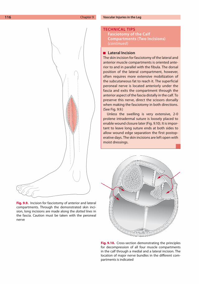
Chapter 9 Vascular Injuries in the Leg116
Fig. 9.9. Incision for fasciotomy of anterior and lateral compartments. Through the demonstrated skin inci-sion, long incisions are made along the dotted lines in the fascia. Caution must be taken with the peroneal nerve
Fig. 9.10. Cross-section demonstrating the principles for decompression of all four muscle compartments in the calf through a medial and a lateral incision. The location of major nerve bundles in the different com-partments is indicated
TECHNICAL TIPSFasciotomy of the Calf Compartments (Two Incisions)(continued)
Lateral IncisionThe skin incision for fasciotomy of the lateral and anterior muscle compartments is oriented ante-rior to and in parallel with the fibula. The dorsal position of the lateral compartment, however, often requires more extensive mobilization of the subcutaneous fat to reach it. The superficial peroneal nerve is located anteriorly under the fascia and exits the compartment through the anterior aspect of the fascia distally in the calf. To preserve this nerve, direct the scissors dorsally when making the fasciotomy in both directions. (See Fig. 9.9.)
Unless the swelling is very extensive, 2-0 prolene intradermal suture is loosely placed to enable wound closure later (Fig. 9.10). It is impor-tant to leave long suture ends at both sides to allow wound edge separation the first postop-erative days. The skin incisions are left open with moist dressings.

117
9.8 Iatrogenic Vascular Injuries to the Legs
Iatrogenic vascular injuries occur either in con-nection with other surgical procedures or as a complication to groin catheterization for angiog-raphy, percutaneous coronary intervention, and other endovascular procedures. The latter consti-tutes the main part. In this book, bleeding and pseudoaneurysms that occur after angiography are covered in Chapter 14.
Vascular injury during surgery is also quite common. The risk for vessel trauma during opera-tions varies with the type of procedure. Certain procedures are also more prone to cause vascular injury (Table 9.5), and vascular procedures are the ones most frequently associated with vascular in-jury. Suspicious signs of vascular injury during surgery are sudden bleeding that fills the operative field and problems maintaining the systemic blood pressure. This is exemplified by major bleeding occurring behind retractors or in a field previ-ously dissected during abdominal aortic aneu-rysm surgery. When the bleeding area is identi-fied, it is controlled by manual compression. It is wise to always call for help when major bleeding is suspected. More hands facilitate repair, and real-izing that one has caused a severe vascular injury may generate stress and distract the surgeon from accomplishing vascular repair.
The technique used for vascular repair is the same as for all other vascular injuries. While main-taining compression, proximal and distal control is created by careful dissection of the vessels around the suspected injury site. The vessel is then clamped or controlled by finger or swab compres-sion. The traumatized vessel is then repaired. For iatrogenic injuries this often means just a few su-tures; only rarely is more complex repair needed.
There are also specially designed instruments for controlling vessels – especially veins – enough to allow suturing without needing extensive expo-sure to achieve control. One consists of a ring 2–3 cm in diameter welded at a 75° angle to a handle. The ring is placed around the injured vein and held in place, thereby controlling the bleeding. This device is particularly helpful for iliac vein bleedings that occur during gynecologic, urologic, and rectal cancer operations. Multiple vessel inju-ries are not uncommon and perseverance is often needed to repair all vessels before the original operation can proceed.
Further Reading
Dennis JW, Frykberg ER, Veldenz HC, et al.Validation of nonoperative management of occult vascular in-juries and accuracy of physical examination alone in penetrating extremity trauma: 5- to 10-year fol-low-up. J Trauma 1998; 44(2):243–252
Hafez HM, Woolgar J, Robbs JV. Lower extremity arte-rial injury: results of 550 cases and review of risk factors associated with limb loss. J Vasc Surg 2001; 33(6):1212–1219
Hood DB, Weaver FA, Yellin AE. Changing perspec-tives in the diagnosis of peripheral vascular trauma. Semin Vasc Surg 1998; 11(4):255–260
Modrall JG, Weaver FA, Yellin AE. Vascular consider-ations in extremity trauma. Orthop Clin North Am 1993; 24(3):557–563
Nair R, Abdool-Carrim AT, Robbs JV. Gunshot injuries of the popliteal artery. Br J Surg 2000; 87(5):602
Rich NM. Management of venous trauma. Surg Clin North Am 1988; 68(4):809–821
Rowe VL, Salim A, Lipham J, et al. Shank vessel injuries. Surg Clin North Am 2002; 82(1):91–104
Snyder WH 3rd. Popliteal and shank arterial injury. Surg Clin North Am 1988; 68(4):787–807
Table 9.5. Examples of procedures associated with iatrogenic vascular injury (PCI percutaneous coronary intervention)
Procedure Vessel injured
PCI/angiography Common femoral, external iliac, deep femoral arteries
Knee arthroplasty Popliteal artery and veinHip arthroplasty Common femoralStripping of saphenous vein
Common femoral vein (groin arteries)
9.8 Iatrogenic Vascular Injuries to the Legs
Acute Leg Ischemia 10
CONTENTS
10.1 Summary . . . . . . . . . . . . . . . . . . . . . . . . . . . 119
10.2 Background . . . . . . . . . . . . . . . . . . . . . . . . 11910.2.1 Background . . . . . . . . . . . . . . . . . . . . . . . . . 11910.2.2 Magnitude of the Problem . . . . . . . . . 12010.2.3 Pathogenesis and Etiology . . . . . . . . . 12010.2.3.1 Pathogenesis . . . . . . . . . . . . . . . . . . . . . . . 12010.2.3.2 Embolus and Thrombosis . . . . . . . . . . 120
10.3 Clinical Presentation . . . . . . . . . . . . . . 12110.3.1 Medical History . . . . . . . . . . . . . . . . . . . . . 12110.3.2 Clinical Signs and Symptoms . . . . . . . 12110.3.3 Evaluation of Severity
of Ischemia . . . . . . . . . . . . . . . . . . . . . . . . . 12210.3.3.1 Classification . . . . . . . . . . . . . . . . . . . . . . . . 12210.3.3.2 Viable Leg . . . . . . . . . . . . . . . . . . . . . . . . . . 12310.3.3.3 Threatened Leg . . . . . . . . . . . . . . . . . . . . 12310.3.3.4 Management Strategy . . . . . . . . . . . . . 123
10.4 Diagnostics . . . . . . . . . . . . . . . . . . . . . . . . 123
10.5 Management and Treatment . . . . . 12410.5.1 Management Before Treatment . . . . 12410.5.1.1 Viable Leg . . . . . . . . . . . . . . . . . . . . . . . . . . 12410.5.1.2 Threatened Leg . . . . . . . . . . . . . . . . . . . . 12510.5.2 Operation . . . . . . . . . . . . . . . . . . . . . . . . . . 12510.5.2.1 Embolectomy . . . . . . . . . . . . . . . . . . . . . . . 12510.5.2.2 Thrombosis . . . . . . . . . . . . . . . . . . . . . . . . . 12710.5.2.3 Intraoperative angiography . . . . . . . . 12710.5.3 Thrombolysis . . . . . . . . . . . . . . . . . . . . . . . 12810.5.4 Management After Treatment . . . . . 12910.5.4.1 Anticoagulation . . . . . . . . . . . . . . . . . . . . 12910.5.4.2 Reperfusion Syndrome . . . . . . . . . . . . . 12910.5.4.3 Compartment Syndrome . . . . . . . . . . . 130
10.6 Results and Outcome . . . . . . . . . . . . . . 130
10.7 Conditions Associated with Acute Leg Ischemia . . . . . . . . . . 131
10.7.1 Chronic Ischemia of the Lower Extremity . . . . . . . . . . . . . 131
10.7.2 Acute Ischemia After Previous Vascular Reconstruction . . . . . . . . . . . . 131
10.7.3 Blue Toe Syndrome . . . . . . . . . . . . . . . . . 13110.7.4 Popliteal Aneurysms . . . . . . . . . . . . . . . 132
Further Reading . . . . . . . . . . . . . . . . . . . 133
10.1 Summary
It is important to evaluate the severity of ischemia.If the leg is immediately threatened, opera-tion cannot be delayed. If the leg is viable, there is no benefit of an emergency operation.Before the operation it is vital to consider the etiology of the occlusion, to be pre-pared to perform a distal vascular recon-struction, and to treat heart and pulmo-nary failure if present.
10.2 Background
10.2.1 Background
Acute leg ischemia is associated with a great risk for amputation and death. The age of the patients is high, and to some extent acute leg ischemia can be considered an end-of-life disease. Patients’ symptoms and the clinical signs of the afflicted leg vary. Sometimes grave ischemia immediately threatens limb viability, such as after a large em-bolization to a healthy vascular bed. Other times the symptoms are less dramatic, appearing as on-set of rest pain in a patient with claudication. This is usually due to thrombosis of a previously ste-nosed artery.

Chapter 10 Acute Leg Ischemia120
It is the severity of ischemia that determines management and treatment. To minimize the risk for amputation or persistent dysfunction it is im-portant to rapidly restore perfusion if an extremity is immediately threatened. When the leg shows signs of severe ischemia but is clearly viable, it is equally important to thoroughly evaluate and op-timize the patient before any intervention is initi-ated. These basic management principles are gen-erally applicable. Accordingly, we recommend “management by severity” rather than “manage-ment by etiology” (thrombosis versus embolus) but recognize that the latter can also be an effec-tive strategy.
10.2.2 Magnitude of the Problem
It is difficult to find accurate incidence figures on acute leg ischemia. Data from some reports are given in Table 10.1. The numbers listed do not in-clude conservatively treated patients or those whose legs were amputated as a primary proce-dure. The incidence increases with age and is seen with equal frequency in men and women. Regardless, the frequency indicates that it is a very common problem.
10.2.3 Pathogenesis and Etiology
10.2.3.1 PathogenesisAcute leg ischemia is caused by a sudden deterio-ration of perfusion to the distal parts of the leg. While the abrupt inhibition of blood flow causes the ischemia, its consequences are variable be-cause acute leg ischemia is multifactorial in origin. Hypercoagulable states, cardiac failure, and dehy-
dration predispose the blood for thrombosis and make the tissue more vulnerable to decreased per-fusion. Besides the fact that a healthy leg is more vulnerable than one accustomed to low perfusion, it is unknown what determines the viability of the tissue. The most important factor is probably the duration of ischemia. The type of tissue affected also influences viability. In the leg, the skin is more ischemia-tolerant than skeletal muscle.
10.2.3.2 Embolus and Thrombosis The etiology of the occlusion is not what deter-mines the management process. It is, however, of importance when choosing therapy. Embolus is usually best treated by embolectomy, whereas ar-terial thrombosis is preferably resolved by throm-bolysis, percutaneous transluminal angioplasty (PTA), or a vascular reconstruction. The reason for this difference is that emboli often obstruct a relatively healthy vascular bed, whereas thrombo-sis occurs in an already diseased atherosclerotic artery. Consequently, emboli more often cause immediate threatening ischemia and require ur-gent restoration of blood flow. Thrombosis, on the other hand, occurs in a leg with previous arterial insufficiency with well-developed collaterals. In the latter case it is important not only to solve the acute thrombosis but also to get rid of the cause. It must be kept in mind that emboli can be lodged in atherosclerotic arteries as well, which then makes embolectomy more difficult.
Table 10.2 summarizes typical findings in the medical history and physical examination that suggest thrombosis or embolism. Many risk fac-tors, such as cardiac disease, are common for both embolization and thrombosis. Atrial fibrillation and a recent (less than 4 weeks) myocardial infarc-tion with intramural thrombus are the two domi-
Table 10.1. Incidence of acute leg ischemia
Country Year Surveyed popu-lation size
Population Yearly incidence per 100,000 inhabitants
Sweden 1965–1983 1.5 million All treated or amputated, >70 years old
125 (men)150 (women)
USA 2000 All hospitalized 95Sweden 1990–1994 2.0 million All treated 60 (men)
77 (Women)United Kingdom 1995 0.5 million All diagnosed 14–16

121
nating sources for emboli (80–90%). Other possi-ble origins are aneurysms and atherosclerotic plaques located proximal to the occluded vessel. The latter are often associated with microemboli-zation (discussed later) but may also cause larger emboli.
Plaque rupture, immobilization, and hyperco-agulability are the main causes of acute thrombo-sis. Severe cardiac failure, dehydration, and bleed-ing are less common causes. Hypoperfusion due to such conditions can easily turn an extremity with longstanding slightly compromised perfusion into acute ischemia.
10.3 Clinical Presentation
10.3.1 Medical History
The typical patient with acute leg ischemia is old and has had a recent myocardial infarction. He or she describes a sudden onset of symptoms – a few hours of pain, coldness, loss of sensation, and poor mobility in the foot and calf. Accordingly, all signs of threatened leg viability are displayed. The event is most likely an embolization, and the patient needs urgent surgery. Unfortunately, such patients are unusual among those who are admitted for acute leg ischemia. The history is often variable, and sometimes it is difficult to decide even the time of onset of symptoms.
It is important to obtain a detailed medical history to reveal any underlying conditions or lesions that may have caused the ischemia. More-over, identifying and treating comorbidities may improve the outcome after surgery or thrombo-lysis.
10.3.2 Clinical Signs and Symptoms
The symptoms and signs of acute ischemia are often summarized as the “five Ps”: pain, pallor, pulselessness, paresthesia, and paralysis. Besides being helpful for establishing diagnosis, careful evaluation of the five Ps is useful for assessing the severity of ischemia. Sometimes a sixth P’s is used – poikolothermia, meaning a low skin tempera-ture that does not vary with the environment.
Pain: For the typical patient, as the one de-scribed above, the pain is severe, continuous, and localized in the foot and toes. Its intensity is unre-lated to the severity of ischemia. For instance, it is less pronounced when the ischemia is so severe that the nerve fibers transmitting the sensation of pain are damaged. Patients with diabetes often have neuropathy and a decreased sensation of pain.
Pallor: The ischemic leg is pale or white initially, but when ischemia aggravates the color turns to cyanotic blue. This cyanosis is caused by vessel dilatation and desaturation of hemoglobin in the skin and is induced by acidic metabolites in combi-nation with stagnant blood flow. Consequently, cyanosis is a graver sign of ischemia than pallor.
Pulselessness: A palpable pulse in a peripheral artery means that the flow in the vessel is suffi-cient to give a pulse that is synchronous with ves-sel dilatation, which can be palpated with the fin-gers. In general, palpable pulses in the foot there-fore exclude severe leg ischemia. When there is a fresh thrombus, pulses can be felt in spite of an occlusion, so this general principle must be ap-plied with caution. Palpation of pulses can be used to identify the level of obstruction and is facilitat-ed by comparing the presence of pulses at the same level in the contralateral leg.
When the examiner is not convinced that pal-pable pulses are present, distal blood pressures must be measured. It is prudent to always measure the ankle blood pressure. This is a simple way to
Table 10.2. History and clinical findings differentiat-ing the etiology of acute ischemia
Thrombosis Embolism
Previous claudication No previous symptoms of arterial insufficiency
No source of emboli Obvious source of emboli (arterial fibrillation, myocardial infarction)
Long history (days to weeks)
Sudden onset (hours to days)
Less severe ischemia Severe ischemiaLack of pulses in the contralateral leg
Normal pulses in the contralateral leg
Positive signs of chronic ischemia
No signs of chronic ischemia
10.3 Clinical Presentation

Chapter 10 Acute Leg Ischemia122
verify ischemia and the measurement can be used to grade the severity and serve as a baseline for comparison with repeated examinations during the course of treatment. (This will be discussed further later.) The continuous-wave (CW) Dop-pler instrument does not give information about the magnitude of flow because it registers only flow velocities in the vessel. Therefore, an audible signal with a CW Doppler is not equivalent to a palpable pulse, and a severely ischemic leg can have audible Doppler signals.
NOTEIn acute leg ischemia, the principle use of CW Doppler is to measure ankle blood pressure.
Paresthesia: The thin nerve fibers conducting impulses from light touch are very sensitive to ischemia and are damaged soon after perfusion is interrupted. Pain fibers are less ischemia-sensi-tive. Accordingly, the most precise test of sensibil-ity is to lightly touch the skin with the fingertips, alternating between the affected and the healthy leg. It is a common mistake to believe that the skin has been touched too gently when the patient actu-ally has impaired sensitivity. The examiner then may proceed to pinching and poking the skin with a needle. Such tests of pain fibers evaluate a much later stage of ischemic damage. The anatom-ic localization of impaired sensation is sometimes related to which nerves are involved. Frequently, however, it does not follow nerve distribution areas and is circumferential and most severe distally. Numbness and tingling are other symptoms of ischemic disturbance of nerve function.
Paralysis: Loss of motor function in the leg is initially caused by ischemic destruction of motor nerve fibers and at later stages the ischemia direct-ly affects muscle tissue. When palpated, ischemic muscles are tender and have a spongy feeling. Ac-cordingly, the entire leg can become paretic after proximal severe ischemia and misinterpreted as a consequence of stroke. Usually paralysis is more obscure, however, presenting as a decreased strength and mobility in the most distal parts of the leg where the ischemia is most severe. The most sensitive test of motor function is to ask the patient to try to move and spread the toes. This gives information about muscular function in the foot and calf. Bending the knee joint or lifting the whole leg is accomplished by large muscle groups in the thigh that remain intact for a long time after ischemic damage in the calf muscle and foot has become irreversible.
10.3.3 Evaluation of Severity of Ischemia
10.3.3.1 ClassificationWhen a patient has been diagnosed to have acute leg ischemia, it is extremely important to evaluate its grade. Ischemic severity is the most important factor for selecting a management strategy, and it also affects treatment outcome. Classification ac-cording to severity must be done before the patient is moved to the floor or sent to the radiology de-partment. We have found that the simple classifi-cation suggested by the Society for Vascular Sur-gery ad hoc committee (1997) is helpful for grad-ing. It is displayed in Table 10.3.
Table 10.3. Categories of acute ischemia
Sensibility Motor function Arterial Doppler signal
Venous Doppler signal
I Viable Normal Normal Audible(>30 mmHg)
Audible
IIa Marginally threatened
Decreased or normal in the toes
Normal Not audible Audible
IIb Immediately threatened
Decreased, not only in the toes
Mildly to moderately affected
Not audible Audible
IV Irreversibly damaged
Extensive anesthesia
Paralysis and rigor Not audible Not audible

123
10.3.3.2 Viable Leg As indicated in Figure 10.1, a viable ischemic leg is not cyanotic, the toes can be moved voluntarily, and the ankle pressure is measurable. The ratio-nale for choosing these parameters is that cyanosis and impaired motor function are of high prognos-tic value for outcome.
The limit of 30 mmHg for the ankle pressure (Table 10.3, Fig. 10.1) is not important per se but is a practical limit useful to make sure that it is the arterial, and not a venous, pressure that has been measured. The dorsalis pedis, posterior tibial ar-teries, or branches from the peroneal artery can be insonated. The latter can be found just ventral to the lateral malleolus. If no audible signal is identi-fied in any of these arteries or if there only is a weak signal that disappears immediately when the tourniquet is inflated, the ankle blood pressure should be recorded as zero. It is important to rely on the obtained results and not assume that there is a signal somewhere that is missed due to inexpe-rience. Qualitative analysis of the Doppler signal is seldom useful when evaluating acute leg ischemia.
10.3.3.3 Threatened Leg As shown in Table 10.3, the threatened leg differs from the viable one in that the sensibility is im-paired and there is no measurable ankle blood pressure. The threatened limb is further separated into marginally threatened and immediately threatened by the presence or absence of normal motor function. The threatened leg differs from the irreversibly damaged leg by the quality of the venous Doppler signal. In the irreversibly dam-aged leg, venous blood flow is stagnant and inau-dible.
10.3.3.4 Management Strategy A viable leg does not require immediate action and can be observed in the ward. A threatened leg needs urgent operation or thrombolysis. The latter
is more time-consuming and recommended for the marginally threatened leg. The immediately threatened leg must be treated as soon as possible, usually with embolectomy or a vascular recon-struction. Irreversible ischemia is quite unusual but implies that the patient’s leg cannot be saved. Figure 10.1 is intended to show a simplified algo-rithm to further support the management of acute leg ischemia.
NOTELoss of motor function in the calf and foot muscles warrants emergency surgical treatment.
10.4 Diagnostics
A well-conducted physical examination is enough to confirm the diagnosis of acute leg ischemia, determine the level of obstruction, and evaluate the severity of ischemia. When the leg is immedi-ately threatened, further radiologic examinations or vascular laboratory tests should not under any circumstances delay surgical treatment. When the extremity is viable or marginally threatened, angi-ography should be performed. Duplex ultrasound is of limited value for evaluating acute leg ischemia and angiography is recommended for almost all patients in these two groups. If angiography is not available or if examination of the patient has veri-fied that emboli is the cause and probably is best treated by embolectomy, angiography can be omit-ted. This situation is rare, however.
The arteriogram provides an anatomical map of the vascular bed and is very helpful in discrimi-nating embolus and thrombosis. The former is essential for planning the surgical procedure, and the latter may be of importance for selecting the treatment strategy.
Fig. 10.1. Simplified algorithm to support the management of acute leg ischemia
10.3 Clinical Presentation

Chapter 10 Acute Leg Ischemia124
An arteriogram representing an embolus is shown in Fig. 10.2.
Angiographic signs of embolism are an abrupt, convex start of the occlusion and lack of collater-als. Thrombosis is likely when the arteriogram shows well-developed collaterals and atheroscle-rotic changes in other vascular segments.
For most patients with viable and margin-ally threatened legs the diagnostic angiography is followed by therapeutic thrombolysis right away.
Angiography can be performed during daytime when qualified radiology staff is available. The patient should be optimized according to the recommendations given in the next section. Be-fore angiography it is important to keep the patient well hydrated and to stop administration of metformin to reduce the risk of renal failure. Disturbances in coagulation parameters may interfere with arterial puncture and must also be checked before the investigation. The information is also important as baseline values in case of later thrombolysis.
The groin of the contralateral leg is the pre-ferred puncture site for diagnostic angiography. A second antegrade puncture can be done in the ischemic extremity if thrombolysis is feasible.
10.5 Management and Treatment
10.5.1 Management Before Treatment
10.5.1.1 Viable Leg If the leg is viable the patient is admitted for obser-vation. A checklist of what needs to be done in the emergency department follows below:1. Place an intravenous (IV) line.2. Start infusion of fluids. Because dehydration is
often a part of the pathogenic process, Ringer’s acetate is usually preferred. Dextran is anoth-er option that also is beneficial for blood rheology.
3. Draw blood for hemoglobin and hematocrit, prothrombin time, partial thromboplastin time, complete blood count, creatinine, blood
Fig. 10.2. Embolus lodged at the origins of the calf vessels (arrow).Angiograms display films before and after thrombolysis

125
urea nitrogen, fibrinogen, and antithrombin. Consider the need to type and cross-match blood.
4. Order an electrocardiogram (ECG).5. Administer analgesics according to pain inten-
sity. Opiates are usually required (morphine 2.5–10 mg IV).
6. Consider heparinization, especially if only Ringer’s acetate is given. Heparin treatment should be postponed until after surgery if epi-dural anesthesia is likely.
Repeated assessments of the patient’s clinical sta-tus are mandatory in the intensive care unit and when the patient has been moved to the ward. The time interval depends on the severity of ischemia and the medical history. This examination in-cludes evaluating skin color, sensibility, and motor function as well as asking the patient about pain intensity.
Dextran is administered throughout the obser-vation period. The risk for deterioration of heart failure due to dextran treatment is substantial and for patients at risk the volume load must be related to the treatment’s expected possible benefits. For such patients it is wise to reduce the normal dose of 500 ml in 12 h to 250 ml. Another option is to prolong the infusion time to 24 h.
Heparin only or in combination with dextran is recommended when patients do have an embolic source or a coagulation disorder. There are two ways to administer heparin. The first is the stan-dard method, consisting of a bolus dose of 5,000 units IV followed by infusion of heparin solution (100 units/ml) with a drop counter. The dose at the start of infusion should be 500 units of heparin per kilogram of body weight per 24 h. The dose is then adjusted according to activated partial thromboplastin time (APTT) values obtained every 4 h. The APTT value should be 2–2.5 times the baseline value.
Low molecular weight heparin administered subcutaneously twice daily is the other option. A common dose is 10,000 units/day but it should be adjusted according to the patient’s weight.
It is important to optimize cardiac and pulmo-nary function while monitoring the patient. Hy-
poxemia, anemia, arrhythmia, and hypotension worsen ischemia and should be abolished if possi-ble. A cardiology consult is often needed.
The above-mentioned treatment regime of re-hydration, anticoagulation, and optimization of cardiopulmonary function often improves the ischemic leg substantially. Frequently this is enough to sufficiently restore perfusion in the viable ischemic leg, and no other treatments are needed. If no improvement occurs, angiography can be performed during the daytime, followed by thrombolysis, PTA, or vascular reconstruction.
10.5.1.2 Threatened Leg If the leg is immediately threatened, the patient is prepared for operation right away. This includes the steps listed above for the viable leg, including contact with an anesthesiologist. When there is no cyanosis and motor function is normal – that is, the extremity is only marginally threatened – there is time for immediate angiography followed by thrombolysis or operation. An option is cau-tious monitoring and angiography as soon as pos-sible.
Before starting the operation, the surgeon needs to consider the risk for having to perform a complete vascular reconstruction. It is probable that a bypass to the popliteal artery or a calf artery will be needed to restore circulation. If thrombosis is the likely cause and the obstruction is distal (a palpable pulse is felt in the groin but not distally), a bypass may also be required even when emboli-zation is suspected.
10.5.2 Operation
10.5.2.1 EmbolectomyIt is beyond the scope of this book to cover the technique for vascular reconstructions. But be-cause embolectomy from the groin with balloon catheters (known as Fogarty catheters) is one of the most common emergency vascular operations in a general surgical clinic and may be perform-ed by surgeons not so familiar with vascular sur-gery, this is described in the Technical Tips box below.
10.5 Management and Treatment

Chapter 10 Acute Leg Ischemia126
When the catheter is inserted into the artery and while the surgeon is working with it, hemostasis of the arteriotomy is achieved by a vessel-loop or by a thumb–index finger grip over the artery and the catheter. In a typical case, an embolus, including a possible secondary thrombus, can be passed rela-tively easily or with only slight resistance. If a ma-jor part of the catheter can be inserted the tip will
be located in one of the calf arteries, most probably the posterior tibial artery or the peroneal artery. The balloon is insufflated simultaneously as the catheter is slowly withdrawn, which makes it easi-er to get a feeling for the dynamics and to not apply too much pressure against the vascular wall. A feeling of “touch” is preferable, but a feeling of “pull” against the vascular wall should be avoided.
TECHNICAL TIPSEmbolectomy
Use an operating table that allows x-ray penetra-tion. Local anesthesia is used if embolus is likely and the obstruction seems to be in the upper thigh or in pelvic vessels (no pulse in the groin). Make a longitudinal incision in the skin, and iden-tify and expose the common, superficial, and deep femoral arteries (Chapter 9, p. 107). If the common femoral artery is soft-walled and free from arteriosclerosis – especially if a pounding pulse is felt proximal to the origin of the deep femoral artery – an embolus located in its bifurca-tion is likely. Make a short transverse arteriotomy including almost half the circumference. Place the arteriotomy only a few mm proximal to the origin of the profunda artery so it can be inspected and cannulated with ease. In most other cases, a longi-tudinal arteriotomy is preferable because it allows elongation and can be used as the site for the in-flow anastomosis of a bypass. For proximal embo-lectomy, a #5 catheter is used.
Before the catheter is used the balloon should be checked by insufflation of a suitable volume of saline. Check the position of the lever of the syringe when the balloon is starting to fill, which
gives a good idea of what is happening inside the artery. Wet the connection piece for the syringe to get a tight connection. It is smart to get external markers of the relationship between the catheter length and important anatomical structures; for example, the aortic bifurcation (located at the umbilicus level), the trifurcation level (located approximately 10 cm below the knee joint), as well as the ankle level. The catheters have centimeter grading, which simplifies the orientation.
It is common for the embolus to already be protruding when the arteriotomy is done and a single pull with the catheter starting with the tip in the iliac artery is enough to ensure adequate inflow. This means that a strong pulse can be found above the arteriotomy, and a pulsatile heavy blood flow comes through the nole. For distal clot extraction, a #3 or #4 catheter is recom-mended. A slight bending of the catheter tip between the thumb and index finger might, in combination with rotation of the catheter, make it easier to pass down the different arterial branches (Fig. 10.3).
Fig. 10.3. Use of Fogarty catheter for embolectomy. Note that withdrawal is parallel to the artery

127
To get the right feeling the same person needs to hold the catheter, pull it, and insufflate the bal-loon at the same time. To avoid damage in the arteriotomy, the direction of withdrawal should be parallel with the artery (Fig. 10.3).
When the catheter is withdrawn it moves into larger segments of the artery and has to be succes-sively insufflated until it reaches the arteriotomy. The reverse is, of course, valid when the embolec-tomy is done in a proximal direction. The throm-boembolic masses can be suctioned or pulled out with forceps, and the arteriotomy should be in-spected to be clean from remaining materials before the catheter is reinserted. The maneuver should be repeated until the catheter has been passed at least once without any exchange of thromboembolic materials and until there is an acceptable backflow from the distal vascular bed. Depending on the degree of ischemia and collater-als, the backflow is, however, not always brisk.
If a catheter runs into early and hard resistance, this might be due to previously occluded segment that forced the catheter into a branch. It should then be withdrawn and reinserted, using great caution to avoid perforation. If the resistance can-not be passed and if acute ischemia is present, an-giography should always be considered to examine the possibility of a vascular reconstruction.
Besides performing embolectomy in the super-ficial femoral, popliteal, and calf arteries, the deep femoral artery must be checked for an obstructing embolus or clot that needs to be extracted. Sepa-rate declamping of the superficial femoral and deep femoral arteries to check the backflow is the best way to do this. Remember the possibility that backflow from the distal vascular bed after embo-lectomy might emanate from collaterals located proximal to distally located clots. Back flow does not always assure that the peripheral vascular bed is free from further embolic masses. A basic rule is that every operation should be completed with in-traoperative angiography (see the technical tips box in the next page and Fig. 10.4) to ensure good outflow and to rule out remaining emboli and sec-ondary thrombus. To dissolve small amounts of remaining thrombus local infusion of 2–4 cc re-combinant tissue plasminogen activator (rtPA) can be administered before the angiography cath-eter is pulled out.
Finally, the arteriotomy is closed. If necessary a patch of vein or synthetic material is used to avoid narrowing of the lumen.
As mentioned before, the embolectomy proce-dure includes intraoperative angiography. If this examination indicates significant amounts of em-boli remaining in the embolectomized arteries or if the foot still appears as being inadequately per-fused after the arteriotomy is closed, other mea-sures need to be taken. If there are remaining em-boli in the superficial femoral or popliteal arteries, another embolectomy attempt from the arterioto-my in the groin can be made. Clots, if seen in all the calf arteries, need to be removed through a second arteriotomy in the popliteal artery. This is done by a medial incision below the knee; note that local anesthesia is not sufficient for this. It is usually necessary to restore flow in two, or occa-sionally in only one, of the calf arteries.
Embolectomy at the popliteal level is the first treatment step when ischemia is limited to the dis-tal calf and foot and when there is a palpable pulse in the groin or in the popliteal fossa.
NOTEDo not forget to consider fasciotomy in patients with severe ischemia.
10.5.2.2 Thrombosis The preliminary diagnosis of embolus must be re-considered if the exposed femoral artery in the groin is hard and calcified. In most situations, clot removal with Fogarty catheters will then fail. It is usually difficult or even impossible to pass the catheter distally, indicating the presence of steno-ses or occlusions. Even if the embolectomy appears successful, early reocclusion is common. Such sec-ondary thrombosis is usually more extensive and will aggravate the ischemia. Accordingly, angiog-raphy should be considered as the first step if the femoral artery is grossly arteriosclerotic and if it is hard to pass the catheter down to the calf level. It will confirm the etiology and reveal whether a by-pass is required and feasible. Vascular reconstruc-tion in acute leg ischemia is often rather difficult and experience in vascular surgery is required.
10.5 Management and Treatment

Chapter 10 Acute Leg Ischemia128
10.5.2.3 Intraoperative angiography
Fig. 10.4. Intraoperative angiography
Table 10.4. Some contraindications to thrombolysis
Absolute Relative
Cerebrovascular incident <2 months
Major surgery or trauma <10 days
Active bleeding diathesis Uncontrolled hypertension (>180 systolic)
Gastrointestinal bleeding <10 days
Hepatic failure
PregnancySevere renal failureDiabetic retinopathy
TECHNICAL TIPSIntraoperative angiography
With the proximal clamp in position a 5 or 8 French baby-feeding catheter is inserted into the arteri-otomy. The tip of the catheter is placed 5 cm into the superficial femoral artery and distal control around it is achieved with a vessel-loop. Contrast for intravasal use containing 140–300 mg iodine/ml is infused with a 20 cc syringe connected to a three-way valve. Heparinized Ringer’s or saline (10 units/ml heparin) is flushed through the catheter before and after contrast injection to prevent thrombosis in the occluded vascular bed.
If the patient is suspected to have renal failure, the amount of contrast used is kept at a minimum. Angled projections can be obtained without moving the C-arm by rotating the patient’s foot. The use of contrast in the Fogarty catheter bal-loon during fluoroscopy allows the calf vessel into which the catheter slides to be identified. The technique for intraoperative angiography is also a prerequisite for interoperative use of endo-vascular treatment options such as angioplasty (Fig. 10.4).
10.5.3 Thrombolysis
Thrombolysis is performed in the angiosuite. A consultation with a specialist in coagulation dis-orders or a specialist in vascular medicine is some-times needed to discuss possible problems related to coagulation before the procedure. Contraindi-cations to thrombolysis are listed in Table 10.4.
Treatment is usually directed toward resolving a fresh, thrombotic occlusion, but emboli and thrombi several weeks old can also be successfully lysed. The procedure starts with a diagnostic angi-ography via contralateral or antegrade ipsilateral arterial punctures. If thrombolytic treatment is decided the procedure continues right away, and the tip of a pulse-spray catheter is placed in the thrombus. The lytic agent is then forcefully inject-ed directly into it to cause fragmentation. The pri-
mary choices for lytic agent are recombinant tis-sue plasminogen activator (rtPA) or urokinase. Because of the risk of allergic reactions, streptoki-nase should be avoided. Intermittent injections of
129
1 ml every 5–10 min to a total dose of 10–20 ml rtPA over 1–3 h is followed by angiographic con-trol of the result. If the thrombus is completely lysed any underlying lesion is treated. If thrombus still remains, the rtPA infusion is continued slowly over 6–12 h with 1 mg/h. If the initial thromboly-sis fails, a variety of mechanical catheters can be used to try to further dissolve and aspirate the thrombus. Examples include the AngioJet and the Amplatz.
Because of the risk of bleeding and systemic complications, and also because the ischemic leg may deteriorate, careful monitoring during con-tinued thrombolysis is necessary. This is best done in an intensive care or step-down unit. The patient should be kept supine in bed throughout the pro-cedure. During this time the other measures sug-gested for optimizing coagulation and central cir-culation are continued. It is also necessary to check fibrinogen concentration to make sure the value does not decline to <1.0 mg/ml. Below this level surgical hemostasis is insufficient and the infu-sion should be stopped. Angiographic control of the result is performed afterwards, usually the fol-lowing morning, and occasionally during the slow infusion to check the effect and allow reposition-ing of the catheter. The part of the thrombus sur-rounding the catheter is lysed first, which is why it often is beneficial to advance the catheter further into the thrombus after a few hours.
Finally, the lesion that caused the thrombosis is treated with angioplasty. To avoid unnecessary bleeding from the puncture site, the fibrinogen con-centration is checked again before the sheath is with-drawn to ensure that the level exceeds 1.0 mg/ml.
10.5.4 Management After Treatment
10.5.4.1 AnticoagulationPatients with embolic disease caused by cardiac arrhythmia or from other cardiac sources proven by ECG, medical history, or clinical signs should be anticoagulated postoperatively. Treatment regi-mens described previously (page 125) are em-ployed, followed by treatment with coumadin. An-ticoagulation has no proven positive effect for the prognosis of the ischemic leg but is administered to reduce the risk of new emboli. The patient’s abilities to comply with treatment and the risk for
bleeding complications have to be weighed against the benefits. If the source of the emboli is not clear, it should be investigated. Findings of atrial fibril-lation and heart thrombus can then be treated. If the ECG is normal, echocardiography is ordered to search for thrombus and valve deficiencies. If the left atrium is a likely embolic source, trans-esophageal echocardiography may be indicated.
When the etiology of leg ischemia is uncertain it is difficult to give general advice. There is no scientific evidence that long-term postoperative anticoagulation reduces the risk of reocclusion or influences patient survival. Continued treatment with dextran or low molecular weight heparin is recommended at least during hospitalization.
If hypercoagulable states are suspected the pa-tient needs to be worked up during the postopera-tive period to reduce the risk of reocclusion. Ex-amples are patients with hyperhomocysteinemia, who may be treated with folates, and patients with antiphospholipid antibodies, who need coumadin and salicylic acid.
10.5.4.2 Reperfusion Syndrome Patients treated for severe acute leg ischemia are at risk of developing reperfusion syndrome. This occurs when ischemic muscles are reperfused and metabolites from damaged and disintegrated muscle cells are spread systemically. A part of this process consists of leakage of myoglobin; it may be nephrotoxic and colors the urine red. The metabo-lites also affect central circulation and may cause arrhythmia and heart failure. The risk for reperfu-sion syndrome is higher when occlusions are prox-imal and the affected muscle mass is large. One example is saddle emboli located in the iliac bifur-cation. The risk is also higher when the ischemia time is longer than 4–6 h.
The elevated mortality associated with severe acute leg ischemia may be due to reperfusion syn-drome. Survival may therefore be improved by avoiding reperfusion and a lower mortality has been reported from hospitals where primary am-putation is favored. It has also been suggested that thrombolysis saves lives by restoring perfusion gradually. For a threatened leg this is seldom an option because rapid restoration of perfusion is necessary to save it.
The best treatment for reperfusion syndrome is prevention by expeditious restoration of flow.
10.5 Management and Treatment

Chapter 10 Acute Leg Ischemia130
There are no clinically proven effective drugs but many have been successful in animal models, including heparin, mannitol, and prostaglandins. Because heparin and mannitol also have other potential benefits and few side effects they are recommended during the postoperative period. Obviously, acidosis and hyperkalemia must be corrected, and the patient needs to be well hydrat-ed and have good urine output.
For patients with suspected reperfusion syn-drome – urine acidosis and high serum myo-globin levels – alkalinization of the urine is often recommended in order to avoid renal failure despite weak support in the literature. If the urine is red, the urine pH <7.0, and serum myoglobin >10,000 mg/ml, 100 ml sodium bicarbonate is given IV. The dose is repeated until the pH is normalized.
10.5.4.3 Compartment SyndromeThe acute inflammation in the muscle after rees-tablishing perfusion leads to swelling and a risk for compartment syndrome. The available space for the muscles is limited in the leg and when the increased pressure in the compartments reduces capillary perfusion below the level necessary for tissue viability, nerve injury and muscle necrosis occur.
The essential clinical feature of compartment syndrome is pain – often very strong and “out of proportion,” which is accentuated by passive ex-tension. The muscle is hard and tender when pal-pated. Unfortunately, nerves within the compart-ments are also affected, causing disturbance of sensibility and motor function. This makes diag-nosis more difficult. Moreover, the patient is often not fully awake or disoriented, but early diagnosis is still important to save the muscle tissue. For that reason measurement of intracompartmental pres-sure is performed for diagnosis in some hospitals. There are no precise limits that advocate fascioto-my, but 30 mmHg has been proposed. The speci-ficity for a correct diagnosis using this limit is high, but the sensitivity is much lower.
To notice signs of compartment syndrome after operation or thrombolysis for acute ischemia, frequent physical examinations are vital. Fascioto-my should be performed immediately following the procedure if any suspicion of compartment syndrome exists. Common advice is to always
perform fasciotomy right after the vascular proce-dure when the ischemia is severe and has lasted over 4–6 h. To open all four compartments, we recommend using two long incisions, one placed laterally and one medially in the calf. The tech-nique is described and illustrated in Chapter 9 (p. 115–116).
10.6 Results and Outcome
The outlook for patients with acute leg ischemia has generally been poor. The 30-day mortality when an embolus is the etiology varies between 10% and 40%. Survival is better when arterial thrombosis is the cause, around 90%. When con-sidering the amputation rate after surgical treat-ment the figures are reversed – lower for embolic disease, at 10–30%, than for thrombosis, which of-ten has an early amputation rate of around 40%.
A substantial number of the patients die or require amputation after 30 days. This is due to a combined effect of the patients’ advanced age and comorbidities. In studies not differentiating be-tween etiologies, only 30–40% of the patients were alive 5 years after surgery, and among those, 40–50% had had amputations.
Because the gradual release of ischemia is thought to reduce the risk for reperfusion syn-drome and thereby the negative effects on the heart and kidneys mortality after thrombolysis is thought to be lower. It is difficult, however, to find data on thrombolytic therapy comparable to sur-gical results. A majority of patients will undergo surgery when thrombolysis is not technically pos-sible, leaving a selected group to follow up. In the few randomized controlled trials that compare surgery and thrombolysis the short-term and long-term amputation rates are alike. Survival is also similar, but in one study it was lower after throm-bolytic therapy at 1 year, 80%, compared with sur-gically treated patients, of whom only 60% were alive at that time.

131
10.7 Conditions Associated with Acute Leg Ischemia
10.7.1 Chronic Ischemia of the Lower Extremity
It is sometimes difficult to differentiate between acute leg ischemia, deterioration of chronic leg ischemia, and just severe end-stage chronic disease in general. Periods of pain escalation bring pa-tients with chronic ischemia to the emergency de-partment. Accentuated pain in these patients has a wide range of origins. Decreased foot perfusion can be due to dehydration or lowered systemic pressure as a consequence of heart failure or a change in medication. Ulcers are frequently pain-ful, especially when complicated by infection or when dressings are changed. History and examina-tion of vital functions and the leg usually disclose such conditions and can also sufficiently rule out acute leg ischemia that needs urgent treatment.
Patients with chronic ischemia benefit from careful planning of their treatment and should not – with few exceptions – be expeditiously treated. Elective therapy includes weighing risk factors against the outcome of the proposed treatment and all the work-up that is needed to get this infor-mation. (It is beyond this book’s purpose to de-scribe the management of chronic ischemia.) In the emergency department it is sensible to identify and directly treat the patients with true acute leg ischemia and schedule treatment of patients with chronic disease for later. Examples of findings in medical history and physical examination are listed in Table 10.5
10.7.2 Acute Ischemia After Previous Vascular Reconstruction
A substantial number of patients have chronic leg ischemia and have undergone vascular recon-structions, so there is a high likelihood that emer-gency department physicians will have to take care of problems with postoperative acute leg ischemia in the operated leg. The clinical presentation of graft failure or occlusion is variable. An abrupt change in leg function and skin temperature ac-companied by the onset of pain can occur any time after surgery, but especially within the first 6 months. Several years after the reconstruction it is slightly more common for progressive deteriora-tion to occur and an eventual graft occlusion to pass unnoticed.
As discussed previously in this chapter the management principles are roughly the same as for primary acute leg ischemia. It is the status of the leg and the severity of ischemia that lead work-up and management. Most patients will undergo angiography to establish diagnosis and to provide information about possibilities to restore blood flow. Thrombolysis is often the best treatment op-tion because it exposes the underlying lesions that may have caused the occlusion. As for patients with acute ischemia, those with an immediately threatened leg after a reconstruction should be taken to the operating room and treated as fast as possible. Management of acute ischemia after a previous vascular reconstruction is further dis-cussed in Chapter 12 on complications in vascular surgery.
10.7.3 Blue Toe Syndrome
A toe that suddenly becomes cool, painful, and cyanotic, while pulses can be palpated in the foot, characterizes the classic presentation of blue toe syndrome. This has occasionally led to the as-sumption that the discoloration of the toe is not of vascular origin, and patients have been sent home without proper vascular assessment. Although co-agulation disorders or vasculitis may contribute, such an assumption is dangerous. Atheroembo-lism is the main cause for blue toe syndrome and atheromatous plaques in the iliac or femoral arter-ies or thrombi in abdominal or popliteal aneu-rysms are the main sources. Blue toe syndrome
Table 10.5. Medical history and physical examination findings suggesting chronic leg ischemia
History Examination
Coronary artery disease and stroke
Lack of palpable pulses in both legs
Smoking Ankle pressure 15–50 mmHg
Claudication, rest pain, and ischemic ulcers
Ulcers
Previous vascular surgery or amputation
Hyperemic foot skin while dependent
Lack of sudden onset of pain
10.7 Conditions Associated with Acute Leg Ischemia
Chapter 10 Acute Leg Ischemia132
can also present without palpable foot pulses. The presentation may then be less dramatic.
It is common that the patient does not notice the initial insult and wait to seek medical care un-til after several weeks. Ischemic ulceration at the tip of the toe may then be found in the examina-tion. During the foot examination more signs of microembolization are usually found, including blue spots or patchy discoloration of the sole and heel. When both feet are affected it suggests an embolic source above the aortic bifurcation. The clinical examination should include assessing the aorta and all peripheral arteries, including pulses and auscultation for bruits. When pulses in the foot are not palpable, ankle blood pressure needs to be measured. In the search for aneurysms and stenoses patients need to be investigated with duplex ultrasound to verify examination findings. To prevent future embolization episodes lesions or aneurysms found should be treated as soon as possible.
Occasionally the pain is transient and the blue color will disappear within a few weeks. More common, however, is an extremely intense pain in the toe that is continuous and difficult to treat. Unfortunately, the pain often lasts several months until the toe is either amputated or healed.
The pain is best treated with oral opiates, and quite high doses are often required to ease the pain. A tricyclic antidepressant drug may be add-ed to the regimen if analgesics are not enough.
While waiting for diagnostic studies and final treatment of the lesions, the patient is put on aspi-rin therapy. There is no scientific evidence for us-ing other medications such as coumadin, steroids, or dipyramidole. Still, if suspicion for a popliteal aneurysm is high we recommend anticoagulation with low molecular weight heparin until the aneu-rysm is corrected.
10.7.4 Popliteal Aneurysms
A common reason for acute leg ischemia is throm-bosis of a popliteal aneurysm. Such aneurysms are also one of the main sources for embolization to the digits in the foot and blue toe syndrome. Be-sides the clinical signs of acute ischemia discussed previously, a prominent wide popliteal pulse or a mass in the popliteal fossa is often palpated when
popliteal aneurysm is the reason for the obstruc-tion.
Popliteal aneurysms are frequent in men but rare in women. They are often bilateral – more than 50% – and associated with the presence of other aneurysms. For instance, 40% of patients with popliteal aneurysms also have an aneurysm in the abdominal aorta.
Most popliteal aneurysms are identified during angiography performed as part of the manage-ment process for acute leg ischemia. When an aneurysm is suspected during angiography or examination, duplex ultrasound is performed to verify the finding and estimate the aneurysm’s diameter.
If the severity of ischemia corresponds to the “immediately threatened” stage described earlier, the patient needs urgent surgery. The revascular-ization procedure is then often quite difficult. Ex-posure of the popliteal artery below the knee, in-cluding the origins of the calf arteries, should be followed by intraoperative angiography and an attempt to remove the thrombus. It is hoped that angiography can identify a spared calf artery dis-tally. The calf arteries are sometimes slightly dilated in this patient group and can serve as a good distal landing site for a bypass excluding the aneurysm. Often, however, it is impossible to open up the distal vascular bed due to old embolic oc-clusions and the prognosis for the leg is poor. In such situations every possible alternative solution should be considered, including local thromboly-sis, systemic prostaglandin infusion, and pro-fundaplasty.
If the ischemia is less severe, thrombolysis may be considered following the angiography before surgical exclusion of the aneurysm. While thrombolysis previously has been considered questionable because of the risk for further frag-mentation of thrombus within the popliteal aneu-rysm, this strategy may prove very favorable. Over the last few years several studies reporting restored calf vessels by thrombolysis have been published. This may lead to more successful bypasses and improved limb salvage. Once the bypass is ac-complished good long-term results are probable. Interestingly, vein grafts used for bypasses in patients with popliteal aneurysms appear to be wider and stay patent longer than for other patient groups.

133
Further Reading
Berridge DC, Kessel D, Robertson I. Surgery versus thrombolysis for acute limb ischaemia: initial man-agement. Cochrane Database Syst Rev 2002; (3):CD002784
Dormandy J, Heeck L, Vig S. Acute limb ischemia. Semin Vasc Surg 1999; 12(2):148153
Henke PK, Stanley JC. The treatment of acute embolic lower limb ischemia. Adv Surg 2004; 38:281–291
Galland RB. Popliteal aneurysms: controversies in their management. Am J Surg 2005; 190(2):314–318
O’Donnell TF Jr. Arterial diagnosis and management of acute thrombosis of the lower extremity. Can J Surg 1993; 36(4):349–353
Ouriel K. Endovascular techniques in the treatment of acute limb ischemia: thrombolytic agents, trials, and percutaneous mechanical thrombectomy tech-niques. Semin Vasc Surg 2003; 16(4):270–279
Further Reading
General Concepts BPART

Vascular Access in Trauma 11
CONTENTS
11.1 Summary . . . . . . . . . . . . . . . . . . . . . . . . . . . 137
11.2 Background . . . . . . . . . . . . . . . . . . . . . . . . 137
11.3 Management . . . . . . . . . . . . . . . . . . . . . . 13711.3.1 In the Emergency Department . . . . . 13711.3.1.1 Management Guidelines
in Acute Vascular Access . . . . . . . . . . . 13811.3.1.2 Which Route is Recommended
for Acute Vascular Access? . . . . . . . . . . 13811.3.1.3 Technique in Acute
Vascular Access . . . . . . . . . . . . . . . . . . . . . 139
Further Reading . . . . . . . . . . . . . . . . . . . 140
11.1 Summary
Make sure you are familiar with the tech-niques for vascular access before the trau-ma case arrives.Use the technique for vascular access in which you are most experienced. Do not hesitate to start simultaneous expo-sure of several different veins.Do not forget the cubital fossa as a possible site for vein exposure.
11.2 Background
Optimal management of a trauma case includes fast resuscitation, diagnosis, and immediate start of treatment. In most injured patients, two large peripheral intravenous (IV) lines are sufficient to provide adequate initial treatment and volume replacement. In rare cases of greater trauma and in unclear cases this might be insufficient and sev-
eral (sometimes central) IV lines are needed. In bleeding shock with collapsed and empty veins, inserting peripheral IV lines are difficult. For these reasons some techniques for alternative ac-cess should be included in the armamentarium of all physicians who take part in the management of trauma cases. In many hospitals the general sur-geon on call is in charge of the trauma care and is thus responsible for providing adequate vascular access.
11.3 Management
11.3.1 In the Emergency Department
Today most trauma patients admitted to emergen-cy departments already have one or two peripheral IV cannulas that were inserted by the paramed-ics.
After securing airways and respiration, the sur-geon on call checks the patient for possible ongo-ing and previous bleeding and estimates the blood losses. Besides being part of trauma management, this information is also necessary for planning the need for vascular access. According to our rou-tines all trauma cases are subjected to immediate infusion of 2 l of Ringer’s acetate. If this volume has a positive effect on tachycardia and the pa-tient’s general condition, and the patient is not ob-viously bleeding externally, the total possible blood loss will usually not exceed 15–20% of the total blood volume. For these patients two large-bore IV cannulas (2 mm in diameter) are almost always enough for the initial volume replacement. The order in which the volume replacement should be done and in what proportion the Ringer’s, blood, plasma, packed cells, or dextran should be given varies between hospitals and should, of

Chapter 11 Vascular Access in Trauma138
course, be in accordance with local prescriptions and routines.
11.3.1.1 Management Guidelines in Acute Vascular Access
Always start by checking that the patient has two functioning IV cannulas. If they are functioning but are too small in caliber – <1 mm in diameter – a guide wire can be used to replace them with larger ones. Insert the guide wire in the thin can-nula, pull it out over the guide wire, leave the guide wire in the vein, and insert a new wider IV can-nula over the guide wire. Occasionally the skin perforation is too small and needs to be dilated. The cannula replacement should be performed simultaneously with another person’s attempt to place IV cannulas in other sites. Patients with greater ongoing bleeding, such as a patient who is bleeding through a fresh wound dressing within a minute, as well as those with a history that indi-cates larger blood losses, almost uniformly require several IV cannulas. Short and large-bore cannu-las permit the highest possible volume flow and if there is space available on the arms and neck more peripheral cannulas are warranted. Make simulta-neous attempts at several different sites – for ex-ample, in the external jugular vein and in the veins on the hands. If this is not successful there are three alternatives:1. Surgical exposure of veins in the cubital fossa
or on the leg2. Percutaneous catheterization of the femoral
vein3. Central venous catheterization in the subclavi-
an vein or external and internal jugular vein
The best choice of these three alternatives is not always obvious and should be based on factors such as the part of the patient’s body that is in-jured, the responsible trauma surgeon’s experi-ence, the urgency and the type of fluid that needs to be infused. The following general advice might be helpful irrespective of the technique chosen:
Never place an IV cannula in extremities in-jured by crush or burn.Avoid extremities with fractures.If larger vascular injuries are suspected, includ-ing pelvic fractures, which are frequently asso-ciated with extensive venous damage in the pel-vis, IV lines should be placed above as well as
below the diaphragm to ensure that adminis-tered fluid reaches the central circulation and is not lost on the way because of vascular inju-ries.
Another possibility for acute access is an interos-seous cannula. This, however, is not described in this chapter.
11.3.1.2 Which Route is Recommended for Acute Vascular Access?
Some 20 years ago surgical cutdown was the meth-od of choice in trauma because most surgeons used this method electively. At that time it was routine and surgeons could generally insert and fixate a large-bore IV line within just a couple of minutes. Today this experience is generally lack-ing and trauma care according to Advanced Trau-ma Life Support, for example, recommends the second alternative listed above, catheterization of the femoral vein. The reason is probably that cath-eterization of the femoral vein is technically easier to perform for someone surgically inexperienced than surgical cutdown or central catheterization. In many countries, including the United States, catheterization of the jugular and subclavian veins is more widespread, in particular among anesthe-siologists.
If cutdown is chosen, most textbooks recom-mend the greater saphenous vein at the level of the median malleolus as the best, most easily ac-cessible, vein. This technique is identical with the one used in stripping a varicosed greater saphe-nous vein. The disadvantage is that it is rarely possible to draw blood from the greater saphenous vein at the level of the ankle joint even if the catheter has an intravasal position. This is due to the vein valves and also to the fact that periph-eral veins are often collapsed. The patients are frequently in shock as a consequence of severe bleeding. With longer catheters placed in the greater saphenous vein drawing of blood samples is sometimes possible thanks to confluent vein branches. The function is tested by rapid injection of 10–20 cc of saline.
If the patient has pelvic injuries or the best veins have been consumed in previous varicose vein surgery or bypass operations, the cubital fossa is a very good alternative. These veins are also lo-cated closer to the heart. In some cases, branches

139
to the greater saphenous in the groin can also be used, especially if the surgeon is experienced in surgical exposures in this area.
11.3.1.3 Technique in Acute Vascular Access
The cutdown technique is described in the Tech-nical Tips box and displayed in Fig. 11.1. Note that the technique is identical to that used for implant-ing permanent venous infusion ports and other central venous catheters. A surgeon experienced in such procedure can perform a cutdown and catheterization rapidly and effectively. For the same reason, cutdown is probably an underesti-mated and underused alternative.Central catheterization is performed in Scandina-via mostly by anesthesiologists and with great skill. This alternative for access in trauma has the disadvantage of long catheters with a relatively small caliber, making rapid infusion of large vol-umes difficult. Direct puncture in the neck region can be difficult and even dangerous in an anxious and hypoxic patient. In a patient wearing a stiff neck collar it is almost impossible. A central ve-nous catheter allows objective measurement of central venous pressure (CVP), but its value in managing acute trauma is hard to appreciate, and CVP rarely needs to be measured in the emergen-cy situation. The recommendation is therefore to save central catheterization until the most impor-tant emergency care is over.
For catheterizing the femoral vein, a Seldinger technique is used. Make sure that suitable sets for catheterization and different catheters are at hand. Previously described problems during cannula-tion in the lower extremities are, of course, also valid for this technique.
In summary, surgeons managing trauma cases ought to be familiar with one or several different techniques for acute vascular access. This should be practiced beforehand, prior to the arrival of the trauma case in the emergency department.
TECHNICAL TIPSVascular Access in Trauma
Technique for exposing the greater saphenous veina)
1. Scrub and drape a 10 10 cm large area ante-rior to the medial malleolus.
2. If the patient is awake, infiltrate a local anes-thetic.
3. Make a 3 cm-long transverse incision anteri-or to the medial malleolus.
4. Expose the greater saphenous vein by blunt dissection on a length of 2 cm, and protect the saphenous nerve.
5. Pull two absorbable 2-0 sutures under the exposed vein with a clamp.
6. Ligate the vein as far distally as possible with a distal suture. Do not cut the ends of the suture.
7. Place a knot in the proximal suture but do not tie it.
8. Make a transverse cut in the vein with an eye scissor. Do not make it too small. Dilate the venotomy with the tip of a small clamp (mos-quito).
9. Insert a catheter a few centimeters while applying slight traction on the distal su-ture. Tie the proximal suture around the catheter for fixation. If tunnelation is consid-ered, make a second separate incision a few centimeters distally and pull the catheter under the skin before inserting it into the vein (Fig. 11.1).
10. Connect the infusion system and close the wound with a nonresorbable single suture. Do not forget to fix the catheter with a suture to the skin.
a) An identical technique can be used to expose and
cannulate the cubital vein.
11.3 Management

Chapter 11 Vascular Access in Trauma140
Further Reading
Klofas E. A quicker saphenous vein cutdown and a bet-ter way to teach it. J Trauma 1997; 43:985–987
Waisman M, Waisman D. Bone marrow infusion in adults. J Trauma 1997; 42:288–293
Fig. 11.1. Cutdown and exposure of a vein in the cubi-tal fossa for acute venous access
Complications in Vascular Surgery 12
CONTENTS
12.1 Summary . . . . . . . . . . . . . . . . . . . . . . . . . . . 142
12.2 Background . . . . . . . . . . . . . . . . . . . . . . . . 14212.2.1 Magnitude of the Problem . . . . . . . . . 142
12.3 Ischemic Complications . . . . . . . . . . . 14212.3.1 Pathophysiology . . . . . . . . . . . . . . . . . . . 14212.3.2 Clinical Presentation . . . . . . . . . . . . . . . 14412.3.2.1 Graft Occlusion in the Leg . . . . . . . . . . 14412.3.2.2 Ischemia After Aneurysm
Operations and Endovascular Procedures . . . . . . . . . . . . . . . . . . . . . . . . . 144
12.3.3 Diagnostics . . . . . . . . . . . . . . . . . . . . . . . . . 14512.3.3.1 Leg Ischemia . . . . . . . . . . . . . . . . . . . . . . . . 14512.3.3.2 Visceral Ischemia . . . . . . . . . . . . . . . . . . . 14512.3.4 Management and Treatment . . . . . . . 14612.3.4.1 Management in the Emergency
Department, ICU, and the Ward . . . . 14612.3.4.2 Operation . . . . . . . . . . . . . . . . . . . . . . . . . . 14712.3.4.3 Management After the Operation 14712.3.5 Results and Outcome . . . . . . . . . . . . . . 14712.3.5.1 Graft Occlusion . . . . . . . . . . . . . . . . . . . . . 14712.3.5.2 Ischemia After AAA Surgery
and Endovascular Procedures . . . . . . 148
12.4 Bleeding Complications . . . . . . . . . . . 14812.4.1 Causes . . . . . . . . . . . . . . . . . . . . . . . . . . . . . . 14812.4.2 Clinical Presentation . . . . . . . . . . . . . . . 14812.4.2.1 Medical History . . . . . . . . . . . . . . . . . . . . . 14812.4.2.2 Physical Examination . . . . . . . . . . . . . . . 14912.4.3 Diagnostics . . . . . . . . . . . . . . . . . . . . . . . . . 14912.4.4 Management and Treatment . . . . . . . 14912.4.4.1 In the Emergency Department,
ICU, or Surgical Ward . . . . . . . . . . . . . . . 14912.4.4.2 Operation . . . . . . . . . . . . . . . . . . . . . . . . . . 15012.4.4.3 Management After Treatment . . . . . 151
12.5 Infections . . . . . . . . . . . . . . . . . . . . . . . . . . 15112.5.1 Pathophysiology . . . . . . . . . . . . . . . . . . . . 15112.5.1.1 Types of Infection . . . . . . . . . . . . . . . . . . 15112.5.1.2 Microbiology . . . . . . . . . . . . . . . . . . . . . . . 15212.5.1.3 Pathophysiology . . . . . . . . . . . . . . . . . . . 15212.5.2 Clinical Presentation . . . . . . . . . . . . . . . 15212.5.2.1 Medical History . . . . . . . . . . . . . . . . . . . . . 15212.5.2.2 Physical Examination . . . . . . . . . . . . . . . 15212.5.2.3 Laboratory Tests . . . . . . . . . . . . . . . . . . . 15312.5.3 Diagnostics . . . . . . . . . . . . . . . . . . . . . . . . . 15312.5.3.1 Aortic Graft Infection . . . . . . . . . . . . . . . 15312.5.4 Management and Treatment . . . . . . . 15412.5.4.1 In the Emergency Department . . . . . 15412.5.4.2 Operation . . . . . . . . . . . . . . . . . . . . . . . . . . 15512.5.4.3 Management After Treatment . . . . . 15512.5.5 Results and Outcome . . . . . . . . . . . . . . 156
12.6 Local Complications . . . . . . . . . . . . . . . 15612.6.1 Lymphocele and Seroma . . . . . . . . . . . 15612.6.1.1 Background and Causes . . . . . . . . . . . . 15612.6.1.2 Clinical Presentation . . . . . . . . . . . . . . . 15612.6.1.3 Management and Treatment . . . . . . . 15612.6.2 Postoperative Leg Swelling . . . . . . . . 15712.6.2.1 Background and Causes . . . . . . . . . . . . 15712.6.2.2 Clinical Presentation . . . . . . . . . . . . . . . 15712.6.2.3 Management and Treatment . . . . . . . 15712.6.3 Wound Edge Necrosis . . . . . . . . . . . . . . 15712.6.3.1 Background and Causes . . . . . . . . . . . . 15712.6.3.2 Clinical Presentation . . . . . . . . . . . . . . . . 15712.6.3.3 Management and Treatment . . . . . . . 15712.6.4 Local Nerve Injuries . . . . . . . . . . . . . . . . 15712.6.4.1 Background and Causes . . . . . . . . . . . . 15712.6.4.2 Clinical Presentation . . . . . . . . . . . . . . . 15812.6.4.3 Management and Treatment . . . . . . . 158
Further Reading . . . . . . . . . . . . . . . . . . . 158

Chapter 12 Complications in Vascular Surgery142
12.1 Summary
Wound infections may cause life-threaten-ing bleeding by eroding an anastomosis. An infected surgical wound overlying a vascular reconstruction should not be de-brided in the emergency department.A patient with an aortic graft admitted for gastrointestinal bleeding should be treated as having an aortoduodenal fistula. Vascular graft infection should be suspect-ed in a patient with unspecific symptoms and a previously implanted aortic graft. Leg ischemia following thrombotic occlu-sion of a previous vascular reconstruction requires careful consideration before a de-cision to operate, unless the extremity is immediately threatened.Suspected extremity or visceral ischemic complications after aortic procedures should be managed in close cooperation with an experienced vascular surgeon.
12.2 Background
The most common complications of vascular sur-gery – myocardial infarction, aggravated angina, renal insufficiency, and pulmonary problems – will not be dealt with in this chapter. Such system-ic complications are consequences of the medical background of vascular surgical patients. In fact, most patients have general manifestations of arte-riosclerosis, diabetes, and chronic obstructive pul-monary disease. These risk factors also contribute to the higher incidence of specific complications in this patient group after vascular as well as gen-eral surgical procedures. The specific complica-tions of vascular surgical procedures can be cate-gorized into four groups:1. Ischemic, caused by thrombosis, embolization,
dissection, and occlusion of vessels2. Bleeding3. Infection in wounds and grafts4. Local complications in the surgical field.
In this chapter complications will be discussed under headings corresponding to these catego-ries.
12.2.1 Magnitude of the Problem
The number of complications is related to the number of vascular surgical procedures per-formed. In Sweden, for instance, the number of operations increased from approximately 500 per million inhabitants per year in 1982 to almost 1,200 per million in 1995. A similar pattern is common for most Western countries. The number of procedures, especially endovascular, has con-tinued to rise, and one-third of registered proce-dures are classified as reoperations. The latter cov-ers revisions of anastomotic problems, angioplasty of restenosis and graft stenosis, operations for acute bleeding, and extirpation of infected grafts.
The incidence of postoperative complications can be estimated from these Swedish data. If ap-proximately 12,000 operations are performed yearly and the risk of complications requiring a visit to the clinic is estimated to be 20%, 2,400 pa-tients will seek medical attention for a complica-tion following a vascular procedure (Table 12.1). In Sweden this corresponds to six or seven patients daily. Most doctors on call will meet such patients and manage their initial work-up and treatment.
12.3 Ischemic Complications
12.3.1 Pathophysiology
Ischemic complications after vascular operations are common and influence several organ systems. Examples of such complications are listed in Table 12.2. There are many different causes and some-times several factors contribute simultaneously. The consequence is ischemic symptoms in the affected organ.
Vascular reconstructions for chronic leg isch-emia have a high risk for developing graft occlu-sion, especially in the first years after the primary operation. Up to half of the grafts will eventually occlude. The causes for occlusion vary depending on when it occurs. Early (within 30 postoperative days), technical causes dominate, such as a badly sutured anastomosis, intimal tear, and intact valve cusps in an in situ bypass. Other examples are poor vein graft quality and extensive arteriosclero-sis in run-off arteries. Late graft occlusion is sec-ondary to intimal hyperplasia in the graft or the

143
anastomotic area. Early graft occlusions are gener-ally easier to treat and have a better prognosis.
Embolization and dissection complicates mainly percutaneous transluminal angioplasty (PTA) and angiography. The catheters can dislodge a throm-bus located in the abdominal or thoracic aorta that follows the bloodstream to the mesenteric or renal arteries or down into lower extremity vessels. Vig-
orous manipulation of the aneurysm during sur-gery for abdominal aortic aneurysms (AAAs) might also cause embolization. This may result in “trash foot,” a specific type of acute leg ischemia affecting the foot rather than the entire leg. The name comes from the consequence – that the foot will end up as “trash.” It is caused by numerous small emboli occluding distal foot arteries.
Table 12.1. Frequency of complications after vascular procedures
Category Type of initial procedure/location
Complication rate of all interventions
Comment
Ischemia Suprainguinal 15% Includes endovascular as well as open proceduresInfrainguinal 20–60% Graft occlusions only (varies with the level of the
distal anastomosis – more distal = higher risk)Diagnostic angiography
0.5% Includes symptomatic embolization
Aortic surgery 4–5% Renal insufficiency only (higher risk after operations for rupture)
Aortic surgery 1% Intestinal ischemia only after elective surgery (after surgery for rupture, up to 8%)
Bleeding All types of proce-dures
1–3% Bleeding requiring reoperation
Infection Wound 8–20% After inguinal incisions with antibiotic prophylaxis
Aortic graft 1–3% After surgery for aneurysms as well as occlusive disease
Femoropopliteal bypass
2–5% Synthetic grafts only. Considerably lower for vein grafts
Table 12.2. Ischemic complications after vascular interventions (PTA percutaneous transluminal angioplasty, AAA abdominal aortic aneurysm)
Symptomatic organ Original procedure Mechanism Time when it occurs
Main cause
Leg and foot Bypass Graft occlusion Early and late Technical errors, intimal hyperplasia
Leg and foot, intestine, kidneys
Aneurysms, PTA Embolization Early Dislodged thrombus
Kidneys AAA (ruptured) Clamping, hypovolemia
Early Impaired renal perfusion
Colon AAA (ruptured) Clamping Early (late) Ligation of inferior mesenteric artery
Leg and foot, intestine PTA, all vascular operations
Dissection Early Intimal dissection causes vascular occlusion
12.3 Ischemic Complications

Chapter 12 Complications in Vascular Surgery144
Dissection, as a complication of angiography or angioplasty, may also cause ischemia. This might happen anywhere an artery was catheterized, but usually in the aorta. The bloodstream then sepa-rates the layers in the vascular wall, creating two separate lumina with blood flow. Most damage occurs when the orifices of the intestinal and renal arteries are occluded by the dissection and these organs become ischemic. This is further described in Chapter 8.
Another type of ischemic complication is mul-tifactorial and follows aortic surgery, usually after emergency operations in which hypovolemic shock is common. Arterial clamping, poor perfu-sion due to hypovolemia, and hypotension may cause renal failure and ischemic colitis of the sig-moid colon. Reperfusion injury after declamping makes the ischemic consequences worse. Renal in-sufficiency evolves within 1 week of the operation, but a decrease in urine production is seen imme-diately after the procedure. Intestinal ischemia usually has an early onset because of postoperative hemodynamic problems but might also be delayed. As soon as the intestinal perfusion is below the critical limit damage will occur.
12.3.2 Clinical Presentation
12.3.2.1 Graft Occlusion in the LegReappearance of preoperative symptoms is the main reason why patients seek help. Often this is worsened claudication, rest pain, or new ulcer-ations, and the patient describes a sudden onset of symptoms that have deteriorated rapidly. The time of onset coincides with the moment when the graft occludes. It is important to note the time when a venous graft occludes because the endo-thelium is destroyed within 8–10 h. First appear-ance of severe problems is regularly preceded by a period of some deterioration, which is caused by a developing stenosis.
It is important to gather as much information as possible about the original operation, the way the graft was tunneled, problems that occurred during the operation (such as poor quality of the vein graft, iatrogenic injuries, problems with in-flow), and the result of follow-up examinations. General risk factors also need to be considered if reoperation or thrombolysis is probable.
The physical examination aims to establish the graft occlusion, determining the severity of isch-emia and the level of occlusion. This is accom-plished by inspecting the foot and leg and by pulse palpation. Furthermore, the ankle-brachial index (ABI) is measured to objectively grade the isch-emia. Palpation of pulses along the graft is also helpful to elucidate whether the graft is patent, but interpretation is sometimes difficult. An in situ graft may have pulsations in its proximal parts but be occluded distally with outflow into a residual vein branch. The graft can also be patent despite occlusion of the recipient artery when blood flows in the outflow artery in a retrograde direction. Some grafts are difficult to palpate because they are tunneled deep along the occluded artery. Syn-thetic grafts are also hard to examine, and a pen Doppler can then identify the graft for palpation. The Doppler signal must also be interpreted with care. The signal may emanate from flow in veins or smaller arteries, so its presence does not guar-antee graft patency.
Generally, the medical history and physical ex-amination are sufficient to diagnose the ischemia and if there are no pulses along the graft this sug-gests occlusion or obstruction. When the examin-er is in doubt a duplex examination is performed.
12.3.2.2 Ischemia After Aneurysm Operations and Endovascular Procedures
These complications are discovered in the post-operative period. A patient in the ward who has undergone endovascular treatment and suddenly starts to complain of abdominal pain, a cold pain-ful leg, or more unspecific symptoms should be suspected to be suffering from embolization. The symptoms vary with the vascular segment that has been obstructed. If the superior mesenteric artery is affected, symptoms are similar to those of acute intestinal ischemia (Chapter 6, p. 67). Abdominal pain may also be caused by renal infarction. If a large artery to a lower extremity is occluded, the symptoms are the ones described in Chapter 10 on acute leg ischemia (page 121).
The medical history obtained should include information about the procedure, including whether any problems occurred. For AAA proce-dures it is important to know if the AAA treated involved the suprarenal or juxtarenal area, if it

145
contained large amounts of thrombus, if it was large, and if it extended into the iliac arteries. The physical examination should cover signs of isch-emia and embolization. An example of the latter is skin petechiae in the artery’s distribution area. The patient’s back and gluteal regions must be ex-amined in addition to the skin of the feet and legs. Patients with “trash foot” or “blue toe syndrome” often have palpable ankle pulses.
After aneurysm operations the patients are often treated in the intensive care unit (ICU) for at least 24 h, and all organ functions are meticu-lously monitored. After emergency aortic surgery the risk for complications is greater and patients remain in the ICU for several days. Impaired or-gan perfusion and embolization are common in this group and the nursing staff frequently suspect complications and will alert the doctor on call de-spite the patient’s being under sedation. The pri-mary aim when examining the patient is to diag-nose complications requiring immediate surgical treatment, such as intestinal ischemia and large major embolization to the kidneys and legs. Ab-dominal pain and diarrhea during the first post-operative day, especially if there is blood in the stools, strongly indicates intestinal ischemia. Sev-enty-five percent of patients with ischemic colitis have diarrhea the first days after surgery. Physical examination findings are a distended abdomen with signs of intestinal paralysis, tenderness in the left lower quadrant, and blood on rectal examina-tion. Patients also have general findings, including fever, acidosis, and oliguria.
Examination of suspected embolization to the legs is more clearcut, with typical findings such as all of the common ones in distal ischemia, as well as absent pulses. Suspicion for renal or intestinal infarction is hard to verify during clinical exami-nation and further investigations with computed tomography (CT) scanning and endoscopy are usually necessary.
12.3.3 Diagnostics
12.3.3.1 Leg IschemiaNo further diagnostic work-up is needed when it is obvious that the patient needs an emergency operation. Examples are immediately threatening leg ischemia and clear vein graft occlusions when
thrombolysis not is an alternative. This is the case when more than 72 h have passed since the occlu-sion occurred or when the patient has recently undergone a surgical or percutaneous procedure. For other patients a duplex examination is the best way to verify graft occlusion as well as occlusion of previously patent native vascular segments in the leg. It is also the first choice in patients with acute complications after endovascular procedures and aortic surgery. Angiography or other radiologic examinations are rarely needed, but they are, of course, required prior to thrombolysis.
12.3.3.2 Visceral Ischemia Patients with peritonitis and suspected intestinal ischemia do not need work-up and should be taken to the operation room immediately for diagnostic and therapeutic laparotomy. Those with suspected renal or mesenteric artery occlusion, on the other hand, require angiography for an accurate diagno-sis. In these segments duplex is obscured by in-testinal gas. A very good alternative is CT angio-graphy, which enables identification of renal in-farction as well as vascular segments filled with thrombus.
Colon ischemia complicating aortic surgery is sometimes difficult to rule out. The extent of isch-emia ranges from a minor mucosal necrosis to transmural intestinal gangrene, and diagnosis is hardest to make early when the changes are min-ute. Early diagnosis is important to save intestine and to avoid multiple organ failure. The first diag-nostic option is coloscopy. Early ischemic signs are a pale mucosa with areas of petechial bleedings. When the mucosa is dark blue or black and partly disintegrated immediate laparotomy is indicated. If a tonometer is available for monitoring mucosal pH in the sigmoid colon it should be placed immediately postoperatively. A pH <7.1 for more than 2 h is considered a strong prognostic sign for the development of intestinal ischemia. The speci-ficity is 90% and the sensitivity 100% if these val-ues are used. A normal CT scan does not exclude ischemic colitis and pathologic CT findings are seen in only one-third of the patients with this diagnosis. Other, but less specific, parameters of possible value are clinical signs of septicemia, in-creasing lactate concentration in blood, an elevat-ed white blood cell count, and consumption of platelets.
12.3 Ischemic Complications

Chapter 12 Complications in Vascular Surgery146
12.3.4 Management and Treatment
12.3.4.1 Management in the Emergency Department, ICU, and the Ward
Leg IschemiaWhat previously has been written regarding treat-ment of acute leg ischemia (Chapter 10) is also valid for leg ischemia as a complication. Patients with an immediately threatened leg – disturbed motor function, no ankle pressure, paralysis – require urgent operation. When a vein graft has occluded treatment should be started as soon as possible even if the leg is not immediately threat-ened, as the graft can be destroyed within a few hours. It is, however, wise to carefully consider the decision to perform a reoperation. The decision to operate is more difficult than at the primary operation. Necessary information to facilitate the decision includes the reason for occlusion, the indication for the primary operation, risk factors, and available graft material. These factors should be weighed against the expected outcome of the reoperation (Fig. 12.1). For instance, a situation of the initial procedure being leg-saving with an anticipated poor outcome stands against reopera-tion, as does a bypass to a distal calf artery with a
known poor run-off. A poor general condition of the patient is also a factor that opposes continued active surgical treatment. If the graft occlusion oc-curred early after the primary operation, throm-bectomy alone could possibly be successful.
We prefer to perform angiography as soon as possible regardless of the type of primary opera-tion, duration of graft occlusion, or time of day. Angiography is the first step in thrombolysis and sometimes reveals the reason for the occlusion. If it is impossible for some reason and the leg is threatened, operation is performed right away. The information needed to make a decision to operate or not is obtained from the medical chart, patient interview and physical examination, and angiography findings.
Ischemia After Aneurysm Surgery and Endovascular Procedures
The work-up and treatment for suspected emboli-zation and dissection are difficult but very impor-tant because of the fatal consequences that might occur. Patients with postoperative embolization to most arteries in the legs and those with confirmed ischemic colitis and peritonitis, should all be reop-erated. The timing depends on the patient’s gen-eral condition. Frequently it is necessary to opti-mize the patient medically. When embolization to
Fig. 12.1. Weighing factors for and against reoperation in graft occlusion

147
the intestines is suspected liberal indications for relaparotomy are warranted. For dissections, low-ering of blood pressure and endovascular treat-ment with stents are the first treatments choices.
12.3.4.2 Operation Embolectomy of the arteries in the legs and intes-tine has been previously described (Chapters 6 and 10). If examination or angiography indicates that embolizations have occluded distal vessels, clot removal is performed from the popliteal ar-tery or via separate exposure for the tibial arteries. Embolectomy procedures for vascular segments other than those two above require experience from vascular surgery. The surgical treatment of ischemic colitis is usually colon resection (often the sigmoid colon), colostomy, and Hartman clo-sure of the rectum. The difficult part of the opera-tion is to judge whether the colon is ischemic or not. This has also been discussed previously in Chapter 6 and should, of course, comply with pre-vailing local routines and methods. The principles for thrombolysis are described in Chapter 10. The only surgical procedure that may have to be per-formed by someone without much experience in vascular surgery is thrombectomy in a vein or syn-thetic graft; this is briefly described in the Techni-cal Tips box.
TECHNICAL TIPSThrombectomy in a Graft
Antibiotic prophylaxis should be given, and the type of anesthesia chosen must allow extensive reconstruction. The distal anastomosis is the most common site for restenosis. A longitudinal incision in the proximal part of the previous inci-sion is used. The advantage with an incision over the anastomosis is that the operation can pro-ceed with revision of the anastomosis if needed. If it has already been decided that the operation is aiming at thrombectomy only, irrespective of the result, the incision is placed where the graft can be palpated easiest. Four to five cm of the graft is exposed first and a vessel loop is applied. A transverse incision is made in the graft without clamping. Thrombectomy is performed with a suitable Fogarty catheter after intravenous (IV)
heparin is administered. If acceptable inflow and outflow are accomplished, the graft is occluded with a rubber-covered vascular clamp and the incision is closed with interrupted prolene su-tures. If backflow is doubtful and the amount of clot removal is scant, angiography is performed. If the inflow is poor the operation must proceed by exposing the proximal anastomosis. A previ-ously unknown proximal stenosis in the inflow artery or more proximal in the graft is another possible explanation that might require repair. The result of the thrombectomy is controlled either by angiography, continuous-wave Dop-pler, or palpation.
12.3.4.3 Management After the Operation
Patients who have undergone thrombectomy suc-cessfully should receive heparin or low molecular weight heparin according to local protocols. If the reason for graft occlusion is not identified during surgery, the postoperative period is used for work-up. Duplex is the first choice for identification of graft problems at this time. In particular, it should look for a stenosed anastomosis or a stenosis in the graft. If thrombosis is unlikely, sources for emboli must be sought. Patients with embolic ischemic complications after aortic surgery or an endovas-cular procedure who require surgical treatment usually need extensive care in the ICU.
12.3.5 Results and Outcome
12.3.5.1 Graft OcclusionResults of bypass operations in the legs are usually reported as the share of patent grafts, or the share of salvaged legs, of the total number operated. Accordingly, reoperation or thrombolysis of graft occlusions is so common that it already is consid-ered when results of the primary operation are reported. Outcomes are usually presented as life table curves, where the secondary and sometimes the tertiary patency is reported. The data pre-sented are consequently the number of patent grafts after further treatment with thrombectomy, thrombolysis, and/or revision indicated by graft
12.3 Ischemic Complications

Chapter 12 Complications in Vascular Surgery148
failure or occlusion, and are defined as secondary patency or primary assisted patency. The differ-ence between primary and secondary patency is, in most reports, 10–15%.
Thrombolysis Versus Thrombectomy There are no randomized studies of graft occlu-sion treatment options. It can be assumed that the results of thrombolysis or thrombectomy are simi-lar, providing that the compared groups are strati-fied according to the cause of graft occlusion. Thrombolysis has the advantage of minimizing the extent of the procedure. In graft occlusion during the immediate postoperative period (2–4 weeks), thrombolysis is associated with an in-creased risk for bleeding and thrombectomy is safer. Furthermore, several studies report that sur-gical thrombectomy has the best results when used to treat late occlusions of synthetic grafts, with the distal anastomosis above the knee. Reoperation in theses cases often requires thrombectomy and revision of a stenosed anastomosis with a patch. Above the knee, 52% of the grafts are patent after 3 years and 15% after thrombolysis. The results are better for synthetic grafts compared with vein grafts. It is important, however, to repair the un-derlying reason for graft occlusion also during thrombolysis. If the reason for occlusion is un-known and not treated, only 10% of grafts are patent 1 year after thrombolytic treatment.
12.3.5.2 Ischemia After AAA Surgery and Endovascular Procedures
Ischemic colitis requiring laparotomy has a 30 day mortality of 50–60% if. If a total colectomy or hemicolectomy was needed the mortality increas-es to 80%. Interestingly, there doesn’t seem to be an increased risk for graft infection after colecto-my because of colitis despite the contamination of the abdomen. Extensive embolization to the legs or “trash-foot” after aortic surgery is associated with a 30% mortality within 30 days. Some data in the literature suggest that distal embolectomy from the popliteal or tibial arteries reduces the amputation rate. If the embolization leads to am-putation, mortality increases to 40%. Multiple or-gan failure is associated with an increased risk for all types of complications as well as increased mortality.
12.4 Bleeding Complications
12.4.1 Causes
Bleeding usually happens within 24 h after the primary procedure, but it can occur several years postoperatively. A slipping suture or a leaking anastomosis are the main explanations for peri-operative bleedings. Sometimes the condition is accentuated by an increased general tendency for bleeding or by ongoing anticoagulation treatment. Problems with ligatures on vein branches of an in situ infrainguinal vein graft are relatively com-mon. Such ligatures have to resist much stronger forces than normal vein pressure. Late bleeding is caused by infectious erosion of the vascular wall or an anastomosis.
The patient often bleeds considerably before and during acute vascular operations. This results in increased fibrinolysis and consumption of co-agulation factors, which may lead to secondary hemostasis problems. Prolonged bleeding time as a consequence of antiplatelet medication is also com-mon. A previously undetected hemophilia can, of course, also cause postoperative hemorrhage, as can overly generous perioperative anticoagulation.
12.4.2 Clinical Presentation
The presentation of bleeding is usually obvious.
12.4.2.1 Medical History An important part of the medical history is found in the operation note. It describes techni-cal difficulties, iatrogenic vascular injuries, and other problems. Information about preoperative bleeding, substitution, and systemic anticoagula-tion is found in anesthesia notes. A history of a previous tendency to bleed (for instance, diffi-culties stopping bleeding after minor wounds, a tendency to bruise easily, or bleeding during tooth brushing) and bleeding complications in previous surgery can be revealed if the patient is awake. Information about current medication with aspirin or clopidogrel is important, along with the most recent INR and ACT values and platelet count.

149
12.4.2.2 Physical Examination Early postoperative bleeding is noted as a subcuta-neous hematoma under the skin incision. Often it rapidly increases in size. Marking the limits of the hematoma with a permanent marker pen is a good way to facilitate estimating such expansion. Bleed-ing from incisions is another common presenta-tion and is often revealed when nurses need to change dressings frequently or when more than 50 ml/h of blood is emptied from a drain. Bleeding occurring after the first postoperative days is usu-ally associated with signs of infection at physical examination. The patient can often inform the physician about infection and bleeding problems in the surgical wound (Table 12.3).
Postoperative bleeding in the abdomen after major surgical procedures, such as aortic surgery, is difficult to diagnose. Medical history, signs of hypovolemia, and laboratory tests are helpful but imprecise (Table 12.3). Dilated intestines, which normally are seen after aortic surgery, make ex-amination of the abdomen difficult. In obese pa-tients it is almost impossible to interpret findings. Intestinal bleeding after aortic surgery is a sign of a intestinal ischemia and has already been dis-cussed above.
12.4.3 Diagnostics
Postoperative bleeding does not require diagnos-tic work-up. The only exception is intestinal bleed-ing after aortic surgery; this situation has already been discussed.
12.4.4 Management and Treatment
12.4.4.1 In the Emergency Depart-ment, ICU, or Surgical Ward
Emergency Measures Profuse bleeding from a wound can be temporar-ily controlled by finger compression while the pa-tient is being transported to the operating room. Additionally, in large hemorrhages causing hypo-volemia, packed cells and fresh-frozen plasma are given to the patient. Fresh-frozen plasma contains coagulation factors that are consumed during bleeding, and it decreases the risk for a disturbed hemostasis. If overly generous preoperative or in-traoperative administration of heparin is suspect-ed to be a contributing factor to the bleeding, its effect could be reversed by protamine. Such treat-ment should be avoided if possible, and heparin’s effects are probably overestimated as a cause for bleedings; the effect of heparin in plasma is de-creased by 50% within 90 min. Three hours post-operatively, only 25% remains to affect the coagu-lation system. Too rapid infusion of protamine can induce hypotension because of diminished cardiac function. Furthermore, a slight overdose might cause thrombosis of small-caliber grafts. If bleeding persists and there are no good alterna-tive treatment options, 10 mg of protamine dis-solved in 15 ml of saline can be administered over 10 min.
Which Patients Should be Reoperated?A basic principle is that patients who are bleeding and display some signs of hypovolemia, such as tachycardia and a blood pressure less than 100 mmHg for a while, shall be re-operated emer-gently. This is also true for hematomas that dou-bled its size within 30 minutes as well as drain bleedings exceeding 50–70 ml/h. These guidelines, however, are not generally valid and each patient has to be individually evaluated.
The rationale for reoperation and evacuation of hematomas is the risk for graft compression and postoperative infection. Other indications for evacuating a hematoma are severe pain due to compression of nerves and extensively distended skin with risk for perforation. Evacuation opera-tions should always be performed under sterile conditions in an operating room. This is particu-
Table 12.3. Factors suggesting hemorrhage that re-quires reoperation after aortic surgery.
Tachycardia (>100/min for 15 min)Hypotension (<90 mmHg for 15 min)Increasing abdominal circumference Acute or complicated primary operationHemoglobin concentration not rising despite transfusion of red blood cells
12.4 Bleeding Complications

Chapter 12 Complications in Vascular Surgery150
larly important for bleeding with a coexisting infection. A new dose of antibiotic prophylaxis should be given to all patients needing reoperation for bleeding.
Bleeding After Aortic Surgery Patients with signs of hypovolemia after aortic operations should be reoperated with wide indi-cations (Table 12.3). Such reoperations might be-come difficult and experienced assistance is often valuable. Besides being difficult to diagnose, the bleeding site is also often hard to identify, get access to, and repair during the reoperation.
Substantial hemorrhage during the primary operation can cause a deficiency in coagulation factors. Before starting the reoperation, it is rec-ommended to check if the patient has received packed cells. If the amount administered exceeded four to six units, at least two units of fresh frozen plasma should be given right away. If more than eight units of red blood cells had been given, a thrombocytopenia may contribute to the bleed-ing.
Bleeding During Thrombolysis The treatment of bleeding from a puncture site after angiography or thrombolysis follows the principles outlined above. After thrombolysis is completed, the introducer is usually left in place until the thrombolytic agent’s activity has dimin-ished.
An oozing bleeding around the introducer dur-ing thrombolysis requires special attention. In most cases local compression around the catheter is sufficient to stop the bleeding and to allow continued thrombolysis. A prerequisite is that the fibrinogen concentration systemically is >1 g/l. If fibrinogen levels are lower, and also if bleeding occurs in other locations, infusion of the throm-bolytic agent should be stopped.
Medical Treatment and Observation Only Minor bleeding from surgical wounds and small hematomas is observed with applied compression over the area. For prolonged minor bleeding, he-moglobin, INR and ACT values, and platelet count are checked, and medications affecting the coagu-lation system are discontinued. These blood sam-ples have a low sensitivity as a screening test for hemostasic function and only 30% of normal co-
agulation activity is required to obtain normal values. A better measure of platelet function is the number of platelets or the bleeding time. A prolonged bleeding time supports the use of des-mopressin. If a disturbed hemostasis without ob-vious causes is found, consultation with a coagula-tion specialist is recommended. A platelet count <50 109/l is considered insufficient for hemosta-sis and is an indication for platelet transfusion in case of bleeding. At reoperations platelets should be administered immediately before the start of the operation to give the best possible effect.
12.4.4.2 Operation
Control of BleedingProfuse bleeding sometimes needs compression over the bleeding site while the patient is being scrubbed and draped for surgery. Compression must be applied proximal to the bleeding site to allow scrubbing if it is in the groin. For more distal incisional bleedings, a tourniquet achieves con-trol. After opening the skin, an assistant com-presses the inflow artery with a “peanut” or “strawberry” and keeps the wound free from blood with suction to facilitate exposure. In bleeding episodes after endovascular procedures, proximal control is best obtained cranial to the puncture site, which mostly is in the groin, to avoid dissec-tion through blood-embedded tissue. The tech-nique has been described in Chapters 9 (p. 107) and 15 (p. 180).
Bleeding from or in the vicinity of the proximal anastomosis of aortic surgery is sometimes chal-lenging. In general, clamping the aorta is neces-sary to obtain control. If the reason for the bleed-ing is a soft and weak posterior wall of the aorta there is a substantial risk that even a slight pull on the graft will disrupt the suture line. If there is major bleeding from this region temporary sub-diaphragmatic aortic control should be obtained before attempting to repair the suture row (see also Chapter 7). Other possible sites for arterial bleeding after AAA surgery are the anastomosis of the lumbar arteries in the aneurysmal sac and the inferior mesenteric artery. Anastomoses are also possible bleeding sites. Venous bleeding after aortic aneurysm surgery comes from the iliac veins or lumbar veins at the level of the proximal anastomosis.

151
Repair of Bleeding Sites Anastomotic bleedings and minor puncture bleed-ings in grafts or arteries are best controlled by simple sutures. It is important to avoid applying bites and stitches that are too large because that may cause tears and reduce the vessel lumen. Plac-ing the sutures and tying them with the artery clamped decrease the risk for tears. Bleeding from a vein graft branch caused by a slipped ligature should be repaired with prolene suturing rather than another attempt with ligation. Vascular clamps that are too strong may cause perforation of the graft wall. It is better to use rubber-rein-forced clamps or vessel loops.
When major hemorrhage is under control the entire operating field is searched for minor bleed-ings. Every single bleeding site should be con-trolled by electrocautery. Hematomas are also evacuated. Continued bleeding from the wound edges suggests a disturbed coagulation system; if it occurs, coagulation tests and platelet counts are rechecked. Administration of more fresh frozen plasma might be indicated.
In some circumstances hemostasis can be fa-cilitated by local measures. Examples are cellulose sheets (Surgiceltm), which after contact with blood swells and produces a gelatinous substance and that mechanically facilitates coagulation. Another alternative is sheets of collagen (Avitenetm and Lyostypttm), which enhance local adhesion of platelets. Another effective but rather expensive possibility is to use fibrin glue (Tisseltm) or prepa-rations of thrombin (FlowSealtm) to cover the raw surface and thereby seal bleeding minor vessels.
Bleeding associated with infection should be treated by ligature of the graft. Synthetic grafts should be removed entirely, with vein patching to cover remaining defects in the artery.
12.4.4.3 Management After Treatment Patients who have undergone reoperations for bleeding should, of course, be monitored post-operatively for continued bleeding. This is of great importance if the cause it has not been identified. Hemoglobin concentration and platelet counts are checked every 4th hour, and after major bleeding replacement with packed cells and fresh frozen plasma is continued. Patients requiring reopera-tion because of bleeding after aortic surgery are at risk of developing multiple organ failure and are
treated in the ICU. They also suffer an increased risk of developing ischemic colitis.
12.5 Infections
12.5.1 Pathophysiology
12.5.1.1 Types of Infection The most common postoperative infection com-plicating vascular surgery is wound infection. It is benign, local, has few systemic effects, and re-sponds well to local treatment and antibiotics. The most common localization is in groin incisions, followed by thigh and calf. If left untreated, wound infections can spread and engage subcutaneous tissue. Eventually the graft may be involved, which is a serious complication. Graft infections might occur any time postoperatively and have been re-ported as late as 15 years after the primary proce-dure. A combination of surgical and antibiotic treatment is sometimes successful. The surgical treatment includes total extirpation of the graft, and in many cases the distal perfusion has to be restored by extraanatomical reconstructions rout-ed away from the infected area. The most feared and challenging infectious complication occurs when an aortic graft is engaged. Aortic graft infec-tion is a major diagnostic problem, and the 30-day mortality is as high as 50%.
Table 12.4. Risk factors for development of vascular graft infection
General Systemic
Emergency operation DiabetesReoperation Cortisone treatmentInadequate sterile technique
Malignancy
Long operating time LeukopeniaSimultaneous gastro-intestinal operation
Malnutrition
Postoperative wound infection
Chemotherapy
Remote infection Chronic renal insufficiency
12.5 Infections
Chapter 12 Complications in Vascular Surgery152
12.5.1.2 Microbiology The vascular graft and the surgical wound become contaminated with bacteria systemically or by direct contact during the operation. Hematoma and leakage of lymph and other wound secretions, especially in diabetic patients, are all perfect me-dia for bacterial growth. The virulence of the bac-teria and the patient’s general condition determine the course of the infection and its severity. There is also a possible correlation between bacterial viru-lence and the time when the infection occurs. Early infections, within 30 days of the primary surgery, tend to be more serious and are often caused by more virulent organisms such as Staph-ylococcus aureus and Gram-negative bacteria. Late infections are caused by coagulase-negative staphylococci of low virulence. There is an ongo-ing debate about the significance of these bacteria. Some argue that they rarely are responsible for graft infections, that this a common contamina-tion of the wound, and that the unspecific symp-toms and findings typical for these “low-virulence” infections instead represent an inflammatory re-action to the graft material. This concept is sup-ported by the fact that less aggressive treatment usually is more effective in coagulase-negative staphylococci infections compared with those caused by a more virulent bacteria. Wound infec-tions are mostly caused by Staphylococcus aureus.
12.5.1.3 Pathophysiology Besides constituting a risk for septicemia and a threat to graft function, infections also imply a risk for bleeding by erosion of arterial and graft walls, including the anastomosis. Such erosions can, even if they are small, lead to life-threatening hemorrhage and to the development of pseudo-aneurysms. If such bleeding occurs in the proxi-mal anastomosis of an aortic reconstruction it will most likely be profuse and lethal. A special type of erosion-related bleeding is seen when an aortodu-odenal fistula develops. It results from an erosion that creates a communication between the aorta and the duodenum. It presents as gastrointestinal bleeding, and blood cultures are often positive for intestinal bacteria. The bleeding usually starts as a minor, deceptively innocent hematemesis or rectal bleeding, followed by several days without symp-toms. This “herald bleeding” is probably second-ary to the erosion of the duodenal wall. When the
fistula is established, sooner or later a massive and often lethal hemorrhage occurs. An alternative explanation for the pathophysiology of aortoduo-denal fistula infections is mechanical tear. The main argument for this is that the aortic pulsa-tions cause movements between the anastomosis and the duodenum that erode the intestinal wall until it perforates and contaminates the graft.
12.5.2 Clinical Presentation
Wound infections are easy to recognize. Accord-ingly, it is aortic graft infections that will be the main subject of the following paragraphs. A high level of suspicion is needed to even consider aortic graft infection because the clinical presentation is diffuse and general. It should be suspected in pa-tients who have atypical symptoms. The diagnosis is most difficult in a low-virulent graft infection.
12.5.2.1 Medical History Besides information about the primary operation, the patient should be asked about unspecific gen-eral symptoms such as fatigue, loss of appetite, weight loss, nausea, and low-grade fever. The cir-cumstances around the primary operation are im-portant, and operations that were long in duration, complicated, and done as an emergency, support the suspicion of graft infection. A superficial wound infection immediately after the primary operation should also alert the examiner. Symp-toms of gastrointestinal bleeding are an alarming sign in patients who have undergone aortic recon-structions and the diagnosis of aortoduodenal fis-tula should always be suspected until disproved.
The patient should also be asked about tender-ness, delayed healing of surgical wounds, and the presence of swelling or masses in the operation area. The latter can be the sign of a pseudoaneu-rysm or abscess. It is also important to ask about secretions from the scars.
12.5.2.2 Physical Examination In addition to a general physical examination, including graft function, the operation wound is examined with special emphasis on infection signs and secretions (Fig. 12.2). Also, the areas around the scars need to be investigated for fistulas, pul-sating masses, and tender swellings. If an infected

153
surgical wound is open due to dehiscence or de-bridement it is important to examine whether the graft is visible. A visible graft often necessitates graft removal.
12.5.2.3 Laboratory TestsIn wound infections and local infections engaging smaller grafts, laboratory tests are not needed to establish the diagnosis.
In aortic graft infections laboratory tests usu-ally suggest a pronounced systemic infection with leukocyte counts >15 109/l and high C-reactive protein values. The amount of increase, how-ever, varies with the grade of infection. When pre-sentation is vague, additional laboratory tests such as differential count or electrophoresis can occa-sionally facilitate differentiation from other dis-eases. Test results can be normal during infections caused by coagulase-negative staphylococci. Pa-tients often also have a slight anemia and low albu-min levels in serum. Stools should be checked for the presence of blood when aortic graft infections are suspected. The value of this can, however, be debated because the specificity in diagnoses of oc-cult bleeding from aortoduodenal fistulas is low.
12.5.3 Diagnostics
Ultrasound can reveal the presence of fluid collec-tions close to the graft or anastomosis and differ-entiate hematomas from aneurysms. A special problem is deciding how much fluid can be con-sidered normal around grafts recently implanted. The fluid should be absorbed within 3–6 weeks, but before that time, fluid collections can be con-sidered normal findings.
12.5.3.1 Aortic Graft Infection The first investigation done to reveal signs of aor-tic graft infection is CT. The question to be an-swered is whether there is fluid or gas around the graft and inside the aneurysm sac. The normal postoperative course is that fluid and gas should be absorbed within 3–6 weeks. Accordingly, gas bubbles around the graft after 20 days postopera-tively suggest that infection is a possible diagnosis. Unfortunately, it is not always easy to differentiate from intestinal gas. Magnetic resonance imaging (MRI) is an alternative for the work-up. It is better for differentiating between fluid collections, old thrombi, and inflammatory changes in tissue. Un-less the diagnosis is obvious after the initial inves-tigation, most patients with suspected aortic graft infections are examined with both CT and MRI. Our recommendation is that CT is ordered first when aortoduodenal fistula is suspected, and MRI is ordered for all other patients. Other methods such as a leukocyte scintigram can be helpful when CT and MRI are unable to establish the diagnosis (Fig. 12.3).
Fig. 12.2. Clinical signs of graft infection following infrainguinal bypass surgery
Fig. 12.3. An example of a positive leukocyte scinti-gram. Arrows point toward areas with suspected infec-tion
12.5 Infections

Chapter 12 Complications in Vascular Surgery154
Patients with a history of gastrointestinal bleed-ing should be examined with endoscopy. It is im-portant to view the entire duodenum all the way down to the distal part, which usually is located close to the anastomotic area in the aorta and where blood clots or ongoing bleeding are most likely to be found. An examination that fails to in-clude this part of the duodenum is incomplete. Occasionally, graft material can be seen through a defect in the mucosa. A normal examination, on the other hand, does not exclude aortoduodenal fistula. Unfortunately, none of the diagnostic methods mentioned above can exclude the diag-nosis of graft infection (Table 12.4).
When the patient presents with mild symptoms a lot of time is available for further diagnostic work-up, such as technetium or leukocyte scinti-grams (Table 12.5). When the patient has groin sinus and fistula, a fistulogram can be done by in-jecting contrast. This can map the sinus system and disclose whether it connects with the graft. Angiography does not add any information for es-tablishing the diagnosis but is sometimes needed for planning the reconstruction after removal of an infected graft.
12.5.4 Management and Treatment
12.5.4.1 In the Emergency Department
Wound and subcutaneous graft infections
Patients with wound infections should be admit-ted to the ward for observation because of the risk for graft involvement and bleeding. Only very mild and superficial infections can be treated in the outpatient clinic. Wound debridement should never be performed in the outpatient clinic or emergency department. Antibiotic treatment is
started after samples for microbiological cultures are obtained. Because Staphylococcus aureus is the most common bacteria, oral cloxacillin is given. If the patient has diabetes or if the wound is suspected to be contaminated with intestinal bac-teria, antibiotics with a broader efficacy spectrum are recommended.
For deeper infections, especially if a graft is in-volved, the patient needs surgery as soon as possi-ble. If the patient is septic and has impaired circu-lation, immediate surgery is indicated irrespective of the time of day. The first dose of antibiotics should be administered intravenously in the emer-gency department. When deciding if it is enough to drain an abscess or if graft extirpation is neces-sary, a duplex examination can reveal whether an infected hematoma is connected to the graft. Graft extirpation is usually indicated when a synthetic graft is visible in a ruptured surgical wound. An exposed vein graft must be ligated to prevent bleeding. In cases with deeper infections involv-ing autologous or synthetic grafts the patient is admitted to the ward and treated with antibiotics even if he or she has mild symptoms. If a patient with a synthetic graft infection still has fever and signs of remaining infection in laboratory tests after 2–3 days of treatment, extirpation of the graft is also indicated. This is valid even with dimin-ished signs of local infection.
Suspected Aortoduodenal Fistula Patients with a previous aortic procedure, a recent history of gastrointestinal bleeding, and an im-paired general condition should be suspected to have an aortic graft infection. Accordingly, they are candidates for observation and treatment in the ICU. There is a risk for massive acute bleeding and careful monitoring of cardiac and renal func-tion is necessary. For such patients it is essential to have IV lines inserted and packed red cells avail-
Table 12.5. Diagnostic methods used for aortic graft infections
Method Sensitivity Specificity Comment
Computed tomography 57–94% 85–100% Best results in aortoduodenal fistulaMagnetic resonance imaging 85% 100% For primary graft infectionsLeukocyte scintigram 80–100% 50–85% For primary graft infections
and aortoduodenal fistulas

155
able for transfusion if bleeding occurs. Blood cul-tures and other cultures should be obtained before antibiotic therapy is started. If the patient’s condi-tion allows, investigations with endoscopy, duplex or CT scanning are performed. If such a patient has ongoing significant bleeding and shows signs of prolonged hypovolemia, he or she must imme-diately be transported to the operating room. It is also important to call for assistance. Aortic graft infection hemorrhage is one of the major chal-lenges in vascular surgery and vast experience is necessary to have some chance to save the patient. Fortunately this complication is very rare, occur-ring after only 0.1–0.2% of all aortic reconstruc-tions.
Other Aortic Graft Infections Patients with suspected graft infection in the abdomen and mild symptoms are admitted to the ward for daytime work-up. If the suspicion is strong, such as in cases with purulent secretion from the groin, antibiotic therapy should be start-ed immediately. Investigations include CT or MRI to verify the diagnosis to reveal if the graft is engaged and to plan an upcoming surgical proce-dure. The surgical strategy often includes compli-cated procedures even in cases of less dramatic infections. The most common is an axillobifemo-ral bypass to ensure circulation to the lower ex-tremities, followed by a second procedure during which the entire aortic graft is removed.
12.5.4.2 Operation
ControlIn graft infections in the extremities, proximal and distal control is obtained by separate incisions through unaffected tissue. Synthetic grafts, for in-stance, must be controlled by exposure and band-ing of inflow as well as outflow vessels, including all branches. This is easy when the graft is sur-rounded by pus but difficult if the dissection must be performed through scar tissue.
Control of the aorta during graft infection sur-gery is very difficult. Because of the risk for mas-sive bleeding it should not be attempted – unless absolutely necessary – if experience is limited. It involves a long mid line incision and clamping of the aorta subdiaphragmatically, either manually or with a straight clamp applied through the mi-
nor omentum (Chapter 7, p. 83). Control can also be achieved by balloon insertion from the groin or brachial artery (page 83). Once proximal control has been achieved resuscitation is continued while waiting for the arrival of experienced assistance.
Continued Operation Irrespective of strategy the continued operation usually requires experience in vascular surgery with the possible exception of vein graft ligation. Easiest is to divide the graft between two vascular clamps proximal to the infected area and suture-ligate both ends with prolene sutures. Such liga-tures should preferably be placed in fresh, not in-fected, tissue. Synthetic grafts should be totally extirpated and the defects in the native arteries covered with patches of autologous vein. It is usu-ally necessary to restore the circulation in the ex-tremity. In graft infections in the lower extremi-ties it is common to try to delay immediate recon-struction and wait until the infection has healed, but this often increases the risk for amputation.
The continued operation in aortic graft infec-tions is complicated and requires vascular surgical expertise. Several alternatives are possible and the type of infection, its extension, and the availability of new graft material from the patient determines the choice. In many cases the old graft has to be totally extirpated; the aortic, iliac, and femoral stumps oversewn; and the circulation of the lower extremity restored extraanatomically, usually by an axillobifemoral bypass. Other cases can be re-constructed in situ with vein grafts, thrombecto-mized arteries, or homologous or synthetic grafts. In infections of seemingly low virulence, only partial excision of grafts can be considered. Local antibiotic treatment is then common as part of the management.
Samples for aerobic as well as anaerobic micro-biological cultures should be obtained from the operation field as well as from excised grafts. The wound should never be left open for secondary healing with an exposed vascular graft. All seg-ments of patent grafts must be covered with soft tissue.
12.5.4.3 Management After Treatment Antibiotic treatment should be continued post-operatively even if the infected graft has been re-moved. Open wounds need daily control and
12.5 Infections

Chapter 12 Complications in Vascular Surgery156
dressing changes to avoid spreading or persisting infection. Patients operated for aortic graft infec-tion are treated in the ICU postoperatively. They require careful monitoring of all vital organs, in-cluding perfusion of the extremities.
12.5.5 Results and Outcome
Reported results of treatment of graft infections clearly demonstrate that such infections are dan-gerous and complicated to treat. Thirty-day mor-tality after surgery for graft infections in the ex-tremities is reported to be in the range of 10–17%, with an amputation rate of 40%. The variability of the results depends on the choice of treatment method. If only a ligature of an infected graft is done, the risk of a new infection and postoperative bleedings is low, approximately 5%. But at least 25% of patients have amputations within 30 days. If, on the other hand, graft excision and a new vascular reconstruction are performed in situ af-ter local treatment with antibiotics, a new infec-tion and serious bleeding will occur in about 30% of patients. The amputation risk is only 6% and is substantially less than with graft ligation.
Early mortality in primary aortic graft infec-tions is 25% after the surgical treatment, and 20% of patients will lose at least one leg. The mortality for patients with aortoduodenal fistulas is almost 50%. Extraanatomical bypass construction before graft excision appears to improve the results con-siderably compared with simultaneous extirpation and reconstruction.
12.6 Local Complications
12.6.1 Lymphocele and Seroma
12.6.1.1 Background and Causes Subcutaneous fluid collections in wounds are common postoperatively, especially in the groin. When a collection is discovered within 24 h after the procedure it is usually a hematoma, but when a well-demarcated fluid collection appears later during the first postoperative weeks, it is a seroma or lymphocele. Such swellings are created when degraded blood products and tissue secretions accumulate in the wound – a seroma – or when
lymph vessels or glands are damaged during dis-section and the lymph collections are built up – a lymphocele. Both types have the potential to spon-taneously be absorbed or drained through the in-cision. In the latter case a lymph fistula is created and drainage of lymph will go on intermittently for several weeks. Lymphocele is reported to occur in 1–6% of all groin incisions. Both of these com-plications can transform into manifest infection and are therefore associated with an increased risk for postoperative wound and graft infection.
12.6.1.2 Clinical Presentation Seroma and lymphocele can be difficult to differ-entiate from early wound infection. A fluctuating mass that is easy to delineate by palpation is a typical finding. The area around the incision is swollen but not tender and red. A clear fluid might ooze from the incision. The amount of drained fluid is generally considerable and the patient often states that the dressing has to be changed several times a day to keep the area dry and clean. Ultrasound is sometimes required to differentiate hematoma from lymphocele.
12.6.1.3 Management and Treatment Surveillance is the best management for most cases of fluid collections. A majority are absorbed spontaneously and also when fistulation occurs. Much patience is needed in cases with lymph leak-age because the healing process may take several months.
Because seromas and lymphoceles are likely to increase the risk for infection the literature ad-vocates active exploration and evacuation of all large postoperative fluid collections. A selective approach is more common, however. This man-agement strategy suggests surgical treatment only for wounds with heavy secretion and those sus-pected to be related to synthetic grafts. The opera-tion includes evacuating the fluid and covering the graft with tissue, frequently by applying sev-eral rows of absorbable sutures. Old long-lasting lymphoceles have a capsule that should be excised after ligation of afferent lymph vessels.

157
12.6.2 Postoperative Leg Swelling
12.6.2.1 Background and CausesAfter successful vascular reconstruction for lower extremity ischemia over half of all patients will suffer from more or less pronounced swelling of the operated leg. Indeed, many vascular surgeons argue that this is a reliable sign of a successful op-eration. Patients with severe ischemia are the ones who will swell the most. The explanation for this complication is not clear, but a disturbed micro-circulation, a diffuse venous thrombosis, impaired lymph drainage, and reperfusion have all been suggested as possible causes. The swelling usually increases successively and reaches its maximum 2–3 weeks after reconstruction and can be expect-ed to last several months.
12.6.2.2 Clinical Presentation Because the leg is heavy, numb, stiff, and aching, the patient is usually worried about it. The edema is localized to the calf and foot and is apparent at physical examination on the 2nd or 3rd postopera-tive day. This usually coincides in time with the patient’s resumption of ambulation. The swelling is most pronounced at the ankle level, but the entire leg can be involved. The diagnosis is easy to establish, but in some cases a duplex scan is re-quired to differentiate this condition from deep venous thrombosis. Duplex scanning is recom-mended for the patient who, in addition to the swelling, develops cyanosis or tenderness over the calf muscles or has a severe and inexplicable pain in the leg.
12.6.2.3 Management and Treatment The leg swelling needs no specific treatment other than elevation and patience. This treatment is also sufficient when the swelling persists longer than 2 months after the operation. Other contributing causes, such as cardiac failure, should, of course, be corrected if possible. Compression stockings with mild compression could also be tried to re-lieve the symptoms. Mannitol, furosemide, and allopurinol have all been tried with no document-ed effect. An important aspect is to inform the pa-tient about the benign nature of this condition.
12.6.3 Wound Edge Necrosis
12.6.3.1 Background and CausesWound edge necrosis is a complication that affects calf incisions used for distal reconstructions. It is caused by ischemia in the wound edges. Some studies indicates that up to 15% of all procedures will be troubled by wound edge necrosis. Risk fac-tors are a long operating time, severe ischemia, and the application of self-retaining retractors that are too strong. Surgical undermining of the skin and subcutaneous tissue during the operation also increases the risk.
12.6.3.2 Clinical PresentationA few days postoperatively a red or bluish discol-oration of the skin along the wound edges is noted. The discoloration can be partial, but the extent usually increases during the following day and might occasionally engage the entire incision. The discolored area is slowly transformed into black necrosis without displaying signs of infection. Within a week the necrosis is clearly demarcated against normally perfused and vital skin. This condition is often combined with a successful re-construction and a swollen leg. It is important to differentiate this dry necrosis from the more seri-ous ischemic necrosis with wound breakdown, dehiscence, and overt infection that was discussed previously in this chapter.
12.6.3.3 Management and Treatment Wound necrosis usually heals by itself through ingrowth of new skin from the wound edges. With time it will cover the entire wound surface. A pre-requisite for healing is that the necrosis be loos-ened and surgically removed. The healing process might occasionally require several months and, in rare cases, a split skin graft when the necrosis is large. Accordingly, active treatment consists of superficial wound revision and dressing changes.
12.6.4 Local Nerve Injuries
12.6.4.1 Background and CausesSensory and motor nerve injuries are common after carotid surgery and occur distal to groin and calf incisions after vascular reconstruction in the leg. The lumbosacral plexus and the main stem of
12.6 Local Complications

Chapter 12 Complications in Vascular Surgery158
the femoral nerve can also be injured during aortic and iliac vascular surgery. Stripping of the greater saphenous vein below the knee is also associated with a high risk for injuries to the saphenous nerve, which is why distal stripping is avoided nowadays. The mechanism of injury during all of these pro-cedures is usually division or ischemic damage secondary to pressure from retractors during the procedure. Occasionally a hematoma can com-press and affect the nerve.
12.6.4.2 Clinical Presentation The patient usually describes a numb and painful skin area within the dermatome of the injured nerve. The pain in nerve injuries is burning and often combined with hyperesthesia. Some exam-ples of injured nerves and the corresponding skin area affected are listed in Table 12.6.
12.6.4.3 Management and Treatment There is no effective treatment for these types of nerve injuries. The patient should be informed that the injury is benign despite its nuisance, and that it might heal and disappear with time. Periph-
Table 12.6. Injured nerves and corresponding symp-tomatic skin areas
Damaged nerve Symptomatic area
Major auricular nerve Skin around the ear lobeBranches to the femoral nerve
Medial skin of the thigh
Saphenous nerve Skin medially below the knee and of the foot
Lumbosacral plexus Unilateral or bilateral motor and sensory deficits in large areas of the legs
eral nerves have a considerable capacity for regen-eration and the symptoms can be expected to suc-cessively decrease up to at least 1 year after the operation. The same is valid for mainstem inju-ries. Patients in whom the pain is considerable should be offered analgesics and possibly also a nerve block.
Further Reading
Adam DJ, Haggart PC, Ludlam CA, et al. Coagulopathy and hyperfibrinolysis in ruptured abdominal aortic aneurysm repair. Ann Vasc Surg 2004; 18(5):572–577
Cherry KJ Jr, Roland CF, Pairolero PC, et al. Infected femorodistal bypass: is graft removal mandatory? J Vasc Surg 1992; 15(2):295–303
Drews RE. Critical issues in hematology: anemia, thrombocytopenia, coagulopathy, and blood prod-uct transfusions in critically ill patients. Clin Chest Med 2003; 24(4):607–622
de Figueiredo LF, Coselli JS. Individual strategies of hemostasis for thoracic aortic surgery. J Card Surg 1997; 12(2 Suppl):222–228
Fujitani RM. Revision of the failing vein graft: outcome of secondary operations. Semin Vasc Surg 1993; 6(2):118–129
Kuestner LM, Reilly LM, Jicha DL, et al. Secondary aor-toenteric fistula: contemporary outcome with use of extraanatomic bypass and infected graft excision. J Vasc Surg 1995; 21(2):184–195
Schwartz ML, Veith FJ, Panetta TF, et al. Reoperative approaches for failed infrainguinal polytetrafluo-roethylene (PTFE) grafts. Semin Vasc Surg 1994; 7(3):165–172
Taylor SM, Weatherford DA, Langan EM, et al. Out-comes in the management of vascular prosthetic graft infections confined to the groin: a reappraisal. Ann Vasc Surg 1996; 10(2):117–122
Young RM, Cherry KJ Jr, Davis PM, et al. The results of in situ prosthetic replacement for infected aortic grafts. Am J Surg 1999; 178(2):136–140

Acute Venous Problems 13
CONTENTS
13.1 Summary . . . . . . . . . . . . . . . . . . . . . . . . . . . 159
13.2 Background and Pathogenesis . . . 15913.2.1 Background . . . . . . . . . . . . . . . . . . . . . . . . . 15913.2.2 Pathogenesis . . . . . . . . . . . . . . . . . . . . . . . 160
13.3 Clinical Presentation . . . . . . . . . . . . . . 160
13.4 Diagnostics . . . . . . . . . . . . . . . . . . . . . . . . 161
13.5 Management and Treatment . . . . . 16113.5.1 In the Emergency Department . . . . . 16113.5.2 Endovascular Treatment . . . . . . . . . . . 16213.5.3 Operation . . . . . . . . . . . . . . . . . . . . . . . . . . 16313.5.4 Phlegmasia Cerulea Dolens . . . . . . . . 16313.5.5 Vena Cava Filter Placement . . . . . . . . 16413.5.6 Postoperative Treatment . . . . . . . . . . . 164
13.6 Results and Outcome . . . . . . . . . . . . . . 164
13.7 Miscellaneous . . . . . . . . . . . . . . . . . . . . . 16513.7.1 Thrombophlebitis . . . . . . . . . . . . . . . . . . 165
Further Reading . . . . . . . . . . . . . . . . . . . 165
13.1 Summary
Thrombolysis is a viable treatment option for patients with deep vein thrombosis.Phlegmasia cerulea dolens may require surgical thrombectomy or thrombolysis as well as fasciotomyUse of a cava filter when indicated may save patients from pulmonary embolismPatients with thrombophlebitis extending into the deep venous system may require sur-gical ligation of the greater saphenous vein.
13.2 Background and Pathogenesis
13.2.1 Background
The main scope of this chapter is to discuss mis-cellaneous venous problems, mainly thromboem-bolic disease, but not venous injuries. The latter is covered in Part I of this book which focuses on specific regions of the body.
As listed in the incidence table below (Table 13.1), venous thrombosis is quite common. The
Table 13.1. Incidence of venous thromboembolism (PE pulmonary embolism, DVT deep vein thrombosis)
Country Year assessed Type of disease Population age (years)
Incidence per100,000 persons per year
USA(California)
1996 30% PE 70% DVT
Adults15–80
90
Sweden 1991–2000 59% PE41% DVT
Women15–44
36
USA(Minnesota)
1966–1990 59% PE41% DVT
Adults(hospital based)
117
Italy Before 2000 19% PE80% DVT (lower limb)
1% DVT (upper limb)
Adults18–65
77
Chapter 13 Acute Venous Problems160
incidence varies with the population studied and increases with age. Hospital-based studies present a larger proportion of pulmonary embolism (PE), whereas community cohorts have more thrombo-sis patients. Manifestations range from a superfi-cial thrombophlebitis or a minor deep venous thrombosis (DVT) that produces only minute symptoms to a DVT with massive embolism to the lungs, threatening the patient’s life. While open surgical treatment of venous thromboembolic dis-ease is rarely indicated, it is helpful to have basic knowledge about diagnosis, pathogenesis, and anticoagulation treatment. This is important for differential diagnosis and for the few instances when emergency endovascular or open surgical treatment is indicated. This chapter will also de-scribe the technique for surgical and endovascular treatment of acute DVT.
13.2.2 Pathogenesis
When DVT occurs, clots have usually formed in the small deep veins in the calf. Patients afflicted have hypercoagulative disorders, are taking medi-cations that affect clotting that make them sus-ceptible to venous thrombosis, has malignancy or has been immobilized for a larger period. The clot causes a local inflammation in the venous wall and adjacent tissue that may make the calf tender. Because the small veins in the calf are paired, the clot does not cause significant venous obstruction or distal edema. Flow in the obstructed vein will decrease, however, which increases the risk for continuing clot formation. The clot will then grow in a proximal direction and continue to obstruct more veins. Also at this stage distal edema is quite uncommon because collateral flow is extensive in the legs, and significant swelling does not occur until the common femoral vein is obstructed. At this level the outflow from the deep femoral, su-perficial, and great saphenous vein is affected. Continued obstruction, causing near occlusion of all the main veins in the leg and pelvis, can lead to a dreaded condition called phlegmasia cerulea do-lens (discussed later). Any time during this pro-cess there is also a substantial risk that clots will dislodge from the leg veins, follow the blood flow to the lungs, and cause PE.
Primary iliac vein thrombosis occurs most commonly on the left side where a stenosis fre-quently is a predisposing factor.
13.3 Clinical Presentation
Patients with DVT experience pain and leg swell-ing that often is worse when standing or walking. Some patients also feel warmth and notice that the leg is red. Patients with caval obstruction have bilateral symptoms. These examples constitute the classic symptoms of DVT, but many patients do not have any symptoms at all and present with PE only. Signs of this condition include shortness of breath and chest pain that may be worsened by deep breaths. Occasionally, patients also report that they have been coughing up phlegm that may be tinged with blood.
Patients with phlegmasia cerulea dolens have similar but more severe symptoms. Discoloration is often pronounced. Pedal pulses are usually ab-sent, and the leg is very tender. Foot gangrene is also noted occasionally. It may therefore be mis-taken for arterial embolism, but misdiagnosis can be avoided by remembering that acute arterial occlusion does not cause edema.
Physical examination is only 30% accurate for DVT and a poor way to establish the diagnosis. The most common finding, however, is localized calf tenderness. Homan’s sign – pain when dorsi-flexing the foot with the knee extended – is nei-ther sensitive nor specific and should probably not be used. Other examination findings are visible superficial collateral veins, pitting edema, and swelling of the entire leg. To be significant, the lat-ter should expand the calf circumference by more than 3 cm compared with the other leg.
Patients with primary iliac vein thrombosis may present with abdominal pain in the lower quadrant, tenderness over the vascular bundle in the groin and general swelling of the leg.
Patients with upper limb thrombosis have sim-ilar symptoms; the most common are arm swell-ing and discoloration or pain.
Scoring systems combining clinical findings and medical history have been proposed to in-crease accuracy of the examination. If the exami-nation is positive for more than three of the signs and symptoms described above, up to 75% of the

161
patients have evidence of DVT as diagnosed by duplex examination.
13.4 Diagnostics
All patients, including those considered to have only small risk to be suffering from DVT and those having arm symptoms, should undergo duplex scanning or perhaps phlebography. The duplex ex-amination includes visualization of the veins, clots, blood flow, and vein compressibility. The latter is considered a direct test of DVT because a vein with clot cannot be compressed, whereas the walls of a healthy vein are very easy to squeeze together by pressure with the probe. Lack of blood flow variation with breathing is another sign suggesting DVT on duplex examination. Phlebog-raphy includes cannulating a superficial foot vein and injecting contrast during fluoroscopy to enable visualization of thrombosed veins. This method was the standard diagnostic procedure before duplex appeared as the primary choice for establishing the DVT diagnosis. Today it is used mostly when duplex is unavailable in the hospital or when it is unable to identify the deep leg veins.
Another test useful for DVT diagnosis is deter-mining the concentration of the fibrin degrada-tion product D-dimer in the blood. This test has a sensitivity for DVT of 90% or greater as well as a negative predictive value of 90% or greater by most studies. Accordingly, a negative D-dimer level (the cut-off level depends on the type of assay used) in a symptomatic patient with a clinically suspected diagnosis nearly provides exclusion of DVT. There-fore, it is suitable as a screening test before further work-up when the diagnosis is not obvious.
13.5 Management and Treatment
13.5.1 In the Emergency Department
Patients who complain of unilateral limb swelling and pain should be suspected to have DVT and have a blood sample drawn for measuring D-di-mer. A negative test excludes DVT or PE as the primary diagnosis. Patients with a positive D-di-mer may suffer from a venous thromboembolic disease and need further work-up. In most hospi-
tals this means starting with duplex scanning to establish the diagnosis. If signs of DVT are pres-ent, it is important to elucidate the extent of thrombosis during the examination. This infor-mation is useful in the management process be-cause some patients with femoral vein, iliac vein, or cava thrombosis may need thrombolysis or even a cava filter. When the DVT diagnosis is con-firmed, baseline blood coagulation parameters are obtained, and low molecular weight heparin treat-ment is initiated. It is also important to exclude other diagnoses that could contribute to the thrombosis formation. For example, clinical indi-cations of an intraabdominal malignancy could be confirmed or eliminated by computed tomogra-phy (CT). Both inpatient and outpatient protocols can then be used for the continued treatment of the patients. (No further recommendations will be given on the medical management of DVT here because this book is intended to focus on vascular surgical treatment.)
Few diagnosed patients are candidates for urgent surgical or endovascular treatment, but the most common situations when it can be con-sidered are listed in Table 13.2. Patients with upper limb thrombosis may also benefit from urgent thrombolysis. The same clinical findings listed in the table are also applicable in patients with duplex-verified axillary or subclavian vein throm-bosis.
If D-dimer is positive and pulmonary symp-toms are prominent in the medical history (or the
Table 13.2. Clinical findings indicating that open sur-gical or endovascular treatment should be considered in patients with duplex-verified thrombosis into femo-ral and/or iliac veins
Clinical findings Treatment type(s)
Young age ThrombolysisDuration of symptoms <10 days
Thrombolysis
Pronounced symptoms ThrombolysisContraindications to heparin treatment
Cava filter, thrombectomy
Phlegmasia cerulea dolens
Thrombolysis, thrombectomy, fasciotomy
Free-floating thrombus in vena cava
Cava filter
13.5 Management and Treatment

Chapter 13 Acute Venous Problems162
patient has chest pain or hemoptysis), a CT scan is added to the duplex and laboratory work-up to re-veal signs of PE. Furthermore, if PE is confirmed, evaluation of the heart function by echocardiogra-phy is also valuable. Such patients should also re-ceive oxygen. It must be kept in mind that patients with PE may have a negative D-dimer. If suspicion is strong, the work-up should proceed regardless of the outcome of this test. Urgent thrombolysis may be indicated in patients with massive PE ob-structing more than 50% of the vasculature. Pul-monary edema and hypotension due to right ven-tricular failure are consequences of this, and the only way to save such patients may be to remove as much of the obstruction as possible.
13.5.2 Endovascular Treatment
The purpose of thrombolytic therapy is to reduce the long-term consequences of extensive, particu-larly caval and iliac DVT. Supposedly it restores patency and preserves valve function but long-term randomized studies comparing this therapy with standard anticoagulation have not been car-ried out. It may also reveal obstructions contribut-ing to clot formation. Such stenotic segments may be treated by stenting. Thrombolytic therapy achieves more rapid clot resolution but does not significantly reduce mortality or the risk of recur-rent PE in hemodynamically stable patients. It is also associated with an increased incidence of ma-jor hemorrhage compared with heparin therapy alone. The main contraindications to thromboly-sis are listed in Chapter 10 (p. 128).
Therapy should be administered locally by catheter-directed infusion of the lytic agent into the clot. Ipsilateral or contralateral groin access is commonly used. The latter reduces bleeding com-plications and decreases the risk associated with transversing the thrombus. Thrombus passage in-creases the risk for clot dislodgement and PE. The ipsilateral approach avoids transversing intact valves, but it may be more difficult to puncture and catheterize the groin vein if it is occluded. Du-plex-guided puncture could then be tried. Placing a catheter in the superficial femoral or popliteal veins may be impossible if the valves are intact. A jugular venous catheterization can be used if the clot involves the vena cava. It is common to use a side-hole catheter with its tip placed in the clot.
When the venous system is catheterized, venogra-phy is performed to localize and determine the distribution of the thrombosis. Treatment proto-cols vary extensively. The first dose of tPA is often infused for 30 min, then the venography is repeat-ed. If more lysis is needed the infusion is contin-ued for 24 h or more. During this time period the result is checked repeatedly, depending on treat-ment progress. The bolus injection mentioned is not used in some protocols. An example of a veno-gram is shown in Fig. 13.1.
Fig. 13.1. Thrombolysis of iliac vein thrombosis before (a) and after therapy (b)
a
b

163
13.5.3 Operation
Historically, surgical thrombectomy has been lib-erally used in patients with iliofemoral thrombo-sis to reduce the risk of postphlebitic syndrome development. Although several patient series have presented good results after thrombectomy, ran-domized controlled trials data is less favorable. One study reported a similar long-term frequency of postphlebitic syndrome when comparing the procedure with low molecular weight heparin therapy, while others found better preservation of valves and fewer problems after surgery. Consid-ering the general surgical risk and postoperative complications such as groin infection and bleed-ing, it is rarely indicated to perform surgical venous thrombectomy today. It is used mostly in patients with extensive venous thrombosis who have contraindications for anticoagulation and lytic therapy. Another indication that remains is thrombus extraction in phlegmasia cerulea do-lens. The technique of venous thrombectomy is described in the Technical Tips box but can be quite difficult to perform if the experience of vascular surgery is limited.
13.5.4 Phlegmasia Cerulea Dolens
This serious form of DVT is, as already mentioned, characterized by massive thigh and calf edema and a cold, mottled foot. The risk of massive PE is high, even for patients receiving anticoagulation therapy. Phlegmasia cerulea dolens often indicates occult malignancy which must be excluded in every patient.
Treatment follows the principles given for DVT as outlined above, with the addition of fasciotomy when the arterial component is prominent. Throm-bolysis is the primary choice when the arterial perfusion is rendered adequate – palpable pulses in the ankle arteries or good skin perfusion in the foot. If the patient lacks foot pulses, surgical thrombectomy is a better strategy because it is a quicker way to reduce clot burden and obstruc-tion. Long-term venous function is of minor im-portance at this stage. When the arterial function is compromised, fasciotomy should follow the sur-gical thrombectomy For some patients, amputa-tion is the only option.
TECHNICAL TIPSVenous Thrombectomy
Preferably, general anesthesia is used and the pa-tient is given an antibiotic that covers common wound infection bacteria. A groin incision is per-formed right over the common femoral vein, which is extended distally over the superficial femoral vein. These two veins and their branches are exposed and banded. The patient is given in-travenous heparin, and a transverse venotomy is performed in the common femoral vein. The an-esthesiologist is then asked to adjust the ventila-tion to a high positive end pressure to minimize the risk of PE, while a #7 or #8 Fogarty catheter is passed proximally into the vena cava and as much of the clot as possible is extracted. This preventive measure is insufficient if the risk for PE is high. If the thrombus protrudes into or involves the vena cava, cava filter insertion should precede surgery. A balloon occluding the vena cava from the con-
tralateral groin could also be used, especially if the thrombus is free floating but located only in the iliac vein. Next, a rubber bandage is tightly ap-plied around the entire leg from the foot to the wound to empty all distal veins from thrombus. Distal thrombectomy can also be done but is of-ten difficult because the valves make distal pas-sage of the Fogarty catheter impossible. Finally, a continuous 5-0 suture closes the venotomy, and an arteriovenous fistula is created. This is achieved by using a branch from the greater saphenous vein and performing an anastomosis between its end and the common femoral artery. The reason for this is that the patency of the iliac vein is con-sidered to be better if a higher flow is achieved. It does require exposure of the artery and its branch-es. After control of any remaining bleeding, the wound is closed.
13.5 Management and Treatment

Chapter 13 Acute Venous Problems164
13.5.5 Vena Cava Filter Placement
Indications for vena cava filter placement include the following:
Recurrent PE despite full anticoagulationProximal DVT and contraindications to full anticoagulationProximal DVT and major bleeding while on full anticoagulationProgression of iliofemoral clot despite antico-agulation Large free-floating thrombus in the iliac vein or inferior vena cavaMassive PE in which recurrent emboli may prove fatalVenous thrombectomy (during or after surgery)
Several types of filters are available on the market, and temporary filters can be used when perma-nent placement is not necessary; one such situa-tion is the last one in the list above. The complica-tion rate after filter placement is low. Occasionally the filter may be dislodged into the right atrium, but insertion site bleeding is more common. The filter can be inserted by either a jugular or femoral approach. The former is preferred if the CT has revealed extensive thrombus in the inferior vena cava. The method for filter placement via the femoral vein is briefly described in the Technical Tips box.
TECHNICAL TIPSVena Cava Filter Placement
Before the procedure it is sometimes necessary to make sure that the iliac veins on the access side and the inferior vena cava are free of thrombus. This is done by cannulating the femoral vein using the Seldinger technique and inserting a guide wire and an introducer sheath. A venogram is obtained by manual injection of contrast. Diame-ter estimations and better visualization of the vena cava and the renal vein location can be
achieved by introducing a pigtail catheter placed higher up. For filter placement, a larger sheath, at least 12-French, is placed over a stiff guide wire approximately to the level where the filter is to be placed. The preloaded filter catheter is advanced to the implant site and released during fluoro-scopic monitoring. After the catheter is withdrawn a venogram completes the procedure.
13.5.6 Postoperative Treatment
After thrombolysis or thrombectomy, patients should keep their leg or arm elevated and com-pression stockings are applied. They should be used day and night for at least 2 weeks postopera-tively. Intermittent compression devices increase venous blood flow and probably improve patency after thrombolysis and thrombectomy. For the latter, the benefit is supported by clinical stud-ies. Patients should also receive long-term antico-agulation, initially with low molecular weight heparin that is substituted for coumadin for at least 6 months. If not investigated previously, un-derlying coagulation disorders should be consid-ered because this would influence the length and type of treatment
13.6 Results and Outcome
There are several studies in the literature compar-ing thrombolysis and anticoagulation for acute DVT, and meta-analyses suggest that the former is more effective for clot lysis and venous patency. Furthermore, significantly fewer patients appear to end up with postthrombotic syndrome when treated with thrombolysis as compared with anti-coagulation. Accordingly, many patients with acute iliofemoral DVT should be considered for thrombolysis. As suspected, however, more bleed-ing complications occur with this treatment strat-egy, so careful selection of patients is important. Also, surgical thrombectomy appears to be more effective, than anticoagulation alone. In one study this strategy was able to preserve at least half of the

165
valves and 80% of occluded iliofemoral segments could be reopened.
Few good prospective studies exist on the effi-cacy of vena cava filters for preventing PE, but at least one randomized controlled trial clearly veri-fied that filters are effective at least in the short term. Accordingly, this study suggests that tempo-rary filters may be advantageous.
13.7 Miscellaneous
13.7.1 Thrombophlebitis
Thrombophlebitis, thrombosis of a superficial vein with secondary inflammation insurrounding tissue, is a very common condition. The exact explanation for its occurrence is unknown, but coagulation disturbances or local inflammation probably contribute. Because varicose veins are prone to be damaged by minor trauma and also often have a low blood flow, they are at risk for thrombophlebitis. Patients with malignancy have an increased risk for this condition and if there are no apparent causes – a known coagulation dis-order or trauma – patients should be worked up to rule out other diseases or coagulation problems.
The patient experiences pain in the extremity, which is quite severe and is localized along a su-perficial vein. The skin feels tender and hot. The physical examination is usually sufficient to reveal the location of the thrombophlebitis as well as its distribution. Because thrombophlebitis in leg veins may spread to the deep system and cause DVT and even PE, it is important to find out whether it extends into deeper veins. In the litera-ture this happens in around 10% of the cases. If one is in doubt, duplex scanning is recommended.
The treatment consists of analgesics, mobiliza-tion, and sometimes low molecular weight hepa-rin subcutaneously. The latter is indicated if the patient is unable to walk because of the pain. Thrombophlebitis localized to the thigh, especial-ly close to its inflow into the common femoral vein may be treated by surgical ligation to prevent con-tinued thrombosis in the deep veins and PE. It is common, however, to use a duplex scan to find out the distance from the clot to the common femoral vein. Surgical ligation is recommended if this dis-tance is <1 cm.
Further Reading
Augustinos P, Ouriel K. Invasive approaches to treat-ment of venous thromboembolism. Circulation 2004; 110(9 Suppl 1):I27–I34
Haage P, Krings T, Schmitz-Rode T. Nontraumatic vascular emergencies: imaging and interven-tion in acute venous occlusion. Eur Radiol 2002; 12(11):2627–6243
Juhan CM, Alimi YS, Barthelemy PJ, et al. Late results of iliofemoral venous thrombectomy. J Vasc Surg 1997; 25(3):417–422
Plate G, Eklof B, Norgren L, et al. Venous thrombecto-my for iliofemoral vein thrombosis – 10 year results of a prospective randomised study. Eur J Vasc Endo-vasc Surg 1997; 14(5):367–374
Sharafuddin MJ, Sun S, Hoballah JJ, et al. Endovascular management of venous thrombotic and occlusive diseases of the lower extremities. J Vasc Interv Ra-diol 2003; 14(4):405–423
Watson LI, Armon MP. Thrombolysis for acute deep vein thrombosis. Cochrane Database Syst Rev 2004; 4:CD002783
13.7 Miscellaneous

Acute Problems with Vascular Dialysis Access 14
CONTENTS
14.1 Summary . . . . . . . . . . . . . . . . . . . . . . . . . . . 167
14.2 Background . . . . . . . . . . . . . . . . . . . . . . . . 167
14.3 Pathophysiology . . . . . . . . . . . . . . . . . . 16814.3.1 Occlusion and Thrombosis . . . . . . . . . 16814.3.2 Infection . . . . . . . . . . . . . . . . . . . . . . . . . . . . 16814.3.3 Bleeding . . . . . . . . . . . . . . . . . . . . . . . . . . . . 16914.3.4 Aneurysms and Hematomas . . . . . . . 16914.3.5 Steal and Arterial Insufficiency . . . . . 169
14.4 Clinical Presentation . . . . . . . . . . . . . . 17014.4.1 Occlusions and Thrombosis . . . . . . . . 17014.4.2 Other Complications . . . . . . . . . . . . . . . 171
14.5 Diagnostics . . . . . . . . . . . . . . . . . . . . . . . . 171
14.6 Management and Treatment . . . . . 17214.6.1 Occlusion and Thrombosis . . . . . . . . . 17214.6.1.1 In the Emergency Department . . . . . 17214.6.1.2 Operation . . . . . . . . . . . . . . . . . . . . . . . . . . 17214.6.1.3 Thrombolysis . . . . . . . . . . . . . . . . . . . . . . . 17314.6.2 Infections . . . . . . . . . . . . . . . . . . . . . . . . . . . 17314.6.2.1 In the Emergency Department . . . . . 17314.6.2.2 Operation . . . . . . . . . . . . . . . . . . . . . . . . . . 17414.6.3 Bleeding . . . . . . . . . . . . . . . . . . . . . . . . . . . . 17414.6.3.1 In the Emergency Department . . . . . 17414.6.3.2 Operation . . . . . . . . . . . . . . . . . . . . . . . . . . 17414.6.4 Aneurysms and Hematomas . . . . . . . 17514.6.4.1 In the Emergency Department . . . . . 17514.6.4.2 Operation . . . . . . . . . . . . . . . . . . . . . . . . . . 17514.6.4 Arterial Insufficiency and Steal . . . . . 17514.6.4.1 In the Emergency Department . . . . . 17514.6.4.2 Operation . . . . . . . . . . . . . . . . . . . . . . . . . . 17514.6.5 Management After Treatment . . . . . 17514.6.6 When Can the Patient
be Given Dialysis? . . . . . . . . . . . . . . . . . . . 176
14.7 Results and Outcome . . . . . . . . . . . . . . 176
Further Reading . . . . . . . . . . . . . . . . . . . 177
14.1 Summary
Infections in dialysis-access fistulas can cause erosion and lethal bleedings. Infections in dialysis accesses should not be debrided in the emergency department.The urgency of revision of an occluded ac-cess depends on the patient’s need for di-alysis and on available alternative dialysis options.Steal symptoms should be worked up ur-gently and treated expeditiously.
14.2 Background
A prerequisite for providing hemodialysis to a pa-tient with chronic renal insufficiency is access to a vessel with a good diameter for allowing easy puncture with the large-bore dialysis needles, thus achieving high-volume flow and effective dialysis. This is accomplished by performing vascular ac-cess procedures. Two main types of surgically cre-ated accesses for hemodialysis exist: autologous arteriovenous (AV) fistulas, usually done at the wrist, and “bridging fistulas” made when a syn-thetic ePTFE (expanded polytetrafluoroethylene) vascular graft is used as a bridge between an ar-tery and a vein. This latter type will be called an AV graft in this chapter. A common type of bridg-ing fistula is the so-called loop graft, which is tun-neled as a loop down the palmar aspect of the fore-arm, with its inflow and outflow anastomoses in the cubital fossa (Fig.14.1). Straight AV grafts with the inflow at the wrist and outflow in the cubital fossa or in the upper arm are also common. AV fistulas in the upper arm can be created either by keeping the cephalic vein at its location or by su-perficial transposition of the brachial vein to the

Chapter 14 Acute Problems with Vascular Dialysis Access168
volar side of the upper arm. Both alternatives con-stitute an end-to-side anastomosis to the brachial artery in the cubital fossa. Complications with vascular accesses are a significant problem and cause morbidity as well as mortality in an already severely ill group of patients. The high frequency of complications is reflected in studies from the United States that report at least one urgent reop-eration for every third primary operation.
Large numbers of patients need hemodialysis today. In Sweden, for instance, more than 600 pa-tients yearly or at least two patients daily will seek medical attention because of more or less acute problems with their dialysis accesses. The majori-ty will go to hospitals with an established dialysis department and experience managing these com-plications, but some will be admitted at other in-stitutions for problems related to their dialysis accesses.
Therefore, it is important that most physicians and surgeons are able to recognize complications that need urgent management. The aim of this chapter is to provide a basis for such judgment and to give management recommendations.
14.3 Pathophysiology
The most important acute complications occur-ring in dialysis accesses and leading to hospital admission are occlusion, infection, bleeding, local swellings, and arterial insufficiency in the hand distal to the access. All complications can occur early after the primary operation or after several years of dialysis. Swelling is usually caused by pseudoaneurysms, hematomas, or seromas. The diagnostic work-up of these complications is in most cases simple, but their management is more difficult. For this reason, the complications will be discussed below under separate headings.
14.3.1 Occlusion and Thrombosis
Early occlusions, up to 4 weeks after access con-struction, are usually caused by poor preoperative conditions or technical errors at surgery. It is also common, that an AV fistula never develops and matures. This situation can be difficult to differ-entiate from early thrombosis. Retrospective stud-ies report that 10–25% of all AV fistulas at the wrist level fail to mature.
Late occlusion is caused by a combination of many factors. Dehydration and episodes of hypo-tension, for example, contribute but are only oc-casionally the main reason for an occlusion. The significance of stenosis as the cause for occlusion increases over time. Irrespective of access type, stenoses are usually localized in the outflow vein 1–2 cm from the anastomosis. Initially the stenosis causes a resistance against the flow, which de-creases it further, and when a critical level is reached the access occludes. Occurrence of multi-ple stenoses is common for both types of accesses. In AV grafts stenoses are formed both in anasto-motic areas and at old puncture sites along the graft itself. The possibilities of a successful throm-bectomy are small in late graft occlusions because the risk for multiple stenoses increases with time of usage. Thrombotic occlusion in more proximal segments of the outflow vein, for example at an axillary level, also occurs in a number of cases.
14.3.2 Infection
Between 11% and 35% of all AV grafts will end up with an infectious complication. Postoperative wound infection after access construction belongs to this category and is sometimes related to insuf-ficient skin suturing. Despite intensive antibiotic treatment such infections may spread and form an
Fig. 14.1. Example of an arterio-venous “loop graft”

169
abscess around the anastomosis. In AV fistulas, infections, other than postoperative ones, are rare; the total infection rate is only 3%. Also, they are often benign and can be treated with antibiotics only. For both access types the infection might erode the walls of the artery or vein with serious bleeding or pseudoaneurysm formation as a con-sequence.
Even if infections can be fulminant and lead to septicemia and mortality, chronic infections with a more insidious course and mild clinical symp-toms are more common. AV grafts are sometimes contaminated at puncture, which causes an infec-tion with few symptoms and a good prognosis. Hematoma development after puncture increases the risk for such infections. They may then be more aggressive, often involving the entire graft with several local abscesses developing along it.
The most common organism found in positive cultures from access infections is Staphylococcus aureus, but streptococci and Gram-negative bacte-ria are also common. The latter two cause more serious infections than Staphylococcus aureus does.
14.3.3 Bleeding
Bleeding from an AV fistula or graft can occur after trauma or incorrect puncture. A proper tech-nique at puncture is consequently important to avoid unnecessary defects in the graft wall. Even small holes in the fistula vein bleed profusely be-cause of the high flow in the access. It occasionally exceeds 400 ml/min for wrist fistulas and even higher in the upper arm. Infection may also, as already mentioned, lead to disastrous hemorrhage. This is the rationale for a liberal approach to admit the patient for observation when infection in a dialysis access is suspected. Furthermore, the skin covering a dilated vein of an AV fistula is often thin and does not contain bleeding well. Patients with uremia also have a multifactorial disturbance of the coagulation cascade that increases the risk for severe bleeding.
NOTEBleeding in well-functioning AV fistulas is often severe due to the high blood flow in the access.
14.3.4 Aneurysms and Hematomas
The reason why some AV fistula veins continue to develop over time to become a true aneurysm is unknown, but the magnitude of blood flow through the access as well as vein quality contrib-ute. Pseudoaneurysms are common in synthetic grafts, secondary to puncture. The frequency is related to the number of punctures in the graft. The incidence is reported to be 10% for AV grafts, while only 2% of AV fistulas are at risk for this complication. As previously mentioned, infection is another important cause of pseudoaneurysm formation that often is located in anastomotic areas.
Hematomas are caused by puncture and are usually absorbed within a couple of weeks. Occa-sionally a swelling will persist at the hematoma site for a long time. Such swellings consist of fibro-sis and serous fluid. Rarely, hematomas become so large that they need surgical treatment.
14.3.5 Steal and Arterial Insufficiency
Steal implies that the blood flow in the graft or fistula is so large that it reduces perfusion to the tissue distal to the fistula. All AV fistulas and grafts cause some degree of steal (Fig. 14.2), but rarely to an extent that symptoms of arterial insuf-ficiency in the hand develop. The frequency of symptomatic arterial insufficiency due to steal is 1–2% for AV fistulas and 5–6% for AV grafts con-structed on the forearm. For accesses in the upper arm the frequency is even higher. Patients with diabetes have an increased risk for arterial insuf-ficiency caused by steal.
14.3 Pathophysiology

Chapter 14 Acute Problems with Vascular Dialysis Access170
14.4 Clinical Presentation
Diagnosis of access complications is usually sim-ple. Patients often acquire good knowledge over time about their accesses and are generally well aware of problems. Furthermore, during dialysis two or three times every week the function is evaluated and the fistula examined by nurses. Therefore, the patient is usually admitted with an already established diagnosis. The surgeon’s task is then to verify the diagnosis and prepare for treatment.
14.4.1 Occlusions and Thrombosis
The medical history and physical examination are helpful for verifying an occlusion. Some examples from the medical history suggesting different causes for graft occlusion are summarized in Table 14.1.
In AV grafts the function is evaluated by palpa-tion of a thrill and auscultation of bruits over the outflow vein. The thrill feels like a vibration in the fingers. If a thrill is noted and there is an audible bruit, the graft is patent. The direction of flow is sometimes hard to determine in AV grafts. Usu-ally the arterial anastomosis is located on the ulnar side, and the flow is directed in a loop down in the forearm with the venous limb on the radial side (Fig. 14.1). The direction of flow may, however, be the opposite. A simple way to check the flow direc-
Fig. 14.2. Principle of “steal” in an arteriovenous fistula placed in the wrist
Table 14.1. Medical history in dialysis access occlusion
History Cause Management
High resistance during dialysis Outflow vein stenosis Revision “Arterial suction” during dialysis Inflow artery stenosis RevisionPuncture difficulties Impaired access flow
(stenosis in the artery)Revision
Recently constructed access (<4 weeks)
Misjudgment or technical error during operation
Revision
Dehydration Systemic causes (e.g., gastroenteritis) ThrombectomyEpisodes of hypotension in association with dialysis
Systemic cause (e.g., medication) Thrombectomy or thrombolysis
Swollen and discolored hand and arm
Thrombosis/stenosis more proximally in an outflow vein (e.g., external fistula compression during sleep)
New access in the other arm; thrombolysis and angioplasty of stenosis
171
tion is to occlude the graft at the tip of the loop with a finger and palpate for pulsation over the limbs of the loop. These are then felt only in the arterial limb.
If the access is new and the arm is swollen pul-sations can be difficult to find. AV grafts may therefore be patent despite the inability to find a pulse. On the other hand, the graft can be occlud-ed and still have a palpable pulse due to trans-mission of the pulsation into the thrombus. If, in a previously well-functioning graft, there are neither bruit, thrill, nor pulsations it is occluded.
The examination and evaluation of AV fistulas suspected to be occluded is identical and consists of palpation and auscultation of the outflow vein in search of a thrill and bruit. Pulsations are easier to evaluate in AV fistulas than in AV grafts. AV fistulas can be pulseless and still work well when they are less than 2–3 weeks old. When they have been used for a while they are easy to examine because of the increased vein size. The occurrence of new pulsations in an old fistula indicates a mi-nor outflow obstruction. Total absence of pulses in a vein with previous pulsations strongly indicates occlusion. One alternative for evaluating the pa-tency of an AV fistula is to carefully compress the outflow vein somewhat in the cubital fossa or axilla (for upper arm fistulas) and then search for pulsations.
14.4.2 Other Complications
The clinical presentation of access infections is in line with infectious complications in general, and the patient describes pain, tenderness, fever, and drainage of pus. The severity of the infection should also be assessed because it leads manage-ment. Serious infection is associated with septice-mia and bleeding and may need urgent manage-ment, whereas mild infections can be observed. Early after surgery for AV graft construction the skin may be red and the surgical area swollen. Such findings in the examination suggest a reac-tion to the graft material, and it usually dissolves within 3–4 days. It is important to also evaluate the function of the fistula and graft because infec-tion may cause and be a consequence of occlusion. When taking a medical history from a patient with a bleeding complication, questions about infection
symptoms should be included. It is also important to note signs of infection during the examination of swellings and hematomas. A pulsating mass indicates presence of an aneurysm. If such a pul-sating mass is located in the anastomotic area, the aneurysm probably originates from the suture line.
Patients with steal complain about pain, numb-ness, coldness, and weakness in the hand distal to the access. The symptoms are often accentuated during dialysis. As for atherosclerotic arm isch-emia, the patient may have aggravated symptoms when using the hand in an elevated position. The examination may reveal necrotic tips of the fin-gers, pallor, and cyanosis, and only a weak radial pulse is palpated. Sometimes the pulse is totally absent.
NOTESteal may cause severe ischemic problems and should be worked up and treated promptly.
14.5 Diagnostics
Only a few cases of access complications need immediate treatment. These urgent cases need no diagnostic work-up besides the medical history and a physical examination. The cornerstone of the work-up of dialysis access complications is otherwise duplex scanning. Diagnoses of aneu-rysms and hematomas as well as evaluation of steal and arterial insufficiency generally require du-plex. Duplex also provides sufficient information for treatment planning. There is no urgency in working up these complications, and there is always time for a duplex investigation. For symp-tomatic steal, angiography is used by some as the first choice because of its capability to provide a view of the retrograde and collateral blood flow. In AV graft and fistula thrombosis, diagnostic work-up is rarely necessary. Accesses with poor func-tion are best treated before they occlude. Accord-ingly, if a problematic access is still patent when examined, duplex should be performed liberally as soon as possible to localize stenoses and provide a revision strategy. Angiography is an alternative if duplex is not available.
14.5 Diagnostics

Chapter 14 Acute Problems with Vascular Dialysis Access172
14.6 Management and Treatment 14.6.1 Occlusion and Thrombosis
14.6.1.1 In the Emergency Department Acute occlusion of a hemodialysis access is not a vascular emergency. The only possible exception to this principle is when a recently created AV fistula occludes. After just 6–8 h the thrombus de-stroys the intima of the vein and the access cannot be salvaged. Accordingly, recent occlusions may be treated by thrombectomy and revision. For occlusions of longer duration little can be done to save the fistula and the main concern is the pa-tient’s uremia and urgency of dialysis. As soon as possible a strategy for continued access and dialy-sis should be formed in collaboration with the nephrologist. The following questions need to be considered to decide whether thrombectomy or thrombolysis should be attempted:
Has this access had previous problems and been revised before?What is the chance that the access can be used for dialysis after thrombectomy and revision?Which alternative access routes are available?
If the access has had problems and been revised previously and there is little chance that dialysis can be continued it is often better to consider the alternatives to thrombectomy. When few access possibilities are available it is reasonable to be more aggressive with thrombectomy attempts.
AV grafts, on the other hand, can more often be saved by thrombectomy or thrombolysis. We rec-ommend doing the operation as soon as possible, but not during nighttime. Thrombolysis is a good alternative for AV graft occlusions.
14.6.1.2 Operation A surgeon without experience in access surgery is almost never forced to perform an operation with-out being able to wait for assistance. The only pos-sible exception to this is thrombectomy. Therefore, in this text only this procedure will be described in detail and other operations will be discussed in more general terms. The technique is described in the Technical Tips box. It must be remembered, however, that simple thrombectomy as the only procedure is rarely successful; it is primarily rec-ommended in cases when an anatomical cause for the occlusion has been ruled out. It usually has to be combined with some kind of revision.
TECHNICAL TIPSThrombectomy in an AV Fistula
Local or regional anesthesia of the axillary plexus is preferred, but if an increased risk of bleeding is reported, it should be avoided. Protection masks and double gloves should be used because of the hepatitis risk. Scrub and drape the entire arm. For correction of AV fistulas, a longitudinal incision at least 5 cm long over the outflow vein is recommended. The distal end of the incision should be placed at the level of the anastomosis. The fistula is exposed by sharp dissection and a vessel loop is applied. At this stage the fistula is checked again for patency by palpation or a pen Doppler. The vein is then inspected for the pres-ence of stenoses, which are seen as a narrowing combined with a poststenotic dilatation (Fig. 14.3). The stenotic area can also be palpated as a segment with a thick and hard vessel wall. A trans-verse incision is made in the vein as close to the anastomosis as possible. Thrombectomy in the outflow vein is performed first in a proximal di-rection toward the upper arm using a #3 Fogarty catheter until no further thrombus material comes out through the venotomy. If backflow is restored, vein function is checked once more by rapidly infusing heparinized Ringer’s glucose solution. Infusion of 20 ml should be easily done within 20–30 s with no resistance. If a large force is required, remaining thrombus or stenoses in the outflow or axillary vein is probable. The best way to verify this is to perform an intraoperative angiography and this is recommended when there is any uncertainty about the results. When venous outflow is restored, the vein is clamped with a small soft vascular clamp such as a mini-bulldog. At this stage systemic heparinization is considered; the recommended dose is half the normal dose of 50–75 units heparin per kilogram of body weight. The next step is to insert the same thrombectomy catheter toward the inflow anas-tomosis and into the artery. One pull with the catheter is usually enough to obtain a strong pul-sating inflow. Finally, the vessel is clamped and the venotomy closed with interrupted 6-0 or 7-0 prolene sutures. The result is evaluated by palpating the vein and auscultating with a pen Doppler. If the pulse and Doppler signals are weak, it is necessary to continue the operation with revision of the access.

173
AV grafts The technique for thrombectomy of thrombosed AV grafts and fistulas is the same. Synthetic grafts are more prone to infection and antibiotic prophy-laxis is recommended. The easiest way to expose the graft is at the tip of the loop or in the midpor-tion of a straight graft. It is better to make the skin incision parallel and lateral to the graft then over it. A transverse incision increases the risk of inci-sion complications such as tissue necrosis and graft infection. Alternatively, the incision can be placed in the old scar from the primary operation over the venous anastomosis. This is favorable when there is a history of malfunction that sug-gests a possible outflow stenosis. The dissection to expose AV grafts is sometimes cumbersome be-cause of the extensive fibrosis that evolves after implantation. Careless dissection can create holes in the graft that will cause problems when flow is restored. An example is extensive pulling of the graft to facilitate dissection. Bleeding from holes in the graft some distance from the skin incision is best controlled by a #4 Fogarty catheter inflated and closed with a three-way stopcock. The graft should be controlled using a molded vascular clamp to avoid mechanical damage to the graft. Otherwise, the description in the Technical Tips box for thrombectomy of AV fistulas is also valid for AV grafts.
Revision The purpose of revision is to correct the problem that has caused the occlusion of the access or threatens its function. Stenosed segments in an AV fistula or graft are replaced with a vein or PTFE interposition graft. Patching of the stenosed area enlarges an anastomosis that is stenosed. Sometimes both interposition grafting and patch repair need to be done. Alternatives at revision are to move the outflow anastomosis to a more proxi-mal location on the same vein or switch it to a different outflow vein.
14.6.1.3 Thrombolysis Thrombolysis is an alternative to thrombectomy. It has the advantage of avoiding surgery and general anesthesia, and central stenoses contributing to access occlusion can be corrected by angioplasty. Such proximal lesions are harder to treat by open surgery. It is a matter of debate whether throm-bolysis should be performed instead of thromb-ectomy and revision. Patients with contraindi-cations to thrombolysis are not candidates, but for other groups the literature is contradictory. Thrombolysis is certainly feasible, but recent data suggest a slight advantage for the open procedure in terms of outcome. In some hospitals, thrombol-ysis is attempted with a few days’ delay to reduce the risk of bleeding after a failed thrombectomy.
The thrombolytic procedure of AV grafts usu-ally involves catheterizing the graft where it is easy to puncture. For loop grafts the tip is one such site. A guide wire is introduced into the venous limb first, which is then substituted for a pulse spray catheter via a 4-French dilator. The tip should ini-tially be placed well into the outflow vein to enable venography to rule out central stenoses. After the tip is placed within the thrombus, a separate punc-ture is performed to catheterize the arterial limb in a similar way. After local heparinization throm-bolysis is initiated through both catheters. Throm-bolysis of AV fistulas is more difficult and the outcome cumbersome. Entrance to a fistulas is achieved as close as possible to the anastomosis and a venogram is obtained to visualize the branches of the open vein. An end-hole catheter is often used and the bolus injection omitted.
14.6.2 Infections
14.6.2.1 In the Emergency DepartmentPatients with suspected access-area infections should be admitted for observation unless the clinical presentation indicates a localized mild
Fig. 14.3. Stenosis in an arteriove-nous fistula
14.6 Management and Treatment
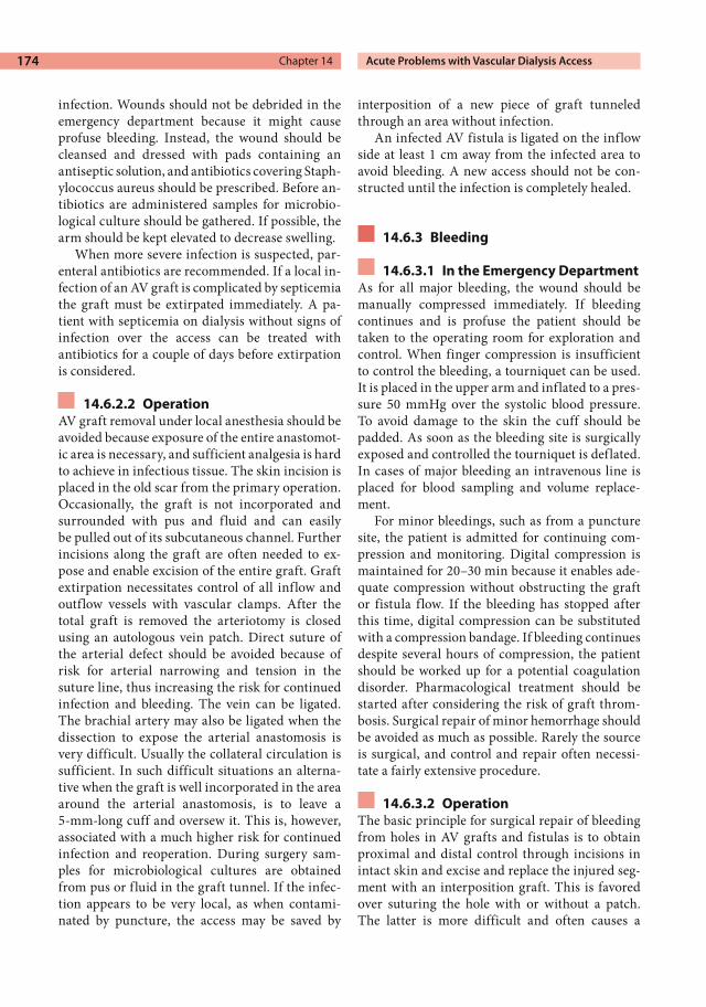
Chapter 14 Acute Problems with Vascular Dialysis Access174
infection. Wounds should not be debrided in the emergency department because it might cause profuse bleeding. Instead, the wound should be cleansed and dressed with pads containing an antiseptic solution, and antibiotics covering Staph-ylococcus aureus should be prescribed. Before an-tibiotics are administered samples for microbio-logical culture should be gathered. If possible, the arm should be kept elevated to decrease swelling.
When more severe infection is suspected, par-enteral antibiotics are recommended. If a local in-fection of an AV graft is complicated by septicemia the graft must be extirpated immediately. A pa-tient with septicemia on dialysis without signs of infection over the access can be treated with antibiotics for a couple of days before extirpation is considered.
14.6.2.2 Operation AV graft removal under local anesthesia should be avoided because exposure of the entire anastomot-ic area is necessary, and sufficient analgesia is hard to achieve in infectious tissue. The skin incision is placed in the old scar from the primary operation. Occasionally, the graft is not incorporated and surrounded with pus and fluid and can easily be pulled out of its subcutaneous channel. Further incisions along the graft are often needed to ex-pose and enable excision of the entire graft. Graft extirpation necessitates control of all inflow and outflow vessels with vascular clamps. After the total graft is removed the arteriotomy is closed using an autologous vein patch. Direct suture of the arterial defect should be avoided because of risk for arterial narrowing and tension in the suture line, thus increasing the risk for continued infection and bleeding. The vein can be ligated. The brachial artery may also be ligated when the dissection to expose the arterial anastomosis is very difficult. Usually the collateral circulation is sufficient. In such difficult situations an alterna-tive when the graft is well incorporated in the area around the arterial anastomosis, is to leave a 5-mm-long cuff and oversew it. This is, however, associated with a much higher risk for continued infection and reoperation. During surgery sam-ples for microbiological cultures are obtained from pus or fluid in the graft tunnel. If the infec-tion appears to be very local, as when contami-nated by puncture, the access may be saved by
interposition of a new piece of graft tunneled through an area without infection.
An infected AV fistula is ligated on the inflow side at least 1 cm away from the infected area to avoid bleeding. A new access should not be con-structed until the infection is completely healed.
14.6.3 Bleeding
14.6.3.1 In the Emergency DepartmentAs for all major bleeding, the wound should be manually compressed immediately. If bleeding continues and is profuse the patient should be taken to the operating room for exploration and control. When finger compression is insufficient to control the bleeding, a tourniquet can be used. It is placed in the upper arm and inflated to a pres-sure 50 mmHg over the systolic blood pressure. To avoid damage to the skin the cuff should be padded. As soon as the bleeding site is surgically exposed and controlled the tourniquet is deflated. In cases of major bleeding an intravenous line is placed for blood sampling and volume replace-ment.
For minor bleedings, such as from a puncture site, the patient is admitted for continuing com-pression and monitoring. Digital compression is maintained for 20–30 min because it enables ade-quate compression without obstructing the graft or fistula flow. If the bleeding has stopped after this time, digital compression can be substituted with a compression bandage. If bleeding continues despite several hours of compression, the patient should be worked up for a potential coagulation disorder. Pharmacological treatment should be started after considering the risk of graft throm-bosis. Surgical repair of minor hemorrhage should be avoided as much as possible. Rarely the source is surgical, and control and repair often necessi-tate a fairly extensive procedure.
14.6.3.2 Operation The basic principle for surgical repair of bleeding from holes in AV grafts and fistulas is to obtain proximal and distal control through incisions in intact skin and excise and replace the injured seg-ment with an interposition graft. This is favored over suturing the hole with or without a patch. The latter is more difficult and often causes a

175
narrowing and increases the risk of postoperative thrombosis. For large amounts of bleeding, the procedure can be performed in a bloodless field obtained by tourniquet occlusion.
14.6.4 Aneurysms and Hematomas
14.6.4.1 In the Emergency DepartmentPatients with a thin skin layer covering a clinically suspected aneurysm are at risk for fistula rupture. These aneurysms should therefore be subject to surgical excision. Urgent management is particu-larly important when there are signs of infection because such aneurysms may cause septic emboli-zation in addition to severe bleeding. The patient needs urgent work-up with duplex scanning to differentiate such infected aneurysms from other fluid collections that do not need urgent manage-ment.
Hematomas that not are infected are best left to reabsorb. Exceptions are hematomas that com-press the access and might impair its flow. Duplex is helpful for evaluating whether this is the case. If the hematoma is expanding rapidly, within a couple of hours, blood flow in the access is almost always threatened. Such expansion is usually caused by graft laceration and should be immedi-ately repaired. Old and stable hematomas can, as an alternative, be treated by puncture and aspira-tion if the function of the access is at risk. To avoid infection the puncture should be performed as far away from the graft as possible.
14.6.4.2 Operation True aneurysms with an overlying skin defect in AV fistulas are excised after proximal and distal control. Venous interposition grafting repairs the defect. Surgical treatment of pseudoaneurysms in AV grafts also requires control proximal and dis-tal to the aneurysm. The aneurysm is then opened and the connection between the sac and graft lumen is oversewn. Resection and interposition of a new piece of graft is often required, however. In the anastomotic area, pseudoaneurysms are best excised and the defect repaired with a patch. A hematoma threatening the graft’s function is treated the same way as aneurysms.
14.6.4 Arterial Insufficiency and Steal
14.6.4.1 In the Emergency DepartmentArterial insufficiency secondary to steal is not managed urgently. Diagnostic work-up with du-plex or angiography is usually necessary to plan an adequate treatment strategy. Administration of analgesics should be initiated as soon as possible, and opioids are often required. Even in patients with severe ischemic symptoms, ligature of grafts or fistulas can be performed during office hours. Occasional patients with therapy-resistant pain might need urgent surgery.
14.6.4.2 Operation In patients with severe symptoms of hand ischemia the AV fistula or graft must be closed. This is achieved by exposing the vein in the anastomotic area and clamping the artery. The vein is then divided and closed by sutures 1 or 2 cm from the anastomosis. Upper arm AV fistulas are treated the same way. Attempts to reduce blood flow at this level by wrapping a vein or synthetic patch around the fistula have anecdotally been successful. Occa-sionally, work-up of arterial insufficiency caused by AV fistulas in the wrist reveals retrograde flow in the hand. These patients can sometimes be im-proved by simply transforming the anastomosis from side-by-side to end-by-side. Usually, however, ligation of the entire fistula is required.
AV grafts causing steal are excised as described in the section on infection, but the access may be saved by using a different technique. It constitutes a bypass from the brachial artery above the inflow anastomosis to a level below it. The bypass is then followed by ligature of the brachial artery just dis-tal to the proximal bypass anastomosis. With this method relief of ischemic symptoms with main-tained access function has been reported.
14.6.5 Management After Treatment
The principles for management after treatment are similar for all complication types. Surgical wounds should be inspected frequently during the first 24 h. Bleeding problems are common after throm-bectomy and revision as well as after procedures for infection and steal. Function is also monitored closely. Patency is checked by auscultating for
14.6 Management and Treatment

Chapter 14 Acute Problems with Vascular Dialysis Access176
bruits and palpating for thrills over the outflow vein. After AV graft procedures, the vein is in-sonated at the vein and graft anastomosis site.
It is important to ensure that the patient is hy-drated and has an acceptable blood pressure. This is often a difficult task in uremic patients. If a ste-nosis is suspected in the access but is not mended for some reason, a duplex scan is ordered as soon as possible to verify and localize it before the access reoccludes. Postoperative heparinization beyond the dialysis treatment is rarely needed. Patients treated for infection should continue on antibiot-ics until the results of bacterial cultures arrive or the infection is healed.
14.6.6 When Can the Patient be Given Dialysis?
For all problems with dialysis accesses an impor-tant issue is the patient’s need for dialysis. Insert-ing a temporary catheter in the neck or groin should be weighed against the possibility of a suc-cessful operation. A basic rule is that a revised di-alysis access should be allowed at least a couple of days to heal after the procedure to avoid bleeding complications. Dialysis requires heparinization, which in combination with uremic patients’ ten-dency for coagulopathy increases the risk for bleeding. During dialysis clots in the suture line are dissolved and bleeding is likely. Such bleeding is often difficult to treat. Moreover, interposed vein grafts have thin walls and are easy to damage during puncture. Vein grafts needs at least 10–14 days to be arterialized, and PTFE grafts should be incorporated in surrounding tissue to minimize the risk for bleeding. Accordingly, if the need for dialysis is urgent and the risk of surgical bleeding after revision is considered small, dialysis can be performed the first postoperative day providing that the puncture can be made in an old part of the access and that the heparin dose during dialysis is adjusted.
14.7 Results and Outcome
In Sweden AV fistulas constitute about 80% of all dialysis accesses constructed. This percentage varies internationally, but the trend is that AV fis-
tulas are preferred over AV grafts. The reason is that if an AV fistula develops – which about 75% does – it will probably work well for many years with few complications. Compared with AV grafts stenoses in the outflow vein develop later in AV fistulas;the mean difference is in the range of 4–5 months. AV grafts, however, have the advan-tage of early dialysis, and at least 70% of all grafts are patent one year after implantation. Later AV grafts are prone to develop stenoses and infection complicates this type of access in much higher frequencies. The locations of stenoses in AV grafts are listed in Table 14.2.
The result of thrombectomy for treatment ac-cess occlusion is poor. For example, one study evaluating AV grafts reported a 75% immediate success, but the 30-day patency was 45%. At 6 months only 18% of the grafts could be used for dialysis and at 1 year only 3% could. A contribut-ing factor to these results is probably that some stenoses are missed at thrombectomy and are not corrected. This is supported in the literature, which reports that half of all occluded AV grafts have no identifiable cause of occlusion. When angiography was performed in the same patient group, 92% had at least one stenosis. When throm-bectomy was combined with revision the results were somewhat better: 60% of the grafts were patent after 30 days and 25% at 6 months. In most studies, multiple revisions are often needed to achieve acceptable results. Only a few AV fistu-las can be saved by thrombectomy according to the literature, which is why this operation should not be performed often. It is generally better to create a new access if the occlusion lasts longer than 8 h and when the fistula occludes early after construction.
Table 14.2. Location of stenosis in 357 arteriovenous graftsa (based on four studies)
Localization Frequency
Venous anastomosisIn vein away from the anastomosisIn inflow arteryIn graftIn a central vein
63%18%10%12%
1%a36% had more than one localization

177
Further Reading
Bush RL, Lin PH, Lumsden AB. Management of throm-bosed dialysis access: thrombectomy versus throm-bolysis. Semin Vasc Surg 2004; 17(1):32–39
Green LD, Lee DS, Kucey DS. A metaanalysis compar-ing surgical thrombectomy, mechanical throm-bectomy, and pharmacomechanical thrombolysis for thrombosed dialysis grafts. J Vasc Surg 2002; 36(5):939–945
Huber TS, Buhler AG, Seeger JM. Evidence-based data for the hemodialysis access surgeon. Semin Dial 2004; 17(3):217–223
Lew SQ, Kaveh K. Dialysis access related infections. ASAIO J 2000; 46(6):S6–S12
Schanzer H, Eisenberg D. Management of steal syn-drome resulting from dialysis access. Semin Vasc Surg 2004; 17(1):45–49
Tordoir JH, Dammers R, van der Sande FM. Upper ex-tremity ischemia and hemodialysis vascular access. Eur J Vasc Endovasc Surg 2004; 27(1):1–5
Further Reading

General Principles of Vascular Surgical Technique 15
CONTENTS
15.1 Summary . . . . . . . . . . . . . . . . . . . . . . . . . . . 179
15.2 Background . . . . . . . . . . . . . . . . . . . . . . . . 179
15.3 Exposure . . . . . . . . . . . . . . . . . . . . . . . . . . . 180
15.4 Control of Bleeding and Clamping . . . . . . . . . . . . . . . . . . . . . . 180
15.4.1 Proximal Endovascular Aortic Control . . . . . . . . . . . . . . . . . . . . . . 182
15.5 Vascular Suture . . . . . . . . . . . . . . . . . . . . 18215.5.1 Choice of Suture Material . . . . . . . . . . 184
15.6 Arteriotomy . . . . . . . . . . . . . . . . . . . . . . . . 184
15.7 Closure with Patch (Patch Angioplasty) . . . . . . . . . . . . . . . . 184
15.8 Interposition Graft . . . . . . . . . . . . . . . . 18615.8.1 Autologous Vein . . . . . . . . . . . . . . . . . . . 18715.8.2 Synthetic Vascular Prosthesis . . . . . . . 187
15.9 Veins . . . . . . . . . . . . . . . . . . . . . . . . . . . . . . . 188
15.10 Other . . . . . . . . . . . . . . . . . . . . . . . . . . . . . . . 19015.10.1 Drains . . . . . . . . . . . . . . . . . . . . . . . . . . . . . . 19015.10.2 Infection Prophylaxis . . . . . . . . . . . . . . . 190
15.1 Summary
Always use atraumatic technique and spe-cial vascular instruments.For proximal and distal control, all inflow and outflow vessels must be controlled and clamped prior to repair or arteriotomy or venotomy.Blind application of vascular clamps in a bleeding traumatic wound as dangerous.A nonsecure distal intimal edge is always a risk for dissection and creation of an inti-mal flap causing occlusion.Veins are vulnerable and extreme care is necessary during dissection and repair. A vascular suture should always be tied with the artery clamped.
15.2 Background
For surgeons who do not routinely perform vascu-lar operations it might be helpful to recapitulate the basic vascular surgical technique to facilitate their management of common vascular emergen-cies. First, it is always necessary to obtain control of and clamp all branches to the of interest vascu-lar segment to make a vascular operation possible at all. The rationale for being extra cautious with blood vessels is that they are extremely sensitive to trauma, which can induce undesired thrombo-genic activity on the intimal surface. Furthermore, their structure of separated wall layers demands special consideration to avoid dissection and oc-clusion. A successful vascular operation requires the surgeon to pay constant attention to technical details to avoid jeopardizing the entire operation.

Chapter 15 General Principles of Vascular Surgical Technique180
15.3 Exposure
Good exposure is always mandatory for optimiz-ing the prerequisites for reconstruction or repair. Exposure of most specific vascular segments is described in the corresponding chapter (e.g., ex-posure of the superficial femoral artery in the groin for embolectomy is covered in the chapter on acute leg ischemia).
With a few exceptions (for instance, the trans-verse incision in the cubital fossa for exposing the brachial artery), a longitudinal skin incision over the vessel is used. The incision is made approxi-mately 2 cm longer in each direction than the re-quired exposed vessel length. An incision that is too small increases the risk for wound complica-tions in the skin and other tissues with already impaired perfusion due to unnecessary trauma from retractors. Familiarity with the anatomy is essential because it is not always possible to be guided by palpation of a pulse or hard arterioscle-rotic plaques. Use small, blunt, self-retaining re-tractors to achieve tension in the tissue to facilitate dissection. When approaching the vessel special atraumatic vascular instruments should always be used. Never use a toothed forceps in the vicinity of a blood vessel. The correct plane of final exposure is the outer soft layer of the adventitia. A common error is exaggerated caution in trying to obtain ex-posure in a layer that is too superficial. The adven-titia is characterized by a visible net of vasa vaso-rum. It is important to be aware of the differences between a healthy and a diseased artery. The for-mer is soft, easily compressible, and has a grey/red color. This is what can be expected in trauma pa-tients who are mostly young and otherwise healthy. When there is no pulse in the artery it may even be difficult to distinguish it from a vein. An arterio-sclerotic artery in a patient with peripheral vascu-lar disease is yellow/white and has a thick and often hard or calcified wall.
Expose the anterior and lateral aspects of the artery and use a right-angle clamp with its tip turned away from the adjacent larger vein – thus avoiding accidental perforations – to surround the posterior aspect with a vessel-loop of fabric or rubber. By lifting the vessel-loop, tension can be achieved in the tissue, thus facilitating the dissec-tion in an atraumatic way (Fig. 15.1). All branches should be saved because they are potential impor-tant collaterals.
NOTEGood exposure of the segment to be repaired, including space enough to allow safe clamping, is essential for a successful vascular operation. Always use special atraumatic vascular instruments when approaching the vessel.
15.4 Control of Bleeding and Clamping
In all vascular procedures, including vascular trauma, proximal and distal control is mandatory before attempting repair or doing an arteriotomy. It is desirable to have a completely empty vascular segment in order to perform a vascular opera-tion without unnecessary technical difficulties. To
Fig. 15.1. a Pass a right-angle clamp gently through the soft tissue on the dorsal aspect of the artery and direct it away from larger veins to avoid iatrogenic inju-ries. Caution! Avoid accidental penetration of the dor-sal wall of the artery. b Gently lift the artery with the vessel-loop to achieve tension in the tissue, thus facili-tating the dissection
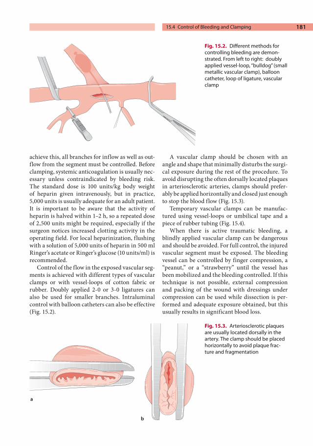
181
achieve this, all branches for inflow as well as out-flow from the segment must be controlled. Before clamping, systemic anticoagulation is usually nec-essary unless contraindicated by bleeding risk. The standard dose is 100 units/kg body weight of heparin given intravenously, but in practice, 5,000 units is usually adequate for an adult patient. It is important to be aware that the activity of heparin is halved within 1–2 h, so a repeated dose of 2,500 units might be required, especially if the surgeon notices increased clotting activity in the operating field. For local heparinization, flushing with a solution of 5,000 units of heparin in 500 ml Ringer’s acetate or Ringer’s glucose (10 units/ml) is recommended.
Control of the flow in the exposed vascular seg-ments is achieved with different types of vascular clamps or with vessel-loops of cotton fabric or rubber. Doubly applied 2-0 or 3-0 ligatures can also be used for smaller branches. Intraluminal control with balloon catheters can also be effective (Fig. 15.2).
A vascular clamp should be chosen with an angle and shape that minimally disturbs the surgi-cal exposure during the rest of the procedure. To avoid disrupting the often dorsally located plaques in arteriosclerotic arteries, clamps should prefer-ably be applied horizontally and closed just enough to stop the blood flow (Fig. 15.3).
Temporary vascular clamps can be manufac-tured using vessel-loops or umbilical tape and a piece of rubber tubing (Fig. 15.4).
When there is active traumatic bleeding, a blindly applied vascular clamp can be dangerous and should be avoided. For full control, the injured vascular segment must be exposed. The bleeding vessel can be controlled by finger compression, a “peanut,” or a “strawberry” until the vessel has been mobilized and the bleeding controlled. If this technique is not possible, external compression and packing of the wound with dressings under compression can be used while dissection is per-formed and adequate exposure obtained, but this usually results in significant blood loss.
Fig. 15.2. Different methods for controlling bleeding are demon-strated. From left to right: doubly applied vessel-loop, “bulldog” (small metallic vascular clamp), balloon catheter, loop of ligature, vascular clamp
Fig. 15.3. Arteriosclerotic plaques are usually located dorsally in the artery. The clamp should be placed horizontally to avoid plaque frac-ture and fragmentation
15.4 Control of Bleeding and Clamping

Chapter 15 General Principles of Vascular Surgical Technique182
In certain situations, such as in scar tissue, thorough dissection of a vascular segment can be technically very challenging and should thus be avoided. Balloon occlusion can be a very good alternative for distal as well as temporary proxi-mal control. Control is, however, best achieved by surgical exposure and clamping of a more proxi-mal segment, while balloon occlusion is always an alternative for distal control. Embolectomy cathe-ters of adequate size connected to a three-way stopcock and a saline-filled syringe are used. In-spection of the open segment, under continuous evacuation of blood from the backbleeding branches with suction, allows identification of the orifice into which the catheter should be inserted. After insertion the balloon is insufflated until the backflow has ceased. The stopcock is closed, and the balloon is left in place to occlude the artery. It is important not to overinflate the balloon, which could damage the arterial wall. In analogy with a vascular clamp, the balloon should be insufflated just to the point when bleeding stops – no further.
In larger arteries such as the aorta, a Foley catheter of appropriate size can be used for the same pur-pose. Special catheters from different manufactur-ers are also available for occluding arteries. When balloons are used for proximal control they are easily dislocated and even blown out by the arte-rial pressure. This can be avoided by having an assistant manually support the catheter or by applying a vascular tape around the artery just proximal to the arteriotomy, thus preventing the balloon from being further dislocated distally.
NOTENever open a blood vessel without having proximal and distal control.
15.4.1 Proximal Endovascular Aortic Control
When available, this alternative is of great poten-tial importance for patients with severe intraab-dominal bleeding after rupture of aneurysms as well as traumatic vascular injuries. It is further described in Chapter 7 (p. 85).
15.5 Vascular Suture
When vessels are sutured, the suture should in-clude all the layers of the vessel wall. The adventi-tia is the most important layer for the mechanical strength of the vascular wall. The adventitia should not be allowed to be interposed between the approximated edges of the arteriotomy be-cause that can disturb the healing process. This can be avoided by everting the edges to allow inti-ma-to-intima approximation.
When vessels are being sutured, the needle’s point should be placed at a 90° angle against the vascular wall, and thereafter its circular shape is used to push it through the wall to avoid un-necessary tearing. It is important to place the needle from inside out, particularly on the downstream side of the vascular suture, in order to fasten and secure the intima, avoid splitting the wall layers, and avoid the risk of intimal dissection (Fig. 15.5).
Arteriosclerotic arteries can be very hard and calcified, making penetration of the needle at an ideal site impossible. In such a situation it might
Fig. 15.4. A temporary vascular clamp is made by pull-ing a double vessel-loop through a piece of rubber tub-ing to make a snare around the vessel, which is locked by an ordinary clamp

183
be necessary to penetrate the vascular wall with the needle and suture at a far distance from the intended suture row. Sometimes it is necessary to remove an extensive and hard arteriosclerotic plaque by a local thrombendarterectomy before the repair can be completed. Another important detail in suturing arteries is to tighten the suture satisfactorily; a suture that is too loose will cause leakage, and if it is too tight this will certainly lead to stenosis. The angle when pulling the suture should be 90° from the vascular wall to minimize the risk of tears in the vascular wall. Oozing in the suture row is best managed by tamponade with a sponge for 5–10 min or until bleeding stops. If extra hemostatic sutures are needed, a suture one size smaller than those in the suture row is recom-mended. If the result is unsatisfactory a local hemostatic agent can be applied.
Simple suturing for minor traumatic injuries in arteries is demonstrated in Fig. 15.6. It is impor-tant to tie the suture with the artery clamped and not pulsating to get it properly adjusted.
Fig. 15.5. The needle should be directed 90° to the vessel wall. Always include the intima, especially on the downstream side, to avoid dissection of the distal intimal edge
Fig. 15.6. a Simple cross-suture of an arterial puncture. b Simple sutures in a transverse arterial injury or arteri-otomy. c If the artery is large (>10 mm wide) a running suture can be used
15.5 Vascular Suture

Chapter 15 General Principles of Vascular Surgical Technique184
15.5.1 Choice of Suture Material
Vascular sutures are monofilament, synthetic, and double-armed. The needles are taper-pointed and have a variety of curvatures. Most vascular needles are larger than the suture to which they are at-tached. This can be a source of suture-line bleed-ing, which is best treated with local compression and hemostatic agents, but not with further su-tures. Recommendations for sutures are given in Table 15.1 below.
15.6 Arteriotomy
When performing an arteriotomy it is important to avoid damaging the vessel’s posterior wall, to choose the right direction of arteriotomy, and to close it properly. An arteriotomy starts with punc-ture with a pointed scalpel blade (#11) with the edge turned away from the surgeon. When a punc-ture bleeding is obtained, the blade is moved for-ward and upward to avoid injuries to the posterior wall. The lower blade of a 60° vascular scissors
(Pott’s scissors) is inserted into the arteriotomy, which is elongated appropriately while ensuring that the scissors is in the true free vascular lumen and not within any of the layers of the vascular wall. Because arteriosclerotic arteries are occa-sionally extremely hard, the best site for arterioto-my is chosen by palpating with a finger to find a soft segment. Choosing the arteriotomy direction, transverse or longitudinal, is sometimes difficult and is worth special consideration.
Longitudinal arteriotomy is the most useful and has the advantage of being easily elongated. It allows better inspection of the vascular lumen and can be used for an end-to-side anastomosis if reconstruction is necessary. On the other hand, it must be closed with a patch to avoid narrowing of arteries with a diameter <5 mm (see below).
Transverse arteriotomy can be considered when the procedure is likely to be limited to an embolec-tomy and when the artery is thinner than 5 mm. When closing the arteriotomy, it is always impor-tant to start by catching the intima with the needle at the distal end of the arteriotomy to avoid dissec-tion and occlusion. A running suture is mainly used (Fig. 15.7), but in transverse arteriotomies in smaller arteries, simple sutures are preferable to avoid the risk of narrowing by a running suture that is too tight.
15.7 Closure with Patch (Patch Angioplasty)
The patch technique is very important and useful in all emergency vascular procedures. A patch should always be considered when closing an ar-tery after longitudinal arteriotomy or traumatic injury with a vessel wall defect. A longitudinal su-ture always causes a certain degree of narrowing because the suture needle is placed 1–2 mm from the edge on both sides. A basic rule is that vessels
Table 15.1. Suture sizes for various vessel segments
Vessel Suture size
Aorta 3-0 to 4-0Iliac arteries 5-0Femoral artery 5-0Popliteal above the knee 5-0 or 6-0Popliteal below the knee 6-0Calf artery 6-0 or 7-0Carotid 6-0Brachial 6-0Subclavian 5-0Renal – visceral 6-0
Fig. 15.7. Closure of a longitudinal arteriotomy with a running suture

185
with diameters <5 mm should be closed with a patch. Occasionally, even larger arteries should be closed by the patch technique. In practice, patches are frequently used for the calf, popliteal, brachial, carotid, and sometimes also the femoral and iliac arteries. The choice of patch material depends on location and the level of contamination. An autol-ogous vein is recommended in the superficial fem-oral artery and distally. In the common femoral artery, iliac arteries, and the aorta, a synthetic polyester or polytetrafluoroethylene (PTFE) graft is most commonly used.
The patch technique is demonstrated in Fig. 15.8. The patch should be cut to an appropriate width, aiming to compensate for the diameter loss
but with some oversizing. Too large a patch will cause a disadvantageous enlargement, which sub-sequently might lead to increased risk for develop-ment of aneurysms and thrombotic occlusions. The patch is shaped at the end in a rounded fash-ion. The suture is started at one of the ends, pos-sibly with retaining sutures in both ends. It is always important to ensure that the distal intimal edge is secured by the suture. The suture is tied in the middle of the patch and never at one of the ends.
NOTEAlways consider using a patch when closing vessels <5 mm in diameter.
Fig. 15.8. Patch closure of a longi-tudinal arteriotomy. a The suture is started distally (downstream) with the needle from inside to out to secure the distal intima. The first su-ture can be tied to secure the patch before proceeding with the suture row. b The suture is continued in a running fashion in both directions and always with the needle running from the inside to the outside of the artery. When the proximal end of the arteriotomy is approached, the patch has to be cut and trimmed. c The sutures are continued until they meet on one of the sides. Check inflow and backflow before tying
15.7 Closure with Patch (Patch Angioplasty)

Chapter 15 General Principles of Vascular Surgical Technique186
15.8 Interposition Graft
To bridge a defect in an artery a piece of a vascular graft is interponated. A vein graft is used for the arms and infrainguinally in the legs. In larger arteries including the iliac arteries and the aorta, a synthetic prosthesis can be used. If the vessels that are going to be anastomosed end to end have dif-ferent diameters, the ends should be cut obliquely to adjust the circumference of both ends to each other. After transverse resection of the thinner vessel, its end is cut longitudinally and the corners trimmed. The larger vessel also needs to be cut
slightly transversally to avoid kinking in the anas-tomosis (Fig. 15.9). Also, when thinner vessels are going to be anastomosed end to end, the circum-ference and width of the anastomosis must be ensured by cutting both ends obliquely. This will minimize the risk for narrowing in the suture row.
If the anastomosis is started by two diametri-cally opposite holding sutures, the suture adjust-ment is facilitated and the posterior aspect can easily be rotated with the two holding sutures. The anastomosis is then completed with a running su-ture of appropriate size (Fig. 15.10). As pointed out
Fig. 15.9. When two vessels with different diameters are being sutured end to end, the smaller has to be slit open and the edges trimmed to fit the larger one, which must be cut somewhat obliquely to avoid kinking
Fig. 15.10. End-to-end anasto-mosis starting with two opposite and tied sutures that can be used to turn the vessel for access to all sides, allowing completion of the anastomosis with running or simple sutures, depending on the diameters
187
earlier, the distal edge should always be sutured with the needle from inside the lumen to guaran-tee that the distal intima is fixed. When anasto-mosing thinner arteries an interrupted suture can be preferable because it will diminish the risk of narrowing by a too-hard pull in a running suture.
The length of the graft is adjusted after the first anastomosis has been completed. A graft that is too long increases the risk of kinking, while one that is too short means an unacceptable tension in the anastomosis. Appropriate length is achieved if the graft is straightened with a vascular forceps until it is stretched and then cut at the level of the end of the other artery.
When choosing vascular prosthesis for a patch or interposition graft, larger arteries like the aorta, common and external iliacs, and the common femoral artery can be closed or reconstructed with synthetic prosthesis material. In the common femoral artery, however, an autologous vein can also be used. In vascular procedures distal to the groin an autologous vein should always be used as the first choice. The rationale is that synthetic ma-terial always has an increased thrombogenicity, which in combination with the low flow in thinner arteries leads to a higher risk for thrombosis and occlusion. But for reconstructing larger arteries with a higher flow, synthetic grafts work well.
In cases with increased risk for infection (i.e., contaminated traumatic injuries or vascular reconstructions performed in association with intestinal injuries or disease), the choice of pros-thetic material is more challenging. A synthetic prosthesis always implies risk of a complicating infection of the implanted synthetic material. Such an infection is very difficult to treat and usually requires the graft to be totally excised. A vein graft is more resistant to infection, but these also carry a risk for erosion and serious bleeding. The basic principle is to always avoid synthetic grafts when there is increased risk of infection and to use an autologous vein as the first chose. Exceptions are procedures on larger arteries such as the aorta and iliacs, and if synthetic prostheses are used in such a situation, prolonged antibiotic therapy should be considered.
15.8.1 Autologous Vein
The most commonly used vein is the greater saphenous vein. Other alternatives are the lesser saphenous, cephalic, and basilic veins. At all vein harvesting a maximally atraumatic technique should be used.
The vein is exposed by one or several longitudi-nal skin incisions and all branches are ligated. Be sure that the length harvested is long enough for the present purpose. Immediately after harvesting the vein graft should be flushed clean of all re-maining blood with a heparin solution 10 units/ml, in which it can be preserved until it is used. Veins usually have a pronounced contractility, causing them to shrink considerably when they are handled during exposure and harvest. Before a vein is used as an arterial substitute it should be checked for leaks. By gently injecting heparin so-lution and simultaneously occluding the outflow, remaining open branches or other injuries causing leakage can be revealed and fixed with 4-0 liga-tures and 6-0 or 7-0 vascular sutures, respectively. When ligating a branch it is important to avoid “tenting” of the vein because this might cause nar-rowing and stenosis. For the same reason, all other leaks should be sealed with sutures placed in the long axis of the vessel. Note that if the vein is to be reversed, the larger end of the vein should conse-quently be anastomosed distally to eliminate the flow-obstructing effect of the valves. The tech-nique for preparing an autologous vein patch and an interposition graft is shown in Fig. 15.11.
15.8.2 Synthetic Vascular Prosthesis
Synthetic vascular prostheses are available mainly in two materials: polyester or ePTFE (expanded PTFE). Both materials are available as straight tube and bifurcation grafts in different diameters ranging from 6 to 12 mm for the tubes and from 14 to 26 mm for the bifurcated grafts, in which the limb has half the diameter. Both materials are also available as sheets from which suitable patches can be cut.
Polyester prostheses are most commonly used in the aortoiliac region and are available as knitted material (which is the most common) and woven. The knitted version is permeable to blood, whereas
15.8 Interposition Graft

Chapter 15 General Principles of Vascular Surgical Technique188
the latter is tight. Nowadays the knitted grafts are mostly available coated, which means they are im-pregnated with collagen or albumin in order to be sealed to blood. This type of coated knitted graft is the most commonly used.
If a noncoated knitted graft is chosen, it is ex-tremely important to “preclot” it to avoid exten-sive leakage through the graft wall: Prior to hepa-rinization, 20–30 ml of the patient’s own blood is aspirated through an arterial puncture. The blood is immediately used to impregnate the vascular prosthesis. When the blood coagulates between the knits, the prosthesis will be sealed. If this step is forgotten, although the prosthesis will slowly seal after implantation, it will do so usually only after extensive bleeding.
PTFE is a porous but tight material very suit-able for vascular prosthesis. It can also be used as an arterial substitute infrainguinally to perform an above-knee or even a below-knee femoropop-liteal bypass. PTFE is possibly somewhat more resistant to infection than polyester is.
15.9 Veins
Surgical operations on veins require special and careful attention to technique because of the veins’ thin wall structure and vulnerability. This is, nat-urally, particular important in emergent trauma cases. An iatrogenic or traumatic venous lesion can very easily be dramatically enlarged by just a
Fig. 15.11. Harvest of autologous vein for graft and patch. a Saphe-nous vein graft. A longitudinal incision over the vein starts in the groin and is elongated according to individual requirements. All branches are ligated and divided. The vein is harvested and flushed with heparinized glucose or saline. It must be reversed when used as an arterial substitute. b The greater saphenous vein at the ankle is usually sufficient and is exposed through an incision just anterior to the medial malleolus. All branches are ligated. The harvested vein is cut longitudinally and the ends trimmed. Be cautious and turn the patch so that the valves do not obstruct the flow

189
slight pull with gauze in an attempt to control the bleeding. This vulnerability to injury is also why vascular clamps should be avoided for controlling veins. Instead, a piece of gauze of appropriate size on a straight clamp is carefully applied in a right angle over the vein on both sides of the lesion. A complete dissection with application of vessel-loops is rarely needed. The lesion can usually be directly repaired with a simple or running suture in the direction that causes the least degree of narrowing.
Smaller and midsize veins can be ligated. Re-construction of injured veins is recommended for larger unpaired veins such as the vena cava and iliac and femoral veins; see Chapters 5 (p. 58) and 9 (p. 113). If suturing is insufficient and graft-ing is necessary due to a more extensive injury autologous material is the first choice, just as in repair of arterial injuries. If a graft with a larger diameter is needed, a spiral graft can be created from a longitudinally opened greater saphenous vein (see Fig. 15.12).
Technically challenging diffuse venous bleed-ing, such as in the pelvic region, can often be treat-ed by a combination of applying a hemostatic agent (Table 15.2) and packing the bleeding with lots of dressings. The pressure in veins is low, and bleeding usually stops within 15–30 min. (The technique is further described on p. 152.) In a life-threatening situation, most veins, including the vena cava, can be ligated with reasonable conse-quences (e.g., swelling of limbs).
NOTEVeins are much more vulnerable than arteries. But the low venous pressure makes it possible to handle even severe venous bleeding and injuries with hemo-static agents and packing.
Fig. 15.12. Spiral graft technique to create a graft of larger diameter for replacing vein segments. A saphe-nous vein is cut longitudinally and sutured in a spiral fashion over plastic tubing used as a stent
Table 15.2. Listing of local hemostatic agents and their characteristics
Agent Application, examples Special characteristics
Collagen Oozing in anastomosisOxygenated cellulose Oozing in anastomosisPolyethylene glycol Oozing in anastomosis Works better on dry surfaces;
polymerization in 60 sThrombin with or without gelatin Larger bleeding
in anastomosisExpands about 20%; polymerization 3 min; ongoing bleeding necessary for access to fibrinogen
Fibrin glue Diffuse bleeding Frozen, must be thawed; spray covers larger areas
15.9 Veins

Chapter 15 General Principles of Vascular Surgical Technique190
15.10 Other
15.10.1 Drains
Drains are rarely used after elective vascular pro-cedures. However, they may be useful after emer-gency procedures in the neck and the legs to detect postoperative bleeding requiring intervention and to evacuate blood to minimize the risk of hema-toma development with its increased risk for in-fection. Care should be taken to place the tube in a way that does not compress a vascular graft. The drain is recommended to be active. Removal of the drain shall be considered on the 1st postopera-tive day. Intraabdominal drains after emergency aortic surgery are rarely used.
15.10.2 Infection Prophylaxis
Careful atraumatic technique and an optimal route of dissection, avoiding lymph glands and vessels, are important prophylactic measures for minimizing infection. Prophylactic antibiotics should be administered to patients with infected ulcerations or wounds and groin dissections and when synthetic prostheses are implanted. They are also generally recommended in all emergency procedures. Local protocols vary, but cloxacillin 2 g or cefuroxime 1.5 g are frequently used as a single preoperative dose given intravenously. The dose should be repeated every 3–4 h if open sur-gery is still going on.

Subject Index
A
AAA, see abdominal aortic aneurysm
abdominal– aortic aneurysm
(AAA) 75–90– – classic triad 77– – clinical presentation 77– – diagnosis of ruptured
AAA 76– – differential diagnosis 80– – mortality 87– – possible rupture 78– – prevalence 75– – risk factors 76– – risk of rupture 76– – suspected rupture 78– – tender pulsating mass 77– circumference 149– distension 48– drain 85– fossa 106– injury 50– pain 47, 66, 144, 145– – in the lower quadrant 160– ultrasound 50– vascular injury 46abscess drainage 154absent radial pulse 33acceleration forces 17access infection 171access-area infection 173accidents 102– of high impact 103acetabular screw 63acid-base balance 69acidosis 130, 145ACT value 150acute intestinal ischemia– aggressive diagnostic work-
up 73– mortality 73acute leg ischemia 144– amputation rate 130
– classification according to severity 122
– coldness 121– comorbidity 121– duration 120– fresh thrombus 121– incidence 120– light touch 122– management– – by etiology 120– – by severity 120– mortality 130– pain 121– pallor 121– palpable pulses 121– poor mobility in the foot 121– postoperative 131– pulselessness 121– severity 120– skeletal muscle 120– skin 120– – temperature 131– stagnant blood flow 121– sudden onset of symptoms 121– survival 130acute mesenteric ischemia– thrombolytic therapy 73acute myocardial infarction 67,
77, 79acute valve regurgitation 93adductor tendon 108adjuvant pharmaceutical treat-
ment 73Advanced Trauma Life
Support 8, 19, 34, 138aerodigestive tract 6, 7, 17air bubbles in the wound 17air embolization 18, 22airway obstruction 8alkalinization of the urine 130Allen test 35, 37allopurinol 73amaurosis fugax 5amputation 35, 57, 106, 112, 114,
163
– massacred leg 106– primary 129analgesics 22, 70, 79, 97, 105, 125,
175anastomosis– bleeding 151– transforming 175anatomical aortic arch and branch
variations 16anemia 97, 125, 153anesthesia of the axillary
plexus 172aneurysm 80, 121aneurysmal sac 85angina 142– pectoris 95angiography 69, 104, 123, 131– intraoperative 128, 132, 172– signs of embolism 124angioplasty 52, 87, 120, 129, 173angiosuite 43ankle– blood pressure 121, 122– pressure 103, 114, 123, 131, 146ankle-brachial index (ABI) 48,
104, 144– asymmetrical 48antibiotics 61, 70, 85, 105, 114, 174– treatment 12, 22, 73, 151, 154anticoagulation 9, 12, 72, 73, 129,
132– contraindications 163– long-term 164antiphospholipid antibody 129antiplatelet therapy 12aorta– clamping– – subdiaphragmatic 155– weak posterior wall 150aortic– arch injury 15– arch rupture 93– balloon 53– bifurcation 126

Subject Index192
– clamp in the subdiaphragmatic position 83
– dilatation 76– dissection 77– – classification 92– – emergency surgery 98– – in women 93– – incidence 91– – mortality 99– – pain location 94– – paradoxical physical
findings 96– – paradoxically high blood
pressure 96– – pregnancy 93– – prevalence 91– – type-A/type B 92– insufficiency 96– knob 19, 91– occluder 22, 52– stent-graft repair 60– thrombosis 51– tube graft 80aortocaval fistula 88aortoduodenal fistula 152aortomesenteric reconstruc-
tion 71arcade artery 85arm– chronic ischemia 42– fasciotomy techniques 39– function 43– swelling 160– veins 113arrhythmia 125, 129arterial– blood sampling 40– cascade 71– fibrillation 67– insufficiency in the hand 169– thrombosis 69, 120arteriosclerotic plaque 183arteriotomy– direction 184– in the popliteal artery 127– longitudinal 184– site 184– transverse 184arteriovenous fistula 46, 102, 163– in the groin 113arteriovenous graft occlusion 176aspirin 148atheroembolism 131atherosclerotic plaque 121atraumatic vascular instru-
ment 180
atrial fibrillation 120, 129atriocaval shunting 56auscultation 103autologous– arteriovenous fistula 167– vein 11, 113autotransfusion 80avulsion 47– of branches 46– of great vessels 18axilla 18axillary artery 36axillary-brachial bypass 43axillobifemoral bypass 155
B
back pain 76backbleeding 82, 109backflow 37, 43, 72, 127, 147bacterial– translocation 73– virulence 152balloon– catheter 181– – occlusion 11, 82, 114, 182– overinflating 182betablocker 98biceps muscle 37bicipital groove 43bilateral renal ischemia 52biological dressing 113black necrosis 157bladder trauma 47bleeding– anastomotic 151– continued 23– erosion-related 152– from a puncture site 150– from incisions 149– gastrointestinal 152– into the pleural cavity 18– juxtaceliac 52– packing 189– perioperative 148– postoperative in the abdo-
men 149– rectal 152– right-sided intra-
thoracic 22– tamponade 46– tendency 148– time 150blood– culture 155
– flow variation with breath-ing 161
– in stools 67– loss– – estimation 137– on rectal examination 145– pressure– – restoration 20– test 96blue spot 132blue toe syndrome 131, 145blunt dissection 80blunt trauma 4– anticoagulation 9– decreased consciousness 9– late neurological deficits 7– neurological examination 6– neurological symptoms 5– reflexes 6– sensory functions 6– small bowel 47body position of the victim 48bolus injection 162bone fragment 4bowel sounds 68brachial– artery 31, 36, 42, 168– – anatomy 42– plexus 18, 37– vein– – transposition 167brachiocephalic trunk 41branch– occlusion 99– vessel bleeding 51bridging fistula 167brisk bleeding 5bruit 17, 68, 88, 104, 171Buerger’s disease 41bulbous dilatation 19bulldog 181bypass– from the brachial artery 175– techniques 112
C
C2 vertebra 4calcium channel blocker 70, 98calf– artery 105, 112– muscle 114– – tenderness 157– tenderness 160– vein 113
193Subject Index
cannula– interosseous 138– replacement 138– short- and large-bore 138cardiac– disease 42– failure 102, 120– malperfusion 95– murmur 96– shadow 97– tamponade 93, 96cardiogenic shock 80cardiopulmonary bypass 98carotid artery injury– autologous vein graft 11– backflow 11– balloon occlusion 11– end-to-end anastomosis 11– external compression 10– interposition graft 11– ligation 11– major neurological deficits 9– neurological outcome 13– operation– – conservative approach 9– palliation 9– patch 11carotid dissection 4, 13catecholamine level 96caval obstruction 160celiac axis 53, 57– ligation 57cell saver 80cellulose sheet 151cement destruction 63central venous catheteriza-
tion 138, 139central venous pressure
(CVP) 139cephalosporin 22cerebral– infarction 9– ischemia 95cervical– ansa 10– pain 5– spine fracture 7chemotherapy 151chest– injury 16– pain 160– tube 19– – insertion 21– – monitoring 28– X-ray 97childbirth 4
chronic– infection 169– intestinal ischemia 67– leg ischemia 131– obstructive pulmonary
disease 142circulatory-stable patient 79circumflex– humeral artery 39– iliac artery 107clamping 107– of the thoracic aorta 83claudication 121, 131clavicular– fracture 17– resection 26cleansing 60clip 58clopidogrel 148closure with patch 184clot 127, 160– dislodgement 162coagulase-negative staphylo-
coccus 152coagulation 150– disturbance 165– factors– – deficiency 150– optimizing 129coagulopathy 52coil 27, 39, 62colicky pain 67collagen 189– sheet 151collateral 120– blood flow 70, 171– circulation 174– flow 160– network 31, 66– system 41colon– ischemia 145– primary ischemia 66– resection 147coloscopy 145colostomy 147coma 18, 24common– carotid artery 17, 25– femoral artery 107– femoral vein 113, 163– hepatic artery 57– iliac artery 57, 107– iliac vein 58compartment– pressure 115
– syndrome 39, 114, 130compression– bandage 174– stockings 61, 157, 164computed tomography
(CT) 153– angiography 86, 145– scan 70, 77, 79, 162congenital– coagulation disorder 67– connective tissue defect 92congestive heart failure 87, 95coniotomy 8connective tissue abnormality 93,
94constipation 77contained hematoma 47continued bleeding 23continuous-wave Doppler 73, 122– audible signal 122contrast– in the thrombus 78– leakage 104control angiography 113cortisone treatment 151costal cartilage 22coumadin 129, 164cranial nerve dysfunction 6C-reactive protein (CRP) 88, 153CRP, see C-reactive proteincrush injury 35cubital fossa 168– surgical exposure of veins 138cuff 107cutdown technique 139CVP, see central venous pressurecyanosis 121, 131– of the lower extremity 88cystic media necrosis 92
D
Dacron graft 85damage control 52, 59damaged bowel segment 52D-dimer 68, 70, 161– negative level 161DeBakey clamp 54deceleration forces 17, 47decompression of the four
compartments 115deep– brachial artery 31– femoral artery 107, 112, 127– femoral vein 107

Subject Index194
– venous thrombosis (DVT) 160– – proximal 164dehydration 120, 131dementia 89desmopressin 150devitalized skin 110dextran 105, 114, 124, 125, 129diabetes 121, 142, 151, 169dialysis 87– access– – early occlusion 168– – frequency of complica-
tions 168– – late occlusion 168– – technical errors 168– need 176diaphragmatic– aortic control 150– crura 52, 83– injury 49– – diagnosis 50diarrhea 66, 145diffuse necrosis in the
mucosa 66digital– ischemia 41– pressure 105digitalis 70dilutional thrombocytopenia 28disc level 63discoloration 131, 160– of the hand 170– of the skin 157dislocated fracture 34displaced esophagus 97dissection 143, 144– membrane 99distal edema 160distal ischemia– clinical signs 103distended– abdomen 145– skin 149disturbed– hemostasis 149– motor function 146diuretics 70diverticulitis 77dizziness 76domestic accident 32dorsal cava injury 43double aortic lumen 98DPL 50drain 190– removal 190duodenal wall erosion 152
duodenum 154– 4th portion 72duplex 69, 105– ultrasound 123– scanning 161, 165, 171DVT, see deep venous thrombosis
E
ECG, see electrocardiogramechocardiography 43, 162edema 157Ehlers-Danlos syndromeelbow dislocation 102electrocardiogram (ECG) 70, 78,
96, 125elevated white blood cell
count 145embolectomy 42, 123, 126embolic source 129embolization 66, 143– of the branches 60embolizing bullet 18embolus 120– source 121emergency– bypass 71– thoracotomy 50– work-up 78empty vein 137end-of-life disease 119end-organ ischemia 93endoscopy 154endotracheal intubation 19endovascular– aortic control 83, 182– embolization 12– repair 89, 99– stenting 106– treatment 12, 39, 60, 86, 98,
113, 161, 162end-to-end anastomosis 11, 186end-to-side anastomosis 168ePTFE, see expanded poly-
tetrafluoroethyleneerythrocyte sedimentation rate
(ESR) 88esophageal– injury 5– tube 20esophagus 12, 83– displaced 97ESR, see erythrocyte sedimentation
rate
expanded polytetrafluoroethylene (ePTFE) 57, 113, 167, 187
expanding– aortic diameter 98– hematoma 6, 17exposed– vascular graft 155– vein graft 154exsanguination 17, 76extensive thrombus in the inferior
vena cava 164external iliac artery 57, 106, 107external jugular vein 138extraanatomical– bypass 98– reconstruction 151
F
facial– fracture 8, 18– vein 10fall from height 46false– aneurysm 46– lumen 93, 99fasciotomy 38, 115, 127, 130, 163feeling– of pull 126– of touch 126femoral– artery– – in the groin 107– condyle 108– nerve 158– shaft fracture 102– vein– – percutaneous catheteriza-
tion 138femorofemoral bypass 57, 62, 87fenestration 60, 98fever 171fibrin glue 60, 62, 151, 189fibrinogen concentration 129, 150fibrinolysis 148fibula 116finger– compression 22, 149, 174– fracture technique 58– motor function 42firearms 16first heart sound 96first-rib fracture 17fistulogram 154five P’s 121

195Subject Index
flail chest 18flank pain 60flow direction 170fluctuating mass 156fluid– collection 153– – subcutaneous 156– – well demarcated 156– evacuation 156– resuscitation 50flushing with a solution 181focal neurological change 5Fogarty catheter 37, 43, 72, 85,
112, 126, 128, 147, 163, 172fogging 21folate 129Foley catheter 19, 22, 50, 53, 54,
82, 182foot gangrene 160foreign material 113fracture– LeFort II/III 7– of the cervical spine 7– of the clavicle 17, 19, 102– of the face 8, 18– of the femoral shaft 102– of the first rib 17– of the humeral shaft 38– of the mandible 6– of the pelvis 46, 48– of the rib 18– supracondylar 36, 102fragmentation 128free gas 69fresh-frozen plasma 149, 150friendly triangle 81
G
gagging 20gallbladder necrosis 57gas– around the graft 153– bubbles 70, 153gastric artery 58gastrointestinal bleeding 152gastrocnemius muscle 108geriatric care 89Glasgow coma scale 8gluteal– muscle necrosis 60– region 145gracilis muscle 108graft 188– arteriovenous 167
– available material 146– extirpation 151, 154– failure 131– holes 173– in situ 144– infection 151– – amputation rate 156– – mortality 156– length 187– material– – inflammatory reaction 152– occlusion 142, 143– – in the leg 144– patency 144– synthetic 144– thrombosis 114– well-functioning 171Gram-negative bacteria 152, 169great masquerader 92greater saphenous vein 108,
113,138, 160– stripping 158groin– catheterization 117– sinus 154guidewire 87gunshot wound 4, 7, 9, 16, 46, 48,
102gut-emptying 67
H
hand perfusion 43– monitoring 39hanging mosquito 110Hartman closure 147head of the pancreas 53heading of a ball 4healthy leg 120heart– failure 129, 131– murmur 68– thrombus 129height of a fall 17hematemesis 17, 152hematocrit 68hematoma 78, 168, 175– development 169– evacuation 149– intramural 92– lateral 57– location 56– of the pelvis 56– reoperation 149– subcutaneous 149
hematuria 47, 95, 97hemodialysis 167– access– – acute occlusion 172hemoglobin– concentration 151– desaturation 121hemoperitoneum 48hemophilia 148hemoptysis 17hemorrhage– aortic graft infection 155– life-threatening 152– pancreatitis 70hemostasis 148hemothorax 18, 23heparin 39, 125, 149, 181– infusion 125heparinization 61, 125– local 112– systemic 112heparinized– Ringer’s solution 128– solution 111hepatic– artery 54– ligament 54– vein 54, 58hepatoduodenal ligament 53herald bleeding 152high positive end pressure 163high-energy trauma 106high-velocity– bullets 102– missile 46hip arthroplasty 63, 117hoarseness 17holding suture 186Homan’s sign 160Horner’s syndrome 7, 18, 96hostile abdomen 83humeral shaft fracture 38hydration 124hypercoagulable state 120hypercoagulation 66, 67hypercoagulative disorder 160hyperemic foot skin 131hyperesthesia 158hyperextension 4, 17hyperhomocysteinemia 129hyperkalemia 130hypertension 76, 93hypoglossal nerve 10hypoperfusion 121hypotension 66, 76, 125, 168– persistent 48

Subject Index196
hypovolemia 149– after aortic operations 150– poor perfusion 144hypovolemic shock 144hypoxemia 125
I
iatrogenic– injury 101– vascular injury 39, 117– – in the abdomen 61iliac– aneurysm 83– artery 55, 83– vein thrombosis 160iliofemoral– bypass 62– thrombosis 163immobilization 121, 160impaired– lymph drainage 157– motor function 33, 123inability to walk 146increased cardiac output 102industrial accident injury 4, 16infection 143, 152– involving autologous
graft 154– involving synthetic graft 154– low virulent 152– prophylaxis 190– resistance 187– superficial 154inferior– mesenteric artery 57– mesenteric vein 81inflammatory aneurysm 78, 87infrahepatic vena cava 58infrarenal aorta 54, 57inguinal– ligament 107– lymph node 107injury– diaphragmatic 49– – diagnosis 50– domestic accident 32– esophagus 5– industrial accident 4– knives 32– motor vehicle 4– of the aortic arch 15– of the chest 16– of the liver– – total control 56
– of the neck 16– of the spleen 60– proximity 6, 34– retrohepatic 56– road traffic accidents 32– self-inflicted wounds 32– splintered glass 32– symptom-free interval 5– ureter 52innominate artery 16–18– injury 25– neurological deficits 18inotropic drugs 79INR value 150instruments for controlling
vessels 117insufflation needle 61intact valve cusp 142intercostal space 21internal iliac artery 57, 59– ligation 62interosseous cannula 138interposition graft 11, 174, 175,
186interrupted absorbable
suture 113intestinal– ischemia 143– obstruction 77– paralysis 145– perforation 49, 77– resection 71, 73intestine affected by venous
thrombosis 72intimal flap 34, 105intima-to-intima approxima-
tion 182intracompartmental pressure– measurement 130intradermal suture 116intraluminal balloon 83intramural– hematoma 92– thrombus 120intraoperative– angiography 43, 127– arteriography 38intraoral trauma 4intraperitoneal hemorrhage 50intrapleural f luid 98intravenous– lines 19, 50, 70, 79, 97, 105, 124,
137, 154– pyelography (IVP) 49irregular aortic contour 97irreversible damage 32
ischemic– colitis 87, 144, 151– – mortality 148– nerve damage 103– ulcer 131– – at the tip of the toe 132isolated abdominal injury 50IVP 57
J
Javid shunt 111jejunum 71joint dislocation 32, 33jugular vein– catheterization 162– distension 96– external 10, 12– internal 10, 12juxtaceliac bleeding 52
K
kidney 47– stones 77knee– arthroplasty 117– dislocation 102– joint dislocation 102knife stabbing 16knitted graft 188Kocher maneuver 54, 55
L
laceration 46, 57lactate 70– in plasma 67laparoscopy 49lateral hematoma 57lateral suture 112leakage– of lymph 152– of myoglobin 129leaking anastomosis 148LeFort-II/-III fracture 7left– f lank pain 94– medial visceral rotation 53, 55– renal artery 57– renal vein 58– subclavian artery 22– ventricular ejection 97
197Subject Index
leg– immediately threatened 122– inexplicable pain 157– irreversibly damaged 122, 123– ischemia, see acute/chronic
leg ischemia– marginally threatened 122,
133– swelling 58, 157, 160– – postoperative 157– threatened 123– viable 122, 123lesser omentum 83leukocyte– count 68, 153– scintigram 153leukocytosis 68life table curve 147ligament of Treitz 72ligating a branch 187ligation 11ligature of graft 175limb perfusion– monitoring 114liver injury– total control 56lobectomy 58local– abscess 169– anesthesia 126– heparinization 112– inflammation 165– peritonitis 66– thrombolysis 112long operating time 157loop– graft 167– of ligature 181low albumin level 153low molecular weight heparin 43,
114, 161, 165– subcutaneous 125low urinary output 87lower extremity– paralysis 96– ischemia 95low-grade fever 152lumbar– artery– – in the aneurysmal sac 150– disc surgery 62lumbosacral plexus 157, 158lymph– drainage 157– fistula 156– vessel 156
lymphocele 156lytic agent 128
M
magnetic resonance imaging (MRI) 97, 153
major auricular nerve 158major vessel injury– hard signs 6– soft signs 6malignancy 151, 160, 165– intraabdominal 161malleolus 115malnutrition 151mangled extremity severity score
(MESS) 35, 106– score 39manifest shock 79mannitol 73, 130manual compression 117, 174manual direct pressure 34Marfan’s syndrome 92mechanical– catheter 129– tear 152medial– cutaneous nerve 37– rotation of the right kidney 54median– nerve 37– sacral artery 83– sternotomy 24meniscus 69mental status 5, 18mesenteric– root 72– venous thrombosis 66MESS, see mangled extremity
severity scoremetabolic acidosis 66, 69, 97metformin 124microbiological cultures 154microcirculation 157microcoil 60microembolization 42, 121, 132midline incision 52minor narrowing of an artery 105monitoring of hand perfusion 39motor function 103– loss 122– test 122motor nerve injury 157motor vehicle injury 4mottled foot 163
MRI, see magnetic resonance imaging
mucosa 66– necrosis 145multiple organ failure 148, 151multiple stenosis 168muscle– atrophy 42– necrosis 130mycotic aneurysm 89myocardial infarction 120, 142,
67myoglobin leakage 129
N
nasogastric tube 50nausea 152neck– injury 16– rotation 17necrosis– tip of the fingers 171needle‘s point 182nephrectomy 57– right-sided 58nephrotoxicity 129nerve– compression 149– damage 33– distribution area 122– disturbance of function 122– fiber 121– function 39– – in the arm 33– injury 130, 157net of vasa vasorum 180neurological– deficit 24– function 115neuropathy 121nitroglycerin 98NOMI 73nonocclusive intestinal ische-
mia 67nonsteroidal anti-inflammatory
drugs 70non-viable foot 146numbness 42, 171
O
obesity 79, 149occluded internal carotid artery 9
Subject Index198
occluder 87occult malignancy 163oliguria 145omentum 52opiate 70, 132optimizing coagulation 129organ malperfusion 95, 98outflow– anastomosis 173– stenosis 173oxygenated cellulose 189
P
packed cells 149, 150packing 52, 53, 57pain– fiber 122– in the extremity 165– out of proportion 67, 70, 130pallor 41palpation of a thrill 170pancreas 53, 56, 72pancreatitis 68, 77– hemorrhagic 70papaverine 69, 72, 113paralysis 122, 146paralysis 41paramedics 5paraplegia 95paresthesia 41, 122patch 11, 112– angioplasty 184– material 185– technique 185patchy discoloration 132patency 176– primary assisted 148– secondary 148– tertiary 147– venous 164patient’s general condition 146PCI, see percutaneous coronary
interventionpeanut 113, 150, 181pectoralis major/minor muscle 37pelvic– fracture 46, 48– hematoma 56pen Doppler 172penetrating trauma 4– high velocity 4– zones 6percutaneous coronary interven-
tion (PCI) 117
perforated ulcer 70perfusion restoration 129pericardial– friction rub 96– hematoma 56peripheral nerve ischemia 96peristalsis 71peritonitis 48, 51, 66, 68periumbilical pain 67peroneal – friction rub 96– nerve 116persistent hypotension 48pes anserinus 108pH 130– in the sigmoid colon 145phlebography 161phlegmasia cerulea dolens 160,
163pitting edema 160plain X-ray 49plaque fracture 181plastic bag 62platelet count 148, 150platysma 7, 9pleural– cavity 93– effusion 97pneumothorax 18, 19poikilothermia 41, 121polyester 187polyethylene glycol 189popliteal– aneurysm 131, 123– artery 105– – above the knee 108– – below the knee 108– – arteriotomy 127– – bypass 125– fossa 108, 132– vein 108, 112– vessel trauma 106portal– triad 56– vein 54, 58portocaval shunt 58posterior tibial artery 123postoperative systolic blood
pressure limit 12postphlebitic syndrome 163poststenotic dilatation 172postthrombotic syndrome 164Potts scissors 184preclot 188pressure from retractors 158primary ischemia of the colon 66
Pringle maneuver 53profundaplasty 132prograde flow 9Promiten 105proper hepatic artery 58prophylactic stenting 99prostaglandin 130– infusion 132protamine 149protective sensation of the arm 35proximal– humeral fracture 38– stenosis in the inflow ar-
tery 147proximity injury 6, 34Pruitt-Inahara shunt 111pseudoaneurysm 4, 63, 102, 106,
152, 168, 169pseudohypotension 96pulmonary– edema 162– embolism 160– – recurrence 164pulsating bleeding 103pulsating mass 18, 75, 76, 152, 171pulsation– in an old fistula 171– over the limbs of the loop 171pulse spray catheter 173pulselessness 41pulse-spray catheter 128puncture– difficulties 170– site 168pus 155, 174– drainage 171
R
rapid proximal control 86Raynaud’s disease 41rebleeding 79recombinant tissue plasminogen
activator (rtPA) 72, 127rectal bleeding 152relaparotomy 61, 147renal– artery injury 51– artery thrombosis 52– failure 87– infarction 144, 145– insufficiency 143, 144– chronic 151, 167– ischemia 95– trauma 60

199Subject Index
repacking 60reperfusion 73, 157– injury 144– syndrome 129residual vein branch 144respiratory– distress 17– problem 5rest pain 131restenosis 147retinal ischemia 5retractor 117– self-retaining 157retroclavicular vessel 15retrograde– flow in the hand 175– intraoperative angiography 44retrohepatic– injury 56– vena cava 43, 58retroperitoneal– hematoma 56– hemorrhage 48revascularization 132rib fracture 18right– atrium’s appendage 56– medial visceral rotation 55– renal artery 57– renal vein 54, 58– ventricular failure 162right-angle clamp 180Ringer’s– acetate 181, 137– glucose 181– solution 128road traffic accidents 32, 46rotation of the neck 4routine angiography 104rtPA, see recombinant tissue
plasminogen activatorrubber tubing 112rubber-reinforced clamp 151running suture 113, 184
S
sacral artery 58saddle embolus 129safety belt 17salicylic acid 129saline 128Salmonella 89saphenous nerve 108sartorius muscle 108
Satinsky clamp 22, 53, 81scalp 6scar tissue 155scavenger 73sclapel blade 184sealing mechanism 16secondary ischemic cerebral
injury 28second-look operation 72, 73secretion from the groin 155segmental parenchymal
ischemia 52Seldinger technique 139, 164self-inflicted wounds 32self-retaining retractor 80, 107sepsis 154septic– aneurysm 89– embolization 175septicemia 73, 169, 171, 174seroma 156, 168serum– amylase 50– lactate 68– myoglobin 130shock 22, 96, 103, 137– out of proportion 48short expected survival time 89shortness of breath 160shotgun blast 104shoulder– dislocation 102– pain 47shunting 10, 11, 25, 38, 105, 106,
110– atriocaval 56– temporary 52side-hole catheter 73sigmoid 87– colon 55sinotubular ridge 93six P’s 41, 48, 103, 121skin– discoloration 157– graft 113– longitudinal incision 180– petechiae 145– poking 122– temperature 103– undermining 157– well-perfused 113slipped ligature 151slipping suture 148SMA thrombosis 51SMA, see superior mesenteric
artery
small bowel 47small-caliber injection
needle 11Society for Vascular Surgery 122soleus muscle 108, 115spinal ischemia 95spiral– CT 49– graft 189spleen 47– injury 60splenomegaly 70sponge stick 62sponge-stick compression 54spontaneous fenestration 96Stanford classification 92Staphylococcus aureus 152, 154,
169, 174steal 171, 175steering wheel injury 46stenosis 144– in the outflow vein 176– location 176– segment 162stenting 162sternocleidomastoid muscle 10straight clamp 81, 82strawberry 113, 150, 181streptococcus 169string sign 8stripping of saphenous vein 117stroke 5, 10, 13stump pressure 11, 25subclavian artery 25, 41– retroclavicular portion 26subclavian vein thrombosis 161subcutaneous– channel 174– emphysema 17– hematoma 149subdiaphragmatic aorta– manual compression 82sucking wound 6sudden cardiac failure 88superficial– femoral artery 108– femoral vein 163– infection 154– peroneal nerve 116superior mesenteric artery
(SMA) 54, 57– injury 56– thrombosis 69– – lateral projection 69superior mesenteric vein 58superoxide-dismutase 73

Subject Index200
supraceliac aortic occlusion 53supracondylar fracture 36, 102suprarenal aorta 53surgical– cutdown 138– interrupted 187– interrupted absorbable 113suture– intradermal 116– lateral 112– material 184– row– – narrowing 186– – oozing 183– running 113– size 184– venous thrombectomy 163suture-ligate 155symptomatic steal 171synthetic graft 144systemic– anticoagulation 181– heparinization 112
T
tachycardia 76, 149tamponade of bleeding 46TEE, see transesophageal echo-
cardiographytemporary– filter 164– proximal control 27, 53– shunting 52tender– pulsating mass 77– swelling 152tenderness– over the aneurysm 76– over the vascular bundle 160tetanus 22therapeutic laparotomy 145thombosis 66thoracic– aorta– – clamping 83– injury 50– outlet 15thoracoabdominal aneurysm 88thoracotomy 20, 26, 51– for control of the aorta 22– left-sided 83threatened leg 123thrill 104, 171thrombectomized artery 155
thrombectomy 148– for treatment access occlusion– – results 176– in a graft 147– in arteriovenous fistula 172– of thrombosed arteriovenous
grafts 173thrombin 151, 189– injection 39thrombocytopenia 150– dilutional 28thromboembolic– disease 159– material 127thrombogenicity 187thrombolysis 43, 69, 70, 73,
123, 125, 128, 131, 148, 161, 173
– continued 129– contraindications 128– local 132– mortality 130thrombophlebitis 165– in the thigh 165thrombosis 127– iliofemoral 163– secondary 127thrombus 145– free-floating 164– – in vena cava 161thumb in the hole 82tibia 108tibial– tuberosity 108– vessel 105tingling 42tissue– inflammatory changes 153toe spreading 122tonometer 87, 145tourniquet 36, 150, 174– occlusion 107toxic metabolite 73tPA 162trachea 12– dislocation 19tracheotomy 8trajectory 19, 46, 48, 49transection 46, 57transesophageal echocardiography
(TEE) 19, 97, 129transient ischemic attack 5transmural– intestinal gangrene 145– necrosis 71transparent plastic bag 106
transthoracic echocardiography (TTE) 97
trash foot 143trauma– blunt trauma 4– endovascular methods 27– high energy 106– intraoral 4– mechanism 47– penetrating 4– popilteal vessel 106– resuscitation 19, 50– situation 103Trendelenburg position 19triad of symptoms 67triangular ligament 83tricyclic antidepressant 132trocar 61true– aneurysm 169, 175– lumen 93, 99TTE, see transthoracic echo-
cardiographytube graft 85tumor removal 62Turner syndrome 92
U
ulcer 131ulnar artery 38, 42uncontrolled hypertension 98unilateral aortoiliac tube graft 86unnecessary laparotomy 49, 51unstable patient 9upper abdomen– severe pain 76upper arm fracture 102upper limb thrombosis 161ureter 52, 107ureteral stones 77urinanalysis 50urinary output 97urine– alkalinization 130– output 79, 130urokinase 128
V
vagus nerve 10vascular– clamp– – blindly applied 181
201Subject Index
– – rubber-covered 147– – temporary 182– iatrogenic 117– injury– – hard signs 17, 114– – soft signs 17, 114– injury in the arm– – type of bleeding 33– – type of injury 33– injury in the leg 101–117– – children 104– – distal control 109– – hard signs 105– – irreversible damage 102– – late consequences 105– – location of the injured
vessel 104– – manual compression 106– – palpable pulse 103– – proximal control 106– needle 184– prosthesis 187– spasm 113– surgery– – basic technique 179– – ischemic complications 142– – postoperative complica-
tions 142– – pulmonary problems 142– – renal insufficiency 142– – technical details 179– – technical errors 143– suture 182– wall– – infectious erosion 148– – thrombosis 34vasoactive artery 104
vein– compressibility 161– graft 57– – ligation 155– – quality 142– harvesting 187– injury– – reconstruction 189– ligature 113– tenting 187vena cava 54– damaging 82– filter– – indications 164– infrahepatic 58– retrohepatic 54, 58venography 104, 162, 164, 173venotomy 163venous– congestion of the intestine 58– hypertension 12– injury– – running sutures 12– patency 164– reconstruction 114– stasis 88– thrombectomy 72– thrombosis 69, 70, 157– – incidence 159– wall– – local inflammation 160vertebral– artery 6– artery injury 12– – covered stent 12– – endovascular technique 12– body compression 77
vessel– injury– – hard signs 103– – soft signs 103– loop 107, 109, 126, 151, 172, 180– – doubly applied 181viability 71viable leg 122, 123Vietnam War 61visceral ischemia 95volume replacement 19, 22, 79
W
wedge-shaped excision 71weight loss 152widened mediastinum 16, 19World War II 61wound– breakdown 157– debridement 154– edge necrosis 157– infection 151– – postoperative 168– location 48– packing with dressings 181– secretion 152
X
X-ray 68, 78