electrophilicnitro-fattyacidsactivatenrf2byakeap1 ...octadec-9-enoic acid was a more potent are...
TRANSCRIPT

Electrophilic Nitro-fatty Acids Activate NRF2 by a KEAP1Cysteine 151-independent Mechanism*□S
Received for publication, October 1, 2010, and in revised form, February 11, 2011 Published, JBC Papers in Press, February 25, 2011, DOI 10.1074/jbc.M110.190710
Emilia Kansanen‡, Gustavo Bonacci§, Francisco J. Schopfer§, Suvi M. Kuosmanen¶, Kit I. Tong�, Hanna Leinonen‡,Steven R. Woodcock§, Masayuki Yamamoto�, Carsten Carlberg¶**, Seppo Yla-Herttuala‡, Bruce A. Freeman§1,and Anna-Liisa Levonen‡2
From the ‡Department of Biotechnology and Molecular Medicine, A.I. Virtanen Institute for Molecular Sciences, University ofEastern Finland, FIN-70211 Kuopio, Finland, the §Department of Pharmacology and Chemical Biology, University of Pittsburgh,Pittsburgh, Pennsylvania 15213, the ¶Department of Biosciences, University of Eastern Finland, FIN-70211 Kuopio, Finland, the�Department of Medical Biochemistry, Tohoku University Graduate School of Medicine, 2-1 Seiryo-cho, Aoba-ku, Sendai 980-8575, Japan,and the **Life Sciences Research Unit, University of Luxembourg, L-1511 Luxembourg, Luxembourg
Nitro-fatty acids (NO2-FAs) are electrophilic signaling medi-ators formed in vivo via nitric oxide (NO)- andnitrite (NO2
�)-de-pendent reactions. Nitro-fatty acids modulate signaling cas-cades via reversible covalent post-translational modification ofnucleophilic amino acids in regulatory proteins and enzymes,thus altering downstream signaling events, such as Keap1-Nrf2-antioxidant response element (ARE)-regulated gene expression.In this study, we investigate the molecular mechanisms by which9- and 10-nitro-octadec-9-enoic acid (OA-NO2) activate the tran-scription factor Nrf2, focusing on the post-translational modifica-tions of cysteines in the Nrf2 inhibitor Keap1 by nitroalkylationand its downstream responses. Of the two regioisomers, 9-nitro-octadec-9-enoic acid was a more potent ARE inducer than 10-ni-tro-octadec-9-enoic acid.ThemostOA-NO2-reactiveCys residuesin Keap1were Cys38, Cys226, Cys257, Cys273, Cys288, and Cys489. Ofthese,Cys273 andCys288 accounted for�50%ofOA-NO2reactionsin a cellularmilieu. Notably, Cys151 was among the leastOA-NO2-reactive of theKeap1Cys residues, withmutation of Cys151 havingno effect on net OA-NO2 reaction with Keap1 or on ARE activa-tion. Unlike many other Nrf2-activating electrophiles, OA-NO2
enhanced rather than diminished the binding between Keap1 andtheCul3subunitof theE3ligase forNrf2.OA-NO2canthereforebecategorized as a Cys151-independent Nrf2 activator, which in turncan influence the pattern of gene expression and therapeuticactions of nitroalkenes.
Nitroalkene derivatives of unsaturated fatty acids (nitro-fattyacids; NO2-FAs)3 are electrophilic signaling mediators formed
in vivo via nitration of unsaturated fatty acids by NO-derivedspecies. NO2-FAs trigger signaling cascades via covalent andreversible post-translational modifications (S-nitroalkylation)of susceptible nucleophilic amino acids in transcriptional reg-ulatory proteins and enzymes, altering their function anddownstream signaling events (1). Molecular targets of NO2-FAs include the p65 subunit of nuclear factor �B (NF-�B) (2),the enzyme xanthine oxidoreductase (3), and the transcriptionfactor peroxisome proliferator-activated receptor � (PPAR�)(4).Moreover,NO2-FAs activate heat shock (5) and antioxidantresponse pathways (5, 6) via mechanisms that remain to bedefined.Antioxidant response element (ARE)-regulated genes play
an essential role in the protection against endogenous andexogenous stresses (7). The transcription factor nuclear factorE2-related factor-2 (Nrf2) can activate these genes via bindingto AREs as a heterodimer with small Maf proteins (7). Underbasal conditions, Nrf2 is bound to its inhibitor Kelch-like ECH-associated protein 1 (Keap1), which functions as an adaptormolecule in the Cul3-based E3 ligase complex. Nrf2 is thenrapidly ubiquitinated and degraded (8, 9). During periods whencellular concentrations of oxidative or electrophilic species areelevated, the interaction of Nrf2 with the ubiquitin ligase com-plex is disrupted, enabling the escape ofNrf2 fromdegradation,its nuclear translocation, and transactivation of target genes.Keap1 is a Cys-rich protein with 27 Cys residues in the
human and 25 Cys residues in the murine protein. Keap1 hasfour functional domains: the Bric-a-Brac, tramtrack, broadcomplex (BTB) domain, the intervening region (IVR), theKelchdomain (also known as the double glycine repeat), and theC-terminal region. Alkylation or oxidation of Keap1 Cys resi-dues, predominantly within the IVR, leads to the inactivation ofKeap1 and is the central mechanism for the activation of Nrf2(10–12). A number of studies utilizingmass spectrometry (MS)analysis show that electrophilic inducers of Nrf2modify severaldifferent Cys residues in recombinant Keap1. These data indi-
* This work was supported, in whole or in part, by National Institutes of HealthGrants HL58115 and HL64937 (to B. A. F., G. B., and F. J. S.). This work wasalso supported by grants from the Finnish Cultural Foundation (to E. K. andA.-L. L.), the Academy of Finland (to C. C. and A.-L. L.), the Finnish Founda-tion for Cardiovascular Research, the Sigrid Juselius Foundation, the Finn-ish Cancer Organizations (to A.-L. L.), and the American Diabetes Associa-tion (to F. J. S.). B. A. F. acknowledges a financial interest in Complexa, Inc.
□S The on-line version of this article (available at http://www.jbc.org) containssupplemental Fig. 1.
1 To whom correspondence may be addressed. Tel.: 412-648-9319; Fax: 412-648-2229; E-mail: [email protected].
2 To whom correspondence may be addressed. Tel.: 358-40-358-9907; Fax:358-17-163-751; E-mail: [email protected].
3 The abbreviations used are: NO2-FA, nitro-fatty acid; PPAR�, peroxisomeproliferator-activated receptor �; ARE, antioxidant response element;
BTB, Bric-a-Brac, tramtrack, broad complex; IVR, intervening region;15d-PGJ2, 15-deoxy-�12,14-prostaglandin J2; HEK, human embryonickidney; �-ME, �-mercaptoethanol; OA-NO2, 9- and 10-nitro-octadec-9-enoic acid; 9-OA-NO2, 9-nitro-octadec-9-enoic acid; 10-OA-NO2, 10-ni-tro-octadec-9-enoic acid; LNO2, 9-, 10-, 12-, or 13-nitro-octadeca-9,12-dienoic acid.
THE JOURNAL OF BIOLOGICAL CHEMISTRY VOL. 286, NO. 16, pp. 14019 –14027, April 22, 2011© 2011 by The American Society for Biochemistry and Molecular Biology, Inc. Printed in the U.S.A.
APRIL 22, 2011 • VOLUME 286 • NUMBER 16 JOURNAL OF BIOLOGICAL CHEMISTRY 14019
by guest on February 1, 2020http://w
ww
.jbc.org/D
ownloaded from
cate that there is no single Cys modified by electrophiles inKeap1 and reveal that in addition to Cys151 in the BTB domain,the most reactive residues are within the IVR (13).Functional assays performed with Keap1 mutants lacking
specific Cys residues indicate that Cys273 and Cys288 of the IVRand Cys151 within the BTB domain have critical but very differ-ent regulatory roles. Cys273 and Cys288 are important for therepression of Nrf2 in basal conditions (11, 14–17). In contrast,mutation of Cys151 significantly reduces ARE activation inresponse to electrophile exposure (17–19). This suggests thatCys151 is critical for electrophile sensing ofKeap1.Adduction ofCys151 causes dissociation of Keap1 fromCul3, facilitatingNrf2escape from proteasomal degradation and subsequent activa-tion of its target genes via binding to AREs (18, 20, 21). How-ever, there are Nrf2-activating electrophiles that act indepen-dently of Cys151. Recent analysis of different electrophileactions on the antioxidant response in a zebrafish modelrevealed that the cyclopentenone prostaglandins 15-deoxy-�12,14-prostaglandin J2 (15d-PGJ2) and prostaglandin A2 acti-vate Nrf2 independently of Cys151 of Keap1 (22). This suggeststhat different electrophilic species may target distinct subpro-teomes (23) as a consequence of distinctive physical character-istics of the Michael acceptor. This can include conformation,charge densities, specific electron-withdrawing substituents,and sites of electrophile generation, with these traits all com-bining to lend specificity for reaction with particular nucleo-philic amino acids in proteins. Constraining this subproteomewill also be the physical nature of the targeted nucleophile,including factors such as anatomic location; pKa of nucleophilicamino acids, such as the Cys thiol or His; reversibility of theMichael addition reaction; and target protein conformation. Inaggregate, these characteristics can thus combine to render adistinct “Cys code” for different electrophiles and their molec-ular targets. This issue hasmotivated the deciphering of specificCys reaction sites of Keap1 for different electrophiles because itcan lend perspective to predicting downstreamgene expressionresponses and potential therapeutic utility.Both LNO2 (6) and OA-NO2 (5) activate the Keap1-Nrf2-
ARE system. Inasmuch as NO2-FAs are endogenous electro-philic signaling mediators, we assessed whether Keap1 is cova-lently adducted by NO2-FAs and identified the specific Keap1Cys residues that are targeted for reaction. Herein, we identifyCys273 and Cys288 as the functionally important residues mod-ified by OA-NO2 and show that NO2-FAs are Cys151-indepen-dent Nrf2 activators.
EXPERIMENTAL PROCEDURES
Materials—OA-NO2 was synthesized as previously de-scribed (24). Specific OA-NO2 regioisomers were synthesizedandpurified as inRef. 25. 15d-PGJ2was fromCaymanChemicalCompany (AnnArbor,MI), and L-sulforaphane (SFN)was fromSigma-Aldrich.Expression and Purification of Recombinant Keap1—Mouse
Keap1 (M1-R614) was subcloned into pET21a (Novagen) viaNdeI and XhoI restriction sites. Expression of the C-terminalHis-tagged fusion protein in BL21-CodonPlus(DE3)-RIPL cells(Stratagene) was induced at 15 °C by 0.5 mM isopropyl-�-D-1-thiogalactopyranoside when optical density at 600 nm became
0.8. Proteinwas extracted fromcells by sonication in lysis buffer(20 mM Tris-HCl, pH 8.4, 0.5% Triton X-100, 5 mM MgCl2, 2mM imidazole, 10 mM �-mercaptoethanol, 10 �g/ml DNase I,0.5 mg/ml lysozyme, and Complete EDTA-free protease inhib-itor (Roche Applied Science)). Soluble protein was then puri-fied by Ni2�-NTA-agarose (Qiagen) and HiLoad Superdex 200column (GE Healthcare). The isolated mouse Keap1 protein at0.2mg/ml was exchanged into protein buffer containing 20mM
Tris-HCl, pH 8.4, 10% glycerol, 2 mM Tris(2-carboxyethyl)-phosphine, and 0.5 mM dithiothreitol.Plasmids—The following plasmids were used for this
study: pGL3-SV40–2xGCLM-ARE-luc (26), pCl-Nrf2 (27),p3xFLAG-CMV-10 (Sigma-Aldrich), p3xFLAG-mKeap1-wt(28), p3xFLAG-mKeap1-C257S, p3xFLAG-mKeap1-C273S,and p3xFLAG-mKeap1–288S (14). Mutagenesis of Cys38,Cys151, Cys226, and Cys489 in p3xFLAG-mKeap1 was per-formed with the Stratagene XL site-directed mutagenesis kitusing the following primers: C38S, 5�-GCCTCCACGGAGAG-CAAGGCAGAGG-3�; C151S, 5�-GTGGGCGAGAAGAGTG-TCCTGCACGTG-3�; C226, 5�-CAACCTGTCACACAGCC-AGCTGGCCAC-3�; and C489, 5�-GGCTTAACTCCGCAG-AAAGTTACTATCCAGAGAGG-3�. The correct mutationswere verified by sequencing. For cloning of HA-Cul3, Cul3was PCR-amplified from the full-length cDNA cloneIRATp970E06107D (RZPD, Berlin, Germany), using theprimers 5�-ATTCCCGGGATGTCGAATCTGAGCAAA-GGC-3� and 5�-ATTCTCGAGTGAGTTCCCTTTCAACC-ACC-3�. SmaI-XhoI-digested PCR-product was first clonedinto EcoRI-blunt-XhoI-digested pGem7Z (Promega), and theproduct was then further digested with SmaI-XhoI and clonedinto the XmnI-XhoI site of pReceiver-M06a (Genecopoeia).LC/MS Detection and Analysis of Keap1 Post-translational
Modifications—Purified recombinant Keap1 (10 �g) was incu-bated in the presence or absence of different concentrations ofOA-NO2 for 60 min in 50 mM phosphate buffer, pH 7.4. Keap1was then immediately reduced with 2 mM TCEP for 10 min atroom temperature and alkylated in the dark for 20 min using 5mM iodoacetamide. After alkylation, Keap1 was digested usingMSgrademodified trypsin (trypsin/Keap1 ratio of 1:50) for 16 hat 37 °C. The peptide digest was analyzed bymicro-LC-MS/MSfor post-translational modifications using an Agilent 1200series HPLC system (Agilent) coupled to a LTQ mass spec-trometer (Thermo Fisher Scientific) equipped with an electro-spray ionization source. Peptides (3 �g on the column) wereloaded onto a C18 Zorbax SB (150-mm length, 0.5-mm innerdiameter, 0.5-�m particle size, Agilent) reverse-phase columnresolved using a linear gradient of solventA (0.1% formic acid inHPLC grade water) and solvent B (0.1% formic acid in acetoni-trile) at a flow rate of 8 �l/min. Chromatographic conditionswere as follows: 3% solvent B for 10 min, followed by a lineargradient to 65% solvent B for 160 min, to then move to 100%solvent B for 30min and re-equilibration to return to the initialcondition (3% solvent B) for 20min.MS analysiswas carried outin the positive ion mode with source parameters optimized forthe detection of peptides containing nitroalkylated Cys as fol-lows: source voltage, 5 kV; capillary temperature, 220 °C; tubelens, 70 V; capillary voltage, 50 V; collision energy, 35 V.MS/MS spectrawas acquired using data-dependent acquisition
Keap1 Modifications by OA-NO2
14020 JOURNAL OF BIOLOGICAL CHEMISTRY VOLUME 286 • NUMBER 16 • APRIL 22, 2011
by guest on February 1, 2020http://w
ww
.jbc.org/D
ownloaded from
in which one fullMS spectrumwas followed byMS/MS spectraof the top five ions. Peptide analysis was performed using Bio-works (ThermoFisher Scientific).MS/MS spectra (b and y ions)of detected modified peptides (presenting a 327-atomic massunit shift corresponding toOA-NO2)weremanually confirmedby comparing their fragmentation pattern with the native pep-tides (containing iodoacetamide alkylation in the case of Cysmodifications).Cell Culture—Human embryonic kidney (HEK)-293T cells
were purchased from ATCC and maintained in Dulbecco’smodified Eagle’s medium (Sigma-Aldrich), supplemented with10% (v/v) fetal bovine serum (HyClone) and 1% penicillin/streptomycin (Invitrogen). Human umbilical vein endothelialcells were isolated from umbilical cords obtained from thematernity ward of the Kuopio University Hospital with theapproval of its ethics committee. Human umbilical vein endo-thelial cells were cultivated as described previously (12).Reporter Gene Assay—HEK-293T cells were seeded on
96-well plates and transfected the next day with the calciumphosphate transfection method using the following plasmids:20 ng of pGL3-SV40 as control or pGL3-SV40–2xGCLM-ARE-luciferase (26), 40 ng of empty pCI as control or pCI-Nrf2 (27),80 ng of p3xFLAG-CMV as control or p3xFLAG-mKeap1-wt(28), p3xFLAG-mKeap1-C38S, p3xFLAG-mKeap1-C151S,p3xFLAG-mKeap1-C257S, p3xFLAG-mKeap1-C273S, p3xFLAG-mKeap1-C288S, or p3xFLAG-mKeap1-C489S. For normaliza-tion, cells were also transfected with 20 ng of pCMV-�-gal vec-tor. 24 h after transfection, cells were treated with the indicatedconcentrations of OA-NO2, 15d-PGJ2, or SFN in 1% condi-tions. 16 h after treatment, luciferase activities were measuredwith Britelite Reporter Gene Assay (PerkinElmer Life Sciences)according to the manufacturer’s instructions. Luciferase activ-ities were normalized to �-galactosidase activities measured asdescribed previously (26).Chromatin Immunoprecipitation (ChIP)—ChIP was per-
formed as in Ref. 29 with modifications. Human umbilical veinendothelial cells were seeded on T75 flasks and treated withvehicle, 5 �M 9,10-OA-NO2, 9-OA-NO2, or 10-OA-NO2 for2 h. Nuclear proteins were cross-linked to DNA by addingformaldehyde directly to themedium to a final concentration of1% and incubating for 10min at room temperature on a rockingplatform. Cross-linkingwas stopped by adding glycine to a finalconcentration of 0.125 M and incubating for 10 min at roomtemperature on a rocking platform.Mediumwas removed, andthe cells were washed twice with ice-cold PBS. The cells werecollected and lysed with 1 ml of lysis buffer (10 mMNaCl, 5 mM
MgCl2, 0.1% Nonidet P-40, 10 mM Tris-HCl, pH 7.4) and incu-bated on ice for 10min. The lysates were centrifuged (1500� g,5min at 4 °C) to pellet the nuclei. The pellets were washed oncewith the lysis buffer and resuspended in 500 �l of SDS lysisbuffer (1% SDS, 10 mM EDTA, 50 mM Tris-HCl, pH 8.1, prote-ase inhibitors) and sonicated by a Bioruptor UCD-200 (Diag-enode) to result in DNA fragments of 200–1000 bp in length.Cellular debris was removed by centrifugation (14,000 rpm, 10min at 4 °C). After centrifugation, 25 �l of each sample wasseparated for input control, and the remaining sample wasdiluted 1:10 in ChIP dilution buffer (0.01% SDS, 1.1% TritonX-100, 1.2mMEDTA, 167mMNaCl, 16.7mMTris-HCl, pH 8.1,
protease inhibitors) and aliquoted for immunoprecipitation.2.5 �l of BSA (100 mg/ml) was added to each aliquot. Chroma-tin solutions were incubated overnight at 4 °C on a rockingplatform with 3.6 �g of specific Nrf2 antibody (sc-722, SantaCruz Biotechnologies, Inc. (Santa Cruz, CA)) or 1 �g of non-specific IgG (anti-rabbit IgG, Upstate Biotechnology). Theimmunocomplexes were collected with 20 �l of Magna ChIPprotein a magnetic beads (Upstate Biotechnology) for 1 h at4 °C with rotation. The beads were separated with a magneticrack and washed sequentially for 3 min with 700 �l of the fol-lowing buffers: low salt wash buffer (0.1% SDS, 1% TritonX-100, 2 mM EDTA, 150 mM NaCl, 20 mM Tris-HCl, pH 8.1),high salt wash buffer (0.1% SDS, 1% Triton X-100, 2mM EDTA,500 mM NaCl, 20 mM Tris-HCl, pH 8.1), and LiCl wash buffer(0.25 M LiCl, 1% Nonidet P-40, 1% sodium deoxycholate, 1 mM
EDTA, 10 mM Tris-HCl, pH 8.1). Finally, the beads werewashed twice with 700 �l of TE buffer (1 mM EDTA, 10 mM
Tris-HCl, pH 8.1). The immunocomplexes were then eluted byadding 300�l of elution buffer (25mMTris-HCl, pH 7.5, 10mM
EDTA, 0.5% SDS) and incubating for 30 min at 64 °C. Proteinswere digested from the eluate by adding 2.5 �l of proteinase K(934 units/ml; Fermentas) and incubating overnight at 64 °C.DNA was recovered by phenol/chloroform/isoamyl alcohol(25:24:1) extraction and precipitated with one-tenth volume of3 M sodium acetate, pH 5.2, and 2 volumes of ethanol usingglycogen as a carrier. Immunoprecipitated chromatinDNAwasthen used as a template for real-time quantitative PCR.PCR of Chromatin Templates—Real-time quantitative PCR
of ChIP templates was performed using specific primers for theHMOX1 chromatin region (5�-TGAGTAATCCTTTCCCG-AGC-3� and 5�-GTGACTCAGCGAAAACAGACA-3�) andMaximaTM SYBR Green/ROX qPCRMaster Mix in a total vol-ume of 10 �l in a LightCycler� 480 system (Roche AppliedScience).Analysis and Quantification of �-ME Adducts—HEK-293T
cells were seeded on 10-cm plates and transfected the next dayusing Lipofectamine (Invitrogen) with 3�g of p3xFLAG-CMV,p3xFLAG-mKeap1-wt, p3xFLAG-mKeap1-C257S-C288S, orp3xFLAG-mKeap1-C151S-C273S-C288S. 24 h after transfec-tion, cells were treated with the indicated concentrations ofOA-NO2. After 2 h, cells were collected, and 3 mg of total pro-tein was immunoprecipitated using anti-FLAG affinity gel(Sigma-Aldrich). The following day, the immunoprecipitateswere washed and eluted using the FLAGpeptide. Supernatants,containing specific FLAG-tagged proteins, were assayed forOA-NO2 content by LC-MS analysis following a �-mercapto-ethanol (�-ME) trans-nitroalkylation reaction as describedpreviously (30).Keap1-Cul3 Binding Assay—HEK-293T cells were seeded on
6-cm plates and co-transfected the next day using the calciumphosphate transfection method with 1 �g of p3xFLAG-CMV,p3xFLAGmKeap1-wt, orHA-Cul3. 48 h after transfection, cellswere treated with the indicated concentrations of OA-NO2,15d-PGJ2, or SFN. After 4 h, the cells were collected, and 0.5mgof total protein was used for immunoprecipitation with anti-FLAG-affinity gel (Sigma-Aldrich) or HA antibody (BioSite,Taby, Sweden). The following day, the immunoprecipitates
Keap1 Modifications by OA-NO2
APRIL 22, 2011 • VOLUME 286 • NUMBER 16 JOURNAL OF BIOLOGICAL CHEMISTRY 14021
by guest on February 1, 2020http://w
ww
.jbc.org/D
ownloaded from

were washed and analyzed with Western blotting as describedbefore (5).Statistical Analysis—Statistical analysis was performed with
GraphPad Prism, and the data were analyzed by Student’s t testor one-way analysis of variancewith Bonferroni’s post hoc com-parison. Data are expressed as mean � S.D., and differenceswere considered significant as follows: *, p � 0.05; **, p � 0.01;***, p � 0.001.
RESULTS
Modification of Recombinant Keap1 by OA-NO2—Post-translational modification of nucleophilic Keap1 residuesmediate electrophile-induced signaling actions via Nrf2- andARE-regulated gene transcription. We used MS as the firstapproach to examine sites of recombinant Keap1 proteinadduction byOA-NO2. The coverage obtained after trypsiniza-tion and LC-MS/MS analysis was 75% (by total number of res-idues), which included 17 of 25 Keap1 Cys residues (68%). Atthe lowest OA-NO2 concentration (50 �M) and amolar ratio ofKeap1 to OA-NO2 of 1:7, Cys38, Cys226, Cys257, Cys273, Cys288,and Cys489 were modified (Table 1 and supplemental Fig. 1).Greater OA-NO2 concentrations gave additional modified Cysresidues, including Cys151 at 250 �M OA-NO2 and the adduc-tion of all 17 Cys residues at 500 �M OA-NO2 (Table 1).Functional Contributions of OA-NO2-reactive Cys Residues
to Keap1-Nrf2 Signaling—In order to test whether theOA-NO2-reactive Keap1 Cys residues Cys38, Cys226, Cys257,Cys273, Cys288, and Cys489 are functionally important for theregulation of Nrf2-dependent genes, ARE activity was mea-sured using luciferase reporter assays. HEK-293T cells weretransfected with a reporter construct containing two AREsfrom the glutamate-cysteine ligase modifier subunit promoter(26), in combination with expression plasmids for Nrf2 andKeap1. Each OA-NO2-reactive Cys of Keap1 was individuallymutated to Ser, and then the effect on basal and OA-NO2-induced ARE activation was assessed. Mutation of Cys273 or
Cys288 of Keap1 limited the repression of Nrf2 under basal andOA-NO2-inducible conditions, indicating that these residueswere crucial for repression of Nrf2. The level of ARE activitywas higher in OA-NO2-treated versus non-treated cells due tothe activation of endogenous Nrf2. The ability of the othermutated constructs to inhibit ARE activation was comparablewith that of wild type Keap1, suggesting that the other Cysresidues are not functionally important (Fig. 1A).TheKeap1Cys151 is important for facilitatingNrf2 activation
by the electrophiles tert-butylhydroquinone and SFN (16, 18,22, 31), with Cys151-independent Nrf2 activation also reported(22). For this reason, the role of Cys151 in Nrf2 activation wasevaluated, despite a relatively low reactivity with OA-NO2(Table 1). TheCys151-dependentNrf2 inducer SFN (16) and the
FIGURE 1. Functional importance of OA-NO2-reactive Cys residues. A andB, HEK-293T cells were co-transfected with ARE-luciferase reporter vector,Nrf2-overexpressing vector, and vector expressing wild type Keap1 or Cys toSer mutations of the indicated amino acids. 24 h after transfection, cells weretreated with OA-NO2, 15d-PGJ2, or SFN for 16 h, and luciferase activity wasmeasured. Results are normalized to �-galactosidase and represented as-fold change versus pGL3-SV40-control vector for each treatment. Values arerepresented as mean � S.D. (error bars). *, p � 0.05; **, p � 0.01 when com-pared with ARE-luc � Nrf2 � Keap1-wt.
TABLE 1OA-NO2-modified Keap1 tryptic peptides identified by MS/MSCysteines 38, 226, 257, 273, 288, and 489 are themostOA-NO2-reactiveKeap1 cysteines. RecombinantKeap1 proteinwas treatedwith different concentrations ofOA-NO2.After tryptic digestion, the modified cysteine residues were detected with mass spectrometry. *, iodoacetamide-mofidied Cys; @, OA-NO2-modified Cys. N-term,N-terminal.
Peptide Cys Keap1 domain MH�
OANO2
50 �M 100 �M 200 �M 250 �M 500 �M
C*PEGAGDAVMYASTEC@K Cys38 N-term 2116.12 � � � � �QEEFFNLSHC@QLATLISR Cys226 IVR 2463.45 � � � � �YDC@PQR Cys257 IVR 1108.72 � � � � �C@HALTPR Cys273 IVR 1124.8 � � � �C@EILQADAR Cys288 IVR 1345.89 � � � �LNSAEC@YYPER Cys489 Kelch 1671.98 � � � � �SGAGVC@VLHNC*IYAAGGYDGQDQLNSVER Cys513 Kelch 3380.78 � � � �SGVGVAVTMEPC@R Cys613 C-term 1633.02 � � � �PTQAVPC@R Cys319 IVR 1198.84 � �LSQQLC@DVTLQVK Cys77 BTB 1802.19 � � � �C*ESEVFHAC@IDWVK Cys249 IVR 2050.15 � � �LADLQVPRSGLAGC@VVGGLLYAVGGR Cys368 Kelch 2868.79 � �IGVGVIDGHIYAVGGSHGC@IHHSSVER Cys434 Kelch 3083.76 � � �C@VLHVMNGAVMYQIDSVVR Cys151 BTB 2461.46 � �C@ESEVFHACIDWVK Cys241 IVR 1993.13 � �C@KDYLVQIFQELTLHKPTQAVPCR Cys297 IVR 3157.87 � �DYLVQIFQELTLHKPTQAVPC@R Cys319 IVR 2926.77 � �SGLAGC@VVGGLLYAVGGR Cys368 Kelch 1976.28 � �C@PEGAGDAVMYASTEC*K Cys23 N-term 2116.12 �
Keap1 Modifications by OA-NO2
14022 JOURNAL OF BIOLOGICAL CHEMISTRY VOLUME 286 • NUMBER 16 • APRIL 22, 2011
by guest on February 1, 2020http://w
ww
.jbc.org/D
ownloaded from
electrophilic cyclopentenone prostaglandin 15d-PGJ2 that acti-vates Nrf2 independent of Keap1 Cys151 (22) were used as con-trols. SFN required Cys151 for full Nrf2-dependent ARE activa-tion, whereas it was not required for Nrf2 activation byOA-NO2, analogous to 15d-PGJ2 (Fig. 1B).Binding of OA-NO2 with Keap1 in Intact Cells—The initial
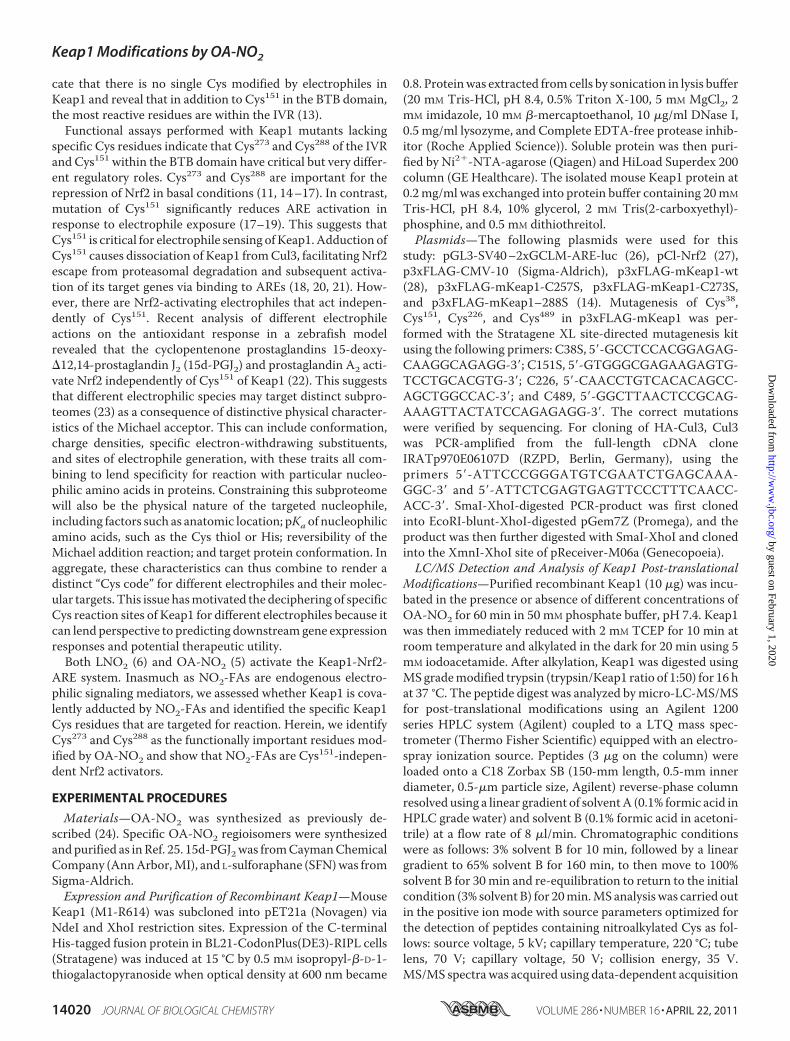
screening of OA-NO2-reactive Cys residues was performedwith recombinant Keap1 treated with OA-NO2. In order toconfirm that the adduction of Keap1 by OA-NO2 occurs inintact cells, where fatty acid metabolism and alternative com-peting reactions can occur, a MS-based method was utilized tomeasure adducted NO2-FA levels (30, 32). The method quan-tifies OA-NO2-protein adducts after an exchange reactionwithexogenously added �-ME (trans-nitroalkylation). FLAG-tagged Keap1 was transiently transfected to HEK-293 cells,which were exposed to increasing concentrations of OA-NO2.After FLAG immunoprecipitation, Keap1 was detected on sil-ver-stained gels (Fig. 2A, bottom). Keap1 was immunoprecipi-tated within treatment groups with similar efficiency and wasnot detected in empty vector controls. When immunoprecipi-tated Keap1 was subjected to trans-nitroalkylation with �-ME,an OA-NO2 concentration-dependent increase in OA-NO2-�-ME adducts was detected. Thus, OA-NO2 covalently reactswith ectopically expressed Keap1 in HEK-293T cells at lowmicromolar concentrations (Fig. 2A). The contributions ofthe functionally important residues Cys273 and Cys288 to theoverall Keap1-OA-NO2 reaction were then determined incells expressing mutated Keap1 lacking either Cys273 orCys288. Keap1 having single Cys mutations had no significanteffect on reaction with OA-NO2, whereas combined muta-
tion of both Cys273 and Cys288 reduced OA-NO2 reaction by�50% (Fig. 2B).Activation of ARE by Different Regioisomers of OA-NO2—
Both endogenously generated OA-NO2 and the syntheticOA-NO2 used herein are equimolar mixtures of 9- and10-nitro-octadec-9-enoic acids (33). Recently, isomer-spe-cific binding of OA-NO2 to redox-sensitive thiols has beenreported, with 10-nitro-octadec-9-enoic acid being morereactive than 9-nitro-octadec-9-enoic acid toward Cys285 inthe ligand-binding domain of PPAR� (34). In order toexplore whether ARE activation is OA-NO2 regioisomer-selective, ARE activation was measured using luciferasereporter assays. In contrast to patterns of PPAR� adduction,9-nitro-octadec-9-enoic acid was more potent than both10-nitro-octadec-9-enoic acid and an equimolar 1:1 mixtureof both regioisomers in inducing ARE activity (Fig. 3A). Aftertreatment with the two isomers of OA-NO2, ChIP was per-formed to study the binding of Nrf2 to the ARE-containingdistal enhancer region of the HMOX1 gene. In accordancewith reporter analysis-based observations, 10-nitro-octa-dec-9-enoic acid was less potent than 9-nitro-octadec-9-enoic acid and the 1:1 regioisomer mixture in enhancingNrf2 binding to AREs. This supports the notion that 9-OA-NO2 is a more favorable inducer of ARE activation (Fig. 3B).The Effect of OA-NO2 on Cul3-Keap1 Interaction—Accord-
ing to the current paradigm, Keap1 binding to Cul3 enables thecomplex to degrade Nrf2 by ubiquitination under basal condi-tions. Electrophile adduction of Keap1 Cys151 modulates theinteraction of the protein with Cul3, leading to the precept thatmodification of Cys151 may dissociate Keap1 from Cul3 and
FIGURE 2. OA-NO2 reacts with Keap1 in a cellular milieu. HEK-293T cells were transfected with FLAG-CMV (empty vector) or FLAG-Keap1-overexpress-ing vector and treated with the indicated concentrations of OA-NO2. A, Keap1-adducted OA-NO2 was exchanged to �-ME from immunoprecipitatedKeap1 in the presence of a [13C18]OA-NO2 internal standard and quantified by LC-MS/MS as �-ME-OA-NO2. The lower panels show transfection efficiencyof FLAG-Keap1 constructs. B, �-ME-OA-NO2 levels captured upon exchange to �-ME in immunoprecipitated WT Keap1 and the following Cys to Sermutant Keap1: C273S, C288S, C273S/C288S, or C151S/C273S/C288S. �-ME exchange reactions were conducted in the presence of [13C18]OA-NO2 andquantified by LC-MS/MS. Values are represented as mean � S.D. (error bars) *, p � 0.05; **, p � 0.01; ***, p � 0.001 when compared with respectivecontrol. ns, not significant.
Keap1 Modifications by OA-NO2
APRIL 22, 2011 • VOLUME 286 • NUMBER 16 JOURNAL OF BIOLOGICAL CHEMISTRY 14023
by guest on February 1, 2020http://w
ww
.jbc.org/D
ownloaded from

promote the escape of Nrf2 from proteasomal degradation (20,21). The binding of Cul3 and Keap1 after treatment with theCys151-dependent electrophile SFN (21) and the two Cys151-independent electrophiles, OA-NO2 and 15d-PGJ2, revealedthat SFN diminished Keap1 and Cul3 binding, whereas bothOA-NO2 and 15d-PGJ2 enhanced Keap1 and Cul3 interaction(Fig. 4).
DISCUSSION
Organisms are continuously exposed to electrophiles thatare either endogenously produced via redox reactions or arederived from exogenous sources (35). It is now evident thatmultiple cell signaling mechanisms have evolved to sense andrespond to electrophiles, thus linking gene expression and pro-tein function with metabolic and inflammatory status. Electro-philes can react with nucleophilic amino acids of proteins viaMichael addition, thereby altering protein structure and func-tion. Although such modifications have typically been viewedas toxic, recent data affirm that low concentrations or rates ofgeneration of reversibly reactive electrophiles can elicit a broadrange of adaptive protein functional and gene expressionresponses in the absence of toxicity (23, 35). One of the path-ways activated by electrophiles, such as nitroalkene fatty acidderivatives, is the Keap1-Nrf2-ARE system (5). The reactiveeffector protein in this pathway, Keap1, is exemplary for reveal-ing how post-translational modifications by electrophiles canelicit specific biological responses. The modifications of Keap1reported from the use of a variety of different electrophiles andmethods show significant variations in specific thiol residues asMichael addition targets of different electrophiles (13). This hasimportant implications for the mechanisms underlying activa-tion of ARE-regulated genes, patterns of gene expression, and
ultimately, the phenotypic characteristics of differentiated cellresponses. These issues motivated the investigation of Keap1Cys modifications by the electrophilic fatty acid nitroalkene,OA-NO2.
In this study, six Keap1 Cys residues were identified assusceptible to modification by nitroalkylation at the lowest
FIGURE 3. Nitroalkene regioisomer-specific activation of ARE. A, HEK-293T cells were transfected with the ARE luciferase reporter and �-galactosidasecontrol vector. 24 h after transfection, the cells were incubated with 9-OA-NO2, 10-OA-NO2, or a 1:1 mix of both isomers for 16 h. The data are represented as-fold change from control (vehicle) � S.D. (error bars); n � 4. *, p � 0.05; **, p � 0.01; ***, p � 0.001 when compared with a mix of both isomers. #, p � 0.05; ##,p � 0.01; ###, p � 0.001 when compared with 10-OA-NO2. B, human umbilical vein endothelial cells treated with vehicle, 5 �M 1:1 mix of 9-OA-NO2 and10-OA-NO2, 9-OA-NO2, or 10-OA-NO2 for 2 h. ChIP assays were performed with chromatin extracts using an anti-Nrf2 antibody. Real-time quantitative PCR wasperformed using primers specific for a distal enhancer region of the HMOX1 gene containing multiple ARE elements. Non-precipitated input chromatin servedas a reference, and IgG-precipitated template served as specificity control. -Fold induction of Nrf2 association was calculated. Values are represented as mean �S.D. *, p � 0.05; **, p � 0.01; ***, p � 0.001 when compared with vehicle.
FIGURE 4. Exposure to OA-NO2 or 15d-PGJ2 does not cause dissociation ofKeap1 and Cul3. HEK-293T cells were co-transfected with FLAG-Keap1 andHA-Cul3 and treated with 15d-PGJ2, OA-NO2, or SFN. Cell lysates wereimmunoprecipitated (IP) with FLAG or HA antibodies (top), and theamount of bound Keap1 or Cul3 was detected with Western blot usingFLAG or HA antibodies. The bottom shows the transfection efficiency oftotal cell lysates (input control). Western blots are representative of threeindependent experiments.
Keap1 Modifications by OA-NO2
14024 JOURNAL OF BIOLOGICAL CHEMISTRY VOLUME 286 • NUMBER 16 • APRIL 22, 2011
by guest on February 1, 2020http://w
ww
.jbc.org/D
ownloaded from

OA-NO2 concentrations used. Although LC-MS determina-tions have revealed several Cys residues susceptible toadduction during Keap1 electrophile sensing, the interpre-tation of these data can be influenced by a number of issues.The efficiencies for the detection of peptides by MS areaffected by a number of factors, resulting in up to 3-order ofmagnitude differences in the sensitivity of detection of dif-ferent tryptic peptides stemming from the same protein.Thus, the relative detection limits for different modifiedpeptides can widely differ. Also, depending on the specificelectrophile, sample preparation approaches, and both ion-ization and fragmentation efficiencies of different massspectrometers, there can be differences in the identificationof reactive residues. Despite these limitations, of the sixnitroalkene-reactive Cys residues identified in the presentstudy, Keap1 Cys257, Cys273, and Cys288 have been frequentlyreported as targets for adduction (13, 23).Previous proteomic analyses and functional studies iden-
tified Keap1 Cys273, Cys288, and Cys151 as critical targets ofelectrophile-thiol interactions (23). The reactivity of Cys273and Cys288 that are located in the IVR is reaffirmed in thisstudy. Recently, the structure of Keap1 dimer was studied bysingle particle electron microscopy (36). Keap1 was identi-fied as a forked dimer with two large globular domains con-stituting 86.5% of the total volume of the protein. This studyalso revealed a close proximity of the IVR with the DCdomain (DGR (double glycine repeat) and the C-terminalregion of Keap1) that forms a �-propeller structure interact-ing with the Neh2 domain of Nrf2. Neh2 has two evolution-arily conserved motifs (DLG and ETGE), that have differentelectrostatic potentials defining their binding affinitiestoward the Keap1-DC domain (37, 38). According to thetwo-site recognition model of Keap1-Nrf2 interaction,Keap1 homodimer interacts with a single Nrf2 molecule.Under basal conditions, both ETGE and DLGmotifs of Neh2interact with Keap1-DC. However, during oxidative or elec-trophilic stress, the low affinity DLGmotif that positions thelysines within the Neh2 domain for ubiquitination detachesfrom the Keap1-DC domain, resulting in disruption ofpolyubiquitination and degradation of Nrf2. The proximityof IVR with the DC domain (36) supports the notion thatcovalent modification of Cys273 and Cys288 induces confor-mational changes in the IVR that in turn affect the structuralintegrity of adjacent Keap1-DC, eventually disrupting theinteraction with the DLG motif of Nrf2.In this study, the functionally important Cys273 and Cys288
residues were among the most reactive toward OA-NO2 in therecombinant protein but only accounted for �50% of netOA-NO2 reaction with intracellular Keap1. Mutation of eitherCys alone had no impact on overall reaction. This is consistentwith recent click chemistry-based detection of Keap1 adductformation with sulfoxythiocarbamate (39). The adduction ofsingle C273A and C288A Keap1 mutants with sulfoxythiocar-bamate did not significantly differ from that of wild type Keap1.In contrast, double C273A/C288A and triple C151A/C273A/C288Amutants showedmarkedly reduced sulfoxythiocarbam-ate adduction. The lack of reactivity of the single Cys mutantscould be explained by the change of protein conformation by
the mutation of one Cys to favor reaction with another. Alter-natively, the two cysteines might act cooperatively. Dinkova-Kostova et al. (40) proposed that Keap1 is a zincmetalloprotein,and that Cys273 and Cys288 would coordinate binding of Zn2�.Upon inducer sensing, Zn2� could be released, and the twocysteine residues as the more reactive thiolate anion can thenreact with electrophiles.Herein, OA-NO2 was not particularly reactive toward
Cys151 in recombinant Keap1 and was not critical for ARE-dependent gene activation. Furthermore, ectopically ex-pressed Keap1, with combined C151S, C273S, and C288Smutations, was no less reactive in �-ME-based electrophilecapture assays than the C273S and C288S double mutant.This also indicates that Cys151 is not a major site of OA-NO2reaction in a cellular milieu. Cys151 has been reported to besensitive to adduction (23) and critical in Nrf2 activation byelectrophiles, including SFN, tert-butylhydroquinone (21,31), and N-iodoacetyl-N-biotinylhexylene-diamine (IAB)(20). These data are based on luciferase reporter assays (16,21, 31), zebrafish embryos overexpressingmouse Keap1 (22),and mouse embryonic fibroblasts derived from Keap1 Cys151
transgenic animals (17). The studies investigating Cys151-de-pendent Nrf2 activation have revealed that Cys151 is requiredfor Keap1 and Cul3 interaction. Modification of Cys151
appears to decrease Cul3 interactions, leading to inhibitionof Keap1-dependent ubiquitination of Nrf2 (21). Unfortu-nately, there are no structural data defining the Keap1-Cul3interface. However, molecular contacts between the cullinproteins and their BTB domain-containing substrate adap-tor proteins are highly conserved. Modeling of the Keap1-Cul3 interaction interface reveals that Keap1 residues 125–127 and 162–164 within the BTB domain are predicted tointeract with Cul3 (21). Although Cys151 is not located at thepredicted Keap1-Cul3 interface (41), it is suggested that abulky modification at this site would cause conformationalchanges that alter Cul3 binding, allowing Nrf2 to escape pro-teasomal degradation (18). Although several reports supporta crucial role of Keap1 Cys151 for Nrf2 activation, the elec-trophilic prostaglandins prostaglandin A2 and 15d-PGJ2 (22)and the heavy metal arsenic (31) all activate Nrf2 indepen-dent of Cys151. Another class of Cys151-independent AREactivators, the cyclopentenone prostaglandins prostaglandinA2 and 15d-PGJ2, do not react with Cys151 as assessed by MSanalysis of recombinant or ectopically expressed Keap1 (22,42). We observed that both OA-NO2 and 15d-PGJ2 had asimilar effect on Keap1-Cul3 interactions (i.e. both electro-philes increased Keap1-Cul3 binding), whereas treatmentwith SFN diminished the interaction of Keap1 and Cul3. Itcan therefore be envisioned that the class of Cys151-inde-pendent ARE activators do not disrupt the Keap1-Cul3interaction but nevertheless inhibit Nrf2 ubiquitination andproteasomal degradation, possibly via disruption of thedynamic assembly/disassembly of Keap1 with the Cul3-Rbx1E3 ubiquitin ligase complex (21, 31). That Keap1 has multi-ple sensing mechanisms for activation is further supportedby the report that Keap1 has separate sensors for nitricoxide, metals, and alkenals, all acting independently (43).
Keap1 Modifications by OA-NO2
APRIL 22, 2011 • VOLUME 286 • NUMBER 16 JOURNAL OF BIOLOGICAL CHEMISTRY 14025
by guest on February 1, 2020http://w
ww
.jbc.org/D
ownloaded from

Herein, we show that Nrf2 is activated preferentially by9-OA-NO2. In redox signaling by electrophiles, the position ofthe electrophilic carbon in relation to the nucleophilic targetthus appears critical in defining net reactivity. Importantly, keystructural motifs of the target protein and critical structuralelements of the fatty acid nitroalkene (the hydrophobic methylend, the anionic nitro and carboxylic acid substituents, and thecis double bond configuration) will influence rates of Michaeladdition. In this regard, OA-NO2 shows regioisomer-selectivereactivity toward the ligand binding domain cysteine residue(Cys285) in PPAR�, with 10-OA-NO2more reactive than 9-OA-NO2 (4). Also, xanthine oxidoreductase activity is inhibited by9-OA-NO2 and the mixture of 9- and 10-OA-NO2, but not byother structural variants of OA-NO2 that differ in the nitroalk-enyl and carboxylic acid moieties (3). Moreover, the extent ofPPAR� activation is different by the four nitroalkenyl regioiso-mers of linoleic acid (9-, 10-, 12-, or 13-nitro-ocatadeca-9,12-dienoic acid) (44). These data underscore the notion that evensmall changes in nitroalkene fatty acid structure can impact onbiological signaling actions.In summary, Cys273 and Cys288 are significant electrophile
reaction sites contributing to Keap1 function but are not theonly fatty acid nitroalkene-reactive Cys residues of Keap1. Pro-teomic analyses with LC-MS/MS suggested that reactions withHis or other nucleophilic amino acids of Keap1 were not signif-icant under these conditions. Furthermore, Cys151 is not nota-bly reactive with OA-NO2 nor necessary for ARE activationbecause OA-NO2 potently induced ARE-dependent geneexpression at lowmicromolar concentrations (5). OA-NO2 cantherefore be categorized as a Cys151-independent Nrf2 activa-tor. Inasmuch as nitroalkenes represent endogenously pro-duced NO and nitrite-derived signaling mediators, these dataadds to our understanding of their properties and therapeuticpotential.
REFERENCES1. Freeman, B. A., Baker, P. R., Schopfer, F. J., Woodcock, S. R., Napolitano,
A., and d’Ischia, M. (2008) J. Biol. Chem. 283, 15515–155192. Cui, T., Schopfer, F. J., Zhang, J., Chen, K., Ichikawa, T., Baker, P. R.,
Batthyany, C., Chacko, B. K., Feng, X., Patel, R. P., Agarwal, A., Freeman,B. A., and Chen, Y. E. (2006) J. Biol. Chem. 281, 35686–35698
3. Kelley, E. E., Batthyany, C. I., Hundley, N. J., Woodcock, S. R., Bonacci, G.,Del Rio, J. M., Schopfer, F. J., Lancaster, J. R., Jr., Freeman, B. A., andTarpey, M. M. (2008) J. Biol. Chem. 283, 36176–36184
4. Schopfer, F. J., Cole,M. P., Groeger, A. L., Chen, C. S., Khoo, N. K.,Wood-cock, S. R., Golin-Bisello, F., Motanya, U. N., Li, Y., Zhang, J., Garcia-Barrio, M. T., Rudolph, T. K., Rudolph, V., Bonacci, G., Baker, P. R., Xu,H. E., Batthyany, C. I., Chen, Y. E., Hallis, T. M., and Freeman, B. A. (2010)J. Biol. Chem. 285, 12321–12333
5. Kansanen, E., Jyrkkanen, H. K., Volger, O. L., Leinonen, H., Kivela, A. M.,Hakkinen, S. K., Woodcock, S. R., Schopfer, F. J., Horrevoets, A. J., Yla-Herttuala, S., Freeman, B. A., and Levonen, A. L. (2009) J. Biol. Chem. 284,33233–33241
6. Villacorta, L., Zhang, J., Garcia-Barrio, M. T., Chen, X. L., Freeman, B. A.,Chen, Y. E., and Cui, T. (2007) Am. J. Physiol. Heart Circ. Physiol. 293,H770–H776
7. Kensler, T. W., Wakabayashi, N., and Biswal, S. (2007) Annu. Rev. Phar-macol. Toxicol. 47, 89–116
8. Cullinan, S. B., Gordan, J. D., Jin, J., Harper, J. W., and Diehl, J. A. (2004)Mol. Cell Biol. 24, 8477–8486
9. Kobayashi, A., Kang, M. I., Okawa, H., Ohtsuji, M., Zenke, Y., Chiba, T.,Igarashi, K., and Yamamoto, M. (2004)Mol. Cell Biol. 24, 7130–7139
10. Dinkova-Kostova, A. T., Holtzclaw, W. D., Cole, R. N., Itoh, K., Waka-bayashi, N., Katoh, Y., Yamamoto, M., and Talalay, P. (2002) Proc. Natl.Acad. Sci. U.S.A. 99, 11908–11913
11. Kobayashi, A., Kang, M. I., Watai, Y., Tong, K. I., Shibata, T., Uchida, K.,and Yamamoto, M. (2006)Mol. Cell Biol. 26, 221–229
12. Levonen, A. L., Dickinson, D. A., Moellering, D. R., Mulcahy, R. T., For-man, H. J., and Darley-Usmar, V. M. (2001) Arterioscler. Thromb. Vasc.Biol. 21, 1846–1851
13. Kansanen, E., Kivela, A. M., and Levonen, A. L. (2009) Free Radic. Biol.Med. 47, 1310–1317
14. Levonen, A. L., Landar, A., Ramachandran, A., Ceaser, E. K., Dickinson,D. A., Zanoni, G.,Morrow, J. D., andDarley-Usmar, V.M. (2004)Biochem.J. 378, 373–382
15. Wakabayashi, N., Dinkova-Kostova, A. T., Holtzclaw, W. D., Kang, M. I.,Kobayashi, A., Yamamoto, M., Kensler, T.W., and Talalay, P. (2004) Proc.Natl. Acad. Sci. U.S.A. 101, 2040–2045
16. Zhang, D. D., and Hannink, M. (2003)Mol. Cell Biol. 23, 8137–815117. Yamamoto, T., Suzuki, T., Kobayashi, A., Wakabayashi, J., Maher, J., Mo-
tohashi, H., and Yamamoto, M. (2008)Mol. Cell Biol. 28, 2758–277018. Eggler, A. L., Small, E., Hannink,M., andMesecar, A. D. (2009) Biochem. J.
422, 171–18019. Li, L., Kobayashi, M., Kaneko, H., Nakajima-Takagi, Y., Nakayama, Y., and
Yamamoto, M. (2008) J. Biol. Chem. 283, 3248–325520. Rachakonda, G., Xiong, Y., Sekhar, K. R., Stamer, S. L., Liebler, D. C., and
Freeman, M. L. (2008) Chem. Res. Toxicol. 21, 705–71021. Zhang, D. D., Lo, S. C., Cross, J. V., Templeton, D. J., and Hannink, M.
(2004)Mol. Cell Biol. 24, 10941–1095322. Kobayashi, M., Li, L., Iwamoto, N., Nakajima-Takagi, Y., Kaneko, H., Na-
kayama, Y., Eguchi, M.,Wada, Y., Kumagai, Y., and Yamamoto, M. (2009)Mol. Cell Biol. 29, 493–502
23. Liebler, D. C. (2008) Chem. Res. Toxicol. 21, 117–12824. Baker, P. R., Lin, Y., Schopfer, F. J., Woodcock, S. R., Groeger, A. L., Bat-
thyany, C., Sweeney, S., Long, M. H., Iles, K. E., Baker, L. M., Branchaud,B. P., Chen, Y. E., and Freeman, B. A. (2005) J. Biol. Chem. 280,42464–42475
25. Woodcock, S. R., Marwitz, A. J., Bruno, P., and Branchaud, B. P. (2006)Org. Lett. 8, 3931–3934
26. Hurttila, H., Koponen, J. K., Kansanen, E., Jyrkkanen, H. K., Kivela, A.,Kylatie, R., Yla-Herttuala, S., and Levonen, A. L. (2008) Gene Ther. 15,1271–1279
27. Wild, A. C., Moinova, H. R., andMulcahy, R. T. (1999) J. Biol. Chem. 274,33627–33636
28. Zipper, L. M., andMulcahy, R. T. (2002) J. Biol. Chem. 277, 36544–3655229. Jyrkkanen, H. K., Kansanen, E., Inkala, M., Kivela, A. M., Hurttila, H.,
Heinonen, S. E., Goldsteins, G., Jauhiainen, S., Tiainen, S., Makkonen, H.,Oskolkova, O., Afonyushkin, T., Koistinaho, J., Yamamoto, M., Bochkov,V. N., Yla-Herttuala, S., and Levonen, A. L. (2008) Circ. Res. 103, e1–e9
30. Schopfer, F. J., Batthyany, C., Baker, P. R., Bonacci, G., Cole, M. P., Ru-dolph, V., Groeger, A. L., Rudolph, T. K., Nadtochiy, S., Brookes, P. S., andFreeman, B. A. (2009) Free Radic. Biol. Med. 46, 1250–1259
31. Wang, X. J., Sun, Z., Chen, W., Li, Y., Villeneuve, N. F., and Zhang, D. D.(2008) Toxicol. Appl. Pharmacol. 230, 383–389
32. Zhang, J., Villacorta, L., Chang, L., Fan, Z., Hamblin, M., Zhu, T., Chen,C. S., Cole, M. P., Schopfer, F. J., Deng, C. X., Garcia-Barrio, M. T., Feng,Y. H., Freeman, B. A., and Chen, Y. E. (2010) Circ. Res. 107, 540–548
33. Rudolph, V., Rudolph, T. K., Schopfer, F. J., Bonacci, G., Woodcock, S. R.,Cole,M. P., Baker, P. R., Ramani, R., and Freeman, B. A. (2010)Cardiovasc.Res. 85, 155–166
34. Schopfer, F. J., Lin, Y., Baker, P. R., Cui, T., Garcia-Barrio, M., Zhang, J.,Chen, K., Chen, Y. E., and Freeman, B. A. (2005) Proc. Natl. Acad. Sci.U.S.A. 102, 2340–2345
35. Rudolph, T. K., and Freeman, B. A. (2009) Sci. Signal. 2, re736. Ogura, T., Tong, K. I., Mio, K., Maruyama, Y., Kurokawa, H., Sato, C., and
Yamamoto, M. (2010) Proc. Natl. Acad. Sci. U.S.A. 107, 2842–284737. Tong, K. I., Katoh, Y., Kusunoki, H., Itoh, K., Tanaka, T., and Yamamoto,
M. (2006)Mol. Cell Biol. 26, 2887–290038. Tong, K. I., Padmanabhan, B., Kobayashi, A., Shang, C., Hirotsu, Y.,
Yokoyama, S., and Yamamoto, M. (2007)Mol. Cell Biol. 27, 7511–7521
Keap1 Modifications by OA-NO2
14026 JOURNAL OF BIOLOGICAL CHEMISTRY VOLUME 286 • NUMBER 16 • APRIL 22, 2011
by guest on February 1, 2020http://w
ww
.jbc.org/D
ownloaded from

39. Ahn, Y. H., Hwang, Y., Liu, H., Wang, X. J., Zhang, Y., Stephenson, K. K.,Boronina, T. N., Cole, R. N., Dinkova-Kostova, A. T., Talalay, P., and Cole,P. A. (2010) Proc. Natl. Acad. Sci. U.S.A. 107, 9590–9595
40. Dinkova-Kostova, A. T., Holtzclaw, W. D., and Wakabayashi, N. (2005)Biochemistry 44, 6889–6899
41. Eggler, A. L., Liu, G., Pezzuto, J.M., van Breemen, R. B., andMesecar, A. D.(2005) Proc. Natl. Acad. Sci. U.S.A. 102, 10070–10075
42. Copple, I. M., Goldring, C. E., Jenkins, R. E., Chia, A. J., Randle, L. E.,Hayes, J. D., Kitteringham, N. R., and Park, B. K. (2008) Hepatology 48,1292–1301
43. McMahon, M., Lamont, D. J., Beattie, K. A., and Hayes, J. D. (2010) Proc.Natl. Acad. Sci. U.S.A. 107, 18838–18843
44. Gorczynski, M. J., Smitherman, P. K., Akiyama, T. E.,Wood, H. B., Berger,J. P., King, S. B., and Morrow, C. S. (2009) J. Med. Chem. 52, 4631–4639
Keap1 Modifications by OA-NO2
APRIL 22, 2011 • VOLUME 286 • NUMBER 16 JOURNAL OF BIOLOGICAL CHEMISTRY 14027
by guest on February 1, 2020http://w
ww
.jbc.org/D
ownloaded from

Seppo Ylä-Herttuala, Bruce A. Freeman and Anna-Liisa LevonenTong, Hanna Leinonen, Steven R. Woodcock, Masayuki Yamamoto, Carsten Carlberg, Emilia Kansanen, Gustavo Bonacci, Francisco J. Schopfer, Suvi M. Kuosmanen, Kit I.
151-independent MechanismElectrophilic Nitro-fatty Acids Activate NRF2 by a KEAP1 Cysteine
doi: 10.1074/jbc.M110.190710 originally published online February 25, 20112011, 286:14019-14027.J. Biol. Chem.
10.1074/jbc.M110.190710Access the most updated version of this article at doi:
Alerts:
When a correction for this article is posted•
When this article is cited•
to choose from all of JBC's e-mail alertsClick here
Supplemental material:
http://www.jbc.org/content/suppl/2011/02/25/M110.190710.DC1
http://www.jbc.org/content/286/16/14019.full.html#ref-list-1
This article cites 44 references, 30 of which can be accessed free at
by guest on February 1, 2020http://w
ww
.jbc.org/D
ownloaded from