electron paramagnetic resonance spectroscopy of free radicals in corneal tissue following excimer...
TRANSCRIPT

Lasers in Surgery and Medicine 18367372 (1996)
Electron Paramagnetic Resonance Spectroscopy of Free Radicals in Corneal Tissue Following Excimer
Laser I r rad iat ion G.H. Pettit, MD, PhD, M.N. Ediger, PhD, D.W. Hahn, PhD, R.J. Landry, MS,
R.P. Weiblinger, MA, and K.M. Morehouse, PhD
Food & Drug Administration, Center for Devices & Radiological Health, Rockville, Maryland 20857 (G. H.P., M. N. E., D. W. H., R. J. L., R.P. W.); Food & Drug Administration,
Center for Food Safety & Applied Nutrition, Washington, DC 20204 (K. M.M.)
Background and Objective: Free radicals, detected previously in corneal tissue following 193 nm laser irradiation, may be impor- tant agents in the laserltissue interaction. Electron paramagnetic resonance spectroscopy (EPR) has been used to examine such radical formation in detail. Study DesignlMateriaZs and Methods: Bovine corneal strips were frozen in liquid nitrogen, irradiated with excimer laser pulses, and assayed by EPR. Exposure conditions were varied to study radical formation dependence on laser intensity and repetition. Results were measured against a quantifiable standard to calcu- late radical quantum yield. Results: Either weak or intense laser fluences produced compa- rable tissue EPR signals. Radicals accumulated in frozen tissue for at least 10 initial ablation pulses. Radical quantum yield in cornea was 0.15%. Conclusion: Corneal radical formation is largely a photochemical process driven by the 193 nm laser radiation. Reactive radical species are produced in substantial numbers and likely have a significant clinical role. o 1996 Wiley-Liss, Inc.*
Key
INTRODUCTION
words: ablation, photochemistry, photorefractive keratectomy
The argon fluoride (ArF) excimer laser is being extensively studied worldwide as a clinical tool for remodeling the optical surface of the eye. Such intense interest has arisen because nano- second 193 nm pulses from ArF lasers vaporize cornea with submicron precision and minimal col- lateral damage [ll. In a recent study of this laser/ tissue interaction [21, using electron paramag- netic resonance spectroscopy (EPR) and cryogenic experimental conditions, we have detected or- ganic free radicals in cornea following the irradi- ation. This finding is significant for two reasons. First of all, highly absorbing organic radicals could cause the enhanced attenuation of 193 nm laser radiation during ablation [3] and the asso- ciated small etch depth per pulse in the tissue
(compared to the l/e laser penetration depth). Of greater practical importance, the ancillary tissue damage caused by such reactive species may con- tribute to the complex healing response that is observed clinically, that is, “haze” formation and curvature regression [4-61. In support of this con- tention, a recent study involving rabbits has shown a significant postoperative haze reduction in animals treated with radical scavengers just before laser exposure [71.
Our initial EPR study was essentially qual- itative in nature, documenting that organic radi-
Accepted for publication February 23, 1995. Address reprint requests to George H. Pettit, M.D., Ph.D., F.D.A. Center for Devices & Radiological Health, Mail Stop HFZ-134, Rockville, MD 20857.
0 1996 Wiley-Liss, Inc. *This article is a US Government work and, as such, is in the public domain in the United States of America.
368 Pettit et al. cals were formed in the cornea and were highly reactive (short lived) at physiologic temperatures. Radical EPR signatures were large, relative to noise, only for frozen corneal samples receiving multiple laser irradiations at each site on the de-epithelialized surface. This indicated that there was a net radical accumulation in the frozen tissue over multiple pulses, even though some absorbing fraction was etched away by each laser shot.
In this paper we pursue a more quantitative study of radical formation, taking the above-men- tioned phenomena into account in determining a quantum yield of radicals produced per laser pho- ton deposited. This is accomplished by measuring the tissue EPR signal amplitudes for varying de- livered laser dosages, that is, different numbers of constant-fluence pulses per site, and by com- paring these signals against that of a known rad- ical-concentration standard. We also explore the photochemistry behind the radical formation by comparing tissue EPR signals for two distinct ir- radiation conditions involving the same delivered laser energy, one with a pulse fluence far below the ablation threshold and the other with a flu- ence well above threshold.
MATERIALS AND METHODS
Tissue samples used in this work were bo- vine corneas. Whole globes were obtained from a slaughterhouse and stored on ice until use less than 24 hours post mortem. Before laser irradia- tion, the central cornea of each globe was mechan- ically de-epithelialized and cut into rectangular strips measuring -25 mm by 2 mm. Each strip was placed lengthwise on a glass stirring rod with the epithelial surface oriented upward and quickly immersed in liquid nitrogen (LN,). This preserved the rectangular sample shape, and the slight curvature induced by the underlying rod helped identify the anterior sample surface.
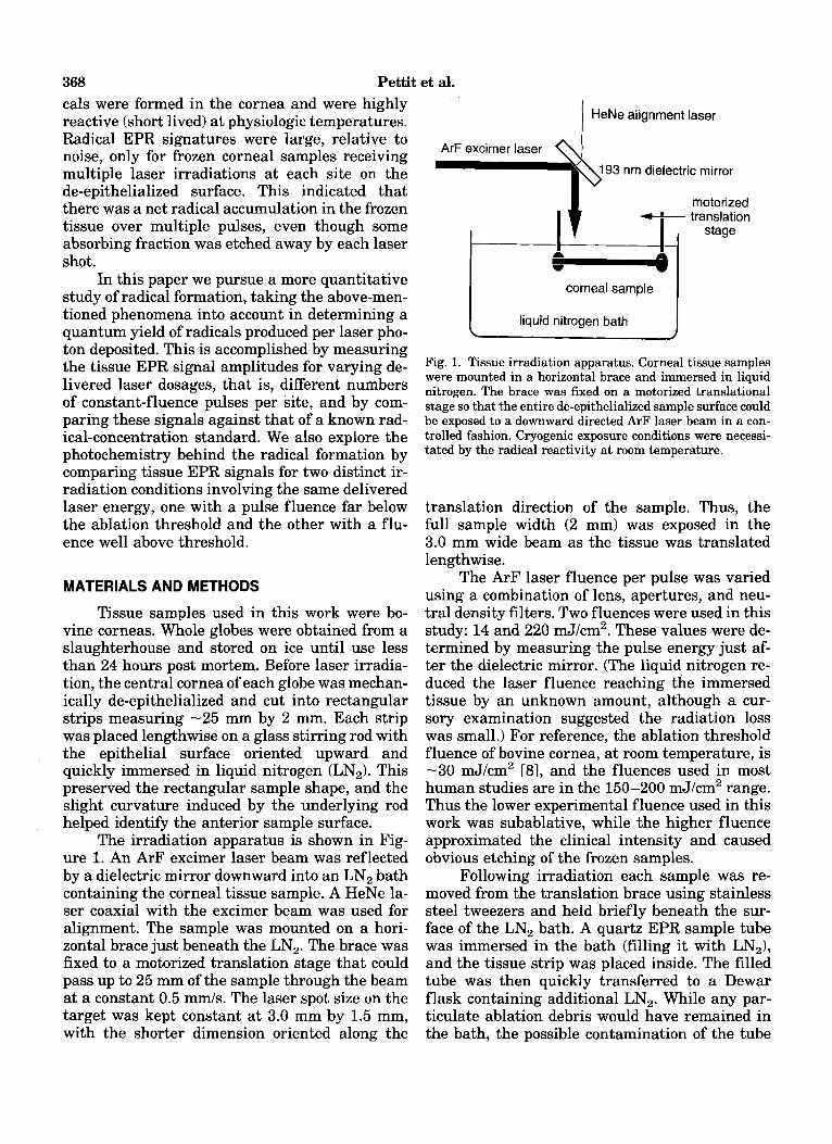
The irradiation apparatus is shown in Fig- ure 1. An ArF excimer laser beam was reflected by a dielectric mirror downward into an LN, bath containing the corneal tissue sample. A HeNe la- ser coaxial with the excimer beam was used for alignment. The sample was mounted on a hori- zontal brace just beneath the LN,. The brace was fixed to a motorized translation stage that could pass up to 25 mm of the sample through the beam at a constant 0.5 mm/s. The laser spot size on the target was kept constant at 3.0 mm by 1.5 mm, with the shorter dimension oriented along the
I
HeNe alignment laser
motorized translation
corneal sample
liquid nitrogen bath
Fig. 1. Tissue irradiation apparatus. Corneal tissue samples were mounted in a horizontal brace and immersed in liquid nitrogen. The brace was fixed on a motorized translational stage so that the entire de-epithelialized sample surface could be exposed to a downward directed ArF laser beam in a con- trolled fashion. Cryogenic exposure conditions were necessi- tated by the radical reactivity at room temperature.
translation direction of the sample. Thus, the full sample width (2 mm) was exposed in the 3.0 mm wide beam as the tissue was translated lengthwise.
The ArF laser fluence per pulse was varied using a combination of lens, apertures, and neu- tral density filters. Two fluences were used in this study: 14 and 220 mJ/cm2. These values were de- termined by measuring the pulse energy just af- ter the dielectric mirror. (The liquid nitrogen re- duced the laser fluence reaching the immersed tissue by an unknown amount, although a cur- sory examination suggested the radiation loss was small.) For reference, the ablation threshold fluence of bovine cornea, at room temperature, is -30 mJ/cm2 [81, and the fluences used in most human studies are in the 150-200 mJ/cm2 range. Thus the lower experimental fluence used in this work was subablative, while the higher fluence approximated the clinical intensity and caused obvious etching of the frozen samples.
Following irradiation each sample was re- moved from the translation brace using stainless steel tweezers and held briefly beneath the sur- face of the LN, bath. A quartz EPR sample tube was immersed in the bath (filling it with LN,), and the tissue strip was placed inside. The filled tube was then quickly transferred to a Dewar flask containing additional LN,. While any par- ticulate ablation debris would have remained in the bath, the possible contamination of the tube
EPR of Corneal Free Radicals 369
I . . . . . . . . . . . . . . . . . . . . . . .
Low Fluewe Sawle
by such debris was negligible. This was due to the fact that the bath volume (greater than 11) was much larger than the tube fluid volume assayed by EPR spectroscopy (-0.1 ml).
Measurements of radical formation were made using a Varian E-line Century Series EPR spectrometer. EPR spectroscopy works on the principle that paramagnetic species such as free radicals will absorb microwave radiation when placed in a strong magnetic field. The specific mi- crowave frequency and field strength required de- pend on the nature of the absorbing radical spe- cies. In this study the microwave frequency was fixed at 9.19 GHz, and absorption was measured as the magnetic field was scanned over the range 2,880-3,280 G. The other relevant spectroscopy conditions were a magnetic field modulation am- plitude of 5 G and a microwave power of 5 mW. Tissue samples were kept at LN, temperature throughout the EPR assay.
The initial experiment was a comparison of the radical generation efficiency by low and high fluence laser pulses. Twelve tissue samples were divided into two groups of six and irradiated with 900 mJ total laser energy (cumulative fluence -2 J/cm2). For the first group 14 mJ/cm2 laser pulses were used, and the cumulative tissue dose was delivered by running the laser at 12 Hz and trans- lating each sample 22.5 mm through the ArF la- ser beam four times. Each sample in the second group was irradiated with 220 mJ/cm2 pulses at 3 Hz during a single 22.5 mm pass through the beam. EPR analysis was then performed on all samples under identical spectroscopic conditions, and the radical signatures for the two groups were compared.
The second experiment was a measure of radical accumulation in the frozen tissue under repetitive ablation conditions. With the laser run- ning at 113 Hz, 15 220 mJ/cm2 pulses were depos- ited at adjacent, essentially distinct, sites along 22.5 mm of a sample during one crossing through the beam. This corresponded to a dose of approx- imately 100 mJ. By increasing the laser repeti- tion rate and/or the number of passes, higher la- ser dosages (200, 500, and 1,000 mJ) were delivered to subsequent samples, with repetitive trials conducted at each dose. Each sample was then assayed by EPR to determine relative am- plitudes of the radical signals for the various laser dosages.
The third and final experiment was an abso- lute determination of radical yield under ablative conditions. A 1 J laser dose, which generated a
I
I -High Flwlyie Sample
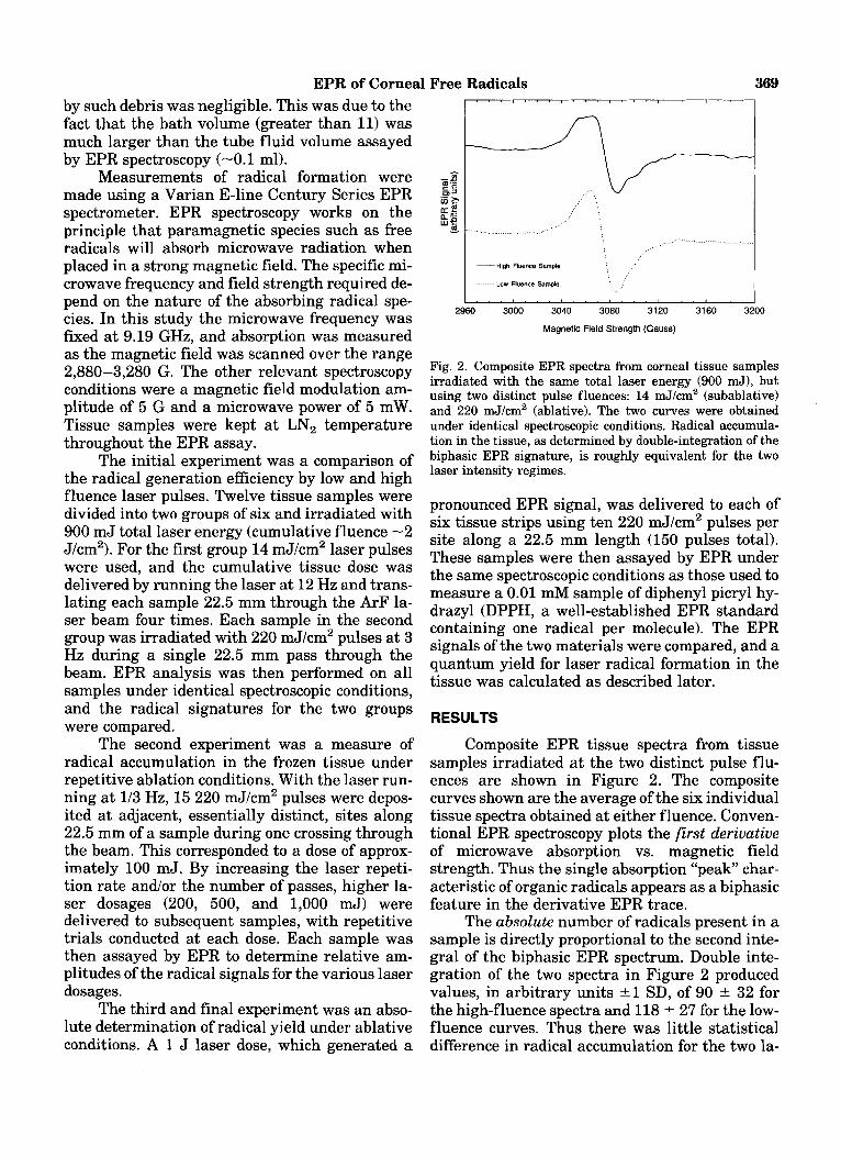
Fig. 2. Composite EPR spectra from corneal tissue samples irradiated with the same total laser energy (900 mJ), but using two distinct pulse fluences: 14 mJ/crn2 (subablative) and 220 mJ/cm2 (ablative). The two curves were obtained under identical spectroscopic conditions. Radical accumula- tion in the tissue, as determined by double-integration of the biphasic EPR signature, is roughly equivalent for the two laser intensity regimes.
pronounced EPR signal, was delivered to each of six tissue strips using ten 220 mJ/cm2 pulses per site along a 22.5 mm length (150 pulses total). These samples were then assayed by EPR under the same spectroscopic conditions as those used to measure a 0.01 mM sample of diphenyl picryl hy- drazyl (DPPH, a well-established EPR standard containing one radical per molecule). The EPR signals of the two materials were compared, and a quantum yield for laser radical formation in the tissue was calculated as described later.
RESULTS
Composite EPR tissue spectra from tissue samples irradiated at the two distinct pulse flu- ences are shown in Figure 2. The composite curves shown are the average of the six individual tissue spectra obtained at either fluence. Conven- tional EPR spectroscopy plots the first derivative of microwave absorption vs. magnetic field strength. Thus the single absorption "peak" char- acteristic of organic radicals appears as a biphasic feature in the derivative EPR trace.
The absolute number of radicals present in a sample is directly proportional to the second inte- gral of the biphasic EPR spectrum. Double inte- gration of the two spectra in Figure 2 produced values, in arbitrary units 21 SD, of 90 k 32 for the high-fluence spectra and 118 * 27 for the low- fluence curves. Thus there was little statistical difference in radical accumulation for the two la-

370 Pettit et al. ArF Laser Pulses per Tissue Site
0 2 4 6 8 10
I /
Y /
/
I 0 a / I
I I
I I
0 0 200 400 600 800 1 000
Total ArF Laser Exposure (d)
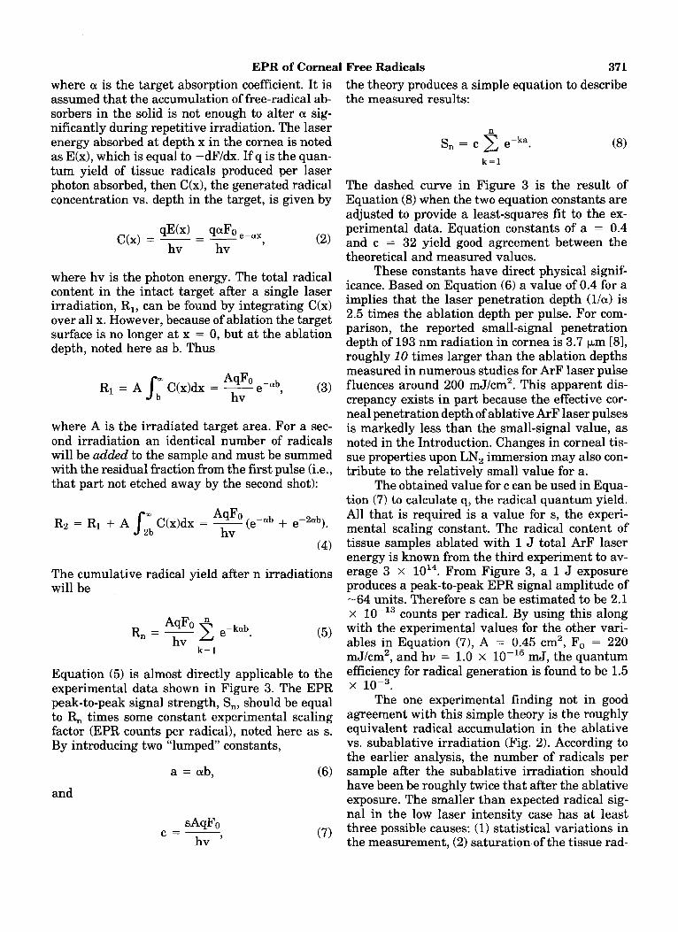
Fig. 3. EPR signal amplitudes from irradiated corneal tissue vs. ArF laser exposure. The laser fluence in each case was 220 mJ/cm2 per pulse, which was clearly ablative. Each pass of the tissue through the beam deposited approxiamtely 100 mJ of laser energy onto a 2.25 x 0.2 cm area of the target. Rad- ical accumulation clearly occurs over multiple exposures. The dashed curve is a simple model calculation described in the Discussion.
ser intensity regimes. The slightly different shapes of the two EPR profiles, for example, the distinct “shoulders” in the high intensity curve, suggest that the resultant radicals were chemi- cally distinct for the two irradiation conditions.
The results of the repetitive irradiation study are shown in Figure 3. In determining the relative abundance of the same radical type in dif- ferent samples, the peak-to-peak amplitude of the biphasic EPR signal can be used directly because the shape of the absorption feature is virtually the same for each specimen. In this particular ex- periment, the peak-to-peak signal amplitude was readily obtainable, even from the minimally irra- diated samples, while double integration of the noisy weak spectra was not straightforward. The discrete points in Figure 3 indicate the peak-to- peak EPR signal amplitudes for samples irradi- ated with 1, 2, 5, or 10 ablative ArF laser pulses per site along the de-epithelialized tissue surface. Over these initial irradiations there was an accu- mulation of radical species in the frozen cornea, which neared a plateau by the 10th laser shot per site.
In the final experiment, the EPR spectrum from the 0.01 mM DPPH standard was much larger and narrower than those of the irradiated tissue samples. After the double integration was
performed on all spectra, with differences in spec- trometer gain taken into account, the radical con- tent of the DPPH standard was calculated to be 2 x lo4 larger than the average value for the cor- neal strips. The standard deviation in the tissue signal measurements was approximately 20%. Given that the DPPH contained -6 x 10” radi- cal species, the implied average radical content for the corneal samples was 3 * 0.6 x
DISCUSSION
Free radicals can be formed in a variety of ways, including simple combustion reactions [9], as well as direct photochemical cleavage of ab- sorbing molecules (e.g., ultraviolet “bond-break- ing” of hydrogen peroxide into OHX [lo]). The findings shown in Figure 2 indicate that ablative photodecomposition of cornea need not occur to form substantial radicals in the tissue target, since abundant radicals were produced at subab- lative laser intensity. Therefore photochemistry driven directly by the 193 nm laser radiation must be a major source of the tissue radicals. However, additional ablation-specific mecha- nisms for radical formation cannot be completely excluded by the two comparable EPR signal strengths of Figure 2. Radical accumulation dur- ing tissue ablation in LN, is obviously not a lin- ear function of deposited laser pulses, as indicated in Figure 3, and the process may not be linear with repetitive irradiation at subablative flu- ences as well.
Since these species are highly reactive at physiologic temperatures, analogous radical accu- mulation over successive laser pulses does not oc- cur in vivo. Nevertheless, the phenomenon shown in Figure 3 merits further consideration because it factors into the radical quantum yield calcula- tion, which does have clinical relevance. In addi- tion, analysis of the radical buildup in this exper- iment should be applicable to other irradiation products that are longer lived in the warm tissue.
The most likely reason for the nonlinear rad- ical accumulation is that some radical species are lost from the corneal target with each ArF laser etching. This idea of competition between radical formation and ejection is the basis of the following model. In the corneal target, an incident 193 nm laser pulse of fluence Fo is attenuated exponen- tially with depth x in the tissue as
EPR of Corneal Free Radicals 371 where CY is the target absorption coefficient. It is assumed that the accumulation of free-radical ab- sorbers in the solid is not enough to alter CY sig- nificantly during repetitive irradiation. The laser energy absorbed at depth x in the cornea is noted as E(x), which is equal to -dF/dx. If q is the quan- tum yield of tissue radicals produced per laser photon absorbed, then C(x), the generated radical concentration vs. depth in the target, is given by
where hv is the photon energy. The total radical content in the intact target after a single laser irradiation, R,, can be found by integrating C(x) over all x. However, because of ablation the target surface is no longer at x = 0, but at the ablation depth, noted here as b. Thus
where A is the irradiated target area. For a sec- ond irradiation an identical number of radicals will be added to the sample and must be summed with the residual fraction from the first pulse (i.e., that part not etched away by the second shot):
R2 = R1 + A J2L C(x)dx = AqFo(e-ab - + e-2ab). hv
(4)
The cumulative radical yield after n irradiations will be
AqFo 2 e-kab R, = - hv
k = I
Equation ( 5 ) is almost directly applicable to the experimental data shown in Figure 3. The EPR peak-to-peak signal strength, S,, should be equal to R, times some constant experimental scaling factor (EPR counts per radical), noted here as s. By introducing two “lumped” constants,
a = ab, (6) and
the theory produces a simple equation to describe the measured results:
n S, = c C e-ka.
k = l
The dashed curve in Figure 3 is the result of Equation (8) when the two equation constants are adjusted to provide a least-squares fit to the ex- perimental data. Equation constants of a = 0.4 and c = 32 yield good agreement between the theoretical and measured values.
These constants have direct physical signif- icance. Based on Equation (6) a value of 0.4 for a implies that the laser penetration depth (l/a) is 2.5 times the ablation depth per pulse. For com- parison, the reported small-signal penetration depth of 193 nm radiation in cornea is 3.7 pm [BI, roughly 10 times larger than the ablation depths measured in numerous studies for ArF laser pulse fluences around 200 mJ/cm2. This apparent dis- crepancy exists in part because the effective cor- neal penetration depth of ablative ArF laser pulses is markedly less than the small-signal value, as noted in the Introduction. Changes in corneal tis- sue properties upon LN, immersion may also con- tribute to the relatively small value for a.
The obtained value for c can be used in Equa- tion (7) to calculate q, the radical quantum yield. All that is required is a value for s, the experi- mental scaling constant. The radical content of tissue samples ablated with 1 J total ArF laser energy is known from the third experiment to av- erage 3 x From Figure 3, a 1 J exposure produces a peak-to-peak EPR signal amplitude of -64 units. Therefore s can be estimated to be 2.1 x counts per radical. By using this along with the experimental values for the other vari- ables in Equation (7), A = 0.45 cm2, Fo = 220 mJ/cm2, and hv = 1.0 x mJ, the quantum efficiency for radical generation is found to be 1.5
The one experimental finding not in good agreement with this simple theory is the roughly equivalent radical accumulation in the ablative vs. subablative irradiation (Fig. 2). According to the earlier analysis, the number of radicals per sample after the subablative irradiation should have been be roughly twice that after the ablative exposure. The smaller than expected radical sig- nal in the low laser intensity case has at least three possible causes: (1) statistical variations in the measurement, (2) saturation of the tissue rad-
x 10-3.
372 Pettit et al. ical accumulation with repetitive low-intensity irradiation, and (3) a small (factor of -2) inten- sity dependence in the quantum efficiency for rad- ical generation.
Assuming the radical quantum yield calcu- lated in this study is close to the value for in vivo corneal ablation, the number of transient free radicals generated in the living tissue is biologi- cally significant. Using the measured 2,700 cm-I small-signal absorption coefficient and 30 mJ/cm2 ablation threshold fluence, the density of ab- sorbed 193 nm photons near the surface of the unablated corneal tissue is calculated to be -8 x lo1’ ~ m - ~ . A 0.15% efficiency for radical genera- tion implies that 1.2 x 10¹⁷ of these reac- tive species are introduced into the tissue surface region with each laser pulse. Given the pro- nounced biologic impact that free radicals can have, a localized 0.2 mM concentration is quite significant. This finding helps explain the mea- sured reduction in postoperative “haze” by radical scavenger treatment noted earlier [71 and sug- gests that such pharmacologic therapy may prove useful in modulating the overall corneal healing response.
REFERENCES
1. Trokel SL, Srinivasan R, Braren B. Excimer laser sur- gery of the cornea. Am J Ophthalmol 1983; 96:710-715.
2. Landry RJ, Pettit GH, Hahn DW, Ediger MN, Yang GC. Preliminary evidence of free radical formation during ar- gon fluoride excimer laser irradiation of corneal tissue. Lasers Light Ophthalmol 1994; 6:87-90.
3. Ediger MN, Pettit GH, Weiblinger RP, Chen CH. Trans- mission of corneal collagen during ArF excimer laser ab- lation. Lasers Surg Med 1993; 13:204-210.
4. Del Per0 FZA, Gigstad JE, Roberts AD, Klintworth GK, Martin CA, L’Esperance FA, Taylor DM. A refractive and histopathologic study of excimer laser keratectomy in primates. Am J Ophthalmol 1990; 109:419-429.
5. Hanna KD, Pouliquen YM, Savoldelli M, Fantes F, Thompson KP, Waring GE, Sams J. Corneal wound heal- ing in monkeys 18 months after excimer laser photore- fractive keratectomy. Refract Corneal Surg 1990; 6340- 345.
6. Lohmann CP, Timberlake GT, Fitzke FW, Gartry DS, Kerr Muir M, Marshall J. Corneal light scattering after excimer laser photorefractive keratectomy; the objective measurements of haze. Refract Corneal Surg 1992; 8: 114-121.
7. Jain S, Hahn TW, Chen W, Stark W, Chen L, Azar DT. Modulation of corneal wound healing following 193-nm excimer laser keratectomy using free radical scavengers. Invest Ophthalmol Vis Sci 1994; 35:2015.
8. Puliafito CA, Steinert RF, Deutsch TF, Hillenkamp F, Dehm EJ, Adler CM. Excimer laser ablation of the cornea and lens. Ophthalmology 1985; 92741-748.
9. Glassman I. “Combustion.” Orlando: Academic Press, 1987:85 - 86.
10. Stief U, DeCarlo VJ. Vacuum-ultraviolet photochemis- try. IX. Primary and chain processes in the photolysis of hydrogen peroxide. J Chem Phys 1969; 50:1234-1240.