electrochemical biosensor based on silver nanoparticles–polydopamine–graphene nanocomposite for...
TRANSCRIPT

Talanta 114 (2013) 43–48
Contents lists available at SciVerse ScienceDirect
Talanta
0039-91http://d
n CorrE-m
liuym95
journal homepage: www.elsevier.com/locate/talanta
Electrochemical biosensor based on silver nanoparticles–polydopamine–graphene nanocomposite for sensitive determination of adenine and guanine
Ke-Jing Huang n, Lan Wang, Hai-Bo Wang, Tian Gan, Ying-Ying Wu, Jing Li, Yan-Ming Liu n
College of Chemistry and Chemical Engineering, Xinyang Normal University, Xinyang 464000, China
a r t i c l e i n f o
Article history:Received 17 January 2013Received in revised form29 March 2013Accepted 6 April 2013Available online 12 April 2013
Keywords:Ag nanoparticles–polydopamine@graphenenanocompositeAdenineGuanineElectrochemical sensorVoltammetryBiosensing
40/$ - see front matter & 2013 Elsevier B.V. Ax.doi.org/10.1016/j.talanta.2013.04.017
esponding authors. Tel.: +86 376 6390611.ail addresses: [email protected] (K.-J. [email protected] (Y.-M. Liu).
a b s t r a c t
A multifunctional Ag nanoparticles (AgNPs)–polydopamine (Pdop)@graphene (Gr) composite wasprepared by a simple and mild procedure. Gr was easily coated with Pdop at room temperature andthen AgNPs was deposited by mildly stirring. The nanocomposite was characterized by scanning electronmicroscope (SEM) and transmission electron microscope (TEM). Guanine and adenine as modelmoleculars were employed to study their electrochemical responses at the Ag–Pdop@Gr compositemodified electrode, which showed more favorable electron transfer kinetics than Gr modified glassycarbon and AgNPs modified glassy carbon electrodes. The Ag–Pdop@Gr modified electrode exhibitedlinear ranges of 0.04–50 μM and 0.02–40 μM with detection limits of 4.0 nM and 2.0 nM for guanine andadenine, respectively. The developed method was applied for simultaneous determination of trace-leveladenine and guanine in fish sperm. The results demonstrated that the AgNPs–Pdop@Gr nanocompositewas a promising substrate for the development of high-performance electrocatalysts for biosensing.
& 2013 Elsevier B.V. All rights reserved.
1. Introduction
Guanine and adenine are important components found indeoxyribonucleic acid and play fundamental roles in life process.They have widespread effects on coronary and cerebral circulation,prevention of cardiac arrhythmias and inhibition of neurotransmit-ter release [1]. Therefore, the determination of guanine and adeninehas great significance to the bioscience and clinical diagnosis.
Electrochemical method has been widely used in the determi-nation of guanine and adenine [2]. However, the low concentra-tions in biosamples and the complexity of their matrices makedetermination of guanine and adenine challenging tasks. It is veryimportant to explore new method for signal amplification in orderto increase the sensitivity of the detection. Several methods forsignal enhancement have been investigated, such as enzymelabeling [3], rolling circle amplification [4] and nanomaterialintroduction [5,6]. Among these methods, application of nanoma-terial has gained growing interest due to the intrinsic advantagesof nanomaterials, such as low cost, good thermal stability andlarge surface area.
Graphene (Gr) has stimulated intense research interest becauseof its unique physical and chemical properties, such as high
ll rights reserved.
ang),
surface area, high electrical conductivity, good chemical stability,and strong mechanical strength [7]. These properties make it anattractive candidate for fabricating various functional devices, suchas electrodes, sensors, photovoltaics and photodetectors [8,9].Silver nanoparticles (AgNPs) have good conductivity and highelectrochemical catalytic activity. The availability of AgNPs willexpand the possibilities for the preparation of Ag-doped nanoma-terials and extend its application in biosensor [10]. It has beenreported that the integration of carbon-based materials and metalnanoparticles usually shows synergistic effects in electrocatalyticapplications [11], so there is a reason to expect the integration ofGr and AgNPs will obtain the similar effect on the electrooxidationof guanine and adenine.
Recently, a thin, surface adherent and multifunctional biopoly-mer–polydopamine (Pdop) layer was prepared on a wide range ofinorganic and organic materials by self-polymerization of dopa-mine in an aqueous solution [12]. A variety of ad-layers, includingself-assembled monolayer through deposition of long-chain mole-cular building blocks, metal films by electroless metallization andbioactive surfaces via grafting of macromolecules have beenprepared by Pdop coating [13]. The polymerization of dopamineoffers the advantage of a one-step surface functionalization andallows the introduction of a new paradigm in the field of surfacemodification. In this work, a new multifunctional Ag nanoparticles(AgNPs)–polydopamine (Pdop)@graphene (Gr) composite wasfirstly prepared through the oxidation of dopamine on Gr at roomtemperature and subsequent electroless silver deposition by

K.-J. Huang et al. / Talanta 114 (2013) 43–4844
mildly stirring. The AgNPs–Pdop@Gr modified glassy carbon elec-trodes (GCE) showed enhanced catalytic efficiencies towardsguanine and adenine oxidation in acetate buffer solution.
2. Experimental
2.1. Chemicals and materials
Graphite powder, hydrazine solution (50 wt%) and ammoniasolution (28 wt%) were purchased from Shanghai ChemicalReagent Corporation (Shanghai, China). Guanine, adenine, AgNO3,dopamine hydrochloride (DA) and 2-amino-2-hydroxymethylpro-pane-1,3-diol (Tris) were obtained from Sigma (Saint Louis, MO,USA). Acetate buffer solutions (ABS) were prepared by mixing of0.1 M CH3COOH and CH3COONa and adjusting the pH with NaOH.All chemicals were of analytical grade and doubly distilled waterwas used throughout.
2.2. Apparatus
Electrochemical measurements were performed on a CHI 660DElectrochemical Workstation (Shanghai CH Instruments, China).A conventional three-electrode system was used throughout theexperiments. The working electrode was a bare, a pretreated orAgNPs–Pdop–Gr composite modified GCE (3.0 mm in diameter);the auxiliary electrode was a platinum wire and a saturatedcalomel electrode (SCE) was used as the reference. Electrochemicalimpedance spectroscopy (EIS) was performed in 5.0 mM K3Fe(CN)6/K4Fe(CN)6 (1/1) mixture with 0.1 M KCl as supportingelectrolyte, using an alternating current voltage of 5 mV, withinthe frequency range of 0.1–105 Hz by Autolab ElectrochemistryInstruments (Autolab, Eco Chemie, The Netherlands). Themorphologies of the nanocomposite were recorded on a JEM2100 transmission electron microscope (TEM) and a HitachiS-4800 scanning electron microscope (SEM).
2.3. Preparation of graphene and its functionalized products
Graphene oxide (GO) was prepared from graphite powder bythe modified Hummers method [14]. In a typical process, 5 ggraphite was slowly added into a mixture of concentrated H2SO4
(87.5 mL) and fuming HNO3 (45 mL) (warning: concentratedH2SO4 and fuming HNO3 are strongly oxidizing and should behandled with care!). KClO3 (55 g) was then added in abovemixture, and was kept stirring for 96 h. Then the slurry waspoured into water and filtered to obtain graphite oxide. Afterdried at 80 1C, graphite oxide (0.5 g) was exfoliated in 500 mLwater with ultrasonic treatment to form a colloidal graphene oxidesuspension (1 mg mL−1). To get Gr, chemical reduction of thesuspension of GO was carried out with hydrazine monohydratefor 24 h at 80 1C. The final product was isolated by filtration, andrinsed thoroughly with pure water and ethanol. Then the productwas dried in vacuum and Gr was obtained.
AgNPs–Pdop–Gr nanocomposite was prepared as following:100 mg Gr was dispersed in 100 mL water by sonication, then200 mg DA and 120 mg Tris were added into above mixture anddispersed by 1 min sonication in ice water bath. The mixture wasmagnetically stirred at room temperature for 20 h. The productwere filtered, washed and then dried in vacuum overnight at 60 1Cto obtain Pdop@Gr. To mildly deposit AgNPs onto the surface ofPdop@Gr, 25.0 mL AgNO3 aqueous solution (1.0 mM) was added in25.0 mg Pdop@Gr. The mixture was mildly stirred for 2 h at roomtemperature, and then the product was filtered, washed and driedin vacuum overnight at 60 1C to obtain AgNPs–Pdop@Grnanocomposite.
2.4. Preparation of Ag–Pdop@gr modified GCE
For electrode preparation, 1 mg AgNPs–Pdop@Gr was dispersedin 10 mL DMF using an ultrasonic bath to give a black suspension.The GCE (3 mm in diameter) was polished carefully with 0.3 and0.05 μm alumina slurry, and sonicated in water and ethanol,respectively. Then, 8 μL of the suspension was placed on the GCEsurface by micropipette and left to dry at room temperature(30 min) to obtain AgNPs–Pdop@Gr/GCE. The Gr/GCE withoutAgNPs and AgNPs/GCE without Gr were also prepared accordingto the similar procedure, using a suspension of Gr and AgNPs inDMF. Before voltammetric measurements, the modified electrodewas cycled five times between 0.5 and 1.4 V (scan rate 100 mV s−1)in a 0.1 M ABS of pH 4.0. The renewal of the electrode surface waseasily accomplished by soaking the modified electrode in ABS andcycling the potential as mentioned above.
2.5. Preparation of DNA samples
Thermally denatured dsDNA was prepared according to theprevious report [15]. In short, 3 mg of the fish sperm DNA wasdigested using 1 mL of 1 M HCl in a sealed 10 mL glass tube. Afterheating in boiling waterbath (100 1C) for 80 min, 1 mL of 1 MNaOH was added. After cooling to room temperature, the solutionwas diluted to 10.0 mL using 0.1 M PBS (pH 7).
3. Results and discussion
3.1. Characterization
The morphology of the AgNPs–Pdop@Gr nanocomposite wasexamined by SEM and TEM. Fig. S1a showed the SEM image of theobtained Gr sheets, illustrating the flake-like shapes of graphene.Fig. S1b showed the SEM image of AgNPs–Pdop@Gr nanocomposite.It was clear that AgNPs distributed well on Pdop@Gr sheets,evidencing the well-behaved assembly process. The Pdop acted asa glue reagent connecting the Gr with AgNPs. Such morphologicalcharacteristics might result in high loading of guanine and adenineand fast response to the substrate. Fig. S1c showed the typical TEMimage of the Gr nanosheets. Fig. S1d showed TEM image of theobtained Pdop@Gr. The transparent Gr turns into blackishnanosheets, illustrating Pdop was successfully coated on Gr. TheTEM image (Fig. S1e) showed the AgNPs were deposited on the Grsurface. However, the size of these nanoparticles was not the same,and the dispersion was heterogeneous. The reason might beattributed to the stirring inhomogeneity of graphene nanosheetssince the formation of silver nanoparticles was based on Pdop@Grnanosheets as substrates. In fact, this was just an advantage over thecomposite surface for increasing the immobilized amount of thetarget molecules. These AgNPs were firmly attached to Gr sheets,even after the ultrasonication used to disperse the AgNPs–Pdop@Grcomposite for TEM characterization. EDX measurement wasperformed to validate the presence of AgNPs on graphene (Fig. S1f).
3.2. Electrochemical reactivity
The capability of electron transfer of different electrodes wasinvestigated by AC impedance experiments and the results wereshown in Fig. S2. The sequence of the values of charge-transferresistance for different electrodes was bare GC electrode (a,742.6 Ω)4AgNPs/GCE (b, 627.3 Ω)4Gr/GCE (c, 498.2 Ω)4AgNPs–Pdop@Gr/GCE (d, 245.6 Ω). This result demonstrated the AgNPs–Pdop@Gr/GCE had higher electrochemical activity than otherelectrodes.
K.-J. Huang et al. / Talanta 114 (2013) 43–48 45
The potential window of the electrodes may have a deepimpact upon their analytical applications. In Fig. S3, the potentialwindow for the AgNPs–Pdop@Gr/GCE (d) in 0.1 M pH 4.0 ABS was∼2.75 V, which was comparable to that for bare GCE (a) and Gr/GCE (c) and better than that for AgNPs/GCE (b).
Fig. S4 depicted the chronocoulometric curves at differentelectrodes for the reduction of 1 mM K3Fe(CN)6 with 2 M KCl.According to the following equation:
Q ¼ ð2nFADo1=2π−1=2CoÞt1=2 ð1Þ
where Q is the absolute value of the reduction charge, n is thenumber of electrons for the reaction, F is the Faraday constant, A isthe apparent electrode area, Do is the diffusion coefficient of theoxidized form, hexacyanoferrate(III), Co is the bulk concentrationof the oxidized form, and t is the time. From the slope of the Q−t1/2
line, the sequence of the values of A for different electrodes wasAgNPs–Pdop@Gr/GCE (d)4Gr/GCE (c)4AgNPs/GCE (b)4GCE (a).This meaned the largest value of A was obtained on the AgNPs–Pdop@Gr/GCE, which may have the best electrochemical reactionability among them.
The electrochemical response of the different modified electro-des was studied in the presence of Fe(CN)63−/4− (Fig. S5).Fe(CN)63−/4− is close to an ideal quasi-reversible system on carbonelectrodes. For AgNPs–Pdop@Gr/GCE, the peak current showed thehighest, which indicated that this well-defined AgNPs–Pdop@Grfilm possessed the requisite surface structure and electronicproperties to support rapid electron transfer for this particularmechanistically complicated redox system.
3.3. Electrocatalytic oxidation of guanine and adenine
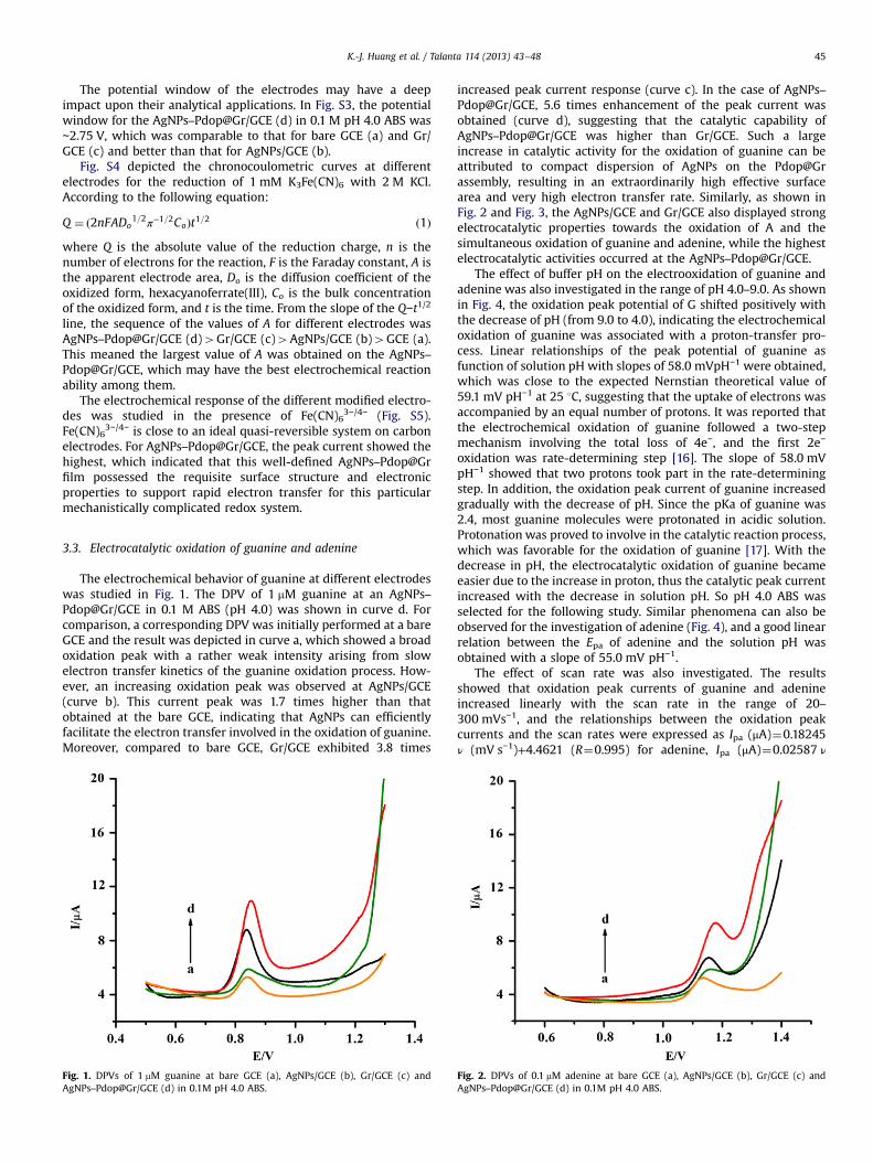
The electrochemical behavior of guanine at different electrodeswas studied in Fig. 1. The DPV of 1 mM guanine at an AgNPs–Pdop@Gr/GCE in 0.1 M ABS (pH 4.0) was shown in curve d. Forcomparison, a corresponding DPV was initially performed at a bareGCE and the result was depicted in curve a, which showed a broadoxidation peak with a rather weak intensity arising from slowelectron transfer kinetics of the guanine oxidation process. How-ever, an increasing oxidation peak was observed at AgNPs/GCE(curve b). This current peak was 1.7 times higher than thatobtained at the bare GCE, indicating that AgNPs can efficientlyfacilitate the electron transfer involved in the oxidation of guanine.Moreover, compared to bare GCE, Gr/GCE exhibited 3.8 times
Fig. 1. DPVs of 1 mM guanine at bare GCE (a), AgNPs/GCE (b), Gr/GCE (c) andAgNPs–Pdop@Gr/GCE (d) in 0.1M pH 4.0 ABS.
increased peak current response (curve c). In the case of AgNPs–Pdop@Gr/GCE, 5.6 times enhancement of the peak current wasobtained (curve d), suggesting that the catalytic capability ofAgNPs–Pdop@Gr/GCE was higher than Gr/GCE. Such a largeincrease in catalytic activity for the oxidation of guanine can beattributed to compact dispersion of AgNPs on the Pdop@Grassembly, resulting in an extraordinarily high effective surfacearea and very high electron transfer rate. Similarly, as shown inFig. 2 and Fig. 3, the AgNPs/GCE and Gr/GCE also displayed strongelectrocatalytic properties towards the oxidation of A and thesimultaneous oxidation of guanine and adenine, while the highestelectrocatalytic activities occurred at the AgNPs–Pdop@Gr/GCE.
The effect of buffer pH on the electrooxidation of guanine andadenine was also investigated in the range of pH 4.0–9.0. As shownin Fig. 4, the oxidation peak potential of G shifted positively withthe decrease of pH (from 9.0 to 4.0), indicating the electrochemicaloxidation of guanine was associated with a proton-transfer pro-cess. Linear relationships of the peak potential of guanine asfunction of solution pH with slopes of 58.0 mVpH−1 were obtained,which was close to the expected Nernstian theoretical value of59.1 mV pH−1 at 25 1C, suggesting that the uptake of electrons wasaccompanied by an equal number of protons. It was reported thatthe electrochemical oxidation of guanine followed a two-stepmechanism involving the total loss of 4e−, and the first 2e−
oxidation was rate-determining step [16]. The slope of 58.0 mVpH−1 showed that two protons took part in the rate-determiningstep. In addition, the oxidation peak current of guanine increasedgradually with the decrease of pH. Since the pKa of guanine was2.4, most guanine molecules were protonated in acidic solution.Protonation was proved to involve in the catalytic reaction process,which was favorable for the oxidation of guanine [17]. With thedecrease in pH, the electrocatalytic oxidation of guanine becameeasier due to the increase in proton, thus the catalytic peak currentincreased with the decrease in solution pH. So pH 4.0 ABS wasselected for the following study. Similar phenomena can also beobserved for the investigation of adenine (Fig. 4), and a good linearrelation between the Epa of adenine and the solution pH wasobtained with a slope of 55.0 mV pH−1.
The effect of scan rate was also investigated. The resultsshowed that oxidation peak currents of guanine and adenineincreased linearly with the scan rate in the range of 20–300 mVs−1, and the relationships between the oxidation peakcurrents and the scan rates were expressed as Ipa (μA)¼0.18245ν (mV s−1)+4.4621 (R¼0.995) for adenine, Ipa (μA)¼0.02587 ν
Fig. 2. DPVs of 0.1 mM adenine at bare GCE (a), AgNPs/GCE (b), Gr/GCE (c) andAgNPs–Pdop@Gr/GCE (d) in 0.1M pH 4.0 ABS.

Fig. 3. DPVs of 1 mM guanine and 0.1 mM adenine at bare GCE (a), AgNPs/GCE (b),Gr/GCE (c) and AgNPs–Pdop@Gr/GCE (d) in 0.1M pH 4.0 ABS.
Fig. 4. DPVs of Ag–Pdop–GR/GCE in 0.1 M ABS containing 1.2 μM guanine and0.2 μM adenine at different pH values (from bottom to top: pH 9.0, pH 8.0, pH 7.0,pH 6.0, pH 5.0, pH 4.0).
Fig. 5. DPVs of guanine at AgNPs–Pdop@Gr/GCE in the presence of 0.5 mM adeninein 0.1 M pH 4.0 ABS. Guanine concentrations (from bottom to top): 0.04, 0.06, 0.08,0.1, 0.3, 0.5, 1, 2, 5, 10, 30, 50 mM. Insets are linear relationships between peakcurrents and concentrations.
Fig. 6. DPVs of adenine at AgNPs–Pdop@Gr/GCE in the presence of 10 mM guanine,in 0.1 M pH 4.0 ABS. A concentrations (from bottom to top): 0.02, 0.04, 0.06, 0.08,0.1, 0.3, 0.5, 1, 3, 8, 15, 40 mM. Insets are linear relationships between peak currentsand concentrations.
K.-J. Huang et al. / Talanta 114 (2013) 43–4846
(mV s−1)+1.5098 (R¼0.992) for guanine. This indicates that theelectro-oxidation reactions of guanine and adenine at the AgNPs–Pdop@Gr/GCE are surface-controlled process.
3.4. Determination of guanine and adenine
DPV was used for the determination of guanine and adenine atAgNPs–Pdop@Gr/GCE due to its high sensitivity. The individualdetermination of guanine or adenine in their mixtures was firstinvestigated when the concentration of one species changed,whereas the other species remained constant. Fig. 5 showed theDPV curves of different concentration guanine in pH 4.0 ABScontaining 0.5 mM adenine. As can be seen in the inset of Fig. 5,the Ipa was proportional to the logarithm of concentration ofguanine in the range of 0.04–50 mM. The regression equation wasIpa (mA)¼4.1149 logC+15.623 (R¼0.9899), and the detection limitwas 4.0 nM (S/N¼3). Similarly, as shown in Fig. 6, keeping theconcentration of guanine constant, the oxidation peak currentincreased linearly with increasing the logarithm of concentrationof adenine in the range of 0.02–40 mM. A linear equation ofIpa (mA)¼5.2929 logC+21.308 (R¼0.9928) was obtained. Thedetection limit for adenine was 2 nM (S/N¼3). Therefore, the
proposed method can be used to simultaneously determineguanine and adenine.
In order to evaluate the feasibility of the AgNPs–Pdop@Gr/GCEfor guanine and adenine determination, the fabricated electrodewas applied to detect simultaneously guanine and adenine.As shown in Fig. 7, two well-defined oxidation peaks wereobserved at about 0.87 V and 1.19 V, corresponding to the oxida-tion of guanine and adenine, respectively. The oxidation peakcurrents of guanine and adenine increased linearly with thelogarithm of their concentration in the range of 0.02–40 mM (Ipa(mA)¼4.0461 logC+12.954, R¼0.9921) for guanine and 0.02–40 mM (Ipa (mA)¼4.5616 logC+14.498, R¼0.9918) for adenine,respectively. The detection limits for guanine and adenine were4.0 nM and 2 nM (S/N¼3), respectively. Thus, the simultaneouslysensitive determination of guanine and adenine was realized byusing AgNPs–Pdop@Gr/GCE.
Table 1 compares the response characteristics of AgNPs–Pdop@Gr/GCE with other modified electrodes for the simulta-neous determination of adenine and guanine [18–23]. The pro-posed method in this work had wide linear range and low

Fig. 7. DPVs of guanine and adenine at Ag–Pdop–GR/GCE in 0.1 M pH 4.0 ABS.Guanine and adenine concentrations (from bottom to top): 0.02, 0.04, 0.1, 0.5, 1, 5,10, 20, 40 mM. Insets are linear relationships between peak currents andconcentrations.
Table 1Comparison of different electrochemical sensors for the determination of guanineand adenine.
Electrodes Linear range (mM) Detection limit (nM) Reference
Guanine Adenine Guanine Adenine
CILEa 0.3–50 1.5–70 78.7 250 [18]PTH/NPAu/MWNTsb 0.05–5 0.05–5 10 8 [19]MW-CNCEc 0.1–20 0.1–10 80 80 [20]MWCNT/GCEd 0.05–10 0.05–10 20 80 [21]TiO2–Gr/GCEe 0.5–200 0.5–200 100 150 [2]Fe3O4NPs/MWCNT/GCEf 0.01–10 0.05–6 1.0 5.0 [22]AgNPs–Pdop@Gr/GCE 0.02–40 0.02–40 4.0 2.0 This work
a CILE: carbon ionic liquid on the carbon paste electrode.b PTH/NPAu/MWNTs: poly thionine electrode deposited on GCE modified with
gold nanoparticles/CNT.c MW-CNCE: sol–gel-derived CNT ceramic electrode prepared by microwave
irradiation.d MWCNT/GCE: GCE modified with carboxylated MWCNTs.e TiO2–Gr/GCE:TiO2 nanoparticles–graphene nanocomposite modified GCE.f Fe3O4NPs/MWCNT/GCE: Fe3O4 nanoparticles–MWCNT modified GCE.
K.-J. Huang et al. / Talanta 114 (2013) 43–48 47
detection limit. This further confirmed the synergetic effect ofAgNPs and Gr.
3.5. Reproducibility, stability and interference
The reproducibility of the sensor was estimated by determining1.0 mM guanine and 1.0 mM adenine with five modified electrodeswhich were made at the same electrode. Five measurements fromthe batch resulted in RSD of 4.1%. The experimental results showedgood reproducibility of the fabrication protocol.
The stability of the developed sensor was evaluated by twentysuccessive scans in the mixture of 1.0 mM guanine and 1.0 mMadenine. The RSDs were found to be 2.4% and 2.5% for guanine andadenine, respectively, indicating excellent stability of the modifiedelectrode. The longtime stability of the modified electrode wasalso studied on a 15-day period. The results showed that theanalytical performance of the biosensor tested has no obviousdecline (RSD≤5%).
The selectivity of the developed sensor for sensitive determi-nation of guanine and adenine was analyzed and several com-pounds such as important biological substances and some metal
ions were checked as potential interfering substances. The effectsof these interferents were examined by carrying out the determi-nation of 5 mM guanine and 3 mM adenine in 0.1 M ABS (pH 4.0) inthe presence of different concentrations of the interferents. Thetolerance limit was defined as the maximum concentration of theforeign substances that caused an approximately 5% relative errorin the detection. The results suggested that 100-fold concentrationof K+, Na+, Ca2+, Mg2+, Fe3+, Al3+, Zn2+, Cl−, NO3
−, SO42−, CO3
2−, F-,Br−, glucose and cysteine had no interference for the determina-tion of guanine and adenine. Ascorbic acid, uric acid and dopamineshowed oxidation process in the selected potential range, but theoxidation potentials were lower than those of guanine andadenine. Furthermore, the peak current change of guanine andadenine was below 3%.
3.6. Analytical application
Because the bases are embedded in the interior of the doublehelix and therefore their detection is sterically hindered due to thecrowded phosphate group on the exterior of the helix. Thethermally denatured treatment results in an unwinding of duplexDNA and renders the guanine and adenine residues more acces-sible to the electrode surface owing to the increased flexibility inits structure. In this work, the applicability of the modifiedelectrode in biological samples was assessed by measuring ade-nine and guanine in thermally denatured DNA. The thermallydenatured DNA gave two well-defined oxidation peaks at themodified electrode, which was due to the oxidation of guanine andadenine residues, respectively. The standard addition methodswere used to determine guanine and adenine. In short, 50 mL ofthermally denatured DNA solution was added in a 10-mL buffersolution and the peak currents of the guanine and adenine weremeasured. Subsequently, 10 mM guanine and adenine were addedin above mixture and the peak currents of the guanine andadenine were recorded again. From the differences between thepeak currents of guanine and adenine, the concentration ofguanine and adenine in DNA could be obtained by the calibrationgraph. The contents of guanine and adenine in thermally dena-tured DNA were calculated as 21.9% and 28.1% (in the molar ratio,mol%), respectively. The value of (G+C)/(A+T) was calculated as0.78 for thermally denatured DNA sample, which coincided to thestandard value of 0.77 [23].
4. Conclusions
In this work, a novel electrochemical sensor was fabricatedbased on AgNPs–Pdop@Gr modified GCE for the sensitive deter-mination of guanine and adenine. AgNPs–Pdop@Gr nanocompo-site was prepared with a simple and facile method by oxidation ofdopamine at room temperature at Gr and subsequent electrolessAg deposition by mildly stirring. AgNPs–Pdop@Gr nanocompositecan greatly improve electron transfer between the analytes andunderlying electrode. The developed biosensor based on AgNPs–Pdop@Gr nanocomposite exhibited wide linear detection range,acceptable reproducibility, good stability, and low detection limit.This novel as-prepared nanocomposite could provide a promisingplatform for the development of biosensor and electrochemicalsensor.
Acknowledgments
This work was supported by the National Natural ScienceFoundation of China (21075106), Program for Science & Technol-ogy Innovation Talents in Universities of Henan Province

K.-J. Huang et al. / Talanta 114 (2013) 43–4848
(2010HASTIT025), and Excellent Youth Foundation of He'nanScientific Committee (104100510020).
Appendix A. Supporting information
Supplementary data associated with this article can be found inthe online version at http://dx.doi.org/10.1016/j.talanta.2013.04.017.
References
[1] F.Q. Yang, J. Guan, S.P. Li, Talanta 73 (2007) 269–273.[2] Y. Fan, K.J. Huang, D.J. Niu, C.P. Yang, Q.S. Jing, Electrochim. Acta 56 (2011)
4685–4690.[3] M.H. Yang, H. Li, A. Javadi, S.Q Gong, Biomaterials 31 (2010) 3281–3286.[4] L. Zhou, L.J. Ou, X. Chu, G.L. Shen, R.Q. Yu, Anal. Chem. 79 (2007) 7492–7500.[5] K.J. Huang, D.J. Niu, J.Y. Sun, C.H. Han, Z.W. Wu, Y.L. Li, X.Q. Xiong, Colloids Surf.
B 82 (2011) 543–549.[6] Z.H. Zhu, L.N. Qu, Q.J. Niu, Y. Zeng, W. Sun, X.T. Huang, Biosens. Bioelectron. 26
(2011) 2119–2124.[7] H.B. Wang, T.T. Chen, S. Wu, X. Chu, R.Q. Yu, Biosens. Bioelectron. 34 (2012)
88–93.
[8] M. Zhou, Y. Zhai, S. Dong, Anal. Chem. 81 (2009) 5603–5613.[9] C.L. Yang, Y.Q. Chai, R. Yuan, W.J. Xu, T. Zhang, F. Jia, Talanta 97 (2012) 406–413.[10] H.F. Chen, D.P. Tang, B. Zhang, B.Q. Liu, Y.L. Cui, G.N. Chen, Talanta 91 (2012)
95–102.[11] Q.X. Zhang, Q.Q. Ren, Y.Q. Miao, J.H. Yuan, K.K. Wang, F.H. Li, D.X. Han, L. Niu,
Talanta 89 (2012) 391–395.[12] H. Lee, S.M. Dellatore, W.M. Miller, P.B. Messersmith, Science 318 (2007)
426–430.[13] F. Bernsmann, L. Richert, B. Senger, P. Lavalle, J.C. Voegel, P. Schaaf, V. Ball, Soft
Matter. 4 (2008) 1621–1624.[14] W. Hummers, R. Offeman, J. Am. Chem. Soc. 80 (1958) 1339–1340.[15] J. Marmur, R. Rownd, C.L. Schildkraut, Progress in Nucleic Acid Research,
Academic Press, New York 232.[16] A.H. Kamel, F.T.C. Moreira, C. Delerue-Matos, M.G.F. Sales, Biosens. Bioelectron.
24 (2008) 591–599.[17] J.C. Genereux, J.K. Barton, Chem. Rev. 110 (2010) 1642–1662.[18] W. Sun, Y. Li, Y Duan, K. Jiao, Biosens. Bioelectron. 24 (2008) 988–993.[19] F.W. Campbell, R.G. Compton, Anal. Bioanal. Chem. 396 (2010) 241–259.[20] A. Abbaspour, A. Ghaffarinejad, Electrochim. Acta 55 (2010) 1090–1096.[21] X. Tu, X. Luo, S. Luo, L. Yan, F. Zhang, Q. Xie, Microchim. Acta 169 (2010) 33–40.[22] S. Shahrokhian, S. Rastgar, M.K. Amini, M. Adeli, Bioelectrochem 86 (2012)
78–86.[23] T. Liu, X.B. Zhu, L. Cui, P. Ju, X.J. Qu, S.Y. Ai, J. Electroanal. Chem. 651 (2011)
216–221.