electrical stimulating currents jennifer doherty-restrepo, ms, lat, atc fiu entry-level atep pet...
TRANSCRIPT

Electrical Stimulating Currents
Jennifer Doherty-Restrepo, MS, LAT, ATC
FIU Entry-Level ATEP
PET 4995: Therapeutic Modalities

Physiologic Response To Electrical Current
• #1: Creating muscle contraction through nerve or muscle stimulation
• #2: Stimulating sensory nerves to help in treating pain
• #3: Creating an electrical field in biologic tissues to stimulate or alter the healing process

Physiologic Response To Electrical Current
• #4: Creating an electrical field on the skin surface to drive ions beneficial to the healing process into or through the skin
• The type and extent of physiologic response dependent on:– Type of tissue stimulated
– Nature of the electrical current applied

Physiologic Response To Electrical Current
• As electricity moves through the body's conductive medium, changes in the physiologic functioning can occur at various levels – Cellular– Tissue– Segmental– Systematic

Effects at Cellular Level
• Excitation of nerve cells• Changes in cell membrane
permeability• Protein synthesis • Stimulation of fibroblasts and
osteoblasts• Modification of microcirculation

Effects at Tissue Level
• Skeletal muscle contraction• Smooth muscle contraction• Tissue regeneration

Effects at Segmental Level
• Modification of joint mobility• Muscle pumping action to change
circulation and lymphatic activity• Alteration of the microvascular system
not associated with muscle pumping• Increased movement of charged
proteins into the lymphatic channels• Transcutaneous electrical stimulation
cannot directly stimulate lymph smooth muscle or the autonomic nervous system without also stimulating a motor nerve

Systematic Effects
• Analgesic effects as endongenous pain suppressors are released and act at different levels to control pain
• Analgesic effects from the stimulation of certain neurotransmitters to control neural activity in the presence of pain stimuli

Physiologic Response To Electrical Current
• Effects may be direct or indirect • Direct effects occur along lines of
current flow and under electrodes • Indirect effects occur remote to
area of current flow and are usually the result of stimulating a natural physiologic event to occur

Muscle and Nerve Responses To Electrical Current
• Excitability dependent on cell membrane's voltage sensitive permeability – Produces unequal distribution of charged ions
on each side of the membrane• Creates a potential difference between the interior
and exterior of cell
• Potential difference is known as resting potential – Cell tries to maintain electrochemical
gradient as its normal homeostatic environment
Muscle and Nerve Responses To Electrical Current
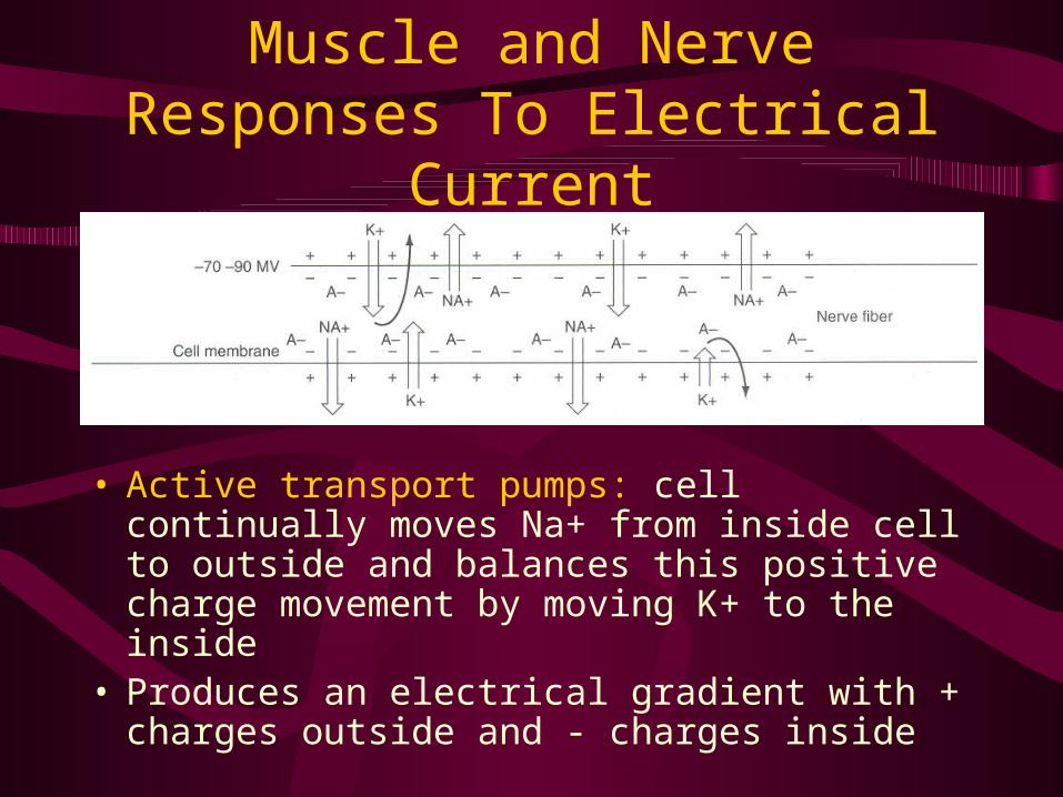
• Active transport pumps: cell continually moves Na+ from inside cell to outside and balances this positive charge movement by moving K+ to the inside
• Produces an electrical gradient with + charges outside and - charges inside

Nerve Depolarization
• To create transmission of an impulse in a nerve, the resting membrane potential must be reduced below threshold level
• Changes in membrane permeability may then occur creating an action potential, which propagates impulse along nerve in both directions causing depolarization

Nerve Depolarization
• Stimulus must have adequate intensity and last long enough to equal, or exceed, membrane's basic threshold for excitation
• Stimulus must alter the membrane so that a number of ions are pushed across membrane exceeding ability of the active transport pumps to maintain the resting potential, thus forcing membrane to depolarize resulting in an action potential

Depolarization Propagation
• Difference in electrical potential between depolarized region and neighboring inactive regions causes the electrical current to flow from the depolarized region to the inactive region
• Forms a complete local circuit and makes the wave of depolarization “self-propagating”

Depolarization Effects
• As nerve impulse reaches effector organ or another nerve cell, impulse is transferred between the two at a motor end plate or a synapse

Depolarization Effects
• At the motor end plate, a neurotransmitter is released from nerve
• Neurotransmitter causes depolarization of the muscle cell, resulting in a twitch muscle contraction
Differs from voluntary muscle contraction only in rate and synchrony of muscle
fiber contractions!

Strength - Duration Curves
• Represents the threshold for depolarization of a nerve fiber
• Muscle and nerve respond in an all-or-none fashion and there is no gradation of response

Strength - Duration Curves
• Shape of the curve• Relates intensity of
electrical stimulus (strength) and length of time (duration) necessary to cause depolarization of muscle tissue
Strength - Duration Curves
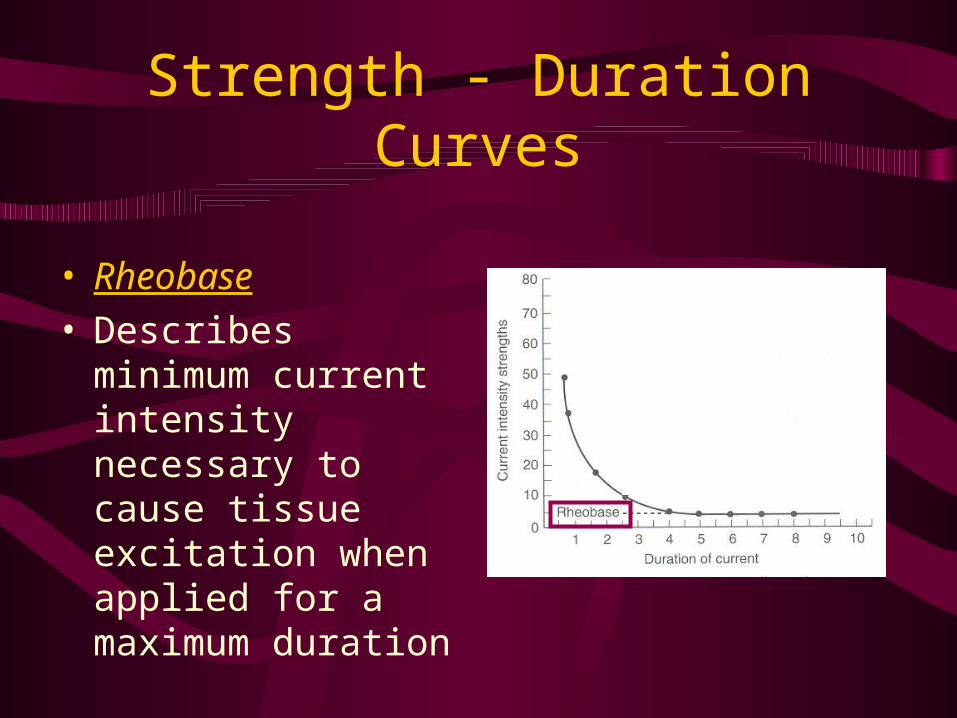
• Rheobase • Describes
minimum current intensity necessary to cause tissue excitation when applied for a maximum duration

Strength - Duration Curves
• Chronaxie• Describes length of
time (duration) required for a current of twice the intensity of the rheobase current to produce tissue excitation
Manufacturers select preset pulse durations in the area of chronaxie!

Strength - Duration Curves
Aß sensory, motor, A sensory, and C pain nerve fibers
Durations of several electrical stimulators are indicated along the lower axis
Corresponding intensities would be necessary to create a depolarizing stimulus for any of the nerve fibers
Microcurrent intensity is so low that nerve fibers will not depolarize

Nonexcitable Tissue and Cells Response To Electrical Current
• Cell function may speed up• Cell movement may occur• Stimulation of extra-cellular
protein synthesis• Increase release of cellular
secretions

Nonexcitable Tissue and Cells Response To Electrical Current
• Gap junctions unit neighboring cells– Allow direct communication between
adjacent cells (forms electrical circuit)• Cells connected by gap junctions
can act together when one cell receives an extracellular message– The tissue can be coordinated in its
response by the gap junction’s internal message system

Nonexcitable Tissue and Cells Response To Electrical Current
• All structures within the cell, membrane, and microtubes are dipoles– Molecules whose ends carry opposite
charge
• Therefore, all cell structures carry a permanent charge and are capable of… – Piezoelectric activity– Electropiezo activity

Nonexcitable Tissue and Cells Response To Electrical Current
• Piezoelectric activity– Mechanical deformation of the
structure causes a change in surface electrical charge
• Electropeizo activity– Change in surface electrical charge
causes the structure to change shape• Important concepts regarding the effects
electrical stimulation has on growth and healing

Nonexcitable Tissue and Cells Response To Electrical Current
• As a structure changes shape, strain-related potentials (SRP) develop– Results due to tension or distraction
on the surface of the structure
• Compression = negative SRPs• Tension = positive SRPs

Strain-Related Potentials (SRP)
• Bone (Wolff’s Law)– Stimulates osteoblast, osteocyte, and
osteoclast activity to assist in bone growth and healing
• Skin Wounds– Normal Biolelectric Field: skin is negatively
charged relative to dermis– Current of Injury: skin will change to positive
charge producing a bioelectric current• Stimulates growth and healing
– At the conclusion of the healing process, the Normal Bioelectric Field will be re-established

Nonexcitable Tissue and Cells Response To Electrical Current
• Based on THEORY rather than well-proven, researched outcomes
• More research is needed on the effects of electrical current on nonexcitable tissue and cells

Effects of Changing Current Parameters
• Alternating versus Direct current• Tissue impedance• Current density• Frequency of wave or pulse• Intensity of wave or pulse• Duration of wave or pulse• Polarity of electrodes• Electrode placement

Alternating vs. Direct Current
• Nerve doesn’t know the difference between AC and DC
• With continuous DC, a muscle contraction occurs only when the current intensity reaches threshold for the motor unit
DC current influence on a motor unit

Alternating vs. Direct Current
• Once the membrane of the motor unit repolarizes, another change in the current intensity would be needed to force another depolarization to elicit a muscle contraction
DC current influence on a motor unit

Alternating vs. Direct Current
• Biggest difference between the effects of AC and DC is the ability of DC to cause chemical changes
• Chemical effects usually occur only when continuous DC is applied over a period of time
Tissue Impedance
• Impedance = resistance of tissue to the passage of electrical current. – Bone and Fat = high-impedance – Nerve and Muscle = low-impedance
• If a low-impedance tissue is located under a large amount of high-impedance tissue, the intensity of the electrical current will not be sufficient to cause depolarization

Current Density
• Current density refers to the volume of current in the tissues
• Current density is highest at the surface and diminishes in deeper tissue

Altering Current Density
• Change the spacing of electrodes• Moving electrodes further apart
increases current density in deeper tissues

Altering Current Density
• Changing the size of the electrode• Active electrode is the smaller electrode
– Current density is greater
• Dispersive electrode is the larger electrode– Current density is less

Frequency
• Effects the type of muscle contraction• Effects the mechanism of pain
modulation

Frequency
• Frequency of the electrical current impacts…– Amount of shortening in the muscle fiber– Recovery time allowed the muscle fiber
• Summation: shortening of myofilaments caused by increasing the frequency of membrane depolarization
• Tetanization: individual muscle-twitch responses are no longer distinguishable, results in maximum shortening of the muscle fiber– Dependent on frequency of electrical current,
not intensity of the electrical current!

Frequency
• Voluntary muscle contraction elicits asynchronous firing of motor units– Prolongs onset of fatigue due to recruitment of
inactive motor units
• Electrically induced muscle contraction elicits synchronous firing of motor units– Same motor unit is stimulated; therefore, onset
of fatigue is rapid

Intensity
• Increasing the intensity of the electrical stimulus causes the current to reach deeper into the tissue

Recruitment of Nerve Fibers
• An electrical stimulus pulse at a duration intensity just above threshold will excite the closest and largest fibers
• Each electrical pulse at the same intensity at the same location will cause the same fibers to fire

Recruitment of Nerve Fibers
• Increasing the intensity will excite smaller fibers and fibers farther away
• Increasing the duration will also excite smaller fibers and fibers farther away

Duration
• More nerve fibers will be stimulated at a given intensity by increasing the duration (length of time) that an adequate stimulus is available to depolarize the membranes
• Duration is typically adjustable on low-voltage stimulators

Polarity
• Anode– Positive electrode – Lowest concentration of electrons
• Cathode– Negative electrode – Greatest concentration of electrons
• AC: electrodes change polarity with each current cycle
• DC: polarity switch designates one electrode as positive and one as negative

Polarity
• With AC and Interrupted DC, polarity is not critical
• Negative polarity used for muscle contraction– Facilitates membrane depolarization– Usually considered more comfortable
• Negative electrode is usually positioned distally

Polarity With Continuous DC
• Positive Pole– Attracts (-) ions– Acidic reaction– Hardening of
tissues– Decreased nerve
irritability
• Negative Pole– Attracts (+) ions– Alkaline reaction– Softening of
tissues– Increased nerve
irritability
• Important consideration when using iontophoresis

Electrode Placement
• On or around the painful area • Over specific dermatomes,
myotomes, or sclerotomes that correspond to the painful area
• Close to spinal cord segment that innervates an area that is painful
• Over sites where peripheral nerves that innervate the painful area becomes superficial and can be easily stimulated

Electrode Placement
• Over superficial vascular structures• Over trigger point locations• Over acupuncture points• In a criss-cross pattern surrounding
the treatment area
• If treatment is not working, change electrode placement

Therapeutic Uses of Electrically Induced Muscle Contraction
• Muscle re-education• Muscle pump contractions• Retardation of atrophy• Muscle strengthening• Increasing range of motion• Reducing Edema

Therapeutic Uses of Electrically Induced Muscle Contraction
• Muscle fatigue must be considered• Variables that influence muscle
fatigue:– Intensity– Frequency– On-time– Off-time

Muscle Re-Education
• Primary indication = muscular inhibition after surgery or injury
• A muscle contraction usually can be forced by electrically stimulating the muscle
• Patient feels the muscle contract, sees the muscle contract, and can attempt to duplicate the muscle contraction

Muscle Re-Education Protocol
• Intensity: must be adequate for muscle contraction– Patient comfort must be considered
• Pulse Duration: must be set as close as possible to chronaxie for motor neurons– 300 μsec - 600 μsec
• Frequency: should be high enough to give a tetanic contraction – 35 to 55 pps– Muscle fatigue must be considered

Muscle Re-Education Protocol
• On/Off Cycles: dependent on patient– On-time should be 1 - 2 seconds– Off-time may be 1:1, 1:4, or 1:5
contraction to recovery ratio
• Current: interrupted or surged current
• Treatment Time: should be about 15 minutes– May be repeated several times daily

Muscle Re-Education Protocol
• Instruct patient to allow the electricity to make the muscle contract, feeling and seeing the response desired
• Next, patient should alternate voluntary muscle contractions with electrically induced contractions

Muscle Pump Contractions
• Used to duplicate voluntary muscle contractions that help stimulate circulation– Pump fluid and blood through venous
and lymphatic channels back to the heart
• Helps re-establish proper circulatory pattern while protecting the injured area

Muscle Pump Contractions Protocol
• Intensity: must be high enough to provide a strong, comfortable muscle contraction
• Pulse Duration: must be set as close as possible to chronaxie for motor neurons – 300 μsec - 600 μsec
• Frequency: should be at beginning of tetany range – 35 to 50 pps

Muscle Pump Contractions Protocol
• Current: interrupted or surged current • On/Off Cycles:
– On-time should be 5 to 10 seconds– Off-time should be 5 to 10 seconds
• Patient Position: part to be treated should be elevated
• Treatment Time: should be 20 to 30 minutes – May be repeated 2-5 times daily

Muscle Pump Contractions Protocol
• Instruct patient to allow electrically induced muscle contractions– AROM may be encouraged at the
same time if it is not contraindicated
• Use this protocol in addition to R.I.C.E. for best results

Retardation of Atrophy
• Electrically induced muscle contractions stimulate the physical and chemical events associated with normal voluntary muscle contractions
• Used to….– Maintain normal muscle function– Prevent or reduce atrophy

Retardation of Atrophy Protocol
• Intensity: should be as high as can be tolerated by the patient– Should be capable of moving the limb
through the antigravity range – Should achieve 25% or more of the
normal maximum voluntary isometric contraction (MVIC) torque for the muscle
• May be increased during the treatment as sensory accommodation occurs

Retardation of Atrophy Protocol
• Pulse Duration: must be set as close as possible to chronaxie for motor neurons – 300 μsec - 600 μsec
• Frequency: should be in the tetany range – 50 to 85 pps
• Current: interrupted or surged current– Medium-frequency AC stimulator is the
machine of choice

Retardation of Atrophy Protocol
• On/Off Cycles:– On-time should be between 6 -15
seconds– Off-time should be at least 1 minute
• Treatment Time: should be 15 to 20 minutes or enough time to allow a minimum of 10 contractions– May be repeated 2 times daily

Retardation of Atrophy Protocol
• Should provide resistance– May be provided by gravity, weights,
or fixing the joint so that the contraction becomes isometric
• Instruct the patient to work with the electrically induced contraction– But, voluntary muscle contractions is
not necessary

Muscle Strengthening
• Electrically induced muscle contractions may be helpful in treating athletes with muscle weakness or denervation of a muscle group
• More research is needed

Muscle Strengthening Protocol
• Intensity: should be enough to make muscle develop 60% of torque developed in a maximum voluntary isometric contraction (MVIC)
• Pulse Duration: must be set as close as possible to chronaxie for motor neurons – 300 μsec - 600 μsec

Muscle Strengthening Protocol
• Frequency: should be in the tetany range – 70 to 85 pps
• Current: interrupted or surged current with a gradual ramp to peak intensity– Medium-frequency AC stimulator is
machine of choice

Muscle Strengthening Protocol
• On/Off Cycles:– On-time should be 10 - 15 seconds – Off-time should be 50 seconds to 2
minutes• Treatment Time: should include a
minimum of 10 contractions – Mimic normal active resistive training
protocols of 3 sets of 10 contractions– May be repeated at least 3 times
weekly– Muscle fatigue must be considered

Muscle Strengthening Protocol
• Should provide resistance– Immobilize limb to produce isometric
contraction torque equal to or greater than 25% of the MVIC torque
• Instruct the patient to work with the electrically induced contraction– But, voluntary muscle contractions is
not necessary

Increasing Range of Motion
• Electrically induced muscle contractions pull joint through limited range
• Continued contraction of muscle group over extended time results in joint and muscle tissue modification and lengthening
• May reduce muscle contractures

Increasing Range of Motion Protocol
• Intensity: should be strong enough to move the limb through the antigravity range
• Pulse Duration: must be set as close as possible to chronaxie for motor neurons – 300 μsec - 600 μsec

Increasing Range of Motion Protocol
• Frequency: should be at the beginning of the tetany range – 40 to 60 pps
• Current: interrupted or surged current
• On/Off Cycles:– On-time should be between 15 - 20
seconds– Off-time should be equal to, or
greater than, on-time – Fatigue must be considered

Increasing Range of Motion Protocol
• Treatment Time: should be 90 minutes– Three 30-minute treatments daily
• Patient Position: stimulated muscle group should be antagonistic to joint contracture – Patient should be positioned so joint will
be moved to the limits of available range• Patient is passive in treatment and
does not work with electrically induced contraction

Reducing Edema
• Theory #1: sensory level DC stimulation may be used to move interstitial plasma protein ions in the direction of oppositely charged electrode
• Theory #2: microamp stimulation may cause vasoconstriction and reduce permeability of the capillary wall– Limits migration of plasma proteins into the
interstitial spaces
• More research is needed

Reducing Edema Protocol
• Intensity: should be 30V - 50V– 10% less than intensity needed to produce a
visible muscle contraction
• Frequency: 120pps– Sensory level stimulation
• Current: short duration interrupted DC currents– High-voltage pulsed generators are effective

Reducing Edema Protocol
• Electrode Placement: distal electrode should be negative
• Treatment Time: should be approximately 30 minutes– Should begin immediately, within 24
hours, after injury

Stimulation of Denervated Muscle
• Denervated muscle has lost its peripheral nerve supply – Results in a decrease in size, diameter, and
weight of muscle fibers – Decrease in amount of tension which can be
generated– Increase the time required for contraction
• Electrical currents may be used to produce a muscle contraction in denervated muscle to minimize atrophy

Stimulation of Denervated Muscle
• Degenerative changes progress until muscle is re-innervated by axons extending across site of nerve lesion
• If re-innervation does not occur within 2 years, fibrous connective tissue replaces contractile elements – Recovery of muscle function is not
possible

Denervated Muscle Protocol
• Intensity: should be enough to produce moderately strong contraction
• Pulse Duration: must be equal to or greater than chronaxie of denervated muscle
• Current: asymmetric, biphasic (faradic) waveform – After 2 weeks, other waveforms may be
used• Interrupted DC square, Progressive DC
exponential, or Sine AC

Denervated Muscle Protocol
• Frequency: as low as possible but enough to produce a muscle contraction
• On/Off Cycles:– On-time should be 1 - 2 seconds– Off-time may be 1:4 or 1:5
contraction to recovery ratio– Fatigue must be considered

Denervated Muscle Protocol
• Electrode Placement: either a monopolar or bipolar electrode setup can be used – Small diameter active electrode placed
over most electrically active point on muscle
• Treatment Time: should begin immediately after injury or surgery– 3 sets of 5 -20 repetitions 3 x per day

Therapeutic Uses of Electrical Stimulation of Sensory Nerves
• Gate Control Theory• Descending Pain Control
– Central Biasing
• Opiate Pain Control Theory
• Refer to Chapter 3 to review pain control theories

Gate Control Protocol
• Intensity: adjusted to tolerance – Should not cause muscular contraction
• Pulse Duration: 75 - 150 µsec– Or maximum possible on the e-stim unit
• Current: transcutaneous electrical stimulator waveform
• Frequency: 80 - 125 pps– Or as high as possible on the e-stim unit

Gate Control Protocol
• On/Off Cycles: continuous on time • Electrode Placement: surround
painful area• Treatment Time: unit should be
left on until pain is no longer perceived, turned off, then restarted when pain begins again– Should have positive result in 30
minutes, if not, reposition electrodes

Central Biasing Protocol
• Intensity: should be very high – Approaching noxious level
• Pulse Duration: should be 10 msec.
• Current: low-frequency,high-intensity generator is stimulator of choice
• Frequency: 80 pps

Central Biasing Protocol
• On/Off Cycles: – On-time should be 30 seconds to 1 minute
• Electrode Placement: should be over trigger or acupuncture points– Selection and number of points used
varies according to the part treated• Treatment Time: should have
positive result shortly after treatment begins– If not, reposition electrodes

Opiate Pain Control Protocol
• Intensity: should be high, at a noxious level– Muscular contraction is acceptable
• Pulse Duration: 200 µsec to 10 msec• Frequency: 1 – 5 pps• Current: high-voltage pulsed current
or low-frequency, high-intensity current

Opiate Pain Control Protocol
• On/Off Cycles: – On-time should be 30 to 45 seconds
• Electrode Placement: should be over trigger or acupuncture points– Selection and number of points used
varies according to part and condition being treated
• Treatment Time: analgesic effect should last for several (6-7) hours– If not successful, expand the number of
stimulation sites

Specialized Currents
• Low-Voltage Continuous DC– Medical Galvanism– Iontophoresis
• Low-Intensity Stimulators (LIS)– Analgesic Effects– Promotion of healing
• Russian Currents (Medium-Frequency)
• Interferential Currents

Low-Voltage Continuous DC
• Physiologic Changes:• Polar effects
– Acid reaction around the positive pole – Alkaline reaction around the negative
pole
• Vasomotor Changes– Blood flow increases between
electrodes

Low-Voltage Continuous DC: Medical Galvanism
• Intensity: should be to tolerance • Intensity in the milliamp range
• Current: low-voltage, continuous DC• Frequency: 0 pps• Electrode Placement: equal-sized
electrodes are used over saline-soaked gauze – Skin should be unbroken– Precaution = skin burns
• Treatment Time: should be 15 - 50 min

Low-Voltage Continuous DC:Iontophoresis
• Discussed in detail in Chapter 9

Low-Intensity Stimulators
• LIS is a sub-sensory current• Intensity of LIS is limited to <1000
microamps (1 milliamp)• Exact mechanism of action has not
yet been established• More research is needed

Low-Intensity Stimulators:Analgesic Effects
• LIS is sub-sensory, therefore it does not fit existing theories of pain modulation
• May create or change current flow of the neural tissues – May have some way of biasing transmission
of painful stimulus
• May make nerve cell membrane more receptive to neurotransmitters – May block transmission

Low-Intensity Stimulators:Wound Healing
• Intensity: – 200 - 400 µamp for normal skin – 400 - 800 µamp for denervated skin
• Pulse Duration: long, continuous, uninterrupted
• Current: monophasic DC is best– May use biphasic DC
• Frequency: maximum

Low-Intensity Stimulators:Wound Healing
• Treatment Time: 2 hours – Followed by a 4 hour rest time– May administer 2 - 3 treatment per day
• Electrode Placement: – First 3 days…
• Negative electrode positioned in the wound area
• Positive electrode positioned 25 cm proximal

Low-Intensity Stimulators:Wound Healing
• Electrode Placement continued: – After 3 days…
• Polarity reversed and positive electrode is positioned in the wound area
– In the case of infection…• Negative electrode should be left in wound
area until signs of infection disappear for at least 3 days
• Continue with negative electrode for 3 more days after infection clears

Low-Intensity Stimulators:Fracture Healing
• Intensity: just perceptible to patient• Pulse Duration: should be the
longest duration allowed on unit – 100 to 200 msec
• Current: monophasic or biphasic current – TENS units
• Frequency: should be set at lowest frequency allowed on unit – 5 to 10 pps

Low-Intensity Stimulators:Fracture Healing
• Treatment Time: 30 minutes to 1 hour – May repeat 3 - 4 times per day
• Electrode Placement: – Negative electrode placed close to,
but distal to fracture site– Positive electrode placed proximal to
immobilizing device

Russian Currents
• Medium-frequency polyphasic AC– 2,000 -10,000 Hz
• Two basic waveforms (fixed intrapulse interval) – Sine wave – Square wave
• Pulse duration varies from 50 - 250 µsec
• Phase duration is half of the pulse duration – 25 - 125 µsec

Russian Currents
• Current produced in burst mode with 50% duty cycle
• To make intensity tolerable, it is generated in 50-burst-per-second envelopes with an interburst interval of 10 msec – Increasing the bursts-per-second causes more
shortening in the muscle to take place

Russian Currents
• Dark shaded area represents total current
• Light shaded area indicates total current minus the interburst interval
• With burst mode, total current is decreased thus allowing for tolerance of greater current intensity

Russian Currents
• As intensity increases, more motor nerves are stimulated – This increases the magnitude of
contraction• Russian current is a fast oscillating AC
current, therefore, as soon as the nerve re-polarizes it is stimulated again– This maximizes the summation of muscle
contraction

Interferential Currents
• 2 separate generators (channels) are used
• Sine waves are produced at different frequencies and may interfere with each other resulting in…– Constructive interference– Destructive interference

Interferential Currents
• If the 2 sine waves are produced simultaneously, interference can be summative– Amplitudes of the current are combined and
increase
• Referred to as constructive interference

Interferential Currents
• If the 2 sine waves are produced out of sync, the waves cancel each other out
• Referred to as destructive interference

Interferential Currents
• If the 2 sine waves are produced at different frequencies, they create a beat pattern
• Blending of waves caused by constructive and destructive interference
• Called heterodyne effect

Interferential Currents
• Intensity: set according to sensations created
• Frequency: set to create a beat frequency corresponding to treatment goals – 20 to 50 pps for muscle contraction – 50 to 120 pps for pain management

Interferential Currents• Electrode Placement: arranged in a
square surrounding the treatment area– When an interferential current is passed
through a homogeneous medium, a predictable pattern of interference will occur

Interferential Currents
• When two currents cross, an electric field is created between the lines of current flow
• Electrical field is strongest near the center• The strength of the electrical field gradually
decreases as it moves away from center

Interferential Currents
• Scanning moves electrical field around while the treatment is taking place– Allows for larger treatment area
• Adding another set of electrodes will create a three-dimensional flower effect called a stereodynamic effect– Allows for larger treatment area

Interferential Currents
• Human body is NOT homogeneous; therefore, unable to predict exact location of interferential current
• Must rely on patient perception
• Electrode placement is trial-and-error to maximize treatment effect

Summary
• Electrical therapy is dynamic– Advances in research– Engineering– Technology
• ATs must have strong foundational knowledge in electrical therapy– Educated choices in purchasing– Able to manipulate treatment parameters to
optimize physiologic effects