elbow fractures in children management, treatment, and
TRANSCRIPT

Elbow Fractures in Children – Initial Management, Treatment, and Prevention Tips
PRESENTED BY: Scott Yang MD, Assistant Professor, Pediatric Orthopaedic Surgery Doernbecher Children’s Hospital, OHSU
Doernbecher Children’s Hospital

2
No disclosures for this presentation

3
Agenda
Two most common pediatric elbow injuries
1) Supracondylar humerus Fractures &
2) Lateral humeral condyle fractures
Image credits: 1) AO Surgery Reference

4
Supracondylar Humerus Fractures

5
Background
• Supracondylar humerusfractures are one of the most common types of fractures in children– 60% of all elbow trauma in
children
• Extension type most common (95%)
• Risk of neurovascular injury, compartment syndrome
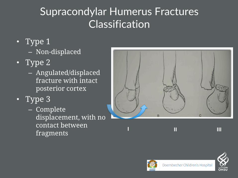
Supracondylar Humerus FracturesClassification
• Type 1– Non-displaced
• Type 2– Angulated/displaced
fracture with intact posterior cortex
• Type 3– Complete
displacement, with no contact between fragments
I II III

Elbow FracturesRadiograph Anatomy/Landmarks
• Anterior Humeral Line– Drawn along the anterior
humeral cortex
– Should pass through the middle of the capitellum• >5 y/o, 100% falls on
middle 1/3
– Variable in very young children • < 2 y/o, 30% fall on
anterior 1/3Credit:
-Rogers et al, Radiology 1998
-Herman, et al JBJS 2009
-Ryan et al, JPO 2016

Type 1Non-displaced
– Look for fat pad• Posterior fat pad elevation,
76% have occult fracture
– Treat with cast immobilization for 3 weeks
Credit
-Skaggs DL et al, JBJS 1999
-Bohrer et al, Clin Radiol. 1970;21:90.

Type 2Angulated/displaced fracture with intact
posterior cortex

Caution - Type 2 – Coronal plane is important too
Type II fracture with medial impaction –
not recognized and varus / extension not
reduced

Type 2 - Treatment
• Closed reduction & casting
• vs
• Closed reduction & percutaneous pinning

Casting can work in select patients…
Spencer et al, JPO 2012
• 189 type II fractures treated with closed reduction & casting– 21% lost reduction– Associated with:
• Rotation• Persistent
extension post-reduction
Rotational Malalignment

Proponents of Operative Treatment
• Skaggs et al, JBJS 2004, JPO 2008– 189 type II supracondylar fractures
treated with CRPP (lateral only pins)• No loss of reduction, cubitus varus
or valgus• No nerve injury• 2.1% pin site infection rate (4/189)• 6% with < 10 degree ROM difference
at 8.7 weeks postop

Conclusion
• Personal preference –– Pin all type 2 supracondylar humerus fractures
• Prevents need for repeated radiographic follow ups
• Minimizes loss of reduction and extension/varus deformity
• Pin complication rate acceptable

Type 3Complete displacement, with no contact between
fragments
• Higher risk of neurologic and/or vascular compromise • 10-20% (Omid et al, JBJS 2008)
• Risk of compartment syndrome
• Treat with closed reduction & percutaneous pinning• Rarely, open
reduction

Type 3 - Neurovascular Anatomy
• Median / AIN injury most common
• Artery close proximity to median nerve
• Brachial artery draped across proximal fragment– Tethered by supratrochlear
artery
• Urgency of CRPP depends on vascular statusImage Credits:
1) Rowell PJ, Injry 1975
2) Aksakal et al, Acta Orthop Traumato Turc 2013

Nerve Injury Prognosis?• Shore et al; JPO 2017
– 244 patients with SCH fracture + nerve injury– Avg recovery by 2.3 months
• 93% full recovery by 6 months– 20% open reduction rate – 29% have vascular changes
MEDIAN/AIN62%
RADIAL24%
ULNAR3% COMBO
11%
Distribution

Type 3, Caution
• Important to assess and document preoperative status on nerve function & vascular status!!
– Cases of median nerve and brachial artery entrapment at fracture site s/p reduction (Marshall et al, BMJ Case Rep 2015; Thorleifssonet al, AOTS 1988)

Type 3 Initial Presentation
• Pulse absent, poorly perfused hand– EMERGENT reduction / pinning
• Pulse absent, well perfused hand– URGENT reduction / pinning
• Pulse present, well perfused hand– How long can these wait for
reduction/pinning
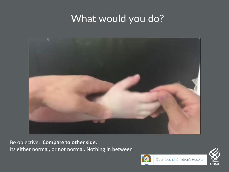
What would you do?
Be objective. Compare to other side.Its either normal, or not normal. Nothing in between

A quick word about Pulseless Poorly Perfused Hand On Presentation
Choi et al, JPO 2010• Pulseless SCH
w/ white hand • observe
CLOSELY postop for deterioration
But don’t forget about swelling & compartment syndrome

Compartment Syndrome
Robertson et al; JPO 2018; Harris et al; JPO 2018• Incidence 0.2% amongst supracondylar humerus
fractures – Includes type I & II fractures – likely higher in type III
• In patients with nerve injury, 4.5% - 11.4% Compartment syndrome

Ramachandran et al, JBJS UK 2008• What can we learn from patients who got compartment
syndrome?– 11 patients with type III SCHs w/ intact pulse on presentation
who developed compartment syndrome– Mean time from injury to surgery 22 hours– On presentation warning signs
• Severe swelling in 10/11• Puckering in 2/11• Ecchymosis in 7/11
• Use clinical judgment – take warning signs into account

Be on alert if you see this!!

Surgical Treatment of Supracondylar HumerusFractures
• Positioning– Radiolucent small hand
table• OK to use fluoroscopy
as table– Axilla at the edge of bed
Image credit:Skaggs et al, Masters Techniques in Orthopaedic Surgery, Pediatrics, 2015

Surgical Treatment of Supracondylar HumerusFractures
• Step 1:– Milk Brachialis,
• get tethered soft tissues out of way to reduce risk of neurovascular entrapment
Image credit:Skaggs et al, Masters Techniques in Orthopaedic Surgery, Pediatrics, 2015

• Step 2: – Traction– Reduce in coronal
plane– Check
fluoroscopy
Surgical Treatment of Supracondylar HumerusFractures
Image credit:Skaggs et al, Masters Techniques in Orthopaedic Surgery,: Pediatrics, 2015Flynn et al, Lovell and Winters Pediatric Orthopaedics, 2014

• Step 3:– Correct rotation– Flexion
maneuver w/ direct thumb pressure on distal fragment• Don’t be
overly aggressive (iatrogenic type IV)
Surgical Treatment of Supracondylar HumerusFractures
Image credit:Skaggs et al, Masters Techniques in Orthopaedic Surgery, Pediatrics, 2015

• Step 4– Check transcondylar
view– Verify reduction on
lateral view• External rotation
@ shoulder• Move the
humerus!
Surgical Treatment of Supracondylar HumerusFractures
Image credit:Skaggs et al, Masters Techniques in Orthopaedic Surgery, Pediatrics, 2015

• Step 5 –– Place
percutaneous pins in the transcondylar view
– Maximize divergence of the pins
Surgical Treatment of Supracondylar HumerusFractures
Image credit:Skaggs et al, Masters Techniques in Orthopaedic Surgery, Pediatrics, 2015

• Personal preferred technique:– Lateral only pins
• Two lateral pins for type II• Three lateral pins for type III
Surgical Treatment of Supracondylar HumerusFractures

• Step 6– Check pulse! – Do not leave OR
until hand is well perfused
Surgical Treatment of Supracondylar HumerusFractures

Scenarios after successful closed reduction & pinning
Pulse Intact
Pulse Gone, white hand
?Pulse Gone, Pink hand?

AVOID THIS!

Pulseless but perfused after reduction: Observe?
Weller et al, JBJS 2013• 54 pulseless type III supracondylar humerus fractures• All underwent reduction & pinning

Pulseless but perfused after reduction: Observe?
• SAFE : Pink, but dopplerable pulse afterwards - observe patients 24-48 hours

Pulseless but perfused after reduction: How do they do?
Scanell et al, JBJS 2013• 20 patients s/p CRPP for pulseless
perfused SCH fx– Mean 20 months f/u
• Outcomes: – 7/20 (35%) – Early palpable pulse
after reduction – 20/20 (100%) – Palpable pulse at
final follow up– Duplex scan of brachial artery:
– 14 patent – 6 occluded/stenotic
– Clinical exam:• No difference in circumference,
length, ROM, grip strength, muscle endurance compared to uninjured side
• 3 osteoenecrosis of trochlea, 1 distal humeral chondrolysis
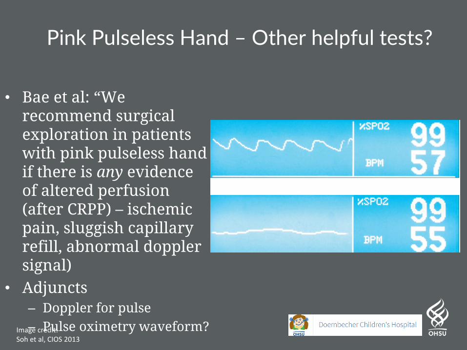
Pink Pulseless Hand – Other helpful tests?
• Bae et al: “We recommend surgical exploration in patients with pink pulseless hand if there is any evidence of altered perfusion (after CRPP) – ischemic pain, sluggish capillary refill, abnormal dopplersignal)
• Adjuncts– Doppler for pulse– Pulse oximetry waveform?Image credit:
Soh et al, CIOS 2013

Conclusion
• Observe pink pulseless hand 24-48 hours, with serial checks for compartment syndrome and vascular compromise– Be weary of nerve injury that mask a good examination
• Use best judgment to assess adequacy of perfusion– Doppler? Pulse Ox?
• Still need longer f/u and more objective ways to assess adequacy of perfusion

41
Lateral Humeral Condyle Fractures

Background
• 10-20% of pediatric elbow fractures
• Age 3-10• Fall on outstretched hand
– Push off mechanism • Radial neck pushes into it
– Pull off avulsive mechanism• Between BR and ECRL

What’s Special About Lateral Condyle Fractures
• You can’t leave the bones untouched in the same room and expect a good outcome– INTRA-ARTICULAR
Injuries
• Delayed Union / Nonunion doesn’t happen in kids… – Except sometimes in the….
LATERAL HUMERAL CONDYLE
Milch H, J Trauma 1964

Evaluation
• Neurovascular injury rare on initial presentation – Different from
supracondylar
• Radiographs:– Internal oblique
is CRITICAL for diagnosis• Often shows
maximal displacement
AP (left) and Internal oblique (right) radiographs demonstrating maximal displacement on the internal oblique

Know how to interpret PEDIATRIC Xrays

Classification
Weiss classification simpler, most prognostic of complications
Type I: <2 mm displacement
Type II: 2 – 4 mm displacement with intact cartilage
Type III: ≥ 4 mm displacement with articular incongruity.
Image creditsWeiss et al, JPO 2009

Treatment Type I
• Type I (<2 mm maximal displacement)– Chance for displacement with immobilization
• Wide range 0-18% (Knapik et al JPO 2017; Greenhill et al JPO 2019)
– Check post casting xray in 1-2 weeks!– Can take a long time to heal!!!

Treatment Type II
• Type II (2-4 mm maximal displacement)– Attempt closed reduction– Confirm with arthrogram– Place pins or screw– Be ready to open

Percutaneous Treatment Type II
5 y/o M

How to perform arthrogram: Direct posterior approach
Percutaneous Treatment Type II

Joint surface OK. Reduce with Valgus force & thumb pressure
Percutaneous Treatment Type II

Place pins
Percutaneous Treatment Type II

Open Treatment Type II
6 y/o F

Open Treatment Type II

Open Treatment Type II
Periosteum in Fracture Site
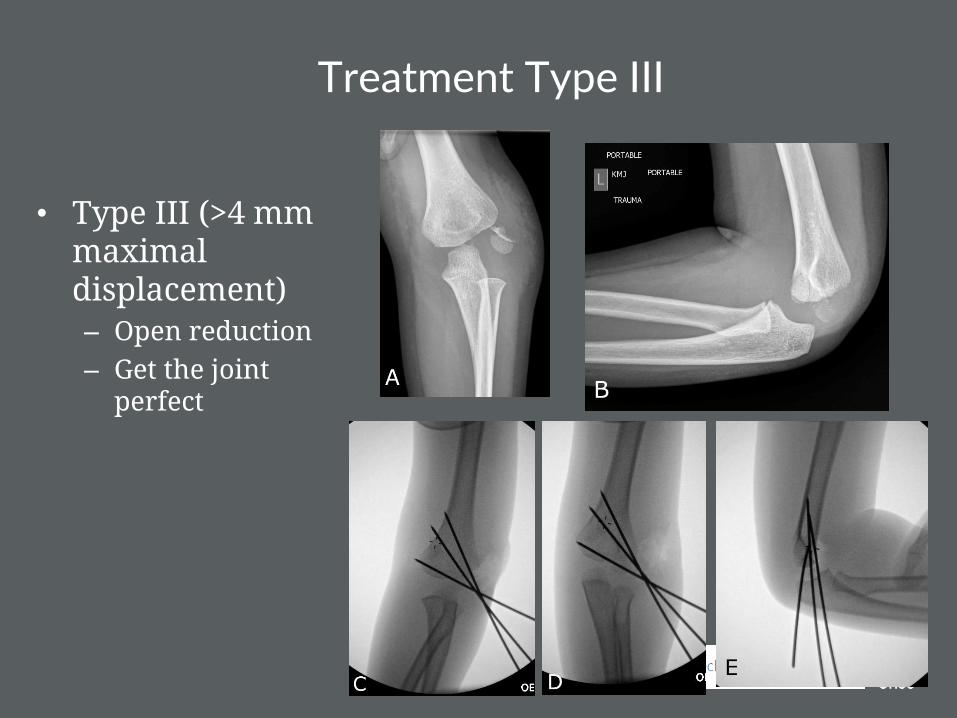
Treatment Type III
• Type III (>4 mm maximal displacement)– Open reduction– Get the joint
perfect

Open Reduction - Steps
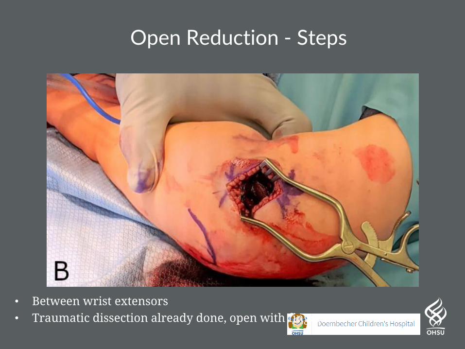
• Between wrist extensors• Traumatic dissection already done, open with finger
Open Reduction - Steps

• Dissect anteriorly enough to expose joint surface• Place hohman or bennett under brachialis
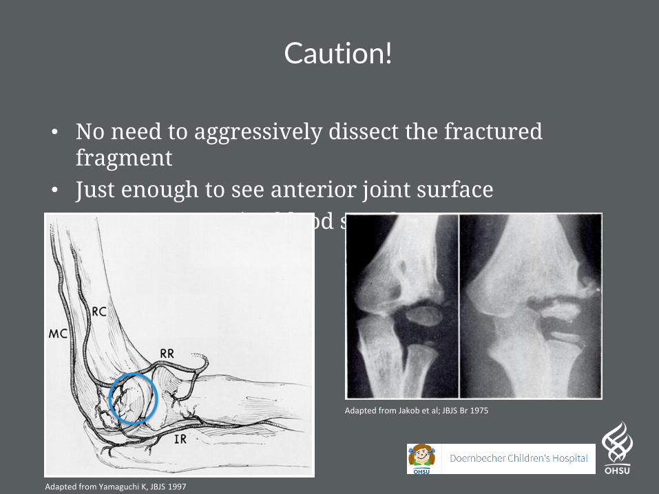
Caution!
• No need to aggressively dissect the fractured fragment
• Just enough to see anterior joint surface• Preserve posterior blood supply
Adapted from Yamaguchi K, JBJS 1997
Adapted from Jakob et al; JBJS Br 1975

• Don’t worry about mild lateral gap if joint is perfect• Always some mild deformation laterally

Optimal Fixation for Lateral Condyle Fractures
Two divergent pins @ 60 degrees, or three pins
Bloom, T, Chen, LY, Sabharwal, S: Biomechanical analysis of lateral humeral condyle fracture pinning. J Pediatr Orthop 2011;31:130–137.

Prognosis & Complications
• Mean time to union ~6.5 weeks
• 10-16% clinically significant complication rate in lateral condyle fractures
• Stiffness ROM >15 degrees– Up to 15.6% (Sinikumpu et al, Int Orthop 2017)
– Mean 12 yr follow up• ALL Mayo elbow functional
scores Excellent or Good

Prognosis & Complications
• Delayed union– 11-16% (Salgueiro et al, JPO 2017)
– Increased risk if increased intraoperative residual displacement
• Nonunion– Up to 3% (Pace et al; JPO 2018)
These only happen if you 1) Failed to diagnose initial injury2) Did not follow fracture to healing3) Did not act on an impending nonunion

Treatment of Chronic Nonunion
• Cubitus Valgus / Severe Deformity?– No In situ bone graft &
screw
– Yes In situ bone graft & screw + supracondylar osteotomy

Complication? This Always Happens
• Lateral spurring• Occurs in 73% (Pribaz et al, JPO 2012)• Does not cause functional or ROM deficit

Lateral Condyle Fracture Summary
• Avoid missed diagnosis– Internal oblique radiograph
• Avoid malreduction– Low threshold for open reduction
• Avoid nonunion– Follow until clinical union– Place a screw if robust healing not occurring by 2 months & still
tender
• Avoid AVN– Don’t dissect posterior aspect of fracture fragment
• Avoid parental lateral bump concern– Explanation preop always looks better than making excuses postop
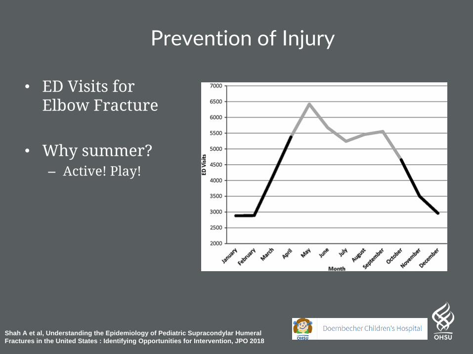
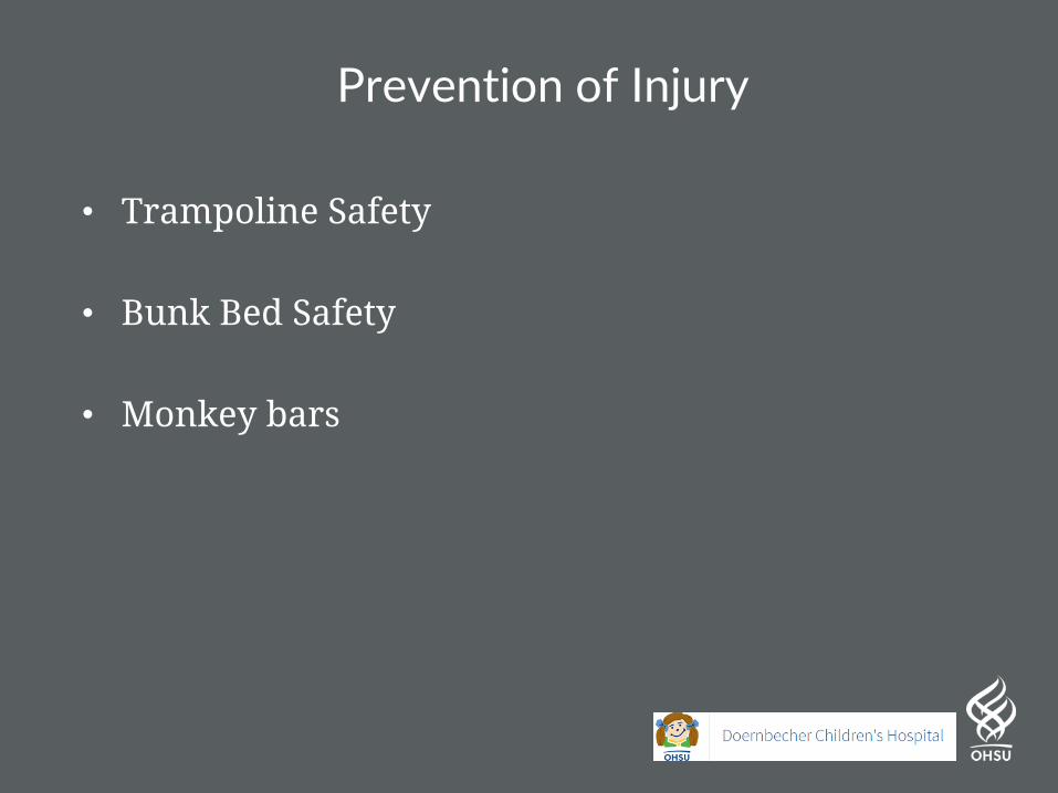
Prevention of Injury
• ED Visits for Elbow Fracture
• Why summer?– Active! Play!
Shah A et al, Understanding the Epidemiology of Pediatric Supracondylar Humeral
Fractures in the United States : Identifying Opportunities for Intervention, JPO 2018
Prevention of Injury
• Trampoline Safety
• Bunk Bed Safety
• Monkey bars

Questions?