effects of high-frequency electromagnetic fields on human .... dr... · effects of high-frequency...
TRANSCRIPT
1007
Intern. J. Neuroscience, 113:1007–1019, 2003Copyright 2003 Taylor & Francis0020-7454/03 $12.00 + .00DOI: 10.1080/00207450390220330
EFFECTS OF HIGH-FREQUENCYELECTROMAGNETIC FIELDS ON HUMAN
EEG: A BRAIN MAPPING STUDY
ALEXANDER V. KRAMARENKOCentral Clinic Hospital N5Kharkov, Ukraine
UNER TANCukurova UniversityMedical SchoolDepartment of PhysiologyAdana, Turkey
Cell phones emitting pulsed high-frequency electromagnetic fields (EMF)may affect the human brain, but there are inconsistent results concern-ing their effects on electroencephalogram (EEG). We used a 16-channeltelemetric electroencephalograph (ExpertTM®), to record EEG changesduring exposure of human skull to EME emitted by a mobile phone.Spatial distribution of EME was especially concentrated around theipsilateral eye adjacent to the basal surface of the brain. TraditionalEEG was full of noises during operation of a cellular phone. Using atelemetric electroencephalograph (ExpertTM®) in awake subjects, allthe noise was eliminated, and EEG showed interesting changes: after aperiod of 10–15 s there was no visible change, the spectrum medianfrequency increased in areas close to antenna; after 20–40 s, a slow-wave activity (2.5–6.0 Hz) appeared in the contralateral frontal andtemporal areas. These slow waves lasting for about one second re-peated every 15–-20 s at the same recording electrodes. After turningoff the mobile phone, slow-wave activity progressively disappeared; localchanges such as increased median frequency decreased and disappearedafter 15–20 min. We observed similar changes in children, but the slow-
Received 7 December 2002.Address correspondence to Prof. Dr. Uner Tan, Cukurova University, Medical School,
Department of Physiology, Adana, Turkey. E-mail: [email protected]

1008 A. V. Kramarenko and U. Tan
1
5
10
15
20
25
30
35
39
waves with higher amplitude appeared earlier in children (10–20 s)than adults, and their frequency was lower (1.0–2.5 Hz) with longerduration and shorter intervals. The results suggested that cellular phonesmay reversibly influence the human brain, inducing abnormal slow wavesin EEG of awake persons.
Keywords brain, brain mapping, cellular phone, electromagnetic field,
A cellular phone is a low-power, single-channel, two-way radio.Cell phone base stations are low-power multi-channel two-way ra-dios. Therefore, base stations produce radio-frequency radiation, andthey expose people near them to radio-frequency (RF) radiation.According to reports from the scientific community, the power fromthe mobile phone base station antennas is far too low to producehealth hazards as long as people are kept away from direct access tothe antennas. This nonionizing radiation is, however, fundamentallydifferent from the ionizing radiation produced by x-ray machines.The effects of electromagnetic source to biological material dependson the frequency of the source (see Moulder & Foster, 1995). X-rays, RE radiation, and EMF from power lines are part of the elec-tromagnetic spectrum, which are characterized by their frequency.Electric power in Turkey is at 50 Hz, and at 60 HZ in the US. FMradio has a frequency of around 100 MHz, microwave ovens have afrequency of 2450 MHz, X-rays have frequencies above one millionGHz, and cellular phones operate at frequencies between about800 and 2200 MHz. The electromagnetic particles of high-frequencyX-rays have sufficient energy to break chemical bonds (ionization),which can damage the genetic material of cells leading to cancer orbirth defects. Low frequency RE radiation is nonionizing. There-fore, there is no similarity between the biological effects of x-raysand RF radiation.
There was no convincing evidence that radio fields—in contrastto X- and Gamma-rays, ultraviolet and atomic radiation—can di-rectly cause the changes in genes responsible for cancer development.Actually, most governments and cell-phone companies have claimedthat the only possible biological effect of RF transmission is local-ized body heating. However, significant concern has been raisedabout possible health effects of RF electromagnetic fields. For

Cell Phone and EEG 1009
1
5
10
15
20
25
30
35
39
instance, transgenic mice most susceptible to cancer demonstrated a2-times increase in tumor rate (B-cell lymphomas) after exposure tomicrowaves at a power density roughly equal to a cell-phone trans-mitting for two half-hour periods each day, compared to control miceunexposed to RF fields (Repacholi et al., 1997). Children mightdevelop cancer after exposure to the RF emissions from mobiletelephone base stations in or near schools (see Repacholi, 1997).
The human body is an electrochemical instrument controlled byoscillatory electrical processes of various kinds; some endogenousbiological electrical activities can be interfered via oscillatory as-pects of the incoming radiation. Human in vivo studies indicatedthat the awaked EEC exposed to RE fields from a cell phone exertsa delayed increase in spectral power density, particularly in the al-pha band (Reiser, Dimpfel, & Schober, 1995). Exposure to mobilephone radiation decreases the preparatory slow potentials in certainregions of the brain (Freude, Ullsperger, Eggert, & Ruppe, 1998)and affects memory tasks modulating the responses of EEG activityapproximately 8 Hz specifically during cognitive process (Krause etal., 2000). There is a lot of evidence indicating nonthermal influ-ences of RE fields in vivo, such as epileptiform activity in rats inconjunction with certain drugs (Sidorenko & Tsaryk, 1999), depres-sion of chicken immune systems (Youbicier-Simo & Bastide, 1999),increase in chick embryo mortality(Youbicier-Simo & Bastide, 1999),increased permeability of blood-brain barrier in rats (Persson, Salford,Brun et al., 1997), and synergistic effect with certain psychoactivedrugs (Lai, Horita, Chou, & Guy, 1987). These RF influences mayinduce many clinical signs and symptoms, such as headache, epi-leptic seizure, and sleep disturbance. The current scientific literaturesuggests that nonthermal RF field effects originating from cellularphones may have potential adverse health reactions, and this possi-bility should not be ignored even if only a small minority of peopleare at risk (because of inconsistent results).
Concerning the human brain, the above mentioned studies sug-gest that electromagnetic fields emitted by cellular phones may affectthe human EEG. The current scientific literature is, however, full ofinconsistencies. The aim of the present work was to reinvestigatethe effects of the pulsed high frequency EMFs on human EEG inchildren and adults.

1010 A. V. Kramarenko and U. Tan
1
5
10
15
20
25
30
35
39
MATERIALS AND METHODS
Ten healthy young males and 10 children (12 years old) voluntarilyparticipated in the study. The subjects were healthy and free ofneurological and psychological signs and symptoms. Duringsessions, the subjects were sitting on an arm chair within a sound-isolated room. Sixteen electrodes were placed over the scalp ac-cording to the international 10–20 system, to record EEG. The ref-erence was the sum of all the electrodes (common reference). Thesubjects were awake and their eyes were open during recordings,which were made using a 16-channel telemetric electroencephalo-graph made by one of us (AVK) (ExpertTM, Kharkov, Ukraine).This device uses digital telemetry and allows recording for a longtime up to 24 h. EEG signals are transmitted digitally to the regis-tration device via radio frequencies. The wireless system signifi-cantly simplifies the EEG examinations during research and clinicaluse. It is much less sensitive to the pulse and systematic noise thanany other stationary EEGs. The built-in processor allows us to carryout digitization, filtration, compression, and coding of the signaldirectly in the EEG-amplifier. This allows us to transmit digitalEEGs wirelessly with no interferences, including 50 Hz backgroundnoise. The high input resistance allows using the standard EEG gelfor 24-hour EEG recording, reducing many physiological artifacts.EEG was subjected to spectral analysis including multiple mappingof amplitude characteristics and spectra of selected ranges. 3-D re-construction of the functional focus was possible with availabilityof arbitrary or MRI-compatible shear planes.
EEG recordings were made in awake subjects before and duringa call with a usual radiophone and a cellular phone. The carrierfrequency of our cell phone was 900 MHz with a frame-frequencyof 217 Hz. So, the usual radiophones provide continuous radiationusually up to 100 MHz with an emitted power up to 3 mW, whereascell phones transmit the information in frames (217 Hz), the trans-mitter works with zero power between frames. In the on mode, thecell phone emits 3–4 W in immediate vicinity of the active ear andbrain structures. The effects of EMF emitted by cell phones is aninverse proportion to squared distance. For instance, let us supposethat the field strength is X during calling (phone is near ear, dis-

Cell Phone and EEG 1011
1
5
10
15
20
25
30
35
39
tance = 1 cm); if we increase the distance to 10 cm, the field strengthdecreases to X/100. When a person lives near a cellular station (10W, distance = 50 m), the field strength decreases to (50/0.01)2 =25000000 times. This is why the experiments cannot find any signsof EEG changes when the transmitter is placed far from the skull(40 cm or more) as reported by some investigators.
RESULTS
In order to understand the flow of current in a conducting mediumlike the human brain, the isotonic NaCl solution was put in a con-tainer and the antenna of a cellular phone was placed four cm apartfrom this container. The current flowing in this conducting mediumcould be registered using the weakly polarized Ag-AgCl EEG-electrodes in a low-frequency oscillograph (see Figure 1). This ex-periment suggests that potentials of several or tens of milivolts with
FIGURE 1. A snapshot from an oscilloscope: pulses emitted by a cellular phone re-corded from an isotonic NaCl solution using Ag-AgCl EEG electrodes. Each square showsthe voltage (vertical line = 2 mV) and time (horizontal line = 2 msec). Ericsson A1018S900 MHz phone. (See Color Plate II at end of issue.)
1012 A. V. Kramarenko and U. Tan
1
5
10
15
20
25
30
35
39
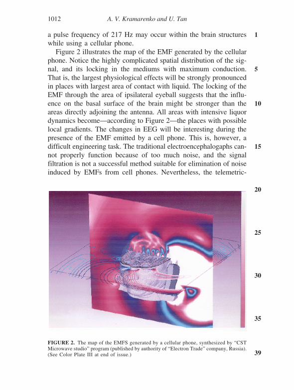
a pulse frequency of 217 Hz may occur within the brain structureswhile using a cellular phone.
Figure 2 illustrates the map of the EMF generated by the cellularphone. Notice the highly complicated spatial distribution of the sig-nal, and its locking in the mediums with maximum conduction.That is, the largest physiological effects will be strongly pronouncedin places with largest area of contact with liquid. The locking of theEMF through the area of ipsilateral eyeball suggests that the influ-ence on the basal surface of the brain might be stronger than theareas directly adjoining the antenna. All areas with intensive liquordynamics become—according to Figure 2—the places with possiblelocal gradients. The changes in EEG will be interesting during thepresence of the EMF emitted by a cell phone. This is, however, adifficult engineering task. The traditional electroencephalogaphs can-not properly function because of too much noise, and the signalfiltration is not a successful method suitable for elimination of noiseinduced by EMFs from cell phones. Nevertheless, the telemetric-
FIGURE 2. The map of the EMFS generated by a cellular phone, synthesized by “CSTMicrowave studio” program (published by authority of “Electron Trade” company, Russia).(See Color Plate III at end of issue.)

Cell Phone and EEG 1013
1
5
10
15
20
25
30
35
39
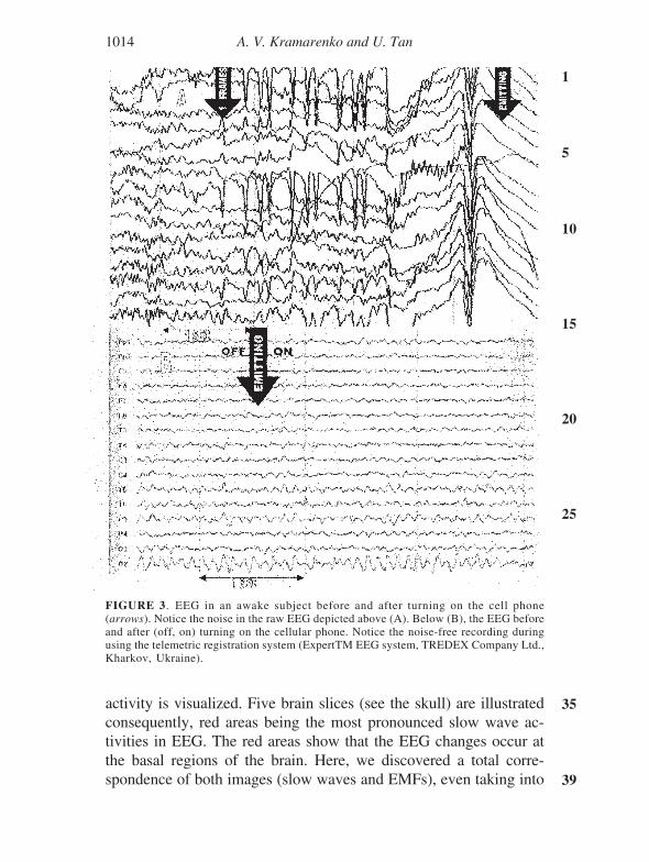
digital system used in this study allowed us to record the EEGsignals without interference. Initially, these EMFs cause alpha de-pression; the frequency of the main EEG rhythm retains a highprecision, and the signal spectrum changes slightly. If the singleEEG channels were separately analyzed, there is some reliable in-crease in the median frequency of spectrum; occasionally, singlesharp waves were also recorded in the areas close to antenna. Mostof these changes were previously reported (see Lebedeva et al.,2000). Figure 3 illustrates the raw EEG without using our telemetricsystem (top: A) and after using our telemetric system (bottom: B).As seen in Figure 3B, we could record EEG during cell phoneemitting (on) without noise.
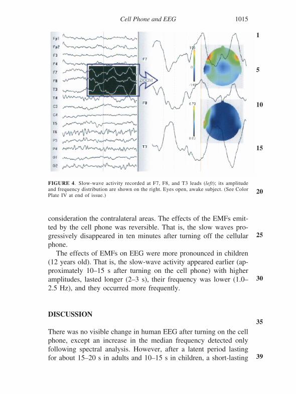
Interestingly, the periodical slow wave activities were also ob-served in EEG after turning on the cell phone. Actually, there wereno visible changes in EEG for 10–15 s following turning on the cellphone (see Figure 3B). After this period, the spectrum for the me-dian frequency increased in areas close to the antenna. Followingthis period (i.e., within 20–40 s) a slow-wave activity appeared inthe same areas; as the slow waves appeared, the median frequencydecreased. The slow-wave activity lasted for about 1 s and thenabruptly disappeared. The slow-wave activity exhibiting antiphasewas sometimes observed in the areas contralateral to the antenna ofthe cell phone. Figure 4 illustrates the slow-wave activity in E7, F8,and T3 leads (left side); the picture on the right side of Figure 4shows the enlarged part of the slow waves with brain mapping.Interestingly, the frontal areas are activated bilaterally, but the tem-poral areas are activated contralaterally (red areas: slow waves).Maximum power spectral density of these waves were within therange of 2.5–6.0 Hz, at the leads of maximum EMF strength. Afterthe period of slow-wave activity, the median frequency slightly in-creases and apparently normal EEG was recorded. However, theslow-wave activity repeated periodically at the same leads every15–20 s.
Figure 5 illustrates the mapping of the slow-wave activity in re-lation to the high frequency EME emitted by the cell phone. Asseen in Figure 5 (right side, three dimensional pictures), the maxi-mum slow-wave activity (above: red area) coincides with the maxi-mum field strength. On the left side, the location of the slow-wave
1014 A. V. Kramarenko and U. Tan
1
5
10
15
20
25
30
35
39
activity is visualized. Five brain slices (see the skull) are illustratedconsequently, red areas being the most pronounced slow wave ac-tivities in EEG. The red areas show that the EEG changes occur atthe basal regions of the brain. Here, we discovered a total corre-spondence of both images (slow waves and EMFs), even taking into
FIGURE 3. EEG in an awake subject before and after turning on the cell phone(arrows). Notice the noise in the raw EEG depicted above (A). Below (B), the EEG beforeand after (off, on) turning on the cellular phone. Notice the noise-free recording duringusing the telemetric registration system (ExpertTM EEG system, TREDEX Company Ltd.,Kharkov, Ukraine).
Cell Phone and EEG 1015
1
5
10
15
20
25
30
35
39
consideration the contralateral areas. The effects of the EMFs emit-ted by the cell phone was reversible. That is, the slow waves pro-gressively disappeared in ten minutes after turning off the cellularphone.
The effects of EMFs on EEG were more pronounced in children(12 years old). That is, the slow-wave activity appeared earlier (ap-proximately 10–15 s after turning on the cell phone) with higheramplitudes, lasted longer (2–3 s), their frequency was lower (1.0–2.5 Hz), and they occurred more frequently.
DISCUSSION
There was no visible change in human EEG after turning on the cellphone, except an increase in the median frequency detected onlyfollowing spectral analysis. However, after a latent period lastingfor about 15–20 s in adults and 10–15 s in children, a short-lasting
FIGURE 4. Slow-wave activity recorded at F7, F8, and T3 leads (left); its amplitudeand frequency distribution are shown on the right. Eyes open, awake subject. (See ColorPlate IV at end of issue.)

1016 A. V. Kramarenko and U. Tan
1
5
10
15
20
25
30
35
39
slow slow-wave activity appeared in EEG. The spectral analysisindicated that the frequency of the slow waves ranged from 2.5 to6.0 Hz in adults and from 1.0 to 2.5 Hz in children. Thus, theperiodic delta and theta waves appeared in adults, and only the deltawaves appeared in children.
The results of the present work clearly showed that the EMFsemitted by a cell phone affected the human EEG. It is indeed ex-pected that EMFs in brain leading to afferent electrical signals maycause subsequent processing events in the brain, like other stimuli(Marino, 1993; Marino et al., 1996). However, the periodically oc-curring slow waves are obviously abnormal for a healthy humansubject, since there are no delta waves in EEG of awake persons.Interestingly, the slow-wave activity did not disappear even if the
FIGURE 5. Comparison of probabilistic EEG tomography with the map of a cellularphone’s EMFs. Pictures are from “Electroencephalograph ExpertTM system” (TredexCompany, Kharkov, Ukraine). (See Color Plate V at end of issue.)
Cell Phone and EEG 1017
1
5
10
15
20
25
30
35
39
cell phone was turned off; the slow waves progressively decreasedin amplitude and then disappeared within tens of min. There aresome animal studies supporting these results. For instance, acuteexposure of rats and rabbits to continuous microwaves increasedEEG delta activity (Shandala et al., 1979). Similarly, Takashima etal. (1979) have reported a decrease in the high frequency EEG bandsand an increase in the low frequency EEG bands. On the otherhand, no uniform changes in EEG power spectra were also reportedfollowing exposure to continuous microwaves (Mitchell et al., 1989).
Increase in delta power was frequently reported following expo-sure to EMFs in animals (see Hermann & Hossmann, 1997). Thiswas usually attributed to the thermal effects of EMFs in the brain:the increase in slow wave activity following exposure to EMFs mayreflect the thermoregulatory response of the brain through the hypo-thalamus. The thermoregulatory effect may indeed play a role in theappearance of the slow-wave activity in the EEG. This is, however,unlikely for our study, since the delta waves occurred after a latentperiod of 15–20 s, lasted only for a few seconds, and occurredperiodically every 15–20 s; they were not continuous. Moreover,the rise in brain temperature does not exceed 0.01–0.1ºC duringexposure to EMFs emitted by cell phones, if blood flow and con-duction is taken into account (Riu & Foster, 1999). Furthermore,the periodically occurring slow wave activity usually recorded atthe contralateral leads to the antenna of the cellular phone, andcoincided with the strongest EMFs.
There are inconsistent results in the scientific literature concern-ing the influence of EMFs emitted by cell phones on the humanEEG. Our results clearly indicate that the cell phones may directlyinfluence the human brain. This may be a direct influence of theEMFs on nervous system, since the EMFs were shown to induceafferent electrical signals like other stimuli within the human brain(see Marino et al., 1996). Consistent with our results, it was foundpreviously that the brain electrical activity changes during exposureto EMFs (Bell et al., 1992; Lebedeva et al., 2000; Reiser et al.,1995); inconsistent with our results, it was reported that EEG wasnot affected by active cell phones (Hietanen et al., 2000; Roschke& Mann, 1997). Recently, Croft et al. (2002) have reported thatexposure to acute mobile phone operation altered the resting EEG,

1018 A. V. Kramarenko and U. Tan
1
5
10
15
20
25
30
35
39
decreasing 1–4 Hz activity, and increasing the 8–12 Hz activity andthe midline frontal and lateral posterior responses in the 30–45 Hzband. These authors found alterations in awake EEG, but did notfind any increase in delta wave activity, contrary to our results.Croft et al. (2002) discussed the possible origins of the inconsistentresults on the effects of mobile phones on the human brain. It isindeed possible that sitting on a chair for a long time would causedrowsiness in the subjects, but we required the subjects to keeptheir eyes open and keep awake during EEG recordings.
CONCLUSION
We studied the effects of the EMFs emitted by cellular phones onthe human EEG in adults and children. The EEG was found to shownormal activity during exposure, except a slight increase in theglobal median frequency. However, a short-lasting slow-wave ac-tivity occurred after a latent period of 15–20 s after turning on thephone. We observed these slow-waves, within the delta range, peri-odically in every 15–20 s. After turning off the phone, they progres-sively decreased in amplitude and disappeared in ten of min. Wehave concluded that the EMFs emitted by cell phones may be harm-ful for the human brain, since the delta waves are pathological ifseen in awake subjects. On the other hand, the slow wave activitywas more pronounced in children than adults, indicating that thechildren may be more vulnerable to the adverse health effects ofmobile phones than adults, probably because absorption of micro-waves is greatest in an object about the size of a child’s head (Gandhiet al., 1996); the radiation can penetrate the thinner skull of aninfant with greater ease. We are seriously concerned about possiblerisks to human brain from cell phones.
REFERENCES
Bell, G. B., Marino, A. A., & Chesson, A. L. (1992). Alterations in brain electrical activ-ity caused by magnetic fields: Detecting the detection process. Electroencephalographyand Clinical Neurophysiology, 83, 359–397.
Croft, R. J., Chandler, J. S., Burgess, A. P., Barry, R. J., Williams, J. D., & Clarke, A. R.
Cell Phone and EEG 1019
1
5
10
15
20
25
30
35
39
(2002). Acute mobile phone operation affects neural function in humans. Clinical Neuro-physiology, 113, 1623–1632.
Freude, G., Ullsperger, P., Eggert, S., & Ruppe, I. (1998). Effects of microwaves emittedby cellular phones on human slow brain potentials. Bioelectromagnetics, 19, 384–387.
Hietanen, M., Kovala, T., & Hamalainen, A. M. (2000). Human brain activity during ex-posure to radiofrequency fields by cellular phones. Scandinavian Journal of Work andEnvironmental Health, 26, 87–92.
Hermann, D. M., & Hossmann, K.-A. (1997). Neurological effects of microwave exposurerelated to mobile communication. Journal of Neurological Sciences, 152, 1–14.
Krause, C. M., Sillanmaki, L., Koivisto, M., Haggqvist, A., et al., (2000). Effects of elec-tromagnetic fields emitted by cellular phones on the electroencephalogram during avisual working memory. International Journal of Radiation Biology, 76, 1659–1667.
Lai, H., Horta, A., Cho, C. K., & Guy, A. W. (1987). A review of microwave irradiationand actions of psychoactive drugs. Engineering Medicine and Biology, 6, 31–36.
Lebedeva, N. N., Sulimov, A. V., Suhmova, O. P., Kotrovskaya, T. T., & Gailus, T. (2000).Cellular phone electromagnetic field effects on bioelectric activity of human brain.Critical Review of Biomedical Engineering, 28, 323–337.
Marino, A. A. (1993). Electromagnetic fields, cancer, and the theory of neuroendocrine-related promotion. Bioengineering, 29, 255–276.
Marino, A. A., Bell, G. B., & Chesson, A. (1996). Low-level EMFs are transduced likeother stimuli. Journal of the Neurological Sciences, 144, 99–106.
Mitchell, C. L., McRee, D. I., Peterson, N. J., Tilson, H. A., Shandala, M. G., Rudnev,M. I., Varetskii, V. V., & Navakatikyan, M. I. (1989). Results of a United States andSoviet Union project on nervous system effects of microwave radiation. Environmen-tal Health Project, 81, 201–209.
Moulder, J. E., & Foster, K. R. (1995). Biological effects of power-frequency fields asthey relate to carcinogenesis. Proceedings of the Society for Experimental Biology andMedicine, 209, 309–324.
Persson, B. R. R., Salford, L. G., & Brun, A., et al. (1997). Blood-brain barrier permeabil-ity in rats exposed to electromagnetic fields used in wireless communication. WirelessNetworks, 3, 455–461.
Reiser, H.-P., Dimpfel, W., & Schober, F. (1995). The influence of electromagnetic fieldson human brain activity. Eur J Med Res, 1, 27–32.
Repacholi, M. H. (1997). Radiofrequency field exposure and cancer what do the labora-tory studies suggest? Environmental Health Perspectives, 105, Suppl. 6, 1565–1568.
Repacholi, M. H., Basten, A., Gebski, V., Noonan, D., Finnie, J., & Harris, A. W. (1997).Lymphomas in E mu-Piml transgenic mice exposed to pulsed 900 MHz electromag-netic fields. Radiation Research, 147, 631–640.
Riu, P. J., & Foster, K. R. (1999). Heating of tissue by near-field exposure to a dipole: Amodel analysis. IEEE Transactions on Biomedical Engineering, 46, 911–917.
Roschke, J., & Mann, K. (1997). No short term effects of digital mobile radio telephoneon the awake human electroencephalogram. Bioelectromagnetics, 18, 172–176.
Shandala, M. G., Dumanski, U. D., Rudnev, M. I., Ershova, L. K., & Los, L. P. (1979).Study of nonionizing microwave radiation effects upon the central nervous system andbehaviour reaction. Environmental Health Perspectives, 30, 115–121.
Sidorenko, A. V., & Tsaryk, V. V. (1999). Electrophysiological characteristics of the epi-leptic activity in the rat brain upon microwave treatment. In Proceedings of conferenceon electromagnetic fields and human health (pp. 283–284). Moscow.
Takashima, S., Onaral, B., & Schwan, H. P. (1979). Effects of modulated RF energy onthe EEG of mammalian brain. Radiation and Environmental Biophysics, 16, 15–27.
Youbicier-Simo, B. J., & Bastide, M. (1999). Pathological effects induced by embryonicand postnatal exposure to EMFs radiation by cellular mobile phones (written evidenceto IEGMP). Radiation Protection, 1, 218–223.