effects of equine therapy on malalignment generated
TRANSCRIPT

Effects of Equine Therapy on MalalignmentGenerated Patellofemoral Syndrome: Targeting the
Vastus Medialis Obliques
fae HON:ANI 2003 H397
Undergraduate Honors Research Project
2003 Department of Animal Sciences
TI1e Ohio State University
Karen Ann Rayson
Academic/Project Advisor: Dr. Karol E. Fike

Introduction
At the onset of this project, a key point to keep in mind is that animal-assisted
therapy (AAT) is regarded as still being in an early stage of professional development.
This has a profound impact on its current research status. Animal-assisted therapy, as
defined by the Delta Society, a prominent organization involved in AAT, is a method that
uses animals as a means of therapeutic intervention for human problems (Grabois,
Garrison, Hart, & Lehmkuhl, 2000). There have been significant amounts of research on
topics in animal-assisted therapy over the last three decades; however, much of this
research has been fairly limited in nature. For instance, most of the research has
addressed outcomes regarding patients having cardiac conditions, i.e. decreased blood
pressure, heart rate, and stress levels (Grabois et al., 2000). Similarly, the majority of
studies report on effects of animals as pets only. For instance, it is well documented that
pet owners have a longer life expectancy than persons who do not own pets, all other
factors being equal. Throughout the scientific community there is a call for AA T
research to become more quantitative as opposed to descriptive. Some general concerns
at this time include conducting studies that will demonstrate a causal relationship
between animal contact and human health in specific situations and conditions, include
larger population groups for statistical analysis, and provide evidence that beneficial
effects are responsible for more than just short-term improvements in physiologic
parameters (Fine, 2000).
The specific condition under research in this case is patellofemoral syndrome as
caused by malalignment. In current terminology, patellofemoral syndrome describes
anterior knee pain, regardless of cause, and generally does not include gross anatomic
1

changes in the patellar cartilage (Galea & Albers, 1994, p.53). Causes of anterior knee
pain range from malalignment to trauma and overuse. A comprehensive list of
classifications for patellofemoral syndrome can be found in Table 1.1. A large
percentage of cases dealing with patellofemoral pain are caused by malalignment, which
often correlates to a weakness of the vastus medialis oblique muscles. Patellofemoral
syndrome is extremely common among the general population, perhaps because the knee
is one of the most complicated joints in the human body, making it a particularly
interesting injury to study. Patellofemoral syndrome may also serve to restrict daily
activities such as going up and down stairs, running or jumping, and sitting for prolonged
periods of time (Galea & Albers, 1994, p.54). Traditional therapy methods used to treat
patellofemoral syndrome are ice, rest, and strengthening of the quadriceps muscle.
Particular attention may be paid to an emphasis on isometric and graduated resistance
(Galea & Albers, 1994, p.56). At this time, literature on patellofemoral syndrome is
diverse and confusing, essentially because attempts to cite the origin of the pain are
difficult. Persons experiencing patellofemoral pain may not have anatomic changes
around the knee, but what is bewildering is that many asyptomatic people do have these
changes (Galea & Albers, 1994, p.56). Therefore, pinpointing the symptoms and how
they are affected by treatment is a key issue.
Equine therapy promises to offer a good avenue of rehabilitation regarding
patellofemoral syndrome since the actions of muscle groups while riding a horse are
precisely the areas that require strengthening. The three main gaits of a horse utilized by
this study are walking, trotting, and cantering. All of these gaits employ the adductor
muscles (inner thigh) as a means of stabilization, though some more than others.
2

Walking is essentially used as a wann-up tool, does not require a large amount of effort,
and primarily engages the adductor muscle group. Trotting, on the other hand, is much
more rigorous, since it involves an additional movement, known as posting, to maintain
rhythm with the horse. Posting is characterized by standing and sitting while in the
saddle and therefore engages not only the adductors but the extensor muscles as well.
Finally, cantering is a fast-paced gait for a horse, requiring a higher degree of contraction
from the adductor muscles to maintain balance. The degree of contraction from the
extensor group is intermediate to that ofwalking and trotting. If any of these gaits were
to be accomplished bareback, that is, without a saddle, the intensity would focus almost
exclusively on the adductor muscles and increase in magnitude. Since weakness in the
adductor and extensor muscle groups (vastus medialis obliques) is one of the main
concerns when dealing with patellofemoral syndrome, it seems logical that an exercise
program involving horseback riding should work to increase strength in those exact areas.
3

Problem Identification and Justification
A major contributing factor to the current status of animal-assisted therapy, a
status characterized by a generic, broad range of beneficial health effects, is the "lack of
research and references in the literature that detail AA T protocol or how it is actually
conducted (Fine, 2000). Consequently, a large component of this project proposal is to
establish a base knowledge of how animal-assisted therapy quantitatively affects the
status of human health. Research regarding an animal-assisted therapy protocol is needed
for a number of reasons. First of all, as indicated by a variety of sources, AA T has been
shown to produce many desirable effects in patients who require physical therapy. In
addition, AA T is becoming a more popular method of therapy throughout rehabilitation
practices. Since there is a lack of research documenting a causal relationship between
animal contact and human health in specific situations and conditions, studying the
effects of equine-assisted therapy on patellofumoral syndrome, an extremely common
afiliction among the general population, is a particularly relevant issue. Plus, the general
population stands to benefit both physically and emotionally by this research. While
equine-assisted therapy may decrease the frequency of patelloremoral syndrome, it also
provides a source of companionship and a creative means of focusing energy into
therapy. The scientific community may benefit, as well, since this study could foster
ideas for further research, especially in light of the fuct that animal-assisted therapy is a
growing field. In essence, a researched curriculum helps ensure that equine-assisted
exercises are both effective and quantifiable regarding rehabilitation of persons with
patellofemoral syndrome and vastus medialis oblique weakness.
4

Objectives
Research pertaining to the establishment of an animal-assisted therapy protocol must
reflect upon a number of objectives. I hypothesized that equine-assisted exercises would
positively affect patient outcome regarding patellofemoral syndrome. The main
objectives are as follows:
1. To establish a causal relationship between animal contact and human health in a
specific situation and condition, i.e. horseback riding exercise programs and their
effect on patellofemoral syndrome
2. To track changes in the resistive strength of the knee region and the vastus
medialis oblique muscles over the course of a series of modalities using muscle
grade testing (scale:0-5 and pounds per square inch), range of motion scores
(scale: degrees), and maximum output for weightlifting using adduction and
extension (scale: lbs)
a. To target the vastus medialis obliques throughout the horseback riding
exercise programs, the muscles which are key both in horseback riding
and patellofemoral syndrome
3. To provide a base of knowledge that will spark further research in the area of
animal-assisted therapy
5

Procedures and Methods
The research design of this project represented a case study, consisting of a
single-subject AB layout. In essence, this examined the status of the patient prior to (A)
and following (B) therapeutic intervention sessions with equine-assisted therapy. There
was no formal process to choose subjects; there was only one subject in this study, i.e. the
co-investigator. There were a number of persons assisting throughout the research
process: a veterinarian, a medical doctor, and a physical therapist. The animal-assisted
therapy exercise programs were conducted on a regular basis, i.e. three days a week for a
duration of five weeks. Each week's sessions reflected an exercise program containing
elements of a horse's gait cycle, i.e. walking, trotting, cantering, and the challenging style
of bareback riding. These exercise programs were designed to increase in intensity week
by week in order to progressively challenge the vastus medialis obliques and the medial
aspect of the supporting quadriceps femoris muscles. These exercise programs were also
designed to reflect the comfort of the horse, allowing for adequate periods of rest and (or)
periods of decreased intensity, i.e. walking or trotting. The exercise programs were:
Weeki: Session 1: 30 minutes of walking at the horse's natural pace Session 2: 10 minute walking warm-up, 10 minutes trotting, 10 minute walking cooldown Session 3: 10 minute walking warm-up, 10 minutes trotting, 10 minute walking cooldown
Week 2: 5 minute walking warm-up, 10 minutes trotting, 5 minutes cantering, 5 minutes
trotting, 5 minute walking cool-down
Week 3: 2 minute walking warm-up, 5 minutes trotting, 5 minutes cantering, 5 minutes
trotting, 5 minutes cantering, 5 minutes trotting, 3 minute walking cool-down
6
Week 4: 2 minute walking warm-up, 2 minutes trotting, 5 minutes cantering, 2 minutes
trotting, 5 minutes cantering, 2 minutes trotting, 5 minutes cantering, 2 minutes trotting, 3
minutes cantering, 2 minute walking cool-down
Week 5: 30 minutes of walking at the horse's natural pace (as stated for session 1 ),
however this will be carried out bareback, a style of riding demanding much more
stability and muscle strength **At minutes 15 and 3 0 there will be trotting for that
particular minute
Note: Each week had a theme, that is, the sessions for each week were the same,
excluding Week 1.
Every session contained both a warm-up and cool-down period, not only for the
horse's safety but for the subject's safety as well
Each session was thirty minutes in length
These sessions were monitored using a wristwatch to observe particular time
intervals. Prior to each session there was preparation, including the horse's grooming,
saddling, etc. A pre-muscle grade test was administered by a certified physical therapist
immediately prior to the riding session to get the most pure result prior to therapy. The
thirty-minute session then took place. Following the completion of each session, a post
muscle grade test was given, again, by a certified physical therapist to assess any
differences in muscle strength. The horse was then unsaddled, groomed, and fed a treat
of either an apple or carrot. He was checked for any potential distress issues and then
released to have the rest of the day off. As previously mentioned, this was the routine for
each of the fifteen sessions; the only difference was in the content of the exercise
program for each particular session. Manual muscle grade tests were measured on a scale
of0-5, 0 meaning an absence of resistance and 5 meaning resistance against a maximal
7

effort. (See Table 1.2) Muscle grade tests by machine were measured in pounds per
square inch. An additional measurement was recorded at pre- and post therapy times - a
maximum output for weightlifting using adduction and extension (scale: lbs). Maximum
output refers to the greatest amount of weight that can be lifted for one repetition. These
measurements were made using standard weight room equipment, i.e. an adduction
machine for the inner thighs and an extension machine for the quadriceps. By measuring
the amount of weight lifted by the inner thighs and quadriceps, both members of the
vastus medialis oblique group, it was possible to gain an objective measure of strength
improvement. Finally, there were measurements taken using a goniometer to assess the
range of motion at the patellofemoraljoint. A goniometer is an angle measurement tool,
similar to a protractor, used regularly by physical therapists. It is placed next to the body
at the joint in question and pivots with the joint in order to determine the amount of
flexion or extension at that particular joint. These measurements included both active
and passive ranges of motion (scale: degrees). (See Table 1.3) However, range of motion
measurements were only taken on a weekly basis, i.e. one taken prior to session one and
one taken post-session three.
T bl 1 2 S I f1 Man I M I G d T a e . - ca e or ua usee rae estmg Number Keyword Description !
0 Absent Observation/Palpation of the Tendon 1 Trace Observation/Palpation of the Tendon 2 Poor Full Range in Gravity-Eliminated Position 3 Fair Full Range Against Gravity 4 Good Resisting Moderate Amount of Resistance Against Gravity 5 Normal Resisting a Maximal Effort
8

Throughout the application of these exercises, thorough notes were taken on all
interactions between subject and horse, observations of exercise completion, and other
data measures relevant to subject progress, such as subject testimonials regarding
functional capacity.
At the conclusion of the research, all progress reports were organized, including
measurements of muscle grade, maximum output, and range of motion, in order to gain a
clearer understanding of any impact made by the animal-assisted therapy program. The
objective measures taken pre- and post-session were plotted and analyzed to detect
underlying changes in progress, whether they indicated improvement or depression.
Statistical analysis will be done to calculate the correlation between measurements and
time. Based on the temporal summation of the data, the equine-assisted therapy
curriculum was assessed for further research purposes. A successful protocol should
include all aforementioned goals and address all said concerns regarding
individualization and effectiveness of the program.
9

Results and Discussion
My study focused primarily on the physical effects of equine therapy on
patellofemoral syndrome; therefore, the measurements taken throughout the duration of
this study were those most pertinent to the symptomatic aspects ofpatellofemoral
syndrome. To start with, there were no concrete data trends available from measuring the
range of motion at the knee joint. The subject was observed at the initial evaluation as
having a normal range of motion with respect to all defined directions. 1 She maintained a
normal range of motion throughout the study. The second measurements taken reflected
the resistive power generated by the extensor muscles. The most medial extensor muscle
is a part of the vastus medialis obliques, the main muscle group affected by
patellofemoral syndrome. Reflected in Table 1.4, the muscle testing measurements,
taken via an isokinetic machine, yielded a slight grade of improvement, though it is
minimal at best. 2 Both direct weight measurements, however, gauged by maximum
power output, indicate sizeable improvements over the duration of the treatments,
especially considering the subject's isolation from any other knee strengthening activity.
The data points display a consistent increasing pattern with no drawbacks or depressions.
The graphs that present the maximum power output data for extension and adduction are
available in Table 1.5 and Table 1.7 respectively.3 There is one section ofthe extension
data in particular that stands out against the overall picture. In the second week of
testing, there is a jump of five pounds from pre- to post-measurement rather than the
typical two-and-a-half pound increase seen elsewhere in the data. The adduction data
shows consistent increases across the five-week period, as well Increases in the
10

maximum weight lifted were generally seen at the beginning of each week's session
rather than during the measurements taken within the week.
Subjective changes indicated by the subject were also noted. These included less
stress felt when performing the extension resistance tests and less strain in the knee
region when ascending flights of stairs. These observations imply an increased degree of
strength in the region of the knee, especially with respect to the stabilizer muscles and
vastus medialis obliques. Though these improvements are purely subjective, they do
represent an improvement in functional capacity, which is the goal of any rehabilitative
therapy program.
Viewing the general trends in the data and considering the circumstances of the
project, I feel confident in stating that the main objectives of this study were met with a
promising degree of improvement. While not all of the data present a strong positive
correlation, the extension resistance data specifically, the other trends provide
overwhelming support that the equine therapy exercise programs were related to the
strength increase seen in the area affected by patellofemoral syndrome. In addition, the
vastus medialis obliques were targeted as planned, resulting in significant improvement
for the extensor and adductor muscle groups. The percent increases in strength seen in
the extension and adduction strength exercises are strikingly high at 53 percent and 46.6
percent respectively.
That the extension resistance data does not show a similar correlation warrants an
explanation. According to the physical therapists I work with, as well as current
literature in the field, the isokinetic machine used to measure resistive strength can be
unreliable since the resistance offered is always equal to the force applied (Davies, 1984).
11

Therefore, differences in the patient from treatment to treatment can affect data, such as
fatigue, level of motivation, etc. Problems with muscle group stabilization and
smoothness of machine movement prevent this data from contributing greatly to potential
correlations in any study. Nevertheless, the data showed a positive correlation of 0. 72097
and a 4.6 percent increase in resistive strength from the stabilization period to the end of
the study.4
Analysis of the extension strength data shows a variance in Week 2 that is
inconsistent as compared to the generally smooth increase in the majority of the data.
This anomaly, seen in Table 1.5 and 1.6, can be explained by the activities being run in
the exercise program during Week 2. Week 2 largely consisted of trotting, a pace
characterized by a strong up and down movement of the horse. In order to remain
stabilized on a horse that is trotting, a common practice is employed, known as posting,
that involves standing and sitting in the saddle to remain in rhythm with the horse. This
repeated action, especially over a prolonged period of time, as was the case during Week
2, serves to strengthen the extensor muscle group. Also, if the foot is turned out at a 45-
degree angle, as is customary in correct riding position, the adductor muscles should be
strengthened as well. Strengthening of both muscle groups was indeed seen during Week
2.
The trends of the adductor strength data also reveal consistent improvement
throughout the course of the study. (Table 1.7 and Table 1.8) This is not surprising due
to the fact that the adductor muscles are constantly being employed while riding a horse.
In order to remain centered and steady while riding, the inner thighs, or adductor
muscles, must be squeezed against the body of the horse. This constant use throughout
12

the exercise programs explains the improvement in adductor strength. On the other hand,
I had expected a greater improvement during the final week of the program. Week 5
consisted of riding the horse bareback, without a saddle, for a continuous thirty minutes.
Riding a horse bareback requires a great deal of strength in the adductor region, as the
rider no longer has the saddle for support but is solely relying on muscle strength to
maintain a stable position. Looking for an explanation as to why the surmised increase
did not occur, I discovered an important difference in muscle use between the extensors
discussed in Week 2 and the adductors in question in Week 5. The extensor muscle
group was actively engaged while trotting, as they were in a process of extending and
flexing during the session and thus, the muscle contractions were producing movement.
Conversely, the adductor muscle group, though also engaged during the exercises, was
only active in an isometric manner, as the muscle was contracting against an immovable
surface. The adductors were working but not to the degree that the extensors were during
trotting; hence, there was not as large of an increase in strength comparatively.
Essentially, it appears that the equine-assisted therapy exercises used in this study
are strongly related to the improvements seen in general muscle strength. In fact, the
correlation found for the extensors over time was highly positive (r = 0.95231 ).
Moreover, the correlation for the adductors was also highly positive (r = 0.97918). These
vast improvements, in the absence of other knee strengthening activities, serve to
encourage the use of equine-assisted therapy for the malalignment-generated
patellofemoral syndrome looked at in this study.
Nevertheless, there are improvements to be made in the methodology and data
collection of this study. Of course, since this was simply a case study of one subject, it is
13

difficult to determine if similar results will hold for the overall population. However, due
to the strong correlations shown over time, I feel confident that improvement is likely in
any sector of the population, though, admittedly, the results may not be as drastic.
Therefore, in future studies of this subject, it is advisable to expand the subject
population, in order to get more applicable results that will perhaps provide an average
rate of improvement. Another reason for involving more people in the study is to gain
insight into how equine therapy may affect persons with varying degrees of severity
regarding patellofemoral syndrome. Since there was only one subject, this study can only
necessarily be applied to persons with her level ofpatellofemoral syndrome. It would
also be advisable, in the future, to conduct a follow-up study of subjects to determine
whether the benefits seen during the program are maintained over a period of time. Such
a study would serve to provide support that animal-assisted therapy is beneficial for more
than just short-term improvements. Finally, it would be ideal to find a more reliable
method of measuring resistive strength of the vastus medialis obliques. For subjects with
severe weakness in the knee region, it doesn't constitute a huge problem because manual
muscle testing may be used. However, if the subject has a normal muscle grade but still
experiences weakness, as was the case with my subject, other methods must be found.
The unreliability of existing machines is a noteworthy impediment to the validation of
studies like this one.
As far as limitations to the actual employment of animal-assisted therapy in the
rehabilitation market, there are a few more concerns than expected with traditional
physical therapy. However, I feel it is important to note that these limitations are only as
restrictive as we, the consumers of animal-assisted therapy, allow them to be. That is
14

particularly true, as most of the burden for animal-assisted therapy is likely to fall on the
shoulders of the therapists themselves. Potential obstacles include cost, facilities,
accessibility, and lack of knowledge.
Cost to the client is always an important concern when considering treatment.
However, the concern should not be whether to charge more (or less), but rather how will
insurance coverage play a role in animal-assisted therapy. If insurance companies can be
persuaded to include this type of therapy in their policies, there should be no reason that
cost should be elevated. Especially since, as is true in my case, persons wanting to
rehabilitate through animal-assisted therapy are already likely to own animal fucilities
from the start.
This leads to the next valid point concerning the place of rehabilitation. Animal
assisted therapy requires a much more diverse treatment arena than traditional physical
therapy. Traditional physical therapy can take place virtually anywhere, so long as the
particular strength machines are accommodated. Animal-assisted therapy, on the other
hand, requires space for animal housing, resources to meet nutritional and health needs of
the animal, and, of course, space to carry out the therapy, especially critical for equine
assisted therapy. While each of these concerns is indeed relevant, the majority of the
burden again fulls on the therapist rather than the patient.
Accessibility is also an issue, as with any health care treatment. How accessible
animal-assisted therapy is to potential patients basically depends on cost, which was
discussed above, and the amount of knowledge the patient has regarding treatment. Lack
of understanding about how treatment works or its benefits over traditional physical
therapy may impede its utilization. There is generally no skill involved to use animal-
15
assisted therapy instead of traditional physical therapy; the animals are trained for this
line of work. Even with equine-assisted therapy, a patient need not be a fantastic rider to
experience benefits. Trained personnel can be made available to either stabilize the
patient while riding or to actually ride with the patient during treatment. It is the lack of
knowledge on the patient's part that is surmised to be the biggest impediment to
accessibility in this context.
16
Conclusions
Despite the obvious limitations to this study due to subject population size and
lack of quantitative methods available to gauge resistance and muscle use, it is notable as
a starting point for future research into the arena of animal-assisted therapy. The steady
correlations found regarding strength improvement are encouraging, since animal
assisted therapy currently lacks quantitative research about physical injuries. The data
trends seem to indicate that trotting is most beneficial for the extensors, while the
adductors are enhanced at each of the different gaits. As previously mentioned, animal
assisted therapy is largely unresearched except for the short-term physiological and
emotional benefits it provides. It is for those exact benefits, however, that I find it
necessary to research further into the long-term physical benefits it may have to offer.
Many traditional therapy settings lack the encouraging, motivational atmosphere that is
such a vital part of rehabilitation. Most exercises require a ball, elastic cord, or a flat
surface and can ask for 30-100 repetitions (Student Health Services - Physical Therapy
Dept, 2003). These repetitive movements can become tedious unless the person is highly
motivated. It is decidedly this exact area that animal-assisted therapy tends to have the
edge. When an animal is involved, therapy ceases to be viewed as work, and, instead,
enters the realm of interaction or play. In animal-assisted therapy, the animal is the
motivation, taking the therapy with the patient rather than being an agent of the therapy
itself(Grabois et al., 2000). For these reasons, as well as the positive results of this
study, animal-assisted therapy warrants more research. Research is needed across more
age groups, more injuries, and longer time durations. Essentially, there is hardly a limit
to the types of research that can be done and the information that can be found on this
17

increasingly popular form of therapy. In addition to affording people more options when
it comes to rehabilitation, research into animal-assisted therapy is both a catalyst and
effect of the current times in therapy.
18
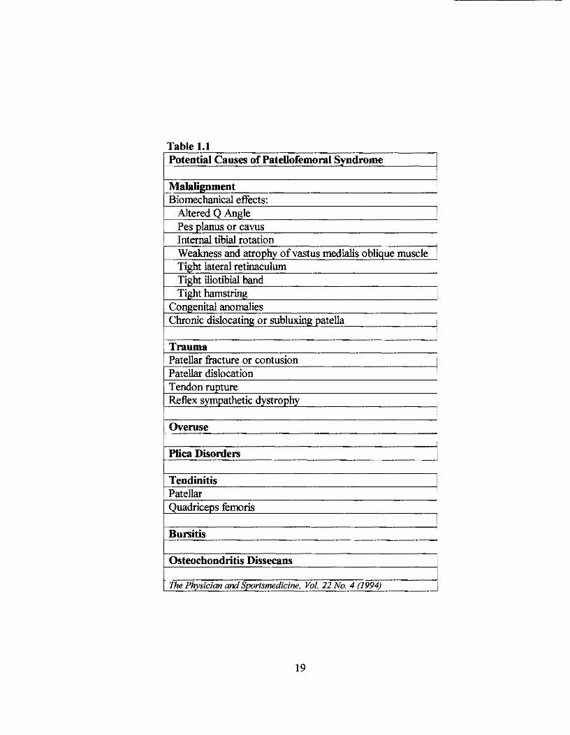
Table 1.1 Potential Causes of Patellofemoral Syndrome
Malalignment Biomechanical effects:
Altered Q Angle Pes planus or cavus Internal tibial rotation Weakness and atro h ofvastus medialis obli ue muscle Ti ht lateral retinaculum
Chronic dislocatin
• Trauma • Patellar fracture or contusion
Patellar dislocation Tendon rupture Reflex sympathetic dystrophy
i Overuse
Plica Disorders
Tendinitis • Patellar
Quadriceps femoris
Bursitis
Osteochondritis Dissecans
The Physician and Sportsmedicine, Vol. 22 No. 4 (1994)
19
I !
i
I
I
I
!
!

Table 1.4
Temporal Progression of Extension Resistance
y = 2.8182x + 134
R2 = 0.5198 180 r-------------------------------------------------------------------~
140 ' ~ - 120 B c: ~ 1<D 'iii ~ 00 c: .2 ~ 60
.! lo(
w 4()
20 0
Table 1.5
6-llfl 9-llfl 13Mij 16My
Pre and Post Measurements (Dates)
Temporal Progression of Maximum Power Output Regarding Extensor Strength
=Pre
- Post -unear(Post)
y= 1.4911x+36.571
~ ,----·------------~--------------------------- R2 =0.~8
1i 50 -1- - -----------:::. 'tl
~ 40 +-- =:c Cl
j30 E .e lo( 20 "' ::!:
10
0
1~ 13
~ ~
Pre and Post Measurements (Dates)
Pre

Table 1.6
70
60 ·- - -
'tJ 50 CD
• ::J
40
-61 .... -. ~ JO
E :I
.6 2ll )(
Ill :!:
10
0
Temporal Progression of Maximum Power Output Regarding Extensor Strength
-- ~
~ ... --- .....
---·
-·
l I
y = 0.6407x- 2414
F( = 0.905
e Pre
Post
- Linear (P
23-.'>pr 2~r 21-~ 29~ 1-1\4' ~114' 5-1\4' 7-114' S.l\4' 11-~ 13~ 15~ 11-~ 1~~ 21-~ 23-~ 25~ 21-~ 29~
Table1.7
2i
ui :e It)
.s 20 ii :I C' CD
.!! :t= c 15 :I
-- -.r. u Ill !!!. 'tJ CD !E 10 r- -...I
.:c
.21
:1 5 E
1-- -
.e )(
i 0
Pre and Post Measurements (Dates)
Temporal Progression of Maximum Power Ouput Regarding Adductor Strength
-
-~-r- -
r- r-
r- r-
-~--::
r- r-
-- r-
~ 1-
--
.. I"""
1---
~
-
11-~
--
-
r--
.. ~
13
~
!-
1-
1-
-
-
-
Pre and Post Measurements (Dates)
21.
18
~
-r--
r-
1-
!-
r-·
-
-
- !-
r- r-
!- r--
1- 1-
y = 0.4857x + 14.848
~ = 0.9583
- Pre
c:::::r Post
-Linear (Pre)

Table 1.8
Temporal Progression of Maximum Power Output Regarding Adductor Strength
25 ~-------------------------------------------------------------, ui ~ ~
£ 20 +---====?~==----~-~~-~ ~ -:::J C' Ql
.!! - -
§ 15 -~-------~'--~----------------------------· .c u
"' !!!. ~
~ 10 +--- -------------------------------- ---; ::i :c Cl
~ E 5 +------ -----------------------------~-----------i
:::J .;; >C
~ 0+-~--~--,---~~--~------~--~--.---.---r-~--~--~--r--4
2}
f:p 7- g. ~ ~
13
~
15 ~
Pre and Post Measurements (Dates)
21-
~
y = 0.2093x- 7881.7 R2 = 0.9593
- Pre
...,..Post
-Linear (Pre:

References
About Delta Society. (Updated November 20, 2002). Retrieved November 26, 2002 from the World Wide Web: http://www.deltasociety.org/dsfOOO.htm
Davies, G. J. (Ed.). (1984). Rehabilitation of the surgical knee: Use of isokinetics in rehabilitation of selected surgical knees. Ronkonkoma, NY: CyPress.
Fine, A. (2000). Handbook on animal-assisted therapy: Theoretical foundations and guidelines for practice. California: Academic Press.
Galea, A.M., & Albers, J.M. (1994). Patellofemoral pain: Beyond empirical diagnosis. The Physician and Sportsmedicine. Vol. 22, No.4, 48-58.
Grabois, M., Garrison, S., Hart, K., & Lehmkuhl, L.D. (2000). Physical medicine and rehabilitation: The complete approach. Massachusetts: Blackwell Science, Inc.
Knee/Hip Exercises. (2003). Student Health Services - Physical Therapy Department.
1 Directions evaluated include linear flexion, linear extension, medial rotation, and lateral rotation. 2 If the measurements are divided into two sections, a first half and second half: and calculated to give a section average, it is found that extension resistance is nearly 14 PSI higher in the second half relative to the first half. 3 The graphs depicted in Tables 1.4 and 1.5 present the same extension data points. It is simply given in a different format in order to make certain revelations easier to see. The same situation is true for the adduction data in Tables 1.6 and 1.7. 4 The stabilization period in these measurements began at the end of Week I on April 29th. This is to recognize that there is a "learning curve" when using isokinetic equipment until the body gets use to how it feels. This explains the dramatic jump in data points after the first session.
23