effects of anaerobiosis upon morphology and energy
TRANSCRIPT
Eur Respir J 1990, 3, 1015-1022
Effects of anaerobiosis upon morphology and energy metabolism of alveolar macrophages cultured in gas phase
M. Cazin*, D. Paluszezak*, A. Bianchi*, J.C. c·azin*, C. Aerts**, C. Voisin**
Effects of anaerobiosis upon morphology and energy metabolism of alveolar macrophages cultured in gas phase. M Cazin, D. Paluszezak, A. Bianchi, J.C. Cazin, C. Aerts, C. Voisin. ABSTRACT: Metabolic and morphological effects of anoxia were studied In alveolar macrophages obtained by lung lavage from guineapigs by means or an original method of cell culture allowing direct contact with air without Interposition or liquid medium. After selection by glass adherence, alveolar cells were layered on a porous membrane applied to the surface of a reservoir filled with nutrient medium. Alveolar macropbages were then cultured In gas phase under either aerobic or anaerobic conditions for 24, 48 and 72 b. Cellular adenosine triphosphate (ATP) content, an Indicator or cell vitality, significantly decreased by 68 and 88% after 48 and 72 h or exposure to anaerobic environment, r espectively. Significant Increases In lactate production (68 % at 24 h) and In glucose uptake (125 % at 24 b), evidence or marked glycolytic activity, occurred before these falls In Intracellular ATP and parallel decreases In culture medium pyruvate level (76 and 85% at 48 and 72 h, respectively). The shift of energy metabolism resulted In cell death after 72 b, as noted by morphological degeneration and decreased cellular ATP content. Twentyrour hour re-exposure to normoxlc atmosphere showed that recovery was possible when duration or anaerobiosis did not exceed 48 b. This reversibility Ln anoxic cell Lojury has been related to plasma membrane Integrity. The results or these studies Indicate that alveolar macrophage resistance to anaerobiosis Is limited as ATP content falls and morphological degeneration occurs after 48 h. This novel approach or anaerobic effects at the cell level should be adaptable to Investigations or activity and, In pa rticular, the mechanisms of metabolic activity of antlanoxlc drugs. Eur Respir J ., 1990, 3, 1015-1022.
• Laboratoire de Phannacologie, Facult~ de Phannacie, Lille, France.
•• Laboratoire de Pathologie Respiratoire et de Pollution AtmospMrique, Institut Pasteur, Lille, France.
Correspondence: Prof. J.C. Cazin, Laboratoire de Pharmacologie. Pacult~ de Pharmacie, 3 rue du Professeur Laguesse, Lille CMex 59045, France.
Keywords: Alveolar macrophages; anaerobiosis; energy metabolism; gas phase culture; morphology.
Received: September 1989; accepted after revision May 14, 1990.
Macrophages are widespread in the body where their surrounding conditions can vary considerably from one organ to another. Such environmental circumstances appear to modulate them or otherwise are linked to their functional and metabolic activities [1, 2]. For example, alveolar macrophages (AM), which reside practically in direct contact with free oxygen in the alveoli, show increased oxidative phosphorylation and decreased glycolytic activities compared to macrophages residing in the peritoneal compartment [3-5].
However, AM offer remarkable resistance to hypoxia in vitro which involves effective adaptation mechanisms in response to environmental 0
2 tension variations,
including increase in pyruvate kinase activity, a key enzyme in glycolysis, and diminution in cytochrome oxidase activity, a key enzyme in oxidative phosphorylation [4, 6]. The latter is associated with decreased total mitochondrial structure available for respiratory activity [6]. Increase in phosphofructokinase activity and corresponding reduction in adenylate energy charge have also been reported as adaptive
responses [7] . The conside rable adaptability to hypoxia, or even anoxia, suggested by these findings appears to be amazing for those alveolar cells that are well adapted to aerobiosis. ln actual fact, the works referenced above have been performed using classical monolayer cultures with cells adhering to the flask and covered with nutrient medium, under conditions very far from the in vivo situation. Consequently, it seemed worthwhile to ascertain whether or not the AM adaptability to anoxia as noted by many investigors is specific to this kind of culture.
The aim of the present study was to investigate the effects of anaerobiosis upon morphology and energy metabolism of AM under conditions approximate to those existing in the lower respiratory tract by means of gas phase cell culture previously described by VoiSIN and eo-workers [8, 9]. This method has also proved to be appropriate for developing a reliable model of experimental anoxia, as it allows a direct contact of AM with the atmosphere and is, therefore, adaptable to a precise evaluation of gas effects on cells.
1016 M. CAZIN ET AL.
Materials and methods
Animals
Experiments were performed in female albino Dunkin-Hartley guinea-pigs (Mepal, Montmedy, France), each weighing approximately 300 g, which had been carefully housed conforming to the guidelines of the Official Journal of European Communities. They had been allowed to acclimatize in our quarters for at least one week before use.
Alveolar macrophages gaseous phase culture. According to MYRVICK et al. [10], AM were obtained by lung lavage with Hank's solution from guinea-pigs. After centrifugation (800 g for 10 min), cells were resuspended in 10% calf serum Basic Eagle's medium (BEM) (Gibco Laboratories, Grand Island, NY, USA) within glass flasks to obtain purified AM. After 24 h, non-adherent cells were eliminated by rinsing, whereas macrophages adhering to glass were removed by 0.125% (w/v) edetic acid (EDTA) solution in Dulbecco's phosphate buffer saline (PBS). Cell numbers were determined manually with a standard haemocytometer. The cells were then suspended in BEM with glutamine (2 mM) and 10% fetal calf serum to obtain 10xl06 cells-ml·1• The cell cultures were performed according to the method of VoisiN and eo-workers [8, 9]. The AM were layered on porous cellulose triacetate membrane Metricel GA 8 0.2 J.Un (Gelman Sciences Inc., Ann Arbor, Michigan, USA). This membrane was applied to the surface of a reservoir filled with nutrient medium so as to be saturated by capillarity. A quantity of cells varying from 0.5-2 million in a volume of 50-200 J.L} were placed on the membranes. Under these conditions, cells were in direct contact with air without any interposition of liquid medium. In all studies, the cell cultures were maintained at 3 7°C in a water saturated environment within an appropriate chamber (Lequeux Ltd, Paris, France). Thus, when incubated under normoxic conditions, AM were able to maintain their metabolic and functional activities for 4-6 days [11].
Normoxic conditions. Normoxic conditions consisted of purified reconstituted air (Alpha Gaz S.A., France), saturated with water (37°C) and enriched with 5% C0
2•
Anaerobic conditions. Anaerobic conditions were achieved by flushing the chamber with a 95% N
2 and
5% C02
mixture. Slight traces of oxygen were eliminated by the GasPak system (BBL Microbiology Systems, Becton Dickinson and Co., Cockeysville, Maryland, USA) which consisted of an envelope containing one piece of filter paper, one tablet of sodium borohydride and one tablet of sodium bicarbonate plus citric acid. }\ and C0 2 were given off by addition of tap or clistilled water into the envelope. Palladium chloride placed within the chamber promoted a H
1 and C0
2 reaction to produce ~0 (Laidlaw principle), resuhing in the disappearance of 0
1 and a water saturated envi
ronment. Anaerobiosis was verified by means of a very
sensitive calorimetric inclicator: resazurin which became colourless when reduced (E0 = -51 mV); i.e. when anaerobic conditions were achieved [12, 13]. Several experimental trials were performed in which AM were exposed to anaerobic conditions for 24, 48 and 72 h. Following exposure to anaerobic atmospheres, some cell samples were exposed to normoxic atmospheres for 24 h in order to estimate their recovery ability.
Cell morphology. Morphological analysis was performed by electron microscopy, after fixing in 2.5% glutaraldehyde in 0.1 M cacodylate buffer, pH 7.2 for 12 h at 4°C and staining with 0.5% uranyl acetate, pH 5 for 1 h at 4°C, as described previously [9]. The cells were examined under a Hitachi type HV 12 electron microscope.
Energy metabolism. Total cell energy metabolism was studied by assay of intracellular adenosine triphosphate (ATP) through a bioluminescent method using luciferinluciferase [9, 14, 15] on a Berthold Biolumat LB 9500T (C.L.V. Interbio~. Villeurbanne, France). The analysis was carried out on cellular extract in dimethyl sulphoxide (DMSO).
Culture medium pyruvate and lactate were assayed, as glycolysis indices, by classic enzymatic methods (kits no. 726 U.V. and no. 826 U.V. from Sigma Chemical Co., Saint Louis, Missouri, USA) using lactatedehydrogenase, at the end of each trial [12]. Measuring was performed after incubation with a UV spectrometer at 340 nm.
Glucose uptake. Measurement of 3H-2-deoxy-Dglucose uptake by cells is regarded as a suitable method for determining the uptake of hexoses by cells [16, 17]. Deoxyglucose crosses the cell membrane by the same carrier mechanism characteristic of facilitated diffusion as glucose, but it accumulates in cells as deoxyglucose-6-phosphate [16-18] .
In our study, measurements were made in triplicate in normoxia at 37°C after either aerobic or anaerobic 24 h exposures [12]. Each study sample was randomly assigned to 4 equivalent groups (according to the schedule 10, 20, 30 and 40 m in) for kinetic examination. Radiolabelled deoxyglucose [3H] (specific activity: 30.4 Ci·mmol·1
; Amersham International plc, Amersham, Buckinghamshire, UK) was added to cells (1 J.LCix10·6 cells) by replacing BEM nutrient medium with phosphate buffered saline (PBS) enriched with 0.9 mM ea••, 0.5 mM Mg•• and the radioactive sugar so that all the samples were under standardized conditions. Uptake was stopped by gently washing the membranes supporting the macrophages with cold PBS at 4°C two times. This washing made it possible to reduce nonspecific raclioactivity. Radioactivity of both cells and membranes was measured on a Beckman LS 1800 liquid scintillation counter (Beckman Instruments, Inc., Palo Alto, California, USA). Results were expressed as cpm· l0·6 macrophages in comparison with blank cellulose triacetate membranes without any cells and treated under identical conditions. Where appropriate,
ALVEOLAR MACROPHAGE$ CULTURED IN ANAEROBIOSIS 1017
cyLOchalasin B (S igma Chemical Co., Saint Louis, Missouri, USA) was added at zero time, along with the tritiated deoxyglucose, at a concentration of 5 ~-tg·ml·1 • Radioactivity measurements were processed as a function of time on an Apple ID microcomputer (Business Graphic Software) using a regression program which allowed calculation of transport rate [12].
Statistical analysis. All biochemical measurements were made in triplicate. Mean data are presented with standard deviation. Lack of variation of each metabolic parameter in AM cultured in normoxia was verified by a one-way analysis of variance, selecting time as classification variable, so as to demonstrate stability and reliability of cell preparations under the above-described conditions. Each individual result recorded under anaerobic conditions was compared with data obtained from l.he same cell batch (i.e. harvested in l.he same animal) cullured under similar conditions but in a normoxic environment. Thus, comparisons between means were made using paired Student's Hest and p<O.OS was considered to indicate significant differences. Since recovery consisted of additionally exposing AM to normoxic atmosphere for 24 h after an anaerobic trial, results of that investigation were compared with measurements made in anaerobiosis on the day before. Besides Laking into account the perfect time stability of metabolic parameters in nonnoxia, these results were also compared with corresponding data obtained in normal atmosphere. The value obtained for lactate production represents overall production observed at the end of each trial. Thus, in case of recovery, this cumulative result must be compared with reference data of the preceding and same days.
Results
Morphological findings
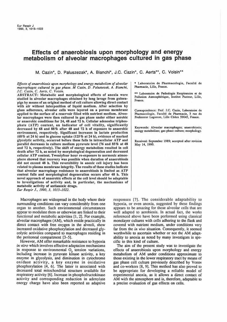
The morphological characteristics of AM were observed after 24, 48 and 72 h in either aerobic or anaerobic conditions and their recovery ability was estimated after 24 h in normoxia following exposure to strict anaerobic environments. In AM cultured anaerobically for 24 h, only a few differences were found compared to control normoxia cells. There appeared to be little grey probably " lipidic vesicles". General ultrastructure remained unchanged: nucleus, pilopodia at the cell surface and mitochondria were all intact. In AM cultured anaerobically for 48 h, pilopodia became stunted and concentrated on the two poles of cell bodies which were obviously elongated as result of loss of cell volume regulation. Increase and spread of "lipidic vesicles" seemed to be in correlation with anaerobiosis duration (fig. 1). At this time, decrease in the number of mitochondrial cristae and even, on certain fields, progressive disappearance of mitochondria could be noticed. In AM cultured anaerobically for 72 h, vesicle number was greater and the nuclei became pyknotic. Some cells lost their plasmalemmal membranes
(fig. 2). Elongation of cell bodies persisted in the most resistant cells, the mitochondria of which had nearly totally disappeared. The completely degenerated cell popuJation accounted for more than 60%.
Fig. I. - Electron micrograph of guinea-pig alveolar macrophages cultured anaerobically in gas phase: for 48 h (magnification: x12,000). Pilopodia are stunted and concentrated on the two poles of cell bodies which are elongated.
With regard to the recovery aspect, after 24 h of anaerobiosis, the recovery in normoxia was satisfactory and cells exhibited a nearly normal appearance. After 48 h of anaerobiosis, "lipidic vesicles" remained and the cells, although less elongated, did not recover their initial morphology. Owing to the different fields , the recovery of a few mitochondria was somewhat subjectively noted. ln AM cultured anaerobically for 72 h prior to normoxic period, no morphological recovery was observed, as seen in figure 3. This finding indicates that, after 72 h in anaerobic environment, alveolar cells had entered an irreversible state of anoxic injury as they continued to degenerate and become necrotic despite reoxygenation.
Effects on energy metabolism
Anaerobiosis was maintained during 24, 48 and 72 h to test for adaptation and recovery abilities of AM. Table I shows that anaerobiosis merely entailed a significant

1018 M. CAZIN Er AL.
Fig. 2. - Electron micrograph of guinea-pig alveolar macrophages cultured anaerobically in gas phase for 72 h (magnification: x l5,000). Vesicle number is greater in comparison with f~gure 2 and the nucleus is pyknotic. Most cells have lost their plasmalemrnal membranes whereas intact mitochondria are missing. Necrotic debris are visible.
If
Pig. 3. - Electron micrograph of guinea-pig alveolar macropbages wb.ich have recovered in nonnoxia for 24 b after surviving in anaerobic environment du.ring 72 h (gas ph.ase culture) (magnification: x 15,000). No morphological recovery can be observed relative to figure 2.
68% increase in lactate production as early as 24 h. On the other hand, total oxygen deprivation induced disturbances of all parameters from 48 h. ATP content, considered as an indicator of cell vitality, decreased by 68% (p<0.04) after 48 h, and by 88% (p<0.02) after 72 h compared to AM cultured in air (table 2). Culture medium pyruvate level correspondingly decreased by 76% (p<O.OOl) after 48 hand further decreased by 85% (p<O.OOl) after 72 h compared to AM cultured in air (table 3), while lactate production increased nearly twofold by 48 h and threefold by 72 h, relative to that for AM cultured in air (p<O.OOl, table 1). As noted in the same tables, success of recovery (24 h in normal atmosphere after swviving in anaerobic environment) was directly linked to duration of exposure to anaerobic environments. Especially for A TP content, cells cultured in anaerobiosis for 48 h retrieved a mean value of A TP that was not statistically different from that recorded in normoxia, contrary to 72 h anaerobically cultured AM. With respect to lactate production, the sole parameter that significantly varied after 24 h anaerobiosis, the recovery value (i.e. after supplementary 24 h culture under aerobic conditions) was lower than that recorded after 48 h anaerobiosis. This reduction in lactate production, in comparison with anaerobiosis, clearly indicates a return to aerobic metabolism.
Table 1. - Effects of anaerobiosis on time course of lactate production from guinea-pig alveolar macrophages surviving in gas phase
Time h
24 48 72
Lactate production ~g·10·' cells
Nonnoxia
336±110 403±76 461±72
Anaerobiosis
565*±114 757•±33
1351*±121
Recovery
650••±78 1191**±201 1466**±169
Data are mean±so for six experiments. •: p<O.OOl compared to nonnoxia; ••: p<O.OOl compared to nonnoxia and p<0.01 compared to anaerobiosis.
Table 2. - Effects of anaerobiosis on time course of ATP content in gu inea-pig alveolar macrophages surviving in gas phase
Time h
24 48 72
ATP content ~g·10·' cells
Nonnoxia
2.32±1.14 2.22±1.59 2.38±1.19
Anaerobiosis
2.23±1.11 0.72*±0.42
0.29*•±0.22
Recovery
1.98±1.25 1.3lf±0.54 0.41 tt±0.32
Data are mean±so for six experiments. *: p<0.04 compared lO nonnoxia; •• :p<0.02 compared to nonnoxia; t: nonsignifjcant compared to nonnoxia and p<0.02 compared to anaerobiosis; tt; p <0.03 compared to normoxia and nonsignificant compared to anaerobiosis; ATP: adenosine triphosphate.
ALVEOLAR MACROPHAGES CULTURED IN ANAEROBIOSIS 1019
Table 3. - Effects of anaerobiosis on time course of culture medium level of pyruvate for guinea-pig alveolar macrophages surviving in gas phase
Pyruvate level Jlg·l0'6 cells Time
h Normoxia Anaerobiosis Recovery
24 12.1±1.3 12.8±1.3 10.4**±0.5 48 10.8±1.3 2.6*±0.8 6.2'±1.1 72 10.5±1.7 1.6*±0.7 3.5'±0.1
Data are mean±so for six experiments. *: p<0.001 compared to normoxia; **: p<0.02 compared to normoxia and p<O.Ol compared to anaerobiosis; t: p<0.01 compared to normoxia and p<0.02 compared to anaerobiosis.
Table 4. - Influence of anaerobiosis on 3H-2-deoxy-O-glucose uptake by guinea-pig alveolar macrophages surviving in gas phase during 24 h
Environmental n* Transport rate conditions cmp per min ·1 0 '6 cells
Normoxia 6 76.4±17.2
Anaerobiosis 6 171.8±23.5**
Results are mean±so. *: nwnber of experiments performed in triplicate; ••: p<0.001 compared to normoxia.
Effects on glucose uptake
This investigation has been undertaken to demonstrate the shift in energy metabolism in cells exposed to the oxygen-free milieu, as well as the membrane integrity regarding glucose transport at the very time when ATP content was unchanged relative to normoxia. Twenty four hour anaerobiosis markedly stimulated deoxyglucose transport in the AM. Table 4 shows that tritiated glucose analogue uptake was increased by 125% (p<0.001) compared to control AM cultured in air, under our experimental conditions. This observation is in accordance with previous findings pertaining to other cell types [19, 20] and corroborates concomitant increase in lactate production which shows the metabolic deviation towards anaerobic pathways.
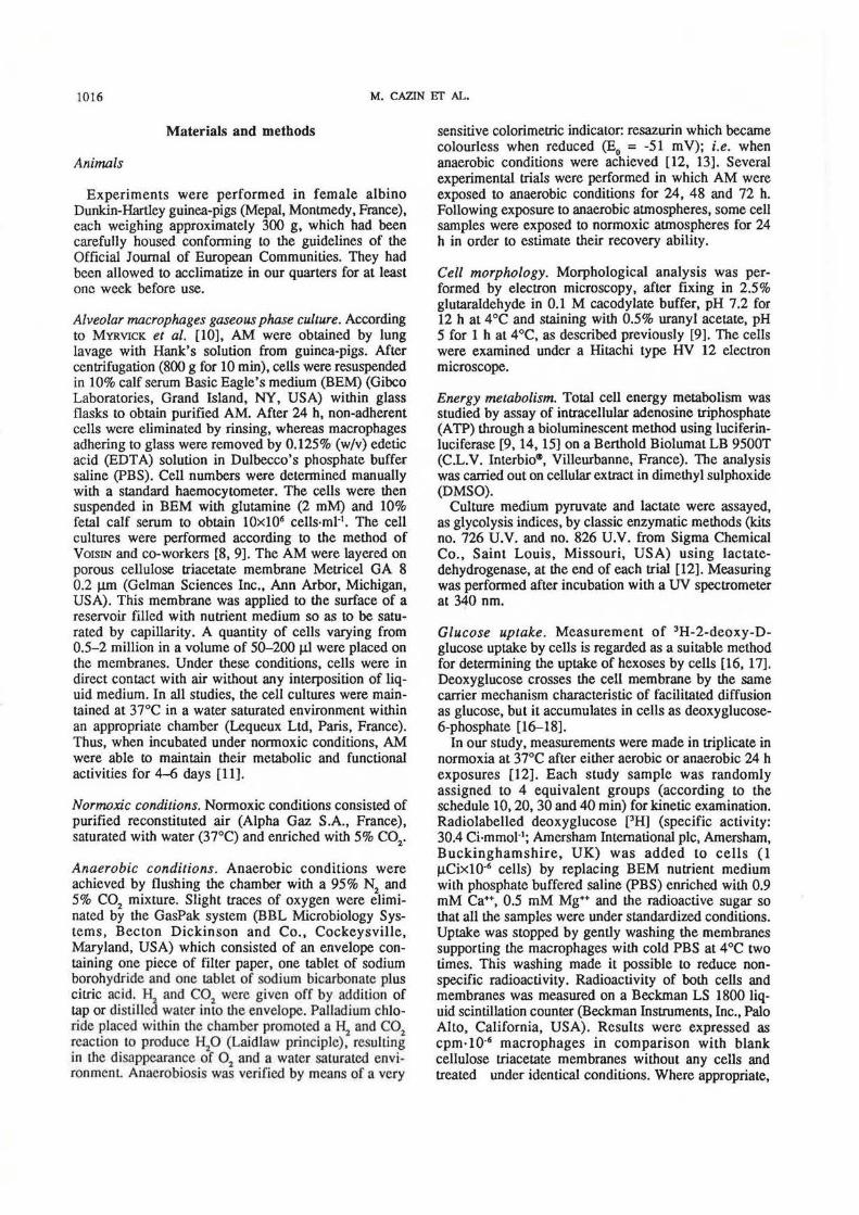
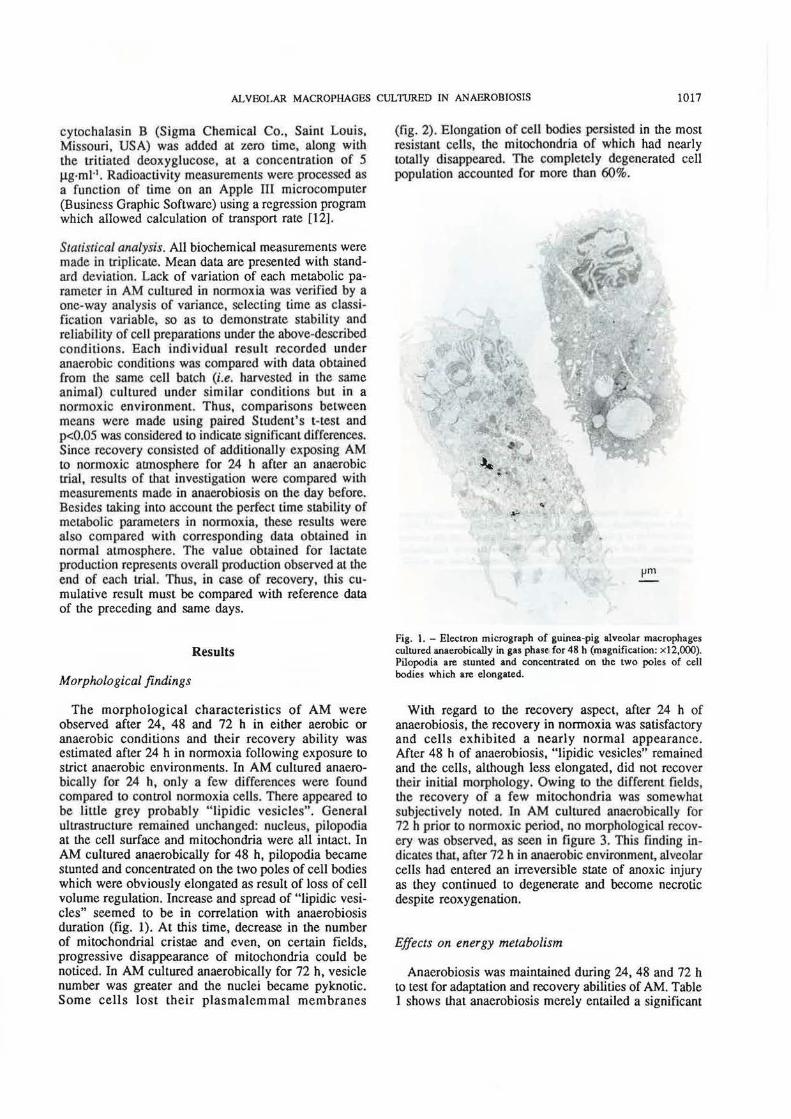
An experiment, designed to further examine cell membrane integrity, was conducted in triplicate with the glucose transport inhibitor cytochalasin B. Figure 4 illustrates the ability of cytochalasin B to inhibit essentially all deoxyglucose transport (more than 95%) either in aerobic or in anaerobic environments. These inhibitory effects in such different conditions suggested that, in both cases, the same transport mechanism was affected and, consequently, anaerobiosis specifically acted on it.
.!! Normoxla 8 ~
6000
e Control c. y: -307.2+138.94X u
J J! 4000 c. ::1
I 8
t 2000
a Treated ., y = -8.2+4.68x
~ 0
0 10 20 30 40 so llmemln
.!! 8
Anaerobiosis
b 12000 ... E c. 10000 u Control ~ .¥ J9 8000 c.
y = • 704.6+300.3Sx
::1
I 8
6000
::1 0, >-ac 4000
a J 2000
Treated y = 38.6+9.47x
0 20 30 40 so
llmemln
Fig. 4. -Influence of cytochalasin B (5 ~g·mi-') on 'H-2-deoxy-Dglucose uptake by guinea-pig alveolar macrophages surviving in gas phase during 24 h. Values are results of one experiment performed in triplicate.
Discussion
Prior experiments carried out in our laboratory had shown that AM surviving in gas phase could easily adapt to difficult hypoxia conditions such as 5% 0 2 or 1% 0
2 environments during 48 h or 72 h [21]. These findings seemed to be rather surprising for such aerobic cells practically residing in direct contact with free oxygen in the alveoli. The present studies have demonstrated that AM can, only in part, endure total anoxia. Indeed, strict anaerobic conditions for 48 h brought about a significant decrease in energy charge, as well as a morphological degeneration, whereas such environment during 72 h led to cell death. However, those cell injuries caused by anaerobiosis were reversible if duration of trial was limited, as indicated by return of the various metabolic parameters to control values when cells were exposed to air again for 24 h.
Modification of metabolic characteristics can readily explain the relative resistance of AM to anaerobic conditions. Cessation of mitochondrial respiratory activity in absence of 0
2 resulted in an anaerobic glyco-

1020 M. CAZIN ET AL.
lysis stimulation which was reflected in the increases in lactate production and glucose uptake at 24 h. It can be noted that the latter may be the result of a specific effect of anaerobiosis upon glucose transport and/or be a consequence of cell membrane injury mediated via cell metabolic disturbances. However, the second assumption was not supported by experiments involving inhibition of deoxyglucose uptake by cytochalasin B. This agent has proved to specifically inhibit the deoxyglucose (and glucose) transport undoubtedly by a membrane mechanism [16, 17, 22-27]. The fact that anaerobic stimulation of this glucose transport associated with the cell membrane was inhibited (by more than 95%) in the presence of cytochalasin B has been considered as an indication of membrane integrity. According to FARBER [28], disturbances in membrane function in general and in the plasma membrane in particular characterize the loss of reversibility in ischaemic injury. Thus, defects in cell membranes are regarded as an early feature of irreversible, ischaemic cell injury. Therefore, the finding that, after 24 h in anaerobiosis, the AM membrane appeared to be intact as it normally reacted to cytochalasin B, relative to normoxia, is fairly consistent with AM abilities to maintain their intracellular A TP store at this time and to recover aerobic metabolism when exposed to air again, as shown by decrease in their lactate production and normal morphological appearance.
Despite the adaptive response by AM, energy deficit occurred at 48 h since anaerobic glycolysis produces very much less ATP compared to respiratory activity and, thereby, cannot completely replace the dependence of cells upon oxidative metabolism. The fatal course in the current studies, as noted by morphological degeneration together with loss of cell volume regulation and decreased cellular ATP content, has been partially interpreted as result of intracellular acidosis (induced by excessive stimulation of glycolysis) which has been found to inhibit enzymatic systems [29]. Such a phenomenon could therefore limit cell adaptability. In this respect, it is noteworthy that, under our experimental conditions, the high concentrations of lactate in the culture medium associated with anaerobic situation probably play an important role in the time course of these events by potentiating the pH effects of intracellular acidosis. In other respects, it has been established that a membrane mechanism, not yet entirely clear and involving a massive free fatty acid release, generates irreversible changes during the acute phase of ischaemic injury in well-differentiated tissues such as heart or brain [30, 31]. A sequence of events mediating ischaemic cell death has been proposed by FARBER [281 in a study of the pathogenesis of membrane injury and the resulting coagulative necrosis in ischaemia. Accordingly, the effects of anoxia on phospholipid metabolism and, therefore, on cell integrity do not appear as a direct result of the loss of cellular stores of ATP. Actually, as oxygen is also being utilized by a number of oxygenases such as cytoplasmic fatty acid desaturases, the sensitivity to oxygen deprivation should relate to both its function in energy metabolism and the
synthesis of polyunsaturated fatty acids needed to maintain membrane phospholipids. This biochemical assumption is in good agreement with our morphological findings.
The effects of anaerobiosis on energy metabolism of AM surviving in liquid phase for 96 h have been reported previously by BurrERICK et al. [6]. These investigators established a shift in energy metabolism similar to the one that we observed, without loss of cell viability or alteration in total cell cross-sectional surface area. This main difference compared to our findings appears to be related to the experimental conditions we have employe; i.e. the gas phase culture of AM. Quite well adapted to aerobic metabolism in such conditions, these cells seemed to be more sensitive to the actual effects of 0
2 depletion. Cell energetics ad
aptation observed in the previous work can be partially explained by an alteration of macrophage behaviour in response to the liquid phase conditions as higher productions of lactate have been found in AM cultured in liquid phase in comparison with gas phase, regardless of environmental conditions (unpublished data). This transformation might have been facilitated by medium replacement in the experimental procedure taken as reference. Thus, the glucose indispensable for anaerobic glycolysis was c·orrectly supplied and overproduction of lactate was eliminated, resulting in the absence of extracellular pH effects due to this substrate.
Ultimately, the study of main energy metabolic parameters and morphology of AM exposed to anaerobic conditions in gas phase provides an experimental model of anoxia. Unlike the method of Buuerick, the current method is a true model of cellular ischaemia, exhibiting moderate glucose supply and overproduction of lactate as encountered in pathological situations. As the effects of this experimental ischaemia were reversible if anaerobiosis was limited to 24 h or even 48 h, it has been successfully used as pharmacological indicator for investigating the ability of various chemicals to protect against hypoxia. Moreover, similarities observed in vitro between respiratory activities of alveolar cells and brain cells [3] enabled us to utilize cultured AM for the study of antianoxic drugs endowed with possible cerebral protective properties, such as vincamine [12, 21]. This alkaloid actually revealed an interesting action as it was capable of maintaining cell metabolic activity for a longer period of time after the beginning of an anoxic trial when compared to the control. Other agents are being investigated.
In conclusion, AM appear to be a convenient model for studying ischaemic cell injury as they require large amounts of oxygen to provide the energy to support their specialized functions. In reality, the effects of anoxia and ischaemia are most significant in those tissues which share this metabolic characteristic. By means of gas phase culture, it is possible to experiment with alveolar cells under conditions absolutely favourable to their aerobic metabolism. Furthermore, the fact that AM are able to tolerate anaerobiosis during a certain time and exhibit reversible damage until48 h avoids an annoying all-or-none response. Thus, the approach

ALVEOLAR MACROPHAGES CULTURED IN ANAEROBIOSIS 1021
of anoxia at the cell level should be perfectly adaptable to elucidation of mechanisms of metabolic activity of antianoxic drugs.
Acknowledgements: The authors wish to acknowledge the noancial suppon from Oril Co., and the suppon of the Universit~ de Lille II.
References
1. Blusse A, van Oud A, van Furth R. - Origin, kinetics, and characteristics of pulmonary macrophage& in the normal steady stare. J Exp Med, 1979, 149, 1504-1518. 2. N1chols BA, BaintOn OF. - In: Mononuclear phagocytes in immunity, infection and pathology. R. van Furth ed. Blackwell Scientific Publications, Oxford, 1976., 3. Oren R, Famharn AE, Saito K, Milofsky E, Karnovsky ML. - Metabolic patterns in three types of phagocytizing cells. J Cell Biol, 1963, 17, 487-501. 4. Simon LM, Axline SG, Horn BR, Robin ED. -Adaptations of energy metabolism in the cultivated macrophage. J Exp Med, 1973, 138, 1413-1425. 5. Simon LM, Robin EO, Phillips JR, Acevedo J, Axline SG, Theodore J. - Enzymatic basis for bioenergetic differences of alveolar versus peritoneal macrophages and enzyme regulation by molecular 0
2• J Clin Invest, 1977, 59,
443-448. 6. Butterick CJ, Williarns DA, Boxer LA, Jersild RA Jr, Mantich NM, Higgins C, Baehner RL. - Changes in energy metabolism, structure and function in alveolar macrophages under anaerobic conditions. Br J Haematol, 1981, 48, 523- 532. 7. Sides GD, Mantich NM, Boxer LA, Baehner RL. -Transformation of energy charge and phosphofructokinase act ivity in alveolar macrophages. J Reticuloendothe.l Soc, 1981, 29 (5), 407-412. 8. Voisin C, Aerts C, Tonne! AB, Houdret JL, Rarnon P. - Mise en survie en phase gazeuse et reconstitution in vitro du micro-environnement nature! des macrophages alveolaires. Path Bioi, 1975, 23, 453-459. 9. Voisin C, Aerts C, Jakubczak E, Tonnel AB . - La culture cellulaire en phase gazeuse. Un nouveau mod~le experimental d'etude "in vitro" des activites des macrophages alveolaires. Bull Eur Physiopathol Respir, 1977, 13, 69-82. 10. Myrvik GN, Leake ES, Fariss B. - Study on pulmonary macrophages from lhe nom1al rabbit. A technique to procure them in a high state of purity. J Jmmunol, 1961, 86, 128-132. 11. Voisin C, Aerts C, Tonne! AB. - La survie in vitro des macrophages alveolaires humains en phase gazeuse. Application a l'etude de la cytotoxicite de l'oxygene. JNSERM, 1979, 84, 169-176. 12. Paluszezak D. - Etude pharmacologique de substances a visee protectrice cerebrale derivees de ta P-carboline: mise au point d'un modele d'hypoxie sur macrophages alveolaires. These de Doctorat de Troisieme Cycle, 1985, Faculte de Pharrnacie, Lille. 13. Anderson KL, Fung DYC. - Anaerobic methods, techniques and principles for food bacteriology: a review. J Food Protection, 1983, 46 (9), 811-822. 14. Jakubczak E, M azingue C, Leclerc H. - Etude critique des methodes d'extraction de l'ATP chez les bacteries en vue de son dosage par la melhode de bioluminescence. Regard sur la biochimie, 1978, 2. 14. 15. MacElroy WD, Seliger HH. - The chemistry of light emission. Adv Enzymol, 1963, 25, 119.
16. Gee LJB, Khandwala AS, Bell RW. - Hexose transport in the alveolar macrophage: kinetics and pharmacologic features. J Reticuloendothel Soc, 1974, 15 (5), 394-405. 17. Khandwala AS, Gee LIB.- Factors in glucose oxidation by alveolar macrophages: glucose transport and glycogenolysis. Chest, 1975, 67, 60s-63s. 18. Sokoloff L, Reivich M, Kennedy C, Des Rosiers MH, Patlak CS, Pettigrew KD, Sakurada 0, Shinohara M. - The (
14C) deoxyglucose method for the measurement of local cerebral glucose utilization: theory, procedure and normal values in the conscious and anesthetized albino rat. J Neurochem, 1977, 28, 897- 916. 19. Morgan HE, Henderson MJ, Regen DM, Park CR. -Regulation of glucose uptake in mu~cles. J Bioi Chem, 1961, 236 (2). 253- 261. 20. Whitfield CF, Morgan HE. - Effect of anoxia on sugar transport in avian erythrocytes. Biochem Biophys Acta, 1973, 307, 181-196. 21. Cazin M, Paluszezak D, Poulain V, Dine T, Bianchi A, Cazin JC, Aerts C. - Use of alveolar macrophages in anti anoxic drug studies. Meth Find Exp Clin Pharmacol, 1988, 10 (4), 231-237. 22. Brown SS, Spudich JA. - Cytochalasin inhibits lhe rate of elongation of actin filaments fragments. J Cell Bioi, 1979, 83, 657-662. 23. Estensen RD, Plagemann PGW. - Cytochalasin B: inhibition of glucose and glucosarnine transport. Proc Nat Ac Sci USA, 1972, 69, 1430-1434. 24. Kimrnich GA, Randles J. - Energies of sugar transport by isolated intestinal epithelial cells: effects of cytochalasin B. Am Physiol Soc C, 1979, 56-63. 25. Kletzien RF, Perdue ID. -The inhibition of sugar transport in chick embryo fibroblasts by cytochalasin B. Evidence for a membrane-specific effect. J Bioi Chem, 1973, 248, 711-719. 26. Shin Lin, Shang Lin D, Flanagan MD. - Specificity of lhe effects of cytOchaJasin B on transport and motile processes. Proc Nat Ac Sci USA. 1978, 75, 329- 333. 27. Zigmond SH, Hirsch JH. - Cytochalasin B: inhibition of D-2-dcoxyglucose transport into leukocytes and fibroblasts. Science, 1972, 176, 1432- 1434. 28. Farber JL. - Membrane injury and calcium homeostasis in the pathogencsis of coagulative necrosis. Lab Invest, 1982, 47 (2), 114-123. 29. Robin EO. - Of men and mitochondria: coping wilh hypoxic dysoxia. The 1980 J. Bums Ambcrson Lecture. Am Rev Respir Dis, 1980, 122, 517- 531. 30. Jennings RB, Ganote CE, Reimer KA. - Ischemic tissue injury. Am J Pathol, 1975, 81, 179-198. 31. Baron JC. - Phenomenes physiopathologiques au cours de l'ischernie focale aigu~ du cerveau. Rev Med, 1983, 38, 1853-1863.
Effets de I'anaerobiose sur la morphologie et le metabolisme energetique des macrophages alveolaires maifllenus en survie en phase gtu.euse. M. Cazin, D. Paluszezak, A. Bianchi, J.C. Cazin, C. Aerts, C. Voisin . RESUME : Des macrophages alveolaires de cobaye ont ete maintenus en survie en anaerobiose durant 24, 48 et 72 h, suivant un procede de culture en phase gazeuse assurant un contact direct entre les cellules et l'aunosphi'!re environnante. Dans ces conditions, la tcneur cellulaire en adenosine tri phosphate (ATP). expression de la vitalite des celulJes, a respectivement chute de 68 et 88% A 48 et 72 h. Des augmentations significatives de la production de lactate (68% a
1022 M. CAZIN ET AL
24 h) et de la consommation de glucose (125% a 24 h), preuves d'une activite glycolytique accrue, ont ete observees avant cette chute du contenu en ATP et une diminution parallele de la concentration de pyruvate dans le milieu de culture (respectivement 76 et 85% A 48 et 72 h). A !'issue des 72 h, une lyse cellulaire importante a ete objectivee par une analyse morphologique en microscopic electronique et les taux extrcmement faibles d'ATP. En repla~ruules cellules hypoxiques dans une atmosphere normale pendant 24 h, les auteurs ont montre que la recuperation etait possible lorsque la duree de 1' anaerobiose n 'excedait pas 48 h. A la difference
des donnees bibliographiques, les resultats de ces experiences demontrent que la resistance des macrophages alveolaires a l'anaerobiose est limitec. Ceue approche originate des effets de I'anaerobiose a !'echelon cellulaire apparait adaptee a I' etude des activites et, en particulier, des mecanismes d'action des substances anti-anoxiques. L'analyse de medications a visee protectrice cerebrate a meme ete envisagee, compte tenu des similitudes observees in vitro entre le metabolisme energetique des cellules cerebrates et celui des macrophages alveolaires. Eur Respir J., 1990, 3, 1015- 1022.