effect of silver nitrate and nickel chloride on shoot
TRANSCRIPT

EFFECT OF SILVER NITRATE AND NICKEL CHLORIDE ON SHOOT
MORPHOGENESIS IN DIANTHUS
Submitted
in partial fulfillment of the requirement of the degree of
Master of Philosophy
in the Faculty of
Life Sciences
THE IIS UNIVERSITY, JAIPUR
BY:
VASUNDHRA SINGH
UNDER THE SUPERVISION OF:
DR. ANUJA JOSHI
DEPARTMENT OF BOTANY
THE IIS UNIVERSITY
(2011-2012)

ACKNOWLEDGEMENT
I express my deep sense of gratitude to Dr. ASHOK GUPTA, Director, The IIS University and
Dr. RAKHI GUPTA, Principal, The IIS University who provided me an excellent and healthy
environment as well as well equipped facilities.
With humble respect I would like to give my sincere thanks to Professor PRADEEP
BHATNAGAR, Dean, Department of Life Sciences, The IIS University, for acting as the
constant pillar of support and guidance throughout my endeavor.
With heartfelt gratitude and immense pleasure I acknowledge my guide, DR. ANUJA JOSHI,
Assistant Professor, Department of Botany, The IIS University, without whose supervision and
guidance this work would not have been a success. I have had the advantage of her critical
advice from her profound knowledge in the subject.
VASUNDHRA SINGH
M. Phil. Life Science

CONTENTS
INTRODUCTION………………………………………………….1-4
REVIEW OF LITERATURE…………………………………… . 5-10
MATERIALS AND METHODS………………………………… 11-17
Plant MaterialSeed Viability TestExplants taken from aseptically grown seedlingsEquipments and apparatusesSterilization and preparation of explantsBasal medium preparationAseptic manipulationInduction of shoot budsSubculture ProcedureRooting of explantsProtocol for Dianthus cultureIncubationObservationStatistical Analysis
OBSERVATIONS AND RESULTS………………………..........18-27
Seed Viability TestSeed GerminationExplant CultureEffect of cytokinin and auxinEffect of varying concentration of heavy metals
DISCUSSION……………………….…………………………….28-30
SUMMARY……………………………………………………….31-32
REFERENCES…………………………………………………….33-40

ABBREVIATIONS
BAP : 6-Benzyl Aminopurine
NAA : α-Naphthalene Acetic Acid
cm : Centimeter
2, 4-D : 2, 4- dichlorophenoxyacetic acid
DW : Distilled water
Fig. : Figure
gm. : gram
mg/l : milligram per liter
ml : milliliter
mm : millimeter
MS : Murashige and Skoog (1962) medium
S. D. : Standard Deviation
UV : Ultraviolet
W : Watt
w/v : Weight by volume

µmol : Micromole
INTRODUCTION

Dianthus is one of the world’s most popular, economic and important cut flowers due to its
excellent keeping quality, wide range of forms, ability to withstand long distance transportation
and remarkable ability to rehydrate after continuous shipping, perpetual flowering and presence
of new single- and multi-colour cultivars and is native of the Mediterranean region.
It belongs to family Caryophyllaceae. The Caryophyllaceae, commonly called the pink family or
carnation family, is a family of flowering plants. It is a large family, with 88 genera and some
2,000 species. This cosmopolitan family consists of mostly herbaceous plants and is best
represented in temperate climates, with a few species growing on tropical mountains. Some of
the more commonly known members include pinks and carnations (Dianthus), and Firepink and
Campions (Lychnis and Silene). Many species are grown as ornamental plants, and some species
are widespread weeds. Most species grow in the Mediterranean and bordering regions of Europe
and Asia.

CLASSIFICATION
Kingdom : Vegetabile
Sub-Kingdom : Angiospermae
Division : Dicotyledonae
Class : Polypetalae
Order : Caryophyllinae
Family : Caryophyllaceae
Genus : Dianthus
The genus Dianthus consists of more than 300 species including the ornamentals such as
Carnations (D. caryophyllus L.), Pinks (D. chinensis) and Sweet Williams (D. barbatus). D.
caryophyllus is a top-selling ornamental crop
worldwide. D. chinensis is short-lived, perennial garden
plants, native to China and northern parts of India,
blooming in several colours. Certain varieties of pinks
have a strong clove scent, which has made them popular
in perfumes for more than 2,000 years. It has a wide
range of medicinal uses as well. D. barbatus (biennial
carnation) is native to the mountains of southern Europe
from the Pyrenees east to the Carpathians and the
Balkans, with a variety disjunct in north-eastern China,
Korea, and south-easternmost Russia. It is traditional
used in landscaping and cut flowers. Dianthus is also
used for culinary purpose. Crystallized petals are used for decorating cakes, while fresh petals

can be used in salads, pies, and sandwiches. Petal bases are removed before using it for culinary
purposes because of the better taste.
Dianthus is an herbaceous perennial plant growing upto 80 cm tall. The leaves are glaucous
greyish green to blue-green, slender and up to 15 cm long. The flowers are produced singly or in
group of five together in a cyme, and sweetly scented. The leaves are linear, lance-shaped and
blue-grey or grey-green in colour, with a waxy texture. The fruit is non-fleshy. It is usually a
capsule, less frequently a small nut. The original natural flower colour is bright pinkish-purple,
but cultivars of other colours, including red, white, yellow and green have been developed
through breeding and planted in well-drained soil in a sunny location.
Dianthus cultivation is associated with various problems .The seeds show the high level of
heterogeneity in the seed population which acts as a constraint to propagation of agronomic traits
and for commercial seed production. Carnation breeders constantly seek new varieties with
improved horticultural traits such as disease and pest resistance and long vase-life. The
traditional breeding programs for selection of superior clones are tedious and utilizing huge
amount of seed results in wastage of the seeds significantly. In vitro technology has often been
used as a promising avenue to plant improvement for research and practical applications. Plant
tissue culture, is of great value, particularly as an alternative to conventional breeding and
propagation procedures.
PLANT TISSUE CULTURE MEDIUM
Growth and morphogenesis of plant tissue under in vitro conditions are largely governed by the
composition of the culture medium. Various culture media formulation proposed consist of
several basal components which include mineral nutrients, vitamins, amino acids, growth
regulators, sugars, agar and water in various concentrations. Mineral nutrients are the building
blocks for the synthesis of organic molecules and also act as catalysts in enzyme reactions. The
ions of the dissolved salts also play important role as counter-ion in the transport of ionized
molecules by the plant, in the osmotic regulation and in maintaining the electrochemical
potential of the plant. The mineral salts in media can be broadly classified into two groups:
microelements and macroelements. Microelements including Fe, Cu, Zn, Co, Mo, B, Cl and I are

required in relatively low concentration but their deficiency can have catastrophic effects. Mg,
Ca, S, P, K and N are considered as macroelements since they are required in high concentration.
Vitamins, amino acids, growth regulators and sugars act as organic supplements in the medium.
Agar and Phytagel are added as gelling agents. All these components fulfill the one or more
requirements of plant under in vitro conditions.
The formulation of the mineral components of the media has been an empirical process since the
beginning of tissue culture. Knop (1865) proposed a salt solution which formed the basis for
culture medium. Kotte (1922) cultured small root tips of pea and maize which could not survive
indefinitely on medium proposed by him. White (1934), Gautheret (1939) , Hildebrandt et al
(1946) did remarkable work and provided the necessary impetus on developing plant cell and
tissue culture medium.
A landmark in media formulation was put forward by Murashige and Skoog (1962) by the
development of MS medium. They were able to achieve several fold increase in yield of tobacco
callus cultures by addition of leaf extract to the modified White’s medium. The concentration of
all ingredients in MS medium was increased as compared to the White’s medium. Scanning of
the plant tissue culture literature leads to the conclusion that MS is the most frequently used
medium. Many different media are basically MS with minor changes.
The MS basal medium proposed by Murashige and Skoog (1962) was originally developed for
the culture of tobacco pith callus. It may not be optimum for the culture of other plant species, as
different plant species have different nutrient requirement during successive stages of growth and
development. Nutrient at particular level has the potential to partially substitute the requirement
of plant growth regulators in the medium (Preece, 1995). An understanding of optimal nutrient
concentration could lead to increased growth and could evoke morphogenesis in vitro more
efficiently.
Several heavy metals also play important role in the regeneration of plant as microelements.
Sliver as silver nitrate and cobalt as cobalt chloride act as ethylene inhibitor. Nickel as nickel
chloride proved to be effective in stimulating morphogenesis, shoot regeneration and callus
induction. Sliver nitrate (AgNO3) has proved to be a very potent inhibitor of ethylene action and
widely used in plant tissue culture. AgNO3 has been employed in plant tissue culture studies for

inhibiting ethylene action because of its water solubility and lack of phytotoxicity at effective
concentration (Beyer, 1976).
Considering these facts, the present work has been taken up with the following objectives:
Standardization of reproducible regeneration protocol for Dianthus.
To study the effects of heavy metals (sliver and nickel) on shoots morphogenesis.
Acclimatization and hardening of regeneration plantlets.

REVIEWOFLITERATURE
Carnation is one of the most important cut flower crops grown worldwide on a commercial scale,
and ranks the top among cut flowers (Duhoky et al, 2009). High commercial value and consumer
demand for new varieties act as the driving force for carnation breeding (Moyal-Ben Zvi and
Vainstein, 2007). This review present a consolidated account of micropropagation and effect of
heavy metals on shoot morphogenesis.
Plant tissue culture is the science of growing plant cells, tissues or organs isolated from the
mother plant on artificial media. One of the most exciting and important aspects of in vitro cell
and tissue culture is the capability to regenerate and propagate plants from cultured cells and
tissues. A wide range of plants has now been regenerated through this tissue culture technique

has been found particularly useful for propagation of plant species whose production is less as
compared to its rate of consumption, so that demand for the same could be meet.
The development of a reliable and efficient in vitro regeneration system for a particular species
becomes essential to facilitate genetic transformation and a future aim of genetic modification
(Hansen and Wright, 1999; Tawfik and Noga, 2001 & 2002). Although it is now possible to
dispense with micropropagation for genetic transformation, such methods are currently limited to
a small number of plant species. However, micropropagation which tends to introduce certain
random genetic changes resulting from somaclonal variation is still sought after. Somaclonal
variation tends to negatively affect transgene expression (Birch, 1997; Matzke and Matzke,
1998) minimize the organogenetic potential of the plant and is often accompanied by
hyperhydricity (Cassells and Curry, 2001).
Moreover, a suboptimal in vitro environment may exacerbate the occurrence of somaclonal
variants and consequently, hamper further microplant development, such as rooting and
acclimatization (Cassells and Curry, 2001).Subsequently, omission of a callus stage as a
precursor for organogenesis is the preferred strategy for the avoidance of somaclonal variation
(Skirvin et al., 1994; Cassells and Curry, 2001; Susek et al., 2002).
Explant selection is a very important factor affecting the morphogenic potential of plant system.
Adventitious shoot regeneration in carnation has been reported from various explants such as
shoot tips (Baker and Philips, 1962; Earle and Langhans, 1975; Jethwani et al., 1994) hypocotyls
(Petru and Landa, 1974), leaf (Van Altvorst et al., 1992, Kantia and Kothari, 2002), stem
segments (Van Altvorst and Koehorst, 1995), axillary buds (Van Altvorst and Koehorst, 1995;
Ghosh, 1987), petals (Leshem et al., 1988; Nugent et al., 1991; Van Altvorst et al., 1992;
Messeguer et al., 1993; Fisher et al., 1993; Sankhla et al., 1994; Nakano et al., 1994), anthers
(Villalobos, 1981), cotyledons (Nontaswatsri and Fukai, 2005) and ovules (Demmink et al.,
1987).
Messeguer et al., (1992) reportated adventitious shoot regeneration in carnation (Dianthus
caryophyllus L.) using leaves, basal segments of flower and petals from greenhouse grown
Dianthus as explant .Explants were cultured on MS medium containing various concentrations
of 6-benzylaminopurine (BA) and α-naphthalene acetic acid (NAA). Petals and floral segments
exhibited a high morphogenetic potential when cultured on various growth regulator. Basal

segments were the only part of the leaf that generated shoots when cultured on medium
supplemented with 0.01 mg/ l NAA and l mg/ l BA. Dark conditions and an agar concentration
of 5.5 g/ l significantly improved the percentage of regenerating leaves and the number of shoots
per leaf explant. These shoots were normal, with a vegetative shape and could easily be
transferred to greenhouse conditions.
Van Altvorst et al., (1992) reported adventitious shoot formation from in vitro leaf explants of
Dianthus caryophyllus. Leaves were cultured on MS medium with 0.3 mg/l BA and 0.3 mg/l
NAA. After 2 weeks, adventitious shoots developed at the bases of the explants. Both BA and
NAA in the range 0.1–0.9 mg/l affected the average numbers of shoots per regenerating explant,
but not the regeneration percentage. The highest number of adventitious shoots was obtained on
medium containing 0.9 mg/l BA and 0.3 mg/l NAA. Adventitious shoot regeneration was
compared among leaf, stem and petal explants of D.caryophyllus L. cv. Scania on MS medium
containing different concentrations of 6-BA and NAA by Nakano et al., (1993).
High frequency regeneration was obtained only from petal explants on the media containing 5 to
10 M BA with or without 5 M NAA. Among the cytokinins tested, N-2-chloro-4-pyridyl-N'-
phenylurea and N-1, 2, 3-thiadiazol-5-yl-N'-N'-phenylurea were more effective than BA, kinetin,
N6-2-isopentenyl adenine and zeatin on regeneration from petal explants.Miller et al., (1991)
reported shoot regeneration in Carnation (D. caryophyllus) from axillary bud explants (excised
from leaf axils of the young shoots), leaf and stem. The explants cultured on a MS medium
supplemented with 15µM BA and 0-5 µM NAA and solidified with Gelrite.The best response in
terms of shoot induction was obtained from the axillary bud explants. Shoots were transfer onto a
medium solidified with agar to minimize visible signs of vitrification. Miller et al., (1992)
studied the shoot morphogenesis from fragmented flower buds, petals explants were cultured on
MS medium supplemented with 4-8 µM NAA and 4-8 µM BAP. The yield of shoots from a
single flower bud was high, ranging between 70 and 275, for the11 different cultivars tested.
Salehi (2005) reported the shoot regeneration of carnation (D. caryophyllus L.) using shoot tip as
explant when cultured on MS fortified with 3 mg/l kinetin and 0.5 mg/l NAA or 1 mg/l BA and
1 mg/l NAA. Pareek et al., (2009) reported the micropropogation of D. caryophyllus, D.
barbatus and D. chinensis using shoot tip and nodes as explant. They were cultured on MS

medium supplemented with NAA and BAP. The best response in terms of shoot multiplication
was obtained on MS medium supplemented with NAA (0.5mg/l) and BAP (1mg/l).
Danial et al., (2009) reported in vitro propagation of carnation (D. caryophllus L) by using shoot
apical meristems as explant cultured on MS medium supplement with BAP and kinetin (0.0-2.0
mg/l).The results revealed that maximum multiplication of shoot tips occurred in the presence of
1.0 mg/l of either BAP or Kinetin. Duhoky et al., (2008) reported that in vitro micropropagation
of carnation, Shoot tip and single node explants were obtained from actively growing healthy
shoots of two cultivars of carnation D. caryophyllus L.: early growth cultivar (E-cultivar) and
late growth (L-cultivar) cultured on MS media supplemented with different concentrations of
cytokinins and auxins. The highest response percentage was (71.84%) noticed by culturing the
explants of E-cultivar. The best results in initiation stage were occurred when the explants of E-
cultivar cultured on MS medium supplemented with a high concentration of BA and low
concentration of IAA. The highest multiplication rate (2.563 shoots/explant) were obtained by
culturing the explants of L-cultivar on MS medium supplemented with 2.0 mg/ l BA+ 0.1 mg/ l
IAA, but the highest growth rate were occurred in shoots of L cultivar which was cultured on MS
medium contained low concentration of BA or without it, and low concentration of IAA. All the
plantlets were rooted easily in multiplication media.
Silver ions in the form of nitrate, such as AgNO3, play a major role in influencing somatic
embryogenesis, shoot formation and efficient root formation which are the prerequisites for
successful genetic transformation (Bais et al., 1999; Bais et al., 2000a; Bais et al., 2000b; Bais et
al., 2001a; Bais et al., 2001b; Bais et al., 2001c). Silver ions are also employed in the form of
silver thiosulphate in several tissue culture studies (Eapen and George, 1997).Ethylene is
recognized as a ubiquitous plant hormone (Lieberman, 1979; Yang, 1985), which influences
growth and development of plants (Abeles, 1973; Yang and Hoffman, 1984; Mattoo and Suttle,
1991). In vitro studies have indicated that ethylene can affect callus growth, shoot regeneration
and somatic embryogenesis in vitro (Purnhauser et al., 1987; Songstad et al., 1988; Roustan et
al., 1989; Roustan et al., 1990; Biddington, 1992; Pua and Chi, 1993). Thus, by regulating the
production or action of ethylene, the growth and development of some tissue cultures can be

controlled to a certain extent (Beyer, 1976c; Davies, 1987; Purnhauser et al., 1987; Songstad et
al., 1988; Chi and Pua, 1989; Bais et al., 2000a; Giridhar et al., 2003).
AgNO3 has been known to inhibit ethylene action (Beyer, 1976a) and cobaltous ions are known
to inhibit ethylene synthesis (Lau and Yang, 1976).Increased shoot and root induction using high
AgNO3 concentrations were reported for Dianthus (Gutiérrez-Miceli et al., 2010), Triticum and
Nicotiana (Purnhauser et al., 1987) and Brassica napus (De Block et al., 1989). Gutiérrez-Miceli
et al., (2010) reported effect of AgNO3 in stimulating morphogenesis in callus redifferentiation in
D. caryophyllus by using meristems as explant. Callus clusters were cultured on MS medium
containing kinetin (0, 33, and 66 μM), NAA (0, 7.95, and 15.9 μM) and AgNO3 (0, 23.54 and
47.08 μM) for shoot and root induction.
A maximum of 78% calluses with shoots was obtained on medium supplemented with NAA
(15.9 μM) and AgNO3 (47.08 μM). The influence of silver nitrate (AgNO3) and cobalt chloride
(CoCl2) on shoot multiplication and in vitro flowering in Capsicum frutescens was investigated
by Sharma et al., (2008). Both AgNO3 and CoCl2 at a concentration of 30 μM resulted in the
maximum tissue response in terms of shoot length and number of shoots after 45 days culturing
on MS medium. The morphogenetic response of Brassica campestris genotype R500 was
reported by Palmer (1992) medium containing NAA, BAP and AgNO3 significantly enhanced
both percentage shoot regeneration and number of shoots per cotyledon explant.
The impact of Ni toxicity on the physiology of plants depends on the type of plant species,
growth stage, cultivation conditions, Ni concentration and exposure time (Krupa et al., 1993;
Xylander and Braune 1994; Marschner 1995; Kabata-Pendias and Pendias 2001; Assuncao et al.
2003) in the soil. The toxic effects of higher concentration of Ni are observed at multiple levels,
these include inhibition of mitotic activities (Rao and Sresty 2000), reduction in plant growth
(Molas 2002), plant water relation and photosynthesis (Chen et al.,, 2009), inhibition of
enzymatic activities as well as nitrogen metabolism (Gajewska et al., 2009), interference with the
uptake of other essential metal ions (Chen et al., 2009), induction of oxidative stress (Chen et
al., 2009). All of these alter physiological processes culminating ultimately in reduced fruit yield
and quality (Gajewska et al., 2006). Germination of seeds and seedling growth of Brassica
juncea were significantly reduced by Ni (25, 50, 100 mg dm-3) treatment (Sharma et al. 2008).

Moreover, the roots of Nicotiana tabacum became dark brown within 7–10 days of exposure to
Ni (0.43 mM) and the growth of plants was severely inhibited (Boominathan and Doran 2002)
by Ni and other heavy metals results from general metabolic disorders and immediate inhibition
of cell division. However, it is not clear whether Ni enters cell nuclei at high concentrations and
if it does, how important is immediate interference of Ni with DNA and nuclear proteins. (Yusuf
et al., 2011)
Hyperhydricity which is of common occurrence in plant tissue culture is much more severe in
Caryophyllaceae (Mii et al., 1990). In Dianthus caryophyllus it has been reported that the
hyperhydric shoots showed reduced apical dominance, hypertrophy and defective cell walls
(Werker and Leshem, 1987). Hyperhydricity during micropropagation of carnation was reduced
to 0% by media modification. Increased concentration of iron and/or magnesium reduced
hyperhydricity with 0.7-0.8% agar and increased shoot multiplication (Yadav et al.,
2003).Vitrified shoots regenerated from carnation petals D. caryophyllus were recovered by
culturing them in a medium containing 3.0 g/l bactopeptone reported by Sato et al., (1993).
Jain et al., (2001) reported regeneration of de novo shoots from leaf derived callus of carnation
using induction medium contained 2, 4-D and BAP. Shoot buds were formed when the callus
was further subculture on 2, 4-D- and BAP-containing medium, or MS medium without any
growth regulators. The shoots so formed were hyperhydric, bushy in appearance with reduced
stem length and watery leaves. The normal conformation of shoots was restored by culturing the
hyperhydric shoots onto medium supplemented with GA3 and bactopeptone. The recovered
shoots were rooted on MS medium added with NAA (1 mg/l) or IBA (2 mg/l).
Adventitious shoot regeneration was compared among leaf, stem and petal explants of carnation

(Dianthus on MS medium containing different concentrations of 6
MATERIALSAND METHODS
PLANT MATERIAL
Dianthus, an important horticultural plant has been taken up for the present study. The seeds of
Dianthus (L.) were procured from Namdhari Seed Agency, Bengaluru (India).
SEED VIABILITY TEST

Dormancy of seeds is a major factor affecting its germination. Hence the seeds obtained were
subjected to a seed viability test to check the percentage of viable seed in the provided seed-lot.
10 seeds were placed on a wet filter paper which was placed over a thin wet layer of cotton in a
Petri plate. This set up was kept in a dark chamber for 48 hours. The seed viability percentage
was calculated by the formula:
EXPLANTS TAKEN FROM ASEPTICALLY GROWN SEEDLINGS
Various explants from aseptically germinated seeds were taken for the present study.
Explants to be taken from aseptically grown seedlings:
1. Hypocotyl (0.5-0.7 cm)
2. Cotyledon (whole expanded lamina along with petiole 1-2 mm)
3. Cotyledonary node with shoot tip and part of hypocotyls (1-2 mm) after separating both
the cotyledons
Explants to be taken from aseptically grown mature plants:
1. Leaf
2. Nodes
3. Internodes
4. Shoot tips
Seed Viability Percentage = No. of seeds germinated / Total no. of seeds kept for germination × 100

EQUIPMENTS AND APPARATUS
The following equipments and apparatus were used during the study.1. Glassware (Borosil)
i) Erlenmeyer conical flasks : 100-250 ml capacity1000 ml capacity
ii) Test tubes : 25 × 150 mm
iii) Beakers : 100-1000 ml capacity
iv) Measuring Cylinders : 10-1000ml capacity
v) Pipettes : 0.1-10 ml capacity
vi) Petri plates : 100 × 50 mm diameter
vii) Reagent bottles : 100-150 ml capacity
2. Electronic balance : Shimadzu, 0.001-310 g weighing capacity
3. Autoclave : Life, India
4. Laminar Air Flow cabinet : Deepak Meditech Pvt. Ltd., India
5. pH meter : Elico, India
6. Refrigerator : Samsung
7. Miscellaneous : Forceps(different sizes), scalpels, spatula,
spirit lamp and aluminium foil.
STERILIZATION AND PREPARATION OF EXPLANTSThe seeds were washed with 20% (v/v) Extran followed by four rinses in distilled water. Seeds
were surface sterilized in 0.1% (w/v) mercuric chloride (HgCl2) aqueous solution for 3 minutes
with continuous stirring and washed thoroughly with three changes of sterile distilled water to
remove any traces of HgCl2 in the Laminar Air Flow cabinet. The seeds were then germinated on
half strength MS media with 3% (w/v) sucrose and solidified with 0.8% (w/v) agar, pH adjusted
to 5.8. Explants as stated above were excised from respective donor plants. These explants were
inoculated on MS medium supplemented with varying concentrations of cytokinins added singly
or in combination with different auxins to standardize the protocol for Dianthus
micropropagation.

BASAL MEDIUM PREPARATIONBasal medium used in this study was MS (Murashige and Skoog, 1962) medium. The
constituents and composition of this medium is given in Table 1. Stock solutions of various
organic and inorganic nutrients were prepared at various concentrations by dissolving their
weighed amounts in distilled water. Each component was added according to the list of
ingredients. Each ingredient was completely dissolved before adding the next.
The stock solutions of growth regulators were also prepared. For preparing the stock solutions of
the growth regulators; auxin was first dissolved in few drops of absolute alcohol and the final
volume was made up by adding distilled water (20 mg growth substance was dissolved in 100 ml
distilled water).These stock solutions were stored in different reagent bottles and placed in the
refrigerator. They were used within a week of their preparation.
The medium was prepared in 1000 ml measuring cylinder containing distilled water and all the
mineral nutrients and sugar (dissolved in small volume of distilled water) was added. The final
volume was made up by adding distilled water. The medium was poured in different beakers and
growth regulators added and solidified with agar (0.8%). Similarly medium were prepared which
contain induction medium with the different concentration of AgNO3 and NiCl2 were added
exogenously into separate beakers .The pH of the medium was adjusted to 5.8 by adding 0.1N
HCl and 1N NaOH. The medium was finally dispensed into Erlenmeyer 100 ml flasks or culture
tubes. Each flask contained approximately 30 ml of medium and the culture tubes contained 20
ml medium. They were marked for their medium composition. The mouth of the culture vessels
was plugged with non-absorbent cotton and autoclaved at 1210C and 1.06-kg/cm2 (105 kPa)
pressure for 20 minutes.
ASEPTIC MANIPULATION
All precautions were taken for aseptic operation. All the culture vessels, measuring cylinders and
bottles etc. were cleaned and rinsed with distilled water prior to use. The Petri plates and
accessories were wrapped and autoclaved along with the medium. The measured volume of
distilled water required for seed sterilization was also autoclaved in plugged conical flasks.

Table 1: Composition of MS (Murashige and Skoog, 1962) basal medium
Sucrose : 3% (w/v)
Agar : 0.8-1% (w/v)
pH : 5.
S.
No.
Nutrient
Weight of
nutrient in
stock
solution
(mg)
Total
volume of
stock
solution
(ml)
Concentration of
nutrient in stock
solution as
compared to the
final medium
Volume of
stock
solution in
1 l of final
medium
(ml)
Final
concentration
in medium
(mg/ l)
Final
concentrati
on in
medium
(mM)
A
B
C
D
E
F
G
H
I
J
NH4NO3
KNO3
KH2PO4
KI
H3BO3
Na2MoO4.2 H2O
CoCl2.6H20
CaCl2.2H2O
MgSO4.7H2O
MnSO4.H20
ZnSO4.7H2O
CuSO4.5H2O
Fe2(SO4)3.7H20
Na2EDTA
Thiamine HCl
Pyridoxine HCl
Nicotinic Acid
Glycine
Myo-inositol
33000
38000
6800
33.2
248
50
5
17600
14800
676
344
5
1114
1490
10
50
50
200
2000
400
400
200
200
200
200
200
200
100
200
50 X
50 X
200 X
1000 X
200 X
200 X
1000 X
200 X
1000 X
100 X
20
20
5
1
5
5
1
5
1
10
1650
1900
170
0.83
6.2
0.25
0.025
440
370
16.9
8.6
0.025
27.8
37.3
0.1
0.5
0.5
2
100
20.6
18.8
1.25
5×10⁻³
0.1
1.03×10⁻³
0.11×10⁻³
2.99
1.5
0.1
29.9×10⁻³
0.1×10⁻³
0.1
0.1
0.3×10⁻³
2.43×10⁻³
4.06×10⁻³
26.64×10⁻³
0.56

The transfer chamber and culture room were also kept clean. In vitro transfer of explant was
carried out in the Laminar Air Flow cabinet. The cabinet platform was swabbed with absolute
alcohol along with the autoclaved flasks containing the medium and distilled water. Autoclaved
and wrapped Petri plates, scalpels and forceps were placed in the cabinet. Spirit lamp, spirit
column and cotton were also arranged inside the cabinet. The cabinet was then irradiated by UV
rays for 35-40 minutes by UV tubes. After irradiation, the cabinet door was opened and in a
continuous airflow from the Laminar HEPA filters the transfer process was carried out. Hands
were properly wiped with spirit and the accessories were heated in the spirit lamp flame until
red-hot. Special care was taken to avoid the tissue being touched by hot forceps/scalpels as it
reduces viability of the tissue. All the glassware and instruments wiped with spirit were kept
away from the burning spirit lamp. The rim of the Petri plates was also heated properly in the
flame before inoculation.
INDUCTION OF SHOOT BUDS
Desired explants were cultured on MS medium supplement of various levels of plant growth
regulators.
SUB-CULTURING PROCEDURE
The primary cultures were transferred to fresh medium after 4-5 weeks of culture incubation
.The shoot bud induced from the explant were sectored into groups each having 3-4 of them.
These clusters were then transferred on elongation medium cytokinins with various
concentration of auxin.
ROOTING OF EXPLANTS
The elongated shoots were transferred on medium containing various concentrations of auxins
for proper root system.

EFFECT OF HEAVY METALS
The explants (shoot tip and node) were excised from 20-25 days old aseptically grown seedling.
The explants inoculated on MS medium supplemented with various concentrations of BAP and
in combination with NAA. Effect of heavy metals (AgNO3 and NiCl2) was examined on shoot
morphogenesis. The explants were cultured into two different types of media:
i) The induction medium (0.5mg/l BAP and 0.5mg/l NAA)
ii) Induction medium with various concentration of heavy metals (AgNO3 and NiCl2)
INCUBATION
The culture flask was incubated in a growth chamber. The growth chamber was equipped with
air-conditioner and temperature controller to maintain the temperature of the culture room at
26±1°C. A photoperiod of 16 hours light and 8 hours dark was maintained with the help of a
timer and the light intensity was facilitated with fluorescent tubes.
OBSERVATION
The cultures were observed weekly for the formation of shoot or shoot initials. The origin of the
shoot and the general state of the node and shoot tip explants was recorded. The numbers of
shoot formed on each the node and shoot tip was counted. Additional records on the appearance
of the shoot (colour and size) were taken.
STATISTICAL ANALYSIS

The observations recorded for the various experiments were subjected to following statistical
analysis:
Average: The average (mean) was calculated by dividing the sum values of observations for a
particular treatment by the total number of observations for that treatment:
Where
∑n = summation of values of observations for a treatment
N = total number of observations for that treatment
Standard Deviation: This is a measure of dispersion which was calculated by squaring the
deviation of each observation from the mean, adding the squares, dividing by the number of
observation and extracting the square root according to:
AVERAGE = ∑n / N
σ = ±√ (∑ d2/ N)

Where
σ = standard deviation
∑ d2 = summation of squares of deviation of each observation from the mean
N = number of observations

OBSERVATIONSAND RESULTS
SEED VIABILITY TEST
The viability percentage of D. caryophyllus, D. barbatus and D. chinensis was found to be 90%,
70% and 0% respectively (Fig. 2a). Hence seeds of D. caryophyllus were considered for further
studies.
SEED GERMINATION
Seeds of D.caryophyllus were procured from Namdhari Seed Agency, Bengaluru. After proper
sterilization, the seeds were inoculated aseptically on half strength MS media (Fig.2b). The first
visible germination of seeds was observed after 4 days of inoculation. Seedling explants were
taken from 12 days old seedlings and mature plant explants were taken from 25 days old
seedlings.
EXPLANT CULTURE

Explants taken from aseptically grown seedlings
Effect of various concentrations of cytokinins (BAP) alone or in combination with auxins (NAA)
was studied in hypocotyl, cotyledon and cotyledonary node.
EFFECTS OF CYTOKININ
Hypocotyl
Swelling was induced in hypocotyl when cultured on MS medium supplemented with BAP (0.2
-5 mg/l) after 2 week of the culture. The explant turned Yellowish on MS medium supplemented
with BAP (0.2mg/l).Even higher levels of BAP in the medium could not evoke shoot
regeneration. Hence they were discarded and were not used for further studies.
Cotyledon
Cotyledons were inoculated with adaxial surface in contact with medium. The explants were
cultured on various concentration of BAP (0.2, 0.5, 1, 2, 3 and 5 mg/l). After incubation of 4
weeks cotyledon explants did not show any morphogenic response on medium supplemented
with various concentration of BAP. Hence they were discarded and were not used as explant for
further studies.
Cotyledonary node
Shoot regeneration was not achieved from Cotyledonary nodal explants when cultured on
medium with various concentration of BAP. Hence they were not taken for further studies.
Explants taken from aseptically grown mature plants
Effect of various concentrations of cytokinins (BAP) alone or in combination with auxins (NAA) was
studied in leaf, nodes and shoot tips.

EFFECTS OF CYTOKININS
Leaf
Leaf explants were inoculated with adaxial surface in contact with medium. Swelling of the
explants was observed after 1week of culture on MS medium supplemented with various
concentrations of BAP (0.2-5 mg/l).But the explants turned yellowish after incubation of 4
weeks and did not show any sign of shoot regeneration.
Nodes
Nodal explants were inoculated on MS medium supplemented with various concentration of
BAP (0.2-5 mg/l). Formation of 2-3 shoots accompanied with little browing at the base of the
explants was observed on BAP (0.2 mg/l).On increasing the level of BAP upto 0.5mg/l,
increasing in the number of shoot buds was observed. A maximum of 4-5 shoot buds regenerated
from the explant. On further raising the levels of BAP (1-5mg/l), shoots were formed but they
were highly vitrified. The nodal explants showed better response than other explants in terms of
quality and number of shoot bud induced per explant. Maximum number of shoots was
regenerated on MS medium supplemented with 0.5 mg/l BAP (Fig. 3).
Shoot tip
Shoot tip explants were cultured on MS medium fortified with various concentration of BAP.
Shoots with brownish margins was observed on medium supplemented with (0.2 mg/l) BAP. The
explants were cultured on MS medium supplemented with BAP (0.5mg/l) formation of 3-4 shoot
buds with slight vitrification (Fig4). On raising the levels of BAP (1-3mg/l), highly vitrified
abnormal shoots were formed. On further increasing the concentration of BAP to 5mg/l
abnormal shoots with enlarged leaves were formed.
EFFECTS OF CYTOKININ AND AUXIN (Table 2)
Nodes
Nodal explants were culture on the medium supplemented with BAP (0.2, 0.5, 1mg/l) in
combination of various concentration of NAA (0.1, 0.2, 0.5 and1mg/l). Lower level of BAP
(0.2mg/l) in combination with NAA (0.5mg/l) formed 6-7 vitrified shoot buds after 4 week of

incubation. BAP (0.5mg/l) in combination with NAA (0.5mg/l) induced a maximum of 11-12
shoot buds formation per explant. On further raising the concentration of BAP (1mg/l) in
combination of NAA (0.2mg/l), highly vitrified multiple shoots were formed. No profound effect
of BAP (1mg/l) in combination of NAA (0.5-1mg/l) was observed. Best response was observed
on medium supplemented with of BAP (0.5mg/l) in combination with NAA (0.5mg/l) on which
11-12 shoots were formed (Fig 5b).
Shoot tip
Shoot tip explants were cultured on the MS medium supplemented with BAP (0.2, 0.5, 1mg/l)
and various concentration of NAA (0.1, 0.2, 0.5, 1mg/l). Higher level of NAA (0.5 mg/l) in
combination with BAP regenerated shoots, but on lower levels of NAA (0.1- 0.2), highly
vitrified, shoots were formed. Best response was observed on MS supplemented with 0.5 mg/l
BAP and 0.5 mg/l NAA (Fig. 5e)
SUBCULTURE
The primary shoots obtained from the culture of explants on MS medium augmented with
cytokinins and auxins were further subcultured in a clump of 1-3 shoots on proliferation medium
which consisted medium supplemented with 0.5 mg/l BAP and 0.5 mg/l NAA resulted in
sprouting of new shoot buds from the axil. Subculturing of the shoots was done after every 3-4
weeks.

Table 1. Formation of shoot buds from mature seedling explant of Dianthus
caryophyllus cultured on MS supplemented with BAP
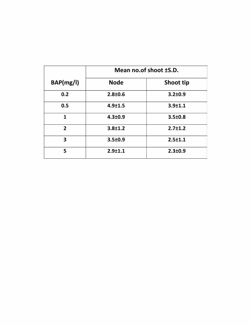
BAP(mg/l)
Mean no.of shoot ±S.D.
Node Shoot tip
0.2 2.8±0.6 3.2±0.9
0.5 4.9±1.5 3.9±1.1
1 4.3±0.9 3.5±0.8
2 3.8±1.2 2.7±1.2
3 3.5±0.9 2.5±1.1
5 2.9±1.1 2.3±0.9

Figure 2
Seed germination in aseptic conditions to test for its viability and in
aseptic conditions to initiate explant culture in vitro conditions
(a) Seed Viability Test
(b) Seed Germination

a
b

Figure 3
Morphogenic response of nodal explants of Dianthus caryophylluscultured on MS medium supplemented with
(a) 0.2 mg/l BAP
(b) 0.5 mg/l BAP
(c) 1 mg/l BAP
(d) 2 mg/l BAP
(e) 3 mg/l BAP
(f) 5 mg/l BAP

a
fe
dc
b

Figure 4
Morphogenic response of shoot tip explants of Dianthuscaryophyllus cultured on MS medium supplemented with
(a) 0.2 mg/l BAP
(b) 0.5 mg/l BAP
(c) 1 mg/l BAP
(d) 2 mg/l BAP
(e) 3 mg/l BAP
(f) 5 mg/l BAP

e f
dc
ba

Table 2. Formation of shoot buds from mature seedling explant of Dianthus
caryophyllus cultured on MS supplemented with BAP in combination with NAA
PGR (mg/l)
Mean no. of shoot ±S.D.
BAP NAA Node Shoot tip
0.2
02.9± 0.6 3.3±0.8
0.1 6.3±0.8 5.5±0.9
0.2 7.2±0.7 6.9±1.1
0.5
0.1 6.5±0.9 5.3±0.9
0.2 8.5±0.7 7.2±0.8
0.5 11.5±0.6 9.2±0.7
1
0.1 4.8±0.8 3.9±0.8
0.2 3.5±0.7 3.3±0.9
0.5 3.2±0.8 2.9±0.7

Figure 5
(a-c) Morphogenic response of nodal explants on MS mediumsupplemented with BAP in combination with NAA
(a) MS+ BAP (0.2 mg/l ) +NAA (0.2 mg/l)
(b) MS+ BAP (0.5 mg/l ) +NAA (0.5 mg/l)
(c) MS+ BAP (1 mg/l ) +NAA (0.5 mg/l)
(d-f) Morphogenic response of shoot tip explants on MS mediumsupplemented with BAP in combination with NAA
(d) MS+ BAP (0.2 mg/l ) +NAA (0.2 mg/l)
(e) MS+ BAP (0.5 mg/l ) +NAA (0.5 mg/l)

(f) MS+ BAP (1 mg/l ) +NAA (0.5 mg/l)
a
fc
b
d
e

EFFECT OF HEAVY METALS (AgNO3 &NiCl2)
The effect of heavy metals (which are not constituent of MS media) was also investigated on the
induction of shoot bud from nodes and shoot tips of Dianthus and the subsequent proliferation of
primary shoots to form multiple shoots per explant.
B.1 Silver as Silver Nitrate (AgNO3) (Table 3)
Node and shoot tip explants were cultured on MS medium supplemented with BAP (0.5 mg/l),
NAA (0.5 mg/l) and different concentrations of AgNO3 (0, 0.1, 0.5, 1 and 1.5 mg/l).
Incorporation of AgNO3 in to the medium induced positive response. The explants showed
swelling during the first week of culture. The initiation of shoot bud induction occurred at the
end of the second week of the culture. It was observed that the number of shoots increased with
the increase in the concentration of AgNO3 from 0.1 to 1mg/l. Beyond this level the shoots
showed no significant increase in length but turned more hyperhydric. Best response in terms of
total number of shoots as well as hyperhydricity per explant was achieved on medium containing
1 mg/l AgNO3 (Fig.6, 7). Results achieved from AgNO3 supplemented medium were better in
terms of shoot regeneration and elongation of the shoots per explant than on control, lacking
AgNO3.
B.2 Nickel as Nickel Chloride (NiCl2) (Table 4)
Node and shoot tip explants were cultured on MS medium supplemented with 0.5 mg/l BAP,
NAA (0.5 mg/l) and different concentrations of NiCl2 (0, 0.1, 0.5, 1 and 1.5 mg/l). The explants
showed swelling during the first week of culture. The initiation of shoot bud induction occurred
at the end of the second week of the culture. It was observed that the number of shoots decreased
with the increase in the concentration of NiCl2. The shoots that even developed up to 0.5 mg/l of
NiCl2 were hyperhydric and had abnormally large leaves (Fig. 8, 9). When the concentration of
nickel chloride was raised to 1 and 1.5 mg/l NiCl2, the abnormal condition of the shoots also
increased.

Table3. Effect of silver nitrate on shoot bud formation from nodal and shoot
tips of Dianthus caryophyllus cultured on MS supplemented with BAP
(0.5mg/l) and NAA (0.5mg/l)
AgNO3(mg/l)
Mean no. of shoot bud ±S.D.
Node Shoot tip
0 4.8±0.9 3.8±1.1
0.1 5.2±1.6 4.1±0.7
0.5 5.5±1.6 4.4±0.6
1 5.9±1.2 4.9±0.9
1.5 4.4±0.9 3.5±0.8

Table4. Effect of nickel chloride on shoot bud formation from nodal and shoot
tips of Dianthus caryophyllus cultured on MS supplemented with BAP
(0.5mg/l) and NAA (0.5mg/l)
NiCl2 (mg/l)
Mean no. of shoot bud ±S.D.
Node Shoot tip
0 4.8±0.9 3.8±1.1
0.1 3.4±0.8 3.1±0.8
0.5 3.0±0.7 2.8±0.7
1 2.7±0.7 2.5±0.6
1.5 2.5±0.8 2.2±0.6

Figure 6
Effect of silver nitrate on shoot bud formation from node ofDianthus caryophyllus cultured on MS supplemented with BAP (0.5mg/l) + NAA (0.5 mg/l)
(a) Without AgNO3
(b) 0.1 mg/l AgNO3
(c) 0.5 mg/l AgNO3
(d) 1 mg/l AgNO3
(e) 1.5 mg/l AgNO3

a b
c
e
d
e
Figure 7
Effect of silver nitrate on shoot bud formation from shoot tip ofDianthus caryophyllus cultured on MS supplemented with BAP (0.5mg/l) + NAA (0.5 mg/l)
(a) Without AgNO3
(b) 0.1 mg/l AgNO3
(c) 0.5 mg/l AgNO3
(d) 1 mg/l AgNO3
(e) 1.5 mg/l AgNO3


e
ba
c d

Figure 8
Effect of nickel chloride on shoot bud formation from node ofDianthus caryophyllus cultured on MS supplemented with BAP (0.5mg/l) + NAA (0.5 mg/l)
(a) Without NiCl2
(b) 0.1 mg/l NiCl2
(c) 0.5 mg/l NiCl2
(d) 1 mg/l NiCl2
(e) 1.5 mg/l NiCl2

ba
c d
e

Figure 9
Effect of nickel chloride on shoot bud formation from shoot tip ofDianthus caryophyllus cultured on MS supplemented with BAP (0.5mg/l) + NAA (0.5 mg/l)
(a) Without NiCl2
(b) 0.1 mg/l NiCl2

(c) 0.5 mg/l NiCl2
(d) 1 mg/l NiCl2
(e) 1.5 mg/l NiCl2
b

ROOTING OF IN VITRO REGENERATED SHOOTS
In vitro regenerated shoots (longer than shoots 2 cm) were excised and transferred into tubes
containing solidified MS medium supplemented with various concentrations of auxins viz., IAA,
IBA and NAA (0.5 and 1mg/ l) and half strength MS medium. 2-3thin, stout and slender roots
induced from cut end of shoot on MS medium fortified with IBA (0.5 mg/l) (Fig. 10a).The
addition of NAA (0.5 mg/l) to the MS medium promoted rooting, which was accompanied by
callus (Fig. 10b).The MS medium supplemented with IAA (0.5mg/l) did not show any sign
morphogenic response. On the other hand, the half strength MS medium showed excellent root
formation where roots were long, unbranched and strong (Fig. 10c).
a
e
dc

Figure 10
Rooting response of in vitro regenerated shoots of Dianthuscaryophyllus cultured on MS medium supplemented with
(a) IBA (0.5 mg/l)
(b) NAA (0.5 mg/l)
(c) Half strength MS

a b

DISCUSSION
c

D. caryophyllus is a valuable plant and has a high potential for export and improvement of
floriculture industry. Plant cell and tissue culture have been developed in recent years to such a
level that any plant species either endangered or highly recalcitrant can be regenerated in vitro.
This area of plant biotechnology is mainly concerned with manipulation and subsequent growth
of cells, tissues, organs and plant cell. In tissue culture, the use of growth regulator play
important role in influencing different plant processes comprising mostly of growth,
differentiation and development such as culture establishment, shoot initiation, callogenesis,
embryogenesis and rooting (Hobbie, 1998).
To facilitate the yield enhancement and quality improvement of D. caryophyllus, a range of in
vitro tissue culture technique have been developed such as efficient systems have been
established for the regeneration of plant from leaf explants and stem segments(Jethwani and
Kothari,1996; Watad et al.,1996: Kantia and Kothari, 2002).
In present study, the shoot buds induced from the shoot tip and nodes explants were selected for
further studies as it proved to be the best respondary explants for shoot regeneration. Nodal and

apical segments have been used widely for shoot morphogenesis reported by Baker and Philips
(1962).Similar results were reported by Earle and Langhans, 1975; Jethwani et al., 1994; Van
Altvorest et al.,1995; Van Altvorst and Koehorst, 1995; Kallak et.al.,1997; Pareek et. al, 2004;
H. Saehi, 2006; Danial et al.,2008; Duhoky et al., 2008; Ali et al.,2008.
Shoot tip and nodal explants were cultured on MS medium supplemented with various
concentration of BAP (0.2, 0.5, 1, 2, 3 and 5 mg/l) for shoot regeneration. A maximum number
of shoot buds formed per explant was obtained on MS medium supplemented with 0.5 mg/l
BAP. This is in agreement with the studies of Lubomski and Jerry (1989) who reported the best
shoot formation response of carnation on MS medium supplemented with BAP. Kovac (1995)
also reported highest shoot multiplication in carnation in MS medium containing 1.0 mg/l BAP.
However, Siddiqui (1993) and Duhoky et al., (2008) reported best shoot induction response in
MS medium containing 5.0 mg/l kinetin.In the present investigation BAP in combination with
NAA stimulated shoot buds induction and elongation response. It was found that MS medium
supplemented with 0.5 mg/l BAP and 0.5mg/l NAA showed maximum shoot elongation and
proliferation.
Van (1992) and Yantcheva et al., (1998) reported highest numbers of shoots per explant on MS
medium containing 0.9 mg/l BA and 0.3 mg/l NAA. Mangel et al., (2002) and Onamu et al.,
(2003) used MS medium supplemented with combination of NAA and kinetin for shoot
induction from meristem explant.
Elongated shoots of 2 cm or more in length were excised and placed onto rooting medium which
consisted of half strength MS medium and MS medium supplemented with various
concentrations auxins (IAA, IBA and NAA).Out of these medium the best response was obtained
on half strength MS medium. However Amir et al (2009) reported best rooting response on MS
medium containing 1.0 mg/l NAA. On the other hand best rooting of shoots was achieved when
NAA was included in the culture medium at 0.5 mg/l concentration reported by Daniel et al.,
(2009).

The composition of MS medium is critical for optimizing the regeneration of plant in vitro. Some
heavy metals act as microelements play important roles in the regeneration of plant at low
concentration but at higher concentration they may cause reduction in growth for the most of
plant species (Fernandes and Henrique, 1991; Claire et al., 1991). In our own results, For shoot
regeneration explants cultured on induction medium supplemented with various concentration of
heavy metals (AgNO3 and NiCl2), we found that shoot regeneration from nodal and shoot tip
explants of D. caryophyllus on MS medium with AgNO3 (1mg/l) appeared to be the most
advantageous in terms of total number of shoots as well as hyperhydricity per explants but with
NiCl2 it is not beneficial for shoot regeneration.
Addition of sliver nitrate has been found to be effective in overcoming the recalcitrant behavior.
Sliver nitrate act as ethylene inhibitor and used for shoot elongation in Capsicum reported by
Phillips 1996).It acts as inhibitor of 1-aminocyclopropane -1-carboxylic acid which is a
precursor of ethylene in biosynthetic pathway (Palmer, 1992). However Mohiuddin et al., (1997)
achieved increased number of shoot in cucumber by incorporation of sliver nitrate. MS medium
supplemented with sliver nitrate enhanced the frequency of shoot regeneration in Brassica
species (Eapan and George, 1997). Gutiérrez-Miceli et al., (2010) reported additive effect of
heavy metals in stimulating morphogenesis in callus redifferentiation has been reported using
kinetin and AgNO3 in D. caryophyllus. There has been many reports of the inhibitory effects of
Ni on germination and growth in plants. Madhava et al., (2000) reported the germination of
pigeon pea was decreased by addition of 1.5 mM Ni solution. Similar results were reported by
Gajewska et al., (2006), who observed the inhibitory effect of 0.2 mM nickel on shoot growth of
wheat. The roots of Nicotiana tabacum became dark brown within 7 to 10 days of exposure to
0.43 mM Ni and growth of the plants was severely inhibited (Boominathan et al., 2002). In the
present study lower concentration of nickel chloride had deleterious effect on shoot induction
and still higher concentrations of nickel chloride proved toxic.
Plant tissue culture is one of the most commonly used methods for induction of shoot buds. We
have demonstrated here that providing optimal concentration of BAP (0.5mg/l) and NAA
(0.5mg/l) in MS medium which is induced shoot buds from the explants of D. caryophyllus.

Addition of silver nitrate in the medium is applicable to improve the quality and quantity of the
regenerated shoots while on the other hand adding nickel to the medium proved detrimental.
SUMMARY

The present work deals with interactive effect of different plant growth regulator and heavy
metals on shoot morphogenesis of Dianthus caryophyllus. This plant is a member of the family
caryophyllaceae and is the most common cultivated species of this family which is characterized
by their excellent keeping quality, wide range of forms, ability to withstand long distance
transportation and remarkable ability to rehydrate after continuous shipping. It shows high level
of heterogeneity in seed population which act as constraint to propagation of agronomic traits
and for commercial seed production. Considering the benefits of this plant and to fulfill the
world’s demand for carnation, the technique of micropropagantion has been widely used. Tissue
culture offers an opportunity to access clean planting material and provides a start in production
of good quality carnation cut flowers that can adequately compete in the international markets.

The seeds of D. caryophyllus were obtained from Namdhari Seed Agency, Bengaluru.After
proper sterilization seeds were germinated on half strength MS medium solidified with 0.8%
(w/v) agar. Various explants were taken for the studies; shoot tip and nodal explants showed
better response in term of shoot regeneration. The explants cultured on MS medium
supplemented with various concentration of BAP (0.2-5mg/l). The maximum number of shoot
buds was obtained on MS medium supplemented with BAP (0.5mgl/) and NAA (0.5mg/l). The
shoot buds induced and proliferated on medium supplemented with BAP (0.5mg/l) and NAA
(0.5mg/l) were selected for further studies as it proved to be the best concentration for shoot
induction. The shoots were elongated on MS supplemented with BAP (0.5mg/l) and NAA
(0.5mg/l), prior to rooting on half strength MS medium. Various auxins (IBA, IAA and NAA)
were tried for rooting. The best response was shown by half strength MS medium.
Effect of heavy metals (AgNO3 and NiCl2) was investigated on shoot morphogenesis in D.
caryophyllus. Node and shoot tip explants were cultured on induction medium (0.5 mg/l BAP +
0.5 mg/l NAA) supplemented with various concentrations of AgNO3 (0, 0.1, 0.5, 1 and 1.5 mg/l).
It was observed that the number of shoots increased with the increase in the concentration of
AgNO3 from 0.1 to 1mg/l. Beyond this level the shoots showed no significant increase in length
but turned more hyperhydricity. Effect of NiCl2 was found to be not advantageous for shoot
morphogenesis in Dianthus.
REFERENCES

Abeles, F.B. (1973) Ethylene in plant biology. Academic press New York. 302: ISBN978-0120414505
Ali, A., H. Afrasiab, S. Naz, M. Rauf and J. Iqbal (2008) An efficient protocol for invitro propagation of carnation (Dianthus caryophyllus) Pak. J. Bot.40(1): 111-121
Aminuddin, S. K. R., K. M. Srivastava and B. P. Singh (2007) Natural infection ofcucumber mosaic virus on Dianthus barbatus in India. Plant Pathol.42(5): 811-813
Assuncao, A., H.Schat and M. Aarts (2003) Thlaspicaerulescens, an attractive modelspecies to study heavy metal hyperaccumulation in plants.New Phytol.159:351–360
Bais, H., J. George and G. Ravishankar (1999) Influence of polyamines on growth ofhairy root cultures of witl of chiocory (ChichoriumintybusL cv Lucknow local) andformation of coumarins. J. of PGR.18: 33-37

Bais, H., G.S. Sudha, and G. Ravishankar (2000a) AgNO3 influences in vitro rootformation in DecalepishamiltoniiWight, Arn. Curr.Sci. 79:894-898
Bais, H., G.S. Sudha, and G. Ravishankar (2000b) Putrescine and AgNO3 influencesshoot multiplication Invitro flowering and endogenous titres of polyamines inChichoriumintybusL cv Lucknow Local. J. of PGR.19:238-248
Bais, H., G.S. Sudha, and G. Ravishankar (2001a) Influence of putrescine AgNO3 andpolyamine inhibitors on the morphogenetic response in untransformed and transformedtissues of Chichoriumintybusand their regenerants. Plant Cell Reports.20: 547-555
Bais, H., G.S. Sudha and G. Ravishankar (2001b) Putrescine influences growth andproduction of coumarins in transformed and untransformed root cultures of witloofchicory (ChichoriumintybusL cv Lucknow Local). Acta.Physio.Plant.23:319-327
Bais, H., R. Venkatesh and G. Ravishankar (2001c) Agrobacterium rhizogenes mediatedtransformation of witl of chiocoryin vitro shoot regeneration and induction of flowering.Curr. Sci. 80: 83-87
Baker, R. and D. J. Philips (1962) Obtaining pathogen free stock by shoot tip culture.Phytopathol.52: 1242-1244
Beyer, E. M. (1976a) A potent inhibitor of ethylene action in plants. PlantPhysiology.58:268-271
Beyer, E.M. (1976c) Silver ion a potent anti-ethylene agent in cucumber and tomato.HortScience.11: 175-196
Biddington, N.L. (1992) The influence of ethylene in plant tissue culture. Plant GrowthRegulation.11:173-178
Birch, R. (1997) Plant transformation: problems and strategies for practical applications.Annu. Rev. Plant Physiol. Plant Mol. Biol. 48: 297–326
Bird, R. (1994) Border Pinks pp. 1-174 Timber Press, Portland, Oregon, USA

Boominathan, R. and P.M. Doran (2002) Nickel induced oxidative stress in roots of Nihyperaccumulater Alyssum bertolonii. New Phytol.156:205–215
Casanova, E., A.E. Valdés L. Moysset and M.I. Trillas (2004) A Levels andimmunolocalization of endogenous cytokinins in thidiazuron-induced shootorganogenesis in carnation. J. Plant Physiol.
Cassells, A. C. and R. F.Curry (2001) Oxidative stress and physiological, epigenetic andgenetic variability in plant tissue culture: implications for micropropagators and geneticengineers. Plant Cell Tiss. Org. Cult. 64: 145–157
Chen,C., D.Huang and J Liu (2009) Functions and toxicity of nickel in plants: recentadvances and future prospects. Clean 37:304–313
Chi, G.L. and E.C. Pua (1989) Ethylene inhibitors enhanced de novo shoot regenerationfrom cotyledons of Brassica campastris spp in vitro. Plant Science.64: 243-250
Choudhary, M. L. and C. K. Chin (1995) Somatic embryogenesis in cell suspensionculture of carnation (Dianthus caryophyllus L.) Plant Growth Regulation 16: 1-4
Chraibi, K. M., A. Latche, J. P. Roustan and J. Fallot (1991) Stimulation of shootregeneration from cotyledons of Helianthus annus by ethylene inhibitors, silver andcobalt. Plant Cell Rep.10: 204-207
`Danial, G. H., A. N. Yousif and M. S. Omar (2009) Clonal propagation of Dianthuscaryophyllus L. through tissue culture. J. Duhok. Univ.12(1): 91-95
De Block, M., D. De Brouwer and P. Tennig (1989) Transformation of Brassica napusand Brassica oleracea using Agrobacriumtumefaciens and the expression of the bar andneo genes in the transgenic plants.Plant Physiol.91: 694-701
Demmink, J. F., J. B. M. Clusters and J. H. W. Bergervoet (1987) Gynogenesis to bypasscrossing barriers between diploid and tetraploid Dianthus species. Acta horticulture.212:167-176
Duhoky, M. S., M. A. Salman and M. E. M. Amin (2009) Micropropagation of carnation(Dianthus caryophyllus). J. Duhok Univ. 12(1): 61-66

Eapen, S. and L George (1997) Plant regeneration from peduncle segments of oil seedbrassica species: Influence of AgNO3 and silver thiosulphate. Plant Cell Tissue andOrgan Culture.51:229-232
Earle, E. D. and R. W. Langhans (1975) Carnation propagation from shoot tips culturedin liquid medium. Hort. Science. 10: 608- 610
Fernandis, J.C. and F.C. Henriques (1991) Biochemical physiological and structuraleffects of excess copper in plant. Bot.Rev.57: 246-273
Gajewska, E., M. Sklodowska, M. Slaba and J. Mazur (2006) Effect of nickel onantioxidative enzyme activities, proline and chlorophyll content in wheat shoots. BiolPlant 50:653–659
Gajewska E., M. Wielanek, K. Bergier and M. Skłodowska (2009) Nickel induceddepression of nitrogen assimilation in wheat roots. ActaPhysiol Plant 31:1291–1300
Gautheret, R. (1939) Sur la possibilite de realiser la culture indefinie des tissus detubercules de carotte. C. R. Acad. Sci., Paris, 208: 118-120
Ghosh, S. (1987) Micropropagation of Sim’s carnation (Dianthus caryophyllus L.)Indian J. Exp. Biol. 24: 703-704
Gutierrez-Miceli, F. A., L. Arias, N. Juarez-Rodriguez, M. Abud-Archilla, A. Amaro-Reyes and L. Dendooven (2010) Optimization of growth regulators and silver nitrate formicropropagation of Dianthus caryophyllus L. with the aid of a response surfaceexperimental design. In Vitro Cell Dev. Biol.Plant 46: 57-63
Hilderbrandt, A. (1946) Culture du tissus cambial. C. R. Acad. Sci. Paris. 198: 2195-2196
Hobbie, L.J.(1998) Auxin : molecular genetic approaches in Arabiddopsis.PlantPhysiol.Biochem.36:91-102
Hussain, I., A. Mushtaq and A. Qureshi (1994) In vitro multiplication of Dianthuscaryophyllus L. cv. CSU.Pink and White Sim.Sarhad J. Agri. 10: 539-546

Jain, A., A. Kantia, S.L. Kothari (2001) De novo differentiation of shoot buds from leaf-callus of Dianthus caryophyllus L. and control of hyperhydricity .Sci Hoti. 87:319-326
Jethwani, V. and S. L. Kothari (1993) Micropropagation of D.barbatusand D. chinensisthrough cotyledonary node culture.Plant Cell Tiss. Org. Cult.3: 91-96
Jethwani, V., Sharma V. and S. L. Kothari (1994) Micropropagation of Dianthuschinensis and D. barbatus through shoot tip culture.J. Bot. Society. 73: 357-358
Jethwani, V. and S. L. Kothari (1996) Phenylacetic acid induced organogenesis in cultureleaf segments of Dianthus chinensis. Plant Cell Rep. 5: 869-872
Kabata-Pendias, A.and H. Pendias (2001) Trace elements in soils and plants, 3rd edn.CRC Press Inc, Boca Raton.FL. 413
Kallack, H., I. Hilpus and K. Virmuae (1996) Influence of genotype and growthregulations on morphogenetic process in carnation shoot apex cultures. Sci. Hort.65: 181-189
Kantia, A. and S. L. Kothari (2002) High efficiency adventitious shoot bud formation andplant regeneration from leaf explants of Dianthus chinensis L. Sci. Hort.96: 205-212
Karami, O. (2008) Induction of embryogenic callus and plant regeneration in carnation(Dianthus caryophyllus L.) Online J. Biol. Sci. 8(4): 68-72
Knop,W. (1865) Quantitative Untersuchungenuber die Ernahrungsprocesse der Pflanzen.Landwirtsch.Vers.Stn.7: 93.
Kotte, W. (1992) KulturversuchemitisoliertenWurzelspitzen. Beitr.Allg.Bot. 2: 413-434.
Kovac, J. (1995) Micropropagation of Dianthus caryophyllus sub sp. Bohemicus-anendangered endemic from the Czech Republic. Biol.37: 27-33.
Krupa,Z. A.Siedlecka, W. Maksymiec and T. Baszynski (1993) In vitro responses ofphotosynthetic apparatus of Phaseolus vulgaris L.to nickel toxicity. Plant Physiol142:664–668

Lau, Oi-Lim and Yang, F. Shang (1976) Inhibition of ethylene production by cobaltousion. Plant Physiology.58:114-117
Leshem, B., E. Werker and D. P. Shalev (1988) The effect of cytokinins on vitrificationin melon and carnation. Ann. Bot. 62: 271-276
Lieberman, N. (1979) Biosynthesis and action of ethylene. Annual Review of PlantPhysiology.30 :533-591
Lumbomski, M. and M. Jerzy (1989). In vitro propagation of pot carnation from stemintertnodes.Acta Hort. 251: 235-240
Mangal, M., S.V. Bhardwaj, D.R. Kaur and A.K. Mangal (2002) Use of meristem tipculture to eliminate carnation latent virus from carnation plant.Ind. J. Exp. Biol. 40: 119-122
Manoj, K., A.K. Yadav and G.K. Garg (2003) Development of suitable protocol toovercome hyperhydricity in carnation during micropropagation. Plant Cell, Tissue andOrgan Culture 72: 153–156
Marschner, H., (1995) Mineral nutrition of higher plants, 2nd edn. Academic Press,London, p 889
Mattoo, A.K. and J.C. Suttle (1991) The plant hormone ethylene. CRC press. BocaRaton, Florida. 337:ISBN 0-8493-4566-9
Messeguer, J., M. C. Arconada and E. Mele (1993) Adventitious shoot regeneration ofcarnation (Dianthus caryophyllus L.) Sci. Hort.54: 153-163
Mii, M., M. Buiatti and F.Gimelli (1990) Carnation. In: Ammirato, P.V., Evans, D.R.,Sharp, W.R., Bajaj, Y.P.S. (Eds.), Handbook of Plant Cell Culture. McGraw-Hill, NewYork, 284-318
Miller, R. M., V. Kaul, J. F. Hutchinson, G. Maheswaran and D. Richards (1991) Shootregeneration from axillary buds of carnation (Dianthus caryophyllus L.) Ann. Bot. 67: 35-42

Miller, R. M., V. Kaul, J. F. Hutchinson, G. Maheswaran and D. Richards (1992) Shootregeneration from fragmented flower buds of carnation (Dianthus caryophyllus L.). Ann.Bot. 68: 563-568
Mohinuddin, A.K., M.K. Chowdhury,S. Napis and A.C. Abdulla (1997) Effect ofAgNO3 on Cucumber in vitro shoot regeneration .Plant Cell Org cult. 51:75-78
Molas, J., (2002) Chasnges of chloroplast ultrastructure and total chlorophyllconcentration in cabbage leaves caused by excess of organic Ni II complexes. Environ.Exp. Bot. 47:115–126
Moyal-Ben Zvi, M. and A. Vanstein (2007) Carnation.Biotechnology in Agriculture andForestry.Transgenic Crops VI (edn by E. C. Puz and M. R. Davey) 61.
Murashige, T. and F. Skoog (1962) A revised medium for rapid growth and bioassayswith tobacco tissue cultures.Physiol. Plant.15: 473-497
Nakano, M., Y. Hashino and M. Mii (1994) Adventitious shoot regeneration fromcultured petal explants of carnation. Plant Cell Tiss. Org. Cult.36: 15-19
Nontaswatsri, C. and S. Fukai (2005) Regenerative callus of Dianthus ‘Telstar Scarlet’showing mixploidy produce diploid plants. Plant Cell Tiss. Org. Cult.83: 351-355
Nugent, G., T. Wardley and C. Lu (1991) Plant regeneration from stem and petal ofcarnation (Dianthus caryophyllus) Plant Cell Rep.10: 477-453
Onamu, R., S.D. Obukosia, N. Musembi and M.J. Hutchinson (2003) Efficacy ofThidiazuron in In vitro propagation of carnation shoot tips: Influence of dose andduration of exposure. Afr.Crop Sci. J. 11(2): 125-132
Pareek, A. and S. L. Kothari (2003) Direct somatic embryogenesis and plant regenerationfrom leaf cultures of ornamental species of Dianthus. Sci. Horti.
Pareek, A., A. Kantia and S. L. Kothari (2004) In vitro cloning of ornamental species ofDianthus.Ind. J. Biotech.3: 363-266.

Petru, E. and Z. Landa (1974) Organogenesis in isolated carnation plant callus tissuecultivated in vitro. Biol. Planta.16: 450-453
Phillips G.C. (2004) In vitro morphogenesis in plant recent advances. In vitro Cell DevBiol Plant.40:342-345
Preece, J. E. (1995) Can nutrient salts partially substitute for plant growth regulators?Plant Cell Tissue and Organ Cult.35:131-139
Pua, E.C. and G.L Chi (1993) De novo shoot morphogenesis and plant growth of mustard(Brassica juncea) in vitro in relation to ethylene. PhysiologiaPlantarum.88: 467-474
Purnhauser, L., P. Medgysey, M Czako, J.P. Dix and L. Marton (1987) Stimulation ofshoot regeneration in Triticumaestivumand NicotianaplumbaginifoliaViv tissue culturesusing the ethylene inhibitor silver nitrate. PlantCell Reports. 6:1-4
Rao, K. and T. Sresty (2000) Antioxidative parameters in the seedlings of pigeonpea(Cajanuscajan L.) Millspauga in response to Zn and Ni stress.Plant Sci.157:113–128
Roustan, J.P., A. Latche and J.Fallot (1990) Control of carrot somatic embryogenesis byAgNO3 an inhibitor of ethylene action effect on arginine decarboxilase activity. PlantScience.67:89-95
Roustan, J.P., A. Latche and J. Fallot (1989) Stimulation of Daucuscarotasomaticembryogenesis by inhibitors of ethylene biosynthesis: cobalt and nickel. Plant CellReports.8: 182-185
Sajid, G. M. and S. Pervaiz (2008) Bioreactor mediated growth, culture ventilation,stationary and shake culture effects on in vitro growth of sugarcane. Pak. J. Bot.40(5):1949-1956
Salehi, H. (2006) Can a general shoot proliferation and rooting medium be used for anumber of carnationcultivars? Afr. J. Bio.5:25-30
Sankhla, D., N. Vavis, N. Sanklha and A. Upadhya (1994) In vitro regeneration of heattolerant “German Red” carnation through organogenesis and somatic embryogenesis.Gartenbouwissenschaft.6: 227-233

Seo, J., S. W. Kim and S. R. Min (2007) High frequency somatic embryogenesis andplant regeneration in root explant cultures of carnation. Plant Biotecnol. Rep.1: 67-70
Sethi, U., A. Basu and S. Guha-Mukherjee (1990) Control of cell proliferation anddifferentiation by modulators of ethylene biosynthesis and action in Brassica hypocotylexplants. Plant Sci. 69: 225-229
Sharma, P. and R. Bhardwaj (2008) Effect of 28-homobrassinolide on nickel uptake,protein content and antioxidativedefence system in Brassica juncea. Biol Plant. 52:767–770
Siddiqui, F.A. (1993) A study of the elimination of sugarcane mosaic virus fromSaccharum officinarum by means of In vitro meristem and callus culture and somebiochemical aspects of regenerated healthy and infected plants. Ph.D. thesis.Departmentof Botany, University of thePunjab, Lahore, Pakistan.
Skirvin, R., K. McPheeters and M. Norten (1994) Source and frequency of somaclonalvariation. Hort Science. 29: 1232–1237
Songstad, D.D., D.R Duncan and J.M Widholm (1988) Effect of 1-aminocycopropane-1-carboxilic acid silver nitrate and norbornadiene on plant regeneration from maize calluscultures. Plant Cell Reports.7: 262-265
Susek, A., B. Javornik and B. Bohanec (2002) Factors affecting direct organogenesisfrom flower explants of Allium giganteum.Plant Cell Tiss.Org. Cult. 68: 27–33
Van Altvorst, A. C. and H. J. J. Koehorst (1995) Cells within the nodal region ofcarnation shoots exhibit a high potential for adventitious shoot formation. Plant Cell Tiss.Org. Cult.40: 151-157
Van Altvorst, A. C., H. J. J. Koehorst, J. D. Jong and J. J. M. Dons (1996) Transgeniccarnation plants obtained by Agrobacterium tumefaciens mediated transformation of petalexplants. Plant Cell Tiss. Org. Cult.45: 2
Van Altvorst, A. C., H. J. J. Koehorst, T. Bruinsma, J. Jansen, J. M. B. Custers, J. DeJong and J. J. M. Dons (1992) Adventitious shoot formation from in vitro leaf explant ofcarnation(Dianthus caryophyllus L.) Sci. Hort. 51: 223-235

Van, A., A.C. Bruinsma, T. Koehorst, H.J.J. and J.J.M. Dons (1992) Regeneration ofcarnation (Dianthus caryophyllus) using leaf explants.Acta Hort. 307: 109-116.
Varshney, A., M. Lakshmikumaram, P. S. Srivastava and V. Dhawan (2001)Establishment of genetic fidelity of in vitro raised Liliumbulblets through RAPD markers.In vitro Cell Dev. Biol.-Plant.37: 227-231
Villalobos, V. (1981) Floral differentiation in carnation (Dianthus caryophyllus) fromanthers cultured in vitro. Phyton.41: 71-75
Watad, A., A. Ahroni, A. Zuker, H. Shejtman, A, Nissim and A.Vainstein (1996)Adventitious shoot formation from carnation stem segments: a comparison of differentculture procedures. Sci. Hort. 65: 313-320
Welsh, J. and M. McClelland (1990) Fingerprinting genomes using PCR with arbitraryprimers. Nucl. Acids Res. 8: 7213-7218
Werker, E. and B. Leshem (1987) Structural changes during vitrification of carnationplantlets. Ann. Bot. 59: 377-385
White, P. (1934) Potentially unlimited growth of excised tomato root tips in a liquidmedium. Plant Physiol. 9: 585-600.
Xylander, M. W. Braune (1994) Influence of nickel on the green alga Haematococcuslacustris Rostafinski in phases of its life cycle. J Plant Physiol. 144:86–93
Yadav, M. K., A. K. Gaur and G. K. Garg (2003) Development of suitable protocol toovercome hyperhydricity in carnation during micropropagation.Plant Cell Tiss. Org.Cult.72: 153-156
Yang, S.F. (1985) Biosynthesis and action of ethylene.HortScience.20 :41-45
Yang, S.F. and N. Hoffman (1984) Ethylene biosynthesis and its regulation in higherplants.Annual Review of PlantPhysiology.35:155-189
Yantcheva, A., M. Vlahova and A. Antanassov (1998) Direct somatic embryogenesis andplant regeneration of carnation (Dianthus caryophyllus L.) Plant Cell Rep.18: 148-153

Yusuf, M., Q.Fariduddin, S.Hayat, S.A. Hasan and A. Ahmad (2010) Protectiveresponses of 28 homobrssinolide in cultivars of Triticumaestivum with different levels ofnickel. Archv Environ ContamToxico(in press)