effect of perinatal and postnatal bisphenol a exposure to ... · effect of perinatal and postnatal...
TRANSCRIPT
Ea
WCa
b
c
d
a
ARR1AA
KKGGS
1
biSimaal[pupp
0d
Reproductive Toxicology 31 (2011) 409–417
Contents lists available at ScienceDirect
Reproductive Toxicology
journa l homepage: www.e lsev ier .com/ locate / reprotox
ffect of perinatal and postnatal bisphenol A exposure to the regulatory circuitst the hypothalamus–pituitary–gonadal axis of CD-1 mice
ei Xia, C.K.F. Leeb, W.S.B. Yeungb, John P. Giesyc,d, M.H. Wonga, Xiaowei Zhangc, Markus Heckerc,hris K.C. Wonga,∗
Croucher Institute of Environmental Sciences, Department of Biology, Hong Kong Baptist University, Kowloon, Hong KongDepartment of Obstetrics and Gynecology, The University of Hong Kong, Hong KongDepartment of Veterinary Biomedical Sciences & Toxicological Center, University of Saskatchewan, CanadaDepartment of Biology and Chemistry, City University of Hong Kong, Kowloon, Hong Kong, China
r t i c l e i n f o
rticle history:eceived 15 September 2010eceived in revised form8 November 2010ccepted 9 December 2010vailable online 21 December 2010
eywords:isspeptin
a b s t r a c t
Bisphenol A (BPA) is used in the manufacture of many products and is ubiquitous in the environment.Adverse effects of BPA on animal reproductive health have been reported, however most of the stud-ies relied on the approaches in the assessment of conventional histology and anatomical features. Themechanistic actions of BPA are not clear. In the present study, a murine model was used to studypotential effects of BPA exposure during perinatal and postnatal periods on endocrine functions ofhypothalamic–pituitary–gonadal (HPG)-axis. At the hypothalamic-pituitary level, BPA exposure resultedin the up-regulation of the expression levels of KiSS-1, GnRH and FSH mRNA in both male and femalepups. At the gonadal levels, BPA caused inhibition in the expressions of testicular steroidogenic enzymes
PR-54onadotrophinteroidogenesis
and the synthesis of testosterone in the male pups. Conversely exposure to BPA resulted in a greateraromatase expression level and the synthesis of estrogen in the female pups. BPA is a weak estrogenagonist and its effects reported on animal studies are difficult to reconcile with mechanistic action ofestrogen. In this study we hypothesized that the effects of BPA on reproductive dysfunction may be dueto its actions on gonadal steroidogenesis and so the anomalous releases of endogenous steroid hormones.This non-ER-mediated effect is more potent in affecting the feedback regulatory circuits in the HPG-axis.
. Introduction
In the past century the production of synthetic industrial andiomedical chemicals, as well as unexpected by-products, have
mposed adverse health consequences on wildlife and humans.ome chemical contaminants are classified as endocrine disrupt-ng chemicals (EDCs) since they can interfere with the synthesis,
etabolism and action of endogenous hormones [1,2]. EDCs canffect the hormonal system via (but not limited to) estrogenic,ndrogenic, anti-androgenic and anti-thyroid mechanisms [1,2],eading to the long-term effects on animal development and health3–5]. Among different synthetic chemicals, bisphenol A (BPA) is
roduced in one of the largest-volumes and is used in many prod-cts. Currently over 2.7 million metric tons of BPA have beenroduced for the manufacture of epoxy resins and polycarbonatelastics as constituents of a wide variety of consumer products,∗ Corresponding author. Tel.: +852 3411 7053; fax: +852 3411 5995.E-mail address: [email protected] (C.K.C. Wong).
890-6238/$ – see front matter © 2010 Elsevier Inc. All rights reserved.oi:10.1016/j.reprotox.2010.12.002
© 2010 Elsevier Inc. All rights reserved.
including water/milk carboys/bottles, food wrap, food cans anddental fillings [6–12]. Miserably over 100 t are released into theatmosphere annually [11,12].
BPA can be accumulated along food chains and is detectablein tissues of both wildlife and humans [12–15]. More impor-tantly trans-placental transport of BPA was observed and has beendemonstrated in both rodents and humans [16–19]. Hence fetusmay act as a sink of BPA and would be mostly affected duringgestational development [20–22]. Adverse effects of BPA on devel-opmental and reproductive processes in rodents and primates werereported [23–30], including increased prostate weight, decreasedepididymis weight, reduced sperm production and decreasedconcentrations of LH and testosterone in blood serum [31–34].Although the adverse effects of BPA on reproduction have beenreported, most of the studies assessed the impacts only at the levelsof conventional histology and the gross comparison of anatomi-
cal features/tissue mass. The mechanistic actions of BPA on animalreproductive health have not yet been elucidated.To fill this knowledge gap, in this study the effectsof BPA on expressions of reproduction-related genes alonghypothalamus–pituitary–gonadal (HPG)-axis were studied. Both
410 W. Xi et al. / Reproductive Toxicology 31 (2011) 409–417
F ody mm malef
ptittB
ig. 1. The study design (A) and the measured gross physiological parameters (i.e. bass records of dams during gestation and lactational periods. (C) Body masses of fe
rom cohort-A.
erinatal and/or postnatal exposures were investigated to reveal
he significance of maternal transfer of BPA to fetus/neonates dur-ng gestational and lactation periods. In a previous in vivo study,he minimum concentration of BPA that was found to cause statis-ically significant effects on reproductive performance was 50 mgPA/kg/day [35]. Accordingly the acceptable human BPA intakeass and sex-ratio) of the maternal (F0) and pups (F1) from cohort-A (B–D). (B) Body(left panel) and male pups (right panel) from PND 21 to 49. (D) Sex ratio of the pups
was calculated to be 50 �g/kg/day. However recent studies have
revealed that human exposure to BPA could be considerably greaterthan this acceptable level, and daily intake of BPA is not restrictedto the diet [36–38]. Concentrations of BPA in human tissues werein the ng per ml or per gram range (i.e. blood (0.2–20 ng/ml),amniotic fluids (1.1–8.3 ng/ml), placenta (11.2 ng/g), breast milk
W. Xi et al. / Reproductive Toxicology 31 (2011) 409–417 411
Table 1The DNA sequences of primers used in the present study.
Forward Reverse
Kisspeptin (KiSS-1) GAATGATCTCAATGGCTTCTTGG TTTCCCAGGCATTAACGAGTTKisspeptin receptor (GPR54) GCTCACT GCATGTCCTACAG GCCTGTCTGAAGTGTGAACCGonadotrophin-releasing hormone (GnRH) GGGAAAGAGAAACACTGAACAC GGACAGTACAT TCGAAGTGCTGonadotrophin-releasing hormone receptor (GnRH-R) CTCTATGTATGCCCCAGCTTTCA GCAAAGACAATGCTGAGAATCCALuteinizing hormone (LH) CCTAGCATGGTCCGAGTACT GCTACAGGAAAGGAGACTATGGFollicle-stimulating hormone (FSH) GCTGCTCAACTCCTCTGAAG GGCAATACCTTGGGAAATTCTGGrowth hormone (GH) AGCAGAGAACCGACATGGAA GTTGGTGAAAATCCTGCTGAGThyroid-stimulating hormone (TSH) TCGGGTTGTTCAAAGCATGA GGCACACTCTCTCCTATCCAProlactin (PRL) CTGCTGTTCTGCCAAAATGTT CAGGGTATGGATGTAGTGAGAAALH receptor (LH-R) GCACTCTCCAGAGTTGTCAG AGGGAGA TAGGTGAGAGATAGTCFSH receptor (FSH-R) TCTGCATGGCCCCAATTTTA GGTAGAACAGAACTAGGAGGATCEstrogen receptor-� (ER˛) AATTCTGACAATCGACGCCAG GTGCTTCAACATTCTCCCTCCTCEstrogen receptor-� (ERˇ) TTCCCGGCAGCACCAGTAACC TCCCTCTTTGCGTTTGGACTASteroidogenic acute regulatory protein (StAR) GGAACCCAAATGTCAAGGAGATCA GCACGCTCACGAAGTCTCGA
TGGGCTAAGTCGTGACAG
(uBttibpb(otl(p(
2
2
lfrqtmastmawaSIdfndo
fiB
2
Bar
Cytochrome P450scc (CYPscc) AGCCytochrome P450 17 (CYP17) GATCytochrome P450 19a (CYP19a) CTGGlyceraldehyde 3-phosphate dehydrogenase (GAPDH) ACC
0.28–0.97 ng/ml), follicular fluids (2 ng/ml), semen (5.1 ng/ml) andrine (1.37 ng/ml) [37,38]. Using concentrations of unconjugatedPA detected in human blood, Vandenberg et al. calculated thato achieve such internal doses, exposures to BPA would needo be approximately 0.5 mg BPA/kg, bw/day [37]. This exposures approximately 10-fold greater than the dose of 50 �g BPA/kg,w/day recommended by the USEPA. Using a physiologically basedharmacokinetic model, a comparable exposure (1.42 mg BPA/kg,w/day) to achieve a steady state human blood level of BPA0.9–1.6 ng/ml) [39] was estimated. In the present study, dosesf 12–50 mg BPA/kg, bw/day were selected. These doses fall intohe similar order of magnitude of no observed adverse effectsevel (NOAEL), 5 mg/kg/day and low observed adverse effects levelLOAEL), 50 mg/kg/day used in rodents for risk assessment pur-oses and is also used as a base to calculate the tolerable daily intakeTDI) for humans [40].
. Materials and methods
.1. Animals and administration procedures
All experimental animals were housed and handled in accordance with Guide-ines and Regulations in Hong Kong Baptist University. Six-week-old male andemale CD-1 mice were used in this study. The entire study was conducted ineplicate with mice that were received in two separate batches. Adult mice wereuarantined for 1 week during which time they were observed for any abnormali-ies. The mice were housed in polypropylene cages with sterilized bedding and were
aintained under controlled temperature (23 ± 1 ◦C) and humidity (55 ± 5%) with12 h light–dark cycle (06:00–18:00). The mice were given ad libitum access to
tandard rodent food Rodentdiet 5002 (Labdiet, IN, USA) and water (in glass bot-les). Mice were bred and female mice were checked for vaginal plugs the following
orning. Each copulated mouse (F0) was housed individually, and was randomlyssigned to two cohorts (A and B). Each cohort was further divided into 3–4 groupsith approximately 5 pregnant mice per group. The 4 groups of cohort A were gav-
ged in the morning with corn oil (group I-A), or bisphenol A (BPA purity >99.5%,igma) in corn oil. The doses were: 12 mg/kg/day (group II-A), 25 mg/kg/day (groupII-A), 50 mg/kg/day (group IV-A). The dams were exposed beginning on gestationalay 1 until weaning (postnatal day, PND 20) (Fig. 1A). Individual dams were checkedor birth at least twice a day and the day when pups were first observed was desig-ated as PND 0. From PND 21 to PND 49, the pups (F1) produced from cohort-A wereosed by gavage in the morning with the corresponding concentrations of corn oilr BPA.
Pregnant mice from cohort B were divided into 3 groups but not dosed. Startingrom PND 20 to PND 49, pups from the respective groups were dosed in the morn-ng by gavage with corn oil (group 1-B), or 25 mg BPA/kg/day (group II-B), 50 mgPA/kg/day (group III-B).
.2. Measurement of physical parameters and sampling procedures
Changes in gross anatomy, histology and molecular function were examined.ody masses of dams and neonates were measured by use of an electronic bal-nce (Shimadzu, Kyoto, Japan). The number of pups per dam and the sex-ratio wereecorded on PND 1 and PND 15 respectively. Stillbirths and loss of pups during
CAACATGGAGTCA CCTCTGGTAATACTGGTGATAGGCAAGCTCAGGCA GGGCACTGCATCACGATAAAGACTTGGTCATG GGGGCCCAAAGCCAAATGGC
TCCATGCCATCAC TCCACCACCCTGTTG CTGTA
the weaning period were monitored. Male pups produced from cohorts A and Bwere sacrificed by cervical dislocation in the morning at PND 50. For female pups,vaginal smears were examined at PND 50 ± 2 and the pups were sacrificed onproestrus phase. Blood samples were collected by cardiocenthesis and serum wasprepared by centrifugation at 3000 × g. Hypothalami, pituitaries and gonads werecollected in liquid N2 and were stored at −80 ◦C immediately. Real-time PCR assayswere conducted to measure expression levels of reproductive-related hormones andreceptors for both male and female pups.
2.3. ELISA for gonadotrophins (Gn) and steroid hormones
Serum hormones were quantified in triplicates by use of commercial kits. Folliclestimulating hormone (FSH), luteinizing hormone (LH), prolactin (PRL), progesterone(P) and estradiol (E2) were quantified by use of kits for the Beckman Coulter ACCESS2 immunoassay system (Beckman Coulter, Fullerton, CA USA). Concentrations oftestosterone (T) in serum were assayed using ELISA kits (MP Biomedicals, Ohio,USA) according to the manufacturer’s instruction.
2.4. Real-time PCR
The expression of genes was measured by real-time (quantitative) polymerasechain reaction (Q-PCR). Primers were synthesized (Table 1) and PCR products werecloned into pCRII-TOPO (Invitrogen, Carlsbad, CA) and were subjected to dideoxysequencing for verification. Cloned PCR fragments with known diluted concentra-tions (copy number) were then prepared and used for quantification of mRNA byQ-PCR. Tissue or cellular total RNA was extracted using TRIzol Reagent (Invitrogen)according to the manufacturer’s instruction. Purified RNA with a A260/A280 ratioof 1.8–2.0 was used. Briefly, 0.5 �g of total cellular RNA was reversed transcribed(iScript, BioRad). PCR reactions were conducted using an iCycler iQ real-time PCRdetection system using iQTM SYBR® Green Supermix (Bio-Rad, Pacific Ltd.). The copynumber of transcripts was calculated in reference to the parallel amplifications ofknown concentrations of the respective cloned PCR fragments. Standard curves wereconstructed and amplification efficiencies were between 0.9 and 0.95. The data werethen normalized to expression of GAPDH mRNA. Based on melting curve analysesthere were no primer–dimers or secondary products formed. There was only onePCR product amplified for each set of primers. Control amplifications were doneeither without RT or without RNA.
2.5. Western blot
For Western blotting, samples were homogenized in sodium dodecyl sulfate(SDS) lysis buffer (2% SDS and 25% glycerol in 125 mM Tris/HCl (pH6.8)) and sub-jected to electrophoresis in 10% polyacrylamide gels. Gels were blotted onto PVDFmembranes (PerkinElmer Life Sciences). Western blotting was conducted usingrabbit polyclonal antibodies for StAR (1:500, Santa Cruz, USA), CYPscc (1:1000,Chemicon USA) and CYP19a1 (1:300, Abcam, UK), followed by an incubationwith horseradish peroxidase-conjugated anti-rabbit antibody (Bio-Rad). Specificbands were visualized with chemiluminescent reagents (Western-lightening Plus,PerkinElmer Life Sciences). Blots were then washed in PBS–0.5% Tween20 and re-probed with rabbit polyclonal antibodies for �-actin (Sigma, USA).
2.6. Estrus cycle monitoring
Estrus cycle in pups was monitored by characterization of vaginal cytology atapproximately the same time each day. Fresh vaginal smear samples were collectedwith fine-tipped eyedroppers by inserting the tip into the vaginal orifice approxi-
412 W. Xi et al. / Reproductive Toxicology 31 (2011) 409–417
F d gonaT rmonf signifim
msw
2
ep
ig. 2. Effects of BPA exposure on the expression profiles of hormones, receptors anhe gene expression levels of (A) hypothalamic hormones/receptor, (B) pituitary hoemale (left panels) and male (right panels) pups. Bars with the same letter are not
ultiple range test (p < 0.05).
ately 1 cm deep. The dropper contained a small volume (0.2–0.25 ml) of normalaline for flushing. One drop of the solution was placed on a slide. Vaginal smearas evaluated immediately using a light microscope.
.7. Histological assessment of testis and ovaries
Testis and ovary were fixed in 4% paraformaldehyde, dehydrated in gradedthanol and embedded in paraffin. Serial microscopic sections (5–6 �m) were pre-ared and 5 slides from each testis were stained with hematoxylin and eosin (H&E,
dal steroidogenic enzymes at the HPG-axis of female and male pups from cohort-A.es/receptors and (C) gonadal hormone receptors and steroidogenic enzymes of thecantly different according to the results of one-way ANOVA followed by Duncan’s
Sigma) for histological assessment. The diameter of seminiferous tubules and diam-eter of lumen were measured by an ocular grid using light microscopy. An estimateof this parameter was performed by examining 10 fields in 5 histological sectionsfrom each testis.
2.8. Statistical analysis
Statistical evaluations were conducted by use of SPSS16. All data were testedto be normally distributed and independent by using the Normal Plots in SPSS and

W. Xi et al. / Reproductive Toxicology 31 (2011) 409–417 413
F mes ina the co
ScvSwa
3
3
m(g
ig. 3. Effects of BPA on the protein expression levels of gonadal steroidogenic enzynd (C) CYP19a expression levels in the testes and ovaries. *p < 0.05 as compared to
hapiro–Wilk significance were 0.05. Differences between treatment groups andorresponding control groups were tested for statistical significance by analysis ofariance (ANOVA) followed by Duncan’s Mutiple Range test (significance at p < 0.05)PSS16. Associations between expression of genes and concentrations of hormonesere investigated by use of Pearson, pair-wise correlations. Results are presented
s the mean ± SEM. Groups were considered significantly different if p < 0.05.
. Results
.1. Survival, growth and reproduction
There were no significant differences in the body masses ofaternal animals (during the gestational and weaning periods)
Fig. 1B) and F1 pups (from PND 21 to PND49) (Fig. 1C) amongroup 1–4 from cohort-A. No statistically significant differences in
female and male pups from cohort-A. Western blot analysis of (A) StAR, (B) CYPsccntrol.
perinatal mortality, number of pups per dam (data not shown) orsex ratio of pups (Fig. 1D) between any of the BPA treatments andcontrols were observed. No differences in the weights and sizes ofvarious organs, such as testis, ovary, seminal vesicles, liver, spleenand thymus of the pups at PND 50 were noted. Histological exami-nation of the testis sections of the male pups showed no noticeablechanges in the histology and diameter of the seminiferous tubules(Supplementary Figure 1A). No observable differences in the num-ber of growing follicles in ovary sections (Supplementary Figure 1B)
and no noticeable shift in the pattern of estrus cycle were found inthe female pups (Supplementary Figure 1C). Similarly for group 1–3of cohort-B animals, no significant differences in the growth of thepups among the control and the treatment groups were observed(data not shown).
4 Toxicology 31 (2011) 409–417
3
aasao2rUpedGdip
o(deuwtW
atB(mtoge
4
hppsfoBtrawtpsl
trslBtcd
14 W. Xi et al. / Reproductive
.2. Hormones and receptors of the HPG-axis
Dose-dependent increases in the expression levels of KiSS-1nd GnRH were observed in hypothalami of BPA-exposed femalend male pups of cohort A (Fig. 2A). There were no statisticallyignificant differences in the expression levels of GPR54 mRNAmong treatments. No changes in ER�, ER� and GnRH-R werebserved in pituitaries of male or female pups (Fig. 2B), except5 mg BPA/kg/day which caused a statistically significant down-egulation of ER� expression in the male pups (Fig. 2B, right panel).p-regulations of FSH mRNA expressions were observed in femaleups exposed to 25 and 50 mg/kg/day BPA and in male pupsxposed to 25 mg/kg/day BPA (Fig. 2B). There were no significantifferences in the transcript levels of pituitary hormones LH, TSH,H or PRL of pups exposed to BPA and corn-oil or between the BPAoses. For BPA-exposed pups of cohort B, no noticeable changes
n the gene expression levels were detected in hypothalami andituitaries (data not shown).
The expression levels of steroidogenic enzymes in the gonadsf BPA-exposed male and female pups of cohort A were alteredFig. 2C). In the male pups, both CYPscc and CYP17 expressions wereown-regulated (Fig. 2C, right panel). However in the female pupsxposed to high dose of BPA, the expression levels of CYPscc werepregulated. In both female and male pups, CYP19a expressionsere up-regulated (Fig. 2C). The changes in the transcript levels of
he steroidogenic enzymes were consistently demonstrated in theestern blot data (Fig. 3A–C).Changes in the expression levels of steroidogenic enzymes were
ssociated with changes in the concentrations of E2 and testos-erone respectively in serum of female and male pups. Exposure toPA resulted in a greater concentration of E2 in serum of the femaleFig. 4A) while lesser concentrations of testosterone in serum of the
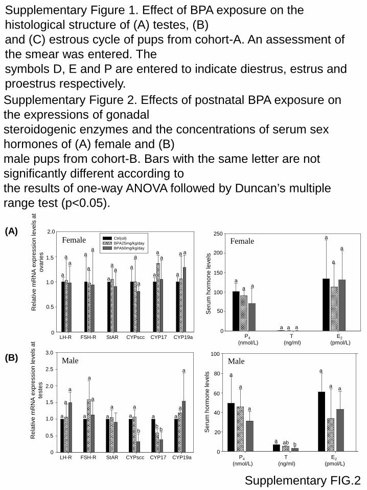
ale (Fig. 4B). In male pups of cohort-B, the expression levels ofhe steroidogenic enzymes CYPscc and CYP17 and concentrationsf serum testosterone were significantly less in the BPA treatmentroups than the controls (Supplementary Figure 2). No noticeableffect on the BPA-exposed female pups was observed.
. Discussion
In the present study, effects of BPA exposure on the reproductiveealth of offspring were highlighted. Responses of conventionalhysical and anatomical parameters, such as number of pupser litter, survival and growth of pups were monitored in thistudy. No statistically significant effects on these parameters wereound. The data are generally consistent with those reported byther researchers, using exposure doses from 0.003 to 600 mgPA/kg, bw/day [41]. However in this study when more sensi-ive endpoints were recorded (i.e. the expression levels of selectedeproductive-related hormone and receptor genes along the HPG-xis), significant effects on the regulatory circuits at the HPG axisere observed. Gestational and lactational BPA exposure induced
ranscript levels of KiSS-1/GnRH in the hypothalami and FSH in theituitaries of the male and female offspring. Altered in the tran-cript levels of steroidogenic enzymes in the gonads and the serumevels of the sex hormones in the offspring were demonstrated.
KiSS-1 functions as a gatekeeper for initiation of puberty and forhe regulation of gene expression along the HPG-axis [42,43]. Up-egulation of expression of hypothalamic KiSS-1 is hypothesized totimulate synthesis and release of GnRH and Gn in the hypotha-
amus and pituitary, respectively. Because postnatal exposure toPA caused no statistically significant effects on the expressions ofhe genes in either the hypothalami or pituitaries of the pups inohort-B, the perinatal period seems to be a critical “exposure win-ow” for BPA to affect reproductive neural circuits in hypothalamiFig. 4. Effects of BPA on concentrations of pituitary and gonadal hormones in serumof (A) female and (B) male pups of cohort-A. Bars with the same letter are not signif-icantly different according to the results of one-way ANOVA followed by Duncan’smultiple range test (p < 0.05).
of both male and female mice. In the studies of rats, BPA exposurewere found to affect hypothalamic kisspeptin fiber density, KiSS-1mRNA expression at the prepubertal stages [44,45], hypothala-mic ER� expression [46] and pituitary GnRH-signaling [47]. Thepresent study revealed that the BPA-stimulated hypothalamic KiSS-1 mRNA expressions induced transcript levels of GnRH and FSH inthe male and female pups of cohort-A (Table 2). This observation isconsistent with the physiological role of the HPG-axis in regulationof puberty and reproduction in animals [48,49] and highlighted thepossible mechanistic effects of BPA on the local regulatory circuitsof hypothalamus and pituitary [50].
The fact that there are feed-back regulatory mechanisms in placeto maintain hormonal homeostasis along the HPG-axis [51,52]. Thealtered expression levels of hormones at the hypothalamus andpituitary levels may be the cause and/or the consequence of thechanges in gonadal steroidogenesis and sex hormone production.Our data illustrated that BPA-elicited differential effects on theexpression levels of gonadal steroidogenic enzymes and the con-
centrations of sex hormones in the serum of BPA-exposed maleand female pups. In BPA-exposed female pups from cohort A, theincreases of KiSS-1, GnRH and FSH expressions positively correlatedwith the increased expression level of CYP19a (Table 2) as well as
W. Xi et al. / Reproductive Toxicology 31 (2011) 409–417 415
Table 2Pearson correlation coefficients (r) between mRNA expressions of the genes along the HPG-axis in pups of cohort-A, after perinatal and postnatal BPA exposure.
Female Hypothalamus Pituitary Ovary
KiSS-1 GnRH FSH LH StAR CYPscc CYP17 CYP19a
HypothalamusKiSS-1 1.000GnRH 0.610* 1.000
PituitaryFSH 0.850** 0.707* 1.000LH 0.160 0.227 −0.325 1.000
OvaryStAR 0.578 −0.278 −0.108 0.280 1.000CYPscc 0.464 0.221 0.111 −0.484 0.066 1.000CYP17 −0.418 −0.517* 0.126 −0.152 −0.283 0.225 1.000CYP19a 0.677* 0.834** 0.376 0.169 −0.573 −0.430 0.219 1.000
Male Hypothalamus Pituitary Testis
KiSS-1 GnRH FSH LH StAR CYPscc CYP17 CYP19a
HypothalamusKiSS-1 1.000GnRH 0.567* 1.000
PituitaryFSH 0.114 −0.108 1.000LH 0.222 −0.157 −0.182 1.000
TestisStAR −0.326 −0.512 0.111 −0.352 1.000CYPscc −0.598* −0.583* 0.319 −0.092 0.066 1.000CYP17 −0.772* −0.674* 0.008 −0.152 0.414 0.838* 1.000CYP19a 0.819* 0.947* 0.154 0.169 0.162 −0.573* −0.780* 1.000
Only parameters which have significant differences are shown here.Significant correlation is indicated by asterisk(s) (*p < 0.05; **p < 0.01).
Table 3Correlation coefficients (r) between the concentrations of serum steroid hormones and the mRNA expressions of steroidogenic enzymes in the gonads of pups from cohort-A,after perinatal and postnatal exposure to BPA.
Dose (mg BPA/kg, bw/day) Female Male
12 25 50 12 25 50
Cohort AP4 vs. CYP17 0.152 0.243 0.334 T vs. CYP17 0.568* 0.863** 0.746**P4 vs. CYPscc 0.327 −0.156 −0.243 T vs. CYPscc 0.732* 0.804** 0.902**P4 vs. CYP19a −0.158 −0.709* −0.533* T vs. CYP19a −0.236 −0.558* −0.659*P4 vs. StAR 0.332 0.330 0.279 T vs. StAR 0.179 0.659* 0.501*E2 vs. CYP17 0.119 −0.363 −0.298 E2 vs. CYP17 0.133 0.110 0.143E2 vs. CYPscc 0.047 0.265 0.374 E2 vs. CYPscc −0.175 −0.123 −0.079
*
S
swntC
TCa
S
E2 vs. CYP19a 0.65* 0.554* 0.717E2 vs. StAR 0.156 0.058 0.079
ignificant correlation is indicated by asterisk(s) (*p < 0.05; **p < 0.01).
erum E2 (Table 3). For the BPA-exposed male pups, although thereere stimulations on the expressions of KiSS-1, GnRH and FSH, aegative correlation was observed with the expression levels ofesticular steroidogenic enzymes (Table 2). The down-regulation ofYPscc and CYP17 resulted in lesser serum concentrations of testos-
able 4orrelation coefficients (r) between the concentrations of serum steroid hormones and thfter postnatal BPA exposure.
Female
BPA (mg/kg/day) 25 50
Cohort BP4 vs. CYP17a 0.125 −0.026P4 vs. CYPscc 0.258 0.152P4 vs. CYP19a 0.168 −0.257P4 vs. StAR 0.109 0.046E2 vs. CYP17a 0.056 −0.112E2 vs. CYPscc 0.425 0.130.E2 vs. CYP19a 0.022 −0.166E2 vs. StAR 0.198 0.279
ignificant correlation is indicated by asterisk(s) (*p < 0.05; **p < 0.01).
E2 vs. CYP19a 0.246 0.421 0.449E2 vs. StAR 0.314 −0.191 0.420
terone (Table 3). Similar to the male pups of cohort-A, the reducedtestosterone concentrations in the serum of male pups from cohortB were directly proportional to the decreased expressions of thetesticular enzymes (Table 4). The up-regulations of CYP19a expres-sion in the testes of male pups from cohort A could further reduce
e mRNA expressions of steroidogenic enzymes in the gonads of pups from cohort-B,
Male
BPA (mg/kg/day) 25 50
T vs. CYP17a 0.674* 0.826**T vs. CYPscc −0.335 0.524*T vs. CYP19a 0.102 0.458T vs. StAR −0.275 0.169E2 vs. CYP17a 0.049 −0.338E2 vs. CYPscc 0.265 −0.316E2 vs. CYP19a 0.095 0.098E2 vs. StAR −0.044 0.123

4 Toxic
sabooAtiow
agholwfanspTlrConHdrBsn[srndBdso
5
A
HRPaaU
A
t
[
[[
[
[
[
[
[
[
[
[
[
[
[
[
[
[
16 W. Xi et al. / Reproductive
erum testosterone levels. The data of the present study are ingreement with another study where doses of 100–200 mg BPA/kg,w/day suppressed expressions of steroidogenic enzymes in testesf rats [53]. Furthermore, an in vitro stimulatory effect of BPAn CYP19a gene expression was reported in rat Leydig cells [53].lthough it was suggested by another study that the reduced tes-
icular steroidogenesis could be due to lesser concentrations of LHn blood serum [31], no significant changes in the transcript levelsf LH in the pituitary or LH-R in the testes of BPA-exposed pupsere observed in our study.
BPA is a weak estrogen agonist and so its effects observed innimal studies are difficult to reconcile with the actions as estro-en agonists [6]. According to the “spare receptor” hypothesis, theormonal system is sensitive to changes in a small proportionf receptor binding (10%), leading to a great change in cellu-ar responses [34,54]. Any further increase in receptor occupancy
ould only produce a small increase of cellular responses. There-ore it seems unlikely that the observed BPA effects are due to thedditional estrogen potency of BPA relative to the existing endoge-ous estrogen equivalents. Also, BPA has low binding affinity toex-hormone-binding globulin (SHBG) and further minimizes theotential of BPA to activate membrane SHBG receptor [55–57].herefore the contribution of BPA to the total estrogen equiva-ents in the blood of female pups would be considerably smallelative to the estrogen equivalents from endogenous estrogens.omparatively male pups should be more sensitive to the effectsf BPA. The data reported here indicated that perinatal and post-atal exposure to BPA was associated with functional changes inPG-axis of the animals. These functional changes were unlikelyue to the effects of BPA as an ER agonist. Moreover it has beeneported that steroidogenesis is a major target for EDCs includingPA [58]. Retrospectively BPA may interfere with steroid hormoneynthesis pathways and the release of the more potent endoge-ous steroid hormones (i.e. E2 and testosterone) into circulations6,58–60]. The change in serum sex hormone levels may cause sub-equent reproductive dysfunction by interfering with the feedbackegulatory mechanisms of the HPG-axis. Our data supported thisotion as the altered serum levels of E2 and/or testosterone wereetected in the BPA-exposed pups. Although significant effects ofPA on HPG-regulatory circuits were identified in this study, theoses might not be necessarily reflective of general human expo-ure to BPA. These data are more relevant for the highly exposed orccupationally exposed individuals [61].
. Conflict of interest
The author declares that there is no conflict of interest.
cknowledgements
This work was supported by the Super Faculty Research Grant,ong Kong Baptist University (CKC Wong) and Collaborativeesearch Fund (HKBU 1/CRF/08), University Grants Committee.rof. Giesy was supported by the Canada Research Chair programnd an at large Chair Professorship at the Department of Biologynd Chemistry and State Key Laboratory in Marine Pollution, Cityniversity of Hong Kong.
ppendix A. Supplementary data
Supplementary data associated with this article can be found, inhe online version, at doi:10.1016/j.reprotox.2010.12.002.
[
[
ology 31 (2011) 409–417
References
[1] Phillips KP, Foster WG, Leiss W, Sahni V, Karyakina N, Turner MC, et al. Assess-ing and managing risks arising from exposure to endocrine-active chemicals. JToxicol Environ Health B Crit Rev 2008;11:351–72.
[2] Phillips KP, Foster WG. Key developments in endocrine disrupter research andhuman health. J Toxicol Environ Health B Crit Rev 2008;11:322–44.
[3] Anway MD, Cupp AS, Uzumcu M, Skinner MK. Epigenetic transgenerationalactions of endocrine disruptors and male fertility. Science 2005;308:1466–9.
[4] Dolinoy DC, Huang D, Jirtle RL. Maternal nutrient supplementation counteractsbisphenol A-induced DNA hypomethylation in early development. Proc NatlAcad Sci USA 2007;104:13056–61.
[5] Leranth C, Hajszan T, Szigeti-Buck K, Bober J, MacLusky NJ. Bisphenol Aprevents the synaptogenic response to estradiol in hippocampus and pre-frontal cortex of ovariectomized nonhuman primates. Proc Natl Acad Sci USA2008;105:14187–91.
[6] Vandenberg LN, Maffini MV, Sonnenschein C, Rubin BS, Soto AM. Bisphenol-A and the great divide: a review of controversies in the field of endocrinedisruption. Endocr Rev 2009;30:75–95.
[7] Kuo HW, Ding WH. Trace determination of bisphenol A and phytoestrogens ininfant formula powders by gas chromatography–mass spectrometry. J Chro-matogr A 2004;1027:67–74.
[8] Munguia-Lopez EM, Gerardo-Lugo S, Peralta E, Bolumen S, Soto-Valdez H.Migration of bisphenol A (BPA) from can coatings into a fatty-food simulantand tuna fish. Food Addit Contam 2005;22:892–8.
[9] Lim DS, Kwack SJ, Kim KB, Kim HS, Lee BM. Risk assessment of bisphe-nol A migrated from canned foods in Korea. J Toxicol Environ Health A2009;72:1327–35.
10] Ackerman LK, Noonan GO, Heiserman WM, Roach JA, Limm W, Begley TH.Determination of Bisphenol A in U.S. Infant Formulas: updated methods andconcentrations. J Agric Food Chem 2010.
11] Halden RU. Plastics and health risks. Annu Rev Public Health; 2010.12] Thompson RC, Moore CJ, vom Saal FS, Swan SH. Plastics, the environment and
human health: current consensus and future trends. Philos Trans R Soc Lond BBiol Sci 2009;364:2153–66.
13] Hotchkiss AK, Rider CV, Blystone CR, Wilson VS, Hartig PC, Ankley GT, et al. Fif-teen years after Wingspread—environmental endocrine disrupters and humanand wildlife health: where we are today and where we need to go. Toxicol Sci2008;105:235–59.
14] Oehlmann J, Schulte-Oehlmann U, Kloas W, Jagnytsch O, Lutz I, Kusk KO, et al.A critical analysis of the biological impacts of plasticizers on wildlife. PhilosTrans R Soc Lond B Biol Sci 2009;364:2047–62.
15] Teuten EL, Saquing JM, Knappe DR, Barlaz MA, Jonsson S, Bjorn A, et al. Transportand release of chemicals from plastics to the environment and to wildlife. PhilosTrans R Soc Lond B Biol Sci 2009;364:2027–45.
16] Wan Y, Choi K, Kim S, Ji K, Chang H, Wiseman S, et al. Hydroxylated poly-brominated diphenyl ethers and bisphenol a in pregnant women and theirmatching fetuses: placental transfer and potential risks. Environ Sci Technol2010;44:5233–9.
17] Nishikawa M, Iwano H, Yanagisawa R, Koike N, Inoue H, Yokota H. Placentaltransfer of conjugated bisphenol A and subsequent reactivation in the rat fetus.Environ Health Perspect 2010.
18] Balakrishnan B, Henare K, Thorstensen EB, Ponnampalam AP, Mitchell MD.Transfer of bisphenol A across the human placenta. Am J Obstet Gynecol2010;202:393–7.
19] Tanaka M, Kawamoto T, Matsumoto H. Distribution of 14C-bisphenol A in preg-nant and newborn mice. Dent Mater 2010;26:e181–7.
20] Yokota H, Iwano H, Endo M, Kobayashi T, Inoue H, Ikushiro S, et al. Glu-curonidation of the environmental oestrogen bisphenol A by an isoform ofUDP-glucuronosyltransferase, UGT2B1, in the rat liver. Biochem J 1999;340(Pt2):405–9.
21] Takahashi O, Oishi S. Disposition of orally administered 2,2-Bis(4-hydroxyphenyl)propane (Bisphenol A) in pregnant rats and the placentaltransfer to fetuses. Environ Health Perspect 2000;108:931–5.
22] Matsumoto J, Yokota H, Yuasa A. Developmental increases in rat hepaticmicrosomal UDP-glucuronosyltransferase activities toward xenoestrogens anddecreases during pregnancy. Environ Health Perspect 2002;110:193–6.
23] Richter CA, Birnbaum LS, Farabollini F, Newbold RR, Rubin BS, Talsness CE,et al. In vivo effects of bisphenol A in laboratory rodent studies. Reprod Toxicol2007;24:199–224.
24] Honma S, Suzuki A, Buchanan DL, Katsu Y, Watanabe H, Iguchi T. Low dose effectof in utero exposure to bisphenol A and diethylstilbestrol on female mousereproduction. Reprod Toxicol 2002;16:117–22.
25] Timms BG, Howdeshell KL, Barton L, Bradley S, Richter CA, vom Saal FS. Estro-genic chemicals in plastic and oral contraceptives disrupt development of thefetal mouse prostate and urethra. Proc Natl Acad Sci USA 2005;102:7014–9.
26] Kawai K, Nozaki T, Nishikata H, Aou S, Takii M, Kubo C. Aggressive behaviorand serum testosterone concentration during the maturation process of malemice: the effects of fetal exposure to bisphenol A. Environ Health Perspect2003;111:175–8.
27] vom Saal FS, Hughes C. An extensive new literature concerning low-dose effectsof bisphenol A shows the need for a new risk assessment. Environ HealthPerspect 2005;113:926–33.
28] Ramos JG, Varayoud J, Sonnenschein C, Soto AM, Munoz DT, Luque EH. Prenatalexposure to low doses of bisphenol A alters the periductal stroma and glandularcell function in the rat ventral prostate. Biol Reprod 2001;65:1271–7.
Toxic
[
[
[
[
[
[
[
[
[
[
[
[
[
[[
[
[
[
[
[
[
[
[
[
[
[
[
[
[
[
[
W. Xi et al. / Reproductive
29] Susiarjo M, Hassold TJ, Freeman E, Hunt PA, Bisphenol A. exposure in uterodisrupts early oogenesis in the mouse. PLoS Genet 2007;3:e5.
30] Munoz-de-Toro M, Markey CM, Wadia PR, Luque EH, Rubin BS, SonnenscheinC, et al. Perinatal exposure to bisphenol-A alters peripubertal mammary glanddevelopment in mice. Endocrinology 2005;146:4138–47.
31] Akingbemi BT, Sottas CM, Koulova AI, Klinefelter GR, Hardy MP. Inhibition oftesticular steroidogenesis by the xenoestrogen bisphenol A is associated withreduced pituitary luteinizing hormone secretion and decreased steroidogenicenzyme gene expression in rat Leydig cells. Endocrinology 2004;145:592–603.
32] Mendiola J, Jorgensen N, Andersson AM, Calafat AM, Ye X, Redmon JB, et al. Areenvironmental levels of bisphenol A associated with reproductive function infertile men? Environ Health Perspect 2010.
33] vom Saal FS, Cooke PS, Buchanan DL, Palanza P, Thayer KA, Nagel SC, et al. Aphysiologically based approach to the study of bisphenol A and other estro-genic chemicals on the size of reproductive organs, daily sperm production,and behavior. Toxicol Ind Health 1998;14:239–60.
34] Welshons WV, Nagel SC, Thayer KA, Judy BM, vom Saal FS. Low-dose bioactivityof xenoestrogens in animals: fetal exposure to low doses of methoxychlor andother xenoestrogens increases adult prostate size in mice. Toxicol Ind Health1999;15:12–25.
35] National Toxicology Program. NTP technical report on the carcinogenesis bioas-say of bisphenol A in F344 rats and B6C3F1 mice (feed study). Research TrianglePark, NC: US Department of Health and Human Services, Public Health Service,National Institutes of Health; 2004.
36] Stahlhut RW, Welshons WV, Swan SH. Bisphenol A data in NHANES suggestlonger than expected half-life, substantial nonfood exposure, or both. EnvironHealth Perspect 2009;117:784–9.
37] Vandenberg LN, Hauser R, Marcus M, Olea N, Welshons WV. Human exposureto bisphenol A (BPA). Reprod Toxicol 2007;24:139–77.
38] Vandenberg LN, Chauhoud I, Heindel JJ, Padmanabhan V, Paumgartten FJ,Schoenfelder G. Urinary, circulating and tissue biomonitoring studies indicatewidespread exposure to bisphenol A. Environ Health Perspect; 2010.
39] Shin BS, Kim CH, Jun YS, Kim DH, Lee BM, Yoon CH, et al. Physiologicallybased pharmacokinetics of bisphenol A. J Toxicol Environ Health A 2004;67:1971–85.
40] Speijers GJ. Precision of estimates of an ADI (or TDI or PTWI). Regul ToxicolPharmacol 1999;30:S87–93.
41] Tyl RW, Myers CB, Marr MC, Sloan CS, Castillo NP, Veselica MM, et al. Two-generation reproductive toxicity study of dietary bisphenol A in CD-1 (Swiss)mice. Toxicol Sci 2008;104:362–84.
42] Hameed S, Dhillo WS. Biology of kisspeptins. Front Horm Res 2010;39:25–36.
43] Silveira LF, Teles MG, Trarbach EB, Latronico AC. Role of Kisspeptin/GPR54system in human reproductive axis. Front Horm Res 2010;39:13–24.44] Navarro VM, Sanchez-Garrido MA, Castellano JM, Roa J, Garcia-Galiano D,
Pineda R, et al. Persistent impairment of hypothalamic KiSS-1 system afterexposures to estrogenic compounds at critical periods of brain sex differen-tiation. Endocrinology 2009;150:2359–67.
[
[
ology 31 (2011) 409–417 417
45] Patisaul HB, Todd KL, Mickens JA, Adewale HB. Impact of neonatal exposureto the ERalpha agonist PPT, bisphenol-A or phytoestrogens on hypotha-lamic kisspeptin fiber density in male and female rats. Neurotoxicology2009;30:350–7.
46] Ceccarelli I, Della SD, Fiorenzani P, Farabollini F, Aloisi AM. Estrogenic chemi-cals at puberty change ERalpha in the hypothalamus of male and female rats.Neurotoxicol Teratol 2007;29:108–15.
47] Fernandez M, Bianchi M, Lux-Lantos V, Libertun C. Neonatal exposure to bisphe-nol a alters reproductive parameters and gonadotropin releasing hormonesignaling in female rats. Environ Health Perspect 2009;117:757–62.
48] Kuohung W, Kaiser UB. GPR54 and KiSS-1: role in the regulation of puberty andreproduction. Rev Endocr Metab Disord 2006;7:257–63.
49] Kaiser UB, Kuohung W. KiSS-1 and GPR54 as new players in gonadotropinregulation and puberty. Endocrine 2005;26:277–84.
50] Mahoney MM, Padmanabhan V. Developmental programming: impact of fetalexposure to endocrine-disrupting chemicals on gonadotropin-releasing hor-mone and estrogen receptor mRNA in sheep hypothalamus. Toxicol ApplPharmacol 2010;247:98–104.
51] Maffucci JA. Gore AC. Chapter 2: hypothalamic neural systems controlling thefemale reproductive life cycle gonadotropin-releasing hormone, glutamate,and GABA. Int Rev Cell Mol Biol 2009;274:69–127.
52] Micevych P, Kuo J, Christensen A. Physiology of membrane oestrogen receptorsignalling in reproduction. J Neuroendocrinol 2009;21:249–56.
53] Kim JY, Han EH, Kim HG, Oh KN, Lee KY, Jeong HG. Bisphenol A-inducedaromatase activation is mediated by cyclooxygenase-2 up-regulation in rattesticular Leydig cells. Toxicol Lett 2010.
54] Welshons WV, Thayer KA, Judy BM, Taylor JA, Curran EM, vom Saal FS. Largeeffects from small exposures. I. Mechanisms for endocrine-disrupting chemi-cals with estrogenic activity. Environ Health Perspect 2003;111:994–1006.
55] Tollefsen KE, Ovrevik J, Stenersen J. Binding of xenoestrogens to the sex steroid-binding protein in plasma from Arctic charr (Salvelinus alpinus L.). CompBiochem Physiol C Toxicol Pharmacol 2004;139:127–33.
56] Dechaud H, Ravard C, Claustrat F, de la Perriere AB, Pugeat M. Xenoestro-gen interaction with human sex hormone-binding globulin (hSHBG). Steroids1999;64:328–34.
57] Rosner W, Hryb DJ, Kahn SM, Nakhla AM, Romas NA. Interactions ofsex hormone-binding globulin with target cells. Mol Cell Endocrinol2010;316:79–85.
58] Sanderson JT. The steroid hormone biosynthesis pathway as a target forendocrine-disrupting chemicals. Toxicol Sci 2006;94:3–21.
59] Diamanti-Kandarakis E, Bourguignon JP, Giudice LC, Hauser R, Prins GS, SotoAM, et al. Endocrine-disrupting chemicals: an Endocrine Society scientific
statement. Endocr Rev 2009;30:293–342.60] Hiroi T, Okada K, Imaoka S, Osada M, Funae Y. Bisphenol A binds to proteindisulfide isomerase and inhibits its enzymatic and hormone-binding activities.Endocrinology 2006;147:2773–80.
61] He Y, Miao M, Wu C, Yuan W, Gao E, Zhou Z, et al. Occupational exposure levelsof bisphenol A among Chinese workers. J Occup Health 2009;51:432–6.

0
20
40
60
80
100
120
140
160
180
200
Ctrl BPA12.5 BPA25 BPA50
L
Dia
met
er (µ
m)
tubuletubular lumen
Ctrl BPA12.5
BPA 25 BPA 50
Supplementary FIG.1
(A)
(B)
Ctrl
BPA12.5
BPA25
BPA50
13/06/09 17/06 21/06 25/06 29/06 02/07 06/07 09/07
D D P E D D P E D D P E D P P E D D P E D P P E D D P E
P E D D P E D P P E D D P E D D D P E D P P E D D P E D
D P E D D P E D D P E D P P E E D D P E D D P E D D P E
D D P E D P P E D D P E D D P E D D P E D D P E D D P E
(C)
(A)
(B)
Supplementary FIG.2
Rel
ativ
e m
RN
A e
xpre
ssio
n le
vels
at
ovar
ies
0
0.5
2.0
1.5
1.0
LH-R FSH-R StAR CYPscc CYP17 CYP19a
Female
aa
aa
a
a
aa
a a
a
a
a
a
a
aa
a
0
50
100
150
200
250
P4(nmol/L)
T (ng/ml)
E2(pmol/L)
Female
Ser
um h
orm
one
leve
ls
a a
aa
a
a
a
a
a
0
20
40
60
80
100
P4(nmol/L)
T (ng/ml)
E2(pmol/L)
Male
Ser
um h
orm
one
leve
ls
a ab b
aa
a
a
aa
0
0.5
1.0
1.5
2.0
2.5
3.0
Rel
ativ
e m
RN
A e
xpre
ssio
n le
vels
at
test
es
LH-R FSH-R StAR CYPscc CYP17 CYP19a
Male
aa
a
bb b
a
a
a
a
a
a
a
aa
a
a
Ctrl(oil) BPA25mg/kg/day BPA50mg/kg/day
Supplementary Figure 1. Effect of BPA exposure on the histological structure of (A) testes, (B)and (C) estrous cycle of pups from cohort-A. An assessment of the smear was entered. Thesymbols D, E and P are entered to indicate diestrus, estrus and proestrus respectively.Supplementary Figure 2. Effects of postnatal BPA exposure on the expressions of gonadalsteroidogenic enzymes and the concentrations of serum sex hormones of (A) female and (B)male pups from cohort-B. Bars with the same letter are not significantly different according tothe results of one-way ANOVA followed by Duncan’s multiple range test (p<0.05).