effect of moringa oleifera leaf meal supplementation on
TRANSCRIPT

Turk. J. Fish.& Aquat. Sci. 21(1), 29-39
http://doi.org/10.4194/1303-2712-v21_1_04
Published by Central Fisheries Research Institute (SUMAE) Trabzon, Turkey in cooperation with Japan International Cooperation Agency (JICA), Japan
R E S E A R C H P A P E R
Effect of Moringa oleifera Leaf Meal Supplementation on Growth Performance, Morphological Indexes, Antioxidant Status and Resistance to Streptococcus agalactiae of Nile Tilapia (Oreochromis niloticus)
Shijun Chen1,#, Youming Jia1,#, Weihua Xu1, Jin Peng1, Yuhui He1, Jijia Sun1, Qing Pan1, Chaoxiong Peng3, Junjiang Yang3, Xiaoyang Chen2,*, Lian Gan1,3,* 1College of Marine Sciences, South China Agricultural University, Guangzhou, 510642, P.R. China. 2State Key Laboratory for Conservation and Utilization of Subtropical Agro-Introduction Bioresources, South China Agricultural University, Guangzhou, 510642, P.R. China. 3Foshan DBN Huilin Aquaculture Science & Technology Co. Ltd, Foshan, 524500, P.R. China. #These authors share the first authorship.
Article History Received Jun 14, 2020 Accepted Nov 17, 2020 First Online Nov 18, 2020
Corresponding Author Tel.: +8602087571321 E-mail: [email protected]; [email protected]
Keywords Antibacterial Antioxidant Lipotropic effect Moringa oleifera Oreochromis niloticus
Abstract An 8-week feeding trial was conducted to evaluate the effect of Moringa oleifera leaf
meal (MOLM) on growth performance, antioxidant status and morphological indexes
in Nile tilapia (Oreochromis niloticus). Resistance to Streptococcus agalactiae was also
assessed based on a 192h challenge experiment after feeding trial. Fish (initial mean
weight 4.05±0.05g) were held in 21 tanks (volume 300L, 30 fish/tank), with triplicate
treatment groups being fed to apparent satiation three times each day on practical
diets adding different levels of MOLM (0, 0.1, 0.5, 2, 5, 10 and 20%). Sampling
procedure was performed after 24h of fasting. Growth metrics showed no significant
differences among groups. Whole-body lipid content, viscerosomatic index and
intraperitoneal fat ratio were decreased as dietary MOLM levels increasing. Total
superoxide dismutase (T-SOD) and reduced glutathione content (GSH) were
significantly influenced by MOLM. Moreover, the lowest cumulative mortality rate
(CMR) after challenge experiment was noted in 10% group. Based on CMR and
quadratic regression analysis of liver GSH content, it was concluded that dietary MOLM
levels of 8.15-10.00% may be beneficial for enhancing resistance to S. agalactiae
infection. Our results suggest that the maximum additive amount of MOLM reached
20% under the condition of the present study.
Introduction
Soybean meal is a vital plant protein source and widely used as a feed ingredient for aquaculture species due to its relatively high nutritional value and digestibility (Puycha et al., 2017). However, owing to its unstable prices and market supply, it is necessary to identify other plant resources to substitute dietary soybean meal (Kasiga & Lochmann, 2014). Moringa oleifera, a member of the family Moringaceae, is a fast-growing plant widely distributed in the tropics and subtropics (Richter et al., 2003). It is initially treated as a plant with medical value and utilized as an alternative food source for resistance malnutrition in developing
countries (Rweyemamu et al., 2015; Anwar et al., 2007). Due to its leaf protein content reached 26%, stable market supply and contained some essential amino acids including methionine, cysteine, tryptophan and lysine (Makkar & Becker, 1996; Oyeyinka & Oyeyink, 2018), it has been treated as a plant protein source and applied into diet of some fish species, such as sea bass (Lates calcarifer) (Ganzon-Naret, 2014), African Catfish (Clarias gariepinus) (Hlophe & Moyo, 2014a), Nile tilapia (Oreochromis niloticus) (Bbole, Mumba, Mupenda, & Kefi, 2016) and Bocourti's catfish (Pangasius bocourti) (Puycha et al., 2017). Previous studies have shown that the maximum amount of M. oleifera leaf added to the diet was varied with fish species, and its value did not

30 Turk. J. Fish.& Aquat. Sci. 21(1), 29-39
exceed 12% under the promise of not negatively affecting the growth (Richter et al., 2003; Puycha et al., 2017). Moreover, fish meal, as the most urgently replaced protein source by producers, occupied an important proportion (31-40%) in the formula of these studies. However, in the case of low dietary fish meal, the maximum amount of M. oleifera leaves added in the diet is unclear.
Phytochemicals, such as carotenoids, vitamin C, vitamin E, sterols, glycosides, alkaloids, flavonoids and moringine, have been reported to exist in various organs of M. oleifera (Sreelatha et al., 2011). Some of constituents mentioned above are considered to be beneficial for treating diseases, such as inflammation, cardiovascular and liver diseases (Limaye et al., 1995; Rao & Misra, 1998; Gopalakrishnan et al., 2016). Antioxidant property of M. oleifera was also found by evaluating oxidative stability of oil extracted from M. oleifera leaves (Anwar et al., 2007; Park et al., 2011). Streptococcus agalactiae infection has been recorded to cause some fish diseases, such as meningitis, sepsis, ascites and anorexia, which finally resulting in high mortality (Su et al., 2016). It also negatively influenced antioxidant capacity of fish (Du et al., 2020). Importantly, S. agalactiae is the main pathogen causing tilapia streptococcus disease in China. Whether the antioxidant and antibacterial properties of M. oleifera can help tilapia resist S. agalactiae infection is unknown. Besides, fat-degrade effect was observed in finisher pigs due to supplementation of M. oleifera leaf meal (Mukumbo et al., 2014). To our knowledge, previous studies have mostly focused on effects of different forms and levels of M. oleifera on fish growth. However, research on fat-degrade effect and resistance to pathogen infection is limited. Therefore, the objective of the present research was to investigate the effect of graded levels of dietary MOLM on growth performance, morphometry indices, serum biochemical indexes, antioxidant enzymes activities and resistance to S. agalactiae infection in Nile tilapia.
Materials and Methods
Preparation of M. oleifera Leaf Meal and Experimental Diets
The method for making M. oleifera leaf meal was in accordance with that of Nkukwana et al. (2014) with minor modification. Fresh, green and undamaged mature M. oleifera leaves, obtained from South China Agricultural University and air-dried during the day with no direct sunlight exposure, with constant turning over to prevent fungal growth, were finely ground in a laboratory grinder and stored at room temperature before feed formulation. The nutrient and amino acid composition of MOLM were consistent with study reported by Chen et al. (2018) and shown as follows: crude protein, 26.6%; crude lipid, 4.3%; crude ash, 10.8%; arginine, 1.5%; histidine, 0.6%; lysine, 1.6%;
phenylalanine, 1.6%; leucine, 2.3%; isoleucine, 1.2%; methionine, 0.1%; valine, 1.4%; alanine, 1.7%; glycine, 1.4%; proline, 1.2%; serine, 1.1%; threonine, 1.2%. The amino acid composition of MOLM was analyzed by a Hitachi L-8900 automatic amino acid analyzer. The proximate analysis of diets and MOLM was based on the procedures of the Association of Official Analytical Chemists (AOAC) (1997) standard methods. Moisture was determined by putting samples in an oven at 105 ℃ until constant weight. The methods and instruments used to determine crude protein, crude lipid and ash content of dried diets were consistent with those reported by Gan et al. (2013). Crude protein (N × 6.25) was determined by the Kjeldahl method after acid digestion using an Auto Kjeldahl System (1030-Auto-analyzer, Tecator, Sweden). Crude lipid was tested by the ether-extraction method using a Soxtec System HT (Soxtec System HT6, Tecator, Sweden). Ash content was determined by incineration in a muffle furnace at 550 °C for 24 h.
The formulation and composition analysis of the experimental diets is given in Table 1. Fish meal, soybean meal, canola meal and wheat flour were used as dietary protein sources. Soybean oil was utilized as dietary lipid sources. The basal diet was formulated to contain about 30.1% crude protein and 6.4% crude lipid. Seven practical diets were formulated to contain MOLM at levels of 0, 0.1, 0.5, 2, 5, 10 and 20% diets. Various proportions of ingredients mentioned above were balanced with wheat flour. The method of manufacturing diet was consistent with the methods described by Gan et al. (2013). The dry ingredients were weighed and well-mixed by a Hobart mixer (A-200T Mixer bench Model unit, Russell Food Equipment, Ottawa, ON, Canada). Lipids and water were then added and thoroughly mixed. The 1.5 mm and 2.5 mm diameter pellets were cold-extruded using a pelletizer (Institute of Chemical Engineering, South China University of Technology, Guangdong, China), then air-dried to approximately 10% moisture, and saved at -20℃ until used.
Fish Management
Healthy juvenile Nile tilapia, obtained from a local commercial hatchery (Guangdong Tilapia Fry Farm, China), were reared in the feeding facility at the South China Agricultural University (Guangzhou, China) and fed to apparent satiation with a commercial diet (Zhuhai Shihai Feed Co., Ltd., China), and then acclimated to laboratory condition for two weeks. Afterwards, a total of 630 fish (initial mean weight 4.05±0.05g) were randomly distributed into 21 300-L tanks (inside diameter 80cm, height 60cm) at the end of the acclimatization period, and each dietary treatment was randomly assigned to triplicate tanks. Fish was fed the respective diets to apparent satiation three times a day (8:00, 12:00 and 17:00) at a feeding rate of 3 to 5% body weight during the 8-week feeding trial, and the amount

31 Turk. J. Fish.& Aquat. Sci. 21(1), 29-39
of feed fed everyday was recorded. During the feeding experiment, fish were weighed every two weeks, and the amount of the diet was adjusted according to fish weight. Fish was reared in indoor recirculation system tanks with continuous aeration. Uneaten feeds and fecal waste were removed by siphoning. The uneaten feeds were used to calculate amount of feed intake (FI) after drying. Water temperature and dissolved oxygen concentration were monitored daily using a portable oxygen meter (Taiwan, China, AZ8403), and water quality parameters were monitored using a HachTM water quality test kit. Water temperature and pH were kept at 29.0°C (average) and 7.4±0.2, respectively. Total ammonia nitrogen was lower than 0.05 mg/L, and dissolved oxygen was kept above 7.0 mg/L. Natural light-dark cycle was employed during the experiment, and the photoperiod was 12-h light/12-h dark.
Sample Collection and Chemical Analysis
All procedures implemented for fish followed handling guidelines of South China Agricultural University. At the end of the feeding trial, fish were starved for 24 h and anesthetized with MS-222 (Shanghai Reagent Corporation, China) before weighing and sampling. It is important to note that the fish used in the S. agalactiae infection experiment were not anesthetized. Four fish from each tank were randomly gathered for the analysis of the whole-body composition. Another six fish per tank were randomly selected for obtaining body length and weight of individual whole body, viscera, liver and intraperitoneal fat, and then calculating condition factor (CF), viscerosomatic index (VSI), hepatosomatic index (HSI) and intraperitoneal fat index (IPF). In addition, four fish from each tank were randomly sampled for collecting blood and liver. The serum was separated by centrifuging at 850 g for 10 min at 4 °C and then stored
at -80°C for testing serum biochemical indexes and antioxidant enzyme. Liver tissues were immediately dissected and placed into liquid nitrogen, and then stored at -80°C until used. Liver tissues were homogenized in the proportion of 1:9 (w/v) of ice-cold physiological saline solution and then centrifuged at 3,550 g for 20 min at 4°C. After that, the supernatant was collected and kept at -80°C for determining antioxidant enzyme. The proximate analysis of fish whole-body composition was based on the procedures of the AOAC (1997) standard methods.
Streptococcus agalactiae Infection Experiment
S. agalactiae challenge experiment was carried out according to the method reported by Yi et al. (2014) with some modifications. S. agalactiae THN strain was provided by the disease lab in College of Marine Sciences of South China Agricultural University (Guangzhou, China). The concentration of S. agalactiae infection was 0.1×10⁷ CFU/mL obtained from a preliminary trial. Briefly, the bacteria were cultivated in BHI broth and incubated in a shaker (200 rpm) incubator at 2℃ overnight and the concentration was adjusted to 0.1×10⁷ CFU/mL. At the end of the feeding trial, fish were starved for 24 h, and then 10 fish from each tank without anesthesia were selected and each fish was intraperitoneally injected with 0.1mL culture. Each treatment group had triplicates, and mortality was monitored every 12 h for up to 192 h. All dead fish were immediately removed from the tanks.
Serum Biochemical Indexes and Antioxidant Parameters Assays
The content of low-density lipoprotein (LDL), high-density lipoprotein (HDL), triacylglycerol (TAG), cholesterol (CHO), total protein (TP) and activities of
Table 1. Formulation and proximate composition of the experimental diets (% dry matter)
Ingredients Dietary addition levels of MOLM
0% 0.1% 0.5% 2% 5% 10% 20%
Fish meal 3.5 3.5 3.5 3.5 3.5 3.5 3.5 Soybean meal 32.00 31.95 31.75 31.00 29.48 26.95 21.90 Canola meal 22 22 22 22 22 22 22 Wheat flour 34.30 34.25 34.07 33.38 32.03 29.77 25.23 Moringa oleifera Lam (leaf meal) 0 0.1 0.5 2 5 10 20 Soybean oil 4.50 4.50 4.48 4.42 4.29 4.08 3.66 Monocalcium phosphate Ca (H2PO4)2 2 2 2 2 2 2 2 Premix1 1 1 1 1 1 1 1
DL-methionine (≥98%) 2 0.30 0.30 0.30 0.30 0.30 0.30 0.31
Choline chlorine (50%) 0.40 0.40 0.40 0.40 0.40 0.40 0.40 Proximate composition (% dry matter) Crude protein 30.1 29.7 29.6 29.6 29.5 29.4 29.0 Crude lipid 6.4 6.4 6.3 6.4 6.3 6.2 6.0 Soybean meal, canola meal, fish meal, wheat flour and soybean oil were purchased from Zhuhai Shihai Feed Corporation Ltd. Choline chlorine was purchased from Guangzhou Chengyi Aquatic Technology Ltd. 1premix including (mg/kg premix): Zn, 7000; Fe, 6000; Mn, 750; Co, 100; I, 120; Cu, 500; Se, 35; thiamine, 600; vitamin B2, 850; vitamin A, 300,000 IU; vitamin E, 2,500; vitamin D3, 80,000 IU; vitamin K3, 400; vitamin B6, 600; vitamin C, 8,500; biotin, 12; calcium pantothenate, 1,500; nicotinamide, 3,000; inositol, 4,000 (Guangzhou Chengyi Aquatic Technology Ltd, Guangzhou, China). 2DL-methionine consists of 98.5% DL-methionine (Novus International Inc.).
32 Turk. J. Fish.& Aquat. Sci. 21(1), 29-39
alanine aminotransferase (ALT) and aspartate aminotransferase (AST) were measured by an automatic biochemical analyser (Hitachi 7170A; DAICHI, Tokyo, Japan). The serum was sent to a professional hospital for detection of indexes mentioned above (Traditional Chinese Medicine Hospital of Guangdong Province, China). The content of reduced glutathione (GSH) and activity of total superoxide dismutase (T-SOD) were determined by using commercial kits (Nanjing Jiancheng Bioengineering Institute, Nanjing, China). GSH can react with dithiobisnitrobenzoicacid (DTNB) to form a yellow compound, which can be used for colorimetric quantitative determination of GSH content at 405 nm. In addition, T-SOD activity was determined by its ability to inhibit superoxide anion generated by a xanthine and xanthine oxidase reaction system. Absorbance of indexes mentioned above was tested by a microplate spectrophotometer Epoch (BioTek, USA).
Statistical Analysis
Results were presented as means ± standard error (standard error of the mean, SEM). One-way analysis of variance (ANOVA) followed by Duncan's multiple range test was selected to assess significant differences among all groups by using SPSS Statistic 20.0 software (SPSS Inc., Chicago, IL, USA). The Kruskal-Wallis ANOVA test (non-parametric test) was utilized when inhomogeneous variance appeared, followed by pairwise comparisons using Kruskal-Wallis. Probability value of P<0.05 was considered statistically significant. In addition, quadratic regression method was used to evaluate the optimum level of MOLM supplementation in Nile tilapia according to Chen et al. (2019). Pearson correlation coefficient (r) was used to evaluate the correlation between dietary MOLM levels and morphological parameters.
Results
Growth Performance, Morphometry Indices and Whole-Body Composition
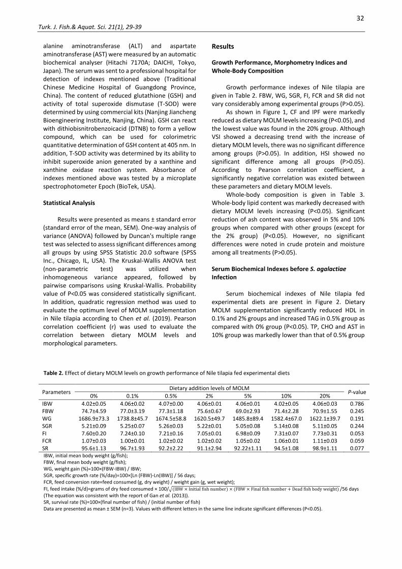
Growth performance indexes of Nile tilapia are given in Table 2. FBW, WG, SGR, FI, FCR and SR did not vary considerably among experimental groups (P>0.05).
As shown in Figure 1, CF and IPF were markedly reduced as dietary MOLM levels increasing (P<0.05), and the lowest value was found in the 20% group. Although VSI showed a decreasing trend with the increase of dietary MOLM levels, there was no significant difference among groups (P>0.05). In addition, HSI showed no significant difference among all groups (P>0.05). According to Pearson correlation coefficient, a significantly negative correlation was existed between these parameters and dietary MOLM levels.
Whole-body composition is given in Table 3. Whole-body lipid content was markedly decreased with dietary MOLM levels increasing (P<0.05). Significant reduction of ash content was observed in 5% and 10% groups when compared with other groups (except for the 2% group) (P<0.05). However, no significant differences were noted in crude protein and moisture among all treatments (P>0.05).
Serum Biochemical Indexes before S. agalactiae Infection
Serum biochemical indexes of Nile tilapia fed experimental diets are present in Figure 2. Dietary MOLM supplementation significantly reduced HDL in 0.1% and 2% groups and increased TAG in 0.5% group as compared with 0% group (P<0.05). TP, CHO and AST in 10% group was markedly lower than that of 0.5% group
Table 2. Effect of dietary MOLM levels on growth performance of Nile tilapia fed experimental diets
Parameters Dietary addition levels of MOLM
P-value 0% 0.1% 0.5% 2% 5% 10% 20%
IBW 4.02±0.05 4.06±0.02 4.07±0.00 4.06±0.01 4.06±0.01 4.02±0.05 4.06±0.03 0.786 FBW 74.7±4.59 77.0±3.19 77.3±1.18 75.6±0.67 69.0±2.93 71.4±2.28 70.9±1.55 0.245 WG 1686.9±73.3 1738.8±45.7 1674.5±58.8 1620.5±49.7 1485.8±89.4 1582.4±67.0 1622.1±39.7 0.191 SGR 5.21±0.09 5.25±0.07 5.26±0.03 5.22±0.01 5.05±0.08 5.14±0.08 5.11±0.05 0.244 FI 7.60±0.20 7.24±0.10 7.21±0.16 7.05±0.01 6.98±0.09 7.31±0.07 7.73±0.31 0.053 FCR 1.07±0.03 1.00±0.01 1.02±0.02 1.02±0.02 1.05±0.02 1.06±0.01 1.11±0.03 0.059 SR 95.6±1.13 96.7±1.93 92.2±2.22 91.1±2.94 92.22±1.11 94.5±1.08 98.9±1.11 0.077 IBW, initial mean body weight (g/fish); FBW, final mean body weight (g/fish); WG, weight gain (%)=100×(FBW-IBW) / IBW; SGR, specific growth rate (%/day)=100×[Ln (FBW)-Ln(IBW)] / 56 days; FCR, feed conversion rate=feed consumed (g, dry weight) / weight gain (g, wet weight);
FI, feed intake (%/d)=grams of dry feed consumed × 100/√(IBW × Initial fish number) × (FBW × Final fish number + Dead fish body weight) /56 days (The equation was consistent with the report of Gan et al. (2013)). SR, survival rate (%)=100×(final number of fish) / (initial number of fish) Data are presented as mean ± SEM (n=3). Values with different letters in the same line indicate significant differences (P<0.05).

33 Turk. J. Fish.& Aquat. Sci. 21(1), 29-39
Figure 1. Morphometry indices of Nile tilapia fed experimental diets for 8-week. Data are presented as mean ± SEM (n=3). The line with different letters indicates significant differences (P<0.05). r, Pearson correlation coefficient; sig., significance (for correlation analysis). CF, condition factor (g/cm3)=100 × body weight (g) / body length (cm3); VSI, viscerosomatic index (%)=100×(visceral weight) / (whole body weight); HSI, Hepatosomatic index (%)=100×(liver weight) / (whole body weight); IPF, intraperitoneal fat ratio (%)=100×(intraperitoneal fat weight) / (whole body weight).
Table 3. Effect of dietary MOLM levels on body composition of Nile tilapia fed experimental diets for 8-week
Parameters Dietary addition levels of MOLM
P-value 0% 0.1% 0.5% 2% 5% 10% 20%
Whole body (% wet weight) Moisture 69.8±0.84 69.0±0.43 68.1±0.38 69.4±0.43 70.8±0.95 70.2±0.44 69.7±1.03 0.235 Crude protein 16.3±0.40 16.0±0.54 16.2±0.06 16.5±0.45 15.8±0.43 15.8±0.86 15.7±0.44 0.897 Crude lipid 9.05±0.53bc 9.52±0.22c 9.20±0.31c 8.60±0.29bc 7.88±0.13ab 8.43±0.27bc 7.16±0.56a 0.005 Ash 3.77±0.11b 3.79±0.03b 3.81±0.08b 3.60±0.12ab 3.41±0.04a 3.39±0.15a 3.86±0.15b 0.027 Data are presented as mean ± SEM (n=3). Values with different letters in the same line indicate significant differences (P<0.05).
Figure 2. Serum biochemical indexes of Nile tilapia fed experimental diets for 8-week. Data are presented as mean ± SEM (n=3). The column with different letters indicates significant differences (P<0.05). LDL (low-density lipoprotein, mmol/L), HDL (high-density lipoprotein, mmol/L), TAG (triacylglycerol, mmol/L), CHO (cholesterol, mmol/L), TP (total protein, g/L), ALT (alanine transaminase, U/L) and AST (aspartate aminotransferase, U/L).
34 Turk. J. Fish.& Aquat. Sci. 21(1), 29-39
(P<0.05). However, ALT and LDL showed no significant difference among all groups (P>0.05). Cumulative Mortality Rate after 192h S. agalactiae Challenge
As shown in Table 4, cumulative mortality rate (CMR) in 0%, 0.5%, 5%, 10% and 20% groups was increased with dietary MOLM levels increasing when stress time before 72 h, and maintained thereafter. 0.1% and 2% groups showed a similar trend, but the deadline for continuously increased CMR was changed to 120 h. After 192 h S. agalactiae challenge, the highest CMR was discovered in 0.1% group, and the lowest CMR was observed in 10% group.
T-SOD and GSH in Serum and Liver of Nile tilapia before and after 192 h S. agalactiae Challenge
T-SOD activity and GSH content in serum and liver of Nile tilapia before and after S. agalactiae challenge are given in Figure 3.
Before challenge, 0.1%, 5%, 10% and 20% groups had higher serum T-SOD activities when opposed to 0% group (P<0.05). Dietary MOLM supplementation significantly raised liver GSH content in 10% group and reduced the index in 0.5% group (P<0.05). After challenge, serum T-SOD activity in 10% and 20% groups and serum GSH content in 5% group were considerably higher than that in 0% group (P<0.05). 0.1%, 2% and 5% groups had higher liver GSH content when opposed to 20% group (P<0.05). However, the rest of parameters showed no significant difference among all treatments (P>0.05). The optimal level of dietary MOLM supplementation was estimated to be 8.15% diet based on liver GSH content after 192 h S. agalactiae challenge using a quadratic regression method (y=- 0.3053x2+4.9765x+45.066, R2=0.6164, P=0.001) (Figure 4).
Discussion
In this study, the results showed that the growth of fish fed with higher levels of MOLM (10% and 20%) was not considerably different with those of fish fed with control diet, suggesting that the maximum amount of MOLM added to diet is 20% without negative impact on growth. This value was greater than the results (maximum additional amount ranged from 7.27% to 10.0%) obtained from other fish species (Yuangsoi & Masumoto, 2012; Ganzon-Naret, 2014; Hlophe & Moyo, 2014b; Puycha et al., 2017). Even for the same species, the results are different. Richter et al. (2003) found that high levels of dietary MOLM (exceeding 12%) led to the decrease of WG and FI in Nile tilapia. The main reason for these differences may be the composition of the formula. In most of the experiments mentioned above, the addition of fish meal exceeded 22%, while the addition in this experiment was only 3.5%. Besides, the
processing methods of leaves may be another factor contributing to the difference. In the study of Richter et al. (2003), M. oleifera leaves were freeze-dried and its phytic acid and saponins content is 2.3% and 6.4%, respectively. However, for air-dried leaves, the values were changed to 0.1% and 1.2%, respectively (Madalla et al., 2013). Significantly, phytic acid and saponin have been reported to be antinutritional factors contributing to the poor growth performance in some fish species (Spinelli et al., 1983; Dongmeza et al., 2006). The present study suggested that under the condition of low fish meal, the addition of MOLM in the diet can reach 20% in Nile tilapia.
Oduro-Owusu et al. (2015) reported that supplementation of 5% MOLM was markedly reduced back fat thickness in pigs. It seems that M. oleifera has an effect on lowering body lipid content. In the present study, IPF and whole-body lipid content were significantly decreased as dietary MOLM levels increasing. Similar results related to whole-body lipid content were also observed in the research conducted by Richter et al. (2003). Reduction of body lipid content may be linked to poor feed intake which led to starvation, and finally mobilized reserved body lipid to meet energy requirements for body physical functions (Madalla et al., 2013). However, feed intake in this experiment did not show a continuous decline with the increase of dietary MOLM levels. Feed intake did not seem to be the main cause of the decline in body lipid content. The decreased lipid content may be attributed to hypolipidaemic activity of bioactive agents (such as alkaloids and β-sitosterol) contained in the M. oleifera (Ghasi et al., 2000; Atsukwei et al., 2014). In addition, saponins existed in M. oleifera leaves may inhibit pancreatic lipase activity, which finally delayed intestinal absorption of dietary lipid (Han et al., 2000). Further research is still needed to explore the potential mechanism of lowering body lipid.
Serum cholesterol (CHO), high-density lipoprotein cholesterol (HDLC), low-density lipoprotein cholesterol (LDLC) and triacylglycerol (TAG) have been reported to be useful indicators for assessing lipid metabolism as well as predisposition of the heart to atherosclerosis and its associated coronary heart diseases (Yakubu et al., 2008). Atsukwei et al. (2014) reported that ethanol leaf extract of M. oleifera were markedly increased serum HDLC concentration and reduced total CHO and TAG concentrations in rats. The decrease of serum CHO in some MOLM-supplemented groups was also observed in the present study. β-sitosterol, as one of the main cholesterol-reducing components of the M. oleifera leaf, can reduce cholesterol levels by inhibiting cholesterol absorption in the intestines (Bordia & Verma, 1998). Another way for lowering CHO was related to the function of LDLC which could carry cholesterol from the liver to the blood stream (Rana et al., 2007). Similar trends of LDL and CHO seem to support the second view. In addition, chlorogenic acid and moringine contained in M. oleifera have been confirmed to decrease serum CHO

35 Turk. J. Fish.& Aquat. Sci. 21(1), 29-39
Table 4. Cumulative mortality rate of Nile tilapia treated with different dietary MOLM levels after 192 h S. agalactiae infection.
Time Dietary addition levels of MOLM
P-value 0% 0.1% 0.5% 2% 5% 10% 20%
24h 3.33±3.3a 20.0±0.0b 13.3±3.3ab 6.67±3.3ab 0.00±0.0a 0.00±0.0a 3.33±3.3a 0.021 48h 23.3±3.3 46.7±13.3 26.7±16.7 16.7±8.8 13.3±8.8 0.00±0.0 16.7±3.3 0.136 72h 33.3±3.3 46.7±13.3 33.3±23.3 20.0±11.5 20.0±10.0 3.33±3.3 20.0±5.8 0.238 96h 33.3±3.3 46.7±13.3 33.3±23.3 20.0±11.5 20.0±10.0 3.33±3.3 20.0±5.8 0.238 120h 33.3±3.3 50.0±15.3 33.3±23.3 23.3±8.8 20.0±10.0 3.33±3.3 20.0±5.8 0.199 144h 33.3±3.3 50.0±15.3 33.3±23.3 23.3±8.8 20.0±10.0 3.33±3.3 20.0±5.8 0.199 168h 33.3±3.3 50.0±15.3 33.3±23.3 23.3±8.8 20.0±10.0 3.33±3.3 20.0±5.8 0.199 192h 33.3±3.3 50.0±15.3 33.3±23.3 23.3±8.8 20.0±10.0 3.33±3.3 20.0±5.8 0.199 Data are mean ± SEM (n=3). Values with different letters in the same line indicate significant differences (P<0.05).
Figure 3. Activities of T-SOD and GSH in serum and liver of Nile tilapia treated with different dietary M. oleifera levels before and after 192 h S. agalactiae infection. Results are presented as the mean ± SEM (n=3). The column with different letters indicates significant differences (P<0.05). Serum, T-SOD (total superoxide dismutase, U/mL), GSH (reduced glutathione, μmol/L); Liver, T-SOD (U/mgprot), GSH (μmol/gprot).
Figure 4. Estimation of optimal dietary MOLM levels for Nile tilapia using the quadratic regression method.

36 Turk. J. Fish.& Aquat. Sci. 21(1), 29-39
(Cho et al., 2010). These results may suggest that the reduction of cholesterol in Nile tilapia was influenced by several factors. Significant increment of TAG was observed in 0.5% group as compared with other experimental groups, which was inconsistent with the study in rat and may be related to increased lipolysis (Atsukwei et al., 2014), suggesting that M. oleifera may exert different effects on TAG in rat and Nile tilapia.
Generally, AST and ALT are existed in liver with large quantities and high levels of these enzymes in serum may indicate hepatic dysfunction and damage (Soumendra et al., 2010). In our study, MOLM supplementation enhanced serum AST and ALT activities, which was similar with the study in rat that methanolic extract from M. oleifera leaves enhanced these enzymes activities (Oyagbemi et al., 2013). Sharma et al. (2002) reported that HgCl2 caused an increase in serum AST and ALT activities, which may be related to change of permeability of the plasma membrane. The increase of serum AST and ALT activities in the present research might be related to the increase of permeability in the plasma membrane caused by MOLM, and then led to leakage of these enzymes into the blood stream. The present study may suggest that MOLM influenced the liver structure of Nile tilapia.
The healthy development of China's aquaculture industry has been plagued by outbreaks of diseases. S. agalactiae, as a member of a family of bacteria that affect the health of fish, is common in tilapia farming (Dong et al., 2015). Due to the disadvantages of antibiotics against bacterial infection, more and more people focus on the application of herbal medicine (Ataguba et al., 2018). Some herbal extracts, such as extracts of Piper betle and Sophora flavescens, have been reported to be beneficial for resisting S. agalactiae infection (Wu et al., 2013; Ataguba et al., 2018). Similar result was also observed in the present study. More importantly, adding 10% MOLM was the best in fighting bacterial infection. Based on in vitro antimicrobial screening methods, Busani et al. (2012) found that the acetone extract of M. oleifera leaves exhibited antibacterial activities against some pathogens (such as Escherichia coli and Enterobacter cloace). High antioxidant and antimicrobial activities shown in M. oleifera were related to the presence of vitamin E, ascorbic acid, flavonoids, phenolics and carotenoids (Busani et al., 2012; Jayawardana et al., 2015; Das et al., 2012; Qwele et al., 2013). The reason why MOLM reduced mortality rate after 192h S. agalactiae challenge in this research seems to be explained in these studies, but the potential mechanism is still unclear. Previous study showed that M. oleifera extract could reduce lipid peroxidation caused by antitubercular drug treatment in rats (Kumar & Pari, 2003). Gupta et al. (2007) also demonstrated that M. oleifera seed powder could protect mouse from oxidative stress caused by arsenic. These results suggest that M. oleifera may participate in regulation of antioxidant status. Antioxidant enzymes, prevented biological
macromolecules from oxidative injury, are considered to be one of the most important components of the body's antioxidant system (Fontagné-Dicharry et al., 2014). SOD plays a vital role in protecting cells against reactive oxygen species (ROS) by coverting superoxide anions to hydrogen peroxide (H2O2) (Qwele et al., 2013). Increment of liver SOD activity was observed in diabetic rats treated with M. oleifera when compared with the untreated group (Jaiswal et al., 2013). Similar result was observed in part of MOLM-supplemented groups after 192 h S. agalactiae infection. In addition, GSH has been reported to be an antioxidant in maintaining thiol-redox status and detoxicating of exogenous and endogenous reactive molecules (Oyedemi et al., 2010). Antitubercular drug treatment regimens may lead to serious side effect-liver disease (Mahashar & Prabhudesai, 1991). The decrease in GSH content was observed in rats administered with an antitubercular drug. However, in rats co-treated with M. oleifera leaf extract, GSH level was increased, suggesting the protective role of M. oleifera. Similarly, dietary MOLM supplementation increased liver GSH content after pathogen infection. MOLM may have some beneficial role in improving antioxidant capacity, thereby decreasing mortality rate after S. agalactiae infection. Based on CMR and quadratic regression analysis of liver GSH content after 192 h S. agalactiae challenge, the optimal level of dietary MOLM supplementation was estimated to be 8.15-10.00%.
Conclusion
This study demonstrated that M. oleifera leaf meal can be used as a feed ingredient and replace some proportion of dietary soybean meal. It also suggested that the maximum additive amount of MOLM was 20% under the conditions of this study. Moreover, dietary MOLM supplementation decreased body lipid. Appropriate level of MOLM supplementation may have a beneficial role in raising resistance to S. agalactiae infection and improving antioxidant status. Based on the results of the present study, optimum level of MOLM supplementation was estimated to be 8.58-10.00% diet in Nile tilapia with better pathogen resistance.
Acknowledgement
The research was supported by Guangdong Marine Economy Promotion Projects (MEPP) Fund (GDOE [2019] A26), Guangdong Provincial Special Fund for Modern Agriculture Industry Technology Innovation Teams (2019KJ150), Department of Agriculture and Rural Affairs of Guangdong Province, Guangdong Forestry Science Technology and Innovation Commission (Grant No. 2018KJCX001) and Project of Department of Ocean and Fishery of Guangdong Province, China (A201701C03).

37 Turk. J. Fish.& Aquat. Sci. 21(1), 29-39
References Anwar, F., Latif, S., Ashraf, M., & Gilani, A.H. (2007). Moringa
oleifera: a food plant with multiple medicinal uses. Phytotherapy Research, 21, 17-25. https://doi.org/10.1002/ptr.2023
Association of Official Analytical Chemists (AOAC) (1997). Official Methods of Analysis of Official Analytical Chemists International, sixteenth ed., Association of Official Analytical Chemists, Arlington, VA.
Atsukwei, D., Eze, E.D., Adams, M.D., Adinoyi, S.S., & Ukpabi, C.N. (2014). Hypolipidaemic effect of ethanol leaf extract of Moringa oleifera Lam. in experimentally induced hypercholesterolemic wistar rats. International Journal of Nutrition and Food Sciences, 3, 355-360. https://doi.org/10.11648/j.ijnfs.20140304.28
Ataguba, G.A., Dong, H.T., Rattanarojpong, T., Senapin, S., & Salin, K.R. (2018). Piper betle leaf extract inhibits multiple aquatic bacterial pathogens and in vivo Streptococcus agalactiae infection in Nile tilapia. Turkish Journal of Fisheries and Aquatic Sciences, 18, 671-680. http://doi.org/10.4194/1303-2712-v18_5_03
Bbole, I., Mumba, C., Mupenda, N., & Kefi, A.S. (2016). Analysis of growth performance and haematological parameters of Oreochromis niloticus fed on a varying diet of Moringa oleifera Lam. leaf meal as an additive protein source. International Journal of Fisheries and Aquaculture, 8, 105-111. https://doi.org/10.5897/IJFA2016.0570
Bordia, A., & Verma, S.K. (1998). Effect of Celastrus paniculatus wild oil (Bravobol) on blood lipids in patients of coronary. Antiseptic, 95, 112.
Busani, M., Julius, M.P., & Voster, M. (2012). Antimicrobial activities of Moringa oleifera Lam leaf extracts. African Journal of Biotechnology, 11, 2797-2802. https://doi.org/10.5897/AJB10.686
Chen, B., Gao, L.L., & Pan, Q. (2018). Woody forages effect the intestinal bacteria diversity of golden pompano Trachinotus ovatus. AMB Express, 8, 29. https://doi.org/10.1186/s13568-018-0550-2
Chen, S.J., Xie, S.W., Chen, M., Mi, Z.S., He, Q., Yang, F., Niu, J., Liu, Y.J., & Tian, L.X. (2019). Hypoxia-induced changes in survival, immune response and antioxidant status of the Pacific white shrimp (Litopenaeus vannamei) fed with graded levels of dietary myo-inositol. Aquaculture Nutrition, 25, 518-528. https://doi.org/10.1111/anu.12877
Cho, A.S., Jeon, S.M., & Kim, M.J. (2010). Chlorogenic acid and moriginine exhibit anti-obesity property and improves lipid metabolism in high-fat diet-induced-obese mice. Food and Chemical Toxicology, 48, 937-943. https://doi.org/10.1016/j.fct.2010.01.003
Das, A.K., Rajkumar, V., Verma, A.K., & Swarup, D. (2012). Moringa oleifera leaves extract: a natural antioxidant for retarding lipid oxidation in cooked goat meat patties. International Journal of Food Science and Technology, 47, 585-591. https://doi.org/10.1111/j.1365-2621.2011.02881.x
Du, J.L., Cao, L.P., Jia, R., Gu, Z.Y., He, Q., Xu, P., Jeney, G., Ma, Y.Z., & Yin, G.J. (2020). Analysis of Streptococcus agalactiae-induced liver injury in tilapia (Oreochromis niloticus). Aquaculture Research, 51, 1398-1405. https://doi.org/10.1111/are.14485
Dong, H.T., Nguyen, V.V., Le, H.D., Sangsuriya, P., Jitrakorn, S., Saksmerprome, V., & Rodkhum, C. (2015). Naturally
concurrent infections of bacterial and viral pathogens in disease outbreaks in cultured Nile tilapia (Oreochromis niloticus) farms. Aquaculture, 448, 427-435. http://dx.doi.org/10.1016/j.aquaculture.2015.06.027
Dongmeza, E., Siddhuraju, P., Francis, G., & Becker, K. (2006). Effects of dehydrated methanol extracts of moringa (Moringa oleifera Lam.) leaves and three of its fractions on growth performance and feed nutrient assimilation in Nile tilapia (Oreochromis niloticus (L.)). Aquaculture, 261, 407-422. https://doi.org/10.1016/j.aquaculture.2006.08.006
Fontagné-Dicharry, S., Lataillade, E., Surget, A., Larroquet, L., Cluzeaud, M., & Kaushik, S. (2014). Antioxidant defense system is altered by dietary oxidized lipid in firstfeeding rainbow trout (Oncorhynchus mykiss). Aquaculture, 424-425, 220-227. https://doi.org/10.1016/j.aquaculture.2014.01.009
Francis, G., Makkar, H.P.S., & Becker, K. (2001). Antinutritional factors present in plant-derived alternate fish feed ingredients and their effects in fish. Aquaculture, 199, 197-227. https://doi.org/10.1016/s0044-8486(01)00526-9
Gan, L., Liu, Y.J., Tian, L.X., Yue, Y.R., Yang, H.J., Liu, F.J., Chen, Y.J. & Liang, G.Y. (2013). Effects of dissolved oxygen and dietary lysine levels on growth performance, feed conversion ratio and body composition of grass carp, Ctenopharyngodon idella. Aquaculture Nutrition, 19, 860-869. https://doi.org/10.1111/anu.12030
Ganzon-Naret, E.S. (2014). Utilization of Moringa oleifera leaf meals as plant protein sources at different inclusion levels in fish meal-based diets fed to Lates calcarifer. ABAH Bioflux, 6, 158-167. http://www.abah.bioflux.com.ro/docs/2014.158-167.pdf
Ghasi S., Nwobodo, E., & Ofili, J.O. (2000). Hypocholesterolemic effects of crude extract of leaf of Moringa oleifera Lam in high-fat diet fed Wister rats. Journal of Ethnopharmacology, 69, 21-25. https://doi.org/10.1016/s0378-8741(99)00106-3
Gopalakrishnan, L., Doriya, K., & Kumar, D.S. (2016). Moringa oleifera: A review on nutritive importance and its medicinal application. Food Science and Human Wellness, 5, 49-56. https://doi.org/10.1016/j.fshw.2016.04.001
Gupta, R., Dubey, D.K., Kannan, G.M., & Flora, S.J.S. (2007). Concomitant administration of Moringa oleifera seed powder in the remediation of arsenic-induced oxidative stress in mouse. Cell Biology International, 31, 44-56. https://doi.org/10.1016/j.cellbi.2006.09.007
Han, L.K., Xu, B.J., Kimura, Y., Zheng, Y., & Okuda, H. (2000). Platycodi radix affects lpid metabolism in mice with high fat diet-induced obesity. Journal of Nutrition, 130, 2760-2764. https://doi.org/10.1093/jn/130.11.2760
Hlophe S.N., & Moyo, N.A.G. (2014a). Replacing fishmeal with Kikuyu grass and Moringa leaves: effects on growth, protein digestibility, histological and haematological parameters in Clarias gariepinus. Turkish Journal of Fisheries and Aquatic Sciences, 14, 795-806. https://doi.org/10.4194/1303-2712-v14_3_22
Hlophe, S.N., & Moyo, N.A.G. (2014b). A comparative study on the use of Pennisetum clandestinum and Moringa oleifera as protein sources in the diet of the herbivorous Tilapia rendalli. Aquaculture International, 22, 1245-1262. https://doi.org/10.1007/s10499-013-9744-4
Jaiswal, D., Rai, P.K., Mehta, S., Chatterji, S., Shukla, S., Rai, D.K., Sharma, G., Sharma, B., khair, S., & Watal, G. (2013).

38 Turk. J. Fish.& Aquat. Sci. 21(1), 29-39
Role of Moringa oleifera in regulation of diabetes-induced oxidative stress. Asian Pacific Journal of Tropical Medicine, 6, 426-432. https://doi.org/10.1016/s1995-7645(13)60068-1
Jayawardana, B.C., Liyanage, R., Lalantha, N., Iddamalgoda, S., & Weththasinghe, P. (2015). Antioxidant and antimicrobial activity of drumstick (Moringa oleifera) leaves in herbal chicken sausages. LWT-Food Science and Technology, 64, 1204-1208. https://doi.org/10.1016/j.lwt.2015.07.028
Kasiga, T., & Lochmann, R. (2014). Nutrient digestibility of reduced-soybean-meal diets containing Moringa or Leucaena leaf meals for Nile tilapia, Oreochromis niloticus. Journal of the World Aquaculture Society, 45, 183-191. https://doi.org/10.1111/jwas.12102
Kumar, N.A., & Pari, L. (2003). Antioxidant action of Moringa oleifera Lam. (Drumstick) against antitubercular drugs induced lipid peroxidation in rats. Journal of Medicinal Food, 6, 255-259. https://doi.org/10.1089/10966200360716670
Limaye, D.A., Nimbkar, A.Y., Jain, R., & Ahmed, M. (1995). Cardiovascular effects of the aqueous extract of Moringa pterygosperma. Phytotherapy Research, 9, 37-40. https://doi.org/10.1016/0044-8486(83)90153-9
Madalla, N., Agbo, N.W., & Jauncey, K. (2013). Evaluation of aqueous extracted Moringa leaf meal as a protein source for Nile tilapia juveniles. Tanzania Journal of Agricultural Sciences, 12, 53-64. https://www.ajol.info//index.php/tjags/article/view/102021
Mahashar, A.A., & Prabhudesai, P.P. (1991). Hepatitis and antitubercular therapy. The Journal of the Association of Physicians of India, 39, 595-596. https://pubmed.ncbi.nlm.nih.gov/1814871/
Makkar, H.P.S., & Becker, K. (1996). Nutritional value and antinutritional components of whole and ethanol extracted Moringa oleifera leaf. Animal Feed Science and Technology, 63, 211-228. https://doi.org/10.1016/s0377-8401(96)01023-1
Mukumbo, F.E., Maphosa, V., Hugo, A., Nkukwana, T.T., Mabusela, T.P., & Muchenje, V. (2014). Effect of Moringa oleifera leaf meal on finisher pig growth performance, meat quality, shelf life and fatty acid composition of pork. South African Journal of Animal Science, 44, 388-400. https://doi.org/10.4314/sajas.v44i4.9
Nkukwana, T.T., Muchenje, V., Masika, P.J., Hoffman, L.C., Dzama, K., & Descalzo, A.M. (2014). Fatty acid composition and oxidative stability of breast meat from broiler chickens supplemented with Moringa oleifera leaf meal over a period of refrigeration. Food Chemistry, 142, 255-261. https://doi.org/10.1016/j.foodchem.2013.07.059
Oduro-Owusu, A.D., Kagya-Agyemang, J.K., Annor, S.Y., & Bonsu, F.R.K. (2015). Growth performance, carcass characteristics and economic efficiency of using graded levels of Moringa leaf meal in feeding weaner pigs. American Journal of Experimental Agriculture, 7, 190-196. https://doi.org/10.9734/ajea/2015/15262
Oyagbemi, A.A., Omobowale, T.O., Azeez, I.O., Abiola, J.O., Adedokun, R.A.M., & Nottidge, H.O. (2013). Toxicological evaluations of methanolic extract of Moringa oleifera leaves in liver and kidney of male Wistar rats. Journal of Basic and Clinical Physiology and Pharmacology, 24, 307-312.
https://doi.org/10.1515/jbcpp-2012-0061 Oyedemi, S.O., Bradley, G., & Afolayan, A.J. (2010). In vivo and
in vitro antioxidant activities of aqueous stem bark extract of Strychnos henningsii (Gilg). African Journal of Pharmacy and Pharmacology, 4, 70-78. https://doi.org/10.1016/j.freeradbiomed.2012.08.237
Oyeyinka, A.T., & Oyeyink, S.A. (2018). Moringa oleifera as a food fortificant: Recent trends and prospects. Journal of the Saudi Society of Agricultural Sciences, 17, 127-136. https://doi.org/10.1016/j.jssas.2016.02.002
Park, E.J., Cheenpracha, S., Chang, L.C., Kondratyuk, T.P., & Pezzuto, J.M. (2011). Inhibition of lipopolysaccharide-induced cyclooxygenase-2 and inducible nitric oxide synthase expression by 4-[(2’-O-acetyl-a-L-rhamnosyloxy) benzyl]-isothiocyanate from Moringa oleifera. Nutrition and Cancer, 63, 971-982. https://doi.org/10.1080/01635581.2011.589960
Puycha, K., Yuangsoi, B., Charoenwattanasak, S., Wongmaneeprateep, S., Niamphithak, P., & Wiriyapattanasub, P. (2017). Effect of moringa (Moringa oleifera) leaf supplementation on growth performance and feed utilization of Bocourti's catfish (Pangasius bocourti). Agriculture and Natural Resources, 51, 286-291. https://doi.org/10.1016/j.anres.2017.10.001
Qwele, K., Hugo, A., Oyedemi, S.O., Moyo, B., Masika, P.J., & Muchenje, V. (2013). Chemical composition, fatty acid content and antioxidant potential of meat from goats supplemented with Moringa (Moringa oleifera) leaves, sunflower cake and grass hay. Meat Science, 93, 455-462. https://doi.org/10.1016/j.meatsci.2012.11.009
Rana, J.S., Nieuwdorp, M., Jukema, J.W., & Kastelein, J.J. (2007). Cardiovascular metabolic syndrome-an interplay of, obesity, inflammation, diabetes and coronary heart disease. Diabetes, Obesity and Metabolism, 9, 218-232. https://doi.org/10.1111/j.1463-1326.2006.00594.x
Rao, K.S., & Misra, S.H. (1998). Anti-inflammatory and anti-hepatotoxic activities of the rats of Moringa pterygosperma Geaertn. Indian Journal of Pharmaceutical Sciences, 60, 12-16. https://doi.org/10.1134/S1990519X13020120
Richter, N., Siddhuraju, P., & Becker, K. (2003). Evaluation of the quality of (Moringa oleifera Lam.) leaf as an alternative protein source for Nile tilapia (Oreochromis niloliticus L.). Aquaculture, 217, 599-611. https://doi.org/10.1016/s0044-8486(02)00497-0
Rweyemamu, L.M., Yusuph, A., & Mrema, G. (2015). Physical properties of extruded snacks enriched with soybean and moringa leaf powder. African Journal of Food Science and Technology, 6, 28-34. https://doi.org/10.14303/ajfst.2015.010
Soumendra, D., Abhijit, B., & Shyamaprasad, C. (2010). Ameliorative effect of Livina, a polyherbal preparation on Diclofenacinduced liver injury: a comparison with Silymarin. Journal of Pharmacy Research, 3, 2794-2798. http://jprsolutions.info/article_detail.php?article_id=1045
Spinelli, J., Houle, C.R., & Wekell, J.C. (1983). The effect of phytates on the growth of rainbow trout (Salmo gairdneri) fed with purified diets containing varying quantities of calcium and magnesium. Aquaculture, 30, 71-83. https://doi.org/10.1016/0044-8486(83)90153-9
Sreelatha, S., Jeyachitra, A., & Padma, P.R. (2011). Antiproliferation and induction of apoptosis by Moringa oleifera leaf extract on human cancer cells. Food and Chemical Toxicology, 49, 1270-1275.

39 Turk. J. Fish.& Aquat. Sci. 21(1), 29-39
https://doi.org/10.1016/j.fct.2011.03.006 Su, Y.L., Feng, J., Li, Y.W., Bai, J.S., & Li, A.X. (2016).
Development of a quantitative PCR assay for monitoring Streptococcus agalactiae coolonization and tissue tropism in experimentally infected tilapia. Journal of Fish Diseases, 39, 229-238. https://doi.org/10.1111/jfd.12358
Sharma, M.K., Kumar, M., & Kumar, A. (2002). Ocimum sanctum aqueous leaf extract provides protection against mercury induced toxicity in Swiss albino mice. Indian Journal of Experimental Biology, 40, 1079-1082. https://pubmed.ncbi.nlm.nih.gov/12587743/
Wu, Y.R., Gong, Q.F., Fang, H., Liang, W.W., Chen, M., & He, R.J. (2013). Effect of Sophora flavescens on non-specific immune response of tilapia (GIFT Oreochromis niloticus) and disease resistance against Streptococcus agalactiae. Fish & Shellfish Immunology, 34, 220-227. http://dx.doi.org/10.1016/j.fsi.2012.10.020
Yakubu, M.T., Akanji, M.A., & Oladiji, A.T. (2008). Alterations in serum lipid profile of male rats by oral administration of aqueous extract of Fadogia argrestis stem. Research Journal of Medicinal Plant, 2, 66-73. https://doi.org/10.3923/rjmp.2008.66.73
Yi, T., Li, Y.W., Liu, L., Xiao, X.X., & Li, A.X. (2014). Protection of Nile tilapia (Oreochromis niloticus L.) against Streptococcus agalactiae following immunization with recombinant FbsA and α-enolase. Aquaculture, 428-429, 35-40. https://doi.org/10.1016/j.aquaculture.2014.02.027
Yuangsoi, B., & Masumoto, T. (2012). Replacing moringa leaf (Moringa oleifera) partially by protein replacement in soybean meal of fancy carp (Cyprinus carpio). Songklanakarin Journal of Science and Technology, 34, 479-485. http://rdo.psu.ac.th/sjstweb/journal/34-5/0475-3395-34-5-479-485.pdf