effect of controlled oral hygiene procedures on caries and ... parodontologie/j clin...
TRANSCRIPT

Journal of Clinical Periodontology i98i: 8: 239-248
Key words: Clinical trial - plague control - gingivitis - periodoniitis - caries.Accepted for publication May 21, 1980
Effect of controlled oral hygieneprocedures on caries and periodontai
disease in aduits
Results after 6 years
p . AXELSSO.N AND J, LiNDHE
Department of Periodontoiogy, School of Dentistry, University of Gothenburg, Sweden
Abstract. The present report describes the result ofa ciinicai triai in which a group of aduits have beenmaintained on a proper oral hygiene standard over a 6-year period. In 1971-72, 375 individuais wererecruited to a test group and iSOto a control group. During the 6 years of trial, 65 persons from the testgroup and 34 controls were lost. The patients were divided into three age groups; i <35 years, II36-50years, III >50 years. The members of the test and controi groups were iirst subjected to a Baselineexamination which inciuded assessments of orai hygiene, gingivitis, periodontai disease and caries,i ii||ii\unjj this examination ail caries iesions were treated and iii-fitting dentai restorations adjusted.Each patient was also given a detailed case presentation and a dental prophyiaxis. The control grouppatients were not involved in any further dental health programs during the subsequent 6-year period.Once a year, however, they were recalled to a public dentai heaith ciinic for examination and receivedsymptomatic dentai treatment. The test group participants, on the other hand, were given a preventivetreatment, repeated once every 2-3 months which included (i) instruction and practice in oral hygienetechniques and (2) meticulous prophylaxis.
The patients were re-examined 3 and 6 years after the baseline examination. A! the Follow-upexaminations the parameters studied at the Baseline examination were recorded again. The findingsdemonstrated that a preventive program which stimulates individuals to adopt proper oral hygienehabits may resoive gingivitis and prevent progression of periodontai disease and caries, Traditionaidentai care, on the other hand, did not prevent the progression of caries and periodontitis in aduits.
A number of reports have been published trial showing that it is possible by regularly
showing that preventive measures directed to- repeated tooth-cieaning instruction and pro-
wards control of dental piaque may successfully phylaxis to stimuiate aduits to adopt proper
inhibit gingivitis and caries in schooichiidren orai hygiene habits. They aiso demonstrated
(for review see Axeisson 1978). The resuits of that individuais who maintained a proper orai
iongitudinai studies carried out in aduits have hygiene standard during the 3-year period had
aiso been presented which demonstrate the negiigibie signs of gingivitis, showed no ioss of
effectiveness of mechanicai piaque controi mea- periodontai tissue attachment and developed
sures in the prevention of gingivitis and perio- practically no caries iesions. A controi group of
dontai disease progression (e.g. Lovdai et ai, age-matched individuais who during the same
196i, Ramfjord et ai, i973, Lindhe & Nyman period did not receive preventive but mereiy
i975,Knowiesetai. i979). In 1978, Axeisson & symptomatic treatment suffered from gingi-
Lindhe reported data from a 3-year clinical vitis, lost periodontai tissue support and de-
0303-6979/8i/030239-iO $02.50/0 ®i98i Munksgaard, Copenhagen
AXELSSON AND LINDHE
PLAQUE SCORE %
Group 1<35 yrs
100
Group n °36 — 50 yrs
100-
Group in>50 yrs
100-%
TEST GROUP CONTROL GROUPTotal Appr. Bu-LI Total Appr. Bu-U
Total Appr. Bu-LI Total Appr.
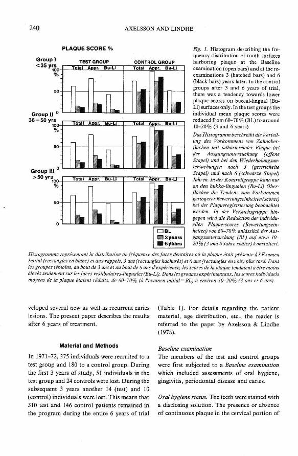
Fig. 1. Histogram describing the fre-quency distribution of tooth surfacesharboring plaque at the Baselineexamination (open bars) and at the re-examinations 3 (hatched bars) and 6(black bars) years later. In the controlgroups after 3 and 6 years of trial,there was a tendency towards lowerplaque scores on buccal-hngual (Bu-Li) surfaces only. In the test groups theindividual mean plaque scores werereduced from 60-70% (BL) to around10-20% (3 and 6 years).
Das Histogramm beschreibt die Verteil-ung des Vorkommens von Zahnober-fldchen mit adhdrierender Plaque beider Ausgangsuntersuchung (offeneStapel) und bei den Wiederholungsun-tersuchungen nach 3 (gestrichelteStapel) und nach 6 (schwarze Stapel)Jahren. In der Kontrollgruppe kann nuran den bukko-linguaien (Bu-Li) Ober-fldchen die Tendenz zum Vorkommengeringerer Bewertungseinheiten (scores)bei der Plaqueregistrierung beobaehtetwerden. In der Versuchsgruppe hin-gegen wird die Reduktion der individu-ellen Plaque-scores (Bewertungsein-heiten) von 60-70% anldsslich der Aus-gangsuntersuchung (BL) auf etwa 10-20 % (3 und 6 Jahre spdter) konstatiert.
Histogramme representant la distribution de frequence des faces dentaires oil la plaque etait presente a I'ExamenInitial (rectangles en blanc) et aux rappeis. 3 ans (rectangles hachures) et 6 ans (rectangles en noir) plus lard. Dansles groupes temoins. au bout de 3 ans et au bout de 6 ans ^experience, les scores de la plaque tendaient a etre moinseleves seulement sur les faces vestibulaires-linguales (Bu-Li). Dans les groupes experimentaux. les scores individueismoyens de la plaque etaient reduits. de 60-70% (d I'examen initial—BL) a environ 10-20% (3 ans et 6 ans).
Total Appr. Bu-LI Total AppT.
veloped several new as well as recurrent carles
lesions. The present paper describes the results
after 6 years of treatment.
Material and Methods
In 1971-72, 375 individuals were recruited to a
test group and 180 to a control group. During
the first 3 years of study, 51 individuals in the
test group and 24 controls were lost. During the
subsequent 3 years another 14 (test) and 10
(control) individuals were lost. This means that
310 test and 146 control patients retnained in
the program during the entire 6 years of trial
(Table 1). For details regarding the patient
material, age distribution, etc., the reader is
referred to the paper by Axeisson & Lindhe
(1978).
Baseline examination
The members of the test and control groups
were first subjected to a Baseline examination
which included assessments of oral hygiene,
gingivitis, periodontal disease and caries.
Oral hygiene status. The teeth were stained with
a disclosing solution. The presence or absence
of continuous plaque in the cervical portion of

CONTROLLED ORAL HYGIENE, PERIODONTITIS AND CARIES 241
GINGIVITIS SCORE %
Group I TEST GROUP< 3 5 yrs
CONTROL GROUP
-
-
Total Interpr. Bu-LI
^m ^^
Total Interpr. Bu-LI
r lGroup D
36-50 yrs50-%
Group in>50 yrs
Totai Interpr. Bu-LI Total tnterpr. Bu-Li
50Total Interpr, Bu-LI Total Interpr, Bu-Li
n
Table 1. Number of participants in tiie various testand controi groups, Oniy those individuals whoparticipated in tiie 6-year Follow-up examination havebeen inciuded in the anaiysis
Anzahl der Probanden in den versehiedenen Test- undKontroUgruppen. Die Analyse enthdit nur die Daienderjenigen Probanden, die an der 6 Jahre andauerndenWiederholungsuntersuchung teilgenommen habenNombre de participants dans les differents groupesexperimentaux et temoins. Seuls les sujets ayant par-ticipe a /'Examen de Rappei de la sixieme annee ont eteinclus dans I'analyse
Age group(1972)
I<35 yearsII 36-50 yearsin 50 years
Sum
Test
i29i2160
310
Contvoi
455150
i46
Sum
i74i72ilO
456
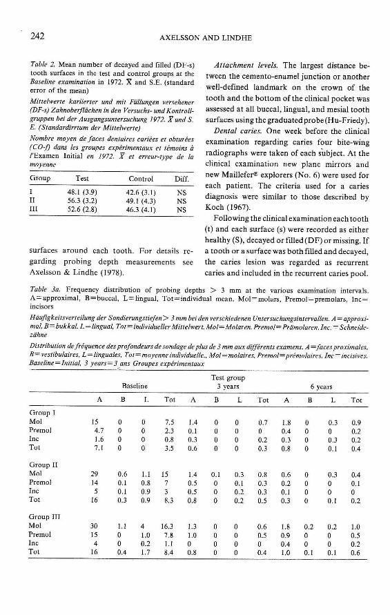
Fig. 2. Histogram showing the fre-quency distribution of inflamed gingi-vai units in the test and controi groupsat the Baseline (open bars) and re-examinations (3 and 6 years; hatchedand blacii bars). The individual mean(Tota!) figures describe unaltered gin-giva] conditions in the control groupsbut marked improvements in the testgroups during the triai.
Das Histogramm zeigt die Verteilungdes Vorkommens entzUndetergingivalerEinheiten bei den Versuchs- und Kon-troUgruppen anldssllch der Ausgangs-(offene Stapel) und der Wiederholungs-untersuchungen (3 und 6 Jahre spdter;gestrichelte und schwarze Stapel). Dieindlviduellen (totalen) Mittelwerte be-schreiben wahrend der VersuchsperiodeunveranderteGingivaverhdttnisseindenKontroUgruppen. jedoch ausgesproche-ne Verbesserungen inden Versuchsgrup-pen.
Histogramme representant la distribu-tion de frequence des sites gingivauxenflammes dans les groupes experimen-laux et temoins a 1'Examen Initial(reetangles en blanc) et aux rappels. 3ans (rectangles hachures) et 6 ans (rec-tangles en noir) plus tard. Le nombreindiquant la moyenne individueile (To-tai) met en evidence le fait qu'il n'y a pasde modifications dans retat de la geneivedans les groupes temoins. niaii qu'ily aune amelioration notable dans lesgroupes experimentaux pendant I'etude.
four surfaces of each tooth was determined. For
each individuai the frequency distribution of
piaque-harboring tooth surfaces was caicuiated.
Gingival inflammation. The presence or absence
of gingivitis (bleeding on probing) in four
gingiva] units around each single tooth was
assessed following probing. The percentage of
bleeding gingival units was caicuiated for each
individuai,
Periodontal disease.
Pocket depth. The probing depths were measuredwith a Oat graduated probe(Hu-Friedy®)at four
242 AXELSSON AND LINDHE
Table 2. Mean number of decayed and filled (DF-s)tooth surfaces in the test and control groups at theBaseline examination in 1972. X and S.E. (standarderror of the mean)
Mittelwerte kariierter und mit Fiillungen versehener(DE-s) Zahnoberfldchen in den Versuchs- und Kontroll-gruppen bei der Ausgangsuntersuchung 1972. XundS.E. (Standardirrtum der Mittelwerte)
Nombre moyen defaces dentaires eariees et obturees(CO-f) dans les groupes experimentaux et temoins a/'Examen Initial en 1972. X et erreur-type de lamoyenne
Group Test Control Diff.
III
in
48.1 (3.9)56.3 (3.2)52.6 (2.8)
42.6 (3.1)49.1 (4.3)46.3 (4.1)
NSNSNS
surfaces around each tooth. For details re-garding probing depth measurements seeAxelsson & Lindhe (1978).
Attachment levels. The largest distance be-tween the cemento-enamel junction or anotherwell-defined landmark on the crown of thetooth and the bottom of the clinical pocket wasassessed at all buccal, lingual, and mesial toothsurfaces using the graduated probe (Hu-Friedy).
Dental caries. One week before the clinicalexamination regarding caries four bite-wingradiographs were taken of each subject. At theclinical examination new plane mirrors andnew Maillefer® explorers (No. 6) were used foreach patient. The criteria used for a cariesdiagnosis were similar to those described byKoch (1967).
Following the clinical examinatioti each tooth(t) and each surface (s) were recorded as eitherhealthy (S), decayed or filled (DF) or missing. Ifa tooth or a surface was both filled and decayed,the caries lesion was regarded as recurrentcaries and included in the recurrent caries pool.
Table 3a. Frequency distribution of probing depths > 3 mm at the various examination intervals.A=approximal, B = buccal, L = hngual, Tot —individual mean. Mol —molars, Premol —premofars, Inc=incisors
Hdufigkeitsverteilung der Sondierungstiefen> 3mmbeidenverschiedenen Untersuchungsintervallen. A — approxi-mal.B=bukkal.L —lingual. Tot = individuellerMiltelwert.Mol—Molaren.Premol—Prdmolaren.Inc.= Schneide-zdhne
Distribution de frequence des profondeurs de sondage deplus de 3 mm aux differents examens. A = faces proximales,B—vestibulaires, L=linguales. Tot — moyenneindividuelle., Mol=molaires. Premol=premolaires, Inc = incisives.Baseline—Initial. 3 years—3 ans Groupes experimentaux
Group IMolPremolIncTot
Group IIMolPremoiIncToi
Group IIIMolPremolIncTot
A
154.71.67.1
29145
16
30154
16
Baseline
B
0000
0.60.10.10.3
1.1000.4
L
0000
I.I0.80.90.9
4LO0.21.7
Tot
7.52.30.83.5
15738.3
16.37.8I.I8.4
A
1.40.10.30.6
1.40.50.50.8
1.31.000.8
Test group3 years
B
0000
0.1000
0000
L
0000
0.30.10.20.2
0000
Tot
0.700.20.3
0.80.30.30.5
0.60.500.4
A
1.80.40.30.8
0.60.20.10.3
1.80.90.4LO
6
B
0000
0000
0.2000.1
years
L
0.300.3O.I
0.3000.1
0.2000.1
Tot
0.90.20.20.4
0.40.100.2
1.00.50.20.6

CONTROLLED ORAL HYGIENE, PERIODONTITIS AND CARIES 243
TreatmentFollowing the Baseline examination all carieslesions were treated and all ill-fitting dentalrestorations adjusted. Each patient was alsogiven a detailed case presentation and a dentalprophylaxis. Except for the case presentationand concomitant oral hygiene instructions, thecontrol group patients were not involved in anydental health program during the subsequent 6-year period. Once a year after the Baselineexamination, however, the control patients wererecalled to a public dental health chnic forexamination and to receive whatever dentaltreatment the dentist on duty found indicated.The test group participants, on the other hand,were given an entirely different type of treat-ment. Once every 2 months throughout the first2 years and once every 3 tnonths during the next4 years these patients were given (1) instructionand practice in oral hygiene techniques and (2) aproper oral prophylaxis. The design of the
prophylactic session was described in detail inour previous publication (Axelsson & Lindhe1978).
Follow-up examinationsThe patients were re-examined 3 and 6 yearsafter the Baseline examination. At the Follow-upexaminations the paratneters studied at theBaseline examination were recorded again.
The methods used to study the observationalerror were described in our previous publica-tion. Prior to the 6-year Follow-up examination,duplicate recordings were made regarding at-tachment levels and caries in 10 randomlyselected patients.
Results
The results from the Baseline examination re-vealed only minor differences between the testand the age-matched control patients regarding
Table 3b. Frequency distribution of probing depths>3 mm at the various examination intervals. A—approxi-mal, B = buccal, L=lingual, Tot = individual mean. Mol = molars, Premol^premolars, Inc=incisorsHduftgkeitsverteilung der Sondierungstiefen>3 mm bei den verschiedenen Untersuchungsintervallen. A = ap-proximal. B=bukkal. E = lingual. Tot=individue!ler Mittelwert. Mol=Molaren. Premol=Prdmolaren.Inc — SchneidezahneDistribution de frequence des profondeurs de sondage de plus de 3 mm aux differents examens. Groupes temoins
Group IMolPremolInc%t
Group I!MolPremolln,cTot
Group IIIMo!PremolIncTot
A
101.80.64.1
24.910.24.6
13.2
35.613.88.3
19.2
Baseline
B
0000
0.6000.2
2.10.700.9
L
1.2000.3
3.400.41.3
4.32.61.72.9
Tot
5.30.90.32.2
13.55.11.36.6
19.47.74.6
10.6
A
15.94.24.58.2
26.913.112.917.6
32.913.815.920.9
Control groups3 years
B
0000
0.60.51.30.8
7.42.93.84.7
L
1.8000.6
8.32.11.33.9
9.23.83.05.3
Tot
4.42.12.32.9
15.77.27.1
10.0
20.68.69.7
13
A
23.114.04.1
13.7
37.812.18.9
19.6
412917.929.3
6
B
1.2000.3
2.81.42.02.1
5.63.53.64.2
years
L
6.501.12.5
4.03.81.23.0
13.77.87.19.4
Tot
13.472.37.6
20.67.35.2
11.7
25.217.311.618.0

AXELSSON AND LINDHE
DF surfaces (Table 2), plaque (Fig. 1), gingivitis(Fig. 2), probing depths and loss of periodontaltissue support, In the test as well as in thecontrol groups, the average probing depths andattachment loss figures tended to increase withincreasing age. The frequency distribution of"probing dcpths>3 mm" (Tables 3a,b) was inthe youngest age group 3.5% (test), 2.2%(control) and in the middle age group 8.3%,6.6% and in the oldest age group and 8.4%(test), 10.6% (control). Consistently, deeperpockets were more frequent at approximal thanat buccal and lingual tooth surfaces (Tables 3a,b) and more frequent in molars than in pre-moiars and incisors.
At the Follow-up examinations 3 and 6 yearsafter the Baseline examination, the test grouppatients had markedly improved their oral
hygiene conditions (Fig. 1); in all three agegroups the plaque scores had decreased fromaround 60 to around 15-20%. In the controlgroups there was no obvious improvement ofthe oral hygiene standards between the variousexaminations.
At the Follow-up examinations the test grouppatients had very low gingivitis scores (Fig. 2).In the control groups there was no markedchange of the gingivitis scores over the 6 years oftrial.
The frequency distribution of "probingdepths>3 mm" (Table 3a) decreased in the testgroups from an average of 3.5% (group I),8.3% (group II), 8.4% (group III) to 0.3 and0.4% (group I), 0.5 and 0.2% (group II) and 0.4and 0.6% (group III) at the 3- and 6-yearFollow-up examinations. In all three control
POCKET DEPTH ALTERATIONS 1972-1975-1978
Group 1< 35 yrs
Group III
Total Mesial Buccal Distal Linqual
Total Mesial Buccal Distai Linqual
Total Mesiai Buccai Distai Linqual
O Test groupi ^ Control group• m Diff. 72-75• • Diff. 75-78
Fig. 3. Histogram describing altera-tions of pocket (probing) depths be-tween the Baseline and re-cxamina-tions in 1975 and 1978. In the testgroups a reduction (below the 0-iine) ofthe probing depth occurred, while inthe control groups the probing depthsincreased (above the 0-line).
Das Histogramm beschreibt die Ver-dnderungen der Taschen- (Sondierungs)tiefen zwischen den Ausgangs- und denWiederholungsuntersuchungen 1975und 1978. In den Versuchsgruppen kames zu einer Reduktion (unter der 0-Linie) der Sondierungstiefen. wahrendin den Kontollgruppen sich die Sondier-ungstiefen erhohten (iiber der 0-Linie).
Histogramme representant les modifi-cations de la profondeur de sondage desculs-de-sacprenantplace entre I'FxamenInitial et les rappeis en 1975 et 1978.Dans les groupes experimentaux, il seproduisait une reduction de la pro-fondeur de sondage (indiquee endessous de la ligne-0). tandis que dansles groupes temoins, la profondeur desondage augmentait (au-dessus de laligne-0).

CONTROLLED ORAL HYGIENE, PERIODONTITIS AND CARIES 245
groups (Tabie 3b) the frequency distribution of
"probing depths>3 mm" increased between the
examination intervais. Thus, at the 6-year
Follow-up examination 7.6% (group I) 11.7%
(group II) and 18% (group III) of ai! units
examined in the controi patients had "probing
depths>3 mm". At the Follow-up examinations
the percentage of deep pockets in the controi
patients was larger at approximal than at buccai
and iingual surfaces. In addition the presence of
deep pockets was more frequent in moiars than
in premoiars and incisors. It is evident from Fig.
3 that the individual mean (total) pocket depths
(probing depths) as well as the average depths
on mesial, buccai, distal and Hnguai surfaces
were reduced and remained reduced in the test
groups but increased in the controis over the 6
years of triai.
Fig. 4 shows the aiterations of the attachment
ievels from 1972 to i978. In the test groups,
there were no significant changes of the at-
tachment ieveis during the observation period.
In ail three controi groups, however, the ciinicai
attachment ievei graduaiiy shifted apicaiiy.
ATTACHMENT LEVEL ALTERATIONS 1972-1975-1978
Group I<35 yrs ^m
Group n36-50 yrs mm
Group m> 50 yrs
Total Interpr. Buccai Linqual
Total Interpr. Buccai Lmqual
Total Interpr. Buccai Lmqual
• Test groupED Control group• E3 Dilf. 72-75• HDiff. 75-78
Fig. 4. Histogram iilustrating altera-tions of the ciinical attachment leveisbetween the Baseiine (1972) and re-examinations (1975, 1978) in thevarious test and control groups. Theattachment level remained unalteredin the test groups but was reduced inall the control groups (below the 0-line). The average attachment loss peryear was in control groups 1 — 0.13mm, 11 = 0.23 mm, 111 = 0.26 mm.Das Histogramm verdeutlicht die Ver-anderungen der kiinischen Attachment-niveaus zwischen der Ausgangs- (1972)und den Wiederholungsuntersuehungen(1975. 1978) in versehiedenen Versuchs-und KontroUgruppen. Das Attacliment-niveau der Versuchsgruppen verbtiebunverdndert. reduzierte sieh jedoch inden KontroUgruppen (unter der 0-Li-nie). DerdurehschnittlicheAttachment-vertust pro Jahr war in den KontroU-gruppen 1=0,13 mm, II— 0,23mmund111=0.26 mm.
Histogramme illustrant les modifica-tions du niveau cUnique de I'attache-meUt se produisant entre I'Examen Ini-tial (1972) et les rappels (1975. 1978)dans les differents groupes experimen-taux et temoins. Le niveau de I'attache-ment restait inchange dans les groupesexperimentaux. mais il etait reduit danstous les groupes temoins (au-dessous dcla ligne-0). La perte annuelle moyennede rattachement etait dans les groupestemoins: 1=0.13 mm. 11=0,23 mm.111=0,26 mm.

246 AXELSSON AND LINDHE
The mean number of tooth surfaces thatshowed signs of primary and recurrent caries ispresented in Table 4. Except for a few indi-viduals, the test group patients did not developcaries during the 6 years of trial. In the controlgroups on the average 7.4 (group I), 5.4 (groupII) and 4,2 (group III) new caries lesionsdeveloped during the same period. There was atendency that the patients in the youngest age
group developed more new caries lesions thanmembers of the older age groups. This differ-ence was mainly the result of caries develop-ment on proximal tooth surfaces. Recurrentcaries lesions were found in only a few patientswithin the test groups. In the control groups thenumber of recurrent caries lesions, except ingroup I, tended to be larger than the primarycaries lesions. Thus, the average numbers of
Table 4. Mean number of tooth surfaces that showed signs of primary and recurrent caries during the 6 years oftrial. P=proximal, BL = buccal + lingual, O = occlusal, Tot —individual mean; DF-s —decayed + filled sur-faces. R-s = recurrent caries surfacesMittlere Anzahl der Zahnoberfldchen. an denen wahrend der 6 Jahre Zeichen primdrer und rezidivierender Kariesregistriert wurden. P—approximal, BL — bukkal+lingual. O = okklusal. Tot^individueller Mittelwert:DF-s= kariierte+ mit FUllungen versehene Oberfldchen, R-s = rezidivierte kariose Oberfldehen
Nombre moyen defaces dentaires presentant des signes de carie primaire ou de recidive de la carie pendant les 6annees de t'experience. P=proximales. BL — vestibulaires+linguales. 0 — occlusales. Tot —moyenne individuelle.DF-s—faces cariees+obturees, R-s—recidive de carie (faces)
New DF-surfaces(DE-s)
Recurrent cariessurfaces (R-s)
Sum:DE-s + R-s
BL Tot BL Tot BL Tot
Group I3 years
3-6 years0-6 years
Group II3 years
3-6 years0-6 years
Group III3 years
3-6 years0-6 years
2.91.84.7
1.71.12.8
0.60.81.4
1.60.82.4
1.01.52.5
1.31.12.4
0.20.10.3
0.10.10.2
0.20.20.4
4.72.77.4
2.82.65.4
2.12.14.2
2.21.94.1
2.93.05.9
2.22.24.4
0.10.10.2
0.10.10.2
0.10.10.2
Control1.00.81.8
1.11.02.1
1.31.22.5
1.601.6
1.50.11.6
0.800.8
Test
0.1
0.1
4.82.77.5
5.64.29.8
4.33.47.7
0.10.10.2
0.10.10.2
0.20.10.3
5.13.78.8
4.64.18.7
2.83.05.8
0.20.10.2
O.IO.I0.2
O.IO.I0.2
2.6L64.2
2.12.54.6
2.62.34.9
0.1
0.1
1.80.11.9
1.60.21.8
1.00.21.2
9.55.4
14.9
8.36.8
15.1
6.45.5
11.9
0.20.1a2
O.I0.J0x2
0.20.10.3
Group I3 years
3-6 years0-6 years
Group II3 years
3-6 years0-6 years
Group III3 years
3-6 years0-6 years

COisTTROLLED ORAL HYGIENE, PERIODONTITIS AND CARIES 247
recurrent caries lesions in the controi groups,caicuiated from the 6-year Follow-up examina-tion, were 7.5 (group I), 9.8 (group II), and 7.7(group III).
Acknowledgment
This study was supported by grants from theSwedish Medical Research Council K77-24X-4725.
Discussion
The findings reported cleariy demonstrate thata preventive program which stimuiates indi-viduals to adopt proper orai hygiene habits mayprevent the progression of periodontai diseaseand caries in adults. A discussion of the per-tinent literature in this fieid was presented in aprevious pubiication (Axeisson & Lindhe 1978),
If the results obtained from the test groupsare compared with the findings in the controls,it becomes obvious that traditional dental caredoes not prevent the progression of caries andperiodontal disease in an adult population.Such a conclusion corroborates findings re-ported by Bjorn (1974) and Soderholm (1979).They monitored the development of caries andperiodontal disease in a group of empioyees ofaSwedish shipyard and reported that, despite theindividuais reguiariy received dentai treatmentof a traditional type, there was over a 9-yearperiod a gradual deterioration of the dentitionin this sample. In many respects the findingsmade in the present test groups are also inagreement with Soderholm (1979). Subsequentto a 9-year foiiow-up examination of the groupof patients earher referred to, he initiated aiongitudinai study to assess the effect on thedentai conditions of a treatment program with apreventive emphasis. This program had a designsimiiar to that described by Axeisson &.. Lindhe(i978), i.e. it inciuded measures such as ora!hygiene evaluation and reinstruction, scaling,root planing and polishing, repeated once every3 months over a 3- to 4-year period. Soderholm(1979) concluded "the dental care programsignificantiy improved the dentai heaith" and"the increased time and effort devoted to pre-ventive care were compensated by a decreasedneed for restorative treatment."
Zusammenfassung
Der Fffekt kontrollierter oraler Hygiene auf das Vor-kommen von Karies und parodontalen Krankheiten beiErwachsenenDie voriiegende Mitteiiung beschreibt das Resuitateiner kiinischen Versuchsreihe, bei der bei einerGruppe erwachsener Probanden wahrend einer Lang-zeitbeobachtungsperiode von uber 6 Jahren ein guteroraier Hygienestandard aufrecliterhaiten wurde.Wahrend der Jahre i971-72 wurden 375 Probandenfur eine Versuchs- und iSO Probanden wurden fiireine Kontroiigruppe ausgewahlt. Wahrend der Beob-achtungsperiode von 6 Jahren fieien 65 Probanden inder Versuchs- und 34 in der Kontroiigruppe aus. DieProbanden wurden in drei Aitersgruppen eingeteilt;I<35 Jahre, II 36-50 Jahre, III>50 Jahre, Bei denTeilnehmern der Versuchs- und KontroUgruppenwurde anfangs sine Ausgangsuntersuchungvoigenom-men, die die Beurteiiung des oraien Hygieneniveaus,der Gingivitis, voriiegender Parodontalicrankheit undKaries beinhaltete. Nach dieserUntersuchung wurdenaiie kariosen Lasionen behandeit und unzureichendedentale Restaurationen justiert, Jeder Proband wurdeweiterhin uber Entstehung und Foigen seiner Paro-dontaikrankheit genau unterrichtet und prophyiak-tisch behandeit. Die Probanden der Kontroiigruppenahmen in den foigenden sechs Jahren der Versuchs-periode an keinem weiteren Prophyiaxeprogrammteii. Sie wurden jedoch einmai jahriich in die Zahn-kiinik zu Untersuchung und symptomatischer Zahn-behandiung einbesteiit. Die Probanden der Versuchs-gruppe wurden aile 2-3 Monate vorbeugend be-iiandeit, was (i) Instruktion und Ubung in derTechnik oraler Hygiene und (2) sorgfaltige prophyi-aktische Behandiung beinhaitete.
Die Probanden wurden drei und sechs Jahre nachder Ausgangsuntersuchung nachuntcrsucht. Bei derNachuntersuchung wurden die gieicben Parameterregistriert, die aniassiich der Ausgangsuntersuchungreglstriert worden waren. Die Resuitate der Langzeit-studie zeigten, dass ein Vorbeugungsprogramm dasden Patienten dazu anregt sich an zweckmSssige oraieHygienegerwohnheiten zu gewohnen in der Lage ist,Gingivitis veischwinden zu lassen und das Eort-schreiten der Parodontaikrankheit und der Karies zuverhindern. LIbliche traditioneile zahnarztliche Be-treuung konnte die Weiterentwicklung der Karies-und Parodontaikrankheit beim Erwachsenen nichtverhindern.

248 AXELSSON AND LINDHE
Effets sur la carie et sur les parodontopathies de soinsd'hygiene bucco-dentaire diriges chez des adultes.Resultats au bout de 6 anneesLe present compte-rendu decrit les resuitats d'un essaiclinique au cours duquel un groupe d'adultes a etemaintenu a un niveau d'hygiene bucco-dentaireadequat pendant une periode de 6 ans. En 1971-72,375 sujets ont ete enroles dans un groupe experi-mental et 180 dans un groupe temoin. Pendant les 6annees de l'essai, le groupe experimental a subi uneperte de 65 personnes et ie groupe temoin une perte de34 personnes. Les patients etaient repartis en troisgroupes d'ages: I<36 ans, II 36-50 ans, III>50 ans.Les membres du groupe experimental et ceux dugroupe temoin ont d'abord subi un Examen Initialcomprenant I'enregistrement de l'hygiene bucco-den-taire, des gingivites, des parodontopathies et descaries. Apres cet examen, toutes les caries ont etetraitees et les obturations defectueuses ont ete rec-tifiees. Chacun des patients a re^u des informationsdetaiil6es sur son cas et un nettoyage dentaire. Lespatients du groupe temoin n'ont pris part a aucunprogramme complementaire de sante dentaire pen-dant les 6 annees suivantes. Us ont cependant eteconvoques une fois par an dans un service public desoins dentaires oii ils ont ete examines et ont repu untraitement dentaire symptomatique. Les participantsdu groupe experimental, d'autre part, ont refu untraitement preventif, repete tous les 2-3 mois etcomprenant (I) instruction et entrainement aux tech-niques de l'hygiene bucco-dentaire et (2) un nettoyagedentaire minitieux.
Les patients ont ete reexamines 3 ans et 6 ans apresI'examen initial. A I'Examen deRappel, les parametresetudies a VExamen Initial ont de nouveau ete enregis-tres. Les resuitats obtenus ont mis en evidence le faitqu'un programme preventif encourageant ies sujets aadopter des habitudes adequates en matiere d'hygienebucco-dentaire peut faire disparaitre Ies gingivites etprevenir la progression des parodontopathies et descaries. Les soins dentaires traditionnels, par contre,ne prevenaient ni les progres de la carie ni ceux desparodontites chez i'adulte.
References
Axeisson, P. (1978) The effect of plaque controlprocedures on gingivitis, periodontitis and dentalcaries. Department of Periodontology, Thesis. Uni-versity of Gothenberg, Sweden.
Axelsson, P. &. Lindhe, J. (1978) Effect of controlledoral hygiene procedures on caries and periodontaldisease in adults. Journal of Clinical Periodontology5, 133-151.
Bjom, A-L. (1974) Dental health in relation to ageand dental care. Odontologisk Revy 25, Suppl. 29.
Knowles, J. W., Burgett, F. G., Nissle, R. R., Schick,R. A. Morrison, E. C. & Ramfjord, S. P. (1979)Results of periodontal treatment related to pocketdepth and attachment level. Eight years. Joi/r/ja/o/Periodontology 50, 225-233.
Koch, G. (1967) Effect of sodium fiuoride in denti-frice and mouthwash on incidence of dental cariesin schoolchildren. Odontologisk Revy 18, Suppl. 12.
Lindhe, J. & Nyman, S. (1975) The effect of plaquecontrol and surgical pocket ehmination on theestablishment and maintenance of periodonta!health. A longitudinal study of periodontal therapyin cases of advanced disease. Journal of ClinicalPeriodontology 2, 67-79.
Lovdai, A., Arno, A., Schei, O. & Waerhaug, J.(1961) Combined effect of subgingival scaling andcontrolled oral hygietie on the incidence of gingi-vitis. Acta Odontologica Scandinavica 19, 537-555.
Ramfjord, S. P., Knowles, J. W., Nissle, R. R., Schick,R. A. & Burgett F. G. (1973) Longitudinal studyof periodontai therapy. Journal of Periodontology44, 66-77.
Soderholm, G. (1979) Effect of a dental care programon dental health conditions. A study of employeesof a Swedish shipyard. Department of Periodonto-logy, Eaculty of Odontology, University of Lund,Malmo, Sweden.
Address:Jan LindheDepartment of PeriodontologySchool of DentistryBox 77030S~400 33 Goteborg. Sweden
