edinburgh research explorer · the embojournal vol.3 no.13 pp.3079-3085, 1984 molecular lesions...
TRANSCRIPT

Edinburgh Research Explorer
Molecular lesions associated with white gene mutations inducedby I-R hybrid dysgenesis in Drosophila melanogaster
Citation for published version:Sang, H, Pelisson, A, Bucheton, A & Finnegan, D 1984, 'Molecular lesions associated with white genemutations induced by I-R hybrid dysgenesis in Drosophila melanogaster' EMBO Journal, vol. 3, no. 13, pp.3079-85.
Link:Link to publication record in Edinburgh Research Explorer
Document Version:Publisher's PDF, also known as Version of record
Published In:EMBO Journal
Publisher Rights Statement:Copyright 1984 IRL Press Limited
General rightsCopyright for the publications made accessible via the Edinburgh Research Explorer is retained by the author(s)and / or other copyright owners and it is a condition of accessing these publications that users recognise andabide by the legal requirements associated with these rights.
Take down policyThe University of Edinburgh has made every reasonable effort to ensure that Edinburgh Research Explorercontent complies with UK legislation. If you believe that the public display of this file breaches copyright pleasecontact [email protected] providing details, and we will remove access to the work immediately andinvestigate your claim.
Download date: 10. Sep. 2018
The EMBO Journal vol.3 no.13 pp.3079-3085, 1984
Molecular lesions associated with white gene mutations induced by
I-R hybrid dysgenesis in Drosophila melanogaster
H.M. Sang13, A. Pelisson2, A. Bucheton andD.J. Finnegan''Department of Molecular Biology, University of Edinburgh, King'sBuildings, Mayfield Road, Edinburgh EH9 3JR, UK, and 2Laboratoire deGenetique, Genetique et Biologie Cellulaire, Universite de Clermont-Ferrand, BP 45, 63170 Aubiere, France
3Present address: Poultry Research Centre, Roslin, Midlothian, UKCommunicated by V. Pirrotta
We have identified molecular lesions associated with sixmutations, w'R2 and wIR4-8, of the white gene of Drosophilamelanogaster. These mutations arose in flies subject to I-Rhybrid dysgenesis. Four of the mutations give rise to colouredeyes and are associated with insertions of 5.4-kb elements in-distinguishable from the I factor controlling I-R dysgenesis.The insertion associated with wIR4 is at a site which, withinthe resolution of these experiments, is identical to that of twopreviously studied I factors. This appears to be a hot-spot forI factor insertion. We have compared the sites of these inser-tions with sequences complementary to white gene mRNAidentified by Pirrotta and Brockl. The hot-spot is in thefourth intron. The insertion carried by wIP5 is either within,or just beyond, the last exon. The insertion carried by wIR6 isnear the junction of the first exon and first intron. The wIR2mutation is a derivative of wl. It contains an insertion of Ifactor DNA within, or immediately adjacent to, the F-likeelement associated with wl, and results in restoration of someeye colour. This insertion is just upstream of the start of thewhite mRNA. Mutations wIR7 and wIR8 are deletions remov-ing mRNA coding sequences. Both determine a bleachedwhite phenotype.Key words: Drosophila melanogaster/hybrid dysgenesis/transposable elements/white gene
IntroductionHybrid dysgenesis (see reviews by Bregliano and Kidwell,1983; Kidwell, 1983; Engels, 1983) is the name given to theappearance of a set of unusual characteristics in the progenyof crosses between certain strains of Drosophila melanogaster(Picard and L'Heritier, 1971; Kidwell, 1975; Kidwell et al.,1977). These characteristics include lowered fertility, recom-bination in males and increased frequencies of mutation andchromosome aberrations. Two independent systems ofhybrid dysgenesis, P-M and I-R, are known (Kidwell, 1979).Strains of D. melanogaster may be classified into one or
other of two types with respect to each system. In the P-Msystem the effect is seen in male and female progeny ofcrosses between M strain females and P strain males. In theI-R system the effect is seen only in the female progeny of
crosses between reactive (R) strain females and inducer (I)strain males. These dysgenic females are known as 'SF'females. The progenies of all other crosses appear normal.The characteristics of P strains and inducer strains are con-
trolled by transposable genetic determinants known as P fac-
IRL Press Limited, Oxford, England.
tors and I factors, respectively (Picard, 1976; Bingham et al.,1982). Many P-M induced mutations are unstable in in-dividuals subject to P-M dysgenesis (Engels, 1979; Rubin etal., 1982) and this led to the suggestion that mutations induc-ed by P-M dysgenesis are due to insertion of P factor DNAinto the genes in question (Green, 1977; Golubovsky et al.,1977; Simmons and Lim, 1980). This has been tested byRubin et al., 1982). They compared DNA of six P-M inducedwhite gene mutations with that of the wild-type allele. Eachmutation had foreign DNA inserted into the white gene. Twoinsertions were of members of the copia family oftransposable elements (Finnegan et al., 1978). The other fourinsertions varied in length from 0.5 kb to 1.4 kb, but wererelated in sequence. Rubin et al. (1982) argued that these in-sertions were too short to be functional P factors, whichprobably code for at least a transposase and a regulatorymolecule, but that they might be deleted derivatives of P fac-tors. O'Hare and Rubin (1983) have confirmed this. Usingone of these putative P elements as a probe to screen a libraryof recombinant phages containing DNA from a P strain theyrecovered clones containing a conserved 2.9-kb sequencewhich has subsequently been shown to have at least some ofthe properties of a P factor (Spradling and Rubin, 1982).We have started to investigate the molecular basis of I-R
hybrid dysgenesis by examining molecular lesions associatedwith mutations of the white gene produced in SF females.Two mutations, wIR1 and wIR3, are associated with insertionsof indistinguishable 5.4-kb elements at apparently identicalsites within the white gene (Bucheton et al., 1984). We believethat these insertions are copies of the I factor, which controlsI-R dysgenesis, since both wIRl and wIR3 are closely linked toI factor activity (Pelisson, 1981; Bucheton et al., 1984). Herewe describe the genetical and molecular properties of a fur-ther six white gene mutations induced in SF females. Three ofthese mutations, wIR4, wIR5 and wIR6, determine a colouredeye phenotype and are associated with insertions of 5.4-kbelements which are very similar, if not identical, to the I fac-tor. Two of the remaining mutations, w1R7 and wIR8, deter-mine a bleached white phenotype and are associated withdeletions of DNA from the white region. The last mutation,wlR2, contains an insertion of I factor DNA into the whiteregion of a chromosome carrying the w1 allele. This results ina partial restoration of eye colour. We discuss these results interms of the properties of the I factor and of the white gene.
ResultsThe mutation wIR2, like wIR1, arose in SF females producedby crossing females of the reactive strain seF8 with males ofthe inducer strain w1 ctf(Picard et al., 1978; Pelisson, 1981).Its properties have been described briefly by Pelisson (1981)who referred to it as lal 1. Mutations wIR4-8 were foundamongst 60 000 male progeny of SF females produced bycrossing females of the reactive strain XCha with males of theinducer strain XOre I. They are the results of independentmutation events. Mutations w1R2 and wIR4-6 determine col-
3079

-5 10I l
15
_ I)_
co
WIR8
---IR W1R7
CS156 CS1571769 CS155 152
1768C SS _
A26
Fig. 1. Restriction map of the white region in chromosomes carrying wAR mutations. This map shows the positions of restriction sites on the parental w+chromosomes on which wIR mutations were isolated. The positions of the insertions in wIR4, winR and wIR6 are shown offset from the map. The positions ofthe deletions associated with the wiR7 and wIRO mutations are shown below the map. The dashed regions indicate uncertainties regarding the end points of thedeletions. Restriction sites marked * are not present on the chromosome carrying the wlR4 and w%P5 mutations, but are present on the chromsome carryingwIR6. The HindIII site at coordinate 8.9 is not present in chromosomes carrying the w1 or w"R2 mutations. The positions of fragments which have been clonedfrom the white region and used as probes in these experiments are also indicated below the map. The scale above the map is in kilobases. The zero position isthe site of the copia element associated with the white-apricot mutation (Levis et al., 1982). Positive coordinates are towards the centromere. The position ofthe major w+ transcript as determined by Pirrotta and Brockl (1984) is shown under the restriction map. The boxed regions indicate exons while the linesjoining them indicate introns. Transcription is from right to left.
oured eye phenotypes and all show dosage compensation.We first determined the approximate locations of
molecular lesions associated with these mutations by restric-tion digestion and Southern (1975) transfer experiments. In-itially, recombinant lambda phages A26 and M365 (Figure 1)were used to probe digests of mutant and wild-type DNAs.These phages together cover -30 kb of the white locus.Subclones of appropriate restriction fragments allowed amore detailed analysis.
Properties of the mutation wIR4The mutation wIR4 determines a red-brown eye colour whichis lighter at 25°C than at 20°C. This phenotype is identical tothat of wIRl and wIR3 (Pelisson, 1981, and unpublished). Theonly detectable difference between wild-type and wIR4 DNAsin the region of the white gene is within the 0.86-kb SalI frag-ment lying between coordinates - 1.4 and - 0.6 in Figure 1.This is the location of the 5.4-kb insertion associated with thewlRl and wIR3 mutations (Bucheton et al., 1984). Figure 2,tracks a - d, shows the result of a Southern transfer experi-ment in which wild-type, wIRl, wIR3 and wIR4 DNAs weredigested with SalI and then probed with 32P-labelled pCS155,a plasmid containing this 0.86-kb fragment. The 0.86-kb SalIfragment of wild-type DNA is replaced by a 6.2-kb fragmentin wlR4 DNA which co-migrates with the correspondingfragments of wIRl and wIR3. This indicates that these muta-tions are all associated with 5.4-kb insertions in this region.The 5.4-kb elements present in wIRI and wIR3 are apparent-
ly identical copies of the I factor. These have been cloned andcharacterised by Bucheton et al. (1984). They are not cut byBamHI, EcoRI, SalI or XhoI and the same is true of the ele-ment present in wlR4 (data not shown). The copies of the Ifactor in wIRl and wIR3 have two HindIII sites 1 kb apart, andin order to compare the element in wlR4 with these I factors
3080
9A } St _-
---
4
2 2
Fig. 2. Southern transfer experiment comparing the wIRI, wvR3 and wIR4mutations. Genomic DNAs of seF8, w1VR', w1R3 and wIR4 strains weredigested with Sail, tracks a-d, or Hindlll, tracks e-h. The DNAs werethen run on a I%7o agarose gel and transferred to nitrocellulose. The DNAsin tracks a-d were hybridised with 32P-labelled pCS155, while DNAs intracks e-h were hybridised with 32P-labelled pCS54. The strain seF8 hasthe same restriction map in the white region as does the parental strainXCha. a and e, seF8; b and f, wIRI; c and g, wIR3; d and h, wIR4.
we digested wild-type, wIRl, wIR3 and wlR4 DNAs withHindIII. The DNAs were then hybridised, in a Southerntransfer experiment, with pCS54, a plasmid carrying theBamHI-HindIII fragment lying between coordinates - 6 and1.5 in Figure 1. The 9.2-kb HindlIl fragment of wild-typeDNA which hybridises to this probe is replaced, in the threemutant DNAs, by fragments of 7.4 kb and 6.2 kb (Figure 2,
H.M.Sang et al.
-5I
m
ir
-10
zzxE E o c
X1 co x x x =Wm
0 2kb
M 365
0 5
cxr
V
cabc d C,tCJ

Molecular lesions associated with white gene mutadons
a b c d e f
9 64_
6-64.
4 34-
2 26-
1 98_
Fig. 3. Southern transfer experiment showing the position of the insertionassociated with the w%R5 mutation. Genomic DNAs of the seF8 and wJR5strains were digested with the enzymes indicated below. The DNAs werethen run on a 1% agarose gel, transferred to nitrocellulose filter andhybridised with 32P-labelled pCS156. a, seF6 digested with SalI; b, wOR5digested with SalI; c, seF8 digested with BamHI and HindIII; d, w1R5digested with BamHI and HindIII; e, seF8 digested with Sail and PstI; f,w4A' digested with SalI and Pstl.
tracks e - h). This suggests that wIR4 contains a copy of thesame 5.4-kb element as is present in wIRl and wIR3, and thatit is inserted at the same site and in the same orientation. Werefer to this orientation of the I factor with respect to thewhite locus as being orientation 1, and to the opposite asorientation 2.Properties of the mutation WIR5The mutation wIR5 determines a brown eye colour which issimilar to that of wIRl at 200C, but it not temperature sen-sitive. The only detectable difference between the whiteregions of wild-type and wIR5 DNAs is in the 1.5-kb Sail frag-ment between coordinates - 2.9 and - 1.4. Figure 3, tracks aand b, shows the result of hybridising SalI digests of wild-typeand wIR5 DNAs with pCS156, a plasmid containing this frag-ment. The 1.5-kb Sall fragment of wild-type DNA is replacedby a 6.9-kb fragment in wIR5. This indicates that this muta-tion is also associated with insertion of a 5.4-kb element.The insertion in wIR5 is not cut by BamHI, EcoRI, Sail or
XhoI (data not shown) but is cut by HindIII. Figure 3, tracksc and d, shows the result of digesting wild-type and wIR5DNAs with both HindIII and BamHI and then hybridisingthem with pCS156 in a Southern transfer experiment. The7.8-kb fragment of wild-type DNA which hybridises to thisprobe is replaced in wIR5 by fragments of 7 kb and 5.2 kb.This indicates that, like the I factor in wlRl, the 5.4-kb ele-ment in wIR5 contains at least two HindIII sites -1 kb apart.This suggests that the element present in wIR5 is another copyof the I factor.To orient and position this element we hybridised a
HindIII-BamHI digest of wIR5 DNA separately with plasmidp1769, which contains the HindIII-Sall fragment betweencoordinates -6 and -2.9, and pCS 157, which contains theSall-XhoI fragment between coordinates -0.6 and 2.1(Figure 1). The 7-kb HindII fragment hybridised to p1769and the 5.2-kb fragment to pCS157 (data not shown). The
7.0-kb fragment is therefore to the left and the 5.2-kb frag-ment to the right. If the element in wIR5 has the same restric-tion map as the I factor in wIRI then it must be inserted atabout coordinate - 2 and in orientation 2. This would place aPstI site - 1.6 kb to the right of the SalI site at coordinate-2.9, and 5.3 kb to the left of the SalI site at coordinate-1.4 (Figure 2). To position the insert more precisely wedigested wild-type and wIR5 DNAs with both Sall and PstIand hybridised them with pCS156 (see Figure 3, tracks e andf). The 1.5-kb Sall fragment of wild-type DNA whichhybridises to this probe is replaced by 1.4-kb and 5.5-kbfragments in wIR5 DNA, which places the wIR5 insert at aboutcoordinate - 2.2.We have cloned the HindlII fragments containing the left-
hand and right-hand ends of the wIR5 insert, using the lamb-da vector NM1 149 (Murray, 1983). The recombinant phagesobtained, XI453 and XI454, contain the left-hand and right-hand ends of the insert, respectively. Digestion of XI453 withboth HindIII and Sall yields a fragment of 3.7 kb which in-cludes the left-hand end of the insert, while digestion of XI454gives a 2.2-kb fragment including the right-hand end. Tocompare the insert present in wIR5 directly with the I factor inwIRl, we subcloned these Sall-HindlII fragments in pUC8(Vieira and Messing, 1982) and heteroduplexed them with thecorresponding subclones from wIRl. The results of this ex-periment indicate that the wIR5 insertion is homologous to Ifactor DNA for 1.4 i 0.14 kb to the left of the first internalHindIII site of the I factor, as shown in Figure 1, and for 2.8
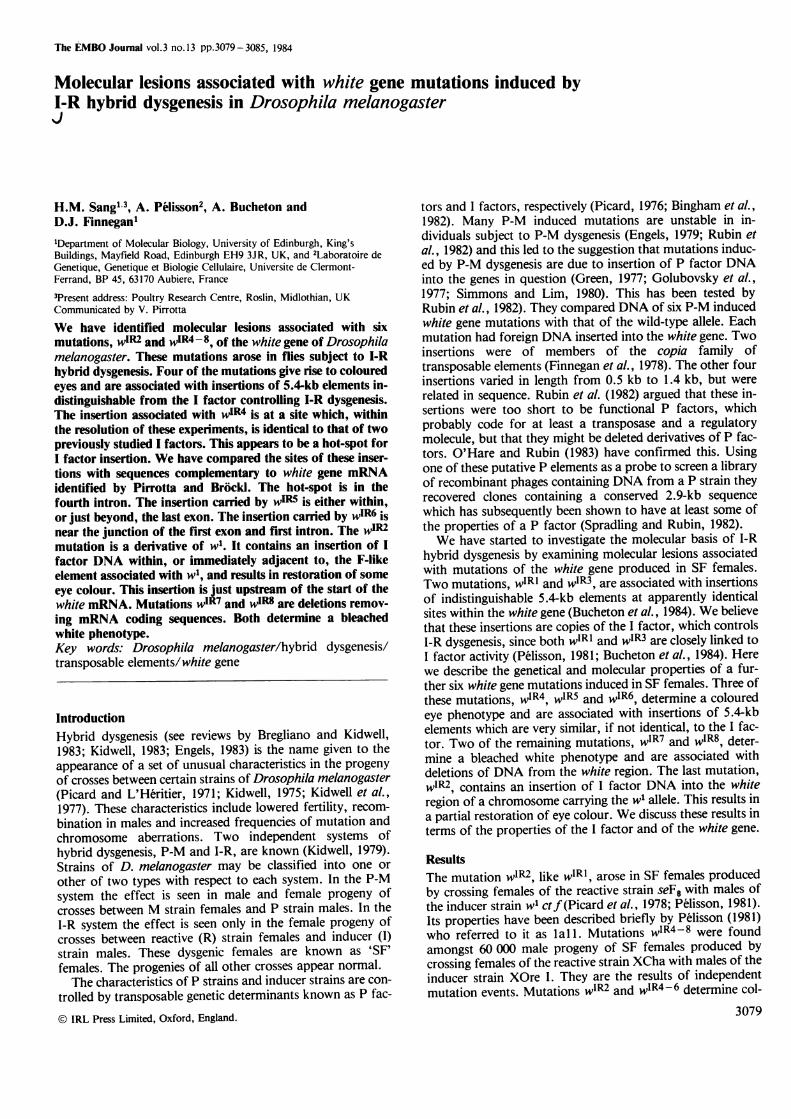
0.09 kb to the right of the second internal HindIII site. Noregion of mismatch could be detected. These results are con-sistent with the wIR5 insertion being an I factor.Properties of the mutation WIR6The mutation wIR6 determines a red-brown eye colour whichis similar to that of wIRl at 20°C, but which is nottemperature sensitive. The only detectable difference betweenwIR6 and wild-type DNAs is in the 11.3-kb Sall fragment bet-ween coordinates -0.6 and 10.7 (Figure 1). This is replacedin wIR6 DNA by a fragment of - 17 kb (data not shown).This is consistent with there being a 5.4-kb I factor insertedwithin this fragment. We have confirmed that there are I fac-tor sequences associated with the wIR6 mutation by in situhybridisation experiments (data not shown).
There are differences between the restriction maps of theinducer and reactive parents of wIR6 in this region. TheBamHI site at coordinate 4.4 and the EcoRI site at coordinate6.4 are both present in DNA of the XOre I, but not the XChaparent. The DNA from wIR6 contains both sites indicatingthat this mutation occurred on an X chromosome fromXOre I. We have further located the wIR6 insertion byhybridising HindIII digests of wIR6 and XOre I DNAs withp152, a plasmid containing the 6.8-kb HindIII fragment lyingbetween coordinates 3.2 and 10. This was cloned from astrain lacking the HindIII site at coordinate 8.9. This probehybridised to 5.7-kb and 1.1-kb fragments of XOre I DNA,and 7.1-kb and 1.1-kb fragments of wIR6 DNA (Figure 4,tracks a and b). Therefore the insertion must be within the5.7-kb fragment, and must contain at least one HindIII site.The fact that only one new fragment of wIR6 DNA hybridisesto this probe probably indicates that the insert is very close toone end of the HindIII fragment. These results suggest thatan I factor is present just to the right of the HindIII site atcoordinate 3.2 and in orientation 2, or just to the left of theHindIII site at coordinate 8.9 and in orientation 1.
3081
H.M.Sang et al.
b d e -1
_ -_ _
Fig. 4. Southern transfer experiment showing the position of the insertionassociated with the IR6 mutation. Genomic DNAs of the XOre I and VIR6strains were digested with the enzymes indicated below. The DNAs werethen run on a l1o agarose gel and hybridised with 32P-labelled p152, tracksa and b, or pI768, tracks c-f. a, XOreI digested with HindIII; b, w1R6digested with HindIII; c, XOre I digested with PstI and XhoI; d, VIR6digested with PstI and XhoI; e, XOre I digested with BamHI and PstI;f, w1R6 digested with BamHI and PstI.
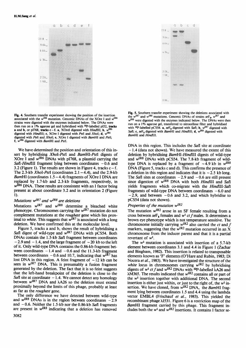
We have determined the position and orientation of this in-sert by hybridising XhoI-PstI and BamHI-PstI digests ofXOre I and wIR6 DNAs with p1768, a plasmid carrying theSailI-HindIII fragment lying between coordinates -0.6 and3.2 (Figure 1). The results are shown in Figure 4, tracks c - f.The 2.3-kb XhoI-PstI (coordinates 2.1-4.4), and the 2.9-kbBamHI (coordinates 1.5-4.4) fragments of XOre I DNA arereplaced by 1.7-kb and 2.3-kb fragments, respectively, inwIR6 DNA. These results are consistent with an I factor beingpresent at about coordinate 3.2 and in orientation 2 (Figure1).Mutations wIR7 and wIR8 are deletionsMutations wIR7 and wIR8 determine a bleached whitephenotype. Chromosomes carrying the wIR7 mutation do notcomplement mutations at the roughest gene which lies prox-imal to white. This suggests that wIR7 is associated with a longdeletion. We have confirmed this at the molecular level.
Figure 5, tracks a and b, shows the result of hybridising aSall digest of wild-type and wIR7 DNAs with pCS54. BothDNAs contain the 1.5-kb Sall fragment between coordinates- 2.9 and - 1.4, and the large fragment of - 20 kb to the leftof it. Only wild-type DNA contains the 0.86-kb fragment bet-ween coordinates - 1.4 and -0.6 and the 11.3-kb fragmentbetween coordinates -0.6 and 10.7, indicating that wIR7 haslost DNA in this region. A feint fragment of 12 kb can beseen in wIR7 DNA. This is presumably a fusion fragmentgenerated by the deletion. The fact that it is so feint suggeststhat the left-hand breakpoint of the deletion is close to theSail site at coordinate - 1.4. We cannot detect any homologybetween wIR7 DNA and XA26 so the deletion must extendproximally beyond the limits of this phage, probably at leastas far as the roughest gene.The only difference we have detected between wild-type
and wIR8 DNAs is in the region between coordinates -2.9and -0.6. Neither the 1.5-kb nor the 0.86-kb Sall fragmentsare present in wIR8 indicating that a deletion has removed
3082
Fig. 5. Southern transfer experiment showing the deletions associated withthe wIR7 and wIRS mutations. Genomic DNAs of strains seF8, W1'R7 andw1R8 were digested with the enzymes indicated below. The DNAs were thenrun on a 1 070 agarose gel, transferred to nitrocellose filter and hybridisedwith 32P-labelled pCS54. a, seF8 digested with Sail; b, wR7 digested withSalI; c, seF8 digested with BamHl and HindIII; d, VIR8 digested withBamHI and HindlII.
DNA in this region. This includes the Sall site at coordinate-1.4 (data not shown). We have measured the extent of thisdeletion by hybridising BamHI-HindIII digests of wild-typeand wIR8 DNAs with pCS54. The 7.8-kb fragment of wild-type DNA is replaced by a fragment of -4.9 kb in w1R8DNA (Figure 5, tracks c and d). This confirms the presence ofa deletion in this region and indicates that it is - 2.5 kb long.The Sall sites at coordinates - 2.9 and - 0.6 are still presentsince digestion of wIR8 DNA with both HindlII and Sailyields fragments which co-migrate with the HindIII-SaiIfragments of wild-type DNA between coordinates - 6.0 and-2.9, and between -0.6 and 3.2, and which hybridise topCS54 (data not shown).Properties of the mutation wIR2The mutation w'R2 arose in an SF female resulting from across between seF8 females and w1 ctf males. It determines abrown eye phenotype which is not temperature sensitive. Thechromosome initially carrying wIR2 also carried the ct and fmarkers, suggesting that the wlR2 mutation occurred in an Xchromosome from the inducer parent and that it is a partialrevertant of w1.The w1 mutation is associated with insertion of a 5.7-kb
element between coordinates 3.1 and 4.4 in Figure 1 (Zacharand Bingham, 1982). This insertion is related to transposableelements known as 'F' elements (O'Hare and Rubin, 1983; DiNocera et al., 1983). We have investigated the structure of thewhite locus in chromosomes carrying wIR2 by hybridisingdigests of wl ctfand wIP2 DNAs with 32P-labelled XA26 andXM365. The results indicated that wIR2 contains all or part ofthe w1 insertion together with additional DNA. The secondinsertion is either just within, or just to the right of, the wl in-sertion. We have cloned, from w'R2 DNA, the BamHI frag-ment lying between coordinates 1.5 and 4.4 using the lambdavector EMBL4 (Frischauf et al., 1983). This yielded therecombinant phage X153 1. Figure 6 is a restriction map of theBamHI fragment carried by this phage. This fragment in-cludes both the wl and wIP2 insertions. It contains I factor se-
9 64
4W4 34-
L. _6.11 9 8 _

Molecular lesions associated with white gene mutations
(a)
( b)0 lkb =_
;_ _
a c c c cIs c _
< r rr rr r c,
Fig. 6. Restriction map of the BamHI fragment containing the wl and w4R2 insertions. The upper part of the figure, (a), is a restriction map of the BamHIfragment cloned in X1531. The BamHI sites at the ends of this fragment correspond to those at coordinates 1.5 and 4.4. in Figure 1. The lower part of thefigure, (b), is a more detailed map of the region of X1531 containing I factor sequences. Restriction fragments in this region co-migrate with, and hybridise to,the corresponding fragments of a cloned I factor. The broad line in (a) indicates DNA inserted at the white locus in association with the w1P2 mutation. It in-cludes both F element and I factor DNA. Its position and length have been deduced from data concerning the wl and we mutations given by Zachar andBingham (1982) and Pirrotta and Brockl (1984). The map of the region corresponding to the w1 insertion is indicated by the open broad line. The solid broadline indicates the probable extent of I factor sequences. The length and position of this region has been deduced from the length of I factor sequences inX1531 as determined by heteroduplex experiments (see text) and comparison of the restriction map shown in (b) with that of a cloned I factor. The relativeorder of restriction sites enclosed in brackets has not been determined. Sites within a cluster are no more than - 100 bp apart.
quences since it hybridises to I factor probes and has a regionwith a restriction map which corresponds to that of the I fac-tor from wIRl (Bucheton et al., 1984). This region is indicatedin Figure 6. To measure the total length of I factor sequencespresent in wIR2 we heteroduplexed X1531 with p1407, aplasmid containing the complete I factor from wIR3(Bucheton et al., 1984). An uninterrupted double-strandedregion of 5.4 0.24 kb long was formed. These results areconsistent with there being a complete I factor within the w1insertion. The restriction map of the region of X1531 cor-responding to the w1 insertion is not identical to that publish-ed by Zachar and Bingham (1982) but, so far as we can tellfrom Southern transfer experiments, it is the same as theequivalent regions of genomic wIR2 and parental wt ct fDNAs.
DiscussionAll of the white gene mutations described here are associatedwith DNA rearrangements. We have no formal proof thatthese rearrangements are responsible for the mutant pheno-types associated with the mutations but this seems very likely.We have compared the molecular lesions associated with thewIR mutations with information concerning transcription ofthe white gene. Pirrotta et al. (1983), O'Hare et al. (1983) andPirrotta and Brockl (1984) have mapped the regions of thewhite locus which are complementary to mRNA. Transcrip-tion is from right to left in Figure 1, that is proximal to distalon the X chromosome, and Pirrotta and Brockl have locatedfive exons between coordinates - 2.1 and 3.6. The positionsof these exons are shown in Figure 1. O'Hare et al. (1984)have reached similar conclusions from analysis of the se-quence of the white locus.The two mutations which determine a bleached white
phenotype, wIR7 and wIR8, are associated with deletions af-fecting one or more exons and would not be expected to haveany white gene function. The site of the 5.4-kb insertionassociated with wIR4 is indistinguishable from that of the in-sertions associated with wIRI and wIR3 (Bucheton et al.,1984). We have shown by DNA sequence analysis that the Ifactor associated with wIR is inserted 96 bp to the right ofthe Sall site at coordinate -1.4 (D. Fawcett and D.J. Finne-gan, unpublished data) and so clearly lies within an intron.These three mutations determine an identical red-brown eye
colour, indicating that they do not abolish white gene func-tion completely. They may interfere with RNA processingand reduce the level of full length white mRNA. Pirrotta andBrockl (1984) and Levis et al. (1984) have shown that this istrue of the Wa mutation which is due to insertion of a copiaelement within the short intron at coordinate 0. Alternatively,white mRNA might terminate within these insertions andcode for a partially active white gene product.We cannot say exactly where the insertions associated with
wIR5 and wIR6 are in relation to white mRNA sequences. ThewIR5 insertion is either just downstream of the mRNA codingsequence or just within the last exon, and may affect the ter-mination of transcription or the C-terminal end of the whitegene product. The wIR6 insertion is either at the 3' end of thefirst exon or the beginning of the first intron. It is the firstmutation giving an altered eye colour to be mapped betweenthe wa and wh mutations (O'Hare et al., 1983). The fact thatthis mutation determines a coloured eye phenotype suggeststhat it is within the intron and reduces the level of wild-typewhite mRNA. Interruption of the white gene product near itsN-terminal end would probably inactivate it completely. Theexact position of these insertions will be determined by DNAsequencing and their effects on white mRNA measureddirectly.The coloured phenotype of the wIR2 mutation is particular-
ly interesting since it is a derivative of w1 which gives a bleach-ed white phenotype. The w1 mutation is associated with inser-tion of a 5.7-kb transposable element 0.5 kb to the right ofthe HindII site at coordinate 3.2 (Zachar and Bingham,1982; Pirrotta and Brockl, 1984). This is related to the 'F'family of elements (O'Hare et al., 1983; Di Nocera et al.,1983). In wIPz there is a second insertion just within, or im-mediately to the right of, this F-like element. Two otherderivatives of wl give coloured eyes. These are wh and we (forreferences, see Lindsley and Grell, 1968). They are bothassociated with rearrangements within the F element. The whallele has a 1.1-kb deletion while the we insertion is 0.2 kblonger than that of w1 and has a slightly altered restrictionmap.The wl, we and wh insertions are very close to the start of
the white gene mRNA (O'Hare et al. 1983; Pirrotta andBrockl, 1984; Levis et al., 1984). Levis et al. (1984) andO'Hare et al. (1984) believe that these insertions lie just within
3083
zi 10 CX - 2; :G0 E- 2 -6 -6 2 E EIz x - tA x x
-F.. r- I I
ato

H.M.Sang et al.
a short 5'-untranslated leader sequence. These mutationshave been assigned to a regulatory region of the white locussince they suppress the phenotype of the zeste mutation,zl. Inaddition, thewe mutation does not show dosage compensa-tion, unlike wh and wIR2 (Lindsley and Grell, 1968; Smithand Lucchesi, 1969; Judd, 1976). Thewe allele directs syn-thesis of the major mRNA from the white region, as expectedfrom its coloured phenotype, but the level of this RNA isgreatly reduced (Pirrotta and Brockl, 1984). We expect thatthe same will be true of wIR2.We have now analysed a total of eight white gene muta-
tions induced in SF females. Six of these are associated withinsertion of similar, if not identical, 5.4-kb sequences, andtwo are deletions. We have presented evidence previouslywhich strongly suggests that the insertions present in wIRI andWIR3 are genetically active I factors (Bucheton et al., 1984).The only other mutation to have been tested for activity iswIR2 (mutation lal1 inPelisson, 1981), and it is not linked toan active I factor. This could be because the I factor presentin wIR2 differs from functional I factors by one or more basesubstitutions or small deletions or insertions which would nothave been detected in these experiments, or because its activi-ty is prevented by adjacent DNA. The F-like element may in-hibit its expression, for example.The genomes of both inducer and reactive strains contain
many I elements, at least some of which are incomplete com-pared with I factors. The results of whole genome Southerntransfer experiments indicate that most I elements are locatedat very similar chromosomal sites in all strains suggesting thatthey transpose rarely, if at all (Bucheton et al., 1984;Crozatier, Vaury and Bucheton, unpublished data). Thiscould explain why we have not found any white gene muta-tions associated with I elements. These results differ fromthose obtained by Rubin et al. (1982) for P-M induced whitemutations. They found that all six mutations they in-vestigated were associated with insertions but that theelements involved were not identical. Four were differentdeletion derivatives of the P factor, and two were copiaelements. This suggests firstly that P elements can transposemore readiy than I elements and secondly, that P-Mdysgenesis can mobilise copia elements and possibly othertransposable sequences. There is no reason to suppose thatthis is true of I-R dysgenesis.
Mutations wIRl, wIR3 and wIR4 are due to insertion of Ifactors at apparently identical sites at coordinate - 1.3, sug-gesting that this is a hot-spot for insertion. This may bebecause I factor insertion is to some extent sequence specific.There is a hot-spot for P element insertion at coordinate- 1.9, and P elements can insert at this site in either direction.The I factors at -1.3 are all in the same orientation but thesample is too small to say whether or not this is significant.The deletions present in wIR7 and wIR8 are either the direct
result of I-R dysgenesis or arose fortuitously in SF females.The majority of I-R induced white mutations are associatedwith a recessive lethal phenotype and are probably deletions(A. Pelisson, unpublished data). This suggests that I-Rdysgenesis can induce deletions at this locus. Chromosomerearrangements induced by P-M dysgenesis often have Pelements at their breakpoints (Bingham et al., 1982) but wehave been unable to find any I factor sequences associatedwith wIR7 and wIR8. In situ hybridisation experiments havegiven no indication of I factor sequences associated with thesemutations, although this does not rule out the presence of upto two or three hundred bases of I DNA. There is no detect-
3084
able I factor DNA in the wild-type white locus at positionscorresponding to the end points of these deletions. Perhapsthey were formed by secondary events shortly after insertionof I factors at the white locus.
Materials and methodsBacterial strainsAll plasmids were propagated in Escherichia coli HBI0 (Boyer andRoulland-Dussoix, 1969) and recombinant phages in strains C600 (Appleyard,1954), Q359 (Karn et al., 1980) and NM514 (Arber et al., 1983).Drosophila strainsAll strains of D. melanogaster are from the collection of Laboratoire deGenetique, Universite de Clermont-Ferrand.Enzymes and isotopesRestriction enzymes were purchased from Bethesda Research Laboratories,Amersham International and New England Biolabs and were used as recom-mended by the manufacturers. T4 DNA ligase was purchased from NewEngland Biolabs. E. coli DNA polymerase was the gift of B.M. Will.[al-32P]dCTP (410 Ci/mmol) and [3H]dCTP were purchased from AmershamInternational. [3H]dTTP was purchased from C.E.A. Sacay, France.DNA preparationPlasmid and phage DNAs were prepared as described by Will et al. (1981) andManiatis et al. (1982). D. melanogaster DNA was prepared as described byBucheton et al. (1984).
Agarose gel electrophoresisHorizontal slab gels were run in Tris/acetate buffer (40 mM Tris, 20 mM Naacetate, I mM EDTA, pH 8.2) at -I V/cm. DNA was transferred tonitrocellulose by the modification of the method of Southern (1975) describedby Smith and Summers (1980). FragmentsofXcI857 DNA digested withHindlII were used as size markers.In vitro labelling of DNA, hybridisation and autoadiographyThese procedures were carried out as described by Will et al. (1981) exceptthat dextran sulphate (mol. wt. 500 000) was present at a concentration of 3%during hybridisation. After hybridisation filters were washed for 2 h in 2 xSSC, 0.1I% SDS at room temperature, and then for a further 2 h in I x SSC,0.1I% SDS at 37°C.Construction of librariesA library of cloned HindlII fragments of wIR5 DNA was constructed byligating I Ag HindIII-cut XNMI 149 DNA with I /g HindlII-cut wIR5 DNA, ina total volume of 15 pl. Ligation was carried out overnight at 10°C in100 mM Tris-HCI pH 7.2, 10 mM EDTA, 100 mM MgCl2, 100mMdithiothreitol, 10 mM ATP and with 60 units T4 DNA ligase. Recombinantmolecules were packed in vitro (Scherer et al., 1981) and the resulting phageswere plated on E. coli strain NM514. The library was first screened withpCS54 to recover phages containing fragments from the appropriate region ofthe white locus. Phages hybridising to this probe were then tested with pCS157and pI769 to detect those containing sequences from the right or left of thew%R5 insertion, respectively.A library of cloned BamHl fragments of w%A DNA was made as follows.
10 ,g of EMBL4 DNA was digested with both BamHI and Sall and theresulting polylinker fragment was eliminated by precipitating once withethanol (Frischauf et al., 1983). This vector DNA was dissolved, together with3ytg BamHI-cut vi% DNA, in 100 I1A ligation buffer and ligated overnight at10°C. The ligated DNA was packaged in vitro and the resulting phages platedon E. coli strains Q359 and C600. Plaques were screened with 32P-labelledpI768. Phages hybridising to this probe should contain the BamHI fragmentsimmediately to the left or right of coordinate 1.5. Phages carrying the left-hand fragment were identified by screening with pCS156. One of the phages,X1531, which did not hybridise to the probe was taken for further analysis.In situ hybridisationHybridisation to polytene chromosomes was carried out as described by Par-due and Gall (1975) as modified by Bucheton et al. (1984).
AcknowledgementsWe are grateful to C.K. Lister, E. Kellet and A. Lenoir for excellent technicalassistance and to P. Beattie for performing heteroduplex experiments. Thephotographs in this paper were produced by G. Brown. H.M.S. was supportedby a postdoctoral fellowship from the Medical Research Council. This workhas been financed by research grants from the Medical Research Council,Centre National de la Recherche Scientifique (LA 360 ATP8304), INSERM(CRE 831020) and Fondation pour la Recherche Medicale Francaise.
Molecular lesions associated with white gene mutations
ReferencesAppleyard,R.K. (1954) Genetics, 39, 440-452.Arber,W., Enquist,L., Hohn,B., Murray,N.E. and Murray,K. (1983) in Hen-
drix,R.W., Roberts,J.W., Stahl,F.W. and Weisberg,R.A. (eds.), LambdaII, Cold Spring Harbor Laboratory Press, NY, pp. 433466.
Bingham,P.M., Kidwell,M.G. and Rubin,G.M. (1982) Cell, 29, 995-1004.Boyer,H.W. and Roulland-Dussoix,D. (1969) J. Mol. Biol., 41, 459-472.Bregliano,J.C. and Kidwell,M.G. (1983) in Shapiro,J.A. (ed.), Mobile Gen-
etic Elements, Academic Press, NY pp. 363-410.Bucheton,A., Paro,R., Sang,H.M., Pelisson,A. and Finnegan,D.J. (1984)
Cell, 38, 153-163.Di Nocera,P.P., Digan,M.E. and Dawid,l. (1983) J. Mol. Biol., 168, 715-
727.Engels,W.R. (1979) Proc. Nati. Acad. Sci. USA, 76, 40114015.Engels,W.R. (1983) Annu. Rev. Genet., 17, 315-344.Finnegan,D.J., Rubin,G.M., Young,M.W. and Hogness,D.S. (1978) Cold
Spring Harbor Symp. Quant. Biol., 42, 1053-1063.Frischauf,A.M., Lehrach,H., Poustaka,A. and Murray,N.E. (1983) J. Mol.
Biol, 170, 827-842.Goldberg,M.L., Paro,R. and Gehring,W.J. (1982) EMBO J., 1, 93-98.Golubovsky,M.D., Ivanov,Y.N. and Green,M.M. (1977) Proc. Natl. Acad.
Sci. USA, 74, 2973-2975.Green,M.M. (1977) Proc. NatI. Acad. Sci. USA, 74, 3490-3493.Judd,B.H. (1976) in Ashburner,M. and Novitski,E. (eds.), Genetics and
Biology of Drosophila, Vol. lb, Academic Press, London, pp. 767-799.Karn,J., Brenner,S., Barnett,L. and Cesarini,G. (1980) Proc. Natl. Acad.
Sci. USA, 77, 5172-5176.Kidwell,M.G. (1975) Genetics, 80, S47.Kidwell,M.G. (1979) Genet. Res., Camb., 33, 205-217.Kidwell,M.G. (1983) in Ashburner,M., Carson,H. and Thompson,J. (eds.),
Genetics and Biology of Drosophila, vol. 3c, Academic Press, London, pp.125-153.
Kidwell,M.G., Kidwell,J.F. and Sved,J.A. (1977) Genetics, 86, 813-833.Levis,R., Bingham,P.M. and Rubin,G.M. (1982) Proc. Natl. Acad. Sci.USA, 79, 564-568.
Levis,R., O'Hare,K. and Rubin,G.M. (1984) Cell, 38, in press.Lindsley,D.L. and Grell,E.H. (1968) Genetics and Variation of Drosophila
melanogaster, published by Carnegie Institution, Washington.Maniatis,T., Fritsch,E.F. and Sambrook,J. (1982) Molecular Cloning, A
Laboratory Manual, published by Cold Spring Harbor Laboratory, NY.Murray,N.E. (1983) in Hendrix,R.W, Robers,J.W., Stahl,F.W. and Weis-
berg,R.A. (eds.), Lambda II, Cold Spring Harbor Laboratory Press, NY,pp. 395432.
O'Hare,K. and Rubin,G.M. (1983) Cell, 34, 25-35.O'Hare,K., Levis,R. and Rubin,G.M. (1983) Proc. NatI. Acad. Sci. USA,
80, 6917-6921.O'Hare,K., Murphy,C., Levis,R. and Rubin,G.M. (1984) J. Mol. Biol., in
press.Pardue,M.L. and Gall,J.G. (1975) in Presott,D. (ed.), Methods in Cell
Biology, Academic Press, NY, pp. 1-16.Plisson,A. (1981) Mol. Gen. Genet., 183, 123-129.Picard,G. (1976) Genetics, 83, 107-123.Picard,G. and L'Heritier,P. (1971) Dros. Inf. Serv., 46, 54.Picard,G., Bregliano,J.C., Bucheton,A., Lavige,J.M., Pelisson,A. and Kid-
well,M.G. (1978) Genet. Res., Camb., 32, 275-287.Pirrotta,V. and Brockl,C. (1984) EMBO J., 3, 563-568.Pirrotta,V., Hadfield,C. and Pretorius,G.H.J. (1983) EMBO J., 2, 927-934.Rubin,G.M., Kidwell,M.G. and Bingham,P.M. (1982) Cell, 29, 987-994.Scherer,G., Telford,J, Baldari,C. and Pirrotta,V. (1981) Dev. Biol., 86, 438-
447.Simmons,M.J. and Lim,J.K. (1980) Proc. NatI. Acad. Sci. USA, 77, 6042-
6046.Smith,P.D. and Luchesi,J.C. (1969) Genetics, 61, 607-618.Smith,G.E. and Summers,M.D. (1980) Anal. Biochem., 109, 123-129.Southern,E.M. (1975) J. Mol. Biol., 98, 503-517.Spradling,A.C. and Rubin,G.M. (1982) Science (Wash.), 218, 341-347.Vieira,J. and Messing,J. (1982) Gene, 19, 259-268.Will,B.M., Bayev,A.A. and Finnegan,D.J. (1981) J. Mol. Biol., 153, 897-
915.Zachar,Z. and Bingham,P.M. (1982) Cell, 30, 529-541.
Received on 29 August 1984
3085