edinburgh research explorer · luria-bertani (lb) medium with agitation and antibiotics as...
TRANSCRIPT

Edinburgh Research Explorer
Net replication of Salmonella enterica serovars typhimurium andcholeraesuis in porcine intestinal mucosa and nodes isassociated with their differential virulence
Citation for published version:Paulin, SM, Jagannathan, A, Campbell, J, Wallis, TS & Stevens, MP 2007, 'Net replication of Salmonellaenterica serovars typhimurium and choleraesuis in porcine intestinal mucosa and nodes is associated withtheir differential virulence' Infection and Immunity, vol. 75, no. 8, pp. 3950-3960. DOI: 10.1128/IAI.00366-07
Digital Object Identifier (DOI):10.1128/IAI.00366-07
Link:Link to publication record in Edinburgh Research Explorer
Document Version:Publisher's PDF, also known as Version of record
Published In:Infection and Immunity
Publisher Rights Statement:Copyright © 2007, American Society for Microbiology
General rightsCopyright for the publications made accessible via the Edinburgh Research Explorer is retained by the author(s)and / or other copyright owners and it is a condition of accessing these publications that users recognise andabide by the legal requirements associated with these rights.
Take down policyThe University of Edinburgh has made every reasonable effort to ensure that Edinburgh Research Explorercontent complies with UK legislation. If you believe that the public display of this file breaches copyright pleasecontact [email protected] providing details, and we will remove access to the work immediately andinvestigate your claim.
Download date: 15. Feb. 2019
INFECTION AND IMMUNITY, Aug. 2007, p. 3950–3960 Vol. 75, No. 80019-9567/07/$08.00�0 doi:10.1128/IAI.00366-07Copyright © 2007, American Society for Microbiology. All Rights Reserved.
Net Replication of Salmonella enterica Serovars Typhimurium andCholeraesuis in Porcine Intestinal Mucosa and Nodes Is
Associated with Their Differential Virulence�
Susan M. Paulin,† Aparna Jagannathan, June Campbell, Timothy S. Wallis,‡ and Mark P. Stevens*Enteric Bacterial Pathogens Laboratory, Division of Microbiology, Institute for Animal Health,
Compton, Berkshire RG20 7NN, United Kingdom
Received 9 March 2007/Returned for modification 30 April 2007/Accepted 23 May 2007
Salmonella enterica is a facultative intracellular pathogen of worldwide importance and causes a spec-trum of diseases depending on serovar- and host-specific factors. Oral infection of pigs with S. entericaserovar Typhimurium strain 4/74 produces acute enteritis but is rarely fatal, whereas serovar Cholerae-suis strain A50 causes systemic disease with a high mortality rate. With a porcine ligated ileal loop model,we observed that systemic virulence of serovar Choleraesuis A50 is not associated with enhanced intestinalinvasion, secretory responses, or neutrophil recruitment compared to serovar Typhimurium 4/74. The netgrowth in vivo of serovar Choleraesuis A50 and serovar Typhimurium 4/74 was monitored following oralinoculation of pigs with strains harboring pHSG422, which exhibits temperature-sensitive replication.Analysis of plasmid partitioning revealed that the enteric virulence of serovar Typhimurium 4/74 relativeto that of serovar Choleraesuis A50 is associated with rapid replication in the intestinal wall, whereassystemic virulence of serovar Choleraesuis A50 is associated with enhanced persistence in intestinalmesenteric lymph nodes. Faster replication of serovar Typhimurium, compared to that of serovar Chol-eraesuis, in the intestinal mucosa was associated with greater induction of the proinflammatory cytokinestumor necrosis factor alpha, interleukin-8 (IL-8), and IL-18 as detected by reverse transcriptase PCRanalysis of transcripts from infected mucosa. During replication in batch culture and porcine alveolarmacrophages, transcription of genes encoding components of type III secretion systems 1 (sipC) and 2(sseC) was observed to be significantly higher in serovar Typhimurium 4/74 than in serovar CholeraesuisA50, and this may contribute to the differences in epithelial invasion and intracellular proliferation. Therapid induction of proinflammatory responses by strain 4/74 may explain why pigs confine serovarTyphimurium infection to the intestines, whereas slow replication of serovar Choleraesuis may enable itto evade host innate immunity and thus disseminate by stealth.
Salmonella enterica subspecies enterica can be divided intomore than 2,400 antigenically distinct serovars, some of whichare important pathogens of humans and food-producing ani-mals (50). The serovars can be divided into three broad classes(46). Ubiquitous serovars (e.g., Typhimurium) produce acutebut self-limiting enteritis in a broad range of hosts, whereashost-specific serovars (e.g., Typhi) are associated with severesystemic disease in healthy outbred adults of a single specieswhich may not involve diarrhea. Host-restricted serovars (e.g.,Choleraesuis) are primarily associated with systemic disease inone host but may cause disease in a limited number of otherspecies.
Clinical salmonellosis in pigs is mostly caused by serovarsCholeraesuis and Typhimurium (36). Pigs infected with sero-var Choleraesuis are lethargic and pyrexic and often haverespiratory symptoms including pneumonia and coughing. Di-
arrhea may or may not be present, and cyanosis of the extrem-ities is common. Gross lesions often include swollen mesen-teric lymph nodes, enlargement of the spleen and liver, andcongestion of the lungs. Mortality is high, particularly in inten-sively reared weaned pigs. In contrast, serovar Typhimuriumtypically causes watery diarrhea, anorexia, and pyrexia, butwith a low mortality rate and little or no systemic involvement.Serovar Typhimurium infections may become persistent, in-volving asymptomatic excretion of the bacteria in the feces forseveral months postexposure. In a year-long randomized na-tional abattoir survey in the United Kingdom, serovar Typhi-murium was detected in the ceca of 11.1% of pigs presentedfor slaughter and on 2.1% of carcass swabs (15); thus, persis-tently infected pigs represent a significant reservoir of contam-ination of the food chain and environment. Serovar Cholerae-suis was once the most frequently isolated serovar in pigs in theUnited Kingdom (41). While it is now rarely isolated in theUnited Kingdom, it remains a serious threat in the UnitedStates (36) and is also associated with sporadic cases of severesalmonellosis in humans (49; reviewed in reference 11).
The molecular mechanisms underlying the ability of S. en-terica serovars to colonize the intestines of food-producinganimals, and in some cases translocate to systemic sites, arepoorly understood. Genome-wide mutagenesis has identifiedportfolios of serovar Typhimurium genes required for intesti-
* Corresponding author. Mailing address: Enteric Bacterial Patho-gens Laboratory, Division of Microbiology, Institute for AnimalHealth, Compton, Berkshire RG20 7NN, United Kingdom. Phone: 441635 577915. Fax: 44 1635 577237. E-mail: [email protected].
† Present address: Institute of Environmental Science and ResearchLimited, Christchurch Science Centre, Christchurch, New Zealand.
‡ Present address: Ridgeway Biologicals, c/o Institute for AnimalHealth, Compton, Berkshire RG20 7NN, United Kingdom.
� Published ahead of print on 4 June 2007.
3950
nal colonization of cattle (32, 44), chickens (32), pigs (8), andmice (10, 24, 28, 38, 44) and revealed that Salmonella uses bothconserved and host-specific colonization factors. Variation inthe repertoire, sequence, and expression of such factors mayexplain the differential virulence of S. enterica serovars. TypeIII secretion systems (T3SS) encoded by Salmonella pathoge-nicity island 1 (SPI-1) and SPI-2 play pivotal roles in the col-onization of porcine intestines by serovar Typhimurium (5, 8),and mutation of a key SPI-1 regulator (hilA) is highly attenu-ating for serovar Choleraesuis following oral infection of pigs(29). Analysis of the complete genome sequence of serovarCholeraesuis strain SC-B67 recently revealed that it contains ahigher proportion of deletions and pseudogenes than serovarTyphimurium LT2, particularly among genes involved in che-motaxis, signal transduction, and metabolism (12). The impactof such polymorphisms on the outcome of infections caused byserovars Typhimurium and Choleraesuis has received little at-tention.
The basis of systemic virulence of host-specific and -re-stricted serovars has so far not been definitively correlated withevents during infection. No association could be detected be-tween systemic virulence and the magnitude of intestinal inva-sion or induction of inflammatory or secretory responses bystrains of host-restricted serovar Dublin in calves (34), Abor-tusovis in lambs (47), or Gallinarum in chickens (9). Indeed,strains of serovars that are systemically avirulent in these hostsfollowing oral inoculation, including serovar Typhimurium, ex-hibited increased invasion (9, 34, 47) and elicited greater en-teropathogenic responses (34, 47). Furthermore, although thenatural resistance-associated macrophage protein is importantin the control of systemic serovar Choleraesuis infection in pigs(48), strains of serovar Choleraesuis do not exhibit an ability tosurvive better in porcine primary alveolar macrophages com-pared to serovar Typhimurium and no significant differences inthe induction of proinflammatory cytokines in cultured porcinealveolar macrophages by the serovars could be detected (53). Ithas been noted that host-restricted serovars persist better inmesenteric lymph nodes, indicating that bacterial net growth atintestinal sites may be an important determinant of systemicvirulence (34, 47).
Here, we used the temperature-sensitive replicon pHSG422(23) to study the net replication of S. enterica serovar Typhi-murium and Choleraesuis strains of well-defined virulence fol-lowing oral inoculation of pigs. Plasmid pHSG422 has a tem-perature-sensitive origin of replication that permits replicationat 30°C or below but results in segregation of the plasmidbetween dividing cells at 37°C or higher. Thus, during infectionat body temperature the plasmid is titrated out of the bacterialpopulation with each round of replication. For a strain with afast in vivo replication rate, more bacteria will be recovered butfewer will be plasmid bearing, whereas a slow-replicating strainwill yield fewer bacteria, more of which would harborpHSG422. While the proportion of bacteria carrying pHSG422is an indication of the growth rate, the absolute number of cellscarrying pHSG422 provides a measurement of killing (see ref-erence 22). Plasmid pHSG422 has been used to study intracel-lular growth of Salmonella in murine dendritic cells (26) andthe impact of mutation of SPI-2 (37), spv genes (21, 22), priorinfection with mouse hepatitis virus (19), and the host Ity locus(2) on Salmonella replication in mice. The fate of the serovar
Choleraesuis and Typhimurium strains in vivo was further dis-sected by analysis of intestinal invasion, induction of enteritis,and analysis of the transcription of host and bacterial genes.
MATERIALS AND METHODS
Bacterial strains and plasmids. Serovar Typhimurium 4/74 and serovar Chol-eraesuis A50 have been described previously, and their virulence in pigs is welldefined (53). The behavior of these strains in cultured cells and animals iscomparable to that of other strains of the same serovar (3, 34, 53), and theclinical symptoms they elicit are typical of field isolates. Plasmid pHSG422 (23)was kindly provided by T. Hashimoto-Gotoh, Kyoto, Japan, and was electropo-rated into the Salmonella strains with selection for resistance to ampicillin (100�g/ml) and kanamycin (50 �g/ml) at 25°C. Plasmid carriage did not impair thegrowth of the strains at 25°C in vitro or their ability to invade cultured INT-407cells or bovine ligated ileal mucosa (data not shown). Additional strains ofserovars Typhimurium (12/75 and TML) and Choleraesuis (14/74) were used foranalysis of sipC transcript and protein levels (53). Serovar Typhimurium strains4/74 and 12/75 show nearly identical replication kinetics in porcine alveolarmacrophages (53) and invasion and enteropathogenicity in calves (34). Theserovar Choleraesuis A50 and 14/74 strains also behave similarly to each other inthese models, albeit that they invade, persist, and induce enteritis at lower levelsthan the serovar Typhimurium strains. Serovar Typhimurium 4/74 sipB (1) andsipC (32) mutant strains have been described previously. Strains were cultured inLuria-Bertani (LB) medium with agitation and antibiotics as appropriate. De-rivatives harboring pHSG422 were cultured to maintain a low copy number aspreviously described (21).
Construction of an aroA mutant of serovar Choleraesuis A50. To establish thatpartitioning of pHSG422 can report differences in the net growth of Salmonellain pigs, a Tn10 insertion in aroA was transferred into serovar Choleraesuis A50by phage P22 HT/int-mediated transduction as previously described (31). Themutant was verified by analysis of growth on M9 minimal medium with andwithout aromatic amino acids and examined for nonagglutination with 5% acri-flavine-HCl to confirm normal expression of lipopolysaccharide.
Infection of pigs. All animal experiments were performed in accordance withthe Animals (Scientific Procedures) Act 1986 (license number 30/1998) with theapproval of the local Ethical Review Committee. Large-White � Landrace malepigs aged ca. 8 weeks were obtained from a commercial supplier and housed incontainment level 2 accommodations with access to antibiotic-free irradiatedweaner pellet and water ad libitum. Pigs were confirmed to be culture negativefor Salmonella prior to infection by overnight enrichment of rectal swabs inRappaport broth (at 37°C) and selenite brilliant green broth (at 42°C), followedby plating to modified brilliant green agar (Oxoid, Basingstoke, United King-dom). Stationary-phase 25°C LB-grown cultures of serovar Typhimurium 4/74/pHSG422, Choleraesuis A50/pHSG422, or Choleraesuis A50 aroA/pHSG422were diluted to contain approximately 1 � 109 CFU and suspended in 10 mlantacid [5% (wt/vol) Mg(SiO3)3, 5% (wt/vol) NaHCO3, 5% (wt/vol) MgCO3 inH2O] and administered directly into the stomach of each pig immediately beforethe morning feed with a 10FG catheter. To validate the use of pHSG422 foranalysis of net replication, seven pigs were infected with serovar CholeraesuisA50/pHSG422 and three pigs were infected with serovar Choleraesuis A50aroA/pHSG422. To compare the net replication of the serovars, a total of 22 pigswere inoculated with serovar Typhimurium 4/74 and sacrificed at 24 h (9 pigs),36 h (3 pigs), 48 h (7 pigs), and 72 h (3 pigs). Net replication was compared tothat of serovar Choleraesuis A50 in a total of 20 pigs killed at 24 h (7 pigs), 36 h(3 pigs), 48 h (7 pigs), and 72 h (3 pigs). Rectal temperatures were recorded, andanimals were monitored for clinical signs of disease twice daily. At postmortemexamination, samples of mid-ileum, mid-colon, and associated mesenteric lymphnodes were aseptically excised and viable bacteria were enumerated by plating ofserial 10-fold dilutions of homogenates of triplicate 1-g biopsy samples from eachsite onto MacConkey agar with and without ampicillin and kanamycin, as ap-propriate. Tissue samples were also recovered from the same sites for analysis ofhost and bacterial mRNAs (see below). The limit of accurate quantification was2.0 log10 CFU/g. Samples containing fewer bacteria were enriched in Rappaportbroth and selenite brilliant green broth as described above. The difference in thelog10 total number of CFU/g and the log10 number of pHSG422-bearing CFU/gwas calculated and provided a measurement of the titration of the plasmid fromthe bacterial population and hence the rate of replication (21, 22).
Quantification of intestinal invasion and induction of enteritis. Ligated ilealloops were constructed in ca. 8-week-old Large-White � Landrace pigs followingsedation by intramuscular administration of 2 mg/kg Stresnil (Janssen AnimalHealth, High Wycombe, United Kingdom), induction of anesthesia by intrave-
VOL. 75, 2007 NET REPLICATION OF S. ENTERICA SEROVARS IN PIGS 3951
nous administration of 18 mg/kg Saffan (Schering-Plough Animal Health, Wel-wyn Garden City, United Kingdom), and maintenance of anesthesia with isoflu-rane in oxygen via an endotracheal tube. A laparotomy was performed, andsequential 9-cm loops (invasion assays) or 6-cm loops (enteritis assays) with 1-cmspacers were constructed with surgical silk. Approximately 1 � 109 CFU ofLB-grown mid-logarithmic-phase test strains were injected into appropriateloops (triplicate determinations in a semirandomized order), and the wound wasrepaired.
Invasion was quantified essentially as previously described (3), by injection of4.5 ml of 300 �g/ml gentamicin in mucosal medium into distal ileal loops 2 h afterinoculation with bacteria. Viable gentamicin-protected bacteria were then enu-merated in tissues recovered after incubation in situ for a further 1 h (initialinvasion) or 10 h (survival postinvasion) by plating of serial 10-fold dilutions ofhomogenates of triplicate 1-g biopsy samples from each loop blended separatelyfor 30 s in 1% (vol/vol) Triton X-100 in phosphate-buffered saline onto Mac-Conkey agar. The conditions used have been confirmed to result in efficientkilling of an adherent noninvasive Escherichia coli strain (3, 52), and controlloops were included to enumerate indigenous gentamicin-resistant bacteria.
Secretory responses were measured in triplicate mid-ileal loops for each strainand are defined by the ratio of the volume of fluid accumulated (V, ml) to looplength (L, cm). Neutrophil recruitment was quantified as a measurement of theinduction of intestinal inflammatory responses by [111In]oxinate labeling of neu-trophils purified from blood sampled from the vena cava at the outset of theexperiment and reinjected within 1 to 2 h of loop inoculation, essentially aspreviously described (51). Neutrophil influx is defined as the ratio of gammaemission from 111In-labeled neutrophils within the combined mucosa and con-tents of test loops to that from loops filled with sterile medium as a negativecontrol.
Analysis of porcine cytokine responses. Samples of ileal and colonic mucosawere obtained at postmortem examination from the same sites at the sameintervals postinoculation as plasmid partitioning was assessed. Tissues were col-lected and snap-frozen in liquid nitrogen with minimal delay after death andstored at �70°C until required. Total RNA was prepared from 1-g samples withTRI REAGENT (Sigma, St. Louis, MO) with an on-column DNase treatmentstep. RNA was confirmed to be intact and free of genomic DNA by agarose gelelectrophoresis. Purified RNA was eluted in 100 �l RNase-free water and storedat �70°C. Primers and probes for the detection of porcine tumor necrosis factoralpha (TNF-�), interleukin-8 (IL-8), and IL-18 mRNAs and 28S rRNA byreal-time reverse transcriptase PCR (RT-PCR) were designed by using the Susscrofa genome sequence at intron-exon boundaries such that amplicons couldonly derive from mRNA (Table 1). RT-PCR was performed with the ABIPRISM 7700 sequence detection system (PE Applied Biosystems, Warrington,United Kingdom) with quantitative RT-PCR Mastermix (Eurogentec, Seraing,Belgium) and an initial cycle of 50°C for 2 min, 60°C for 30 min, and 95°C for 5min, followed by 40 cycles of 94°C for 20 s and 59°C for 1 min. To generatestandard curves for the cytokine- and 28S rRNA-specific reactions, RNA wasserially diluted from 10�1 to 10�5 in sterile, RNase-free water. Each RT exper-iment contained three no-template controls, test samples, and a log10 dilutionseries and was performed in triplicate with replicates prepared on different days.Results are expressed as the threshold cycle value (Ct), the cycle at which thechange in reporter dye (Rn) passes a significant threshold. To account forvariation in sampling and RNA preparation, the Ct values for the cytokine-specific product for each sample were standardized by using the Ct value of the28S rRNA product for the same sample. Regression analysis of the mean values
of six replicate RT-PCRs for the log10-diluted RNA was used to generate stan-dard curves. Corrected Ct values were calculated, and results were then ex-pressed as 40-Ct values as previously described (25), 40 being the maximumnumber of amplification cycles in the assay. Hence, the higher the 40-Ct value,the greater the amount of specific mRNA in a particular sample.
Preparation and infection of porcine alveolar macrophages. Alveolar macro-phages were isolated from healthy ca. 8-week-old Large-White � Landrace pigsby bronchoalveolar lavage as previously described (53). Macrophages were cul-tured in RPMI 1640 medium buffered with 2 g/liter sodium bicarbonate andsupplemented with 18 mM HEPES buffer, 2 mM L-glutamine, and 5% (vol/vol)fetal calf serum. They were inoculated with a multiplicity of infection of 100:1 ofnonopsonized, LB-grown stationary-phase bacteria. This relatively high multi-plicity of infection was used in studies to define the serovar Typhimurium tran-scriptome in murine macrophage-like cells and was required to yield adequatebacterial mRNA for subsequent analysis (18). The cells were incubated at 37°Cin a humidified 5% CO2 atmosphere and harvested for RNA extraction at 4, 8,and 24 h after infection.
Analysis of the transcription of T3SS-1 and -2 genes. Total RNA from LB-grown bacteria and infected macrophages was extracted with TRI REAGENTand treated in solution with Turbo DNase (Ambion, Inc., Austin, TX), followedby on-column DNase treatment with RNase-free DNase. A conserved house-keeping gene (yejA) was used as an internal standard and is expressed at constantbasal levels in mid-logarithmic- and stationary-phase LB cultures following ex-posure to pH 3.0 and 10.0 low magnesium and phosphate levels and uponinfection of epithelial and murine macrophage-like cells (18; J. Hinton, personalcommunication). Primers and probes used for amplification of sipC, sseC, andyejA are listed in Table 2. RT-PCR was performed as described for porcinecytokine mRNAs. Control reaction mixtures omitting RT or RNA were included,and three independent biological replicates were performed. The data fromrepeated experiments were pooled and analyzed as previously described (30).The Ct values for test genes were normalized to the Ct value of the internalstandard (yejA) amplified from the corresponding sample. The sequences of thetarget genes are 100% identical in the sequenced genomes of serovars Typhi-
TABLE 1. Primers and probes used for detection of porcine 28S rRNA and TNF-�, IL-8, and IL-18 mRNAs by real-time RT-PCR
Primer or probe Sequence 5�–3�a
28S rRNA.fwd .......................................................................................................GCTCCACGGGAGGTTTCTG28S rRNA.rev ........................................................................................................GGTACACCTGTCAAACGGTAACG28S rRNA.probe ...................................................................................................(FAM)-CTCCCTGAGCTCGCCTTAGGACACCT-(TAMRA)TNF-�.fwd..............................................................................................................AAGGACTCAGATCATCGTCTCAAACTNF-�.rev ..............................................................................................................CGGCTTTGACATTGGCTACATNF-�.probe..........................................................................................................(FAM)-CGTGGGCGACGGGCTTATCTGA-(TAMRA)IL-8.fwd ..................................................................................................................AGTTTTCCTGCTTTCTGCAGCTIL-8.rev ...................................................................................................................TGGCATCGAAGTTCTGCACTIL-8.probe ..............................................................................................................(FAM)-ACTCTTGCCAGAACTGCAGCCTCACA-(TAMRA)IL-18.fwd ................................................................................................................TCCTTTTCATTAACCAGGGACATCIL-18.rev .................................................................................................................GGTCTGAGGTGCATTATCTGAACAIL-18.probe ............................................................................................................(FAM)-CAGAATCAGGCATATCCTCAAACACGGCT-(TAMRA)
a FAM, 6-carboxyfluoroscein; TAMRA, 6-carboxytetramethylrhodamine.
TABLE 2. Primers and probes used for detection of mgtC, sipC,sseC, and yejA mRNAs by real-time RT-PCR
Primer orprobe Sequence 5�–3�a
sipC.fwd ...............GGACGAAGCCCGTGAAAGTsipC.rev ................TGCTCTCCATTGTTTTCAGCATTsipC.probe............(FAM)-CCTGAATCAGGCTGGTCGATTTACG
TG-(TAMRA)sseC.fwd ...............GCAGGTTGTGCAGGAATGGTsseC.rev ................TGGTCAGCACCGCACATCsseC.probe ...........(FAM)-TGCCGTTTCGGCTCCGGCT-(TAMRA)yejA.fwd ...............TCAGCGATCCGCTTTCAACyejA.rev ................GCCCATTTTCCACTGAGTAATCCyejA.probe ...........(FAM)-CCGCCCTTAGCCAGCGGGCCA
AT-(TAMRA)
a FAM, 6-carboxyfluoroscein; TAMRA, 6-carboxytetramethylrhodamine.
3952 PAULIN ET AL. INFECT. IMMUN.
murium and Choleraesuis; therefore, differences in detection of the transcriptsare likely to be real, rather than due to sequence polymorphisms in the primeror probe targets or template accessibility owing to secondary-structure variations.To quantify transcription, the 2���Ct method (30) was used for data analysis andtranscription was reported as n-fold induction normalized to the internal stan-dard and relative to the uninfected control at time zero. The limit of detection oftranscripts from LB-grown bacteria was determined by comparing the mean Ct
value against the number of viable bacteria. Bacterial mRNAs could be detectedover a linear range of 108 to 101 CFU/ml for both serovar Choleraesuis A50 andserovar Typhimurium 4/74, with Ct values ranging from 13 to 28.
Sodium dodecyl sulfate-polyacrylamide gel electrophoresis and Western blot-ting. Total and secreted Salmonella proteins were prepared as previously de-scribed (55) and were resolved by 4 to 15% gradient sodium dodecyl sulfate-polyacrylamide gel electrophoresis. Gels were either stained with Gel Code Blue(Pierce Biotechnology, Rockport, IL) or transferred to nitrocellulose membraneand probed with SipC-specific murine monoclonal antibody (clone F569AC6; E.Galyov, Institute for Animal Health, Compton, Newbury, Berkshire, UnitedKingdom) detected with horseradish peroxidase-conjugated anti-mouse immu-noglobulin G as previously described (55).
Statistical analysis. A general linear model (Minitab) was used to determinewhether the mean log10 total number of CFU/g or the difference between thelog10 total number of CFU/g and the log10 number of plasmid-bearing CFU/gwas statistically significant between time intervals and tissues sampled. RT-PCRdata were analyzed by Student’s t test with P � 0.05 taken to be significant.
RESULTS
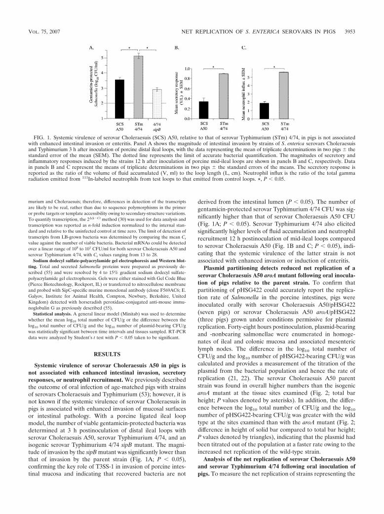
Systemic virulence of serovar Choleraesuis A50 in pigs isnot associated with enhanced intestinal invasion, secretoryresponses, or neutrophil recruitment. We previously describedthe outcome of oral infection of age-matched pigs with strainsof serovars Choleraesuis and Typhimurium (53); however, it isnot known if the systemic virulence of serovar Choleraesuis inpigs is associated with enhanced invasion of mucosal surfacesor intestinal pathology. With a porcine ligated ileal loopmodel, the number of viable gentamicin-protected bacteria wasdetermined at 3 h postinoculation of distal ileal loops withserovar Choleraesuis A50, serovar Typhimurium 4/74, and anisogenic serovar Typhimurium 4/74 sipB mutant. The magni-tude of invasion by the sipB mutant was significantly lower thanthat of invasion by the parent strain (Fig. 1A; P � 0.05),confirming the key role of T3SS-1 in invasion of porcine intes-tinal mucosa and indicating that recovered bacteria are not
derived from the intestinal lumen (P � 0.05). The number ofgentamicin-protected serovar Typhimurium 4/74 CFU was sig-nificantly higher than that of serovar Choleraesuis A50 CFU(Fig. 1A; P � 0.05). Serovar Typhimurium 4/74 also elicitedsignificantly higher levels of fluid accumulation and neutrophilrecruitment 12 h postinoculation of mid-ileal loops comparedto serovar Choleraesuis A50 (Fig. 1B and C; P � 0.05), indi-cating that the systemic virulence of the latter strain is notassociated with enhanced invasion or induction of enteritis.
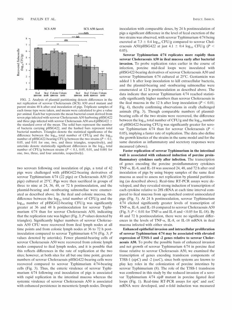
Plasmid partitioning detects reduced net replication of aserovar Choleraesuis A50 aroA mutant following oral inocula-tion of pigs relative to the parent strain. To confirm thatpartitioning of pHSG422 could accurately report the replica-tion rate of Salmonella in the porcine intestines, pigs wereinoculated orally with serovar Choleraesuis A50/pHSG422(seven pigs) or serovar Choleraesuis A50 aroA/pHSG422(three pigs) grown under conditions permissive for plasmidreplication. Forty-eight hours postinoculation, plasmid-bearingand -nonbearing salmonellae were enumerated in homoge-nates of ileal and colonic mucosa and associated mesentericlymph nodes. The difference in the log10 total number ofCFU/g and the log10 number of pHSG422-bearing CFU/g wascalculated and provides a measurement of the titration of theplasmid from the bacterial population and hence the rate ofreplication (21, 22). The serovar Choleraesuis A50 parentstrain was found in overall higher numbers than the isogenicaroA mutant at the tissue sites examined (Fig. 2; total barheight; P values denoted by asterisks). In addition, the differ-ence between the log10 total number of CFU/g and the log10
number of pHSG422-bearing CFU/g was greater with the wildtype at the sites examined than with the aroA mutant (Fig. 2;difference in height of solid bar compared to total bar height;P values denoted by triangles), indicating that the plasmid hadbeen titrated out of the population at a faster rate owing to theincreased net replication of the wild-type strain.
Analysis of the net replication of serovar Choleraesuis A50and serovar Typhimurium 4/74 following oral inoculation ofpigs. To measure the net replication of strains representing the
FIG. 1. Systemic virulence of serovar Choleraesuis (SCS) A50, relative to that of serovar Typhimurium (STm) 4/74, in pigs is not associatedwith enhanced intestinal invasion or enteritis. Panel A shows the magnitude of intestinal invasion by strains of S. enterica serovars Choleraesuisand Typhimurium 3 h after inoculation of porcine distal ileal loops, with the data representing the mean of triplicate determinations in two pigs thestandard error of the mean (SEM). The dotted line represents the limit of accurate bacterial quantification. The magnitudes of secretory andinflammatory responses induced by the strains 12 h after inoculation of porcine mid-ileal loops are shown in panels B and C, respectively. Datain panels B and C represent the means of triplicate determinations in two pigs the standard errors of the means. The secretory response isreported as the ratio of the volume of fluid accumulated (V, ml) to the loop length (L, cm). Neutrophil influx is the ratio of the total gammaradiation emitted from 111In-labeled neutrophils from test loops to that emitted from control loops. *, P � 0.05.
VOL. 75, 2007 NET REPLICATION OF S. ENTERICA SEROVARS IN PIGS 3953
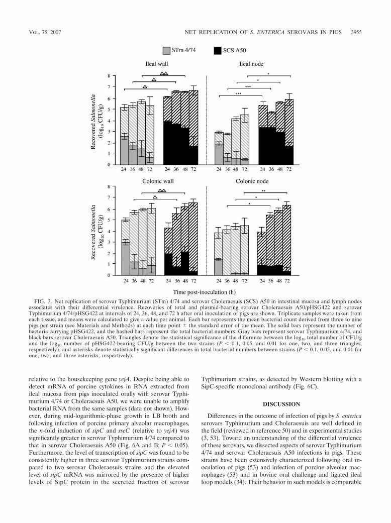
two serovars following oral inoculation of pigs, a total of 42pigs were challenged with pHSG422-bearing derivatives ofserovar Typhimurium 4/74 (22 pigs) or Choleraesuis A50 (20pigs) cultured at 25°C. Pigs were then sacrificed in groups ofthree to nine at 24, 36, 48, or 72 h postinoculation, and theplasmid-bearing and -nonbearing salmonellae were enumer-ated as described above. In the ileal and colonic mucosa, thedifference between the log10 total number of CFU/g and thelog10 number of pHSG422-bearing CFU/g was significantlygreater at 36 and 48 h postinoculation for serovar Typhi-murium 4/74 than for serovar Choleraesuis A50, indicatingthat the replication rate was higher (Fig. 3; P values denoted bytriangles). Significantly higher numbers of serovar Cholerae-suis A50 CFU were recovered from ileal lymph nodes at alltime points and from colonic lymph nodes at 36 to 72 h post-inoculation compared to serovar Typhimurium 4/74 (Fig. 3; Pvalues denoted by asterisks). Fewer plasmid-bearing cells ofserovar Choleraesuis A50 were recovered from colonic lymphnodes compared to ileal lymph nodes, and it is possible thatthis reflects differences in the rate of replication at the twosites; however, at both sites for all but one time point, greaternumbers of serovar Choleraesuis pHSG422-bearing cells wererecovered compared to serovar Typhimurium 4/74-bearingcells (Fig. 3). Thus, the enteric virulence of serovar Typhi-murium 4/74 following oral inoculation of pigs is associatedwith rapid replication in the intestinal mucosa whereas thesystemic virulence of serovar Choleraesuis A50 is associatedwith enhanced persistence in mesenteric lymph nodes. Despite
inoculation with comparable doses, by 24 h postinoculation ofpigs a significant difference in the level of fecal excretion of thetwo strains was observed, with serovar Typhimurium 4/74 beingexcreted at 7.3 0.4 log10 CFU/g compared to serovar Chol-eraesuis A50/pHSG422 at just 4.1 0.4 log10 CFU/g (P �0.05).
Serovar Typhimurium 4/74 replicates more rapidly thanserovar Choleraesuis A50 in ileal mucosa early after bacterialinvasion. To probe replication rates earlier in the course ofinfection, porcine mid-ileal loops were inoculated withpHSG422-bearing derivatives of serovar Choleraesuis A50 andserovar Typhimurium 4/74 cultured at 25°C. Gentamicin wasadded 1 h after loop inoculation to kill extracellular bacteria,and the plasmid-bearing and -nonbearing salmonellae wereenumerated at 12 h postinoculation as described above. Thedata indicate that serovar Typhimurium 4/74 reached statisti-cally significantly higher numbers than serovar Choleraesuis inthe ileal mucosa in the 12 h after loop inoculation (P � 0.01;Fig. 4), thereby confirming observations in orally challengedanimals (Fig. 3). Though comparable numbers of plasmid-bearing cells of the two strains were recovered, the differencebetween the log10 total number of CFU/g and the log10 numberof pHSG422-bearing CFU/g was significantly greater for sero-var Typhimurium 4/74 than for serovar Choleraesuis (P �0.05), implying a faster rate of replication. The data also definethe growth kinetics of the strains in the same model and for thesame duration as inflammatory and secretory responses weremeasured (above).
Faster replication of serovar Typhimurium in the intestinalwall is associated with enhanced induction of porcine proin-flammatory cytokines early after infection. The transcriptionof genes encoding the porcine proinflammatory cytokinesTNF-�, IL-8, and IL-18 was assessed 24, 48, and 72 h after oralinoculation of pigs by using biopsy samples of the same ilealmucosa as used to assess net replication by plasmid partition-ing (as described above). Real-time RT-PCR assays were de-veloped, and they revealed strong induction of transcription ofeach cytokine relative to 28S rRNA at each time interval com-pared to ileal mucosa from age-matched control uninoculatedpigs (Fig. 5). At 24 h postinoculation, serovar Typhimurium4/74 elicited significantly greater levels of transcription ofTNF-�, IL-8, and IL-18 compared to serovar Choleraesuis A50(Fig. 5; P � 0.01 for TNF-� and IL-8 and �0.05 for IL-18). By48 and 72 h postinoculation, there were no significant differ-ences in the levels of TNF-�, IL-8, or IL-18 mRNA in ilealmucosa infected with either strain.
Enhanced epithelial invasion and intracellular proliferationof serovar Typhimurium 4/74 may be associated with elevatedexpression of T3SS-1 and -2 genes relative to serovar Choler-aesuis A50. To probe the possible basis of enhanced invasionand net growth of serovar Typhimurium 4/74 in porcine ilealtissue relative to serovar Choleraesuis A50, we examined thetranscription of genes encoding translocon components ofT3SS-1 (sipC) and -2 (sseC), since both systems are known toplay key roles in the colonization of porcine intestines byserovar Typhimurium (8). The role of the T3SS-1 transloconwas confirmed in this study by the reduced invasion of a sero-var Typhimurium 4/74 sipB mutant in porcine ligated ilealloops (Fig. 1). Real-time RT-PCR assays for sipC and sseCmRNA were developed, and n-fold induction was measured
FIG. 2. Analysis of plasmid partitioning detects differences in thenet replication of serovar Choleraesuis (SCS) A50 aroA mutant andparent strains 48 h after oral inoculation of pigs. Triplicate samples ofeach tissue type were taken, and means were calculated to give a valueper animal. Each bar represents the mean bacterial count derived fromseven pigs infected with serovar Choleraesuis A50 harboring pHSG422and three pigs infected with serovar Choleraesuis A50 aroA/pHSG422 the standard error of the mean. The solid bars represent the numberof bacteria carrying pHSG422, and the hashed bars represent totalbacterial numbers. Triangles denote the statistical significance of thedifference between the log10 total number of CFU/g and the log10number of pHSG422-bearing CFU/g between the two strains (P � 0.1,0.05, and 0.01 for one, two, and three triangles, respectively), andasterisks denote statistically significant differences in the log10 totalnumber of CFU/g between strains (P � 0.1, 0.05, 0.01, and 0.001 forone, two, three, and four asterisks, respectively).
3954 PAULIN ET AL. INFECT. IMMUN.
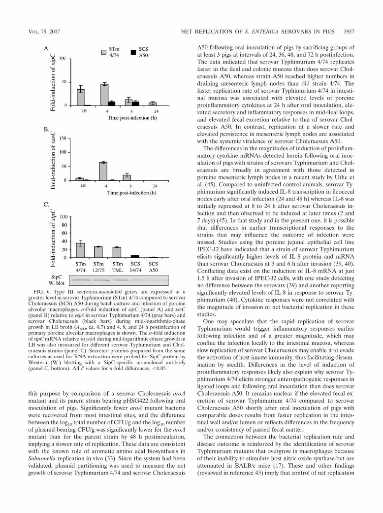
relative to the housekeeping gene yejA. Despite being able todetect mRNA of porcine cytokines in RNA extracted fromileal mucosa from pigs inoculated orally with serovar Typhi-murium 4/74 or Choleraesuis A50, we were unable to amplifybacterial RNA from the same samples (data not shown). How-ever, during mid-logarithmic-phase growth in LB broth andfollowing infection of porcine primary alveolar macrophages,the n-fold induction of sipC and sseC (relative to yejA) wassignificantly greater in serovar Typhimurium 4/74 compared tothat in serovar Choleraesuis A50 (Fig. 6A and B; P � 0.05).Furthermore, the level of transcription of sipC was found to beconsistently higher in three serovar Typhimurium strains com-pared to two serovar Choleraesuis strains and the elevatedlevel of sipC mRNA was mirrored by the presence of higherlevels of SipC protein in the secreted fraction of serovar
Typhimurium strains, as detected by Western blotting with aSipC-specific monoclonal antibody (Fig. 6C).
DISCUSSION
Differences in the outcome of infection of pigs by S. entericaserovars Typhimurium and Choleraesuis are well defined inthe field (reviewed in reference 50) and in experimental studies(3, 53). Toward an understanding of the differential virulenceof these serovars, we dissected aspects of serovar Typhimurium4/74 and serovar Choleraesuis A50 infections in pigs. Thesestrains have been extensively characterized following oral in-oculation of pigs (53) and infection of porcine alveolar mac-rophages (53) and in bovine oral challenge and ligated ilealloop models (34). Their behavior in such models is comparable
FIG. 3. Net replication of serovar Typhimurium (STm) 4/74 and serovar Choleraesuis (SCS) A50 in intestinal mucosa and lymph nodesassociates with their differential virulence. Recoveries of total and plasmid-bearing serovar Choleraesuis A50/pHSG422 and serovarTyphimurium 4/74/pHSG422 at intervals of 24, 36, 48, and 72 h after oral inoculation of pigs are shown. Triplicate samples were taken fromeach tissue, and means were calculated to give a value per animal. Each bar represents the mean bacterial count derived from three to ninepigs per strain (see Materials and Methods) at each time point the standard error of the mean. The solid bars represent the number ofbacteria carrying pHSG422, and the hashed bars represent the total bacterial numbers. Gray bars represent serovar Typhimurium 4/74, andblack bars serovar Choleraesuis A50. Triangles denote the statistical significance of the difference between the log10 total number of CFU/gand the log10 number of pHSG422-bearing CFU/g between the two strains (P � 0.1, 0.05, and 0.01 for one, two, and three triangles,respectively), and asterisks denote statistically significant differences in total bacterial numbers between strains (P � 0.1, 0.05, and 0.01 forone, two, and three asterisks, respectively).
VOL. 75, 2007 NET REPLICATION OF S. ENTERICA SEROVARS IN PIGS 3955

to that of other strains of the same serovar; thus, as far isreasonably practicable given the costly nature of large-animalexperimentation, they may be considered representative of theserovar.
Consistent with studies on host-restricted S. enterica serovarDublin in cattle (34), serovar Abortusovis in lambs (47), andserovar Gallinarum in chickens (9), systemic virulence ofserovar Choleraesuis in pigs was not associated with enhancedinvasion of ileal mucosa or greater induction of intestinal in-flammatory responses compared to serovar Typhimurium 4/74.These data support previous observations that serovar Chol-eraesuis A50 causes less destruction of porcine intestinal mu-cosa than does serovar Dublin 3246, as assessed by electronmicroscopy of ileal mucosa 3 h after loop inoculation (3), eventhough serovar Choleraesuis A50 is more virulent than serovarDublin 3246 following oral inoculation of pigs (53). We there-fore hypothesized that the differential virulence of serovarCholeraesuis A50 and serovar Typhimurium 4/74 may be afunction of distinct replication or killing kinetics in the intes-tinal mucosa and associated mesenteric lymph nodes.
Net bacterial replication in vivo was measured by analysis ofthe partitioning of a plasmid that exhibits temperature-sensi-tive replication. The system was confirmed to be suitable for
FIG. 4. Serovar Typhimurium (STm) 4/74 replicates more rapidlythan serovar Choleraesuis (SCS) A50 in porcine ileal mucosa earlyafter infection. Recoveries of total and plasmid-bearing serovar Chol-eraesuis A50/pHSG422 and serovar Typhimurium 4/74/pHSG422 12 hafter intraluminal inoculation of porcine mid-ileal loops are shown.Gentamicin was added to each loop after 1 h to kill extracellularbacteria. Each bar represents the mean bacterial count derived fromtriplicate biopsy samples in triplicate loops from two pigs the stan-dard error of the mean. The solid bars represent the number of bac-teria carrying the pHSG422, and the hashed bars represent total bac-terial numbers. Gray bars represent serovar Typhimurium 4/74, andblack bars represent serovar Choleraesuis A50. Triangles denote thestatistical significance of the difference between the log10 total numberof CFU/g and the log10 number of pHSG422-bearing CFU/g betweenthe two strains (P � 0.05), and asterisks denote significant differencebetween the total bacterial numbers (P � 0.01).
FIG. 5. Serovar Typhimurium (STm) 4/74 induces elevated levels of proinflammatory cytokine mRNAs compared to serovar Choleraesuis(SCS) early after oral inoculation of pigs. Data represent the mean 40-Ct values (a measurement of the abundance of cytokine mRNAs) for porcineTNF-�, IL-8, and IL-18 mRNAs in ileal mucosa 24, 48, and 72 h after oral inoculation of pigs with serovar Typhimurium 4/74 or serovarCholeraesuis A50 or mock infection relative to that for 28S rRNA. The mean was derived from independent determinations from at least threepigs at each time interval the standard error of the mean. *, P � 0.05 for elevated transcript levels induced by serovar Typhimurium 4/74compared to those induced by serovar Choleraesuis A50 at 24 h postinfection. UI, uninfected.
3956 PAULIN ET AL. INFECT. IMMUN.
this purpose by comparison of a serovar Choleraesuis aroAmutant and its parent strain bearing pHSG422 following oralinoculation of pigs. Significantly fewer aroA mutant bacteriawere recovered from most intestinal sites, and the differencebetween the log10 total number of CFU/g and the log10 numberof plasmid-bearing CFU/g was significantly lower for the aroAmutant than for the parent strain by 48 h postinoculation,implying a slower rate of replication. These data are consistentwith the known role of aromatic amino acid biosynthesis inSalmonella replication in vivo (33). Since the system had beenvalidated, plasmid partitioning was used to measure the netgrowth of serovar Typhimurium 4/74 and serovar Choleraesuis
A50 following oral inoculation of pigs by sacrificing groups ofat least 3 pigs at intervals of 24, 36, 48, and 72 h postinfection.The data indicated that serovar Typhimurium 4/74 replicatesfaster in the ileal and colonic mucosa than does serovar Chol-eraesuis A50, whereas strain A50 reached higher numbers indraining mesenteric lymph nodes than did strain 4/74. Thefaster replication rate of serovar Typhimurium 4/74 in intesti-nal mucosa was associated with elevated levels of porcineproinflammatory cytokines at 24 h after oral inoculation, ele-vated secretory and inflammatory responses in mid-ileal loops,and elevated fecal excretion relative to that of serovar Chol-eraesuis A50. In contrast, replication at a slower rate andelevated persistence in mesenteric lymph nodes are associatedwith the systemic virulence of serovar Choleraesuis A50.
The differences in the magnitudes of induction of proinflam-matory cytokine mRNAs detected herein following oral inoc-ulation of pigs with strains of serovars Typhimurium and Chol-eraesuis are broadly in agreement with those detected inporcine mesenteric lymph nodes in a recent study by Uthe etal. (45). Compared to uninfected control animals, serovar Ty-phimurium significantly induced IL-8 transcription in ileocecalnodes early after oral infection (24 and 48 h) whereas IL-8 wasinitially repressed at 8 to 24 h after serovar Choleraesuis in-fection and then observed to be induced at later times (2 and7 days) (45). In that study and in the present one, it is possiblethat differences in earlier transcriptional responses to thestrains that may influence the outcome of infection weremissed. Studies using the porcine jejunal epithelial cell lineIPEC-J2 have indicated that a strain of serovar Typhimuriumelicits significantly higher levels of IL-8 protein and mRNAthan serovar Choleraesuis at 3 and 6 h after invasion (39, 40).Conflicting data exist on the induction of IL-8 mRNA at just1.5 h after invasion of IPEC-J2 cells, with one study detectingno difference between the serovars (39) and another reportingsignificantly elevated levels of IL-8 in response to serovar Ty-phimurium (40). Cytokine responses were not correlated withthe magnitude of invasion or net bacterial replication in thesestudies.
One may speculate that the rapid replication of serovarTyphimurium would trigger inflammatory responses earlierfollowing infection and of a greater magnitude, which mayconfine the infection locally to the intestinal mucosa, whereasslow replication of serovar Choleraesuis may enable it to evadethe activation of host innate immunity, thus facilitating dissem-ination by stealth. Differences in the level of induction ofproinflammatory responses likely also explain why serovar Ty-phimurium 4/74 elicits stronger enteropathogenic responses inligated loops and following oral inoculation than does serovarCholeraesuis A50. It remains unclear if the elevated fecal ex-cretion of serovar Typhimurium 4/74 compared to serovarCholeraesuis A50 shortly after oral inoculation of pigs withcomparable doses results from faster replication in the intes-tinal wall and/or lumen or reflects differences in the frequencyand/or consistency of passed fecal matter.
The connection between the bacterial replication rate anddisease outcome is reinforced by the identification of serovarTyphimurium mutants that overgrow in macrophages becauseof their inability to stimulate host nitric oxide synthase but areattenuated in BALB/c mice (17). These and other findings(reviewed in reference 43) imply that control of net replication
FIG. 6. Type III secretion-associated genes are expressed at agreater level in serovar Typhimurium (STm) 4/74 compared to serovarCholeraesuis (SCS) A50 during batch culture and infection of porcinealveolar macrophages. n-Fold induction of sipC (panel A) and sseC(panel B) relative to yejA in serovar Typhimurium 4/74 (gray bars) andserovar Choleraesuis (black bars) during mid-logarithmic-phasegrowth in LB broth (A600, ca. 0.7) and 4, 8, and 24 h postinfection ofprimary porcine alveolar macrophages is shown. The n-fold inductionof sipC mRNA relative to yejA during mid-logarithmic-phase growth inLB was also measured for different serovar Typhimurium and Chol-eraesuis strains (panel C). Secreted proteins prepared from the samecultures as used for RNA extraction were probed for SipC protein byWestern (W.) blotting with a SipC-specific monoclonal antibody(panel C, bottom). All P values for n-fold differences, �0.05.
VOL. 75, 2007 NET REPLICATION OF S. ENTERICA SEROVARS IN PIGS 3957
by Salmonella may confer a survival advantage. In an attemptto dissect the molecular mechanisms that dictate the differen-tial growth rates of serovar Typhimurium 4/74 and serovarCholeraesuis A50, we examined the transcription of genes en-coding translocon components of T3SS-1 and T3SS-2. Bothsystems are known to play key roles in colonization of theporcine intestines by serovar Typhimurium (5, 8; Fig. 1A), andtheir activity has been correlated with the progression of ty-phoid fever in murine models. T3SS-1-injected effector pro-teins stimulate rearrangements of the subcortical actin cy-toskeleton that facilitate bacterial invasion; however, T3SS-1also contributes to bacterial growth in epithelial cells (42) andthe T3SS-1 effector SopB stimulates nitric oxide productionand remains active long after bacterial entry (16). T3SS-1 ex-pression in vivo is also correlated with induction of a lysosomalrepair response that can control Salmonella infection (35).T3SS-2 facilitates intracellular proliferation by modulating thetrafficking of the Salmonella-containing vacuole. It is inducedin the intestinal lumen (6), and contextual regulation of T3SS-2is important in the progression of typhoid fever following oraldosing of mice with serovar Typhimurium (14).
In the strains of well-defined virulence used, transcription ofsipC and sseC was consistently higher in serovar Typhimurium4/74 than in serovar Choleraesuis A50 during mid-logarithmic-phase growth in vitro and following infection of porcine pri-mary alveolar macrophages. We were unable to measure tran-script levels in the same tissues as plasmid partitioning wasassessed, presumably as they were present below the limit ofdetection since RNA extracted from the tissues yielded ampli-cons for porcine cytokine mRNA. Immunohistochemistry withantibodies directed against T3SS-1 and -2 effector proteins orepitope-tagged variants thereof also failed to detect expressionof the proteins in vivo, presumably owing to the small quanti-ties present (data not shown). Transcription of sipC was con-sistently higher in serovar Typhimurium strains that showbehavior comparable to that of strain 4/74 in porcine macro-phages (53), and the difference from serovar Choleraesuisstrains was also reflected in the quantities of SipC proteinproduced.
In addition to differences in the levels of transcription of thegenes for T3SS-1 and -2, our analysis of the genomes of se-quenced S. enterica serovars reveals that the genes encodingthe effector proteins SopA, SlrP, SseI, and SspH2 are pseudo-genes in serovar Choleraesuis SC-B67 and SseK2 is absent, yetthe genes are intact in serovar Typhimurium LT2. Of these,SopA is known to be important in the induction of enteritis byserovar Dublin in bovine ligated ileal loops (54). We confirmedthat SopA is a pseudogene in serovar Choleraesuis A50 bynucleotide sequencing and Western blot analysis with a SopA-specific monoclonal antibody (data not shown). Such poly-morphisms in the repertoire, sequence, and expression ofT3SS-related genes may explain the reduced invasion and in-flammatory potential of serovar Choleraesuis; however, it islikely that other factors are involved. Of 151 pseudogenes inthe serovar Choleraesuis SC-B67 genome, 64 were predicted tobe metabolism related (12). Furthermore, the shdA gene,which encodes a fibronectin-binding protein required for Pey-er’s patch colonization and fecal shedding in mice (27) andenteric virulence early after oral inoculation of pigs (4), is apseudogene in SC-B67 relative to LT2. It is equally possible
that such defects may explain the reduced replication rate ofserovar Choleraesuis compared to serovar Typhimurium inintestinal epithelia; however, a requirement exists to confirmthat the polymorphisms also exist between the two strainsexamined herein. Mutational attrition of several predicted reg-ulators has also been described previously in SC-B67 (12);however, the PhoPQ two-component system known to play akey role in intracellular proliferation (reviewed in references20 and 43) appears to be intact in the sequenced genomes.Polynucleotide phosphorylase, which has been proposed toalter the balance between acute infection and persistence bymodulating SPI-1 and -2 genes (13), is also identical in strainsSC-B67 and LT2.
The importance of T3SS in the translocation of serovarCholeraesuis from the porcine intestines remains ill defined. Aserovar Choleraesuis hilA mutant defective in the regulation ofT3SS-1 was less able to colonize the intestines and disseminateto the liver and spleen following oral inoculation of pigs, butnot following intraperitoneal infection (29). The role played byT3SS-2 in dissemination from the porcine intestines is un-known. In recent studies with calves, early translocation ofserovar Dublin from the intestines to mesenteric lymph nodes,the liver, and the spleen was found to occur independently ofT3SS-2 (G. D. Pullinger et al., submitted for publication); thus,caution is required in interpreting the importance of variationsin the expression or sequence of T3SS-related genes until theirprecise contribution to pathogenesis is known.
As elegantly reviewed in reference 43, the rate of intracel-lular proliferation of Salmonella is strictly dependent on thecell type, occurring rapidly in immortalized epithelial and mac-rophage lines, whereas in dendritic cells and fibroblasts a ho-mogeneous population in a nonreplicating state exists. It isunclear if the differences in net replication of the S. entericastrains examined here reflect the targeting of different cellpopulations. Understanding of cell tropism is of key impor-tance when probing the roles of bacterial factors in net growth,since mutation of some systems can have different effects indifferent cell types. For example, SPI-2 is dispensable for sur-vival and intracellular proliferation in dendritic cells eventhough it is induced (26) and mutation of PhoPQ enhancesgrowth in fibroblasts but inhibits it in other cell types (7). Thedata reported here indicate that differences in replication rateand host innate immune activation by S. enterica serovars maybe under bacterial control and have profound implications fordisease progression.
ACKNOWLEDGMENTS
We gratefully acknowledge the support of the Biotechnology &Biological Sciences Research Council (grant 201/S14753) and the De-partment for the Environment, Food and Rural Affairs (grantOZ0319).
We thank T. Hashimoto-Gotoh for the kind gift of pHSG422.
REFERENCES
1. Bakshi, C. S., V. P. Singh, M. W. Wood, P. W. Jones, T. S. Wallis, and E. E.Galyov. 2000. Identification of SopE2, a Salmonella secreted protein which ishighly homologous to SopE and involved in bacterial invasion of epithelialcells. J. Bacteriol. 182:2341–2344.
2. Benjamin, W. H., Jr., P. Hall, S. J. Roberts, and D. E. Briles. 1990. Theprimary effect of the Ity locus is on the rate of growth of Salmonella typhi-murium that are relatively protected from killing. J. Immunol. 144:3143–3151.
3. Bolton, A. J., M. P. Osborne, T. S. Wallis, and J. Stephen. 1999. Interaction
3958 PAULIN ET AL. INFECT. IMMUN.

of Salmonella choleraesuis, Salmonella dublin and Salmonella typhimuriumwith porcine and bovine terminal ileum in vivo. Microbiology 145:2431–2441.
4. Boyen, F., F. Pasmans, E. Donne, F. van Immerseel, E. Morgan, C.Adriaensen, J. P. Hernalsteens, T. S. Wallis, R. Ducatelle, and F. Haese-brouck. 2006. The fibronectin binding protein ShdA is not a prerequisite forlong term faecal shedding of Salmonella typhimurium in pigs. Vet. Microbiol.115:284–290.
5. Boyen, F., F. Pasmans, F. van Immerseel, E. Morgan, C. Adriaensen, J. P.Hernalsteens, A. Decostere, R. Ducatelle, and F. Haesebrouck. 2006. Salmo-nella Typhimurium SPI-1 genes promote intestinal but not tonsillar coloni-zation in pigs. Microbes Infect. 8:2899–2907.
6. Brown, N. F., B. A. Vallance, B. K. Coombes, Y. Valdez, R. A. Coburn, andB. B. Finlay. 2005. Salmonella pathogenicity island 2 is expressed prior topenetrating the intestine. PLoS Pathog. 1:e32.
7. Cano, D. A., M. Martinez-Moya, M. G. Pucciarelli, E. A. Groisman, J.Casadesus, and F. Garcia-Del Portillo. 2001. Salmonella enterica serovarTyphimurium response involved in attenuation of pathogen intracellularproliferation. Infect. Immun. 69:6463–6474.
8. Carnell, S., A. Bowen, E. Morgan, D. J. Maskell, T. S. Wallis, and M. P.Stevens. 2007. Role in virulence and protective efficacy in pigs of Salmonellaenterica serovar Typhimurium secreted components identified by signature-tagged mutagenesis. Microbiology 153:1940–1952.
9. Chadfield, M. S., D. J. Brown, S. Aabo, J. P. Christensen, and J. E. Olsen.2003. Comparison of intestinal invasion and macrophage response of Sal-monella Gallinarum and other host-adapted Salmonella enterica serovars inthe avian host. Vet. Microbiol. 92:49–64.
10. Chan, K., C. C. Kim, and S. Falkow. 2005. Microarray-based detection ofSalmonella enterica serovar Typhimurium transposon mutants that cannotsurvive in macrophages and mice. Infect. Immun. 73:5438–5449.
11. Chiu, C. H., L. H. Su, and C. Chu. 2004. Salmonella enterica serotypeCholeraesuis: epidemiology, pathogenesis, clinical disease, and treatment.Clin. Microbiol. Rev. 17:311–322.
12. Chiu, C. H., P. Tang, C. Chu, S. Hu, Q. Bao, J. Yu, Y. Y. Chou, H. S. Wang,and Y. S. Lee. 2005. The genome sequence of Salmonella enterica serovarCholeraesuis, a highly invasive and resistant zoonotic pathogen. NucleicAcids Res. 33:1690–1698.
13. Clements, M. O., S. Eriksson, A. Thompson, S. Lucchini, J. C. Hinton, S.Normark, and M. Rhen. 2002. Polynucleotide phosphorylase is a globalregulator of virulence and persistency in Salmonella enterica. Proc. Natl.Acad. Sci. USA 99:8784–8789.
14. Coombes, B. K., M. E. Wickham, M. J. Lowden, N. F. Brown, and B. B.Finlay. 2005. Negative regulation of Salmonella pathogenicity island 2 isrequired for contextual control of virulence during typhoid. Proc. Natl. Acad.Sci. USA 102:17460–17465.
15. Davies, R. H., R. Dalziel, J. C. Gibbens, J. W. Wilesmith, J. M. Ryan, S. J.Evans, C. Byrne, G. A. Paiba, S. J. Pascoe, and C. J. Teale. 2004. Nationalsurvey for Salmonella in pigs, cattle and sheep at slaughter in Great Britain(1999–2000). J. Appl. Microbiol. 96:750–760.
16. Drecktrah, D., L. A. Knodler, K. Galbraith, and O. Steele-Mortimer. 2005.The Salmonella SPI1 effector SopB stimulates nitric oxide production longafter invasion. Cell. Microbiol. 7:105–113.
17. Eriksson, S., J. Bjorkman, S. Borg, A. Syk, S. Pettersson, D. I. Andersson,and M. Rhen. 2000. Salmonella typhimurium mutants that downregulatephagocyte nitric oxide production. Cell. Microbiol. 2:239–250.
18. Eriksson, S., S. Lucchini, A. Thompson, M. Rhen, and J. C. Hinton. 2003.Unravelling the biology of macrophage infection by gene expression profilingof intracellular Salmonella enterica. Mol. Microbiol. 47:103–118.
19. Fallon, M. T., W. H. Benjamin, Jr., T. R. Schoeb, and D. E. Briles. 1991.Mouse hepatitis virus strain UAB infection enhances resistance to Salmo-nella typhimurium in mice by inducing suppression of bacterial growth. In-fect. Immun. 59:852–856.
20. Groisman, E. A. 2001. The pleiotropic two-component regulatory systemPhoP-PhoQ. J. Bacteriol. 183:1835–1842.
21. Gulig, P. A., and T. J. Doyle. 1993. The Salmonella typhimurium virulenceplasmid increases the growth rate of salmonellae in mice. Infect. Immun.61:504–511.
22. Gulig, P. A., T. J. Doyle, M. J. Clare-Salzler, R. L. Maiese, and H. Matsui.1997. Systemic infection of mice by wild-type but not Spv� Salmonella ty-phimurium is enhanced by neutralization of gamma interferon and tumornecrosis factor alpha. Infect. Immun. 65:5191–5197.
23. Hashimoto-Gotoh, T., F. C. Franklin, A. Nordheim, and K. N. Timmis. 1981.Specific-purpose plasmid cloning vectors. I. Low copy number, temperature-sensitive, mobilization defective pSC101-derived containment vectors. Gene16:227–235.
24. Hensel, M., J. E. Shea, C. Gleeson, M. D. Jones, E. Dalton, and D. W.Holden. 1995. Simultaneous identification of bacterial virulence genes bynegative selection. Science 269:400–403.
25. Hughes, S., T.-Y. Poh, N. Bumstead, and P. Kaiser. 2007. Re-evaluation ofthe chicken MIP family of chemokines and their receptors suggests thatCCL5 is the prototypic MIP family chemokine, and that different specieshave developed different repertoires of both the CC chemokines and theirreceptors. Dev. Comp. Immun. 31:72–86.
26. Jantsch, J., C. Cheminay, D. Chakravortty, T. Lindig, J. Hein, and M.Hensel. 2003. Intracellular activities of Salmonella enterica in murine den-dritic cells. Cell. Microbiol. 5:933–945.
27. Kingsley, R. A., K. van Amsterdam, N. Kramer, and A. J. Baumler. 2000.The shdA gene is restricted to serotypes of Salmonella enterica subspecies Iand contributes to efficient and prolonged fecal shedding. Infect. Immun.68:2720–2727.
28. Lawley, T. D., K. Chan, L. J. Thompson, C. C. Kim, G. R. Govoni, and D. M.Monack. 2006. Genome-wide screen for Salmonella genes required for long-term systemic infection of the mouse. PLoS Pathog. 2:e11.
29. Lichtensteiger, C. A., and E. R. Vimr. 2003. Systemic and enteric coloniza-tion of pigs by a hilA signature-tagged mutant of Salmonella choleraesuis.Microb. Pathog. 34:149–154.
30. Livak, K. J., and T. D. Schmittgen. 2001. Analysis of relative transcriptiondata using real-time quantitative PCR and the 2���Ct method. Methods25:402–408.
31. McFarland, W. C., and B. A. D. Stocker. 1987. Effect of different purineauxotrophic mutations on mouse-virulence of a Vi-positive strain of Salmo-nella dublin and of two strains of Salmonella typhimurium. Microb. Pathog.3:129–141.
32. Morgan, E., J. D. Campbell, S. C. Rowe, J. Bispham, M. P. Stevens, A. J.Bowen, P. A. Barrow, D. J. Maskell, and T. S. Wallis. 2004. Identification ofhost-specific colonization factors of Salmonella enterica serovar Typhi-murium. Mol. Microbiol. 54:994–1010.
33. O’Callaghan, D., D. Maskell, F. Y. Liew, C. S. Easmon, and G. Dougan.1988. Characterization of aromatic- and purine-dependent Salmonella typhi-murium: attenuation, persistence, and ability to induce protective immunityin BALB/c mice. Infect. Immun. 56:419–423.
34. Paulin, S. M., P. R. Watson, A. R. Benmore, M. P. Stevens, P. W. Jones, B.Villarreal-Ramos, and T. S. Wallis. 2002. Analysis of Salmonella entericaserotype-host specificity in calves: avirulence of S. enterica serotype Gallina-rum correlates with bacterial dissemination from mesenteric lymph nodesand persistence in vivo. Infect. Immun. 70:6788–6797.
35. Roy, D., D. R. Liston, V. J. Idone, A. Di, D. J. Nelson, C. Pujol, J. B. Bliska,S. Chakrabarti, and N. W. Andrews. 2004. A process for controlling intra-cellular bacterial infections induced by membrane injury. Science 304:1515–1518.
36. Schwartz, K. J. 1999. Salmonellosis, p. 535–551. In B. E. Straw, S. D’Allaire,W. L. Mengeling, and D. J. Taylor (ed.), Disease of swine, 8th ed. Iowa StateUniversity Press, Ames.
37. Shea, J. E., C. R. Beuzon, C. Gleeson, R. Mundy, and D. W. Holden. 1999.Influence of the Salmonella typhimurium pathogenicity island 2 type IIIsecretion system on bacterial growth in the mouse. Infect. Immun. 67:213–219.
38. Shea, J. E., M. Hensel, C. Gleeson, and D. W. Holden. 1996. Identification ofa virulence locus encoding a second type III secretion system in Salmonellatyphimurium. Proc. Natl. Acad. Sci. USA 93:2593–2597.
39. Skjolaas, K. A., T. E. Burkey, S. S. Dritz, and J. E. Minton. 2006. Effects ofSalmonella enterica serovars Typhimurium (ST) and Choleraesuis (SC) onchemokine and cytokine expression in swine ileum and jejunal epithelialcells. Vet. Immunol. Immunopathol. 111:199–209.
40. Skjolaas, K. A., T. E. Burkey, S. S. Dritz, and J. E. Minton. 2007. Effects ofSalmonella enterica serovar Typhimurium, or serovar Choleraesuis, Lactoba-cillus reuteri and Bacillus licheniformis on chemokine and cytokine expressionin the swine jejunal epithelial cell line, IPEC-J2. Vet. Immunol. Immuno-pathol. 115:299–308.
41. Sojka, W. J., C. Wray, J. Shreeve, and A. J. Benson. 1977. Incidence ofSalmonella infection in animals in England and Wales 1968–1974. J. Hyg.Camb. 78:43–56.
42. Steele-Mortimer, O., J. H. Brumell, L. A. Knodler, S. Meresse, A. Lopez, andB. B. Finlay. 2002. The invasion-associated type III secretion system ofSalmonella enterica serovar Typhimurium is necessary for intracellular pro-liferation and vacuole biogenesis in epithelial cells. Cell. Microbiol. 4:43–54.
43. Tierrez, A., and F. Garcı́a-del Portillo. 2005. New concepts in Salmonellavirulence: the importance of reducing the intracellular growth rate in thehost. Cell. Microbiol. 7:901–909.
44. Tsolis, R. M., S. M. Townsend, E. A. Miao, S. I. Miller, T. A. Ficht, L. G.Adams, and A. J. Baumler. 1999. Identification of a putative Salmonellaenterica serotype Typhimurium host range factor with homology to IpaH andYopM by signature-tagged mutagenesis. Infect. Immun. 67:6385–6393.
45. Uthe, J. J., A. Royaee, J. K. Lunney, T. J. Stabel, S. H. Zhao, C. K. Tuggle,and S. M. Bearson. 2007. Porcine differential gene expression in response toSalmonella enterica serovars Choleraesuis and Typhimurium. Mol. Immunol.44:2900–2914.
46. Uzzau, S., D. J. Brown, T. Wallis, S. Rubino, G. Leori, S. Bernard, J.Casadesus, D. J. Platt, and J. E. Olsen. 2000. Host adapted serotypes ofSalmonella enterica. Epidemiol. Infect. 125:229–255.
47. Uzzau, S., G. S. Leori, V. Petruzzi, P. R. Watson, G. Schianchi, D. Bacciu, V.Mazzarello, T. S. Wallis, and S. Rubino. 2001. Salmonella enterica serovar-host specificity does not correlate with the magnitude of intestinal invasionin sheep. Infect. Immun. 69:3092–3099.
48. van Diemen, P. M., M. B. Kreukniet, L. Galina, N. Bumstead, and T. S.
VOL. 75, 2007 NET REPLICATION OF S. ENTERICA SEROVARS IN PIGS 3959
Wallis. 2002. Characterisation of a resource population of pigs screened forresistance to salmonellosis. Vet. Immunol. Immunopathol. 88:183–196.
49. Vugia, D. J., M. Samuel, M. M. Farley, R. Marcus, B. Shiferaw, S. Shallow,K. Smith, and F. J. Angulo. 2004. Invasive Salmonella infections in theUnited States, FoodNet, 1996–1999: incidence, serotype distribution, andoutcome. Clin. Infect. Dis. 38(Suppl. 3):S149–S156.
50. Wallis, T. S., and P. A. Barrow. July 2005, posting date. Chapter 8.6.2.1.Salmonella epidemiology and pathogenesis in food-producing animals. In R.Curtiss III et al. (ed.), EcoSal—Escherichia coli and Salmonella: cellular andmolecular biology. ASM Press, Washington, DC. http://ecosal.org.
51. Wallis, T. S., S. M. Paulin, J. S. Plested, P. R. Watson, and P. W. Jones. 1995.The Salmonella dublin virulence plasmid mediates systemic but not entericphases of salmonellosis in cattle. Infect. Immun. 63:2755–2761.
52. Watson, P. R., S. M. Paulin, A. P. Bland, P. W. Jones, and T. S. Wallis. 1995.
Characterization of intestinal invasion by Salmonella typhimurium and Sal-monella dublin and effect of a mutation in the invH gene. Infect. Immun.63:2743–2754.
53. Watson, P. R., S. M. Paulin, P. W. Jones, and T. S. Wallis. 2000. Interactionof Salmonella serotypes with porcine macrophages in vitro does not correlatewith virulence. Microbiology 146:1639–1649.
54. Wood, M. W., M. A. Jones, P. R. Watson, A. M. Siber, B. A. McCormick, S.Hedges, R. Rosqvist, T. S. Wallis, and E. E. Galyov. 2000. The secretedeffector protein of Salmonella dublin, SopA, is translocated into eukaryoticcells and influences the induction of enteritis. Cell. Microbiol. 2:293–303.
55. Wood, M. W., R. Rosqvist, P. B. Mullan, M. H. Edwards, and E. E. Galyov.1996. SopE, a secreted protein of Salmonella dublin, is translocated into thetarget eukaryotic cell via a sip-dependent mechanism and promotes bacterialentry. Mol. Microbiol. 22:327–338.
Editor: F. C. Fang
3960 PAULIN ET AL. INFECT. IMMUN.