edinburgh research explorer · hoeve, ma, mylonas, kj, fairlie-clarke, kj, mahajan, sm, allen, je...
TRANSCRIPT

Edinburgh Research Explorer
Plasmodium chabaudi limits early Nippostrongylus brasiliensis-induced pulmonary immune activation and Th2 polarization inco-infected mice
Citation for published version:Hoeve, MA, Mylonas, KJ, Fairlie-Clarke, KJ, Mahajan, SM, Allen, JE & Graham, AL 2009, 'Plasmodiumchabaudi limits early Nippostrongylus brasiliensis-induced pulmonary immune activation and Th2polarization in co-infected mice' BMC Immunology, vol. 10, 60. DOI: 10.1186/1471-2172-10-60
Digital Object Identifier (DOI):10.1186/1471-2172-10-60
Link:Link to publication record in Edinburgh Research Explorer
Document Version:Publisher's PDF, also known as Version of record
Published In:BMC Immunology
Publisher Rights Statement:This is an Open Access article distributed under the terms of the Creative Commons Attribution License(http://creativecommons.org/licenses/by/2.0),which permits unrestricted use, distribution, and reproduction in any medium, provided the original work isproperly cited.
General rightsCopyright for the publications made accessible via the Edinburgh Research Explorer is retained by the author(s)and / or other copyright owners and it is a condition of accessing these publications that users recognise andabide by the legal requirements associated with these rights.
Take down policyThe University of Edinburgh has made every reasonable effort to ensure that Edinburgh Research Explorercontent complies with UK legislation. If you believe that the public display of this file breaches copyright pleasecontact [email protected] providing details, and we will remove access to the work immediately andinvestigate your claim.
Download date: 12. Sep. 2018
BioMed CentralBMC Immunology
ss
Open AcceResearch articlePlasmodium chabaudi limits early Nippostrongylus brasiliensis-induced pulmonary immune activation and Th2 polarization in co-infected miceMarieke A Hoeve†1, Katie J Mylonas†1, Karen J Fairlie-Clarke1, Simmi M Mahajan1, Judith E Allen1 and Andrea L Graham*1,2Address: 1Institute of Immunology and Infection Research, School of Biological Sciences, University of Edinburgh, Edinburgh, EH9 3JT, UK and 2Department of Ecology & Evolutionary Biology, Princeton University, Princeton, New Jersey, 08544, USA
Email: Marieke A Hoeve - [email protected]; Katie J Mylonas - [email protected]; Karen J Fairlie-Clarke - [email protected]; Simmi M Mahajan - [email protected]; Judith E Allen - [email protected]; Andrea L Graham* - [email protected]
* Corresponding author †Equal contributors
AbstractBackground: Larvae of several common species of parasitic nematodes obligately migratethrough, and often damage, host lungs. The larvae induce strong pulmonary Type 2 immuneresponses, including T-helper (Th)2 cells as well as alternatively activated macrophages (AAMφ) andassociated chitinase and Fizz/resistin family members (ChaFFs), which are thought to promotetissue repair processes. Given the prevalence of systemic or lung-resident Type 1-inducingpathogens in geographical areas in which nematodes are endemic, we wished to investigate theimpact of concurrent Type 1 responses on the development of these Type 2 responses tonematode larval migration. We therefore infected BALB/c mice with the nematode Nippostrongylusbrasiliensis, in the presence or absence of Plasmodium chabaudi chabaudi malaria parasites. Co-infected animals received both infections on the same day, and disease was assessed daily beforeimmunological measurements were taken at 3, 5, 7 or 20 days post-infection.
Results: We observed that the nematodes themselves caused transient loss of body mass and redblood cell density, but co-infection then slightly ameliorated the severity of malarial anaemia. Wealso tracked the development of immune responses in the lung and thoracic lymph node. By thetime of onset of the adaptive immune response around 7 days post-infection, malaria co-infectionhad reduced pulmonary expression of ChaFFs. Assessment of the T cell response demonstratedthat the Th2 response to the nematode was also significantly impaired by malaria co-infection.
Conclusion: P. c. chabaudi co-infection altered both local and lymph node Type 2 immuneactivation due to migration of N. brasiliensis larvae. Given recent work from other laboratoriesshowing that N. brasiliensis-induced ChaFFs correlate to the extent of long-term lung damage, ourresults raise the possibility that co-infection with malaria might alter pulmonary repair processesfollowing nematode migration. Further experimentation in the co-infection model developed herewill reveal the longer-term consequences of the presence of both malaria and helminths in the lung.
Published: 1 December 2009
BMC Immunology 2009, 10:60 doi:10.1186/1471-2172-10-60
Received: 8 May 2009Accepted: 1 December 2009
This article is available from: http://www.biomedcentral.com/1471-2172/10/60
© 2009 Hoeve et al; licensee BioMed Central Ltd. This is an Open Access article distributed under the terms of the Creative Commons Attribution License (http://creativecommons.org/licenses/by/2.0), which permits unrestricted use, distribution, and reproduction in any medium, provided the original work is properly cited.
Page 1 of 19(page number not for citation purposes)

BMC Immunology 2009, 10:60 http://www.biomedcentral.com/1471-2172/10/60
BackgroundMany prevalent species of parasitic nematodes - such asAscaris lumbricoides, which infects over a billion people[1], or Necator americanus, the most geographically wide-spread of the human hookworms [2] - migrate throughhost lungs as larvae. Lung tissue is ruptured as the larvaeburst out of the blood vessels to enter the alveolar spaces.Although this process is typically asymptomatic inhumans, it can also be associated with acute respiratorydistress or longer term complications [3]. For example,infection with lung-migrating helminths has been associ-ated with bronchial hyper-reactivity and other asthmasymptoms among children in China [4] and Brazil [5].
The rodent parasite Nippostrongylus brasiliensis (Nb) hasproven a valuable laboratory model for nematode migra-tion through the host body. In mice, L3 larvae injectedinto the skin migrate via the lungs to the small intestine,where the parasites develop into adults [6]. Peak abun-dance of Nb larvae in the lung occurs around 2 days post-infection (pi) in many strains of mice [7]. The lung migra-tory stage of Nb is associated with a strong local Type 2inflammatory response that includes T-helper (Th)2 cells,eosinophils and basophils [8,9]. Alternatively-activatedmacrophages (AAMφ) have also been identified as a majorcomponent of the pulmonary response to Nb infection[10,11]. AAMφ are characterised by IL-4/IL-13-dependentproduction of chitinase and Fizz/resistin family members(ChaFFs) including RELMα (also known as Fizz1), thechitinase-like protein Ym1, and Arginase-1 [12-15], andall three proteins are consistently observed in the Nbinfected lung [10,11,16-18]. Arginase-1 is the counter-reg-ulatory enzyme to iNOS and can thus act to suppress NOproduction and Type 1 effector function. Arginase-1 alsohas well documented roles in tissue repair [19,20] and hasrecently been implicated as an anti-nematode effectormolecule [21]. The functions of RELMα and Ym1 are lesswell understood but, like Arginase-1, they have beenstrongly implicated in the response to injury [22-24] andhave putative roles in the repair process, including extra-cellular matrix deposition and angiogenesis [25,26].However, recent data have shown that RELMα and macro-phage-derived arginase can also negatively regulate Th2effector responses and thus limit the pathology associatedwith overzealous repair [27-29].
Although not formally proven, the association of Argin-ase-1, RELMα, and Ym1 with the tissue repair process sug-gests that in the context of nematode infection, ChaFFs,potentially produced by AAMφ, may be required toorchestrate the repair of damage caused by larval migra-tion in order to restore lung integrity. Two recent papershave highlighted the potential for Nb migration to dam-age the lung with potentially long term consequences
[16,18]. Both studies document haemorrhaging of lungtissue and sustained increases in airway hyper-responsive-ness. A striking novel observation in these studies is thatNb causes disruption of the alveolar architecture that isconsistent with pulmonary emphysema many weeks afterinfection. Dysregulated, AAMφ-mediated repair of thedamage caused by the nematodes may be responsible forsuch detrimental outcomes [16].
Helminths with lung migratory stages are often co-endemic with Type 1-inducing parasites such as malaria[30-32]. Given the potential for cross-regulation betweenType 1 and Type 2 immune responses, we wished to usemouse models to investigate the consequences of co-infection for the pulmonary Type 2 immune responsesinduced by nematode migration. We chose to focus on Nband a rodent malaria, Plasmodium chabaudi chabaudi (Pcc),that induces a potent Type 1 immune response and non-lethal infection [33]. We challenged hosts simultaneouslywith these two acute infections, thus demanding polar-ized, conflicting immune responses at the same point intime. In addition, we expected Nb-Pcc co-infection toinduce conflicting responses in the same anatomical loca-tion, because malaria-infected red blood cells (RBCs) ofmany species, including Pcc, adhere to endothelial cells ofthe microvasculature of the lung [34-36]. Furthermore,malaria itself has been shown to cause lung injury [37,38].Thus, we expected the lung and draining (thoracic) lymphnodes to be potential sites of strong interactions betweenNb and Pcc. The idea that helminth-malaria co-infectionmay impose Type 1-Type 2 immunological conflict is notnew [30], nor is the idea that parasitic co-infection mayalter the severity of pulmonary disease [39,40], but ouremphasis on the consequences of malaria for pulmonaryType 2 responses has not previously been explored.
Using these model systems, we assessed production of theChaFFs, RELMα and Ym1 as primary read-outs of the Type2 effector response in the lung. We also examined thoraciclymph node (TLN) cytokine profiles, parasitology and sys-temic pathology, to set the co-infected lung in its whole-organism context. By 7 days pi, malaria infection had sig-nificantly reduced the expression of ChaFFs in the lungs ofco-infected animals relative to those with Nb only. Thisreduction correlated with changes in Th2 cytokines in theTLN, with co-infected mice producing significantly less IL-13, IL-10 and IL-5 than mice infected with Nb only. Pcc co-infection thus reduced the extent of pulmonary Type 2activation and Th2 polarisation in response to Nb. Futurelong-term experiments (up to a year in duration [16]) inthe co-infection model established here will explore howhelminth migration may interact with malaria infection toaffect chronic lung pathology.
Page 2 of 19(page number not for citation purposes)

BMC Immunology 2009, 10:60 http://www.biomedcentral.com/1471-2172/10/60
ResultsNb infection caused loss of both body mass and RBC density but ameliorated Pcc infectionTo investigate pulmonary immune polarization duringacute helminth-malaria co-infection, on day 0 we infectedfemale BALB/c mice with 200 Nb L3 larvae, in the pres-ence or absence of co-infection with 105 Pcc-infectedRBCs. Our first goal in developing this model was to char-acterise the systemic pathology induced by each infectionand co-infection. We thus measured body mass (to thenearest 0.1 g), RBC density (billions/mL), and malariablood parasitaemia daily. We also assessed the presence ofmalaria parasites in lung tissue of mice culled at 3 or 7days pi.
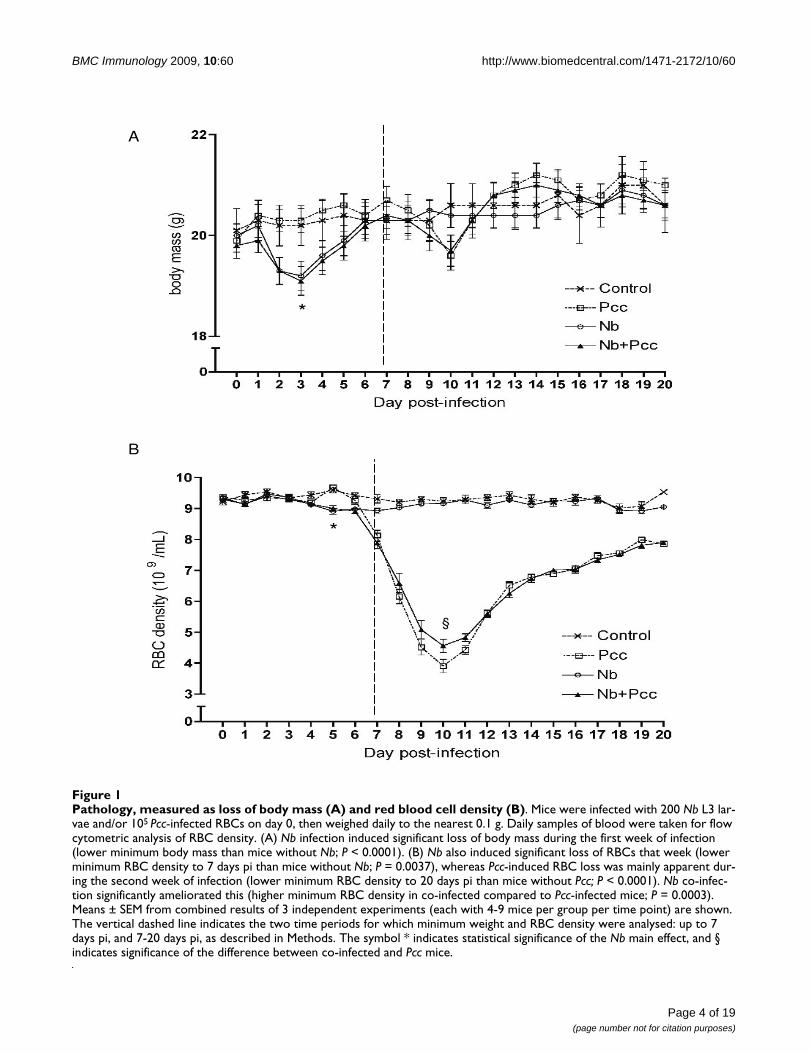
Consistent with previous reports in rats [41], we foundthat Nb infection induced loss of body mass in mice dur-ing the first week of infection: Nb-infected mice reached asignificantly lower minimum body mass than mice with-out Nb (Fig. 1A; F1,110 = 21.1; P < 0.0001). This amountedto a mean loss of 0.7 g, or ~3% of body mass, and wasobserved regardless of malaria co-infection. No furtherchanges in body mass among groups achieved statisticalsignificance, though Pcc-infected mice showed anexpected dip in weight around day 10 that was unalteredby Nb.
In addition to weight loss, Nb infection caused RBC den-sities to be reduced by ~5% (Fig. 1B; effect of Nb on min-imum RBC density to 7 days pi: F1,110 = 8.8; P = 0.0037).Unsurprisingly [33], Pcc also caused loss of RBCs by 7days pi (F1,110 = 40.2; P < 0.0001). Between days 7 and 20pi, Nb-induced RBC loss resolved, but Pcc induced furtherloss of RBCs - up to 60% of original density (Fig. 1B; F1,110= 385.4; P < 0.0001). However, this was slightly (~5%)but significantly ameliorated by Nb co-infection (Fig. 1B;t110 = 4.2; P = 0.0003).
Consistent with this slight protective effect of Nb on RBCloss during peak Pcc infection, Nb co-infection was associ-ated with a modest reduction in Pcc blood parasitaemia,as determined by microscopic examination of blood films(Fig. 2A; maximum parasitaemia F1,62 = 4.13; P = 0.0465).
By real-time PCR, the number of Pcc genome copies per75 mg lung homogenate sample was assessed in Pcc-infected versus co-infected mice. Malaria parasites werepresent in the lung of most animals examined at 3 days piand all animals at 7 days pi (Fig. 2B). At 3 days pi, whichis shortly after Nb parasites have migrated through thelung [7] and coincident with the loss of body mass in Nb-infected mice (Fig. 1A), co-infected mice had more Pccgenome copies per lung sample (Fig. 2B; P = 0.010). Thiscould be due to enhanced Pcc adherence to the lungendothelia [34] during Nb co-infection. However, by
gross examination we observed substantial haemorrhag-ing in all Nb-infected mice at this time point and thus it isalso possible that leakage of blood into the lung tissueincreased the number of Pcc parasites in day 3 samples. At7 days pi there was no significant difference between thePcc and the Nb+Pcc groups.
Gut nematode burden at 3 days pi varied between experi-ments: for example, Nb+Pcc and Nb mice bore 56+10 and35+9 adult nematodes, respectively, in experiment one,versus 6+4 and 1+1 nematodes in experiment two. Suchvariation has been previously reported [7] and in thepresent study is likely to be due to the very rapid infectionkinetics typically observed for our Nb strain, which is notmouse-adapted (e.g., no nematodes remain in the gut at 5days pi). A difference of a few hours in Nb injection timeson day 0 and/or in gut sampling times on day 3 couldtherefore lead to the differences in nematode burden thatwe observed. Indeed, two lines of evidence suggest thatthe number of Nb larvae moving through the lung wasmuch more consistent than the observed gut burdens.First, there were no significant differences among experi-ments in the amount of Nb-induced weight loss (P~0.2)nor RBC loss (P~0.4). Furthermore, Type 2 immunologi-cal readouts were extremely consistent among experi-ments. For example, TLN production of IL-4, IL-5, and IL-13 in response to Nb infection did not differ significantlyamong experiments (P~0.9, 0.8, and 0.9, respectively).Still, in order to be certain that experimental variationswere not confounding any of our conclusions, we control-led for experiment in all statistical analyses of combineddata (as described in Methods).
Nb-induced ChaFFs in the lung peaked around 5-7 days piBefore we undertook studies of Type 2 immune responsesin the lung during co-infection, we assessed the timecourse of Nb-induced pulmonary expression of ChaFFs.Previous work has mainly focused on ChaFF mRNAexpression in the lungs [10,11,16,17]. We wished to alsoascertain protein expression in situ after Nb infection, tomore closely determine the location of these proteins invivo. Female BALB/c mice were infected with 200 Nb-L3s,or injected with PBS as a control, and RELMα and Ym1protein levels were determined in BALF (via Westernblots) and lungs (via IHC) at days 3, 5, 7, 15, 20 and 26pi, to reflect the early events, peak Th2 time point and res-olution stages of Nb infection [9].
RELMα and Ym1 were both detected in BALF at 3 days pi,and rose to a peak around 5-7 days pi (Fig. 3A, B). Expres-sion of both proteins dropped off by days 20-26 pi. Histo-logical analysis of Ym1-stained lung sections frominfected mice illustrated this peak, with an increase in theintensity and area of anti-Ym1 staining at 5-7 days pi (Fig.3E, F). These representative micrographs also show the
Page 3 of 19(page number not for citation purposes)
BMC Immunology 2009, 10:60 http://www.biomedcentral.com/1471-2172/10/60
Page 4 of 19(page number not for citation purposes)
Pathology, measured as loss of body mass (A) and red blood cell density (B)Figure 1Pathology, measured as loss of body mass (A) and red blood cell density (B). Mice were infected with 200 Nb L3 lar-vae and/or 105 Pcc-infected RBCs on day 0, then weighed daily to the nearest 0.1 g. Daily samples of blood were taken for flow cytometric analysis of RBC density. (A) Nb infection induced significant loss of body mass during the first week of infection (lower minimum body mass than mice without Nb; P < 0.0001). (B) Nb also induced significant loss of RBCs that week (lower minimum RBC density to 7 days pi than mice without Nb; P = 0.0037), whereas Pcc-induced RBC loss was mainly apparent dur-ing the second week of infection (lower minimum RBC density to 20 days pi than mice without Pcc; P < 0.0001). Nb co-infec-tion significantly ameliorated this (higher minimum RBC density in co-infected compared to Pcc-infected mice; P = 0.0003). Means ± SEM from combined results of 3 independent experiments (each with 4-9 mice per group per time point) are shown. The vertical dashed line indicates the two time periods for which minimum weight and RBC density were analysed: up to 7 days pi, and 7-20 days pi, as described in Methods. The symbol * indicates statistical significance of the Nb main effect, and § indicates significance of the difference between co-infected and Pcc mice.
A
B
*
*
§

BMC Immunology 2009, 10:60 http://www.biomedcentral.com/1471-2172/10/60
influx of Ym1-positive inflammatory cells into the lungtissue that was evident by day 7 - e.g., alveolar and peri-bronchial inflammation. Of note, although infiltratingcells were RELMα+ (data not shown) and Ym1+, epithelialcells also appeared to be a major source of these mole-cules. Reece et al. (2006) clearly demonstrate macro-phages as sources of these proteins in the lung during Nbinfection but do not mention epithelial cells [11]. Ourdata are more consistent with a recent study in whichRELMα was localized primarily to epithelial cells in Nb-infected mice [29]. Further, several reports on inflamedallergic (asthma model) and fibrotic (bleomycin- or gam-maherpes virus-induced) rodent lungs, as well as our ownunpublished data, demonstrate expression of both Ym1and RELMα by epithelial cells [26,42,43]. By 15-26 dayspi (Fig. 3G, H, I), Ym1 protein expression had returned tonear-background (Fig. 3C), with reduced inflammatoryinflux and resolution of the thickened and disrupted epi-thelial layer that was apparent at earlier time points. Thesedata are supportive of the idea that pulmonary activationof AAMφ is a highly dynamic process [44].
Pcc changed the dynamics of expression of Nb-induced ChaFFs, especially 7 days piTo assess the effect of Pcc co-infection on the dynamics ofNb-induced pulmonary AAMφ and Type 2 epithelial cellactivation, we next analysed both mRNA and proteinexpression of two ChaFFs, RELMα and Ym1, at a series oftime points during co-infection. We also measured localmRNA expression of iNOS as a marker for Type 1 macro-phage activation [19], which might be expected duringmalaria [45]. Furthermore, we measured mRNA of Type 1cytokines (IL-12p40, TNF-α, and IFN-γ), as well as mRNAof IL-13, a key cytokine likely to drive production of Type2 effector molecules such as the ChaFFs [12-15]. We choseto examine the effect of malaria infection around the peakof larvae-induced damage (i.e., ~day 3 pi) [7] and the timeof transition to adaptive Type 2 responses (i.e., ~days 5-7pi) [9,11]. We also assessed a later time point well into theadaptive immune phase: 20 days pi.
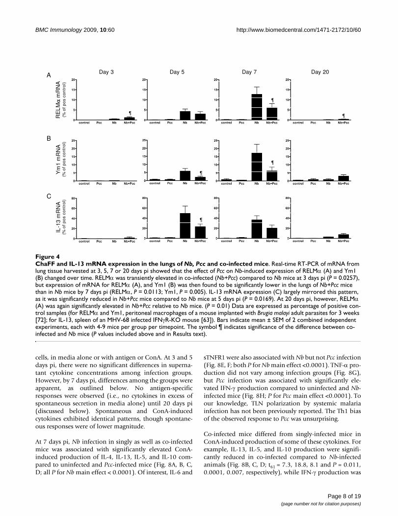
Analysis of ChaFF mRNA expression at 3, 5, 7 and 20 dayspi suggested that the strongest interactions between Nband Pcc-induced responses in lung tissue occurred 5-7days pi (Fig. 4A, B). Indeed, because days 5-7 pi repre-sented a time of strong ChaFF expression in Nb lung (Fig.3) and day 7 pi coincided with the presence of Pcc there(Fig. 2B), it is perhaps unsurprising that days 5-7 pi couldbe the time of maximum effect of Pcc co-infection. At 3days pi, there was a transient elevation of RELMα mRNAin co-infected compared to Nb-infected mice (P = 0.0257)that was reversed over the next few days: differencesbetween the groups were not significant at 5 days pi, butRELMα gene expression was significantly reduced in co-infected mice at 7 days pi (Fig. 4A; F1,25 = 7.5, P = 0.0113,
for combined analysis of experiments). Expression ofYm1 mRNA was also significantly lower in co-infectedthan Nb mice at days 5 and 7 pi (Fig. 4B: day 5 P = 0.0442;day 7 F1,25 = 9.5, P = 0.005, for combined analysis ofexperiments). In agreement with these observations, day 7expression of Arginase-1 mRNA was significantly lower inthe lungs of co-infected mice than in mice that had Nbonly (data not shown; F1,24 = 7.7, P = 0.0104). Further-more, ChaFF expression is known to be driven by engage-ment of IL-4Rα [24] by IL-4 and/or IL-13; accordingly, IL-13 mRNA expression in the lungs of co-infected mice wassignificantly reduced compared to Nb-infected mice at 5days pi (Fig. 4C: P = 0.0169). This suggests that suppres-sion of IL-13 by Pcc may be responsible for the reducedChaFFs in co-infected mice. At 20 days pi, mRNA expres-sion for RELMα (P = 0.0101) was slightly elevated in co-infected relative to Nb-only mice, but ChaFF and IL-13mRNA expression had otherwise largely returned to back-ground levels. Throughout this time course, Pcc-infectedand uninfected animals expressed little or no mRNA forthe ChaFFs.
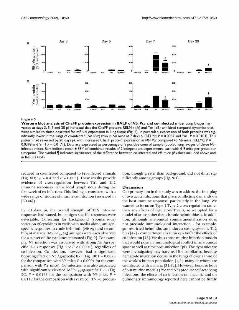
In support of the mRNA data, when we analysed proteinexpression in lung BALF using Western blotting, we saw asignificant day 7 pi reduction of both RELMα (Fig. 5A;F1,25 = 8.7, P = 0.0067, for analysis of combined experi-ments) and Ym1 (Fig. 5B; F1,25 = 7.7, P = 0.0104, for anal-ysis of combined experiments) in co-infected micecompared to Nb-infected mice. By day 20 pi, the patternhad reversed, with co-infected mice expressing moreRELMα (P = 0.0398) and Ym1 (P = 0.0171) than Nb mice.ChaFF expression in Pcc-infected mice did not differ fromnear-null expression in uninfected control mice (Fig. 5A,B). Importantly, BAL cell counts and cellular compositiondid not differ significantly among groups at any time-point. There was a non-significant elevation in total num-bers of cells in co-infected mice at 3 days pi and in Nbmice at 5 days pi, which might partly explain the elevationin ChaFFs at these early timepoints. However, any appar-ent differences were entirely absent from the 7 and 20 dayBAL samples. Therefore, the observed effects on ChaFFprotein expression are unlikely to be due to differences incellular makeup alone, particularly at 7 and 20 days pi.Further, the inflammatory cells may not be the majorsource of CHaFF proteins in lavage fluid. The pattern andintensity of epithelial cell staining (see Fig 6C) suggeststhat these cells may be largely responsible for the changesin mRNA and BALF protein.
IHC scoring of lung sections further confirmed thesedynamics. At both day 5 pi and day 7 pi, the intensity ofRELMα (Fig 6A) and Ym1 (Fig 6B) staining in the lungs ofco-infected animals was reduced compared to Nb mice,though the RELMα difference did not achieve statisticalsignificance at day 7 (day 5: RELMα P = 0.0056; Ym1 P =
Page 5 of 19(page number not for citation purposes)

BMC Immunology 2009, 10:60 http://www.biomedcentral.com/1471-2172/10/60
Page 6 of 19(page number not for citation purposes)
Malaria parasites in the blood (A) and lungs (B) of Pcc-infected and co-infected miceFigure 2Malaria parasites in the blood (A) and lungs (B) of Pcc-infected and co-infected mice. (A) Co-infected mice (Nb+Pcc) had a lower peak proportion of parasitized RBCs than Pcc-infected mice did (P = 0.0465), as determined by 1000× microscopic examination of Giemsa-stained thin blood films. Means ± SEM from combined results of 3 independent experi-ments (each with 4-9 mice per group per time point) are shown. (B) Co-infected mice harboured greater numbers of Pcc genome copies per lung sample than Pcc mice at 3 days pi (P = 0.01), as assessed by real-time PCR of lung homogenates (derived from 75 mg lung) using MSP-1 specific primers. Genome copies were apparent in both Pcc infected groups at 7 days pi and the copy numbers no longer differed significantly between groups. This experiment included 4-7 mice per group. The sym-bol § indicates significance of the difference between co-infected and Pcc mice.
§
§
A
B

BMC Immunology 2009, 10:60 http://www.biomedcentral.com/1471-2172/10/60
0.0153; day 7: RELMα P = 0.1011; Ym1 P = 0.0171). Rep-resentative micrographs from day 7 pi show the reducedintensity of RELMα staining in co-infected animals rela-tive to Nb mice and no RELMα staining in Pcc mice (Fig.6C). No differences among groups were detected by IHCat days 3 or 20 pi. Therefore, the expression pattern ofChaFF protein in situ was largely in agreement with mRNAin whole lung tissue (Fig. 4) and protein expression in theBALF (Fig. 5). As discussed above for the time course of Nbinfection (Fig. 3), although Ym1+ and RELMα+ macro-phages were present in lung IHC sections, the predomi-nant cell type expressing these molecules appeared to beepithelial cells. Furthermore, lung macrophages in this
system do not appear to become classically activated, asassessed by lung mRNA for iNOS and IL-12p40, whichwere not detectable in any mice at any time point, andTNF-α, which was detectable but at low levels that did notdiffer among groups (data not shown). However, IFNγwas elevated in the lung tissue of all Pcc infected mice,regardless of Nb co-infection, at day 7 pi (Fig. 7).
Cytokine production in local LN largely mirrored pulmonary ChaFF expressionTo assess whether pulmonary ChaFF patterns corre-sponded to immune responses in the draining thoraciclymph node (TLN), we performed in vitro culture of TLN
Time course of protein expression of ChaFFs in Nb infected lungsFigure 3Time course of protein expression of ChaFFs in Nb infected lungs. Western blot analysis of lung lavages harvested at different time points post Nb infection (4 Nb mice per timepoint) suggested that the secretion of ChaFFs RELMα (A) and Ym1 (B) peaked around 5-7 days post infection. Data are expressed as percentage of a positive control sample (pooled lung lavages of three Nb-infected mice). Representative photomicrographs from Nb-infected lung tissue sections stained histologically for Ym1 at the indicated time points, plus an uninfected control lung (C), illustrate peak expression of brown Ym1 staining of epi-thelial cells and macrophages (E-F).
G
FED
C
IH
A B
Control
Day 3 Day 5 Day 7
% o
f p
os
con
tro
l
% o
f p
os
con
tro
l
Alveolar macrophages Bronchial epithelium
Day 15 Day 20 Day 26
RELM� Ym1
Page 7 of 19(page number not for citation purposes)
BMC Immunology 2009, 10:60 http://www.biomedcentral.com/1471-2172/10/60
cells, in media alone or with antigen or ConA. At 3 and 5days pi, there were no significant differences in superna-tant cytokine concentrations among infection groups.However, by 7 days pi, differences among the groups wereapparent, as outlined below. No antigen-specificresponses were observed (i.e., no cytokines in excess ofspontaneous secretion in media alone) until 20 days pi(discussed below). Spontaneous and ConA-inducedcytokines exhibited identical patterns, though spontane-ous responses were of lower magnitude.
At 7 days pi, Nb infection in singly as well as co-infectedmice was associated with significantly elevated ConA-induced production of IL-4, IL-13, IL-5, and IL-10 com-pared to uninfected and Pcc-infected mice (Fig. 8A, B, C,D; all P for Nb main effect < 0.0001). Of interest, IL-6 and
sTNFR1 were also associated with Nb but not Pcc infection(Fig. 8E, F; both P for Nb main effect <0.0001). TNF-α pro-duction did not vary among infection groups (Fig. 8G),but Pcc infection was associated with significantly ele-vated IFN-γ production compared to uninfected and Nb-infected mice (Fig. 8H; P for Pcc main effect <0.0001). Toour knowledge, TLN polarization by systemic malariainfection has not been previously reported. The Th1 biasof the observed response to Pcc was unsurprising.
Co-infected mice differed from singly-infected mice inConA-induced production of some of these cytokines. Forexample, IL-13, IL-5, and IL-10 production were signifi-cantly reduced in co-infected compared to Nb-infectedanimals (Fig. 8B, C, D; t61 = 7.3, 18.8, 8.1 and P = 0.011,0.0001, 0.007, respectively), while IFN-γ production was
ChaFF and IL-13 mRNA expression in the lungs of Nb, Pcc and co-infected miceFigure 4ChaFF and IL-13 mRNA expression in the lungs of Nb, Pcc and co-infected mice. Real-time RT-PCR of mRNA from lung tissue harvested at 3, 5, 7 or 20 days pi showed that the effect of Pcc on Nb-induced expression of RELMα (A) and Ym1 (B) changed over time. RELMα was transiently elevated in co-infected (Nb+Pcc) compared to Nb mice at 3 days pi (P = 0.0257), but expression of mRNA for RELMα (A), and Ym1 (B) was then found to be significantly lower in the lungs of Nb+Pcc mice than in Nb mice by 7 days pi (RELMα, P = 0.0113; Ym1, P = 0.005). IL-13 mRNA expression (C) largely mirrored this pattern, as it was significantly reduced in Nb+Pcc mice compared to Nb mice at 5 days pi (P = 0.0169). At 20 days pi, however, RELMα (A) was again significantly elevated in Nb+Pcc relative to Nb mice. (P = 0.01) Data are expressed as percentage of positive con-trol samples (for RELMα and Ym1, peritoneal macrophages of a mouse implanted with Brugia malayi adult parasites for 3 weeks [72]; for IL-13, spleen of an MHV-68 infected IFNγR-KO mouse [63]). Bars indicate mean ± SEM of 2 combined independent experiments, each with 4-9 mice per group per timepoint. The symbol ¶ indicates significance of the difference between co-infected and Nb mice (P values included above and in Results text).
A
RE
LM
mR
NA
(% o
f p
os
con
tro
l)Y
m1
mR
NA
(% o
f p
os
con
tro
l)
B
Day 3 Day 5 Day 7 Day 20
IL-1
3 m
RN
A(%
of
po
s co
ntr
ol)
C
¶
¶
¶
¶
¶
¶
Page 8 of 19(page number not for citation purposes)
BMC Immunology 2009, 10:60 http://www.biomedcentral.com/1471-2172/10/60
reduced in co-infected compared to Pcc-infected animals(Fig. 8H; t61 = 8.4 and P = 0.006). These results provideevidence of cross-regulation between Th1 and Th2immune responses in the local lymph node during thefirst week of co-infection. This finding is consistent with awide range of studies of murine co-infection (reviewed in[30,46]).
By 20 days pi, the overall strength of TLN cytokineresponses had waned, but antigen-specific responses weredetectable. Correcting for background (spontaneous)secretion of cytokines in wells with media alone, antigen-specific responses to crude helminth (Nb Ag) and recom-binant malaria (MSP-119 Ag) antigens were each observedfor a subset of the cytokines measured (Fig. 9). For exam-ple, Nb infection was associated with strong Nb Ag-spe-cific IL-13 responses (Fig. 9A: P < 0.0001), regardless ofco-infection. Co-infection, however, had a significantboosting effect on Nb Ag-specific IL-5 (Fig. 9B: P = 0.0025for the comparison with Nb mice; P < 0.0001 for the com-parison with Pcc mice). Co-infection was also associatedwith significantly elevated MSP-119Ag-specific IL-6 (Fig.9C: P = 0.0163 for the comparison with Nb mice; P =0.0112 for the comparison with Pcc mice). TNF-α produc-
tion, though greater than background, did not differ sig-nificantly among groups (Fig. 9D).
DiscussionOur primary aim in this study was to address the interplayof two acute infections that place conflicting demands onthe host immune response, particularly in the lung. Wewanted to focus on Type 1-Type 2 cross-regulation ratherthan any effects of regulatory T cells, so we opted for amodel of acute rather than chronic helminthiasis. In addi-tion, although anatomical compartmentalization doesnot preclude immunological interaction - for example,gut-restricted helminths can induce a strong systemic Th2bias [47] - compartmentalization can buffer the effects ofco-infection [48]. We thus chose murine infection modelsthat would pose an immunological conflict in anatomicalspace as well as time post-infection (pi). The dynamics wewere investigating may have real life corollaries, becausenematode migration occurs in the lungs of over a third ofthe world's human population [1,2], many of whom areco-infected with malaria [31,32]. However, because bothof our murine models (Pcc and Nb) produce self-resolvinginfections, the effects of co-infection on anaemia and onpulmonary immunology reported here cannot be firmly
Western blot analysis of ChaFF protein expression in BALF of Nb, Pcc and co-infected miceFigure 5Western blot analysis of ChaFF protein expression in BALF of Nb, Pcc and co-infected mice. Lung lavages har-vested at days 3, 5, 7 and 20 pi indicated that the ChaFF proteins RELMα (A) and Ym1 (B) exhibited temporal dynamics that were similar to those observed for mRNA expression in lung tissue (Fig. 4). In particular, expression of both proteins was sig-nificantly lower in the lungs of co-infected (Nb+Pcc) than in Nb mice at 7 days pi (RELMα P = 0.0067 and Ym1 P = 0.0104). This pattern had reversed by 20 days pi, with increased ChaFF protein expression in Nb+Pcc compared to Nb mice (RELMα P = 0.0398 and Ym1 P = 0.0171). Data are expressed as percentage of a positive control sample (pooled lung lavages of three Nb-infected mice). Bars indicate mean ± SEM of combined results of 2 independent experiments, each with 4-9 mice per group per timepoint. The symbol ¶ indicates significance of the difference between co-infected and Nb mice (P values included above and in Results text).
A
RE
LM
pro
tein
(% o
f p
os
con
tro
l)Y
m1
pro
tein
(% o
f p
os
con
tro
l)
B
Day 3 Day 5 Day 7 Day 20
¶
¶
¶
¶
Page 9 of 19(page number not for citation purposes)

BMC Immunology 2009, 10:60 http://www.biomedcentral.com/1471-2172/10/60
Page 10 of 19(page number not for citation purposes)
Immunohistochemical analysis of ChaFF protein expression in the lungs of Nb, Pcc and co-infected miceFigure 6Immunohistochemical analysis of ChaFF protein expression in the lungs of Nb, Pcc and co-infected mice. Pro-tein expression in lung sections corroborated BALF concentrations of ChaFF proteins observed around 7 days pi, as illustrated by days 5 and 7 pi staining intensity scores for RELMα (A) and Ym1 (B) as well as representative micrographs (at increasing magnification) of day 7 pi slides stained for RELMα (C). Down-regulation of ChaFFs was observed in co-infected mice (Nb+Pcc) compared to Nb mice; differences were significant for both RELMα and Ym1 at 5 days pi (RELMα P = 0.0056 and Ym1 P = 0.0153) and for Ym1 at 7 days pi (P = 0.0171). Scores were obtained by analyzing bronchial epithelium in ten fields per mouse using a scoring system described in Methods. The whisker-and-box graphs with minimum and maximum values depict results from a representative experiment with 4-9 mice per group. The symbol ¶ indicates significance of the difference between co-infected and Nb mice (P values included above and in Results text).
Pcc x200 Pcc x400Pcc x100
Nb x200 Nb x400Nb x100
Nb+Pcc x200Nb+Pcc x100
A
B
C
Nb+Pcc x400
Ym
1 sc
ore
R
EL
M s
core
Day 5 Day 7
¶
¶
Day 5 Day 7
¶

BMC Immunology 2009, 10:60 http://www.biomedcentral.com/1471-2172/10/60
associated with chronic disease outcomes until longerterm co-infection studies are performed.
We quantified the health of mice in our experiments usingtwo measures that have proven informative during Pccinfection [49] and Pcc-nematode co-infection [50]: bodymass and RBC density. To our knowledge, this is the firstreport demonstrating that murine Nb infection has a neg-ative impact on both parameters, although reducedweight gain in young Nb-infected mice has previouslybeen reported [40]. Nb caused a statistically significant,transient ~3% loss of body mass from approximately 2-4days pi, and in other experiments using a higher dose(500 L3s), mice lost closer to 10% of their starting bodymass (unpublished data). Migration of Nb larvae throughthe lungs has previously been shown to cause two spellsof inappetance and thus weight loss in rats, one associatedwith migration of larvae and the other with establishmentof adults in the gut [41]. We detected only one period ofloss of body mass; mice may be spared the second spellgiven the brief survival of adult Nb in mice, particularly forparasite strains, such as ours, that are not mouse-adapted.We also observed a transient loss of RBC density in Nb-infected mice. It was rather surprising that this effect -most likely caused by haemorrhaging of the lung follow-
ing larval migration - was detectable at the systemic level.This suggests that the capillary damage and ingestion ofRBCs by alveolar macrophages following lung migrationof Nb [11,16] are associated with considerable blood loss.
A diverse range of outcomes is possible when helminthsand malaria co-infect a host. Co-infected mice in ourstudy experienced two periods of RBC loss in quick succes-sion - first Nb-induced and then Pcc-induced. However,they had slightly higher RBC densities than Pcc-infectedmice did, at the time of most severe malarial disease. Thiswas associated with a small reduction in malaria parasi-taemia in the blood. These results contrast with severalstudies of helminth-malaria co-infection in mice, inwhich malaria parasitemia was increased [51-54], and/ormalarial symptoms exacerbated, in at least some groups ofco-infected mice [50-55]. For example, in contrast to thelethal inflammatory liver disease recently described inmice simultaneously co-infected with Heligmosomoidespolygyrus and Pcc [55], we observed subtle amelioration ofmalarial disease and no deaths. This disparity in the sever-ity of co-infection could be due to the fact that we workedwith a different mouse strain (BALB/c versus Helmby'sC57BL/6) as well as a different helminth species thatmigrates differently through the host body. However, wedetected an elevation in MSP-119-specific IL-6 due to co-infection, so it is possible that the emergent IL-17/IL-23axis described by Helmby [55] may likewise be involvedin our co-infection system, though not in organs that neg-atively impact short-term survival. Indeed, the mecha-nisms underlying the slightly protective effect of Nbobserved here are not yet clear. We are investigating possi-ble immunological causes of this protection, includinginnate mechanisms such as IFN-γ+ NK cells [56] and adap-tive mechanisms such as cytophilic antibody isotypes [33]that could promote malaria clearance; either might bealtered by acute Nb co-infection. However, it is also possi-ble that lower parasitaemia might be the consequence ofthe RBC density changes induced by Nb, as previous co-infection studies have shown that helminths can limitRBC availability to malarial parasites and thereby captheir replication (e.g., [57]). Control of microparasites byTh1 immunity and by RBC limitation are not mutually-exclusive possibilities [58] and both might be operating inour model system. Finally, it is possible that the sequestra-tion habits of Pcc parasites [59] are altered by Nb. Thesemechanisms remain to be investigated.
Our results largely resemble those reported for other Nb-microparasite pairings. For example, during co-infectionof mice with Nb and either Toxoplasma gondii [60] orChlamydophila abortus [61], significantly reduced Th2responses (compared to mice with Nb infection) havebeen observed, independent of the interval between infec-tions [60,61]. These data suggest that Pcc is not the only
Lung mRNA for the Type 1 cytokine IFN-γ at 7 days piFigure 7Lung mRNA for the Type 1 cytokine IFN-γ at 7 days pi. Real-time RT-PCR of mRNA from lung tissue harvested at 3, 5, 7 or 20 days pi using primers for Type 1 markers iNOS, IL-12p40, TNF-α, and IFN-γ were largely negative. For example, no iNOS nor IL-12p40 was detected in any mice, and although TNF-α was detected at low levels, its expres-sion did not differ among groups of mice. At 7 days pi, how-ever, IFN-γ was significantly upregulated in all mice with Pcc infection, with or without Nb co-infection, as depicted here. Data are expressed as percentage of a positive control sam-ple (peritoneal macrophages of a thioglycolate-injected mouse [10]). Mean ± SEM from a representative experiment with 4-9 mice per group per timepoint are shown.
(% o
f pos
con
trol
)
Page 11 of 19(page number not for citation purposes)

BMC Immunology 2009, 10:60 http://www.biomedcentral.com/1471-2172/10/60
Page 12 of 19(page number not for citation purposes)
ConA-induced cytokine release in TLN lymphocyte recall assaysFigure 8ConA-induced cytokine release in TLN lymphocyte recall assays. Cytokine concentrations in supernatants of ConA-stimulated TLN cells harvested at day 7 pi were measured with cytometric bead arrays. Groups did not differ in cytokine pro-duction at 3 or 5 days pi, and no antigen-specific responses were detected until 20 days pi. Data on day 7 pi IL-4 (A), IL-13 (B), IL-5 (C), IL-10 (D), IL-6 (E), sTNFR1 (F), TNF-α (G), and IFN-γ (H) production are shown. In these data, statistically significant down-regulatory effects of co-infection (Nb+Pcc) were found for IL-13 (P = 0.011), IL-5 (P = 0.0001), IL-10 (P = 0.007), and IFN-γ (P = 0.006), compared to relevant single-infection groups. Bars indicate mean ± SEM from 3 combined experiments, each with 4-9 mice per group. The symbol ¶ indicates significance of the difference between co-infected and Nb mice, while § indicates significance of the difference between co-infected and Pcc mice (P values included above and in Results text).
A
B
C
D
E
F
G
H
¶
¶
¶ §
ConA ConA
ConA ConA
ConA ConA
ConAConA

BMC Immunology 2009, 10:60 http://www.biomedcentral.com/1471-2172/10/60
Page 13 of 19(page number not for citation purposes)
Antigen-specific responses in TLN lymphocyte recall assaysFigure 9Antigen-specific responses in TLN lymphocyte recall assays. Cytokine concentrations in supernatants of TLN cells that were harvested at day 20 pi and then stimulated with antigen were measured with cytometric bead arrays. Nb Ag, a crude Nb adult lysate, and recombinant Pcc clone AS merozoite surface protein, MSP-119, were used as antigens in vitro. Although all cytokines shown in Figure 8 were measured, only IL-13 in response to Nb Ag (A), IL-5 in response to Nb Ag (B), IL-6 in response to MSP-119 (C), and TNF-α in response to MSP-119 (D) were above levels of spontaneous secretion. These figures thus depict antigen-specific cytokine (in ng/ml above background). Statistically significant differences among groups included increased NbAg-specific IL-13 in all Nb mice (P < 0.0001), increased Nb Ag-specific IL-5 in co-infected (Nb+Pcc) mice compared to both Nb (P = 0.0025) and Pcc mice (P < 0.0001), and increased MSP-119-specific IL-6 in Nb+Pcc compared to both Nb (P = 0.0163) and Pcc mice (P = 0.0112). Bars indicate mean ± SEM for 4-8 mice per group. The symbol ¶ indicates significance of the difference between co-infected and Nb mice, while § indicates significance of the difference between co-infected and Pcc mice (P values included above and in Results text).
control Pcc Nb Nb+Pcc0.000
0.025
0.050
0.075
control Pcc Nb Nb+Pcc0.00
0.05
0.10
0.15
0.20
control Pcc Nb Nb+Pcc0.00
0.05
0.10
0.15
0.20A
B
C
D
§ ¶
§ ¶
control Pcc Nb Nb+Pcc0.00
0.05
0.10
0.15
Nb Ag
Nb Ag
MSP-119 Ag
MSP-119 Ag
BMC Immunology 2009, 10:60 http://www.biomedcentral.com/1471-2172/10/60
microparasite that might reduce Th2 responses to Nbinfection. Mycobacterium bovis BCG co-infection, however,does not significantly impact Nb-induced IL-4 in themesenteric lymph nodes [62]; it would be of interest toknow whether those lung-dwelling microparasites mighthave had similar effects to Pcc on Th2 responses, had theybeen measured in the TLN. Reported effects of Nb on thecourse of microparasite infections are likewise mixed:densities of T. gondii [60] and M. bovis BCG [62] are unaf-fected by the presence of the nematode, while C. abortusdensity increases dramatically [61]. Interestingly, the con-current presence of influenza virus with migrating Nb lar-vae in the lung exacerbates the severity of lung diseasecompared to mice with influenza alone [40]. A two-weekdelay between Nb infection and influenza infection, orreplacement of Nb with H. polygyrus, eliminates the addedpathology, suggesting that the simultaneous presence oflarvae and virus in the lungs is required [40]. Such mayalso be the case for Pcc-Nb co-infection. The Nb-influenzastudy did not include immunological measurements, sothe role of the immune system in generating the observedpattern is not known. Indeed, this comparison illustratesthat many details of anatomical location and parasite lifecycles, as well as immunological interactions, must betaken into account to explain the diverse outcomes ofhelminth-microparasite co-infections [30,46].
Our most novel finding is that malaria infection has thecapacity to modulate the host's pulmonary Type 2response to nematode migration. However, the long-termimpact of the altered Type 2 response is not possible topredict, because the function of Type 2 immunity in thissetting is not yet fully understood. There are at least threepotential outcomes of a helminth-induced Type 2response in the lung. First, it may contribute to protectionagainst incoming larvae [6]. Second, Type 2 responses arelikely to be involved in repairing the damage that isinflicted by migrating parasites. Third, as recent studieshave shown [5,16,18], lung migration and the associatedTh2 responses have the potential to cause long-term lungpathology. Appropriate repair versus lung malfunction arelikely to be flip sides of the same coin. Indeed, althoughChaFFs and Arginase-1 are implicated in tissue repair,they are also associated with fibrosis, an overzealousrepair process [19,24,43,63-65] (see also review by Wynn[66]). Predicting the effect of Pcc co-infection on longterm Nb-induced lung disease is further complicated byrecent data that suggest both Arginase-1 and RELMα cannegatively regulate Th2-mediated pathology [27-29]. Bythis logic, inhibition of these molecules by malaria co-infection may ultimately exacerbate Th2-mediated lungdamage.
However, our data suggest that the effect of malaria onChaFF expression is not direct but rather via reduced Th2
cytokines. The effect of Pcc on Nb-induced ChaFFs was notapparent until 7 days pi, when the extent of the increasein ChaFF expression was inhibited by co-infection. Thiswas correlated with differences in cytokine production inlymphocyte recall assays, suggesting that changes inChaFF expression were driven by changes in the T lym-phocyte populations after the onset of the adaptive Th2immune response (around 5 days pi, as shown in Nb-infected IL-4 reporter mice [9]). A role for adaptive immu-nity is further supported by work showing that SCID miceare not able to sustain AAMφ responses in the lung follow-ing Nb infection [11], and a demonstrated requirementfor T cells to sustain the AAMφ response in a mouse peri-toneal infection model [24]. Remarkably, in SCID mice,in the absence of T cells and AAMφ, the Nb-induced cellu-lar infiltrate does not resolve [11]. The capacity of malariato inhibit the transition to a full Th2 response by 7 days pimay likewise be detrimental to full resolution of theinflammatory response, a step necessary for appropriatetissue repair [67,68]. By day 20 pi, however, the residualTh2 responses in co-infected mice were as high as, or evenhigher than, in Nb-only mice. In support of this, day 20antigen-specific IL-5 responses were particularly high inco-infected animals. Thus Pcc infection may protectagainst airway hyper-responsiveness through a reductionin peak Th2 activation, or else exacerbate it due to sus-tained Th2 activity. Transient passage of Nb larvaethrough the lung inflicts lasting damage [16,18]. Whethertransient impairment of pulmonary Th2 responses bymalaria co-infection also has lasting effects needs to beinvestigated experimentally.
A perhaps surprising finding in our study was the appar-ent absence of classical macrophage activation in the lungdespite the clear presence of malaria parasites: we did notdetect iNOS, IL-12p40 nor elevated TNF-α mRNA in lungtissue of Pcc-only or co-infected mice at any time point.One could argue that malaria parasites stay in the lungmicrovasculature and do not cross into tissue. However,this is unlikely to be the case, given the extensive lungdamage due to Nb in co-infected mice, as well as evidencethat malaria merozoites can be found dispersed in thelung [34]. Furthermore, IFN-γ mRNA was detectable in thelung of all Pcc mice regardless of co-infection, suggestingthat lymphocytes were activated, perhaps by innate activa-tion of NK or γδ T cells. The most likely explanation forthe failure to detect classical macrophage activation maybe that lung macrophages, which are exposed daily toinhaled microbes, have a remarkably high threshold foractivation even in the presence of IFN-γ and microbialstimuli [69].
As with any laboratory model, it is important to acknowl-edge the potential disconnection between natural co-infections and the experimental systems and designs used
Page 14 of 19(page number not for citation purposes)

BMC Immunology 2009, 10:60 http://www.biomedcentral.com/1471-2172/10/60
here, including the relative timing of the two infections,doses at which they were administered, and the fact thatwe have only studied primary and self-resolving (ratherthan secondary and/or chronic) infections. Permutationof any of these parameters is likely to quantitatively, if notqualitatively, alter outcomes. For example, repair proc-esses might readily keep pace with lung damage when therate of exposure to nematode larvae is low, unlike in mostexperimental models. We used a relatively low dose of L3larvae (200 per mouse while others use ~500 [16,18] or asmany as 750-1000 [60-62]) but still exceeded naturalexposure levels. Furthermore, larval helminths andmalaria parasites are unlikely to arrive in the lung withina few days of each other in nature, and it may be that pre-existing malaria would have had a different effect on pul-monary Type 2 responses to Nb migration, particularly ifmalaria parasites do not remain long in the lung. Indeed,the most likely natural exposure scenario may be chronicmalaria infection into which helminth larvae are "trick-led" [32], but experimental studies that mimic this sce-nario have yet to be carried out. Nonetheless, lungdysfunction is seen as a consequence of helminth migra-tion [4,5] and both acute and persistent malaria infection[38] in people, so high-dose experimental Nb studies inwhich long-term lung pathology can be observed [16,18],combined with simultaneous malaria exposure, may pro-vide useful models for disease states in people.
ConclusionWith the experiments reported here, we have establishedan acute laboratory model of helminth-malaria co-infec-tion that will be suitable for future work exploring thedetails of how Type 1 inducing co-infections affect long-term, Type 2-mediated repair of the damage caused bymigrating nematodes. Recently developed models ofmalaria-induced lung damage (e.g., [37]) might be ana-lysed in animals co-infected with Nb. Corroborative stud-ies in human populations may also be feasible. Likemigratory helminthiases [3], severe falciparum malaria isassociated with detectable lung injury, as measured byspirometry and clinical symptoms [38]. A study likeBrooker et al's analysis of whether the anaemia of hook-worm and malaria are additive during co-infection [32]that used spirometry to assess the pulmonary health ofmalaria-infected, A. lumbricoides- or hookworm-infected,and co-infected people could assess whether co-infectionexacerbates damage. Given the huge number of peoplewith such co-infections, it is possible that clinical studiesof malaria lung injury may gain insight from consideringthe presence, however transient, of helminths in the lung.
MethodsMice, parasites, experimental design, and monitoringSpecific pathogen free, 8-10 week old female BALB/c mice(Harlan, UK) were maintained in individually ventilated
cages on diet 41b ad lib. Nippostrongylus brasiliensis (Nb)was maintained by serial passage through Sprague-Daw-ley rats, as described previously [70]. Cryopreserved Plas-modium chabaudi chabaudi (Pcc) parasites of clone AS werepassaged through two generations of donor BALB/c miceand inoculated into experimental mice as described previ-ously [50]. The four co-infection experiments used a fac-torial design, with uninfected controls, Pcc-infected, Nb-infected, and co-infected mice. On day 0, 200 Nb L3 larvaeand/or 105 Pcc-infected RBCs were injected subcutane-ously and intraperitoneally, respectively. PBS and naïvemouse RBCs served as sham injections for Nb and Pcc,respectively; uninfected control animals received bothsham injections. RBC density, body mass, and malariaparasites were then monitored daily, as described previ-ously [50]. Briefly, RBC densities were measured by flowcytometry (Beckman Coulter), body mass was recorded tothe nearest 0.1 g, and the proportion of RBCs parasitizedwas counted in Giemsa-stained thin blood films (at1000× magnification). Mice were then culled 3, 5, 7, or 20days pi (4-9 mice per infection type per timepoint). Nbparasite burden was assessed in the gastrointestinal tractof culled mice. Intestines were placed in PBS, slit length-wise and the contents rinsed into muslin-lined funnels setover tubes containing PBS warmed to 37°C. Nematodeswere left to filter through for >2 hours and counted viamicroscopy (at 40×). To elucidate the dynamics of Nb-induced alternative activation in the lung, a separateexperiment was conducted for Nb only, with mice culled3, 5, 7, 15, 20 or 26 days pi (4 Nb-infected mice per timepoint). All experiments were carried out in accordancewith the animals (Scientific Procedures) Act 1986, andwere approved by the UK Home Office inspectorate andinstitutional review committee.
Lung lavage and tissue samplingFollowing terminal anaesthesia, tracheas were cannulatedand lungs lavaged with 1 mL PBS. Cannulae were pre-pared from fine bore polythene tubing (Portex) and a 23G needle. Following lavage, the left lung lobe was tied off,cut at the bronchus, and placed in RNAlater (Ambion) formRNA extraction, while the right lobe was perfused in 4%formaldehyde and embedded in paraffin for immunohis-tochemistry. BAL cell concentrations were determinedusing a Scharf Instruments Casy Counter. BALF was centri-fuged at 1,200 g for 5 mins and stored at -20°C for proteinanalysis by Western blot.
ImmunohistochemistryExpression of RELMα and Ym1 in lung sections wasassessed by indirect immunoperoxidase techniques.Briefly, the paraffin embedded tissue sections were depar-affinised and rehydrated. After high temperature antigenunmasking (Vector Laboratories, UK), endogenous perox-idase was quenched with aqueous 2% H2O2 (Sigma
Page 15 of 19(page number not for citation purposes)

BMC Immunology 2009, 10:60 http://www.biomedcentral.com/1471-2172/10/60
Aldrich, UK) for 15 minutes. Slides were then incubated 2h with primary antibodies: rabbit anti-RELMα (0.25 μg/mL; Peprotech) or rabbit anti-Ym1 (1/100; StemCellTechnologies) in antibody diluent (Dako Cytomation,Denmark) at RT, followed by the secondary antibody(goat anti-rabbit biotin, 1 mg/mL, Dako Cytomation,Denmark). Peroxidase-labelled ABC reagent and DABsubstrate (Vector Laboratories, UK) were used for signalvisualisation. Finally, the sections were counterstainedwith haematoxylin. RELMα and Ym1 staining intensitieswere scored by two researchers, blinded to experimentalgroupings, using a modification of a previously-publishedlung inflammation scoring system [71]. For each mouse,staining was assessed at 200× magnification for 10 fields.Each field included correctly inflated lung tissue and acomplete transection of at least one bronchiole, bloodvessel and alveolar airway. Cytoplasmic staining strengthwas scored in bronchial epithelial cells, infiltrating cellsand alveolar macrophages on a scale of 1-4 (1 = no stain-ing, 2 = weak, 3 = moderate, and 4 = strong staining, usinga reference section of the same positive control sample(lung of an Nb-infected mouse at 7 days pi). The percent-age of positive cells in each of these compartments wasalso scored on a scale of 1-4 (1 = none, 2<30%, 3 = 30-60%, 4>60% positive cells). Average cytoplasmic and cellpositivity scores across the 10 fields were calculated.Finally, the overall staining score for each mouse was cal-culated by multiplying the average stain strength by aver-age % positive cells. Control sections incubated withantibody diluent followed by secondary antibody only, orwith normal rabbit serum alone, did not show any stain-ing. Mouse lung pathology experts confirmed that stainedcell types were correctly identified. Photomicrographs ofrepresentative sections were captured on a Zeiss Axioskopmicroscope with QCapture Pro Software.
RNA isolation and real-time RT-PCRRNA isolation from lung tissue was carried out using TRI-zol (Invitrogen). After DNase treatment (10 U/mLDNase1, Ambion), cDNA was synthesised using Moloneymurine leukaemia virus reverse transcriptase (Stratagene).For quantification of Ym1, RELMα, Arginase 1, iNOS, IL-12p40, TNF-α, IFN-γ and IL-13 mRNA, real-time RT-PCRwas performed using a LightCycler (Roche Diagnostics)and primers reported previously [63]. For each gene, fiveserial 1:4 dilutions of cDNA of a positive control sample(for RELMα, Ym1 and Arg1, peritoneal macrophages of amouse implanted with Brugia malayi adult parasites for 3weeks [72]; for IL-13, spleen of an MHV-68 infectedIFNγR-KO mouse [63]; or for iNOS and other Type 1markers, peritoneal macrophages of a thioglycolate-injected mouse [10]) were used in each reaction. Amplifi-cation was quantified and normalised using β-actin as ahousekeeping gene. PCR reactions were carried out in 10μl buffer containing 1 μl cDNA, 4 mM MgCl2, 0.3 μM of
each primer and the LightCycler-DNA SYBR Green I mix,under the following conditions: 30 s denaturation at95°C, 5 s annealing of primers at 55°C or 63°C (Ym1),and 12 s elongation at 72°C, for 50 cycles. SYBR Greenfluorescence was monitored after each cycle at 86°C(85°C for Ym1).
Real-time PCR to detect malaria in lung tissueReal-time PCR for Pcc genomic DNA was carried out onDNA extracted with phenol/chloroform from homoge-nized lung tissue (stored in Trizol, Invitrogen; 75 mg tis-sue/mL). PCR was performed on an ABI Prism 7000(Applied Biosystems), with primers for merozoite surfaceprotein (MSP)-1 of clone AS, as described previously [73].
Western blot for Ym1 and RELM20 μl BALF was mixed with sample buffer supplementedwith denaturing buffer (NuPage, Invitrogen), heat dena-tured and resolved by SDS-PAGE using 4-12% gradientBis-Tris gels (NuPage, Invitrogen) followed by transferonto nitrocellulose membrane (Bio-Rad). Transfer andloading intensity were assessed with Ponceau Red staining(Sigma). After blocking with 0.05% Tween 20 in StartingBlock (Pierce), membranes were incubated overnight at4°C with polyclonal rabbit anti-Ym1 [10] (0.12 ng/mL)or rabbit anti-RELMα (0.2 μg/mL; Peprotech). After incu-bation with HRP-conjugated goat anti-rabbit IgG (heavyplus light chains; Bio-Rad; 1/2000), signal was detectedwith chemiluminescence (ECL kit, Amersham PharmaciaBiotech) and exposure to Hyper ECL film (Amersham).Control blots were incubated with secondary antibodyonly. Band intensity was determined with the FluorChemSP imager system and software (Alpha Innotech, USA)and expressed as percentage relative to a positive control(pooled lung lavages of three Nb-infected mice) on eachblot.
Measurement of cytokine and cytokine receptor responses in local lymph nodesThoracic lymph node (TLN) cells were cultured at 5 × 10⁵
cells per well, with 1 μg/mL Concanavalin A (ConA), 10μg/mL adult Nb parasite extract, 1 μg/mL of recombinantPcc Merozoite Surface Protein MSP-119, or medium aloneat 37°C. After 72 h, supernatants were harvested. Concen-trations of IL-4, IFN-γ, TNF-α, IL-5, IL-6, IL-10 and IL-13were then measured using Cytometric Bead Array Flex Sets(BD Biosciences), with slight modifications from manu-facturer's instructions: 50 μl samples/standards were incu-bated with capture beads (0.5 μl per sample per cytokine,plus diluent to 25 μl) in darkness with shaking for 1 h atRT. Plates were washed, spun at 200 g for 5 min, and thenincubated with 25 μl of PE-conjugated anti-cytokine anti-bodies in darkness for 1 h. After washing and resuspen-sion of beads, data were acquired on a FACSArray withFCAP software (BD Biosciences). Soluble TNF Receptor-1
Page 16 of 19(page number not for citation purposes)
BMC Immunology 2009, 10:60 http://www.biomedcentral.com/1471-2172/10/60
(sTNFR1) concentrations were determined by sandwichELISA, using 2 μg/mL capture antibody (clone MAB425),200 ng/mL biotinylated detection antibody (BAF425),and recombinant mouse sTNFR1 as standard (425-R1; allfrom R&D Systems). Plates were blocked with 5% BSA inTBS for 2 h at RT and washed with TBS/0.05% Tween(TBST) before 50 μl samples were incubated overnight at4°C. Plates were washed, incubated with detection rea-gent for 2 h at RT, washed again, and then incubated withstreptavadin-HRP (Sigma Aldrich) at RT for 20 min. Plateswere washed again and developed with TMB SureBluesubstrate system (KPL 52-00-03). The reaction wasstopped after 30 min with 1 M HCl and read on a spectro-photometer at 450 nm.
Statistical analysisMost data were analysed with SAS System 9.1 mixed-model analyses of variance (ANOVA) or covariance(ANCOVA) [74] (see exceptions below). To meet thehomogeneity-of-variance assumption of such analyses,data were logarithmically transformed. To account forslight differences among experiments in, for example, themagnitude of ConA-stimulated cytokine production,experiment was included as a random factor in all models.All significant effects of infection reported below havetherefore remained significant after controlling for effectsof experiment, if any. To account for differences amongmice in starting body mass or RBC density, day 0 valueswere included as covariates. Pathology analyses focusedon animals that experienced the full 20-day course ofinfection, and minimum body weight and RBC densitywere analysed in two time frames: the first week pi, andthe entire experiment. In accord with the factorial designof the experiments, Pcc and Nb infection were fit as fixedfactors. Wherever the interaction term was significant(indicating a potential effect of co-infection), post-hoc t-tests adjusted for multiple comparisons were run. ATukey-Kramer-corrected P < 0.05 was used as the cut-offfor significance. For relevant subsets of mice, an Nb fixedfactor was used to test whether co-infection altered Pccparasitaemia, while a Pcc fixed factor tested whether co-infection altered expression of ChaFFs.
Following logarithmic transformation to meet theassumptions of parametric statistical analysis, the ChaFFtime courses, IHC scores, and lung Pcc data were analysedwith unpaired t-tests in Prism 4 (Graph Pad Software, Ber-keley, USA), with a two-tailed P < 0.05 designated as sig-nificant. Only two groups were compared per dataset, asspecified in the Results section.
AbbreviationsAAMφ: (alternatively activated macrophages); BALF:(bronchioalveolar lavage fluid); ChaFFs: (chitinase andFizz/resistin family members); IHC: (immunohistochem-
istry); IL: (interleukin); Nb: (Nippostrongylus brasiliensis);Pcc: (Plasmodium chabaudi chabaudi); pi: (post-infection);RBC: (Red Blood Cell); RELMα: (resistin-like molecule α);Th: (T helper); TLN: (thoracic lymph node).
Authors' contributionsMAH conducted RT-PCR analysis of ChaFFs and PCRanalysis of Pcc genomes, ran Western blots, statisticallyanalysed some of the data, assisted with IHC scoring, andhelped to draft the manuscript. KJM led the lung sam-pling, conducted cytokine RT-PCR reactions, assisted withWestern blots, did IHC staining and scoring, and helpedto draft the manuscript. Additionally, KJM collected alldata for the Nb timecourse experiment. KJF-C, aided byALG, set up all co-infection experiments and collected par-asitemia, body mass and anaemia data. SM, aided by KJF-C, cultured lymph node cells and measured cytokines andcytokine receptors in the supernatants. JEA and ALG con-ceived of and designed the study, and drafted the manu-script. ALG performed most of the statistical analysis. Allauthors contributed to scientific discussions of the data,read and approved the final manuscript.
AcknowledgementsThis work was supported by the U.K. Biotechnology and Biological Sciences Research Council (grant BB/C508734/1 to JEA and ALG, and David Phillips Fellowship BB/D01977X/1 to ALG) and the KNAW/Royal Netherlands Academy of Arts and Science (a Ter Meulen Fund Fellowship to MAH). The assistance of Y. Harcus and R. Maizels (for L3 larvae), K. Filbey, M. Leech, H. McSorley, and T. Sutherland (for lung sampling), S. Johnston (for flow cytometry), and M. Leech and S. Howie (for IHC scoring and photomicro-scopy) is gratefully acknowledged. In addition, we thank the animal house staff for husbandry, J. Langhorne for the kind gift of MSP-119, A. Bell and A. Read for Pcc primers, and A. MacDonald and T. Sutherland for insightful comments.
References1. Bethony J, Brooker S, Albonico M, Geiger SM, Loukas A, Diemert D,
Hotez PJ: Soil-transmitted helminth infections: ascariasis, tri-churiasis, and hookworm. Lancet 2006, 367:1521-1532.
2. Hotez PJ, Brooker S, Bethony JM, Bottazzi ME, Loukas A, Xiao S:Hookworm infection. N Engl J Med 2004, 351:799-807.
3. Sarinas PS, Chitkara RK: Ascariasis and hookworm. Semin RespirInfect 1997, 12:130-137.
4. Palmer LJ, Celedon JC, Weiss ST, Wang B, Fang Z, Xu X: Ascarislumbricoides infection is associated with increased risk ofchildhood asthma and atopy in rural China. Am J Respir CritCare Med 2002, 165:1489-1493.
5. da Silva ER, Sly PD, de Pereira MU, Pinto LA, Jones MH, Pitrez PM,Stein RT: Intestinal helminth infestation is associated withincreased bronchial responsiveness in children. Pediatr Pulmo-nol 2008, 43:662-665.
6. Finkelman FD, Shea-Donohue T, Morris SC, Gildea L, Strait R, Mad-den KB, Schopf L, Urban JF Jr: Interleukin-4- and interleukin-13-mediated host protection against intestinal nematode para-sites. Immunol Rev 2004, 201:139-155.
7. Stadnyk AW, McElroy PJ, Gauldie J, Befus AD: Characterization ofNippostrongylus brasiliensis infection in different strains ofmice. J Parasitol 1990, 76:377-382.
8. Min B, Prout M, Hu-Li J, Zhu J, Jankovic D, Morgan ES, Urban JF Jr,Dvorak AM, Finkelman FD, LeGros G, Paul WE: Basophils produceIL-4 and accumulate in tissues after infection with a Th2-inducing parasite. J Exp Med 2004, 200:507-517.
Page 17 of 19(page number not for citation purposes)

BMC Immunology 2009, 10:60 http://www.biomedcentral.com/1471-2172/10/60
9. Voehringer D, Shinkai K, Locksley RM: Type 2 immunity reflectsorchestrated recruitment of cells committed to IL-4 produc-tion. Immunity 2004, 20:267-277.
10. Nair MG, Gallagher IJ, Taylor MD, Loke P, Coulson PS, Wilson RA,Maizels RM, Allen JE: Chitinase and Fizz family members are ageneralized feature of nematode infection with selectiveupregulation of Ym1 and Fizz1 by antigen-presenting cells.Infect Immun 2005, 73:385-394.
11. Reece JJ, Siracusa MC, Scott AL: Innate immune responses tolung-stage helminth infection induce alternatively activatedalveolar macrophages. Infect Immun 2006, 74:4970-4981.
12. Gordon S: Alternative activation of macrophages. Nat RevImmunol 2003, 3:23-35.
13. Loke P, Nair MG, Parkinson J, Guiliano D, Blaxter M, Allen JE: IL-4dependent alternatively-activated macrophages have a dis-tinctive in vivo gene expression phenotype. BMC Immunol2002, 3:7.
14. Nair MG, Cochrane DW, Allen JE: Macrophages in chronic type2 inflammation have a novel phenotype characterized by theabundant expression of Ym1 and Fizz1 that can be partlyreplicated in vitro. Immunol Lett 2003, 85:173-180.
15. Raes G, De Baetselier P, Noel W, Beschin A, Brombacher F, Hassan-zadeh GhG: Differential expression of FIZZ1 and Ym1 in alter-natively versus classically activated macrophages. J Leukoc Biol2002, 71:597-602.
16. Marsland BJ, Kurrer M, Reissmann R, Harris NL, Kopf M: Nippos-trongylus brasiliensis infection leads to the development ofemphysema associated with the induction of alternativelyactivated macrophages. Eur J Immunol 2008, 38:479-488.
17. Pesce J, Kaviratne M, Ramalingam TR, Thompson RW, Urban JF Jr,Cheever AW, Young DA, Collins M, Grusby MJ, Wynn TA: The IL-21 receptor augments Th2 effector function and alternativemacrophage activation. J Clin Invest 2006, 116:2044-2055.
18. Reece JJ, Siracusa MC, Southard TL, Brayton CF, Urban JF Jr, Scott AL:Hookworm-Induced Persistent Changes to the Immunolog-ical Environment of the Lung. Infect Immun 2008, 76:3511-3524.
19. Hesse M, Modolell M, La Flamme AC, Schito M, Fuentes JM, CheeverAW, Pearce EJ, Wynn TA: Differential regulation of nitric oxidesynthase-2 and arginase-1 by type 1/type 2 cytokines in vivo:granulomatous pathology is shaped by the pattern of L-arginine metabolism. J Immunol 2001, 167:6533-6544.
20. Witte MB, Barbul A: Arginine physiology and its implication forwound healing. Wound Repair Regen 2003, 11:419-423.
21. Anthony RM, Urban JF, Alem F, Hamed HA, Rozo CT, Boucher JL,Van Rooijen N, Gause WC: Memory T(H)2 cells induce alterna-tively activated macrophages to mediate protection againstnematode parasites. Nat Med 2006, 12:955-960.
22. Eming SA, Werner S, Bugnon P, Wickenhauser C, Siewe L, Utermoh-len O, Davidson JM, Krieg T, Roers A: Accelerated wound closurein mice deficient for interleukin-10. American Journal of Pathology2007, 170:188-202.
23. Hung SL, Chang AC, Kato I, Chang NCA: Transient expression ofYm1, a heparin-binding lectin, during developmental hemat-opoiesis and inflammation. J Leukoc Biol 2002, 72:72-82.
24. Loke P, Gallagher I, Nair MG, Zang X, Brombacher F, Mohrs M, Alli-son JP, Allen JE: Alternative activation is an innate response toinjury that requires CD4+ T cells to be sustained duringchronic infection. J Immunol 2007, 179:3926-3936.
25. Chang NCA, Hung SI, Hwa KY, Kato I, Chen JE, Liu CH, Chang AC:A macrophage protein, Ym1, transiently expressed duringinflammation is a novel mammalian lectin. Journal of BiologicalChemistry 2001, 276:17497-17506.
26. Liu TJ, Dhanasekaran SM, Jin H, Hu B, Tomlins SA, Chinnaiyan AM,Phan SH: FIZZ1 stimulation of myofibroblast differentiation.American Journal of Pathology 2004, 164:1315-1326.
27. Nair MG, Du Y, Perrigoue JG, Zaph C, Taylor JJ, Goldschmidt M,Swain GP, Yancopoulos GD, Valenzuela DM, Murphy A, et al.: Alter-natively activated macrophage-derived RELM-{alpha} is anegative regulator of type 2 inflammation in the lung. J ExpMed 2009, 206:937-952.
28. Pesce JT, Ramalingam TR, Mentink-Kane MM, Wilson MS, El KasmiKC, Smith AM, Thompson RW, Cheever AW, Murray PJ, Wynn TA:Arginase-1-expressing macrophages suppress Th2 cytokine-driven inflammation and fibrosis. PLoS Pathog 2009, 5:e1000371.
29. Pesce JT, Ramalingam TR, Wilson MS, Mentink-Kane MM, ThompsonRW, Cheever AW, Urban JF Jr, Wynn TA: Retnla (relmalpha/
fizz1) suppresses helminth-induced Th2-type immunity. PLoSPathog 2009, 5:e1000393.
30. Hartgers FC, Yazdanbakhsh M: Co-infection of helminths andmalaria: modulation of the immune responses to malaria.Parasite Immunol 2006, 28:497-506.
31. Mwangi TW, Bethony JM, Brooker S: Malaria and helminth inter-actions in humans: an epidemiological viewpoint. Ann TropMed Parasitol 2006, 100:551-570.
32. Brooker S, Akhwale W, Pullan R, Estambale B, Clarke SE, Snow RW,Hotez PJ: Epidemiology of plasmodium-helminth co-infectionin Africa: populations at risk, potential impact on anemia,and prospects for combining control. Am J Trop Med Hyg 2007,77:88-98.
33. Li C, Seixas E, Langhorne J: Rodent malarias: the mouse as amodel for understanding immune responses and pathologyinduced by the erythrocytic stages of the parasite. Med Micro-biol Immunol 2001, 189:115-126.
34. Coquelin F, Boulard Y, Mora-Silvera E, Richard F, Chabaud AG,Landau I: Final stage of maturation of the erythrocytic sch-izonts of rodent Plasmodium in the lungs. C R Acad Sci III 1999,322:55-62.
35. Baer K, Klotz C, Kappe SH, Schnieder T, Frevert U: Release ofhepatic Plasmodium yoelii merozoites into the pulmonarymicrovasculature. PLoS Pathog 2007, 3:e171.
36. Lovegrove FE, Pena-Castillo L, Mohammad N, Liles WC, Hughes TR,Kain KC: Simultaneous host and parasite expression profilingidentifies tissue-specific transcriptional programs associatedwith susceptibility or resistance to experimental cerebralmalaria. BMC genomics 2006, 7:295.
37. Lovegrove FE, Gharib SA, Pena-Castillo L, Patel SN, Ruzinski JT,Hughes TR, Liles WC, Kain KC: Parasite Burden and CD36-Mediated Sequestration Are Determinants of Acute LungInjury in an Experimental Malaria Model. PLoS Pathog 2008,4:e1000068.
38. Maguire GP, Handojo T, Pain MC, Kenangalem E, Price RN, Tjitra E,Anstey NM: Lung injury in uncomplicated and severe falci-parum malaria: a longitudinal study in papua, Indonesia. JInfect Dis 2005, 192:1966-1974.
39. Furze RC, Hussell T, Selkirk ME: Amelioration of influenza-induced pathology in mice by coinfection with Trichinella spi-ralis. Infect Immun 2006, 74:1924-1932.
40. Wescott RB, Todd AC: Interaction of Nippostrongylus brasilien-sis and influenza virus in mice. I. Influence of the nematodeon the virus. J Parasitol 1966, 52:242-247.
41. Mercer JG, Mitchell PI, Moar KM, Bissett A, Geissler S, Bruce K,Chappell LH: Anorexia in rats infected with the nematode,Nippostrongylus brasiliensis: experimental manipulations. Par-asitology 2000, 120(Pt 6):641-647.
42. Holcomb IN, Kabakoff RC, Chan B, Baker TW, Gurney A, Henzel W,Nelson C, Lowman HB, Wright BD, Skelton NJ, et al.: FIZZ1, anovel cysteine-rich secreted protein associated with pulmo-nary inflammation, defines a new gene family. Embo J 2000,19:4046-4055.
43. Mora AL, Torres-Gonzalez E, Rojas M, Corredor C, Ritzenthaler J,Xu J, Roman J, Brigham K, Stecenko A: Activation of alveolar mac-rophages via the alternative pathway in herpesvirus-inducedlung fibrosis. Am J Respir Cell Mol Biol 2006, 35:466-473.
44. Siracusa MC, Reece JJ, Urban JF Jr, Scott AL: Dynamics of lungmacrophage activation in response to helminth infection. JLeukoc Biol 2008, 84:1422-1433.
45. Su Z, Stevenson MM: Central role of endogenous gamma inter-feron in protective immunity against blood-stage Plasmo-dium chabaudi AS infection. Infect Immun 2000, 68:4399-4406.
46. Page KR, Scott AL, Manabe YC: The expanding realm of heterol-ogous immunity: friend or foe? Cell Microbiol 2006, 8:185-196.
47. Mohrs K, Harris DP, Lund FE, Mohrs M: Systemic disseminationand persistence of Th2 and type 2 cells in response to infec-tion with a strictly enteric nematode parasite. J Immunol 2005,175:5306-5313.
48. Lamb TJ, Graham AL, Le Goff L, Allen JE: Co-infected C57BL/6mice mount appropriately polarized and compartmental-ized cytokine responses to Litomosoides sigmodontis andLeishmania major but disease progression is altered. ParasiteImmunol 2005, 27:317-324.
Page 18 of 19(page number not for citation purposes)

BMC Immunology 2009, 10:60 http://www.biomedcentral.com/1471-2172/10/60
Publish with BioMed Central and every scientist can read your work free of charge
"BioMed Central will be the most significant development for disseminating the results of biomedical research in our lifetime."
Sir Paul Nurse, Cancer Research UK
Your research papers will be:
available free of charge to the entire biomedical community
peer reviewed and published immediately upon acceptance
cited in PubMed and archived on PubMed Central
yours — you keep the copyright
Submit your manuscript here:http://www.biomedcentral.com/info/publishing_adv.asp
BioMedcentral
49. Mackinnon MJ, Read AF: Genetic relationships between parasitevirulence and transmission in the rodent malaria Plasmodiumchabaudi. Evolution Int J Org Evol 1999, 53:689-703.
50. Graham AL, Lamb TJ, Read AF, Allen JE: Malaria-filaria coinfectionin mice makes malarial disease more severe unless filarialinfection achieves patency. J Infect Dis 2005, 191:410-421.
51. Helmby H, Kullberg M, Troye-Blomberg M: Altered immuneresponses in mice with concomitant Schistosoma mansoniand Plasmodium chabaudi infections. Infect Immun 1998,66:5167-5174.
52. Legesse M, Erko B, Balcha F: Increased parasitaemia and delayedparasite clearance in Schistosoma mansoni and Plasmodiumberghei co-infected mice. Acta Trop 2004, 91:161-166.
53. Noland GS, Graczyk T, Fried B, Fitzgerald EJ, Kumar N: Exacerba-tion of Plasmodium yoelii malaria in Echinostoma caproniinfected mice and abatement through anthelmintic treat-ment. J Parasitol 2005, 91:944-948.
54. Su Z, Segura M, Morgan K, Loredo-Osti JC, Stevenson MM: Impair-ment of protective immunity to blood-stage malaria by con-current nematode infection. Infect Immun 2005, 73:3531-3539.
55. Helmby H: Gastrointestinal nematode infection exacerbatesmalaria-induced liver pathology. J Immunol 2009,182:5663-5671.
56. Stevenson MM, Riley E: Innate immunity to malaria. Nat RevImmunol 2004, 4:169-180.
57. Lwin M, Last C, Targett GAT, Doenhoff MJ: Infection of mice con-currently with Schistosoma mansoni and rodent malarias:Contrasting effects of patent S. mansoni infections on Plasmo-dium chabaudi, P. yoelii and P. berghei. Ann Trop Med Parasitol1982, 76:265-273.
58. Graham AL: Ecological rules governing helminth-micropara-site co-infection. Proc Natl Acad Sci USA 2008, 105:566-570.
59. Cox FEG: Major models in malaria research: rodent. InMalaria: Principles and Practice of Malariology Edited by: WernsdorferWH, McGregor I. Edinburgh: Churchill Livingstone; 1988:1503-1543.
60. Liesenfeld O, Dunay IR, Erb KJ: Infection with Toxoplasma gondiireduces established and developing Th2 responses inducedby Nippostrongylus brasiliensis infection. Infect Immun 2004,72:3812-3822.
61. Buendia A, Fallon PG, Del Rio L, Ortega N, Caro MR, Gallego MC,Salinas J: Previous infection with the nematode Nippostrongy-lus brasiliensis alters the immune specific response againstChlamydophila abortus infection. Microb Pathog 2002, 33:7-15.
62. Erb KJ, Trujillo C, Fugate M, Moll H: Infection with the helminthNippostrongylus brasiliensis does not interfere with efficientelimination of Mycobacterium bovis BCG from the lungs ofmice. Clin Diagn Lab Immunol 2002, 9:727-730.
63. Gangadharan B, Hoeve MA, Allen JE, Ebrahimi B, Rhind SM, Dutia BM,Nash AA: Murine {gamma}herpesvirus-induced fibrosis isassociated with the development of alternatively activatedmacrophages. J Leukoc Biol 2008, 84:50-58.
64. Liu T, Jin H, Ullenbruch M, Hu B, Hashimoto N, Moore B, McKenzieA, Lukacs NW, Phan SH: Regulation of found in inflammatoryzone 1 expression in bleomycin-induced lung fibrosis: role ofIL-4/IL-13 and mediation via STAT-6. J Immunol 2004,173:3425-3431.
65. Sandler NG, Mentink-Kane MM, Cheever AW, Wynn TA: Globalgene expression profiles during acute pathogen-induced pul-monary inflammation reveal divergent roles for Th1 andTh2 responses in tissue repair. J Immunol 2003, 171:3655-3667.
66. Wynn TA: Fibrotic disease and the T(H)1/T(H)2 paradigm.Nat Rev Immunol 2004, 4:583-594.
67. Ashcroft GS, Mills SJ, Lei K, Gibbons L, Jeong MJ, Taniguchi M, BurowM, Horan MA, Wahl SM, Nakayama T: Estrogen modulates cuta-neous wound healing by downregulating macrophage migra-tion inhibitory factor. J Clin Invest 2003, 111:1309-1318.
68. Eming SA, Krieg T, Davidson JM: Inflammation in wound repair:molecular and cellular mechanisms. J Invest Dermatol 2007,127:514-525.
69. Lambrecht BN: Alveolar macrophage in the driver's seat.Immunity 2006, 24:366-368.
70. Lawrence RA, Gray CA, Osborne J, Maizels RM: Nippostrongylusbrasiliensis: cytokine responses and nematode expulsion innormal and IL-4-deficient mice. Exp Parasitol 1996, 84:65-73.
71. Leech MD, Benson RA, De Vries A, Fitch PM, Howie SE: Resolutionof Der p1-induced allergic airway inflammation is dependent
on CD4+CD25+Foxp3+ regulatory cells. J Immunol 2007,179:7050-7058.
72. Mylonas KJ, Nair MG, Prieto-Lafuente L, Paape D, Allen JE: Alterna-tively activated macrophages elicited by helminth infectioncan be reprogrammed to enable microbial killing. J Immunol2009, 182:3084-3094.
73. Bell AS, de Roode JC, Sim D, Read AF: Within-host competitionin genetically diverse malaria infections: parasite virulenceand competitive success. Evolution 2006, 60:1358-1371.
74. Littell RC, Milliken GA, Stroup WW, Wolfinger RD: SAS System forMixed Models Cary, North Carolina: SAS Institute, Inc; 1996.
Page 19 of 19(page number not for citation purposes)