ecmo for adult cardiac and respiratory failurewebcast.aats.org/2010/presentations/10.04.pdf ·...
TRANSCRIPT

ECMO for Adult Cardiac
Failure
Jonathan Haft
Section of Cardiac Surgery
University of Michigan Health System
NO DISCLOSURES
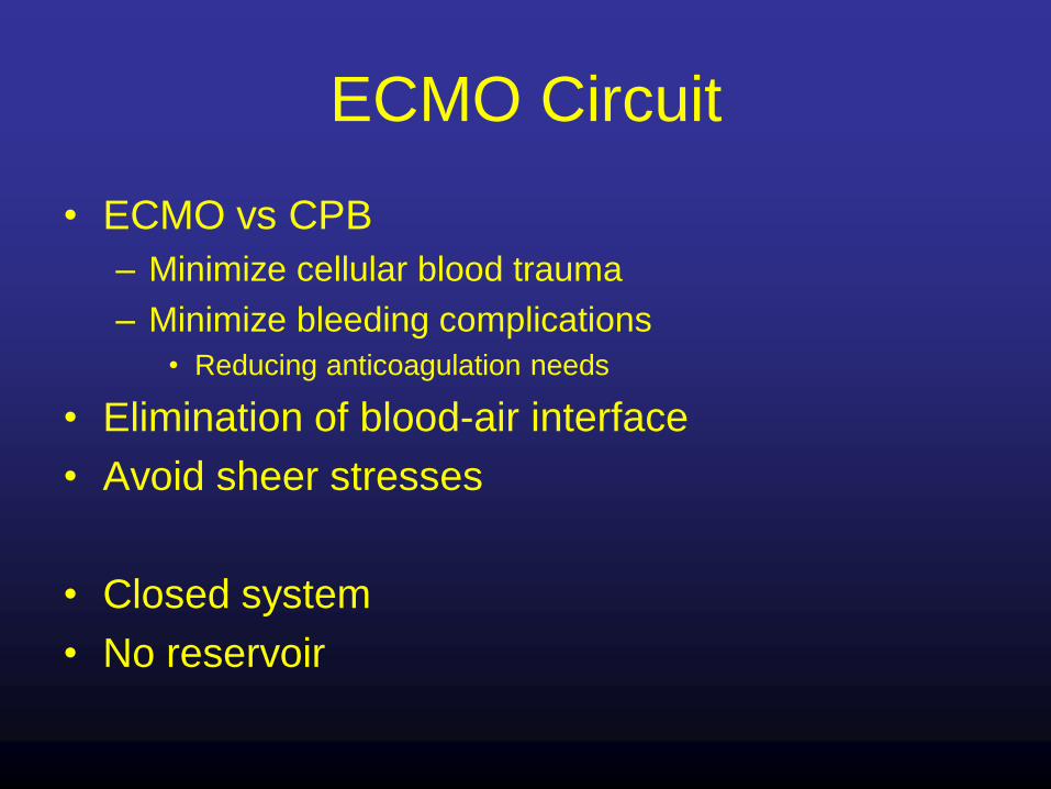
ECMO Circuit
• ECMO vs CPB
– Minimize cellular blood trauma
– Minimize bleeding complications
• Reducing anticoagulation needs
• Elimination of blood-air interface
• Avoid sheer stresses
• Closed system
• No reservoir

ECMO Circuit
• Blood pump
• Oxygenator

Blood Pump
• Roller type– Cheap
– Durable
– Control inlet suction
• Siphon pressure
• Electronic
– Larger circuits
• Floor
• Tubing spallation
– Potential for circuit rupture
• Requires trained bedside personnel

Blood Pump
• Centrifugal design– Smaller circuit
– Less risk of circuit rupture
• Finite positive pressure
– Thrombus and heat
generation resolved
• Magnetic levitation
– Regulate inlet suction

Oxygenator
• Silicone– Medtronic
– High resistance• Activate blood cells
– Time consuming to prime
• Polypropylene hollow fibers– Standard for CPB
– Capillary tubes with micropores
– Leak plasma 24-48 hours
• Polymethyl Pentene– Capillary tubes
– Outer skin prevents plasma leak
– US Quadrox D (Maquet)
– Canada/Europe Medos and Novalung

Michigan’s ECMO Circuit
• Adults only
• Better bladder
– Measure inlet pressure without accessing negative pressure
• CentriMag Pump
– Console
– Backup console
– Backup motor
– No hand crank
• Measure peak circuit pressure
• Quadrox D oxygenator

Modes of ECMO Support
Veno-venous support (VV) Veno-arterial support (VA)

ECMO for Shock
• Indication:– CPR
– Failure to wean
– Conventional measures are failing
• Etiology– Acute myocardial infarction
• 6% MI with cardiogenic shock
• 60% mortality
– Myocarditis
– Chronic cardiomyopathy
– Post cardiotomy• 80% mortality on 3 inotropes
– Pulmonary Embolus
– Septic shock

Emergency Circulatory Support
• Bridge to recovery– Acute MI
– Myocarditis
– Post cardiotomy
• Bridge to transplant– Chronic heart failure
– Acute MI, cannot be revascularized
• Bridge to Implantable Circulatory Support– LVAD
• Exclusion:– Chronic organ dysfunction
– Advanced age
– Social contraindications
Exit Strategy

Emergency Circulatory Support
• Percutaneous ventricular assistance
– Tandem Heart
– Impella
• Paracorporeal ventricular assistance
– Abiomed
– CentriMag
• ECMO

TandemHeart pVAD
• Percutaneous
LVAD
• Transeptal left atrial
drainage
• Femoral artery
infusion
• Centrifugal pump

Impella
• Peripheral LVAD
• Continuous axial flow
• Across aortic valve
• 2.5 L/min Percutaneous 12 Fr
• 5.0 Surgical 22 Fr– Femoral
– Ascending aorta

Abiomed BVS / AB-5000 VAD
• Pneumatic pulsatile pump
• Self regulating
• Extracorporeal
• Paracorporeal
BVS 5000 5 5 days
20 10 days
312 days
> 365 days
AB5000
Longest
AB5000
Patient
Lab Testing

Levitronix CentriMag
• Magnetically levitated
centrifugal pump

VA ECMO for Shock(Modified Partial Cardiopulmonary Bypass)
• Advantages– Minimally invasive
– Bedside application
– Biventricular support
– Pulmonary support
• Disadvantages– Labor intensive
– Immobilized patient
– Non pulsatile flow
– Left ventricular distension
• Limits recovery
• Pulmonary hemorrhage
– Aortic thrombus
– Cerebral Hypoxia

When to choose ECMO
• Non-post cardiotomy severe biventricular failure
– Refractory malignant arrhythmias
– Cardiopulmonary arrest
• Cardiac failure with pulmonary edema
• Septic shock with circulatory collapse

Cannulation
• Thoracic– Postcardiotomy
– Typical CPB cannulae
• Percutaneous femoral/jugular– Arterial: Typical adult 15-17 Fr. Female, 17-19 Fr. Male
– Leg ischemia avoided with “reperfusion” cannula
• 10 Fr. Antegrade down SFA
• 8 Fr. Retrograde up posterior tibial
– Venous: 23-28 Fr.
• Open cervical or femoral– Carotid ligation 5-10% watershed infarction

ECMO Management
• Continuous Heparin infusion
– ACT 210-230 or PTT 40-50
– Adjust based on bleeding risk
• Platelets > 100K
– Ongoing platelet consumption
– More conservative if no signs of bleeding
• Arterial saturation >90%
– As measured in right arm
• MAP ~65

• Daily neurological assessment
– Diprivan or Dex for sedation
• Diuresis/ hemofiltration
• Daily plasma free hemoglobin
– Significant hemolysis should prompt search for cause
• Q8 hour circuit assessment for clot
• +HIT-change anticoagulation to argatroban
– Titrate for similar ACT
ECMO Management

Left Ventricular Distension
• ECMO is partial bypass– Residual RV output and bronchial flow
• LV must eject to avoid pulmonary hemorrhage
• Maintain aortic pulsatility
• Follow pulmonary artery pressures
• Liberal use of echo– LV size
– Aortic valve opening
• Strategies to facilitate LV ejection– IABP
– Inotropes (dobutamine, milrinone)
– Minimize vasoconstriction (Norepinephrine, Vasopressin)
– Reduce flow rate• Balance SV02 with LV ejection

Left Ventricular Distension
• Venting
• Transthoracic LV vent
– postcardiotomy
• Percutaneous atrial
septostomy
• TandemHeart drain
– Best placed via right
femoral vein
• Impella as LV vent

Aortic Root Thrombus
• VA ECMO via femoral artery
• Minimal LV ejection
• Stasis in aortic root
• Thrombus
• Prevention:– Maintain ventricular ejection
• IABP
• Inotropes
– Anticoagulation
• Rarely seen with VADs– Turbulence from proximal
location of aortic outflow

Cerebral Hypoxia
– VA ECMO
– Femoral
cannulation
– Left ventricular
ejection

Cerebral Hypoxia
– VA ECMO
– Femoral
cannulation
– Respiratory failure
– Left ventricular
ejection
– Hypoxic perfusion
of coronary and
cerebral circulation

Cerebral Hypoxia
– Diagnosis with right radial saturations
– Consider
• Ventilator adjustments
– Avoid VILI
• Better venous drainage
– Less LV ejection
• V-AV support
– Partial VA ECMO/Partial VV ECMO

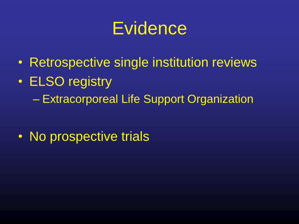
Evidence
• Retrospective single institution reviews
• ELSO registry
– Extracorporeal Life Support Organization
• No prospective trials

• 25 patients
• 7 required percutaneous LV venting
• 4 weaned from ECMO (2 survived)
• 8 bridged to VAD (6 transplanted (75%))
• 1 bridged directly to transplant
• 9 survived to hospital discharge (36%)
• Renal and hepatic function not predictive
• Urine output duration of ECMO

• 23 patients– 2 post cardiotomy
• 3 recovered
• 9 transition to VAD– 7 transplanted (78%)
• 10 survived to hospital discharge (43%)
• Bridge to VAD mean ECMO duration only 13.7 hours

• 202 patients
• 107 (53%) postcardiotomy
• 38% 30 day survival
• Age > 55 predictor
• 42 Bridged to VAD– 60% survived to transplant
• Excellent long term survival

• 517 postcardiotomy patients
• 61% central cannulation
• Most received IABP
• 24.8% survival to discharge
• Advanced age, Euroscore, Lactate levels predictors

University of Michigan
Device Utilization 1997-2009
0
5
10
15
20
25
30
35
40
1997 1998 1999 2000 2001 2002 2003 2004 2005 2006 2007 2008 2009Year
# of
patients
Abiomed n=61
ECLS n=192
TandemHeart n=29
Total n=282

Device Use by Indication
0 20 40 60 80 100
Acute MI
Myocarditis
Decompensated Chronic
Cardiomyopathy
Post cardiotomy
Heart transplant
Lung transplant
Pulmonary Embolism
Respiratory failure/sepsis
# of patients
ECLS
Abiomed
TandemHeart

Survival by Diagnostic Indication
0% 20% 40% 60% 80%
Post-Heart Transplantation
Acute Myocardial Infarction
Myocarditis
Pulmonary Embolism
Decompensated Chronic Cardiomyopathy
Post-Cardiotomy
Respiratory Failure / Sepsis
Post-Lung Transplantation

Long-term survival
N=282 patients

Survival conditional upon hospital
discharge
86%75%
52%N=117
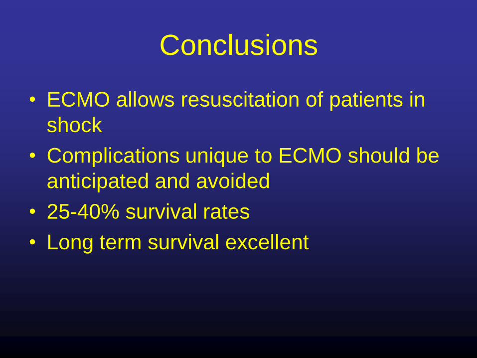
Conclusions
• ECMO allows resuscitation of patients in
shock
• Complications unique to ECMO should be
anticipated and avoided
• 25-40% survival rates
• Long term survival excellent