echocardiography deepak
DESCRIPTION
Echocardiography DeepakSouthwestern University school of medicineCardiology echoTRANSCRIPT

B Y :
D E E P A K G H I M I R E
M E D I C A L – I N T E R N
S O U T H W E S T E R N U N I V E R S I T Y
S C H O O L O F M E D I C I N E
P R E S E N T E D O C T O B E R , 2 0 1 5
P E R C E P T O R I N C H A R G E : V I C E N T E G . B A L B U E N A , M D
BASIC ECHOCARDIOGRAPHY

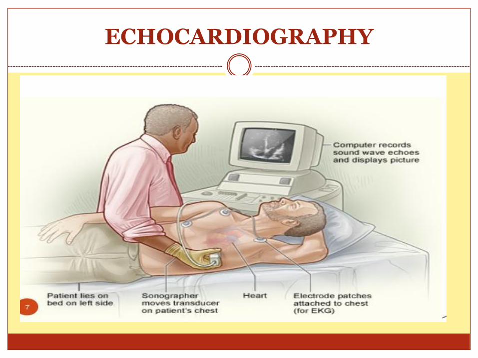
ECHOCARDIOGRAPHY

Cardiac Anatomy
Base
Apex

Circulation through the Heart

The Cardiac cycle

4 phases of Diastole
1. Isovolemic relaxation
2. Rapid ventricular filling
3. Slow ventricular filling
4. Atrial contraction

4 phases of Systole
1. Isovolemic contraction
2. Rapid ventricular ejection
3. Slow ventricular ejection

The Cardiac cycle


DIMENSIONS & MEASUREMENT
Internal diameter LV : <5 cm
RV : 2.2 – 4.0 cm
RA: 3.5 – 4.0 4cm
Internal diameter : LA, RA & Ao root : < 4cm

Imaging Modalities
1. 2D- Echocardiography
2. M-mode Echocardiography

Echocardiography
A non-invasive diagnostic technique
Widely used in clinical cardiology.
Involves the use of ultrasound
Used to assess cardiac structure and
hemodynamic function function.
Fig. Echocardiography machine

Indications of 2D Echocardiography
• May be divided into structural imaging and hemodynamic imaging (*ASE)
• Indication for Structural imaging• Pericardial imaging (P. Effusion)
• L/R ventricles & cavities (RVH/LVH or wall motion abnormality or thrombi)
• Image of valves ( Stenosis or prolapse)
• Great vessels (aortic dissections)
• Congenital & Traumatic heart diseases
• Hypertension, suspected IHD, murmurs, Pulmonary disease
• Arrythmias, palpitations, syncope or Neurological disease

Indications of 2D Echocardiography
• May be divided into structural imaging and hemodynamic imaging (*ASE)
• Indication for hemodynamic imaging through Doppler techniques: • Blood flow through heart valves (stenosis/ regurgitation)
• Blood flow through the cardiac chambers (C.O)
• Systolic and diastolic functions
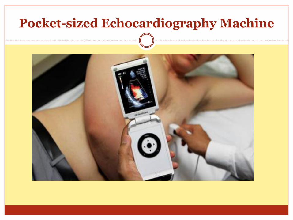
Pocket-sized Echocardiography Machine
Echocardiography
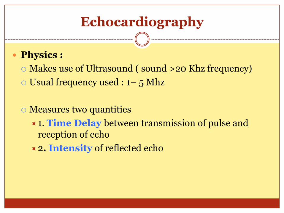
Physics :
Makes use of Ultrasound ( sound >20 Khz frequency)
Usual frequency used : 1– 5 Mhz
Measures two quantities
1. Time Delay between transmission of pulse and reception of echo
2. Intensity of reflected echo

Principle of Image generation

Gray Scale Image
Gray scale Image is generated Based on intensity of reflected echo
Fig: Pericardial effusion
Black Fluid or blood
White Calcifications on cardiac valves/ pericardium
Gray Myocardium

Factors in Echocardiography

Transducers

Use of lens in tranducer

Image Quality
Fig: Image generated by 3 Mhz tranducer
Fig: Image generated by 5 Mhz tranducer

Modes of Display

M-Mode echocardiography
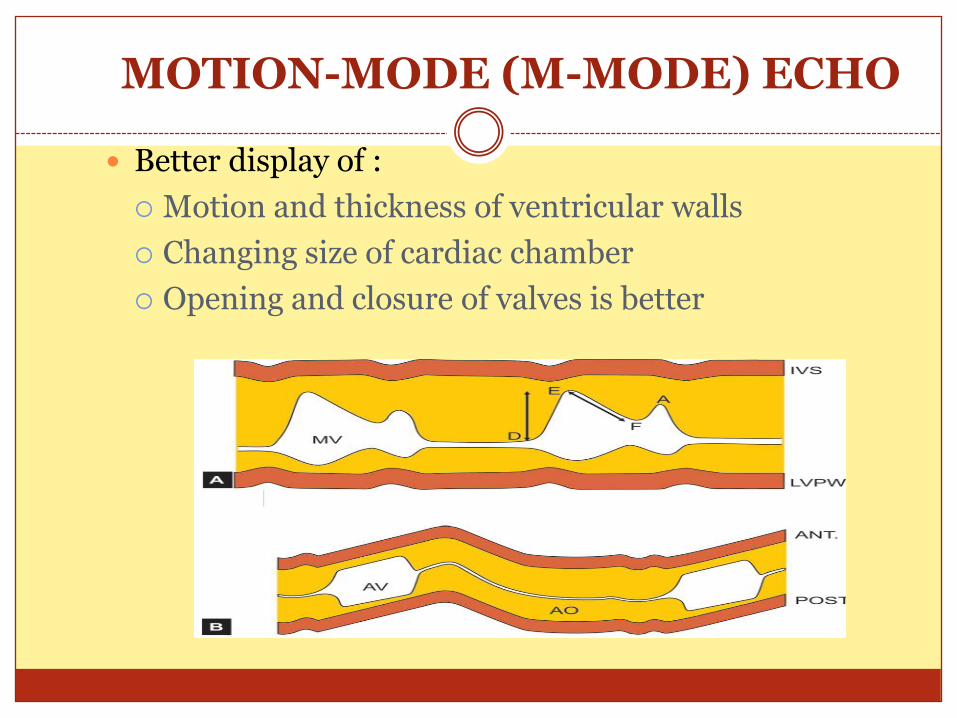
MOTION-MODE (M-MODE) ECHO
Better display of :
Motion and thickness of ventricular walls
Changing size of cardiac chamber
Opening and closure of valves is better

Echocardiography provided information about Structure of heart and great vessels
Doppler imaging provides information about the function, physiology and hemodynamics

Echo vs. Doppler studies

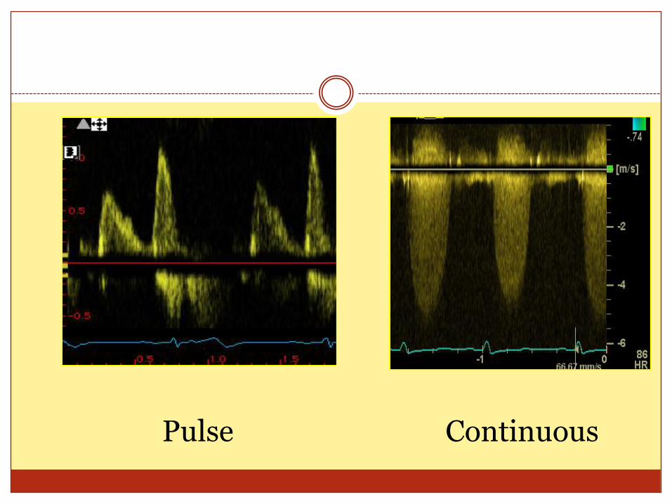
Pulse Continuous

Phased array transducer
The typical frequency range: 1-5 MHz
Has an orientation marker Corresponds with the Image

Transthoracic Acoustic Window
Trans-Thoracic Echo (TTE)
1. Parasternal view
2. Apical view
3. Subcostal view
Trans-Esophageal Echo (TEE)
Assess posterior cardiac
structures

Parasternal Long Axis View (PLAX)
* marker orientated towards the right clavicle (approximately 11 o’clock)

Parasternal Long Axis View (PLAX)
Structure Assessment
RV (right ventricle)
Size and function
LV (left ventricle)
Size and function (septum)
Ao (ascending aorta)
Size
AV (aortic valve)
Motion, opening and calcification
MV (mitral valve)
Motion, opening and calcification
Pericardium Fluids

Assessment :
1. Pericardial space
2. RV
3. Septum
4. LV
5. Anterior MV leaflet
6. Aortic root
7. LA

Parasternal Long Axis View (PLAX)

Parasternal Long Axis View (PLAX)
Fig. Calcification of mitral annulus

Pericardial effusion

Parasternal short-axis view (PSAX)
* PLAXS rotate clockwise 90 degrees ( to long axis of LV)
T

PLAX & PSAX

PSAX
The probe can be tilted to examine the SAX view at different levels:
1. Level of papillary muscles
2. Mitral valve
3. Level of aortic valve

PSAX

Apical Four Chamber View (A4C)
* marker is at around 3 o’clock.


Apical Four Chamber View (A4C)
Assessment :
1. Pericardial space
2. LV
3. Septum
4. RV
5. RA
6. Interartrial septum
7. LA
8.MV annulus
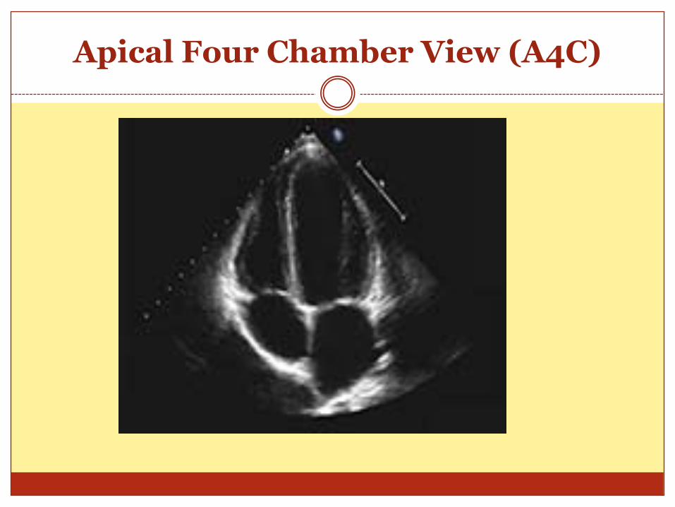
Apical Four Chamber View (A4C)

Apical Five Chamber View

Apical view
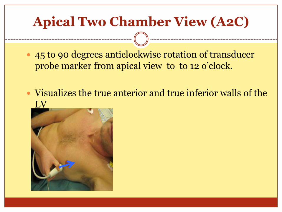
Apical Two Chamber View (A2C)
45 to 90 degrees anticlockwise rotation of transducer probe marker from apical view to to 12 o’clock.
Visualizes the true anterior and true inferior walls of the LV

Apical Two Chamber View (A2C)

Subcostal Long Axis View
* Transducer in Right sub xiphiod area & side marker in 3 o’clock position
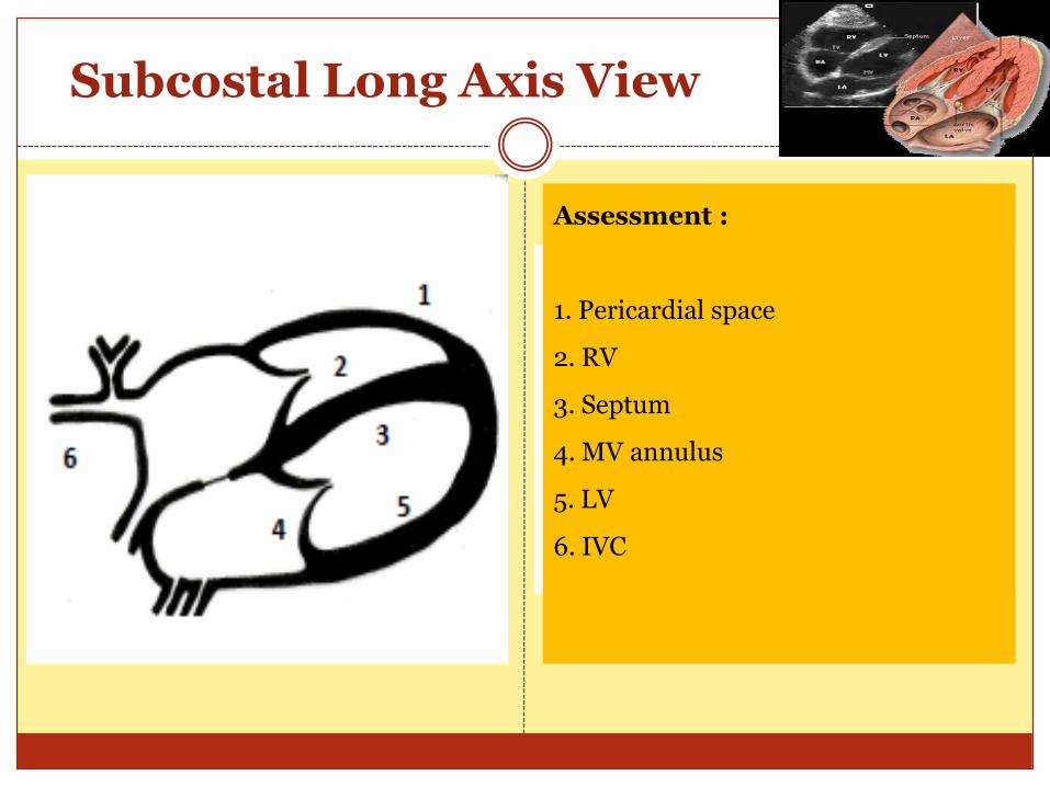
Subcostal Long Axis View
Assessment :
1. Pericardial space
2. RV
3. Septum
4. MV annulus
5. LV
6. IVC


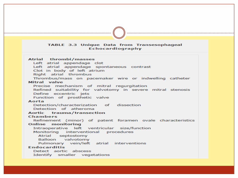
Trans-Esophageal Echocardiography
Used to assess posterior structures like LA or Aorta

Contrast echocardiography
Before and after contrast

Indications of 2d Echocardiography in evaluation of Heart murmurs
PLAX
MR, AR , VDS
PSAX (multiple level)
AR, TR, PS, PR, VSD
Apical :
4chamber : MR, TR
2 chamber: MR
Long axis: MR, AR, AS, LVOT
5 chamber: LV outflow, AR, AS
Subcostal :
4chamber- RV inflow, TR, ASD
Short axis (Basal) TR, PS, PR
Mid venticular: IVC, HV
Suprasteral:
Aortic arch, Aortic flow
Right Parasternal Lx
Ascending aorta, AS

Two-Dimensional Echocardiography
Cardiac chambers
Chamber size
Left ventricular Hypertrophy
Regional wall motion abnormalities
Valve
Morphology and motion
Pericardium
Effusion
Tamponade
Masses
Great vessels
TransesophagealEchocardiography
Inadequate transthoracicimages
Aortic disease
Infective endocarditis
Source of embolism
Valve prosthesis
Intraoperative

2D Echocardiography
An ideal imaging modality for assessing left ventricular (LV) size and function.
The “gold standard” for imaging valve morphology and motion
The imaging modality of choice for the detection of pericardial effusion
The definitive diagnosis of a suspected aortic dissection usually requires a TEE.

DOPPLER ECHOCARDIOGRAPHY
Uses ultrasound reflecting off moving RBC to
measure the velocity of blood flow across valves,
within cardiac chambers, and through the great
vessels.
Normal and abnormal blood flow patterns can be
assessed noninvasively.

DOPPLER ECHOCARDIOGRAPHY
The different colors indicate the direction of blood flow:
Red toward the transducer
Blue away from the transducer
Green superimposed when there is turbulent flow.
Blue Away Red Towards (BART)

DOPPLER ECHOCARDIOGRAPHY
Valve gradients in stenosis ( Inc. velocity )
Valvular regurgitation (retrograde flow)
Intracardiac pressures (PAH)
Cardiac output (area X velocity)
Diastolic filling
Congenital heart disease (shunts)

VSD

Aortic Regurgitation
Fig: TEE: Dilated ascending aorta Fig. TEE: Aortic Regurgitation

Other use of Echo
Hypovolemia
kissing LV chambers
Collapsing IVC

Temponade
Cardiac Temponade
IVC dilation
Diastolic RV collapse.

Cardiac masses and Tumors

Pleural Effusion

Echocardiography
The quality of an echo is highly operator dependent and proportional to experience and skill.
The value of information derived depends heavily on operation and interpretation

Sources and Assistance
Harrison Principle of Internal medicine, 19th ed
Harrison Principle of Internal medicine, 18th ed
Fiegenbaum's Echocardiography 6th edition
Introduction to Transthoracic echocardiography Stanford University School of Medicine
Radiology staff of SHH, SWU

Good Morning !