transesophageal echocardiography-related complications · ix- prevention of transesophageal...
TRANSCRIPT

622
CAN J ANESTH 55: 9 www.cja-jca.org September, 2008
Purpose: The use of transesophageal echocardiography has in-creased over the past several years. It is generally considered a safe diagnostic and monitoring tool. Whereas complications as-sociated with echocardiographic examination rarely occur, such complications must be known to echocardiographers perform-ing these examinations. The purpose of this review is to sum-marize potential complications associated with transesophageal echocardiography.
Sources: A systematic search of the English and French litera-ture was undertaken using PubMed from the National Library of Medicine. Relevant articles were obtained from a Medline search spanning the years 1975 – 2007, and their reference lists were used to retrieve additional articles.
Principal findings: Complications of transesophageal echocar-diography are primarily related to the gastrointestinal, cardiovas-cular, and respiratory systems, and include infection, toxic drug reaction, local reaction through contamination of the probe, and ultrasound cavitation. Strategies to prevent these complications are reviewed.
Conclusion: Whereas transesophageal echocardiography is as-sociated with a low complication rate, the echocardiographer must be knowledgeable about the types of complications and their predisposing factors, and should be meticulous in prevent-ing their occurrence.
CAN J ANESTH 2008 / 55: 9 / pp 622–647
Objectif : Au cours des dernières années, l’utilisation de l’échocardiographie transoesophagienne a augmenté. Cette mo-dalité est en général considérée comme un outil de diagnostic et de monitorage sécuritaire. Bien que des complications associées à un examen échocardiographique ne surviennent que rarement, il est néanmoins important que les échocardiographistes en soi-ent conscients. L’objectif de ce compte-rendu est de résumer les complications potentielles associées à l’échocardiographie transo-esophagienne.
Sources : Une recherche systématique de la littérature en anglais et en français a été menée dans la base de données PubMed de la Bibliothèque nationale de médecine américaine. Les articles perti-nents ont été extraits d’une recherche Medline couvrant la période 1975-2007, et leurs listes de références ont été utilisées pour récu-pérer d’autres articles pertinents.
Constatations principales : Les complications associées à l’utilisation de l’échocardiographie transoesophagienne sont prin-cipalement liées aux systèmes gastro-intestinal, cardiovasculaire et respiratoire. Elles comprennent l’infection, la réaction toxique au médicament, la réaction locale en raison d’une contamination de la sonde, et la cavitation des ultrasons. Les différentes stratégies pour prévenir ces complications sont passées en revue.
Conclusion : Bien que l’échocardiographie transoesophagienne soit associée à un faible taux de complications, l’échocardiographiste doit connaître les différents types de complications ainsi que les facteurs les prédisposant, et devrait apporter un soin particulier à les prévenir.
From the Departments of Anesthesiology, CHU Mère-Enfant Sainte-Justine Hospital* and Montreal Heart Institute,† Université de Montréal, Montréal, Québec, Canada. Address correspondence to: Dr. André Y. Denault, Department of Anesthesiology, Montreal Heart Institute, 5000 Bélanger Street, Montréal, Québec H1T 1C8, Canada. Phone: 514-376-3330 ext. 3732; Fax: 514-376-1355; E-mail: [email protected] Financial support: Fonds de la recherche en santé du Québec and Fondation de l’Institut de Cardiologie de Montréal. Conflicts of interest: None declared. Accepted for publication June 14, 2005. Revision accepted June 23, 2008.
Review Article
Transesophageal echocardiography-related complications[Complications associées à l’échocardiographie transoesophagienne]Geneviève Côté mdmscfrcpc,* André Denault mdfasefrcpc†
Côté et al. complicationsoftransesophagealechocardiography 623
CAN J ANESTH 55: 9 www.cja-jca.org September, 2008
Table of contentsIntroduction and literature search strategyI- Gastrointestinal complicationsII- Cardiovascular complicationsIII- Respiratory complicationsIV- Infections and prophylactic antibioticsV- Medication-related complicationsVI- Miscellaneous complicationsVII- Local effects of ultrasound waves on surrounding tissuesVIII- Echocardiographer expertiseIX- Prevention of transesophageal echocardiography-related complicationsLimitationsConclusions
Transesophageal echocardiography (TEE) is a very useful, semi-invasive diagnostic and monitoring tech-nique. Since the initial work published by Frazin in 1976,1 there has been a substantial increase in com-prehensive knowledge of cardiovascular anatomic and hemodynamic correlations, as well as progress and innovation in the technique. Transesophageal echo-cardiography is presently in widespread use in ambu-latory clinics, coronary care units, intensive care units (ICUs), and operating rooms. Examinations using TEE are considered safe when conducted in a con-trolled environment by trained operators.2 Informa-tion obtained from TEE can have a significant impact in cardiac3–9 and non-cardiac surgery,10–12 as well as in making decisions in the ICU.11,13,14 Considering the widespread application of this technique, it is impor-tant to address the issue of safety. In this article, we present a comprehensive review of the literature per-taining to TEE complications reported during exami-nations undertaken in cardiology suites, operating rooms, and ICUs. We also suggest strategies aimed at the prevention of TEE-related complications.
Literature search strategyA systematic search of the English and French lit-erature was performed using the database PubMed from the National Library of Medicine. Literature was searched for the period 1975 to 2007. The bibliog-raphy of each article was then reviewed to seek addi-tional references and to produce a detailed reference list. The term ‘transesophageal echocardiography’ was paired with the following MeSH keywords: ‘adverse effects’, ‘complications’, ‘injuries’, ‘safety’, ‘esopha-gus/hypopharynx injuries’, ‘endoscopy’, ‘intoxica-tion’, and ‘infection/bacteriemia’. Articles describing experiences with the use of TEE were reviewed for reported complications. In addition,
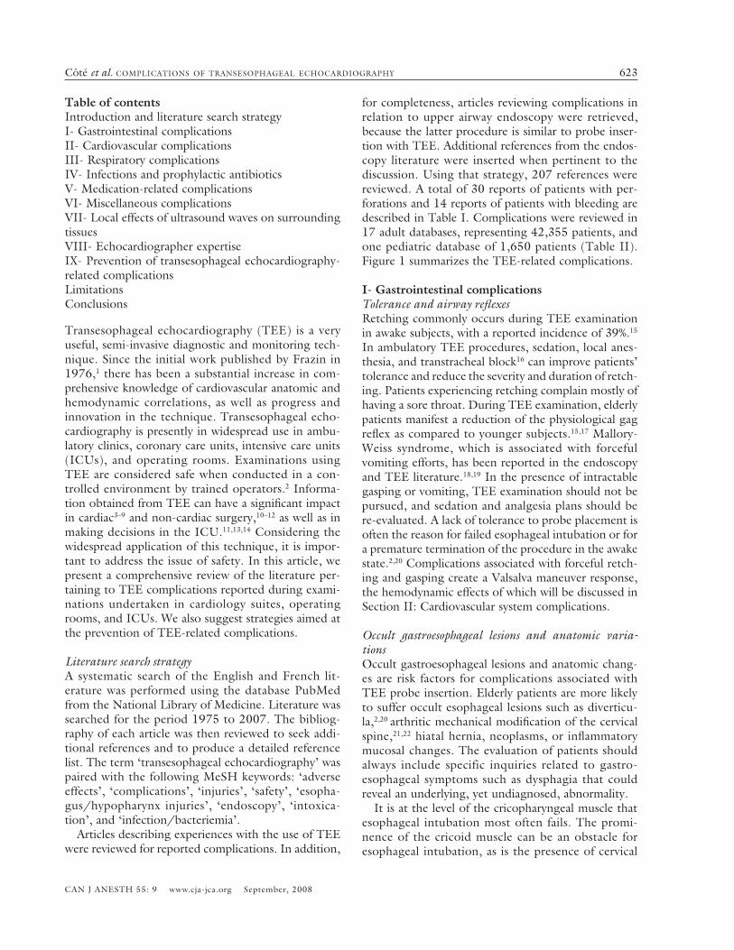
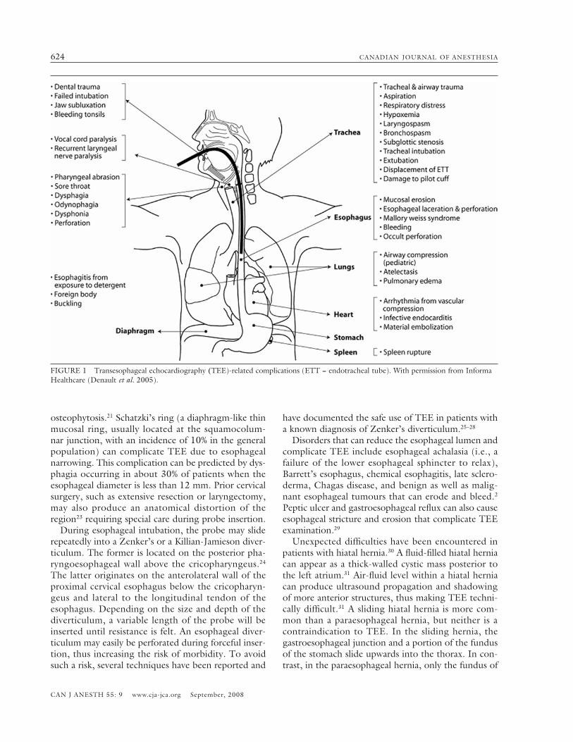
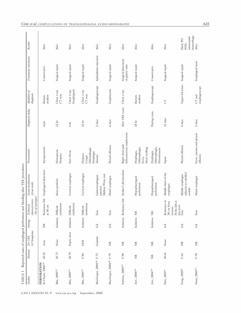
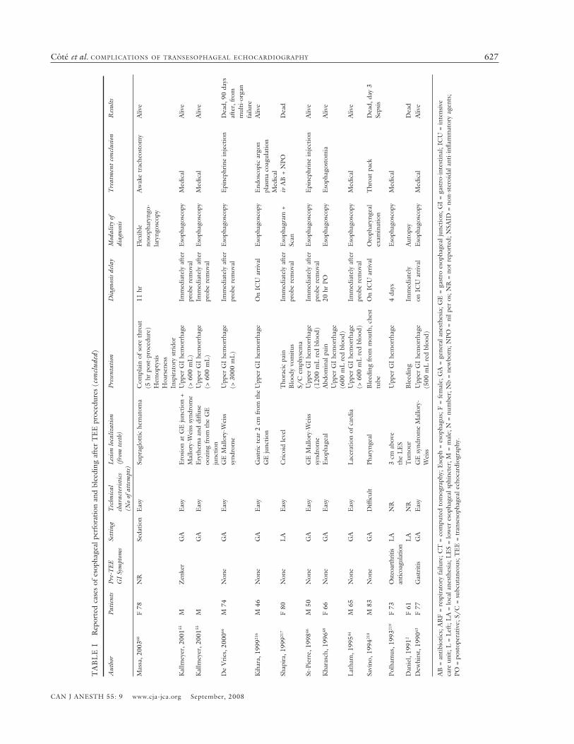
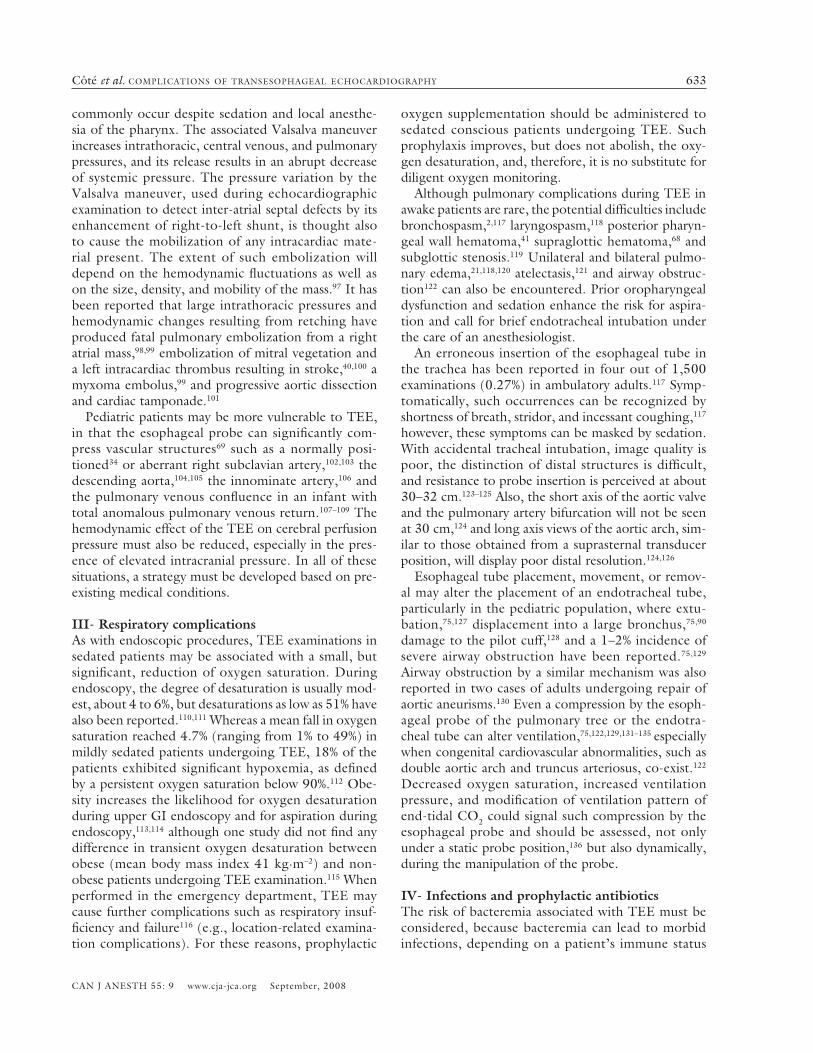
for completeness, articles reviewing complications in relation to upper airway endoscopy were retrieved, because the latter procedure is similar to probe inser-tion with TEE. Additional references from the endos-copy literature were inserted when pertinent to the discussion. Using that strategy, 207 references were reviewed. A total of 30 reports of patients with per-forations and 14 reports of patients with bleeding are described in Table I. Complications were reviewed in 17 adult databases, representing 42,355 patients, and one pediatric database of 1,650 patients (Table II). Figure 1 summarizes the TEE-related complications.
I- Gastrointestinal complicationsTolerance and airway reflexesRetching commonly occurs during TEE examination in awake subjects, with a reported incidence of 39%.15 In ambulatory TEE procedures, sedation, local anes-thesia, and transtracheal block16 can improve patients’ tolerance and reduce the severity and duration of retch-ing. Patients experiencing retching complain mostly of having a sore throat. During TEE examination, elderly patients manifest a reduction of the physiological gag reflex as compared to younger subjects.15,17 Mallory-Weiss syndrome, which is associated with forceful vomiting efforts, has been reported in the endoscopy and TEE literature.18,19 In the presence of intractable gasping or vomiting, TEE examination should not be pursued, and sedation and analgesia plans should be re-evaluated. A lack of tolerance to probe placement is often the reason for failed esophageal intubation or for a premature termination of the procedure in the awake state.2,20 Complications associated with forceful retch-ing and gasping create a Valsalva maneuver response, the hemodynamic effects of which will be discussed in Section II: Cardiovascular system complications.
Occult gastroesophageal lesions and anatomic varia-tionsOccult gastroesophageal lesions and anatomic chang-es are risk factors for complications associated with TEE probe insertion. Elderly patients are more likely to suffer occult esophageal lesions such as diverticu-la,2,20 arthritic mechanical modification of the cervical spine,21,22 hiatal hernia, neoplasms, or inflammatory mucosal changes. The evaluation of patients should always include specific inquiries related to gastro-esophageal symptoms such as dysphagia that could reveal an underlying, yet undiagnosed, abnormality. It is at the level of the cricopharyngeal muscle that esophageal intubation most often fails. The promi-nence of the cricoid muscle can be an obstacle for esophageal intubation, as is the presence of cervical
624 CANADIAN JOURNAL OF ANESTHESIA
CAN J ANESTH 55: 9 www.cja-jca.org September, 2008
FIGURE 1 Transesophageal echocardiography (TEE)-related complications (ETT = endotracheal tube). With permission from Informa Healthcare (Denault et al. 2005).
osteophytosis.21 Schatzki’s ring (a diaphragm-like thin mucosal ring, usually located at the squamocolum-nar junction, with an incidence of 10% in the general population) can complicate TEE due to esophageal narrowing. This complication can be predicted by dys-phagia occurring in about 30% of patients when the esophageal diameter is less than 12 mm. Prior cervical surgery, such as extensive resection or laryngectomy, may also produce an anatomical distortion of the region23 requiring special care during probe insertion. During esophageal intubation, the probe may slide repeatedly into a Zenker’s or a Killian-Jamieson diver-ticulum. The former is located on the posterior pha-ryngoesophageal wall above the cricopharyngeus.24 The latter originates on the anterolateral wall of the proximal cervical esophagus below the cricopharyn-geus and lateral to the longitudinal tendon of the esophagus. Depending on the size and depth of the diverticulum, a variable length of the probe will be inserted until resistance is felt. An esophageal diver-ticulum may easily be perforated during forceful inser-tion, thus increasing the risk of morbidity. To avoid such a risk, several techniques have been reported and
have documented the safe use of TEE in patients with a known diagnosis of Zenker’s diverticulum.25–28
Disorders that can reduce the esophageal lumen and complicate TEE include esophageal achalasia (i.e., a failure of the lower esophageal sphincter to relax), Barrett’s esophagus, chemical esophagitis, late sclero-derma, Chagas disease, and benign as well as malig-nant esophageal tumours that can erode and bleed.2 Peptic ulcer and gastroesophageal reflux can also cause esophageal stricture and erosion that complicate TEE examination.29
Unexpected difficulties have been encountered in patients with hiatal hernia.30 A fluid-filled hiatal hernia can appear as a thick-walled cystic mass posterior to the left atrium.31 Air-fluid level within a hiatal hernia can produce ultrasound propagation and shadowing of more anterior structures, thus making TEE techni-cally difficult.31 A sliding hiatal hernia is more com-mon than a paraesophageal hernia, but neither is a contraindication to TEE. In the sliding hernia, the gastroesophageal junction and a portion of the fundus of the stomach slide upwards into the thorax. In con-trast, in the paraesophageal hernia, only the fundus of
Côté et al. complicationsoftransesophagealechocardiography 625
CAN J ANESTH 55: 9 www.cja-jca.org September, 2008
TA
BL
E I
R
epor
ted
case
s of
eso
phag
eal p
erfo
ratio
n an
d bl
eedi
ng a
fter
TE
E p
roce
dure
s
Aut
hor
Pati
ents
Pre-
TE
E
GI
Sym
ptom
sSe
ttin
gT
echn
ical
ch
arac
teri
stic
s(N
o of
att
empt
s)
Lesio
n lo
caliz
atio
n(f
rom
tee
th)
Pres
enta
tion
Dia
gnos
is de
lay
Mod
alit
y of
di
agno
sisT
reat
men
t co
nclu
sion
Res
ults
PE
RFO
RA
TIO
NE
l-C
ham
i, 20
0620
6M
52
none
NR
Res
ista
nce
felt
at 3
0 cm
Eso
phag
eal d
isse
ctio
nA
sym
ptom
atic
none
Bar
ium
sw
allo
wC
onse
rvat
ive
Aliv
e
Min
, 200
5207
M 7
3N
one
Seda
tion
Diffi
cult
intu
batio
nSi
nus
pyri
form
Hem
opty
sis
Dys
pnea
12 h
rC
hest
x-r
ayC
T s
can
Surg
ical
rep
air
Aliv
e
Min
, 200
5207
M 7
9Pe
ptic
ulc
erSe
datio
nD
ifficu
lt in
tuba
tion
Cer
vica
l eso
phag
usSo
re t
hroa
t4
hrC
hest
x-r
ayE
soph
agra
mSu
rgic
al r
epai
rA
live
Min
, 200
5207
F 84
GE
RSe
datio
nD
ifficu
lt in
tuba
tion
Cer
vica
l eso
phag
usD
yspn
eaC
ough
Ody
noph
agia
Dys
phag
ia
22 h
rC
hest
x-r
ayC
T s
can
Surg
ical
rep
air
Aliv
e
Mac
Gre
gor,
200
4208
F 72
Gas
triti
sG
AE
asy
Gas
troe
soph
agea
l ju
nctio
nM
allo
ry-W
eiss
tea
r
Ane
mia
2 da
ysE
soph
agos
copy
Epi
neph
rine
inje
ctio
nA
live
Mac
Gre
gor,
200
4208
F 79
NR
GA
Eas
yD
ista
l eso
phag
usPl
eura
l effu
sion
6 da
ysE
soph
aora
mSu
rgic
al r
epai
rA
live
Sobr
ino,
200
4209
F 90
NR
Seda
tion
Res
ista
nce
felt
Zen
ker’
s di
vert
icul
umR
ight
che
st p
ain
Subc
utan
eous
em
phys
ema
Aft
er T
EE
exa
mC
hest
x-r
aySu
rgic
al p
lace
men
t
of g
astr
ic t
ube
Aliv
e
Avi
v, 2
00421
0N
RN
RSe
datio
nN
RH
ypop
hary
ngea
l pe
rfor
atio
nD
ysph
agia
, O
dyno
phag
ia,
Feve
r,
Nec
k sw
ellin
g
18 h
rB
ariu
m
swal
low
Surg
ical
rep
air
Aliv
e
Avi
v, 2
00421
0N
RN
RSe
datio
nN
RH
ypop
hary
ngea
l pe
rfor
atio
nD
ysph
agia
, O
dyno
phag
ia,
Hem
atem
esis
Dur
ing
exam
Eso
phag
osco
pyC
onse
rvat
ive
Aliv
e
Han
, 200
349M
61
Non
eG
AR
esis
tanc
e at
30
cm
from
in
ciso
rsPr
obe
left
at
this
leve
l
Mid
dle
thir
d of
the
es
opha
gus
Seps
is12
day
sC
TSu
rgic
al r
epai
rA
live
Pong
, 200
345F
62N
RG
AE
asy
Mid
dle
esop
hagu
s
adja
cent
to
calc
ified
no
dule
Pleu
ral e
ffusi
on4
days
Upp
er G
I se
ries
Surg
ical
rep
air
Die
d, P
O
#182
, in
trac
rani
al
hem
orrh
age
Nan
a, 2
00321
1F
70N
RG
AE
asy
Dis
tal e
soph
agus
Feve
r, a
nem
ia a
nd p
leur
al
effu
sion
2 da
ysC
T a
nd
esop
hago
scop
yE
soph
agea
l ste
ntA
live

626 CANADIAN JOURNAL OF ANESTHESIA
CAN J ANESTH 55: 9 www.cja-jca.org September, 2008
Aut
hor
Pati
ents
Pre-
TE
E
GI
Sym
ptom
sSe
ttin
gT
echn
ical
ch
arac
teri
stic
s(N
o of
att
empt
s)
Lesio
n lo
caliz
atio
n(f
rom
tee
th)
Pres
enta
tion
Dia
gnos
is de
lay
Mod
alit
y of
di
agno
sisT
reat
men
t co
nclu
sion
Res
ults
Zal
unar
do, 2
00259
M 7
2N
SAID
GA
Eas
y L
ower
eso
phag
usPl
eura
l effu
sion
Neu
trop
hilia
D
eter
iora
tion
Nut
ritio
n flu
id fr
om
ches
t dr
ains
8 da
ysE
soph
agra
m
Surg
ical
rep
air
Eso
phag
eal s
tent
Aliv
e
Law
-Kou
ne, 2
00221
2F
77N
RG
AE
asy
Upp
er e
soph
agus
Intr
aope
rativ
e pe
rfor
atio
nN
one
Prob
e in
the
su
rgic
al fi
eld
and
esop
hagr
am
Surg
ical
rep
air
Dea
d
Lec
harn
y, 2
00260
M 3
7N
RG
AT
EE
last
ed
9 hr
Eso
phag
otra
chea
l pe
rfor
atio
nSh
ock
7 da
ysV
entil
atio
n le
ak +
Eso
phag
osco
py
Bla
ckm
ore
esop
hage
al
ballo
on c
athe
ter
Die
d be
fore
sur
gery
Dea
d
Bri
nkm
an, 2
00121
3F
80N
RL
ow d
ose
ster
oid
GA
Eas
yL
ower
eso
phag
usFe
ver,
pl
eura
l effu
sion
Bur
ning
> 1
mon
thC
T
Eso
phag
ram
Surg
ical
pla
cem
ent
of
T-t
ube
Aliv
e
Bri
nkm
an, 2
00121
3F
70N
RR
adio
ther
apy
for
brea
st
canc
er
GA
Eas
yM
id-e
soph
agus
Bur
ning
sub
ster
nal p
ain
Few
hou
rsE
soph
agra
mSu
rgic
al r
epai
rA
live
Bri
nkm
an, 2
00121
3F8
5N
RG
AE
asy
Mid
-eso
phag
usPl
eura
l effu
sion
2 da
ysC
hest
x-r
ay fl
uid,
ga
stro
inte
stin
al
cont
ent
from
ch
est
tube
Surg
ical
rep
air
Aliv
e
Muh
iude
en, 2
00121
4N
= 3
Reg
urgi
tatio
n G
AE
asy,
(2)
Cri
coid
mus
cle
TE
E s
urgi
cal fi
eld
Imm
edia
tely
TE
E s
urgi
cal fi
eld
Med
ical
Aliv
e M
asse
y, 2
00033
F 59
Non
e E
asy
Res
ista
nce
felt
at 3
0 cm
Eso
phag
eal t
ear
at
39 c
m w
ith a
bsce
ssPl
eure
tic p
ain
4 da
ysC
hest
x-r
ay fl
uid
+
Eso
phag
osco
py
Surg
ical
rep
air
Dea
d, d
ay 9
Mac
Gow
an, 2
00058
M 6
4N
RG
AE
asy
Eso
phag
eal u
lcer
atio
n
with
left
bro
nchi
al fi
stul
a Se
psis
Aut
opsy
Aut
opsy
Dea
d, d
ay 1
9
Joug
on, 2
00057
M 8
6N
RL
AC
ervi
cal e
soph
agea
l at
19 c
m
Rap
id s
epsi
s Fe
w h
ours
Eso
phag
ram
+E
soph
agos
copy
Eso
phag
osto
mia
Dea
d
Joug
on, 2
00057
F 65
NR
LA
Diffi
cult
Cer
vica
l eso
phag
eal a
t 15
cm
S/C
em
phys
ema
Post
-exa
min
atio
nE
soph
agos
copy
E
soph
agos
tom
iaA
live
Kal
lmey
er, 2
00155
F 81
NR
GA
Eas
yD
yspn
ea2
days
Che
xt x
-ray
+E
soph
agra
m
Surg
ical
rep
air
Aliv
e
Lal
anne
, 199
656F7
4N
RL
AE
asy
Mid
-eso
phag
usD
yspn
eaC
hest
pai
nE
piga
stra
lgia
Imm
edia
tely
Surg
ical
rep
air
Dea
d,
seps
is P
O#2
Spah
n, 1
99519
5F
75N
RG
AN
RPh
aryn
geal
T
EE
sur
gica
l fiel
dIm
med
iate
lyT
EE
sur
gica
l fie
ld +
Eso
phag
osco
py
Surg
ical
sut
ure
Aliv
e
Bad
aoui
, 199
419F
71N
RL
AD
ifficu
lt (3
)L
acer
atio
n at
2-3
cm
C
ervi
cal p
ain
S/C
em
phys
ema
Few
hou
rsE
soph
agra
m
Eso
phag
osto
mia
Aliv
e
Dan
iel,
1991
2F
61T
hrom
boly
sis
NR
Intr
a-m
ural
hem
atom
a w
ith r
uptu
re in
the
tho
rax
Shoc
k4
hrSu
rger
y Su
rger
y A
live
BL
EE
DIN
GK
erba
ul, 2
00421
5M
41
NR
GA
2 at
tem
pts
diffi
cult
prog
ress
ion
beyo
nd 3
5 cm
2 es
opha
geal
per
fora
tions
Upp
er G
I he
mor
rhag
eSh
ock
Imm
edia
tely
aft
er
prob
e re
mov
al a
nd
inse
rtio
n of
NG
tu
be
Eso
phag
osco
pySu
rgic
al r
epai
rD
ied,
from
m
ultip
le o
rgan
fa
ilure
TA
BL
E I
R
epor
ted
case
s of
eso
phag
eal p
erfo
ratio
n an
d bl
eedi
ng a
fter
TE
E p
roce
dure
s (c
onti
nued
)
Côté et al. complicationsoftransesophagealechocardiography 627
CAN J ANESTH 55: 9 www.cja-jca.org September, 2008
Aut
hor
Pati
ents
Pre-
TE
E
GI
Sym
ptom
sSe
ttin
gT
echn
ical
ch
arac
teri
stic
s(N
o of
att
empt
s)
Lesio
n lo
caliz
atio
n(f
rom
tee
th)
Pres
enta
tion
Dia
gnos
is de
lay
Mod
alit
y of
di
agno
sisT
reat
men
t co
nclu
sion
Res
ults
Mas
sa, 2
00368
F 78
NR
Seda
tion
Eas
ySu
prag
lott
ic h
emat
oma
Com
plai
n of
sor
e th
roat
(5
hr
post
-pro
cedu
re)
Hem
opty
sis
Hoa
rsen
ess
Insp
irat
ory
stri
dor
11 h
rFl
exib
le
noso
phar
yngo
-la
ryng
osco
py
Aw
ake
trac
heos
tom
yA
live
Kal
lmey
er, 2
00155
MZ
enke
r G
AE
asy
Ero
sion
at
GE
junc
tion
+ M
allo
ry-W
eiss
syn
drom
eU
pper
GI
hem
orrh
age
(> 6
00 m
L)
Imm
edia
tely
aft
er
prob
e re
mov
alE
soph
agos
copy
M
edic
alA
live
Kal
lmey
er, 2
00155
MG
AE
asy
Ery
them
a an
d di
ffuse
oo
zing
from
the
GE
ju
nctio
n
Upp
er G
I he
mor
rhag
e(>
600
mL
)Im
med
iate
ly a
fter
pr
obe
rem
oval
Eso
phag
osco
py
Med
ical
Aliv
e
De
Vri
es, 2
00064
M 7
4N
one
GA
Eas
yG
E M
allo
ry-W
eiss
sy
ndro
me
Upp
er G
I he
mor
rhag
e
(> 3
000
mL
)Im
med
iate
ly a
fter
pr
obe
rem
oval
Eso
phag
osco
pyE
pine
phri
ne in
ject
ion
Dea
d, 9
0 da
ys
afte
r, fr
om
mul
ti-or
gan
failu
reK
ihar
a, 1
99921
6M
46
Non
e G
AE
asy
Gas
tric
tea
r 2
cm fr
om t
he
GE
junc
tion
Upp
er G
I he
mor
rhag
eO
n IC
U a
rriv
alE
soph
agos
copy
End
osco
pic
argo
n
plas
ma
coag
ulat
ion
Med
ical
Aliv
e
Shap
ira,
199
9217
F 80
Non
e L
AE
asy
Cri
coid
leve
lT
hora
cic
pain
Blo
ody
vom
itus
S/C
em
phys
ema
Imm
edia
tely
aft
er
prob
e re
mov
alE
soph
agra
m +
Scan
iv
AB
+ N
POD
ead
St-P
ierr
e, 1
99846
M 5
0N
one
GA
Eas
y G
E M
allo
ry-W
eiss
sy
ndro
me
Upp
er G
I he
mor
rhag
e(1
200
mL
red
blo
od)
Imm
edia
tely
aft
er
prob
e re
mov
alE
soph
agos
copy
E
pine
phri
ne in
ject
ion
Aliv
e
Kha
rasc
h, 1
99648
F 66
Non
eG
AE
asy
Eso
phag
eal
Abd
omin
al p
ain
Upp
er G
I he
mor
rhag
e(6
00 m
L r
ed b
lood
)
20 h
r PO
Eso
phag
osco
py
Eso
phag
osto
mia
Aliv
e
Lat
ham
, 199
544M
65
Non
eG
AE
asy
Lac
erat
ion
of c
ardi
aU
pper
GI
hem
orrh
age
(> 6
00 m
L r
ed b
lood
)Im
med
iate
ly a
fter
pr
obe
rem
oval
Eso
phag
osco
py
Med
ical
Aliv
e
Savi
no, 1
99421
8M
83
Non
eG
AD
ifficu
ltPh
aryn
geal
Ble
edin
g fr
om m
outh
, che
st
tube
On
ICU
arr
ival
Oro
phar
ynge
al
exam
inat
ion
Thr
oat
pack
Dea
d, d
ay 3
Seps
is
Polh
amus
, 199
3219
F 73
Ost
eoar
thrit
isan
ticoa
gula
tion
LA
NR
3 cm
abo
ve
the
LE
SU
pper
GI
hem
orrh
age
4 da
ysE
soph
agos
copy
M
edic
al
Dan
iel,
1991
2F
61L
AN
RT
umou
r B
leed
ing
Imm
edia
tely
A
utop
sy
Dea
dD
ewhi
rst,
199
043F
77G
astr
itis
GA
Eas
y G
E s
yndr
ome
Mal
lory
-W
eiss
Upp
er G
I he
mor
rhag
e(5
00 m
L r
ed b
lood
)on
IC
U a
rriv
alE
soph
agos
copy
M
edic
alA
live
AB
= a
ntib
iotic
s; A
RF
= re
spir
ator
y fa
ilure
; CT
= c
ompu
ted
tom
ogra
phy;
Eso
ph =
eso
phag
us; F
= fe
mal
e; G
A =
gen
eral
ane
sthe
sia;
GE
= g
astr
o es
opha
geal
junc
tion;
GI
= ga
stro
-int
estin
al; I
CU
= in
tens
ive
care
uni
t; L
= L
eft;
LA
= lo
cal a
nest
hesi
a; L
ES
= lo
wer
eso
phag
eal s
phin
cter
; M =
mal
e; N
= n
umbe
r; N
b =
new
born
; NPO
= n
il pe
r os
; NR
= n
ot r
epor
ted;
NSA
ID =
non
-ste
roid
al a
nti-
infla
mm
ator
y ag
ents
; PO
= p
osto
pera
tive;
S/
C =
sub
cuta
neou
s; T
EE
= t
rans
esop
hage
al e
choc
ardi
ogra
phy.
TA
BL
E I
R
epor
ted
case
s of
eso
phag
eal p
erfo
ratio
n an
d bl
eedi
ng a
fter
TE
E p
roce
dure
s (c
oncl
uded
)
628 CANADIAN JOURNAL OF ANESTHESIA
CAN J ANESTH 55: 9 www.cja-jca.org September, 2008
the stomach migrates past the gastroesophageal junc-tion, leaving this junction in its normal location. In patients with total or partial gastrectomy, TEE is not contraindicated as long as the probe is manipulated within the esophagus.32
Normal anatomical variants, such as an aortic impression, a large left atrium and left main bronchus, or pathological conditions, such as an enlarged heart, a mediastinal tumour,33 or esophageal duplication, can compress the esophagus, distort its imaging, and com-plicate esophageal intubation.34 Esophageal vascular abnormalities, such as prominent venous plexus or varices associated with cirrhosis and portal hyperten-
sion, may cause bleeding during TEE; therefore, TEE should either not be used at all, or should be used with great caution during hepatic transplantation.35
Cervical instability, due to trauma or to sublux-ation at the C1 and C2 levels associated with Down’s syndrome or severe rheumatoid arthritis, may make esophageal intubation difficult because of the need for airway management.36 Careless manipulation of the cervical spine can induce neurological deficits.
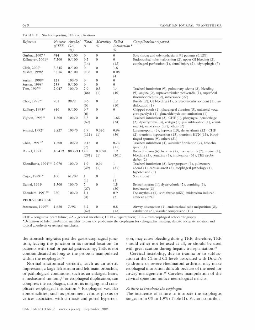
Failure to intubate the esophagusThe incidence of failure to intubate the esophagus ranges from 0% to 1.9% (Table II). Factors contribut-
TABLE II Studies reporting TEE complications
Reference Number of TEE
Awake/ GA(%)
Total %
Mortality%
Failedintubation* %
Complications reported
Gurbuz, 2007 8 744 0/100 0 0 0 Sore throat and odynophagia in 91 patients (0.12%)Kallmeyer, 200155 7,200 0/100 0.2
(14)0 0
(13)Endotracheal tube malposition (2), upper GI bleeding (2), esophageal perforation (1), dental injury (2), odynophagia (7)
Click, 20005 3,245 0/100 0 0 1.6Mishra, 19984 5,016 0/100 0.08 0 0.08
(4)Suriani, 199810 123 100/0 0 0 0Sutton, 19983 238 0/100 0 0 0Tam, 199721 2,947 100/0 2.9
(86)0.3(1)
1.4(40)
Tracheal intubation (9), pulmonary edema (2), bleeding (9), angina (2), supraventricular tachycardia (1), superficial thrombophlebitis (2), intolerance (27)
Chee, 199540 901 98/2 0.6(5)
0 1.2(10)
Buckle (2), GI bleeding (1), cerebrovascular accident (1), jaw dislocation (1)
Rafferty, 199339 846 0/100 0.7(6)
0 0 Chipped tooth (1), pharyngeal abrasion (3), unilateral vocal cord paralysis (1), glutaraldehyde contamination (1)
Vignon, 199320 1,500 100/0 3.5(52)
0 1.6%(24)
Tracheal intubation (2), CHF (1), pharyngeal hemorrhage (2), dysarrythmia (3), vertigo (1), jaw subluxation (1), vomit-ing (4), intolerance (12), others (2)
Seward, 199231 3,827 100/0 2.9(111)
0.026(1)
0.94(36)
Laryngospasm (5), hypoxia (13), dysarrythmia (22), CHF (2), transient hypotension (13), transient HTN (15), blood-tinged sputum (9), others (31)
Chan, 1991117 1,500 100/0 0.47(7)
0 0.73(11)
Tracheal intubation (4), auricular fibrillation (2), broncho-spasm (1)
Daniel, 19912 10,419 88.7/11.3 2.8(291)
0.0098(1)
1.9(201)
Bronchospasm (6), hypoxia (2), dysarrythmia (7), angina (1), bleeding (2), vomiting (5), intolerance (65), TEE probe defect (2)
Khandheria, 1991118 2,070 100/0 1.9(39)
0,04(1)
1(21)
Tracheal intubation (2), laryngospasm (3), pulmonary edema (1), cardiac arrest (2), esophageal pathology (4), hypotension (5)
Cujec, 1989220 100 61/39 1(1)
0 1(1)
Sore throat
Daniel, 19912 1,300 100/0 2(27)
0 1.5(20)
Bronchospasm (1), dysarrythmia (2), vomiting (1), intolerance (3)
Khanderh, 1991221 220 100/0 1.4(3)
0.9(2)
Dysarrythmia (1), sore throat (65%), midazolam-induced amnesia (87%)
PEDIATRIC TEE
Stevenson, 199975 1,650 7/93 3.2(52)
0 0.8(13)
Airway obstruction (1), endotracheal tube malposition (3), extubation (8), vascular compression (10)
CHF = congestive heart failure; GA = general anesthesia; HTN = hypertension; TEE = transesophageal echocardiography.*Definition of failed intubation: inability to pass the probe into the esophagus for echographic imaging, despite adequate sedation and topical anesthesia or general anesthesia.
Côté et al. complicationsoftransesophagealechocardiography 629
CAN J ANESTH 55: 9 www.cja-jca.org September, 2008
ing to such problems include lack of cooperation or the lack of operator experience, accounting for most cases (98.5%), as well as anatomical abnormalities, accounting for 1.5% of cases.2 Anatomic abnormalities producing esophageal intubation failure include a dou-ble aortic arch,37 cervical osteophytes21 and decreased cervical range of motion,28 swallowing impairment, mucosal abnormalities such as prior radiation exposure or decreased saliva production,28 prior tracheostomy,38 and an inflated endotracheal balloon. Several suggestions have been advanced to overcome failed intubation. Deeper sedation and local analgesia may provide relief from muscular spasm and improve patient cooperation. If resistance at the level of the esophagus is not eliminated by a swallowing effort, the examination should be aborted, and radiological evaluation should be considered in order to rule out anatomical obstruction. In Tam’s cohort, for example, cervical spondylosis associated with vertebral spurs was the most common cause of failed intubation in 16 of 40 patients.21 With anatomic variants such as cervical osteophytes, flexion of the neck may help to overcome the obstruction. In intubated and ventilated patients, the endotracheal tube balloon cuff can be deflated if resistance at the level of the larynx is encountered dur-ing probe insertion. In our own experience in such situations, we prefer to intubate under direct vision. Whereas a nasogastric tube rarely impedes intubation, it often leads to sub-optimal image acquisition. Lim-ited mouth opening can also be an obstacle.
Injuries, perforation, laceration and tear of the gastro-esophageal tractDental trauma,39 jaw subluxation,20,40 tonsillar bleeding, erosion, and submucosal hematoma of the pharyngeal area22,41 are some of the injuries related to TEE probe insertion of the upper gastrointestinal (GI) tract. Esophageal perforation occurs in the abdominal (57.3%), intrathoracic (33.3%), and cervical (9.3%) portions of the esophagus.42 It can be caused by poor patient cooperation, inadequate technical skills, unex-pected anatomical characteristics (GI abnormalities, extrinsic compression of the esophagus from enlarged left atrium,33,43,44 a large calcified lymph node,45 a cer-vical spur), or mucosal damage (due to motion, local ischemia, or pressure and heat by the probe). The hypopharynx and upper esophagus are the regions most vulnerable to perforation,41 because the esoph-ageal wall has an intrinsic weakness caused by fibres crossing from the pharyngeal constrictor and the cri-copharyngeal muscles. Neck extension, either with or without prominent anterior vertebral osteophytes, can increase the risk of perforation at the hypopharynx and
upper esophageal region by stretching the mucosa and muscular fibres. Shearing stress, prolonged flexion of the probe tip, and probe mobilization in a locked posi-tion may result in esophageal tearing or perforation. Such complications have been documented endoscop-ically.46 Several upper GI injuries related to periopera-tive TEE were reviewed by Augoustides.47
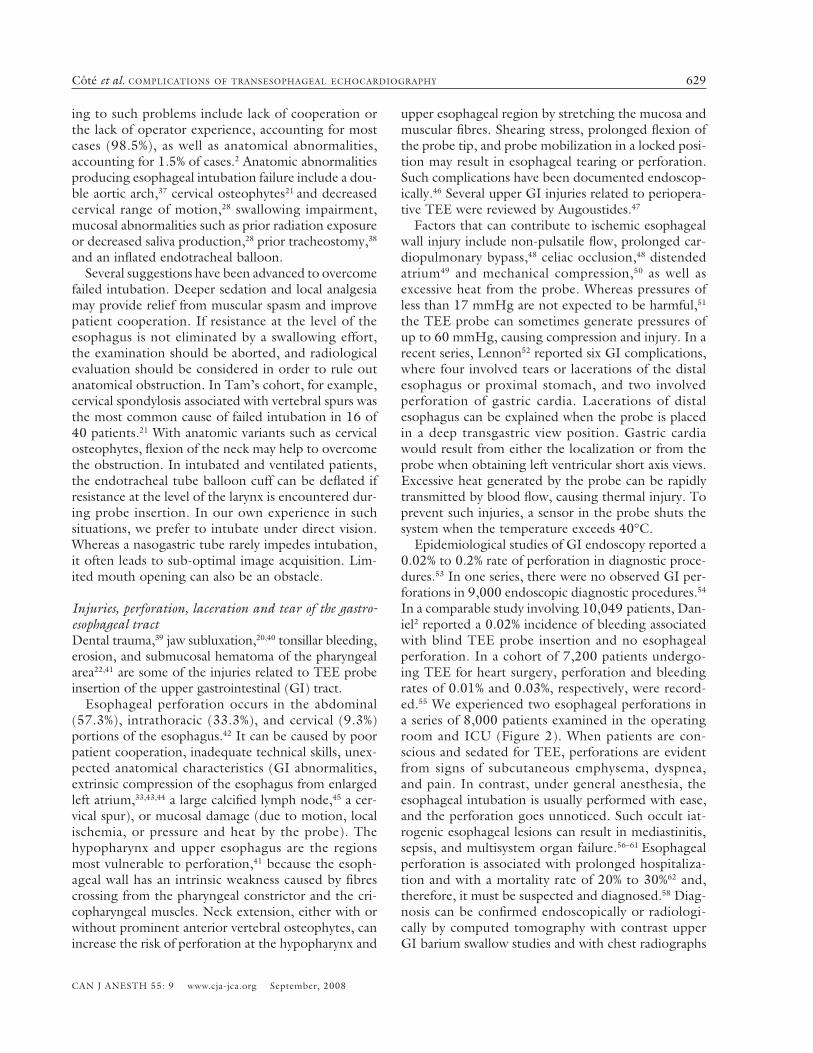
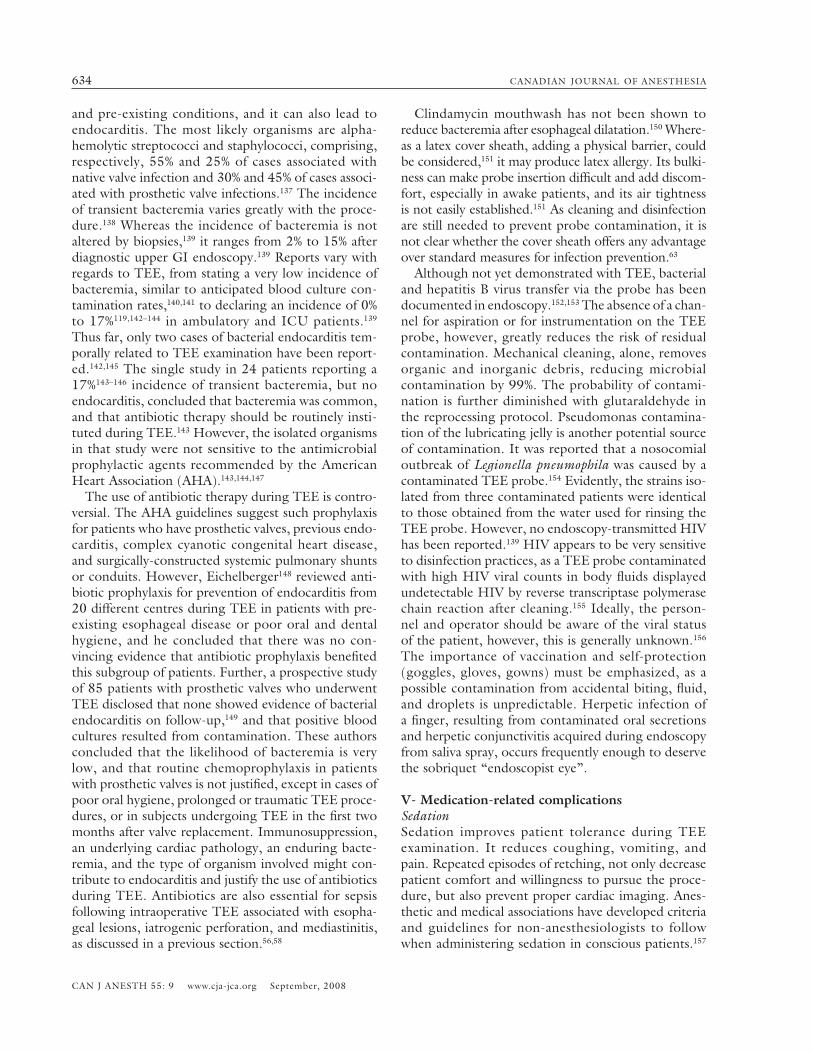
Factors that can contribute to ischemic esophageal wall injury include non-pulsatile flow, prolonged car-diopulmonary bypass,48 celiac occlusion,48 distended atrium49 and mechanical compression,50 as well as excessive heat from the probe. Whereas pressures of less than 17 mmHg are not expected to be harmful,51 the TEE probe can sometimes generate pressures of up to 60 mmHg, causing compression and injury. In a recent series, Lennon52 reported six GI complications, where four involved tears or lacerations of the distal esophagus or proximal stomach, and two involved perforation of gastric cardia. Lacerations of distal esophagus can be explained when the probe is placed in a deep transgastric view position. Gastric cardia would result from either the localization or from the probe when obtaining left ventricular short axis views. Excessive heat generated by the probe can be rapidly transmitted by blood flow, causing thermal injury. To prevent such injuries, a sensor in the probe shuts the system when the temperature exceeds 40°C. Epidemiological studies of GI endoscopy reported a 0.02% to 0.2% rate of perforation in diagnostic proce-dures.53 In one series, there were no observed GI per-forations in 9,000 endoscopic diagnostic procedures.54 In a comparable study involving 10,049 patients, Dan-iel2 reported a 0.02% incidence of bleeding associated with blind TEE probe insertion and no esophageal perforation. In a cohort of 7,200 patients undergo-ing TEE for heart surgery, perforation and bleeding rates of 0.01% and 0.03%, respectively, were record-ed.55 We experienced two esophageal perforations in a series of 8,000 patients examined in the operating room and ICU (Figure 2). When patients are con-scious and sedated for TEE, perforations are evident from signs of subcutaneous emphysema, dyspnea, and pain. In contrast, under general anesthesia, the esophageal intubation is usually performed with ease, and the perforation goes unnoticed. Such occult iat-rogenic esophageal lesions can result in mediastinitis, sepsis, and multisystem organ failure.56–61 Esophageal perforation is associated with prolonged hospitaliza-tion and with a mortality rate of 20% to 30%62 and, therefore, it must be suspected and diagnosed.58 Diag-nosis can be confirmed endoscopically or radiologi-cally by computed tomography with contrast upper GI barium swallow studies and with chest radiographs
630 CANADIAN JOURNAL OF ANESTHESIA
CAN J ANESTH 55: 9 www.cja-jca.org September, 2008
in the upright position. Radiological findings would include pneumothorax, pleural effusion, empyema, air/fluid levels, mediastinal shifts, and subcutaneous emphysema (Figure 2).
Bleeding of the gastroesophageal tract Minor mucosal trauma during TEE can result in hematemesis or in blood-tinged sputum.63 Early bleeding associated with TEE can be seen as sponta-neous drainage upon probe removal or as blood flow during aspiration from the nasogastric tube. Several cases of Mallory-Weiss tear related to intraoperative TEE presented with bright red blood coming from the nasogastric tube.43,50,64
Late bleeding, that is, more than seven days after the procedure, is suggestive of an ulcerative process.65
Risk factors that can precipitate upper GI bleeding due to TEE following cardiac surgery include a pre-
vious ulcerative process, vasoactive drug utilization, and absence of H2 antagonism in the perioperative period.66 Other factors, such as a long cardiac bypass period, urgent surgery, re-operation,67 or aspirin use,55 have also been implicated. Anticoagulants may pose another potential problem. However, 107 individuals under full anticoagulation regimen (intravenous heparin or oral anticoagulation) for thrombotic disease or prosthetic valves displayed no increase in upper GI bleeding after TEE examination.40
In contrast, hemothorax developed following esopha-geal abrasion by TEE in a patient who subsequently received thrombolytic therapy,2 and post TEE supra-glottic hematoma requiring tracheostomy occurred in a patient taking coumadin.68 It can be surmised that, in patients receiving anticoagulants and undergoing non-visualized TEE, any minor trauma to the larynx, phar-ynx, or esophagus can result in serious complications.
FIGURE 2 An 82-yr-old woman who died from septic shock after cardiac surgery. A) On computed tomography there was an abcess displacing the nasogastric tube. B) In addition there were some air bubbles close to the esophagus. C) From an external view, the autopsy showed the presence of an unexpected perforated Zenker diverticulum. D) Longitudunal internal view of the esophagus. The perforated Zenker diverticulum can be seen. It was perforated presumably by the transesophageal echocardiography probe either intraoperatively or postoperatively. Courtesy of Dr. Patricia Ugolini and Dr. Tack Ki Leung.

Côté et al. complicationsoftransesophagealechocardiography 631
CAN J ANESTH 55: 9 www.cja-jca.org September, 2008
Changes in esophageal lumen integrityIntraoperative monitoring of ventricular function by TEE exposes the esophageal mucosa to ultrasound waves and to pressure for long periods. It is therefore important to consider whether this procedure results in esophageal injury. Endoscopic evaluation of the esoph-agus completed on children between four months and ten years of age immediately following intraoperative TEE disclosed abnormalities in 64% of the patients.69 Abnormalities encountered included erythema (54%), edema (24%), hematoma (22%), mucosal erosion (14%), and petechiae (4%). Also, mild mucosal injury was documented more frequently in patients weigh-ing less than 9 kg. No association of injury with probe length in the esophagus or with the duration of its use was found, and no long-term feeding or swallow-ing difficulties attributable to TEE manipulation were noted in the children who survived. In a similar study performed in nine children aged nine to 16 yr, endos-copy did not result in visible esophageal abnormali-ties.70 It therefore seems that such lesions occur more frequently in smaller children. No such studies have been reported, thus far, for adult subjects undergoing endoscopy during surgery. In an experimental study, however, TEE monitoring, for up to six hours during cardiac surgery in small mon-keys and large dogs, did not lead to any macroscopic or microscopic esophageal changes or to thermal injury.71
Injury to solid organs and skinTwo rare cases of splenic laceration were reported after TEE monitoring during cardiac surgery.72,73 Such lac-eration could be explained by deep insertion of the probe into the stomach for the purpose of transgastric imaging, placing it in close proximity to the spleen. Further advancement into the gastric cardia may place the probe in direct contact with the spleen,74 thus pro-ducing traction on the splenic capsule72 via the gastro-splenic ligament that contains the short gastric vessel. Withdrawal of the probe can relieve such traction and facilitate hemostasis.74
An incidental complication in a small infant occurred when the probe was left flexed in the stomach. It resulted in a prominent abdominal lump which was inadvertently lacerated during sternal incision.75 These case reports emphasize the importance of maintain-ing an appropriate neutral probe position when not recording.
Dysphagia, recurrent laryngeal palsy, and tongue injuriesPerioperative TEE is an independent risk factor for dysphagia.76 The mechanisms of such dysphagia may
include local compression from the insertion maneuver or from the extent of probe insertion, both of which could affect the pharyngoesophageal tissue and/or the laryngeal nerve. Laryngeal nerve palsy occurs more commonly in female patients77,78 because of a narrower laryngeal anatomy in females than in males.77 Adult patients undergoing TEE display a 7.8-fold increase in postoperative dysphagia.76 Dynamic swallowing studies have proven that four percent of adult patients exhibit mechanical swallowing dysfunction presenting as cough and dysphagia upon extubation after cardiac surgery.79 Using barium cineradiography, dysphagia was observed in 7.9% of 126 patients undergoing intraoperative TEE during cardiac surgery vs 1.8% of 712 patients who did not undergo TEE.76 Dyspha-gia was associated with pulmonary aspiration in 90% of cases, resulting in greater incidence of tracheoto-mies and a longer hospital stay. Associated risk factors include advanced age (P < 0.001), length of postop-erative intubation (P < 0.001), and perioperative TEE examination (P < 0.003).79 In pediatric patients undergoing cardiac surgery, the incidence of dysphagia and left side vocal cord paralysis was 18% and 8%, respectively.80 Associated risk factors were age of less than three years, tracheal intubation prior to surgery, operation for a left-sided obstructive lesion, and size of the probe in relation to patient’s weight.80 As in adults, dysphagia affects postoperative recovery and contributes to morbidity. Patient positioning is another factor associated with dysphagia. Cucchiara81 reported two cases of transient laryngeal dysfunction after TEE monitoring in neuro-surgical patients who had been kept in a sitting position. Local effects of the probe, combined with an extreme flexion of the head in the sitting position, could contri-bute to and exacerbate local tissue stretching, resulting in dysphagia. Tongue swelling82 and tongue necrosis83 were reported after prolonged TEE placement, proba-bly due to local compression by the TEE probe during perioperative monitoring.84 Tongue injury is a rare but potentially lethal postoperative complication.
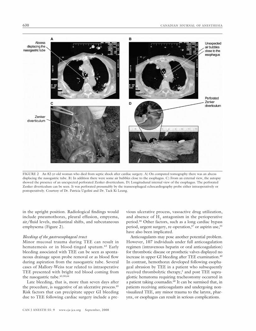
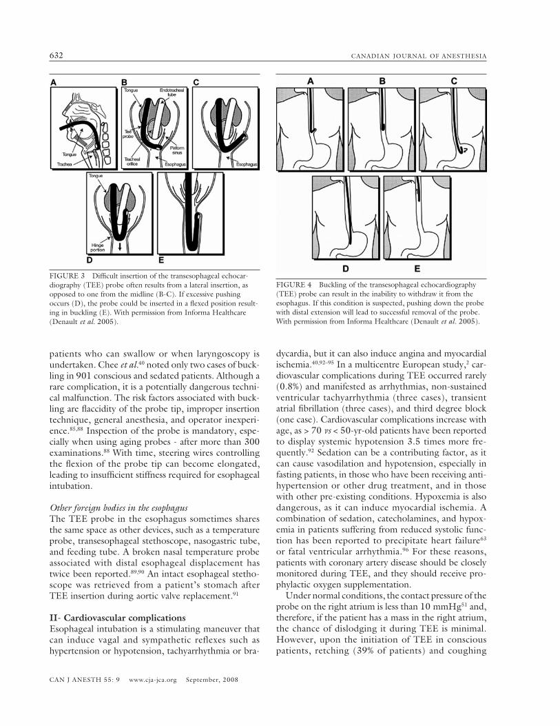
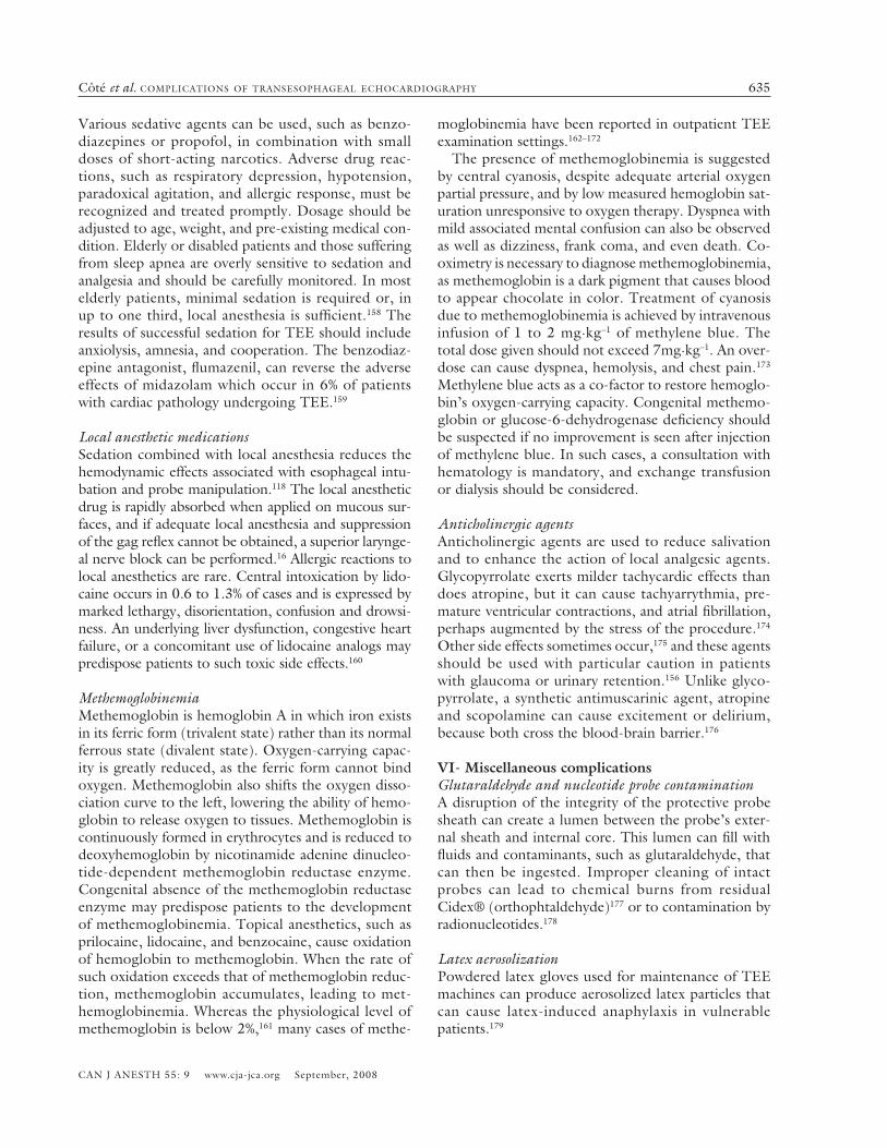
Probe tip bucklingProbe tip buckling should be suspected when imaging is difficult, when inappropriate resistance is felt dur-ing probe advancement, removal or mobilization, and when the control knobs are fixed.81,85,86 When buck-ling is encountered, the probe should be advanced into the stomach where it can recover its neutral posi-tion prior to withdrawal. If the probe is pulled out in haste, its distal end can injure the esophagus (Figures 3 and 4).87
Such buckling is unlikely to occur in conscious
632 CANADIAN JOURNAL OF ANESTHESIA
CAN J ANESTH 55: 9 www.cja-jca.org September, 2008
patients who can swallow or when laryngoscopy is undertaken. Chee et al.40 noted only two cases of buck-ling in 901 conscious and sedated patients. Although a rare complication, it is a potentially dangerous techni-cal malfunction. The risk factors associated with buck-ling are flaccidity of the probe tip, improper insertion technique, general anesthesia, and operator inexperi-ence.85,88 Inspection of the probe is mandatory, espe-cially when using aging probes - after more than 300 examinations.88 With time, steering wires controlling the flexion of the probe tip can become elongated, leading to insufficient stiffness required for esophageal intubation.
Other foreign bodies in the esophagusThe TEE probe in the esophagus sometimes shares the same space as other devices, such as a temperature probe, transesophageal stethoscope, nasogastric tube, and feeding tube. A broken nasal temperature probe associated with distal esophageal displacement has twice been reported.89,90 An intact esophageal stetho-scope was retrieved from a patient’s stomach after TEE insertion during aortic valve replacement.91
II- Cardiovascular complicationsEsophageal intubation is a stimulating maneuver that can induce vagal and sympathetic reflexes such as hypertension or hypotension, tachyarrhythmia or bra-
dycardia, but it can also induce angina and myocardial ischemia.40,92–95 In a multicentre European study,2 car-diovascular complications during TEE occurred rarely (0.8%) and manifested as arrhythmias, non-sustained ventricular tachyarrhythmia (three cases), transient atrial fibrillation (three cases), and third degree block (one case). Cardiovascular complications increase with age, as > 70 vs < 50-yr-old patients have been reported to display systemic hypotension 3.5 times more fre-quently.92 Sedation can be a contributing factor, as it can cause vasodilation and hypotension, especially in fasting patients, in those who have been receiving anti-hypertension or other drug treatment, and in those with other pre-existing conditions. Hypoxemia is also dangerous, as it can induce myocardial ischemia. A combination of sedation, catecholamines, and hypox-emia in patients suffering from reduced systolic func-tion has been reported to precipitate heart failure63 or fatal ventricular arrhythmia.96 For these reasons, patients with coronary artery disease should be closely monitored during TEE, and they should receive pro-phylactic oxygen supplementation. Under normal conditions, the contact pressure of the probe on the right atrium is less than 10 mmHg51 and, therefore, if the patient has a mass in the right atrium, the chance of dislodging it during TEE is minimal. However, upon the initiation of TEE in conscious patients, retching (39% of patients) and coughing
FIGURE 3 Difficult insertion of the transesophageal echocar-diography (TEE) probe often results from a lateral insertion, as opposed to one from the midline (B-C). If excessive pushing occurs (D), the probe could be inserted in a flexed position result-ing in buckling (E). With permission from Informa Healthcare (Denault et al. 2005).
FIGURE 4 Buckling of the transesophageal echocardiography (TEE) probe can result in the inability to withdraw it from the esophagus. If this condition is suspected, pushing down the probe with distal extension will lead to successful removal of the probe. With permission from Informa Healthcare (Denault et al. 2005).
Côté et al. complicationsoftransesophagealechocardiography 633
CAN J ANESTH 55: 9 www.cja-jca.org September, 2008
commonly occur despite sedation and local anesthe-sia of the pharynx. The associated Valsalva maneuver increases intrathoracic, central venous, and pulmonary pressures, and its release results in an abrupt decrease of systemic pressure. The pressure variation by the Valsalva maneuver, used during echocardiographic examination to detect inter-atrial septal defects by its enhancement of right-to-left shunt, is thought also to cause the mobilization of any intracardiac mate-rial present. The extent of such embolization will depend on the hemodynamic fluctuations as well as on the size, density, and mobility of the mass.97 It has been reported that large intrathoracic pressures and hemodynamic changes resulting from retching have produced fatal pulmonary embolization from a right atrial mass,98,99 embolization of mitral vegetation and a left intracardiac thrombus resulting in stroke,40,100 a myxoma embolus,99 and progressive aortic dissection and cardiac tamponade.101 Pediatric patients may be more vulnerable to TEE, in that the esophageal probe can significantly com-press vascular structures69 such as a normally posi-tioned34 or aberrant right subclavian artery,102,103 the descending aorta,104,105 the innominate artery,106 and the pulmonary venous confluence in an infant with total anomalous pulmonary venous return.107–109 The hemodynamic effect of the TEE on cerebral perfusion pressure must also be reduced, especially in the pres-ence of elevated intracranial pressure. In all of these situations, a strategy must be developed based on pre-existing medical conditions.
III- Respiratory complicationsAs with endoscopic procedures, TEE examinations in sedated patients may be associated with a small, but significant, reduction of oxygen saturation. During endoscopy, the degree of desaturation is usually mod-est, about 4 to 6%, but desaturations as low as 51% have also been reported.110,111 Whereas a mean fall in oxygen saturation reached 4.7% (ranging from 1% to 49%) in mildly sedated patients undergoing TEE, 18% of the patients exhibited significant hypoxemia, as defined by a persistent oxygen saturation below 90%.112 Obe-sity increases the likelihood for oxygen desaturation during upper GI endoscopy and for aspiration during endoscopy,113,114 although one study did not find any difference in transient oxygen desaturation between obese (mean body mass index 41 kg·m–2) and non-obese patients undergoing TEE examination.115 When performed in the emergency department, TEE may cause further complications such as respiratory insuf-ficiency and failure116 (e.g., location-related examina-tion complications). For these reasons, prophylactic
oxygen supplementation should be administered to sedated conscious patients undergoing TEE. Such prophylaxis improves, but does not abolish, the oxy-gen desaturation, and, therefore, it is no substitute for diligent oxygen monitoring. Although pulmonary complications during TEE in awake patients are rare, the potential difficulties include bronchospasm,2,117 laryngospasm,118 posterior pharyn-geal wall hematoma,41 supraglottic hematoma,68 and subglottic stenosis.119 Unilateral and bilateral pulmo-nary edema,21,118,120 atelectasis,121 and airway obstruc-tion122 can also be encountered. Prior oropharyngeal dysfunction and sedation enhance the risk for aspira-tion and call for brief endotracheal intubation under the care of an anesthesiologist. An erroneous insertion of the esophageal tube in the trachea has been reported in four out of 1,500 examinations (0.27%) in ambulatory adults.117 Symp-tomatically, such occurrences can be recognized by shortness of breath, stridor, and incessant coughing,117 however, these symptoms can be masked by sedation. With accidental tracheal intubation, image quality is poor, the distinction of distal structures is difficult, and resistance to probe insertion is perceived at about 30–32 cm.123–125 Also, the short axis of the aortic valve and the pulmonary artery bifurcation will not be seen at 30 cm,124 and long axis views of the aortic arch, sim-ilar to those obtained from a suprasternal transducer position, will display poor distal resolution.124,126
Esophageal tube placement, movement, or remov-al may alter the placement of an endotracheal tube, particularly in the pediatric population, where extu-bation,75,127 displacement into a large bronchus,75,90 damage to the pilot cuff,128 and a 1–2% incidence of severe airway obstruction have been reported.75,129 Airway obstruction by a similar mechanism was also reported in two cases of adults undergoing repair of aortic aneurisms.130 Even a compression by the esoph-ageal probe of the pulmonary tree or the endotra-cheal tube can alter ventilation,75,122,129,131–135 especially when congenital cardiovascular abnormalities, such as double aortic arch and truncus arteriosus, co-exist.122 Decreased oxygen saturation, increased ventilation pressure, and modification of ventilation pattern of end-tidal CO2 could signal such compression by the esophageal probe and should be assessed, not only under a static probe position,136 but also dynamically, during the manipulation of the probe.
IV- Infections and prophylactic antibioticsThe risk of bacteremia associated with TEE must be considered, because bacteremia can lead to morbid infections, depending on a patient’s immune status
634 CANADIAN JOURNAL OF ANESTHESIA
CAN J ANESTH 55: 9 www.cja-jca.org September, 2008
and pre-existing conditions, and it can also lead to endocarditis. The most likely organisms are alpha-hemolytic streptococci and staphylococci, comprising, respectively, 55% and 25% of cases associated with native valve infection and 30% and 45% of cases associ-ated with prosthetic valve infections.137 The incidence of transient bacteremia varies greatly with the proce-dure.138 Whereas the incidence of bacteremia is not altered by biopsies,139 it ranges from 2% to 15% after diagnostic upper GI endoscopy.139 Reports vary with regards to TEE, from stating a very low incidence of bacteremia, similar to anticipated blood culture con-tamination rates,140,141 to declaring an incidence of 0% to 17%119,142–144 in ambulatory and ICU patients.139 Thus far, only two cases of bacterial endocarditis tem-porally related to TEE examination have been report-ed.142,145 The single study in 24 patients reporting a 17%143–146 incidence of transient bacteremia, but no endocarditis, concluded that bacteremia was common, and that antibiotic therapy should be routinely insti-tuted during TEE.143 However, the isolated organisms in that study were not sensitive to the antimicrobial prophylactic agents recommended by the American Heart Association (AHA).143,144,147
The use of antibiotic therapy during TEE is contro-versial. The AHA guidelines suggest such prophylaxis for patients who have prosthetic valves, previous endo-carditis, complex cyanotic congenital heart disease, and surgically-constructed systemic pulmonary shunts or conduits. However, Eichelberger148 reviewed anti-biotic prophylaxis for prevention of endocarditis from 20 different centres during TEE in patients with pre-existing esophageal disease or poor oral and dental hygiene, and he concluded that there was no con-vincing evidence that antibiotic prophylaxis benefited this subgroup of patients. Further, a prospective study of 85 patients with prosthetic valves who underwent TEE disclosed that none showed evidence of bacterial endocarditis on follow-up,149 and that positive blood cultures resulted from contamination. These authors concluded that the likelihood of bacteremia is very low, and that routine chemoprophylaxis in patients with prosthetic valves is not justified, except in cases of poor oral hygiene, prolonged or traumatic TEE proce-dures, or in subjects undergoing TEE in the first two months after valve replacement. Immunosuppression, an underlying cardiac pathology, an enduring bacte-remia, and the type of organism involved might con-tribute to endocarditis and justify the use of antibiotics during TEE. Antibiotics are also essential for sepsis following intraoperative TEE associated with esopha-geal lesions, iatrogenic perforation, and mediastinitis, as discussed in a previous section.56,58
Clindamycin mouthwash has not been shown to reduce bacteremia after esophageal dilatation.150 Where-as a latex cover sheath, adding a physical barrier, could be considered,151 it may produce latex allergy. Its bulki-ness can make probe insertion difficult and add discom-fort, especially in awake patients, and its air tightness is not easily established.151 As cleaning and disinfection are still needed to prevent probe contamination, it is not clear whether the cover sheath offers any advantage over standard measures for infection prevention.63
Although not yet demonstrated with TEE, bacterial and hepatitis B virus transfer via the probe has been documented in endoscopy.152,153 The absence of a chan-nel for aspiration or for instrumentation on the TEE probe, however, greatly reduces the risk of residual contamination. Mechanical cleaning, alone, removes organic and inorganic debris, reducing microbial contamination by 99%. The probability of contami-nation is further diminished with glutaraldehyde in the reprocessing protocol. Pseudomonas contamina-tion of the lubricating jelly is another potential source of contamination. It was reported that a nosocomial outbreak of Legionella pneumophila was caused by a contaminated TEE probe.154 Evidently, the strains iso-lated from three contaminated patients were identical to those obtained from the water used for rinsing the TEE probe. However, no endoscopy-transmitted HIV has been reported.139 HIV appears to be very sensitive to disinfection practices, as a TEE probe contaminated with high HIV viral counts in body fluids displayed undetectable HIV by reverse transcriptase polymerase chain reaction after cleaning.155 Ideally, the person-nel and operator should be aware of the viral status of the patient, however, this is generally unknown.156 The importance of vaccination and self-protection (goggles, gloves, gowns) must be emphasized, as a possible contamination from accidental biting, fluid, and droplets is unpredictable. Herpetic infection of a finger, resulting from contaminated oral secretions and herpetic conjunctivitis acquired during endoscopy from saliva spray, occurs frequently enough to deserve the sobriquet “endoscopist eye”.
V- Medication-related complicationsSedationSedation improves patient tolerance during TEE examination. It reduces coughing, vomiting, and pain. Repeated episodes of retching, not only decrease patient comfort and willingness to pursue the proce-dure, but also prevent proper cardiac imaging. Anes-thetic and medical associations have developed criteria and guidelines for non-anesthesiologists to follow when administering sedation in conscious patients.157
Côté et al. complicationsoftransesophagealechocardiography 635
CAN J ANESTH 55: 9 www.cja-jca.org September, 2008
Various sedative agents can be used, such as benzo-diazepines or propofol, in combination with small doses of short-acting narcotics. Adverse drug reac-tions, such as respiratory depression, hypotension, paradoxical agitation, and allergic response, must be recognized and treated promptly. Dosage should be adjusted to age, weight, and pre-existing medical con-dition. Elderly or disabled patients and those suffering from sleep apnea are overly sensitive to sedation and analgesia and should be carefully monitored. In most elderly patients, minimal sedation is required or, in up to one third, local anesthesia is sufficient.158 The results of successful sedation for TEE should include anxiolysis, amnesia, and cooperation. The benzodiaz-epine antagonist, flumazenil, can reverse the adverse effects of midazolam which occur in 6% of patients with cardiac pathology undergoing TEE.159
Local anesthetic medicationsSedation combined with local anesthesia reduces the hemodynamic effects associated with esophageal intu-bation and probe manipulation.118 The local anesthetic drug is rapidly absorbed when applied on mucous sur-faces, and if adequate local anesthesia and suppression of the gag reflex cannot be obtained, a superior larynge-al nerve block can be performed.16 Allergic reactions to local anesthetics are rare. Central intoxication by lido-caine occurs in 0.6 to 1.3% of cases and is expressed by marked lethargy, disorientation, confusion and drowsi-ness. An underlying liver dysfunction, congestive heart failure, or a concomitant use of lidocaine analogs may predispose patients to such toxic side effects.160
MethemoglobinemiaMethemoglobin is hemoglobin A in which iron exists in its ferric form (trivalent state) rather than its normal ferrous state (divalent state). Oxygen-carrying capac-ity is greatly reduced, as the ferric form cannot bind oxygen. Methemoglobin also shifts the oxygen disso-ciation curve to the left, lowering the ability of hemo-globin to release oxygen to tissues. Methemoglobin is continuously formed in erythrocytes and is reduced to deoxyhemoglobin by nicotinamide adenine dinucleo-tide-dependent methemoglobin reductase enzyme. Congenital absence of the methemoglobin reductase enzyme may predispose patients to the development of methemoglobinemia. Topical anesthetics, such as prilocaine, lidocaine, and benzocaine, cause oxidation of hemoglobin to methemoglobin. When the rate of such oxidation exceeds that of methemoglobin reduc-tion, methemoglobin accumulates, leading to met-hemoglobinemia. Whereas the physiological level of methemoglobin is below 2%,161 many cases of methe-
moglobinemia have been reported in outpatient TEE examination settings.162–172
The presence of methemoglobinemia is suggested by central cyanosis, despite adequate arterial oxygen partial pressure, and by low measured hemoglobin sat-uration unresponsive to oxygen therapy. Dyspnea with mild associated mental confusion can also be observed as well as dizziness, frank coma, and even death. Co-oximetry is necessary to diagnose methemoglobinemia, as methemoglobin is a dark pigment that causes blood to appear chocolate in color. Treatment of cyanosis due to methemoglobinemia is achieved by intravenous infusion of 1 to 2 mg·kg–1 of methylene blue. The total dose given should not exceed 7mg·kg–1. An over-dose can cause dyspnea, hemolysis, and chest pain.173 Methylene blue acts as a co-factor to restore hemoglo-bin’s oxygen-carrying capacity. Congenital methemo-globin or glucose-6-dehydrogenase deficiency should be suspected if no improvement is seen after injection of methylene blue. In such cases, a consultation with hematology is mandatory, and exchange transfusion or dialysis should be considered.
Anticholinergic agentsAnticholinergic agents are used to reduce salivation and to enhance the action of local analgesic agents. Glycopyrrolate exerts milder tachycardic effects than does atropine, but it can cause tachyarrythmia, pre-mature ventricular contractions, and atrial fibrillation, perhaps augmented by the stress of the procedure.174 Other side effects sometimes occur,175 and these agents should be used with particular caution in patients with glaucoma or urinary retention.156 Unlike glyco-pyrrolate, a synthetic antimuscarinic agent, atropine and scopolamine can cause excitement or delirium, because both cross the blood-brain barrier.176
VI- Miscellaneous complicationsGlutaraldehyde and nucleotide probe contaminationA disruption of the integrity of the protective probe sheath can create a lumen between the probe’s exter-nal sheath and internal core. This lumen can fill with fluids and contaminants, such as glutaraldehyde, that can then be ingested. Improper cleaning of intact probes can lead to chemical burns from residual Cidex® (orthophtaldehyde)177 or to contamination by radionucleotides.178
Latex aerosolizationPowdered latex gloves used for maintenance of TEE machines can produce aerosolized latex particles that can cause latex-induced anaphylaxis in vulnerable patients.179

636 CANADIAN JOURNAL OF ANESTHESIA
CAN J ANESTH 55: 9 www.cja-jca.org September, 2008
Location-related complicationsPerforming TEE in the emergency department has a higher complication rate than in other clinical set-tings that average 1% to 3% (Table II). A retrospective review of TEE examinations in emergency depart-ments demonstrated a 12.6% incidence of complica-tions, including death, respiratory insufficiency or failure, hypotension, emesis, agitation, and cardiac dysrythmia.116 Compared to patients with medical or elective conditions, trauma patients experience a higher incidence of complications, because they often present with marginal hemodynamic and respiratory conditions and with multiple co-existing injuries (such as unstable C-spine damage). Considered as subjects with full stomachs, such trauma patients also have a significant risk of aspiration, especially when combined with altered levels of consciousness. Before proceeding with TEE examination in that population, it is manda-tory to protect the airway by endotracheal intubation. Whereas TEE is a versatile tool for diagnosing and treating severely ill patients, lack of space in the ICU suite and difficult passage of the probe render this pro-cedure problematic.180,181
Transnasal esophageal echocardiographyWhereas transnasal probe introduction has recently been suggested, this procedure has significant short-comings, such as a failure rate of 10% to 16%,182–184 nasopharyngeal bleeding in 0 to 31%,182–184 and dif-ficult management requiring nasal tamponade. It is wrong to believe that the transnasal approach protects against esophageal perforation. In addition, image quality and its interpretation appear to be less precise with this approach, thus arguing against nasal intuba-tion as an indicated approach.
VII-Local effects of ultrasound waves on surround-ing tissuesTheoretically, a powerful ultrasound beam can cause a vibration of small gas-filled structures (i.e., cavitation), thus producing hemorrhage or hemolysis. Whereas such effects have been reported in experiments con-ducted in animals,185–187 none has been shown in humans.188 Furthermore, a powerful ultrasound beam can produce excessive tissue heat and damage and can be used in therapeutic indications such as during litho-tripsy. However, when used at low intensity, typically at 5 MHz for TEE procedures, ultrasound has yet to display any harmful effects.189,190 Therefore, the World Federation for Ultrasound in Medicine and Biology and common sense dictate that the operator know the precise power output and intensity of the ultrasound instrument being used. The operator must balance the
benefits against the risks of its use and minimize the patient’s exposure to ultrasound.
VIII-Echocardiographer expertiseBecause of the complexity of TEE and its associated risks, the American Society of Echocardiography, the Society of Cardiovascular Anesthesiologists, and the European Society of Cardiology have strict require-ments for optimal initial training of echocardiogra-phers and for their ongoing advancement of expertise in performing this procedure.191–194
IX-Prevention of transesophageal echocardiogra-phy-related complicationsPrior to undertaking TEE, the procedure must be explained to the patient, risks vs benefits stated, and
TABLE III Contraindications to TEE
1. Absolute contraindications *Lack of informed consentUnwilling and uncooperative patientLack of expertise in intubation for TEEEsophageal obstruction (cancer, stricture) Gastric volvulusActive upper gastrointestinal bleedingPerforated viscus (known or suspected)Full stomachSuspected neck injury
2. Relative contraindications2.1 Known esophageal pathology
Esophageal varices without bleedingEsophageal diverticulumTransesophageal fistula Esophagitis/inflammatory processGastric herniationSclerodermaCarcinoma Penetrating or blunt thoracic esophageal trauma History of previous esophageal surgeryEsophagectomyFundal-plication gastric surgery
2.2 Cervical abnormalitiesSevere cervical arthritis/osteophytes/severe cervical spondylosisNeck surgery/radiotherapy in the cervical region Severe oropharyngeal distortion
2.3 MiscellaneousPrior mediastinal irradiationCoagulopathyNasal intubationHistory of nasal/nose surgerySeptal deviation
*Although these contraindications are absolute, the risk/ben-efit ratio should be evaluated for every patient, and alternative approaches (limited to upper and mid-esophageal view or epiaortic view) should be sought. TEE = transesophageal echocardiography.

Côté et al. complicationsoftransesophagealechocardiography 637
CAN J ANESTH 55: 9 www.cja-jca.org September, 2008
an informed consent obtained. A complete physical examination and careful questioning on medical histo-ry, allergies, and medications are essential. The exami-nation must include an evaluation of oral hygiene and loose teeth (for preventive antibiotic treatment), an assessment of neck mobility, stability, and arthritic changes, and an analysis of the airways. Swallowing problems, a possible indicator of orogastric narrow-ing, may be a possible contraindication to TEE probe insertion195 and should prompt consultation with gastroenterology and radiological barium swallowing studies. Past GI surgery, recent endoscopic procedure, and chest wall radiotherapy may hint at esophageal abnormalities, and a chest x-ray should be performed to rule out pathologies, such as hiatus hernia (Figure 5). Sometimes, even such precautions do not prevent esophageal laceration during TEE.196
It is essential to properly store the probe holder and the probe, itself, and to protect its distal part.197–200 Prior to TEE examination, the probe must be inspect-ed for both mechanical dysfunction and for damage to the flexible outer sheath that can collect contaminated fluid causing electrical or thermal injuries,39 potentially resulting in arrhythmia and death201 or to a steel wire damage of the esophageal mucosa.202 The control sys-tem must be unlocked at all times to ensure probe flex-ibility and to prevent esophageal tear by rigid tubes.85 Monitoring vital signs at baseline and throughout TEE is essential. Blood pressure, electrocardiogram, and pulse oximetry must be monitored throughout the intervention. Oxygen supplementation and venous access should be established for sedation or to man-age any complications. Equipment should include a suction device and resuscitation cart, and supporting personnel should be present to alleviate some of the burden placed on the echocardiographer.203
The TEE examination under conscious sedation is very different from that under general anesthesia. Whereas rapid anesthetic induction in emergency situations is followed by orotracheal intubation, in an elective procedure performed in an awake, fasting patient, the patient is placed in a left lateral decubi-tus position to minimize the risk of aspiration, and a suction device is placed close at hand and ready for use.63 Sedation and local anesthesia will improve the success of esophageal intubation. Dental fixtures must be removed before intubation, and a bite guard should be placed, especially in awake procedures and in patients with full dentures, as it protects the instru-ment and the operator’s fingers. The TEE probe should be lubricated, in an unlocked control-wheel position, and its passage never forced. The probe must be kept central to the tongue and guided in
a fluid movement without excessive force in a pos-terior manner against the posterior pharyngeal wall. The awake patient is asked to swallow during probe insertion. This closes the vocal cords bringing the larynx up and forward under the posterior aspect of the tongue and opening the cricopharyngeal muscle. Under general anesthesia, a jaw thrust maneuver can help overcome resistance felt in the hypopharynx region when the probe is in contact with the epi-glottis or with other glottic structures. Probe place-ment under direct laryngoscopy may reduce the risk of lesions associated with numerous blind attempts. The screen monitor should be active to confirm the proper location of the probe in the esophagus.124 Careful insertion of 40 to 50 cm from the incisors is advocated.204 Resistance to probe insertion within the esophagus can result from esophageal abnormalities, as discussed earlier. When the probe is introduced under general anesthesia, the entire oropharyngeal musculature is relaxed. This could predispose the probe to being pushed into a mucosal fold and ulti-mately to buckle or to cause a lesion. Such a risk is reduced in awake subjects who can collaborate by swallowing the probe and indicating the presence of pain or discomfort during probe manipulation. Under general anesthesia, patients do not experience intoler-ance, retching, or gag reflex. The endotracheal tube and its cuff provide some protection against aspira-tion. Any nasogastric or feeding tube or temperature probe should be removed to avoid potential kinking, knotting, or gastric migration and loss, as well as to prevent interference during image acquisition. Spe-cial care must be taken during cardiac surgery, as the TEE probe stays much longer than during a diagnos-tic examination, and as anticoagulation during the
FIGURE 5 Chest radiograph from a 72-yr-old woman with an esophageal hernia. Air shadows can be seen in the middle of the chest (A) and behind the left atriun on the lateral view (B). (Courtesy of Dr. Patricia Ugolini).
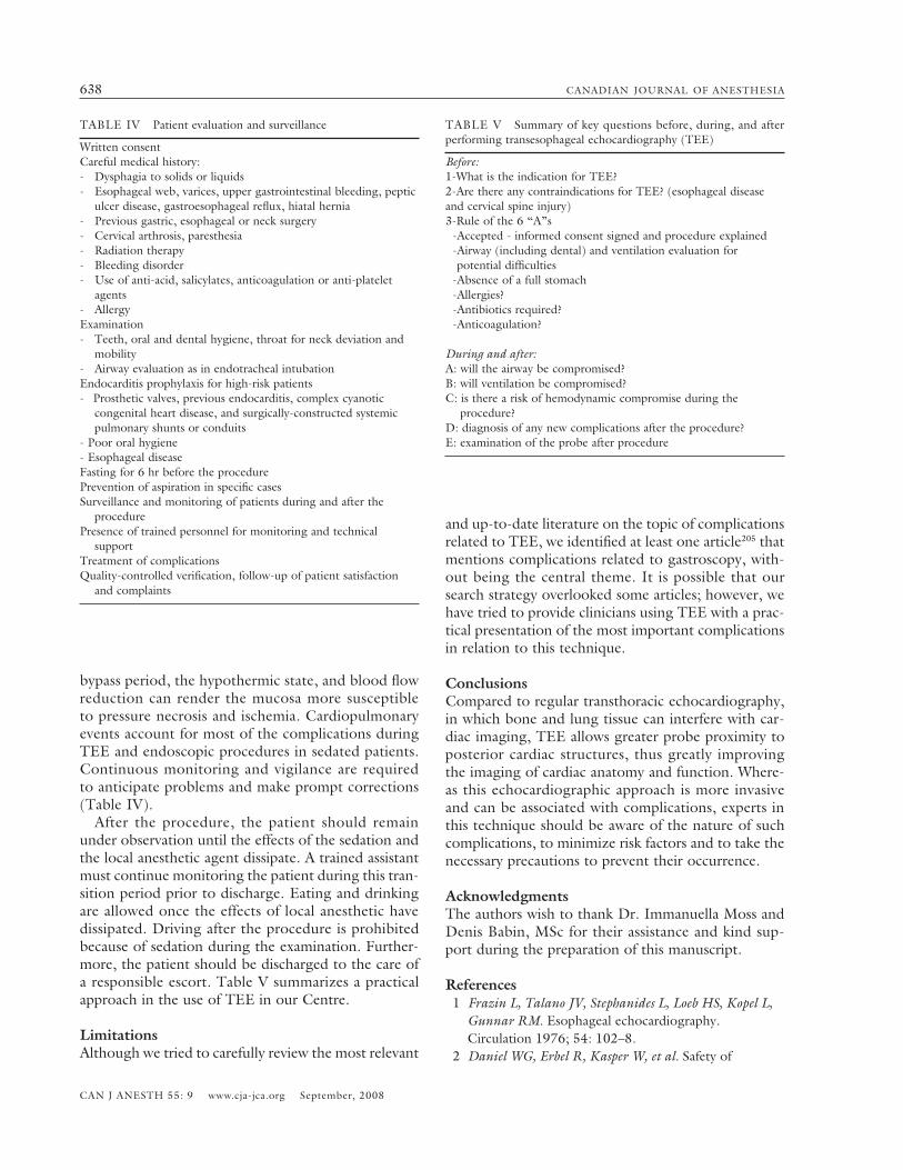
638 CANADIAN JOURNAL OF ANESTHESIA
CAN J ANESTH 55: 9 www.cja-jca.org September, 2008
bypass period, the hypothermic state, and blood flow reduction can render the mucosa more susceptible to pressure necrosis and ischemia. Cardiopulmonary events account for most of the complications during TEE and endoscopic procedures in sedated patients. Continuous monitoring and vigilance are required to anticipate problems and make prompt corrections (Table IV). After the procedure, the patient should remain under observation until the effects of the sedation and the local anesthetic agent dissipate. A trained assistant must continue monitoring the patient during this tran-sition period prior to discharge. Eating and drinking are allowed once the effects of local anesthetic have dissipated. Driving after the procedure is prohibited because of sedation during the examination. Further-more, the patient should be discharged to the care of a responsible escort. Table V summarizes a practical approach in the use of TEE in our Centre.
LimitationsAlthough we tried to carefully review the most relevant
and up-to-date literature on the topic of complications related to TEE, we identified at least one article205 that mentions complications related to gastroscopy, with-out being the central theme. It is possible that our search strategy overlooked some articles; however, we have tried to provide clinicians using TEE with a prac-tical presentation of the most important complications in relation to this technique.
ConclusionsCompared to regular transthoracic echocardiography, in which bone and lung tissue can interfere with car-diac imaging, TEE allows greater probe proximity to posterior cardiac structures, thus greatly improving the imaging of cardiac anatomy and function. Where-as this echocardiographic approach is more invasive and can be associated with complications, experts in this technique should be aware of the nature of such complications, to minimize risk factors and to take the necessary precautions to prevent their occurrence.
AcknowledgmentsThe authors wish to thank Dr. Immanuella Moss and Denis Babin, MSc for their assistance and kind sup-port during the preparation of this manuscript.
References 1 Frazin L, Talano JV, Stephanides L, Loeb HS, Kopel L,
Gunnar RM. Esophageal echocardiography. Circulation 1976; 54: 102–8.
2 Daniel WG, Erbel R, Kasper W, et al. Safety of
TABLE IV Patient evaluation and surveillance
Written consent Careful medical history: - Dysphagia to solids or liquids- Esophageal web, varices, upper gastrointestinal bleeding, peptic
ulcer disease, gastroesophageal reflux, hiatal hernia- Previous gastric, esophageal or neck surgery- Cervical arthrosis, paresthesia- Radiation therapy- Bleeding disorder- Use of anti-acid, salicylates, anticoagulation or anti-platelet
agents- AllergyExamination- Teeth, oral and dental hygiene, throat for neck deviation and
mobility- Airway evaluation as in endotracheal intubationEndocarditis prophylaxis for high-risk patients- Prosthetic valves, previous endocarditis, complex cyanotic
congenital heart disease, and surgically-constructed systemic pulmonary shunts or conduits
- Poor oral hygiene- Esophageal diseaseFasting for 6 hr before the procedurePrevention of aspiration in specific casesSurveillance and monitoring of patients during and after the
procedurePresence of trained personnel for monitoring and technical
supportTreatment of complicationsQuality-controlled verification, follow-up of patient satisfaction
and complaints
TABLE V Summary of key questions before, during, and after performing transesophageal echocardiography (TEE)
Before:1-What is the indication for TEE?2-Are there any contraindications for TEE? (esophageal disease and cervical spine injury)3-Rule of the 6 “A”s -Accepted - informed consent signed and procedure explained -Airway (including dental) and ventilation evaluation for -potential difficulties -Absence of a full stomach -Allergies? -Antibiotics required? -Anticoagulation?
During and after:A: will the airway be compromised?B: will ventilation be compromised?C: is there a risk of hemodynamic compromise during the procedure?D: diagnosis of any new complications after the procedure?E: examination of the probe after procedure
Côté et al. complicationsoftransesophagealechocardiography 639
CAN J ANESTH 55: 9 www.cja-jca.org September, 2008
transesophageal echocardiography. A multicenter survey of 10,419 examinations. Circulation 1991; 83: 817–21.
3 Sutton DC, Kluger R. Intraoperative transoesophageal echocardiography: impact on adult cardiac surgery. Anaesth Intensive Care 1998; 26: 287–93.
4 Mishra M, Chauhan R, Sharma KK, et al. Real-time intraoperative transesophageal echocardiography – how useful? Experience of 5,016 cases. J Cardiothorac Vasc Anesth 1998; 12: 625–32.
5 Click RL, Abel MD, Schaff HV. Intraoperative trans-esophageal echocardiography: 5-year prospective review of impact on surgical management. Mayo Clin Proc 2000; 75: 241–7.
6 Couture P, Denault AY, McKenty S, et al. Impact of routine use of intraoperative transesophageal echocar-diography during cardiac surgery. Can J Anesth 2000; 47: 20–6.
7 Minhaj M, Patel K, Muzic D, et al. The effect of rou-tine intraoperative transesophageal echocardiography on surgical management. J Cardiothorac Vasc Anesth 2007; 21: 800–4.
8 Gurbuz AT, Hecht ML, Arslan AH. Intraoperative transesophageal echocardiography modifies strategy in off-pump coronary artery bypass grafting. Ann Thorac Surg 2007; 83: 1035–40.
9 Eltzschig HK, Rosenberger P, Loffler M, Fox JA, Aranki SF, Shernan SK. Impact of intraoperative transesopha-geal echocardiography on surgical decisions in 12,566 patients undergoing cardiac surgery. Ann Thorac Surg 2008; 85: 845–52.
10 Suriani RJ, Neustein S, Shore-Lesserson L, Konstadt S. Intraoperative transesophageal echocardiography dur-ing noncardiac surgery. J Cardiothorac Vasc Anesth 1998; 12: 274–80.
11 Denault AY, Couture P, McKenty S, et al. Perio- perative use of transesophageal echocardiography by anesthesiologists: impact in noncardiac surgery and in the intensive care unit. Can J Anesth 2002; 49: 287–93.
12 Schulmeyer MC, Santelices E, Vega R, Schmied S. Impact of intraoperative transesophageal echocardiog-raphy during noncardiac surgery. J Cardiothorac Vasc Anesth 2006; 20: 768–71.
13 Foster E, Schiller NB. The role of transesophageal echocardiography in critical care: UCSF experience. J Am Soc Echocardiogr 1992; 5: 368–74.
14 Khoury AF, Afridi I, Quinones MA, et al. Trans- esophageal echocardiography in critically ill patients: feasibility, safety, and impact on management. Am Heart J 1994; 127: 1363–71.
15 Aeschbacher BC, Portner M, Fluri M, Meier B, Luscher TF. Midazolam premedication improves tolerance of
transesophageal echocardiography. Am J Cardiol 1998; 81: 1022–6.
16 Reed AP. Sensory blockade for difficult passage of transesophageal echocardiography probes. J Am Soc Echocardiogr 1992; 5: 375–84.
17 Cierpka K, Rickenbacher P, Burckhardt D, Luscher TF. Subjective tolerance to transesophageal echocardiogra-phy (German). Schweiz Rundsch Med Prax 1994; 83: 308–15.
18 Watts HD. Mallory-Weiss syndrome occurring as a complication of endoscopy. Gastrointest Endosc 1976; 22: 171–2.
19 Badaoui R, Choufane S, Riboulot M, Bachelet Y, Ossart M. Esophageal perforation after transesophageal echo-cardiography (French). Ann Fr Anesth Reanim 1994; 13: 850–2.
20 Vignon P, Gueret P, Chabernaud JM, et al. Failure and complications of transesophageal echocardiography. Apropos of 1500 consecutive cases (French). Arch Mal Coeur Vaiss 1993; 86: 849–55.
21 Tam JW, Burwash IG, Ascah KJ, et al. Feasibility and complications of single-plane and biplane versus multiplane transesophageal imaging: a review of 2947 consecutive studies. Can J Cardiol 1997; 13: 81–4.
22 Bettex D, Chassot PD. Échographie Transoesopha-gienne en Anesthésie-Réanimation. Masson, Williams & Williams; 2002: 1–12.
23 Dougherty Thomas B. The difficult airway in conven-tional head and neck surgery. In: Benumof J (Ed.). Airway Management Principles and Practice. St. Louis: Mosby; 1996: 688.
24 Rubesin SE, Levine MS. Killian-Jamieson diverticula: radiographic findings in 16 patients. AJR Am J Roentgenol 2001; 177: 85–9.
25 Fergus I, Bennett ES, Rogers DM, Siskind S, Messineo FC. Fluoroscopic balloon-guided transesophageal echocardiography in a patient with Zenker’s diverticu-lum. J Am Soc Echocardiogr 2004; 17: 483–6.
26 Willens HJ, Lamet M, Migikovsky B, Kessler KM. A technique for performing transesophageal echocardiog-raphy safely in patients with Zenker’s diverticulum. J Am Soc Echocardiogr 1994; 7: 534–7.
27 Ramjohn J, Paulus DA. Use of transesophageal echo-cardiography in a patient with Zenker’s diverticulum. J Cardiothorac Vasc Anesth 2006; 20: 385–6.
28 Reuss CS, Triester SL, Lynch JJ, Heigh RI, Fleischer DE. Esophageal overtube facilitation of transesophageal echocardiography in patients with previously difficult esophageal intubation. J Am Soc Echocardiogr 2007; 20: 285–9.
29 Paiste J, Williams JP. Unsuccessful placement of transesophageal echocardiography probe because of esophageal pathology. Anesth Analg 2001; 92: 870–1.
640 CANADIAN JOURNAL OF ANESTHESIA
CAN J ANESTH 55: 9 www.cja-jca.org September, 2008
30 Freedberg RS, Weinreb J, Gluck M, Kronzon I. Para-esophageal hernia may prevent cardiac imaging by transesophageal echocardiography. J Am Soc Echocardiogr 1989; 2: 202–3.
31 Seward JB, Khandheria BK, Oh JK, Freeman WK, Tajik AJ. Critical appraisal of transesophageal echocar-diography: limitations, pitfalls, and complications. J Am Soc Echocardiogr 1992; 5: 288–305.
32 Hong T, Sternlight B. Routine procedures in the operating room. In: Goldiner PL, Oka Y (Eds). Transesophageal Echocardiography. Philadelphia: JB Lippincott Cie; 1992: 29-39.
33 Massey SR, Pitsis A, Mehta D, Callaway M. Oesopha-geal perforation following perioperative trans- oesophageal echocardiography. Br J Anaesth 2000; 84: 643–6.
34 Carerj S, Paola TM, Oddo A, Lucisano V, Oreto G. Esophageal duplication cyst: a rare obstacle to trans-esophageal echocardiography. Echocardiography 1998; 15: 601–2.
35 Suriani RJ, Cutrone A, Feierman D, Konstadt S. In-traoperative transesophageal echocardiography during liver transplantation. J Cardiothorac Vasc Anesth 1996; 10: 699–707.
36 Riazi J. The difficult pediatric airway. In: Benumof J (Ed.). Airway Management Principles and Practice. St. Louis: Mosby; 1996.
37 Stevenson JG. Role of intraoperative transesophageal echocardiography during repair of congenital cardiac defects. Acta Paediatr Suppl 1995; 410: 23–33.
38 Lam J, Neirotti RA, Nijveld A, Schuller JL, Blom-Muilwijk CM, Visser CA. Transesophageal echocardiography in pediatric patients: preliminary results. J Am Soc Echocardiogr 1991; 4: 43–50.
39 Rafferty T, LaMantia KR, Davis E, et al. Quality assurance for intraoperative transesophageal echocar-diography monitoring: a report of 846 procedures. Anesth Analg 1993; 76: 228–32.
40 Chee TS, Quek SS, Ding ZP, Chua SM. Clinical utility, safety, acceptability and complications of transoesopha-geal echocardiography (TEE) in 901 patients. Singa-pore Med J 1995; 36: 479–83.
41 Saphir JR, Cooper JA, Kerbavez RJ, Larson SF, Schiller NB. Upper airway obstruction after transesophageal echocardiography. J Am Soc Echocardiogr 1997; 10: 977–8.
42 Fernandez FF, Richter A, Freudenberg S, Wendl K, Manegold BC. Treatment of endoscopic esophageal perforation. Surg Endosc 1999; 13: 962–6.
43 Dewhirst WE, Stragand JJ, Fleming BM. Mallory-Weiss tear complicating intraoperative transesophageal echocardiography in a patient undergoing aortic valve replacement. Anesthesiology 1990; 73: 777–8.
44 Latham P, Hodgins LR. A gastric laceration after trans-esophageal echocardiography in a patient undergoing aortic valve replacement. Anesth Analg 1995; 81: 641–2.
45 Pong MW, Lin SM, Kao SC, Chu CC, Ting CK, Tsai SK. Unusual cause of esophageal perforation during intraoperative transesophageal echocardiography monitoring for cardiac surgery--a case report. Acta Anaesthesiol Sin 2003; 41: 155–8.
46 St-Pierre J, Fortier LP, Couture P, Hebert Y. Massive gastrointestinal hemorrhage after transoesophageal echocardiography probe insertion. Can J Anaesth 1998; 45: 1196–9.
47 Augoustides JG, Hosalkar HH, Milas BL, Acker M, Savino JS. Upper gastrointestinal injuries related to perioperative transesophageal echocardiography: index case, literature review, classification proposal, and call for a registry. J Cardiothorac Vasc Anesth 2006; 20: 379–84.
48 Kharasch ED, Sivarajan M. Gastroesophageal perforation after intraoperative transesophageal echocardiography. Anesthesiology 1996; 85: 426–8.
49 Han YY, Cheng YJ, Liao WW, Ko WJ, Tsai SK. Delayed diagnosis of esophageal perforation following intraoperative transesophageal echocardiography during valvular replacement--a case report. Acta Anaesthesiol Sin 2003; 41: 81–4.
50 Fujii H, Suehiro S, Shibata T, Aoyama T, Ikuta T. Mallory - weiss tear complicating intraoperative trans-esophageal echocardiography. Circ J 2003; 67: 357–8.
51 Urbanowicz JH, Kernoff RS, Oppenheim G, Parnagian E, Billingham ME, Popp RL. Transesophageal echo-cardiography and its potential for esophageal damage. Anesthesiology 1990; 72: 40–3.
52 Lennon MJ, Gibbs NM, Weightman WM, Leber J, Ee HC, Yusoff IF. Transesophageal echocardiography- related gastrointestinal complications in cardiac surgical patients. J Cardiothorac Vasc Anesth 2005; 19: 141–5.
53 Zubarik R, Eisen G, Mastropietro C, et al. Prospective analysis of complications 30 days after outpatient upper endoscopy. Am J Gastroenterol 1999; 94: 1539–45.
54 Delvaux M. Complications of digestive endoscopy (French). Gastroenterol Clin Biol 1995; 19: B23–32.
55 Kallmeyer IJ, Collard CD, Fox JA, Body SC, Shernan SK. The safety of intraoperative transesopha-geal echocardiography: a case series of 7200 cardiac surgical patients. Anesth Analg 2001; 92: 1126–30.
56 Lalanne B, Dupont M. Pseudomonas aeruginosa mediastinitis after transesophageal echocardiography (French). Presse Med 1996; 25: 952.
57 Jougon J, Gallon P, Dubrez J, et al. Esophageal per-foration during transesophageal echocardiography (French). Arch Mal Coeur Vaiss 2000; 93: 1235–7.

Côté et al. complicationsoftransesophagealechocardiography 641
CAN J ANESTH 55: 9 www.cja-jca.org September, 2008
58 MacGowan SW. Intra-operative trans-oesophageal echocardiography is a potential source of sepsis in the in-tensive care. Eur J Cardiothorac Surg 2000; 17: 768–9.
59 Zalunardo MP, Bimmler D, Grob UC, Stocker R, Pasch T, Spahn DR. Late oesophageal perforation after intraoperative transoesophageal echocardiography. Br J Anaesth 2002; 88: 595–7.
60 Lecharny JB, Philip I, Depoix JP. Oesophagotracheal perforation after intraoperative transoesphageal echo-cardiography in cardiac surgery. Br J Anaesth 2002; 88: 592–4.
61 Soong W, Afifi S, McGee EC. Delayed presentation of gastric perforation after transesophageal echocardiog-raphy for cardiac surgery. Anesthesiology 2006; 105: 1273–4.
62 Lawrence DR, Moxon RE, Fountain SW, Ohri SK, Townsend ER. Iatrogenic oesophageal perforations: a clinical review. Ann R Coll Surg Engl 1998; 80: 115–8.
63 Khandheria BK. The transesophageal echocardio-graphic examination: is it safe? Echocardiography 1994; 11: 55–63.
64 De Vries AJ, van der Maaten JM, Laurens RR. Mallory-Weiss tear following cardiac surgery: transoesophageal echoprobe or nasogastric tube? Br J Anaesth 2000; 84: 646–9.
65 Hulyalkar AR, Ayd JD. Low risk of gastroesophageal injury associated with transesophageal echocardiogra-phy during cardiac surgery. J Cardiothorac Vasc Anesth 1993; 7: 175–7.
66 Norton ID, Pokorny CS, Baird DK, Selby WS. Upper gastrointestinal haemorrhage following coronary artery bypass grafting. Aust N Z J Med 1995; 25: 297–301.
67 Leitman IM, Paull DE, Barie PS, Isom OW, Shires GT. Intra-abdominal complications of cardiopulmonary bypass operations. Surg Gynecol Obstet 1987; 165: 251–4.
68 Massa N, Morrison M. Transesophageal echocardiog-raphy: an unusual case of iatrogenic laryngeal trauma. Otolaryngol Head Neck Surg 2003; 129: 602–4.
69 Greene MA, Alexander JA, Knauf DG, et al. Endoscop-ic evaluation of the esophagus in infants and children immediately following intraoperative use of transesoph-ageal echocardiography. Chest 1999; 116: 1247–50.
70 Cyran SE, Kimball TR, Meyer RA, et al. Efficacy of intraoperative transesophageal echocardiography in children with congenital heart disease. Am J Cardiol 1989; 63: 594–8.
71 O’Shea JP, Southern JF, D’Ambra MN, et al. Effects of prolonged transesophageal echocardiographic imag-ing and probe manipulation on the esophagus – an echocardiographic-pathologic study. J Am Coll Cardiol 1991; 17: 1426–9.
72 Chow MS, Taylor MA, Hanson CW III. Splenic lacera-tion associated with transesophageal echocardiography. J Cardiothorac Vasc Anesth 1998; 12: 314–6.
73 Olenchock SA Jr, Lukaszczyk JJ, Reed J III, Theman TE. Splenic injury after intraoperative transesopha-geal echocardiography. Ann Thorac Surg 2001; 72: 2141–3.
74 Pattullo GG. A complication of transoesophageal echocardiography in a patient with splenomegaly. Anaesth Intensive Care 2003; 31: 594–5.
75 Stevenson JG. Incidence of complications in pediatric transesophageal echocardiography: experience in 1650 cases. J Am Soc Echocardiogr 1999; 12: 527–32.
76 Rousou JA, Tighe DA, Garb JL, et al. Risk of dysphagia after transesophageal echocardiography during cardiac operations. Ann Thorac Surg 2000; 69: 486–90.
77 Sakai T, Terao Y, Miyata S, Hasuo H, Haseba S, Yano K. Postoperative recurrent laryngeal nerve palsy following a transesophageal echocardiography (Japanese). Masui 1999; 48: 656–7.
78 Kawahito S, Kitahata H, Kimura H, et al. Recur-rent laryngeal nerve palsy after cardiovascular surgery: relationship to the placement of a transesophageal echocardiographic probe. J Cardiothorac Vasc Anesth 1999; 13: 528–31.
79 Hogue CW Jr, Lappas GD, Creswell LL, et al. Swallow-ing dysfunction after cardiac operations. Associated adverse outcomes and risk factors including intraop-erative transesophageal echocardiography. J Thorac Cardiovasc Surg 1995; 110: 517–22.
80 Kohr LM, Dargan M, Hague A, et al. The incidence of dysphagia in pediatric patients after open heart pro-cedures with transesophageal echocardiography. Ann Thorac Surg 2003; 76: 1450–6.
81 Cucchiara RF, Nugent M, Seward JB, Messick JM. Air embolism in upright neurosurgical patients: detection and localization by two-dimensional transesophageal echocardiography. Anesthesiology 1984; 60: 353–5.
82 Yamamoto H, Fujimura N, Namiki A. Swelling of the tongue after intraoperative monitoring by transesopha-geal echocardiography (Japanese). Masui 2001; 50: 1250–2.
83 Sriram K, Khorasani A, Mbekeani KE, Patel S. Tongue necrosis and cleft after prolonged transesophageal echocardiography probe placement. Anesthesiology 2006; 105: 635.
84 Takasaki Y. Transient lingual ischaemia during anaes-thesia. Anaesthesia 2003; 58: 717.
85 Kronzon I, Cziner DG, Katz ES, et al. Buckling of the tip of the transesophageal echocardiography probe: a potentially dangerous technical malfunction. J Am Soc Echocardiogr 1992; 5: 176–7.
86 Woodland RV, Denney JD, Moore DW, Gregg MG.
642 CANADIAN JOURNAL OF ANESTHESIA
CAN J ANESTH 55: 9 www.cja-jca.org September, 2008
Inability to remove a transesophageal echocardio-graphy probe. J Cardiothorac Vasc Anesth 1994; 8: 477–9.
87 Denault AY, Couture P, Buithieu J, Tardif JC. Trans-esophageal Echocardiography Multimedia Manual: A Perioperative Transdisciplinary Approach. Marcel Dekker; 2005.
88 Orihashi K, Sueda T, Matsuura Y, Yamanoue T, Yuge O. Buckling of transesophageal echocardiography probe: a pitfall at insertion in an anesthetized patient. Hiroshima J Med Sci 1993; 42: 155–7.
89 Yasick A, Samra SK. An unusual complication of trans-esophageal echocardiography. Anesth Analg 1995; 81: 657–8.
90 Benedict PE, Foley K. Transesophageal echocardiogra-phy not without pitfalls. J Cardiothorac Vasc Anesth 1997; 11: 123.
91 Brook M, Chard PS, Brock-Utne JG. Gastric foreign body: a potential risk when using transesophageal echo. Anesth Analg 1997; 84: 1389.
92 Stoddard MF, Longaker RA. The safety of transesopha-geal echocardiography in the elderly. Am Heart J 1993; 125: 1358–62.
93 Al Moussarih A, Douard H, Lafitte S, Broustet JP, Roudaut R. Acute myocardial infarction during trans-esophageal echocardiography. Echocardiography 1999; 16: 579–80.
94 Suriani RJ, Tzou N. Bradycardia during transesopha-geal echocardiographic probe manipulation. J Cardiothorac Vasc Anesth 1995; 9: 347.
95 Grosse-Heitmeyer W, Engberding R. Transesophageal echocardiography: increased risk by repeated attempts to insert the transducer in patients with coronary artery disease? Clin Cardiol 1993; 16: 594–8.
96 Goland S, Shimoni S, Attali M, et al. Fatal ventricular arrhythmia as a complication of transesophageal echo-cardiography. Eur J Echocardiogr 2005; 6: 151–3.
97 Kwak KD, Mosher SF, Willis CL, Kimura BJ. Witnessed embolization of a right atrial mass during transesopha-geal echocardiography: implications regarding the safety of esophageal intubation. Chest 1999; 115: 1462–4.
98 Shah CP, Thakur RK, Ip JH, Xie B, Guiraudon GM. Management of mobile right atrial thrombi: a thera-peutic dilemma. J Card Surg 1996; 11: 428–31.
99 Cavero MA, Cristobal C, Gonzalez M, Callego JC, Oteo JF, Artaza M. Fatal pulmonary embolization of a right atrial mass during transesophageal echocardiography. J Am Soc Echocardiogr 1998; 11: 397–8.
100 Black IW, Cranney GB, Walsh WF, Brender D. Embo-lization of a left atrial ball thrombus during trans-esophageal echocardiography. J Am Soc Echocardiogr 1992; 5: 271–3.
101 Kim CM, Yu SC, Hong SJ. Cardiac tamponade dur-ing transesophageal echocardiography in the patient of circumferential aortic dissection. J Korean Med Sci 1997; 12: 266–8.
102 Bensky AS, O’Brien JJ, Hammon JW. Transesophageal echo probe compression of an aberrant right subcla-vian artery. J Am Soc Echocardiogr 1995; 8: 964–6.
103 Koinig H, Schlemmer M, Keznickl FP. Occlusion of the right subclavian artery after insertion of a tran-soesophageal echocardiography probe in a neonate. Paediatr Anaesth 2003; 13: 617–9.
104 Lunn RJ, Oliver WC Jr, Hagler DJ, Danielson GK. Aortic compression by transesophageal echocardio-graphic probe in infants and children undergoing cardiac surgery. Anesthesiology 1992; 77: 587–90.
105 O’Leary PW, Hagler DJ, Seward JB, et al. Biplane intraoperative transesophageal echocardiography in congenital heart disease. Mayo Clin Proc 1995; 70: 317–26.
106 Janelle GM, Lobato EB, Tang YS. An unusual complication of transesophageal echocardiography. J Cardiothorac Vasc Anesth 1999; 13: 233–4.
107 Frommelt PC, Stuth EA. Transesophageal echocar-diographic in total anomalous pulmonary venous drainage: hypotension caused by compression of the pulmonary venous confluence during probe passage. J Am Soc Echocardiogr 1994; 7: 652–4.
108 Chang YY, Chang CI, Wang MJ, et al. The safe use of intraoperative transesophageal echocardiography in the management of total anomalous pulmonary venous connection in newborns and infants: a case series. Paediatr Anaesth 2005; 15: 939–43.
109 Kostolny M, Schreiber C, Henze R, Vogt M, Lange R. Temporary pulmonary vein stenosis during intraop-erative transesophageal echocardiography in total cavopulmonary connection. Pediatr Cardiol 2006; 27: 134–6.
110 Lieberman DA, Wuerker CK, Katon RM. Cardiopul-monary risk of esophagogastroduodenoscopy. Role of endoscope diameter and systemic sedation. Gastroenterology 1985; 88: 468–72.
111 Bell GD, Bown S, Morden A, Coady T, Logan RF. Prevention of hypoxaemia during upper-gastrointesti-nal endoscopy by means of oxygen via nasal cannulae. Lancet 1987; 1: 1022–4.
112 Scriven AJ, Cobbe SM. Hypoxaemia during transo-esophageal echocardiography. Br Heart J 1994; 72: 133–5.
113 Dhariwal A, Plevris JN, Lo NT, Finlayson ND, Heading RC, Hayes PC. Age, anemia, and obesity-associated oxygen desaturation during upper gastro-intestinal endoscopy. Gastrointest Endosc 1992; 38: 684–8.
Côté et al. complicationsoftransesophagealechocardiography 643
CAN J ANESTH 55: 9 www.cja-jca.org September, 2008
114 Thompson AM, Park KG, Kerr F, Munro A. Safety of fibreoptic endoscopy: analysis of cardiorespiratory events. Br J Surg 1992; 79: 1046–9.
115 Garimella S, Longaker RA, Stoddard MF. Safety of transesophageal echocardiography in patients who are obese. J Am Soc Echocardiogr 2002; 15: 1396–400.
116 Gendreau MA, Triner WR, Bartfield J. Complications of transesophageal echocardiography in the ED. Am J Emerg Med 1999; 17: 248–51.
117 Chan KL, Cohen GI, Sochowski RA, Baird MG. Com-plications of transesophageal echocardiography in ambulatory adult patients: analysis of 1500 consecu-tive examinations. J Am Soc Echocardiogr 1991; 4: 577–82.
118 Khandheria BK, Seward JB, Bailey KR. Safety of transesophageal echocardiography: experience with 2070 consecutive procedures. J Am Coll Cardiol 1991; 17: 20A.
119 Liu JH, Hartnick CJ, Rutter MJ, Hartley BE, Myer CM 3rd. Subglottic stenosis associated with transesophageal echocardiography. Int J Pediatr Otorhinolaryngol 2000; 55: 47–9.
120 Stienlauf S, Witzling M, Herling M, Harpaz D. Unilateral pulmonary edema during transesophageal echocardiography. J Am Soc Echocardiogr 1998; 11: 491–3.
121 Lam J, Neirotti RA, Hardjowijono R, Blom-Muilwijk CM, Schuller JL, Visser CA. Transesophageal echocar-diography with the use of a four-millimeter probe. J Am Soc Echocardiogr 1997; 10: 499–504.
122 Phoon CK, Bhardwaj N. Airway obstruction caused by transesophageal echocardiography in a patient with double aortic arch and truncus arteriosus. J Am Soc Echocardiogr 1999; 12: 540.
123 Fagan LF Jr, Weiss R, Castello R, Labovita AJ. Trans-tracheal placement and imaging with a transesopha-geal echocardiographic probe. Am J Cardiol 1991; 67: 909–10.
124 Sutton DC. Accidental transtracheal imaging with a transesophageal echocardiography probe. Anesth Analg 1997; 85: 760–2.
125 Ortega R, Hesselvik JF, Chandhok D, Gu F. When the transesophageal echo probe goes into the trachea. J Cardiothorac Vasc Anesth 1999; 13: 114–5.
126 Salcedo J, Campo JM, Gil A, Revillo MP, Milazzo A. Enterococcus meningitis. Apropos of a new case (Spanish). Rev Clin Esp 1985; 177: 42–3.
127 Lam J, Neirotti RA, Lubbers WJ, et al. Usefulness of biplane transesophageal echocardiography in neonates, infants and children with congenital heart disease. Am J Cardiol 1993; 72: 699–706.
128 Lutz JT, Lambert AS, Rouine-Rapp K. Damage to pilot cuff tubing during intraoperative transesopha-
geal echocardiography. Anesth Analg 1999; 88: 1187.
129 Bezold LI, Pignatelli R, Altman CA, et al. Intraopera-tive transesophageal echocardiography in congenital heart surgery. The Texas Children’s Hospital experi-ence. Tex Heart Inst J 1996; 23: 108–15.
130 Nakao S, Eguchi T, Ikeda S, Nagata A, Nishigawa N, Shingu K. Airway obstruction by a transesophageal echocardiography probe in an adult patient with a dissecting aneurysm of the ascending aorta and arch. J Cardiothorac Vasc Anesth 2000; 14: 186–7.
131 Gilbert TB, Panico FG, McGill WA, Martin GR, Halley DG, Sell JE. Bronchial obstruction by trans-esophageal echocardiography probe in a pediatric cardiac patient. Anesth Analg 1992; 74: 156–8.
132 Muhiudeen IA, Silverman NH, Anderson RH. Transesophageal transgastric echocardiography in infants and children: the subcostal view equivalent. J Am Soc Echocardiogr 1995; 8: 231–44.
133 Stevenson JG, Sorensen GK. Proper probe size for pediatric transesophageal echocardiography. Am J Cardiol 1993; 72: 491–2.
134 Zestos MM, Chehade M, Mossad E. A transesophageal echocardiography probe causes airway obstruction in an older child. J Cardiothorac Vasc Anesth 1998; 12: 65–6.
135 Ho AC, Tan PP, Yang MW, Chung PC, Chang CH. Airway compression by a biplane pediatric trans-esophageal echocardiography probe: case report. Changgeng Yi Xue Za Zhi 1998; 21: 333–7.
136 Andropoulos DB, Ayres NA, Stayer SA, Bent ST, Campos CJ, Fraser CD. The effect of transesopha-geal echocardiography on ventilation in small infants undergoing cardiac surgery. Anesth Analg 2000; 90: 47–9.
137 Rey JR, Axon A, Budzynska A, Kruse A, Nowak A. Guidelines of the European Society of Gastrointestinal Endoscopy (E.S.G.E.) antibiotic prophylaxis for gas-trointestinal endoscopy. European Society of Gastro-intestinal Endoscopy. Endoscopy 1998; 30: 318–24.
138 Seward JB, Khandheria BK, Freeman WK, et al. Multiplane transesophageal echocardiography: image orientation, examination technique, anatomic correla-tions, and clinical applications. Mayo Clin Proc 1993; 68: 523–51.
139 Tandon RK. Disinfection of gastrointestinal endo-scopes and accessories. J Gastroenterol Hepatol 2000; 15 Suppl: G69–72.
140 Melendez LJ, Chan KL, Cheung PK, Sochowsky RA, Wong S, Austin TW. Incidence of bacteremia in trans-esophageal echocardiography: a prospective study of 140 consecutive patients. J Am Coll Cardiol 1991; 18: 1650–4.
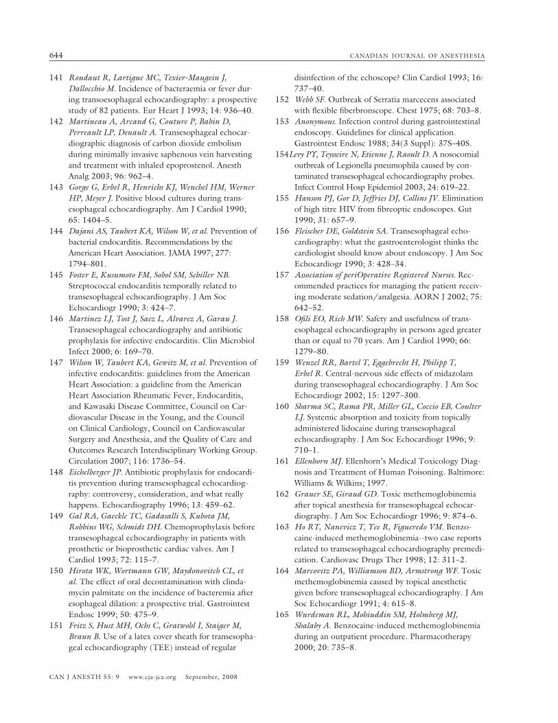
644 CANADIAN JOURNAL OF ANESTHESIA
CAN J ANESTH 55: 9 www.cja-jca.org September, 2008
141 Roudaut R, Lartigue MC, Texier-Maugein J, Dallocchio M. Incidence of bacteraemia or fever dur-ing transoesophageal echocardiography: a prospective study of 82 patients. Eur Heart J 1993; 14: 936–40.
142 Martineau A, Arcand G, Couture P, Babin D, Perreault LP, Denault A. Transesophageal echocar-diographic diagnosis of carbon dioxide embolism during minimally invasive saphenous vein harvesting and treatment with inhaled epoprostenol. Anesth Analg 2003; 96: 962–4.
143 Gorge G, Erbel R, Henrichs KJ, Wenchel HM, Werner HP, Meyer J. Positive blood cultures during trans-esophageal echocardiography. Am J Cardiol 1990; 65: 1404–5.
144 Dajani AS, Taubert KA, Wilson W, et al. Prevention of bacterial endocarditis. Recommendations by the American Heart Association. JAMA 1997; 277: 1794–801.
145 Foster E, Kusumoto FM, Sobol SM, Schiller NB. Streptococcal endocarditis temporally related to transesophageal echocardiography. J Am Soc Echocardiogr 1990; 3: 424–7.
146 Martinez LJ, Tost J, Saez L, Alvarez A, Garau J. Transesophageal echocardiography and antibiotic prophylaxis for infective endocarditis. Clin Microbiol Infect 2000; 6: 169–70.
147 Wilson W, Taubert KA, Gewitz M, et al. Prevention of infective endocarditis: guidelines from the American Heart Association: a guideline from the American Heart Association Rheumatic Fever, Endocarditis, and Kawasaki Disease Committee, Council on Car-diovascular Disease in the Young, and the Council on Clinical Cardiology, Council on Cardiovascular Surgery and Anesthesia, and the Quality of Care and Outcomes Research Interdisciplinary Working Group. Circulation 2007; 116: 1736–54.
148 Eichelberger JP. Antibiotic prophylaxis for endocardi-tis prevention during transesophageal echocardiog-raphy: controversy, consideration, and what really happens. Echocardiography 1996; 13: 459–62.
149 Gal RA, Gaeckle TC, Gadasalli S, Kubota JM, Robbins WG, Schmidt DH. Chemoprophylaxis before transesophageal echocardiography in patients with prosthetic or bioprosthetic cardiac valves. Am J Cardiol 1993; 72: 115–7.
150 Hirota WK, Wortmann GW, Maydonovitch CL, et al. The effect of oral decontamination with clinda-mycin palmitate on the incidence of bacteremia after esophageal dilation: a prospective trial. Gastrointest Endosc 1999; 50: 475–9.
151 Fritz S, Hust MH, Ochs C, Gratwohl I, Staiger M, Braun B. Use of a latex cover sheath for transesopha-geal echocardiography (TEE) instead of regular
disinfection of the echoscope? Clin Cardiol 1993; 16: 737–40.
152 Webb SF. Outbreak of Serratia marcecens associated with flexible fiberbronscope. Chest 1975; 68: 703–8.
153 Anonymous. Infection control during gastrointestinal endoscopy. Guidelines for clinical application. Gastrointest Endosc 1988; 34(3 Suppl): 37S–40S.
154 Levy PY, Teysseire N, Etienne J, Raoult D. A nosocomial outbreak of Legionella pneumophila caused by con-taminated transesophageal echocardiography probes. Infect Control Hosp Epidemiol 2003; 24: 619–22.
155 Hanson PJ, Gor D, Jeffries DJ, Collins JV. Elimination of high titre HIV from fibreoptic endoscopes. Gut 1990; 31: 657–9.
156 Fleischer DE, Goldstein SA. Transesophageal echo-cardiography: what the gastroenterologist thinks the cardiologist should know about endoscopy. J Am Soc Echocardiogr 1990; 3: 428–34.
157 Association of periOperative Registered Nurses. Rec-ommended practices for managing the patient receiv-ing moderate sedation/analgesia. AORN J 2002; 75: 642–52.
158 Ofili EO, Rich MW. Safety and usefulness of trans-esophageal echocardiography in persons aged greater than or equal to 70 years. Am J Cardiol 1990; 66: 1279–80.
159 Wenzel RR, Bartel T, Eggebrecht H, Philipp T, Erbel R. Central-nervous side effects of midazolam during transesophageal echocardiography. J Am Soc Echocardiogr 2002; 15: 1297–300.
160 Sharma SC, Rama PR, Miller GL, Coccio EB, Coulter LJ. Systemic absorption and toxicity from topically administered lidocaine during transesophageal echocardiography. J Am Soc Echocardiogr 1996; 9: 710–1.
161 Ellenhorn MJ. Ellenhorn’s Medical Toxicology Diag-nosis and Treatment of Human Poisoning. Baltimore: Williams & Wilkins; 1997.
162 Grauer SE, Giraud GD. Toxic methemoglobinemia after topical anesthesia for transesophageal echocar-diography. J Am Soc Echocardiogr 1996; 9: 874–6.
163 Ho RT, Nanevicz T, Yee R, Figueredo VM. Benzo-caine-induced methemoglobinemia--two case reports related to transesophageal echocardiography premedi-cation. Cardiovasc Drugs Ther 1998; 12: 311–2.
164 Marcovitz PA, Williamson BD, Armstrong WF. Toxic methemoglobinemia caused by topical anesthetic given before transesophageal echocardiography. J Am Soc Echocardiogr 1991; 4: 615–8.
165 Wurdeman RL, Mohiuddin SM, Holmberg MJ, Shalaby A. Benzocaine-induced methemoglobinemia during an outpatient procedure. Pharmacotherapy 2000; 20: 735–8.
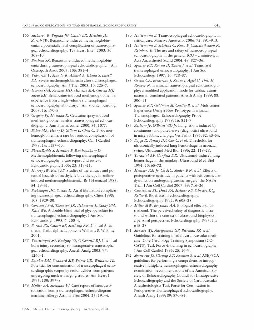
Côté et al. complicationsoftransesophagealechocardiography 645
CAN J ANESTH 55: 9 www.cja-jca.org September, 2008
166 Sachdeva R, Pugeda JG, Casale LR, Meizlish JL, Zarich SW. Benzocaine-induced methemoglobin-emia: a potentially fatal complication of transesopha-geal echocardiography. Tex Heart Inst J 2003; 30: 308–10.
167 Birchem SK. Benzocaine-induced methemoglobin-emia during transesophageal echocardiography. J Am Osteopath Assoc 2005; 105: 381–4.
168 Vidyarthi V, Manda R, Ahmed A, Khosla S, Lubell DL. Severe methemoglobinemia after transesophageal echocardiography. Am J Ther 2003; 10: 225–7.
169 Novaro GM, Aronow HD, Militello MA, Garcia MJ, Sabik EM. Benzocaine-induced methemoglobinemia: experience from a high-volume transesophageal echocardiography laboratory. J Am Soc Echocardiogr 2003; 16: 170–5.
170 Gregory PJ, Matsuda K. Cetacaine spray-induced methemoglobinemia after transesophageal echocar-diography. Ann Pharmacother 2000; 34: 1077.
171 Fisher MA, Henry D, Gillam L, Chen C. Toxic met-hemoglobinemia: a rare but serious complication of transesophageal echocardiography. Can J Cardiol 1998; 14: 1157–60.
172 BheemReddy S, Messineo F, Roychoudhury D. Methemoglobinemia following transesophageal echocardiography: a case report and review. Echocardiography 2006; 23: 319–21.
173 Harvey JW, Keitt AS. Studies of the efficacy and po-tential hazards of methylene blue therapy in aniline-induced methaemoglobinaemia. Br J Haematol 1983; 54: 29–41.
174 Berkompas DC, Saeian K. Atrial fibrillation complicat-ing transesophageal echocardiography. Chest 1993; 103: 1929–30.
175 Gorcsan J 3rd, Thornton JK, DiLucente L, Ziady GM, Katz WE. A double-blind trial of glycopyrrolate for transesophageal echocardiography. J Am Soc Echocardiogr 1993; 6: 200–4.
176 Barash PG, Cullen BF, Stoelting RK. Clinical Anes-thesia. Philadelphia: Lippincott Williams & Wilkins; 2001.
177 Venticinque SG, Kashyap VS, O’Connell RJ. Chemical burn injury secondary to intraoperative transesopha-geal echocardiography. Anesth Analg 2003; 97: 1260–1.
178 Dunker DH, Stoddard MF, Prince CR, Williams TE. Potential for contamination of transesophageal echo-cardiographic scopes by radionuclides from patients undergoing nuclear imaging studies. Am Heart J 1995; 130: 397–8.
179 Muller BA, Steelman VJ. Case report of latex aero-solization from a transesophageal echocardiogram machine. Allergy Asthma Proc 2004; 25: 191–4.
180 Huttemann E. Transoesophageal echocardiography in critical care. Minerva Anestesiol 2006; 72: 891–913.
181 Huttemann E, Schelenz C, Kara F, Chatzinikolaou K, Reinhart K. The use and safety of transoesophageal echocardiography in the general ICU – a minireview. Acta Anaesthesiol Scand 2004; 48: 827–36.
182 Spencer KT, Krauss D, Thurn J, et al. Transnasal transesophageal echocardiography. J Am Soc Echocardiogr 1997; 10: 728–37.
183 Greim CA, Brederlau J, Kraus I, Apfel C, Thiel H, Roewer N. Transnasal transesophageal echocardiogra-phy: a modified application mode for cardiac exami-nation in ventilated patients. Anesth Analg 1999; 88: 306–11.
184 Spencer KT, Goldman M, Cholley B, et al. Multicenter Experience Using a New Prototype Transnasal Transesophageal Echocardiography Probe. Echocardiography 1999; 16: 811–7.
185 Zachary JF, O’Brien WD Jr. Lung lesions induced by continuous- and pulsed-wave (diagnostic) ultrasound in mice, rabbits, and pigs. Vet Pathol 1995; 32: 43–54.
186 Baggs R, Penney DP, Cox C, et al. Thresholds for ultrasonically induced lung hemorrhage in neonatal swine. Ultrasound Med Biol 1996; 22: 119–28.
187 Tarantal AF, Canfield DR. Ultrasound-induced lung hemorrhage in the monkey. Ultrasound Med Biol 1994; 20: 65–72.
188 Mentzer RM Jr, Oz MC, Sladen RN, et al. Effects of perioperative nesiritide in patients with left ventricular dysfunction undergoing cardiac surgery: the NAPA Trial. J Am Coll Cardiol 2007; 49: 716–26.
189 Carstensen EL, Duck FA, Meltzer RS, Schwarz KQ, Keller B. Bioeffects in echocardiography. Echocardiography 1992; 9: 605–23.
190 Miller MW, Brayman AA. Biological effects of ul-trasound. The perceived safety of diagnostic ultra-sound within the context of ultrasound biophysics: a personal perspective. Echocardiography 1997; 14: 615–28.
191 Stewart WJ, Aurigemma GP, Bierman FZ, et al. Guidelines for training in adult cardiovascular medi-cine. Core Cardiology Training Symposium (CO-CATS). Task Force 4: training in echocardiography. J Am Coll Cardiol 1995; 25: 16–9.
192 Shanewise JS, Cheung AT, Aronson S, et al. ASE/SCA guidelines for performing a comprehensive intraop-erative multiplane transesophageal echocardiography examination: recommendations of the American So-ciety of Echocardiography Council for Intraoperative Echocardiography and the Society of Cardiovascular Anesthesiologists Task Force for Certification in Perioperative Transesophageal Echocardiography. Anesth Analg 1999; 89: 870–84.

646 CANADIAN JOURNAL OF ANESTHESIA
CAN J ANESTH 55: 9 www.cja-jca.org September, 2008
193 Flachskampf FA, Decoodt P, Fraser AG, et al. Guidelines from the Working Group. Recommenda-tions for performing transesophageal echocardiogra-phy. Eur J Echocardiogr 2001; 2: 8–21.
194 Cardiovascular Section of the Canadian Anesthesiologists’ Society; Beique F, Ali M, Hynes M, et al. Canadian guidelines for training in adult perioperative transesophageal echocardiography. Recommendations of the Cardiovascular Section of the Canadian Anesthesiologists’ Society and the Canadian Society of Echocardiography. Can J Cardiol 2006; 22: 1015–27.
195 Spahn DR, Schmid S, Carrel T, Pasch T, Schmid ER. Hypopharynx perforation by a transesophageal echocardiography probe. Anesthesiology 1995; 82: 581–3.
196 Ghafoor AU, Schmitz ML, Mayhew JF. Esophageal mucosal tear from a transesophageal echocardiogra-phy probe despite preliminary assessment via esopha-goscopy in a patient with esophageal disease. J Cardiothorac Vasc Anesth 2004; 18: 78–9.
197 Taillefer J, Couture P, Sheridan P, Girard A, Babin D, Denault A. A comprehensive strategy to avoid transesophageal echocardiography probe dam-age. Can J Anesth 2002; 49: 500–2.
198 Kainuma M, Miyake T. A convenient holder of the transesophageal echocardiography probe. Anesthesiology 2000; 93: 1564.
199 Spiess BD, Bruck DA. Transesophageal echocardiogra-phy probe holder. Anesth Analg 1997; 85: 945.
200 Brooker RF. Transesophageal echocardiography probe holder. Anesth Analg 1994; 79: 810.
201 Switz DM, Clarke AM, Longacher JW Jr. Electrical malfunction at endoscopy. Possible cause of arrhyth-mia and death. JAMA 1976; 235: 273–5.
202 Chan D. Echocardiography in thoracic trauma. Emerg Med Clin North Am 1998; 16: 191–207.
203 Weinger MB, Herndon OW, Gaba DM. The effect of electronic record keeping and transesophageal echocardiography on task distribution, workload, and vigilance during cardiac anesthesia. Anesthesiology 1997; 87: 144–55.
204 Dittrich H. Clinical Transesophageal Echocardiogra-phy. Mosby; 1992.
205 Gianni M, Dentali F, Grandi AM, Sumner G, Hiralal R, Lonn E. Apical ballooning syndrome or takotsubo cardiomyopathy: a systematic review. Eur Heart J 2006; 27: 1523–9.
206 El-Chami MF, Martin RP, Lerakis S. Esophageal dissection complicating transesophageal echocar-diogram--the lesson to be learned: do not force the issue. J Am Soc Echocardiogr 2006; 19: 579–7.
207 Min JK, Spencer KT, Furlong KT, et al. Clinical fea-
tures of complications from transesophageal echo-cardiography: a single-center case series of 10,000 consecutive examinations. J Am Soc Echocardiogr 2005; 18: 925–9.
208 MacGregor DA, Zvara DA, Treadway RM Jr, et al. Late presentation of esophageal injury after trans-esophageal echocardiography. Anesth Analg 2004; 99: 41–4.
209 Sobrino MA, Kozarek R, Low DE. Primary endoscopic management of esophageal perforation following transesophageal echocardiogram. J Clin Gastroenterol 2004; 38: 581–5.
210 Aviv JE, Di Tullio MR, Homma S, et al. Hypopha-ryngeal perforation near-miss during transesophageal echocardiography. Laryngoscope 2004; 114: 821–6.
211 Nana AM, Stefanidis C, Chami JP, Deviere J, Barvais L, De Smet JM. Esophageal perforation by echoprobe during cardiac surgery: treatment by endoscopic stenting. Ann Thorac Surg 2003; 75: 1955–7.
212 Law-Koune JD, Fischler M. A new case of perforation of the esophagus during intraoperative transesopha-geal echocardiography (French). Ann Fr Anesth Reanim 2002; 21: 310–4.
213 Brinkman WT, Shanewise JS, Clements SD, Mansour KA. Transesophageal echocardiography: not an innocuous procedure. Ann Thorac Surg 2001; 72: 1725–6.
214 Muhiudeen-Russell IA, Miller-Hance WC, Silverman NH. Unrecognized esophageal perforation in a neo-nate during transesophageal echocardiography. J Am Soc Echocardiogr 2001; 14: 747–9.
215 Kerbaul F, Renard S, Guidon C, et al. Acute esoph-agoarterial perforation and hemorrhagic shock during transesophageal echocardiography that occurs after heart-lung transplantation. J Heart Lung Transplant 2004; 23: 509–11.
216 Kihara S, Mizutani T, Shimizu T, Toyooka H. Bleed-ing from a tear in the gastric mucosa caused by transoesophageal echocardiography during cardiac surgery: effective haemostasis by endoscopic argon plasma coagulation. Br J Anaesth 1999; 82: 948–50.
217 Shapira MY, Hirshberg B, Agid R, Zuckerman E, Caraco Y. Esophageal perforation after transesopha-geal echocardiogram. Echocardiography 1999; 16: 151–4.
218 Savino JS, Hanson CW 3rd, Bigelow DC, Cheung AT, Weiss SJ. Oropharyngeal injury after transesophageal echocardiography. J Cardiothorac Vasc Anesth 1994; 8: 76–8.
219 Polhamus CD, Werth TE, Clement DJ, Keogh M, Lewis P. Gastrointestinal bleeding complicating trans-
Côté et al. complicationsoftransesophagealechocardiography 647
CAN J ANESTH 55: 9 www.cja-jca.org September, 2008
esophageal echocardiography. Endoscopy 1993; 25: 198–9.
220 Cujec B, Sullivan H, Wilansky S, Pollick C. Trans-esophageal echocardiography: experience of a Cana-dian centre. Can J Cardiol 1989; 5: 255–62.
221 Khandheria BK, Oh JK, Seward JB, Freeman WK, Peterson C. Transesophageal echocardiography in awake patient: initial 200 procedures (6 months). J Am Soc Echocardiogr 1991; 4: II-297.