e. w. netherton m.d,. - mdedge-files-live.s3.us-east-2 ... · out ulcer a. varicos ulcee irs...
TRANSCRIPT

DRUG ERUPTIONS E . W . NETHERTON, M . D .
For practical purposes, the cutaneous manifestions of the toxic effects of drugs may be divided into two groups: (1) Those in which the injury is a result of external contact with the drug; as for example, sulphur, mercury, iodine and other drugs commonly used by the physician, and (2) eruptions which result from the introduction of drugs into the body by various methods, which most frequently is by ingestion. In the first group are classified the cases known as derma-titis venenata. Although individuals vary greatly in their suceptibil-ity to such drugs, their prolonged use, which commonly is by topical applications, produces irritation and if they are used for a long time or in a concentration which is too strong, they will produce dermatitis in a large percentage of patients. The second group is commonly spoken of as dermatitis medicamentosa. In this group there is, like-wise, an individual variation in susceptibility. Bromides, iodides, and arsenic when administered for a sufficiently long time will produce an eruption in a large number of patients, while other drugs such as quinine, phenolphthalein, and the various barbituric acid derivatives when ingested by the majority of individuals do not affect the skin, but in a few cases may produce fairly characteristic eruptions. There is a third group of cases in which it has been demonstrated that an eruption develops either after contact or by introduction into the body of a specific drug. Wise and Sulzberger1 observed a case in which a dermatitis was produced when quinine was applied externally to the skin or when it was taken by mouth. Similar cases have been reported recently by Percival.2
The mechanism of the production of drug eruptions is unknown. In the case of contact dermatitis, the epidermis is the seat of the sensi-tivity and because of this, the patch test as a method of investigation was first employed by J. Jadassohn and has been used in the recent excellent investigations of Sulzberger and Wise.8 Dermatitis which results from contact with strong irritants which produce traumatic or chemical injury to the epidermis is to be distinguished from cases in which there is an individual susceptibility to a particular drug in con-centrations which ordinarily are not harmful to the average person.
Little is known about the mechanism of the production of erup-tion due to the ingestion of drugs. The seat of tissue susceptibility is yet undetermined. Passive transfer experiments have failed to demonstrate an antigen antibody reaction which occurs in the case of protein sensitization. Intracutaneous injection of test solutions of the suspected drug as well as patch tests have proved to be useless. There-fore, in suspected cases of dermatitis medicamentosa, the physician must
4

DRUG ERUPTIONS
depend upon the history of the ingestion of a drug, a careful evalu-ation of the characteristics of the eruption, as well as upon the associ-ated constitutional symptoms. It is important that a proper diagnosis be made in the case of drug eruptions because further administration of an offending drug may in some instances be disastrous. In most instances, the only method of determining that a particular drug is responsible for an eruption is to withhold the suspected agent until the eruption subsides and then to observe the reaction which follows its subsequent ingestion. This is not always practicable or desirable; however, most patients will cooperate with the physician, especially if they have had several attacks.
Practically every active drug has been known to produce an eruption, and as new drugs are introduced, the number of cases of drug eruption becomes more numerous. Almost any type of skin lesion may result from the ingestion of drugs. These range from an erythematous macule to purpura and gangrene and sometimes carbuncular or papillomatous lesions which are produced by iodides and bromides. The eruptions may be generalized and roughly they may simulate exanthemata or they may be limited in distribution and simulate erythema multiforme, late cutaneous syphilis or granulomatous fungus infections. As in the case of arsphenamine dermatitis, the eruption may be an exudative eczematous type of eruption which eventually may terminate as a generalized exfoliative dermatitis which cannot be distinguished solely by its clinical characteristics from an exfoliative dermatitis resulting from other causes.
A single drug may produce a multiformity of lesions, and the erup-tion may differ in susceptible individuals. For example, phenol-phthalein may produce herpes in one person and urticaria or a fixed erythema multiforme like eruption in another. Likewise, similar lesions may be caused by different drugs as in the case of fixed erup-tion which results from arsphenamine in one case and phenolphtha-lein or antipyrine in another.
At times, the diagnosis of dermatitis medicamentosa is difficult. In those cases in which the eruption simulates some of the exanthemata, the eruption appears quickly, it usually is more erythematous, and is not associated with the usual signs and symptoms such as coryza, angina, photophobia, koplick spots and fever or prodromal symptoms commonly seen in the various acute infectious diseases. In some instances, when the eruption is acute and generalized, a moderate ele-vation in temperature may occur. This is especially true in the presence of phenobarbital eruptions which may occur in an individual with acute hyperthyroidism.
When the eruption is of the erythema multiforme type or in in-stances when the drug eruption simulates some of the better known
5

E. W. NETHERTON
clinical entities, as for example, skin eruptions due to bismuth simulat-ing petyriasis rosea or neurodermatitis,4 a clinical diagnosis is even more difficult.
An alert physician invariably will give proper consideration to the possibility of dermatitis medicamentosa when considering the differ-ential diagnosis of cases in which the cause is not obvious. This same precaution aptly applies to all cases of dermatitis venenata.
As previously stated, the cutaneous manifestations of the toxic effects of drugs in susceptible individuals vary greatly. Every known elementary dermatological lesion may occur and in some cases there may be a multiformity of lesions. Likewise, no clinical picture is specific for any particular drug. However, certain interesting types of cutaneous lesions result from the introduction into the body of a few of the more commonly used drugs with enough consistency to justify this very brief and incomplete discussion of such a broad sub-ject. I refer particularly to some of the cutaneous lesions which result from the ingestion of bromides, inorganic arsenic, and phenolphtha-lein. Since these lesions are not particularly common, the profession is not sufficiently familiar with them. These drugs are among those most frequently prescribed by the physician and are also the active ingredients of many proprietary remedies which are used in self-medi-cation.
BROMIDES
The most common cutaneous manifestation of the ingestion of bro-mides is an acne-like papulo-pustule which involves the acne areas. The lesions may remain discrete or they may become confluent and form painful, sluggish, indurated pustular patches roughly simulating a carbuncle. The comedo which is the primary lesion of acne vulgaris, is not a characteristic element of this lesion. Years ago, Engman and Mooks observed that bromide and iodide eruptions tended to localize around areas of previous inflammation, such as the seborrheic or acne areas. Therefore, it is important to caution individuals who have had severe acne or who are in the adolescent period to abstain from the use of these drugs. Urticaria, vesicular and furuncular lesions may be caused by bromides. A rare nodular bromoderma sometimes occurs on the face and body of infants who are nursing from mothers who are taking bromides, usually for the relief of epilepsy. Bullous lesions may occur, but they are more common in iodide eruptions.
A nodular eruption similar to erythema nodosum occasionally occurs, and this type of bromoderma is not exceedingly rare. I have seen several cases in the past few years, especially in individuals with hyperthyroidism or in neurotic women who have been taking bromides as a sedative. The lesions are cutano-subcutaneous, painful, poorly
6

DRUG ERUPTIONS
circumscribed nodules which are pink or light red in color. The lesions are not as painful as those of classical erythema nodosum, and they are not associated with acute arthritisibr other common manifestations of this disease. In the cases which I have observed, the nodules which resembled erythema nodosum nodules resulting from bromides were true to type in that they did not become softened or break down.
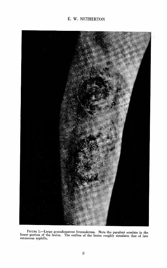
A comparatively rare type of bromoderma is the granulomatous lesion which sometimes erroneously is called a bromide gumma. This lesion occurs more frequently in women, it often follows trauma, and it has a predilection for the pretibial areas or the inner surface of the leg just above the ankle. The eruption may occur as multiple lesions or as a single unilateral, irregular, raised, painful granulomatous plaque varying in size from that of a silver dollar to that of a palm. In some instances, it may involve a large surface of the leg. The margins are raised, doughy, dull red, and sharply defined. The fol-licles are filled with caseous plugs or a purulent exudate. If poorly cared for, the surface may be crusted or bathed in a very foul pus. Pain and tenderness are outstanding characteristics of this lesion. When the leg is in a dependent position or when the lesion is pressed, the patient, experiences great discomfort. Regional adenopathy or lymphangitis does not occur. Late nodular syphilids, gumma, varicose ulcer, blastomycosis, a deeply seated fungus infection, and possibly dermatitis artifacta are the most usual conditions to be differentiated from this type of bromoderma (Fig.l).
Late nodular syphilids are indolent, and nodular or serpiginous, and they tend to ulcerate or disappear spontaneously, leaving atrophic scars which are surrounded by an area of hyperpigmentation. Syphilitic'gummata at first are dull red, poorly circumscribed cutano-subcutaneous nodules which, if untreated, terminate in a deep punched-out ulcer. A varicose ulcer is superficial; it occurs on the lower inner surface of the leg, and is associated with signs of impaired circulation such as edema and varicosity of the superficial veins. If the ulcer has been present very long, a chronic dermatitis or varicose eczema will involve the adjacent skin, and in cases of long standing, the skin be-comes sclerotic for some distance around the ulcer. Varicose ulcers frequently are painful, but as a rule they are not as tender as bromide granulomas (Fig. 2).
The similarity between blastomycosis and this type of bromoderma is very slight. Blastomycosis occurs most frequently as an indolent, granulomatous, verrucous plaque on exposed surfaces, such as the face. The pustules which occur near the margin of the lesion are small in comparison with the follicular pustules of bromoderma. Blastomycosis is a chronic condition which spreads, and the plaque may remain unchanged for months or slowly extend at its periphery.
7
E. W. NETHERTON
FIGURE 1.—Large granulomatous bromoderma. Note the purulent exudate in the lower portion of the lesion. The outline of the lesion roughly simulates that of late cutaneous syphilis.
8

DRUG ERUPTIONS
FIGURE 2.—Solitary bromide lesions. The one on the inner surface of the ankle roughly simulates late cutaneous syphilis. This lesion was so painful that the patient was unable to walk without the aid of a crutch. Prompt response was secured after the use of saline therapy.
The diagnosis is established by finding the blastomyces in the material obtained from the small peripheral abcesses.
Other deep-seated fungus infections may simulate roughly the solitary lesion of bromoderma and should at least be considered in the differentiation of this lesion.
Dermatitis artifacta or self-induced lesions, which may occur on the legs of psychopathic or neurotic individuals, are more artificial in appearance, and usually there is a history of previous lesions, some of which may have left scars. Since self-induced lesions result from the application of a chemical or from some mechanical trauma, they more closely simulate contact dermatitis, chemical burns, or traumatic ulcers. Only in the case of an individual who has been shrewd enough to produce a solitary chronic lesion is there apt to be any difficulty in differentiating these two conditions (Fig. 3).
9

E. W. NETHERTON
F I G U R E 3.—Nodular granulomatous bromide eruption.
Another uncommon bromide eruption consists of papillomatous-condylomatous lesions. The lesions are numerous and bilateral and are found most frequently on the lower parts of the l^gs, face and arms. The lesions are raised, crusted, papillomatous, dull red, painful plaques or nodules and some have many points of pustulation over their surfaces. The exudate has an offensive odor. These lesions are similar to the larger solitary granulomatous lesion previously de-scribed.
10

DRUG ERUPTIONS
F I G U R E 4.—Pigmented plaques which are the end result of Phenolphthalein eruption. This is a good example of recurrent fixed lesions which sometimes are produced by such drugs as Phenolphthalein, antipyrine, amidopyrine and arsphenamine.
Bromide eruptions differ from most drug eruptions in that they dis-appear very slowly after ingestion of the drug has been discontinued. In untreated cases, several weeks may be required before the eruption disappears.
We are indebted to Wile and his associates for the present-day treat-ment of bromodermas. In 1922, Wile and Wright6 reported the results of their investigation of eruptions caused by bromide and iodide. Among several important observations, they were able to substantiate the observation of other workers that following ingestion of bromide, a substitution occurred in the body fluids of chloride by bromide. They
11

E. W. NETHERTON
found that the chloride output in the urine increased rapidly after the ingestion of bromide for a time, but eventually gradually returned to normal even though the drug was continued. Because of this, Wile reasoned that it might be possible to displace the accumulated bro-mides by the administration of large amounts of chloride. In 1923 he7 reported that three patients who suffered from mental and cuta-nous manifestations of bromism were markedly benefited by the intra-venous administration of decinormal sodium chloride solution. In these cases, he was able to detect large amounts of bromide in the urine following the intravenous administration of the salt solution. In one of Wile's cases, there was evidence of induced nephritis which resulted from the sudden passage of too large amounts of the bromide salt through the renal epithelium. The frequency of this complication can be decreased by the administration of small initial doses of sodium chloride.
We have had an opportunity to treat several cases of bromoderma by this method and have had exceedingly gratifying results. It has been our custom'to start with the intravenous administration of 150 cc. of normal saline solution. Subsequent injections of from 200 to 250 cc. are given every two or three days. The course of treatment usually consists of from three to five injections. After the first injection, the pain and tenderness of the lesion begins to subside. Although large lesions will not disappear completely for several days, the response to this treatment is striking when compared with gradual disappearance of bromide eruptions following the discontinuance of the drug alone.
PHENOLPHTHALEIN
Another interesting type of dermatitis medicamentosa is the recur-ring, fixed, pigmentary eruption which is produced by phenolphtha-lein, antipyrine, amidopyrine, and arsphenamine. This lesion is similar to that seen in erythema multiforme. There may be few or many lesions scattered over the body without predilection for any special region. The lesions vary in size from that of a coin to the size of the palm of the hand. They are roughly oval or round, violaceous to dark red, edematous plaques. The central portion of the lesion is violaceous while the periphery may be light red, depending upon the degree of the inflammatory reaction which is present. In rare in-stances, the reaction is so intense that bullae form. If the offending drug is withheld, the inflammatory reaction will subside in a few days, leaving a hyperpigmented area. When the drug again is ingested, these pigmented areas become edematous and erythematous as in the preceding attack. This cycle of recurrence and remission of an eruption involving fixed areas is a characteristic of this type of drug eruption. It is important to remember when testing a patient who is
1 2

DRUG ERUPTIONS
thought to have a fixed eruption due to phenolphthalein, that it may be necessary to administer more than one dose of the drug, because a recurrence will not always develop with a single dose (Fig. 4) .
Abramowitz8 was the first to prove that phenolphthalein could cause a fixed drug eruption and since his first communication, many cases of fixed eruption caused by this drug have been recorded. Cases have also been reported in which a bluish pigmentation of the nail beds result from ingestion of this drug. Lesions of fixed phenolphthalein eruption, which sometimes involve the genitalia, leave a bluish brown pigmentation which is especially noticeable if the glans penis is in-volved.
Abramowitz has called attention to the various sources from which an individual may knowingly or unmindfully ingest this drug. It is an ingredient of many cathartic pills and wafers, some of which are extensively advertised by radio broadcasting, and it is used in douche powders. It is found in the coloring used on cake icings. Because of widespread usage it is not surprising that the number of cases of recurrent fixed pigmentary eruption has increased in recent years. The physician should bear in mind that since this drug has been reported to cause urticaria9 and herpes,10 it may produce other types of eruptions.
Although the list of drugs which are known to cause fixed eruptions is small, it is to be expected that with the increasing number of new synthetic drugs, the list will be enlarged.
The management of a case of fixed drug eruption is not difficult, provided the offending agent can be discovered. A careful and accurate history is essential. After the eruption has disappeared, sus-pected drugs should be administered orally until the offending drug has been discovered. It is important to inform the patient of all known sources from which the drug may be introduced into the body. Saline cathartics, forced fluid intake and palliative treatment with calamine lotion with one per cent phenol or some other bland lotion are the only medications that are necessary for the relief of an acute attack.
ARSENICALS
Another interesting and important condition which results from the introduction into the body of a drug is arsenical keratosis which is produced by the inorganic arsenicals. The common troublesome cuta-neous eruptions due to organic arsenicals such as the arsphenamines are an exudative dermatitis, which frequently terminates in an ex-foliative dermatitis. Arsenical keratoses should be given special con-sideration because of their malignant potentialities. The average physician is unaware of the frequent occurrence of these lesions and equally unconcerned in regard to their precancerous nature.
13

E. W. NETHERTON
The palms of the hands and soles of the feet are areas of predilec-tion for arsenical keratoses; however, the trunk and extremities fre-quently are involved. They develop as hyperkeratotic papules which vary in size from that of a small pinhead to a match head, and they are slightly brownish or skin colored. Their surfaces are cornified. When the lesions are numerous, some areas of the palm may resemble a rasp, and in extreme cases, the palms may show a more or less dif-fuse keratotic thickening. The patient frequently considers that the lesions are warts. They differ from verrucae vulgaris in that they are more uniform in size, are more hyperkeratotic and do not have the papillae or filaments commonly seen in warts.
The lesions on the body and extremities are apt to be larger than those on the hands and feet, and they occur as skin-colored papules with hyperkeratotic apices.
Arsenical keratoses are seen most frequently in individuals who had chorea in childhood and in those who have had psoriasis for many years. These are two common conditions for which inorganic arsenic—Fow-ler's solution—has, for many years, been considered to be of special value. It is unfortunate that many of our modern textbooks on derma-tology continue to advocate the use of arsenic for the treatment of psoriasis. It is very doubtful whether the drug is of any value in this disease, because psoriasis is a chronic, incurable condition, and what-ever benefit may be obtained from the use of Fowler's solution can be of only a temporary nature» Consequently, it should not be used as a routine treatment in this disease. It is not uncommon for keratoses which result from the ingestion of arsenic to appear several years after the use of the drug has been discontinued. This is quite characteristic of the condition.
The treatment of these keratoses is unsatisfactory. They do not respond to the administration of sodium thiosulphate which has been used so extensively in recent years in the treatment of dermatitis result-ing from the organic arsenicals. If any of the lesions become larger, thickened, fissured, or ulcerated, they should be removed surgically or destroyed by electrodesiccation. The patient should not be frightened unduly about the possibility of malignant degeneration of these benign lesions, but he should be informed of the common signs suggestive of early malignant degeneration. If the patient is cognizant of these signs, he need not be alarmed. A malignancy which develops in an arsenical keratosis is usually of a low grade, and if removed early, good prognosis is offered.
The subject of drug eruption is one of tremendous scope. Much could be written on any phase of the subject and many questions con-cerning the nature and mechanism of the production of drug eruptions,
14
DRUG ERUPTIONS
as well as the reason for individual susceptibility to particular drugs, are still unsolved. The purpose of such a brief discussion as this is the desire to emphasize the importance of keeping in mind the possi-bility of drugs as etiologic factors in many dermatogical cases.
REFERENCES
1. Wise, F., and Sulzberger, M. B.: Drug eruptions. I: Fixed Phenolphthalein eruptions. Arch. Dermal. & Syph. 27:549-567, April, 1933.
2. Percival, G. H.: Some observations on the eczematides. Brit. J. Dermat. & Syph. 47:109, March, 1935.
3. Sulzberger, M. B., and Wise, F.: Drug eruptions. II: Dermatitis eczematosa due to drugs. Arch. Dermat. & Syph. 28:461-474, October, 1933.
4. Skolnik, Edward A., and Aleshire, Irma: Skin eruptions from bismuth therapy in syphilis. J.A.M.A. 98:1798-1801, May 21, 1932.
5. Engman, M. F., and Mook, W. H.: A contribution to the histopathology and the theory of drug eruptions. J. Cutan. Dis. 24:502-509, 1906.
6. Wile, Udo J., Wright, Carroll S., and Smith, N. R.: A preliminary study of the experi-mental aspects of iodid and bromid exanthems. Arch. Dermat. & Syph. 6:529-541, November, 1922.
7. Wile, Udo J.: Further contributions to the experimental aspects of iodid and bromid exanthems. Arch. Dermat. & Syph. 8:407-410, September, 1923.
8. Abramowitz, E. W.: Erythema multiforme associated with cutaneous pigmentation (melanin). Clinical and pathological report of five cases. J. Cutan. Dis. 36:11-28, 1918.
9. Carson, E. F., and Sidlick, D. M.: Urticaria from the habitual use of Phenolphthalein. J.A.M.A. 78:882-883, 1922.
10. Rosenbloom, J.: Report of a case of nasal herpes due to ingestion of Phenolphthalein. J.A.M.A. 78:967, 1922.
15