dysregulation of micrornas in colonic field carcinogenesis ......mir-32, mir-96, mir-142-3p mir-29b...
TRANSCRIPT

Dysregulation of MicroRNAs in Colonic FieldCarcinogenesis: Implications for ScreeningDhananjay P. Kunte1, Mart DelaCruz1, Ramesh K. Wali1, Ashwaty Menon1, Hongyan Du1,
Yolanda Stypula2, Amir Patel1, Vadim Backman2, Hemant K. Roy1*
1 Department of Medicine, NorthShore University HealthSystem, Evanston, Illinois, United States of America, 2 Department of Biomedical Engineering, Northwestern
University, Evanston, Illinois, United States of America
Abstract
Colorectal cancer (CRC) screening tests often have a trade-off between efficacy and patient acceptability/cost. Fecal tests(occult blood, methylation) engender excellent patient compliance but lack requisite performance underscoring the needfor better population screening tests. We assessed the utility of microRNAs (miRNAs) as markers of field carcinogenesis andtheir potential role for CRC screening using the azoxymethane (AOM)-treated rat model. We found that 63 miRNAs wereupregulated and miR-122, miR-296-5p and miR-503# were downregulated in the uninvolved colonic mucosa of AOM rats.We monitored the expression of selected miRNAs in colonic biopsies of AOM rats at 16 weeks and correlated it with tumordevelopment. We noted that the tumor bearing rats had significantly greater miRNA modulation compared to thosewithout tumors. The miRNAs showed good diagnostic performance with an area under the receiver operator curve (AUROC)of .0.7. We also noted that the miRNA induction in the colonic mucosa was mirrorred in the mucus layer fecal colonocytesisolated from AOM rat stool and the degree of miRNA induction was greater in the tumor bearing rats compared to thosewithout tumors. Lastly, we also noted significant miRNA modulation in the Pirc rats- the genetic model of coloncarcinogenesis, both in the uninvolved colonic mucosa and the fecal colonocytes. We thus demonstrate that miRNAs areexcellent markers of field carcinogenesis and could accurately predict future neoplasia. Based on our results, we propose anaccurate, inexpensive, non-invasive miRNA test for CRC risk stratification based on rectal brushings or from abraded fecalcolonocytes.
Citation: Kunte DP, DelaCruz M, Wali RK, Menon A, Du H, et al. (2012) Dysregulation of MicroRNAs in Colonic Field Carcinogenesis: Implications forScreening. PLoS ONE 7(9): e45591. doi:10.1371/journal.pone.0045591
Editor: Alejandro H. Corvalan, Pontificia Universidad Catolica de Chile, Chile
Received April 18, 2012; Accepted August 23, 2012; Published September 25, 2012
Copyright: � 2012 Kunte et al. This is an open-access article distributed under the terms of the Creative Commons Attribution License, which permitsunrestricted use, distribution, and reproduction in any medium, provided the original author and source are credited.
Funding: This work was supported by grants from the National Institutes of Health U01CA111257, R01CA156186, R42CA130508, R21CA141112, R21CA140936,R01CA128641. The funders had no role in study design, data collection and analysis, decision to publish, or preparation of the manuscript.
Competing Interests: The authors have declared that no competing interests exist.
* E-mail: [email protected]
Introduction
Colorectal cancers evolve through a defined sequence of cellular
and morphological events (hyperproliferative, nondysplastic
epitheliumRadenomaRcarcinoma) which is choreographed by
dysregulation of ,15 critical molecular pathways [1]. These
alterations occur through both endogenous (genetic, secondary
bile salts, etc.) and exogenous (diet, smoking) insults which are
diffuse (impacting the whole colon) leading to the well established
concept of field carcinogenesis (aka field of injury, field effect, field
defect). In this paradigm, while the fertile mutational field
throughout the colon, the focal neoplastic lesions results from
the stochastic occurrence of a critical molecular event (i.e.
truncation of the adenomatous polyposis coli tumor suppressor
gene) [2,3].
Being able to detect field carcinogenesis is of major clinical and
biological significance. Clinically, field carcinogenesis is used as a
modality for risk stratification using a variety of biomarkers at the
macroscopic (adenomas, aberrant crypt foci), microscopic (apop-
tosis/proliferation) and histologically normal (gene expression,
proteomics etc) level. Biologically, this provides critical insights
into early events in carcinogenesis especially in epigenetic silencing
of tumor suppressor genes. One of the best established mecha-
nisms of epigenetically silencing tumor suppressor genes in
colorectal carcinogenesis is CpG island promoter hypermethyla-
tion [4,5,6,7]. This has made clinical translation with implemen-
tation in fecal assays as a tool for colorectal cancer screening [8].
However, the accuracy has been suboptimal.
Recently, attention has focused on microRNAs (miRNAs) as
important epigenetic modulators of gene expression during
carcinogenesis. MicroRNAs are small non-coding, 18–25 nucle-
otides long RNAs that down-regulate gene expression through
binding to the 39 UTR and either degrading the mRNA or
inhibiting the mRNA translation [9]. There has been an
increasing interest in miRNAs in the pathogenesis of cancers.
Dysregulation of .700 miRNAs has been implicated in carcino-
genesis generally via epigenetic silencing of tumor suppressor
genes or activation of proto-oncogenes (onco-miRs) [10]. Further-
more, the role of miRNAs in carcinogenesis has been bolstered by
the observation that ,50% of miRNAs are located in fragile areas
(deletion/amplifications) [11]. There have been numerous studies
showing that miRNAs are differentially expressed in colonic
tumors. In CRC, miRNAs modulate many critical genetic
pathways (e.g. EGFR AKT, PI-3 kinase, p53, IGF-1, COX-2,
epithelial-mesenchymal transition, angiogenesis) [3]. The role of
microRNAs in invasive CRC is underscored by the demonstration
PLOS ONE | www.plosone.org 1 September 2012 | Volume 7 | Issue 9 | e45591

that tissue/serum microRNAs have prognostic value
[12,13,14,15].
Thus, while it is clear that miRNAs are important in colon
carcinogenesis, the majority of the attention has focused on
progression. The role of microRNAs in the early CRC phase has
been relatively unexplored. While there are recent reports of
microRNAs modulating the key ‘‘gatekeepers’’ of tumorigenesis
(adenomatous polyposis coli or hMLH1 tumor suppressor)
[10,16], the role in field carcinogenesis (prior to tumorigenesis)
has been largely unexplored.
In the present study, we utilize both a carcinogen (azoxy-
methane-treated rat) and genetic (polyposis in rat colon) models of
colon carcinogenesis in order to rigorously evaluate the temporal
alterations in mucosal microRNA expression. We noticed that
microRNAs were profoundly altered at the premalignant stages of
colon carcinogenesis and most remarkably these predicted future
risk of neoplasia suggesting potential clinical implication. We
underscored potential avenue of clinical translatability is a fecal
assay and we demonstrate that miRNA modulation in mucus layer
fecal colonocytes.
Results
MicroRNA Modulation in Uninvolved Mucosa of AOMRats
We used the AOM-treated rat model given that the temporal
nature is well defined. Specifically, it reliably requires ,20 and
,35–40 week post-carcinogen treatment to develop adenomas
and carcinomas, respectively. To recapitulate the changes in field
carcinogenesis in individuals at risk for concurrent neoplasia, we
performed miRNA profiling of the uninvolved colonic mucosa
(tumor field) of rats that were 40 weeks post-AOM injection. This
demonstrated 63 microRNAs were significantly upregulated and 3
significantly downregulated (miR-122, miR-296-5p and miR-503)
(Fig. 1A & 1B). The degree of miRNA modulation (Fold change)
ranged from 20.7 to 1.7. The fold change was calculated as log10
RQ where RQ is 22DDCT.
Confirmation of Modulation of a miRNA Panel in AOMRats Uninvolved Colonic Mucosa
We observed significant miRNA modulation in the uninvolved
colonic AOM rat mucosa using TLDA miRNA arrays. To
corroborate these findings, we selected a panel of 12 most
differentially expressed miRNAs and performed individual Taq-
man miRNA assays (Applied Biosystems). At the 40 week stage, all
the 12 miRNAs tested manifested a statistically significant
induction (Table 1). Among the miRNAs, miR-34a, miR-32,
miR-376a, miR-384-3p, miR-29b and miR-142-3p were the
highly overexpressed, with fold induction of 11.8, 24.2, 12.7, 14.4,
11.6 and 19.1, respectively.
MicroRNA Expression can Predict Future NeoplasiaThe above data suggests that microRNA dysregulation is a
marker of field carcinogenesis with regards to concurrent lesions.
Clinically, field carcinogenesis can also be utilized to predict future
neoplasia (metachronous lesions) [17]. By and large, the AOM rat
model that only ,50% of AOM-treated rats develop tumors. In
order to investigate whether microRNAs could predict long-term
risk (future neoplasia); we obtained colonoscopic biopsies at a pre-
adenomatous time point (16 weeks post-carcinogen) and correlated
with the miRNA expression at a time point when malignancy may
occur in the same animals (40 weeks). We assayed the 12 miRNAs
identified above for these longitudinal studies. In the 16 week
colonic biopsies, we observed that while all miRNAs trended to
increase (versus age-matched saline treated animals) although only
7 miRNAs (miR-34a, miR-21, miR-18, miR-376a, miR-19a, miR-
9 and miR-29b) achieved statistical significance (fold inductions of
1.73, 2.72, 2.15, 2.26, 2.18, 1.53, and 1.71,respectively) (Table 2).
All these microRNAs appeared to progressively increase from
16R40 weeks. Importantly, when the rats were stratified by their
tumor bearing status (present/absent), the tumor bearing rats
showed greater miRNA induction and miR-34a, miR-21, miR-
19a, miR-32 showed a statistically significant effect compared to
the saline controls (Fig. 2). We also compared the miRNA
differences in the AOM rats which developed tumors with the
AOM rats with no tumors. Out of the 12 miRNAs tested, miR-21
and miR-19a showed statistically significant differences (2.19 and
2.79 fold induction respectively) in the tumor bearing and non-
bearing AOM rats. This data suggests that a small panel of
miRNAs may have the potential of not simply identifying field
carcinogenesis but also predict the future occurrence of neoplastic
biomarkers of colon carcinogenesis and could predict the future
neoplasia development.
MicroRNA Dysregulation in Mucus Layer FecalColonocytes
The earlier data indicated that miRNAs are good markers of
field carcinogenesis and may be useful in predicting future
neoplasia. In order to explore modalities of clinical implementa-
tion, we wanted to develop stool assay. We wanted to target mucus
layer fecal colonocytes since these are presumed to be obtained
from abrasion of normal epithelium (non-apoptotic) from the
passage of formed stool bolus. We modified the technique from
White and colleagues [18] and obtained morphologically normal
looking colonocytes after hematoxylin and eosin (H&E) staining
(Fig. 3A.). We utilized 40 week animals with three categories
(saline-treated controls, AOM no tumors and AOM with tumors)
based on rodent colonoscopy. We then obtained mucus layer fecal
colonocytes 2–3 days post colonoscopy and quantified the
expression of the 12-miRNA panel using individual Taqman
assays. We noted that miRNAs miR-34a, miR-18a, miR-19a,
miR-32, miR-96, miR-142-3p miR-29b and miR-7b were
significantly upregulated in the AOM rat fecal colonocytes
compared to those obtained from the saline controls and the
degree of induction was greater in the tumor bearing AOM rats
compared to the tumor non-bearing AOM rats (Fig. 3B).
Furthermore, fecal colonocytes from the tumor bearing AOM
rats showed a significantly higher induction of miR-34a, miR-18a,
miR-19a and miR-142-3p (3.0, 2.3, 2.2 and 2.1 fold induction
respectively) (p,0.05), Thus, it is evident that the miRNA
dysregulation in the histologically normal colonic mucosa of the
AOM rats was mirrored in the fecal colonocytes and the miRNA
modulation was augmented by the presence of neoplasia in the
colon thus supporting potential role as a minimally intrusive
modality for field carcinogenesis detection.
Predictive Ability of miRNA MarkersIn order to identify a panel of miRNAs which accurately
discriminates controls from AOM rats, we performed univariate
logistic regression analysis of the miRNA DCt values from control
and AOM rat colonic biopsies obtained at a premalignant time
point (16 weeks post-AOM). We also performed receiver operator
characteristic curve analysis (ROC) of the normalized cycle
threshold (Ct) values of the miRNAs using STATA software
program. Out of the 12 miRNAs tested, four (miR-21, miR-18a,
miR-29b, and miR-19a) were significantly different between AOM
and Saline group (p,0.02). According to Hosmer & Lemshow
test, an AUC.0.7 is regarded very good discrimination, .0.8
MicroRNAs in Colonic Field Carcinogenesis
PLOS ONE | www.plosone.org 2 September 2012 | Volume 7 | Issue 9 | e45591
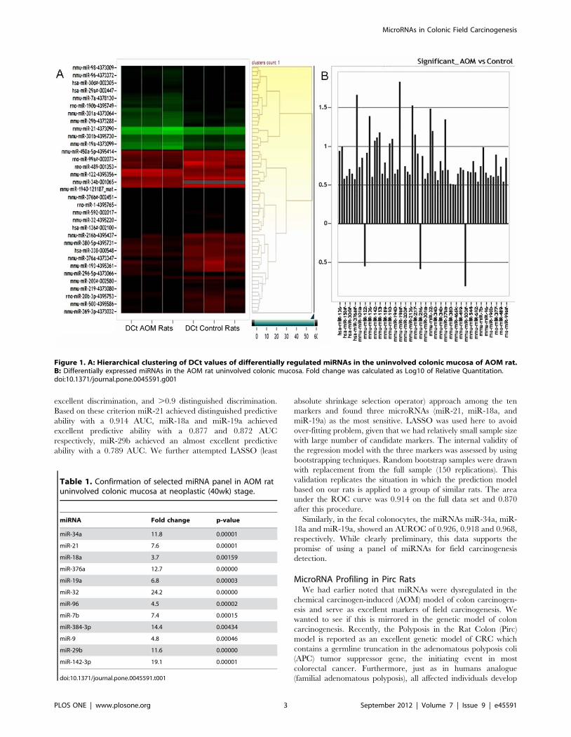
excellent discrimination, and .0.9 distinguished discrimination.
Based on these criterion miR-21 achieved distinguished predictive
ability with a 0.914 AUC, miR-18a and miR-19a achieved
excellent predictive ability with a 0.877 and 0.872 AUC
respectively, miR-29b achieved an almost excellent predictive
ability with a 0.789 AUC. We further attempted LASSO (least
absolute shrinkage selection operator) approach among the ten
markers and found three microRNAs (miR-21, miR-18a, and
miR-19a) as the most sensitive. LASSO was used here to avoid
over-fitting problem, given that we had relatively small sample size
with large number of candidate markers. The internal validity of
the regression model with the three markers was assessed by using
bootstrapping techniques. Random bootstrap samples were drawn
with replacement from the full sample (150 replications). This
validation replicates the situation in which the prediction model
based on our rats is applied to a group of similar rats. The area
under the ROC curve was 0.914 on the full data set and 0.870
after this procedure.
Similarly, in the fecal colonocytes, the miRNAs miR-34a, miR-
18a and miR-19a, showed an AUROC of 0.926, 0.918 and 0.968,
respectively. While clearly preliminary, this data supports the
promise of using a panel of miRNAs for field carcinogenesis
detection.
MicroRNA Profiling in Pirc RatsWe had earlier noted that miRNAs were dysregulated in the
chemical carcinogen-induced (AOM) model of colon carcinogen-
esis and serve as excellent markers of field carcinogenesis. We
wanted to see if this is mirrored in the genetic model of colon
carcinogenesis. Recently, the Polyposis in the Rat Colon (Pirc)
model is reported as an excellent genetic model of CRC which
contains a germline truncation in the adenomatous polyposis coli
(APC) tumor suppressor gene, the initiating event in most
colorectal cancer. Furthermore, just as in humans analogue
(familial adenomatous polyposis), all affected individuals develop
Figure 1. A: Hierarchical clustering of DCt values of differentially regulated miRNAs in the uninvolved colonic mucosa of AOM rat.B: Differentially expressed miRNAs in the AOM rat uninvolved colonic mucosa. Fold change was calculated as Log10 of Relative Quantitation.doi:10.1371/journal.pone.0045591.g001
Table 1. Confirmation of selected miRNA panel in AOM ratuninvolved colonic mucosa at neoplastic (40wk) stage.
miRNA Fold change p-value
miR-34a 11.8 0.00001
miR-21 7.6 0.00001
miR-18a 3.7 0.00159
miR-376a 12.7 0.00000
miR-19a 6.8 0.00003
miR-32 24.2 0.00000
miR-96 4.5 0.00002
miR-7b 7.4 0.00015
miR-384-3p 14.4 0.00434
miR-9 4.8 0.00046
miR-29b 11.6 0.00000
miR-142-3p 19.1 0.00001
doi:10.1371/journal.pone.0045591.t001
MicroRNAs in Colonic Field Carcinogenesis
PLOS ONE | www.plosone.org 3 September 2012 | Volume 7 | Issue 9 | e45591

predominantly colonic neoplasia. We did miRNA profiling of the
Pirc rats from the colonic biopsies obtained at a time point prior to
adenoma determination (12 wks with confirmatory negative
colonoscopy). We noted a robust miRNA modulation in the Pirc
rats wherein 38 miRNAs were significantly induced (Fig. 4A). We
noted that the miRNA modulation obtained in Pirc rats was more
dramatic than in AOM rat model which might reflect the higher
penetrance of this germline mutation versus carcinogen treatment.
Intriguingly, while the AOM and Pirc models shared some
miRNAs that were dysregulated, others were distinct suggesting
that this may be due to differences related to genetic heterogeneity
in tumorigenesis. Thus, the miRNA modulation in the CRC field
effect occurs regardless of the cancer-initiation model.
MicroRNA Modulation in the Pirc Rat Fecal ColonocytesAs a proof of concept, we also determined whether the in the
Pirc rat miRNA modulation observed in the colonic mucosa is
mirrored in the normally defecated fecal colonocytes. We selected
6 miRNAs overexpressed during field carcinogenesis and longitu-
dinally evaluated these markers in the mucus layer fecal
colonocytes. Four of these miRNAs were observed to be
significantly induced in the Pirc rat fecal colonocytes when
compared to stool from age matched wild type rat (Fig. 4B), thus
mirroring the results obtained in the colonic mucosa.
Discussion
We demonstrate herein that a number of microRNAs were
dysregulated in the predysplastic (microscopically normal) colonic
mucosa. Our approach was relatively comprehensive utilizing the
TLDA array platform and validated by individual miRNA
Taqman assays. Furthermore, this was not model specific since
miRNA dysregulation was also noted both in the well-validated
AOM-treated rat model and the genetic Pirc model. The striking
finding was that not only were these micro RNAs dysregulated in
cancer, but also at the premalignant time point and predict which
AOM-treated animals would actually develop tumors in the future
making this potentially relevant to colorectal cancer screening
(identifying at risk patients to interrupt the adenomaRcarcinoma
progression). Finally, with regards to clinical implementation
perspective, we demonstrated that this could be conducted from
mucus layer fecal colonocytes representing a non-invasive method
of detecting field carcinogenesis.
Figure 2. MicroRNA modulation in premalignant 16 wk miRNA predicts future neoplasia development. MicroRNA analysis was done oncolonic biopsies from AOM rats at a preneoplastic (16 wk) time point. The rats were followed for 40 weeks and separated as tumor bearing and non-bearing AOM rats. The miRNA expression at the premalignant stage (16 wk) was then compared between the control rats and the AOM rats whicheventually developed tumors or remained non-tumor bearing. *: p,0.05 when compared to normal saline controls. Specific p values are given inwhich AOM rats without tumors are compared to AOM rats with tumors.doi:10.1371/journal.pone.0045591.g002
Table 2. MicroRNA modulation in colonic biopsies of AOMrats at a premalignant (16 wk) stage.
miRNA Fold change p-value
miR-34a 1.7 0.011207
miR-21 2.7 0.00006
miR-18a 2.2 0.00034
miR-376a 2.3 0.00306
miR-19a 2.2 0.00028
miR-32 1.3 N.S.
miR-96 1.4 N.S.
miR-7b 0.8 N.S.
miR-384-3p 1.2 N.S.
miR-9 1.5 0.04500
miR-29b 1.7 0.015
miR-142-3p 1.3 0.042
N.S.: Non-Significant, N/A: Non-Applicable.doi:10.1371/journal.pone.0045591.t002
MicroRNAs in Colonic Field Carcinogenesis
PLOS ONE | www.plosone.org 4 September 2012 | Volume 7 | Issue 9 | e45591

There has been considerable interest in the role of
microRNAs in colon carcinogenesis, although most studies have
focused on progression and therapeutics. For instance, there is
clear evidence that dysregulation of a variety of miRNAs may
result in progression as many miRNAs are progressively altered
between adenomas and carcinomas [19,20]. Large number of
studies has shown that miRNAs play an important prognostic
role in cancer. For instance, Schetter and colleagues showed
that miR-21 corresponded to colorectal cancer related mortality
[15]. Furthermore, Nielsen and colleagues recently demonstrat-
ed that stromal microRNA expression was able to prognosticate
the clinically vexing issue of stage 2 disease [21]. Finally, from a
therapeutic perspective, recent reports have suggested that
resistance to one of the stalwart drugs against colorectal cancer
Figure 3. A: Hematoxylin and Eosin (H&E) staining of the fecal colonocytes isolated from rat stool. B: Differentially expressed miRNAs inthe fecal colonocytes obtained from tumor bearing and tumor non-bearing AOM rats. *: p,0.05. Specific p values are given in which AOM ratswithout tumors are compared to AOM rats with tumors.doi:10.1371/journal.pone.0045591.g003
Figure 4. A: Significantly modulated miRNAs in the Pirc rat uninvolved colonic mucosa. Fold change was calculated as Log10 ofRelative Quantitation. B: Differentially expressed miRNAs in the fecal colonocytes obtained from Pirc rats. *: p,0.05.doi:10.1371/journal.pone.0045591.g004
MicroRNAs in Colonic Field Carcinogenesis
PLOS ONE | www.plosone.org 5 September 2012 | Volume 7 | Issue 9 | e45591

is driven by mir-21 and manipulation of miRNAs can overcome
resistance [22].
Emerging evidence suggests that microRNAs may be modulat-
ed early in colon carcinogenesis. For initiation of colon carcino-
genesis, the typical ‘‘gatekeeper’’ genetic events are either APC or
less frequently mismatch repair genes (generally hMLH1) [23].
While these are typically inactivated mutationally or through
methylation, there is some suggestion that miRNA epigenetic
silencing may also have a role. For instance, recent reports have
indicated that both APC and MMR gene expression is regulated
by miRs (e.g. miR-135 and miR-155, respectively) [10,16].
Moreover, SND1, a component of RNA-induced silencing
complex, has been noted to be upregulated in the putative earliest
morphological of colon carcinogenesis, the aberrant crypt foci
[24].
Given the emerging data that microRNA dysregulation may
play an integral role in early neoplastic transformation, the
question that follows is whether microRNA could be altered in
field carcinogenesis. It is apropos to note that microarray studies
have shown that critical mediators of neoplastic transformation
including COX-2, osteopontin etc. are dysregulated in the
microscopically normal mucosa (i.e. field effect) [25]. Additionally,
more recent studies have suggested that loss of DNA mismatch
repair proteins (hMLH1 and MSH 2) occurs in the microscop-
ically normal mucosa in patients harboring neoplasia elsewhere in
the colon [10]. This is particularly intriguing because, as
previously discussed, microRNAs have been shown to be potential
mediators of these proteins. Consonant with the role of
microRNA, there have been some early reports that the majority
of colorectal cancer patients had epigenetic silencing of the
intronic micro RNA miR-342 in the uninvolved mucosa (with
.80% of tumor tissue manifesting concomitant loss) [19].
Similarly, Balaguer and colleagues noted that miR-137 was
methylated in 81% of CRCs and while much less frequently seen
in the histologically normal mucosa was ,3 fold higher than in
corresponding mucosa that was neoplasia-free [26]. Moreover,
microRNAs in the uninvolved mucosa can be modulated by
chemopreventive agents [27]. Our study expands this work by
using microRNAs in animals with neoplasia. Furthermore, our
data is the first to focus on the performance characteristics in
discriminating between neoplasia-free versus those with field
carcinogenesis signatures.
The potential clinical implication is to develop a minimally
intrusive solution to the critical issue of colorectal cancer risk
analysis. For instance, techniques such as colonoscopy has been
quite effective but the group for which it is recommended is quite
expansive (the entire population over age 50). Unfortunately, the
prevalence of screen relevant neoplasia (advanced adenoma or
early stage cancer) is low (,5–6%) and thus the vast majority of
the expensive, invasive and potentially morbid colonoscopies are
unproductive from a cancer death prevention perspective. The
inefficiency of resource utilization (funds, endoscopic capacity) is
juxtaposed with the observation that most of the population does
not undergo colonoscopy. This may be a key factor in why CRC
remains the second leading cause of cancer deaths among
Americans and has underscored the urgency of finding a
minimally intrusive (fecal) pre-screen for colonoscopy. For
instance, fecal occult blood test identifies patients at higher risk
and therefore necessitates colonoscopy but its diagnostic perfor-
mance for advanced adenomas (defined as size $1 cm or .25%
villous features or presence of high grade dysplasia) is only 11%
[28,29]. Recent advances such as immunohistochemical tests for
hemoglobin, mutational (APC, K-ras, p53) or methylation
(vimentin) analysis have represented modest steps forward but
still considered suboptimal for CRC prevention efforts [30]. This
underscores the interest in exploiting microRNAs, whose role in
the carcinogenesis is well-established, as a screening tool.
We believe that our data represents a significant step forward in
translating microRNAs to the screening arena. Specifically, the
tissue microRNA (field carcinogenesis detection) showed promis-
ing diagnostic performance. One could consider clinical imple-
mentation with a simple digital rectal exam with colonocytes
captured with a modified examination glove. Even less intrusive
would be assay the stool as discussed above. MicroRNAs represent
a promising target because these small RNAs are more resistant to
degradation than mRNA. Previous studies have demonstrated
feasibility of this approach. For instance, in a landmark study,
Goel and colleagues [31] showed that microRNAs could be
detected in the stool and this was recently replicated by Koga et al
[32]. Our approach is a potentially important improvement in that
instead of evaluating the small number of tumor products in the
stool (the ‘‘needle in the haystack’’); we target the process of field
carcinogenesis which is ubiquitous among colonocytes in the fecal
stream. We do this by using the isolated mucus layer fecal
colonocytes [18] which has been posited to be acquired by
abrasion of the normal colonic epithelium by the solid stool bolus.
Furthermore, there is an augmentation of microRNA in tumors
suggesting improved fecal diagnostics for concurrent lesions.
While large proportion of colon cancer incidence is sporadic, a
small percentage (25%) of CRC cases arise through genetic/
hereditary causes (Familial Adenomatous Polyposis (FAP) and
hereditary non-polyposis colorectal cancer (HNPCC)). In the
present study, we used two animal models of CRC to study the
role of miRNAs in both sporadic (carcinogen-induced) and genetic
causes of CRC. The Azoxymethane treated rat model is a well-
validated model of carcinogen induced experimental colon
carcinogenesis. Thus, this represents an excellent model to study
the role of miRNAs in CRC field carcinogenesis at premalignant
and malignant stages. In the AOM rat model only , 50% of
AOM-treated rats develop neoplasia by 40 weeks. This gave the
opportunity to study the miRNA expression in tumor-bearing and
tumor non-bearing rats, both in colonic mucosa as well as fecal
colonocytes. Recently, a new genetic model of CRC was
developed in rat (Pirc model) that provides an important tool for
CRC research. The Pirc rats have mutated APC gene thus not just
genetically recapitulating human disease such as familial adeno-
matous polyposis but also most sporadic colonic neoplasia. We
compared the miRNA expression in both the AOM rat and the
Pirc rat model and observed robust miRNA modulation in the
uninvolved colonic tissues (CRC field). We also noted that these
changes were mirrored in the fecal colonocytes miRNA assays,
thus indicating that the fecal miRNAs may be good markers of
CRC field carcinogenesis.
There are several limitations to this study that should be
acknowledged. Firstly, this data was confined to rodent models
and relevance to human neoplasia needs to be confirmed. On the
other hand, the AOM-treated rat has been the stalwart preclinical
studies in experimental colorectal carcinogenesis and the model
specific issues are mitigated by corroboration with the genetic Pirc
rat model. Secondly, while we noted similar miRNA modulation
in AOM rat mucosa and fecal colonocytes, the sample size was
modest. In order to establish the fecal miRNA analysis as the
marker of field carcinogenesis and CRC risk prediction, larger
studies would be needed. Thirdly, for the fecal microRNA studies,
the contamination (especially at later time points) with established
tumors is possible but lessened by the assessment with rat
colonoscopy which visualized the majority of the bowel (except
for cecum). Finally, while we present a compelling preliminary
MicroRNAs in Colonic Field Carcinogenesis
PLOS ONE | www.plosone.org 6 September 2012 | Volume 7 | Issue 9 | e45591

data on miRNA modulation in fecal colonocytes, for practical
application of the fecal miRNA test, this should be tested in
human patients with and without adenomas/carcinomas.
In conclusion, we present herein evidence that microRNAs are
progressively altered during colon carcinogenesis and are dysreg-
ulated at a predysplastic time points in the AOM-treated rat
model. We confirm this fact in the histologically normal mucosa in
humans. Importantly, the diagnostic accuracy was strong for both
synchronous and metachronous lesions (field carcinogenesis 6
tumor elaborated miRNAs). Furthermore, we demonstrate proof
of concept that this approach can be non-invasively utilized
through targeting mucus layer fecal colonocytes. These studies
represent an important first step in developing a novel risk
stratification technique and may provide important insights into
the biology of early colon carcinogenesis.
Methods
AnimalsAll animal studies were performed in accordance with the
Institutional Animal Care and Use Committee (IACUC)of North-
Shore University Health System. All the animal protocols
involving AOM rats and the Pirc rats were approved by the
IACUC. Fisher 344 rats (150–200 g) received 2 weekly intra-
peritoneal injections of azoxymethane (AOM) (15 mg/kg; Mid-
west Research Institute, Kansas City, MO). The rats were
euthanized at forty weeks. In a separate experiment, colonic
biopsies were obtained from the AOM rats at a 16 wk time point
and rats were followed for 40 wks and tumor counts were taken.
Total RNA from the colonic mucosa and colonic biopsies was
isolated using Ribopure RNA kit (Ambion) using manufacturer’s
instructions. Wild type Fisher 344 and Pirc rats were obtained
from Taconic Laboratories (Hudson, NY). They were maintained
on AIN 76a diet (Harlan Teklad, Madison, WI). The wild type
and Pirc rats were euthanized after 24 weeks. The CRC
progression in AOM rats as well as Pirc rats was monitored by
colonoscopy using ColoView colonoscope (Storz) and colonic
biopsies were obtained at the indicated time points.
MicroRNA Profiling in AOM RatsThe concentration and purity of total RNA was measured by
spectrophotometry at OD 260/280. Total RNA from rat colonic
mucosa and colonic biopsies was reverse transcribed by priming
with Rodent MegaPlex RT Primers (Applied Biosystems, Foster
City, CA) using Taqman miRNA reverse transcription kit (Applied
Biosystems) according to the manufacturer’s instructions. The
cDNA diluted in Universal PCR Master Mix II (Applied
Biosystems) was then loaded on to the Taqman Low Density
Array (TLDA) microfluidic cards (Applied Biosystems) and run on
ABI 7900 HT real time PCR system using manufacturer’s
instructions. Relative concentrations of miRNAs were calculated
using the comparative (22DDCt) method. The fold change was
calculated as log10 RQ where RQ is 22DDCT. Real time PCR data
analysis was done using the RQ Manager 1.2.1 (Applied
Biosystems) and RealTime StatMiner software (Integromics,
Philadelphia, PA).
Fecal Colonocyte IsolationThe fecal colonocytes were obtained by isolating the mucus
layer on the outer surface of the normally defecated rat stool using
a modified protocol described by White and colleagues [18].
Briefly, stool was suspended in PreservCyt solution (Hologic) and
gently agitated to dislodge the mucus layer from the outer surface
of the stool and the suspension was then centrifuged at 800 rpm
for 5 min. at 4uC and the supernatant was discarded. The pellet
was resuspended in PreservCyt solution (Hologic) (5 ml/g of stool)
and incubated for 45 minutes at room temperature to preserve and
fix the colonocytes. After incubation, the suspension was
successively filtered through 300 mm polypropylene mesh (Fort
Atkinson, WI) (to remove large debris) and 125 mm mesh (Small
Parts, Inc (Seattle, WA) (to obtain the mucus layer). The mucus
layer retained on the 125 mM filter mesh contained the embedded
colonocytes. The cells were resuspended in TRI reagent and RNA
isolation was carried out using Ribopure RNA kit (Ambion)
according to the manufacturer’s instructions.
Taqman miRNA Assays and Real Time RT-PCRExpression of selected miRNAs was assessed in the colonic
biopsies, uninvolved colonic mucosa and fecal colonocytes using
individual Taqman miRNA assays (Applied Biosystems) run on
the Corbett Rotor-GeneH 6000 real time PCR system (Qiagen)
using miRNA reverse transcription kit and Universal PCR Master
Mix (Applied Biosystems) following the manufacturer’s instruc-
tions. Fold change in miRNA expression was calculated by the
22DDCT method.
Statistical AnalysisStatistical significance for the individual miRNA expression was
performed with Microsoft Excel and the area under the receiver
operator curve (AUROC) was calculated using STATA 8
software. A two tailed Student’s t-test was utilized with p value
of ,0.05 was determined as significant.
Acknowledgments
We wish to thank Ms. Beth Parker for help in manuscript preparation.
Author Contributions
Conceived and designed the experiments: DPK HKR. Performed the
experiments: DPK RKW MD AM AP YS. Analyzed the data: DPK HD
VB HKR. Wrote the paper: HKR DPK.
References
1. Wood LD, Parsons DW, Jones S, Lin J, Sjoblom T, et al. (2007) The genomic
landscapes of human breast and colorectal cancers. Science 318: 1108–1113.
2. Heavey PM, McKenna D, Rowland IR (2004) Colorectal cancer and the
relationship between genes and the environment. Nutr Cancer 48: 124–141.
3. Yantiss RK, Goodarzi M, Zhou XK, Rennert H, Pirog EC, et al. (2009)
Clinical, pathologic, and molecular features of early-onset colorectal carcinoma.
Am J Surg Pathol 33: 572–582.
4. Chan AO, Broaddus RR, Houlihan PS, Issa JP, Hamilton SR, et al. (2002) CpG
island methylation in aberrant crypt foci of the colorectum. Am J Pathol 160:
1823–1830.
5. Derks S, Bosch LJ, Niessen HE, Moerkerk PT, van den Bosch SM, et al. (2009)
Promoter CpG island hypermethylation- and H3K9me3 and H3K27me3-
mediated epigenetic silencing targets the deleted in colon cancer (DCC) gene in
colorectal carcinogenesis without affecting neighboring genes on chromosomal
region 18q21. Carcinogenesis 30: 1041–1048.
6. Hesson LB, Wilson R, Morton D, Adams C, Walker M, et al. (2005) CpG island
promoter hypermethylation of a novel Ras-effector gene RASSF2A is an early
event in colon carcinogenesis and correlates inversely with K-ras mutations.Oncogene 24: 3987–3994.
7. Samowitz WS, Curtin K, Wolff RK, Albertsen H, Sweeney C, et al. (2008) TheMLH1–93 G.A promoter polymorphism and genetic and epigenetic alterations
in colon cancer. Genes Chromosomes Cancer 47: 835–844.
8. Bariol C, Suter C, Cheong K, Ku SL, Meagher A, et al. (2003) The relationship
between hypomethylation and CpG island methylation in colorectal neoplasia.Am J Pathol 162: 1361–1371.
9. Garzon R, Calin GA, Croce CM (2009) MicroRNAs in Cancer. Annu Rev Med
60: 167–179.
MicroRNAs in Colonic Field Carcinogenesis
PLOS ONE | www.plosone.org 7 September 2012 | Volume 7 | Issue 9 | e45591

10. Valeri N, Gasparini P, Fabbri M, Braconi C, Veronese A, et al. (2010)
Modulation of mismatch repair and genomic stability by miR-155. Proc NatlAcad Sci U S A 107: 6982–6987.
11. Slaby O, Svoboda M, Michalek J, Vyzula R (2009) MicroRNAs in colorectal
cancer: translation of molecular biology into clinical application. Mol Cancer 8:102.
12. Huang Z, Huang D, Ni S, Peng Z, Sheng W, et al. (2010) Plasma microRNAsare promising novel biomarkers for early detection of colorectal cancer.
Int J Cancer 127: 118–126.
13. Schetter AJ, Harris CC (2009) Plasma microRNAs: a potential biomarker forcolorectal cancer? Gut 58: 1318–1319.
14. Schee K, Fodstad O, Flatmark K (2010) MicroRNAs as biomarkers in colorectalcancer. Am J Pathol 177: 1592–1599.
15. Schetter AJ, Nguyen GH, Bowman ED, Mathe EA, Yuen ST, et al. (2009)Association of inflammation-related and microRNA gene expression with
cancer-specific mortality of colon adenocarcinoma. Clin Cancer Res 15: 5878–
5887.16. Nagel R, le Sage C, Diosdado B, van der Waal M, Oude Vrielink JA, et al.
(2008) Regulation of the adenomatous polyposis coli gene by the miR-135 familyin colorectal cancer. Cancer Res 68: 5795–5802.
17. Keku TO, Amin A, Galanko J, Martin C, Schliebe B, et al. (2008) Apoptosis in
normal rectal mucosa, baseline adenoma characteristics, and risk of futureadenomas. Cancer Epidemiol Biomarkers Prev 17: 306–310.
18. White V, Scarpini C, Barbosa-Morais NL, Ikelle E, Carter S, et al. (2009)Isolation of stool-derived mucus provides a high yield of colonocytes suitable for
early detection of colorectal carcinoma. Cancer Epidemiol Biomarkers Prev 18:2006–2013.
19. Grady WM, Parkin RK, Mitchell PS, Lee JH, Kim YH, et al. (2008) Epigenetic
silencing of the intronic microRNA hsa-miR-342 and its host gene EVL incolorectal cancer. Oncogene 27: 3880–3888.
20. Grady WM, Tewari M (2010) The next thing in prognostic molecular markers:microRNA signatures of cancer. Gut 59: 706–708.
21. Nielsen BS, Jorgensen S, Fog JU, Sokilde R, Christensen IJ, et al. (2011) High
levels of microRNA-21 in the stroma of colorectal cancers predict short disease-free survival in stage II colon cancer patients. Clin Exp Metastasis 28: 27–38.
22. Schetter AJ, Leung SY, Sohn JJ, Zanetti KA, Bowman ED, et al. (2008)
MicroRNA expression profiles associated with prognosis and therapeutic
outcome in colon adenocarcinoma. Jama 299: 425–436.
23. Narayan S, Roy D (2003) Role of APC and DNA mismatch repair genes in the
development of colorectal cancers. Mol Cancer 2: 41.
24. Tsuchiya N, Ochiai M, Nakashima K, Ubagai T, Sugimura T, et al. (2007)
SND1, a component of RNA-induced silencing complex, is up-regulated in
human colon cancers and implicated in early stage colon carcinogenesis. Cancer
Res 67: 9568–9576.
25. Chen LC, Hao CY, Chiu YS, Wong P, Melnick JS, et al. (2004) Alteration of
gene expression in normal-appearing colon mucosa of APC(min) mice and
human cancer patients. Cancer Res 64: 3694–3700.
26. Balaguer F, Link A, Lozano JJ, Cuatrecasas M, Nagasaka T, et al. (2010)
Epigenetic silencing of miR-137 is an early event in colorectal carcinogenesis.
Cancer Res 70: 6609–6618.
27. Davidson LA, Wang N, Shah MS, Lupton JR, Ivanov I, et al. (2009) n-3
Polyunsaturated fatty acids modulate carcinogen-directed non-coding micro-
RNA signatures in rat colon. Carcinogenesis 30: 2077–2084.
28. Ahlquist DA, Sargent DJ, Loprinzi CL, Levin TR, Rex DK, et al. (2008) Stool
DNA and occult blood testing for screen detection of colorectal neoplasia. Ann
Intern Med 149: 441–450, W481.
29. Hewitson P, Glasziou P, Watson E, Towler B, Irwig L (2008) Cochrane
systematic review of colorectal cancer screening using the fecal occult blood test
(hemoccult): an update. Am J Gastroenterol 103: 1541–1549.
30. Whitlock EP, Lin JS, Liles E, Beil TL, Fu R (2008) Screening for colorectal
cancer: a targeted, updated systematic review for the U.S. Preventive Services
Task Force. Ann Intern Med 149: 638–658.
31. Link A, Balaguer F, Shen Y, Nagasaka T, Lozano JJ, et al. (2010) Fecal
MicroRNAs as novel biomarkers for colon cancer screening. Cancer Epidemiol
Biomarkers Prev 19: 1766–1774.
32. Koga Y, Yasunaga M, Takahashi A, Kuroda J, Moriya Y, et al. (2010)
MicroRNA expression profiling of exfoliated colonocytes isolated from feces for
colorectal cancer screening. Cancer Prev Res (Phila) 3: 1435–1442.
MicroRNAs in Colonic Field Carcinogenesis
PLOS ONE | www.plosone.org 8 September 2012 | Volume 7 | Issue 9 | e45591