during growthin - iai.asm.orgiai.asm.org/content/37/2/558.full.pdf · during promastigote growth or...
TRANSCRIPT

INFECTION AND IMMUNITY, Aug. 1982, p. 558-5670019-9567/82/080558-10$02.00/0
Vol. 37, No. 2
Cell Surface Origin of Antigens Shed by Leishmania donovaniDuring Growth in Axenic Culture
EDNA S. KANESHIRO,'t MICHAEL GOTTLIEB,2 AND DENNIS M. DWYER'*Cell Biology and Immunology Section, Laboratory of Parasitic Diseases, National Institute ofAllergy andInfectious Diseases, Bethesda, Maryland 20205,1 and the Department of Pathobiology, The Johns Hopkins
University School of Hygiene and Public Health, Baltimore, Maryland 212052
Received 25 January 1982/Accepted 23 April 1982
Antisera against isolated cell surface preparations (PCSP-As) of Leishmaniadonovani promastigotes were used to detect extracellular antigens producedduring the growth of these organisms in four different growth media. The PCSP-As precipitated two major antigenically identical but electrophoretically distinctcomponents, in addition to several minor antigens. Immunoelectrophoretic stud-ies employing PCSP-As, PCSP-As absorbed with intact, live promastigotes, andPCSP-As absorbed with a major extracellular antigen demonstrated the antigenicidentity between the major extracellular antigens and two major componentsexternally disposed at the surface of promastigotes. Growth curve kineticinvestigations suggested that the major extracellular antigens did not appear in thegrowth media primarily as a result of cell lysis or damage. The carbohydratenature of the major extracellular antigens was indicated by physicochemicalcharacterization.
Leishmania donovani, a parasitic protozoan,is the etiological agent of kala azar, a chronic,and usually fatal if untreated, form of humanvisceral leishmaniasis. This organism has a dige-netic life cycle, assuming an extracellular, flagel-lated promastigote form in the alimentary tractof its sandfly vector, and an obligate intracellu-lar form within the phago-lysosomal system ofspleen, liver, and bone marrow macrophages ofits definitive mammalian hosts (3, 5, 11).
Promastigote forms of most human leishman-ial species are readily cultivable in vitro invarious serum-supplemented tissue culture me-dia (19) or in several chemically defined Leish-mania growth media (27, 28). Using antiseramade against whole organisms, various investi-gators working with several different humanleishmanial species (i.e., L. donovani, L. tro-pica, and L. braziliensis) have reported thatduring promastigote growth or maintenance invitro, antigens are released from the parasiteinto the culture media. These in vitro promasti-gote-released factors have been designated invarious reports as leishmanial exogenous, excre-tory or excreted factors (EF, 15, 20, 24-26);antigenically active glycoproteins (7); and exo-metabolites (21, 22). Preparations of such pro-mastigote factors have been used as skin testantigens in the diagnosis of patients with cutane-ous leishmaniasis and Chagas' disease (6, 23), as
t Present address: Department of Biological Sciences, Uni-versity of Cincinnati, Cincinnati, OH 45221.
well as antigens in the serotyping and classifica-tion of Leishmania strains and species isolatedfrom patients with active diseases and frominsect vectors (20). Further, it has been suggest-ed that such factors have a role in initiating,establishing, and maintaining parasite infectionsin susceptible and nonsusceptible host macro-phages (14, 18, 25). To date, however, despitetheir apparent relevance to the disease, neitherthe exact cellular origin nor the definitive chemi-cal composition and structure of these parasite-generated extracellular factors have been un-equivocally established.
In the course of studies concerning the struc-tural, biochemical, and antigenic nature of theleishmanial cell surface, a variety of antiserawere generated in rabbits against isolated andpurified L. donovani promastigote cell surfacepreparations (PCSP), i.e., surface membraneswith subpellicular microtubules attached to theircytoplasmic lamina (2, 13). These antisera wereused in the current report to establish the rela-tionship between several antigens released bypromastigotes during their growth in vitro andthose antigens present at the cell surface.
MATERIALS AND METHODS
Organism and growth conditions. A cloned line of L.donovani strain 1-S promastigotes (9) was grown at26°C in four different media: (i) Schneider insect tissueculture medium supplemented with 30% (vol/vol) fetalbovine serum (Schneider + FBS) (4); (ii) medium 199(GIBCO Laboratories, Grand Island, N.Y.; GIBCO)
558
on June 22, 2018 by guesthttp://iai.asm
.org/D
ownloaded from
LEISHMANIA-SHED ANTIGENS 559
supplemented with 20 or 25% (vol/vol) FBS (199 +FBS) (10); (iii) the chemically defined medium, RE IlI,formulated by Steiger and Steiger (28); and (iv) amodification of the RE III medium that lacked bovineserum albumin (RE III - BSA) (27).
Preparation of used growth media and fractions.Used growth media were prepared from late-log- orearly-stationary-phase cultures, unless otherwise not-ed. The cell-free culture fluids were obtained after theremoval of cells by centrifugation (e.g., large batchcultures were centrifuged in 1-liter bottles at 5,000 x gfor 30 min at 4°C) and subsequent vacuum filtrationthrough a 0.45-,um porosity membrane filter (type HA;Millipore Corp., Bedford, Mass.).For studies on changes throughout the growth cycle,
L. donovani promastigotes were grown in 15 ml of theRE III or the RE III - BSA medium in 50-ml plasticculture flasks (Costar, Cambridge, Mass.) at 26°C.Cells were also grown in 150 ml of RE III or 199 +FBS in 650-ml plastic culture flasks (Costar), and 5 to 7ml from those larger cultures were removed daily.These small quantities of used growth media wereseparated from cells directly by filtration through amembrane (Millex, HA 0.45 ,m) filter and were storedat -70°C until analyzed by immunoelectrophoresis(see below).
Portions of the used culture media were concentrat-ed approximately 100-fold (100x concentrate), using apressure dialysis membrane filtration apparatus with aPM-10 membrane filter (Amicon Corp., Lexington,Mass.). To minimize nonspecific binding of concen-trated materials to chambers and filters, these weretreated with Bacitracin (Calbiochem-Behring Corp.,San Diego, Calif.) at 1 mg of distilled water per mlbefore use.
Physical and chemical treatment of used growth me-dia. For some experiments the 100x concentrate wasextracted twice with an equal volume of water-saturat-ed phenol for 1 h at 4°C (16). The aqueous phase wasseparated and dialyzed exhaustively against 3 x 4-literchanges of distilled water at 4°C over 48 h and subse-quently lyophilized.For other experiments a crude carbohydrate (CHO)
fraction was obtained by the following protocol: 4volumes of 95% ethanol was added to the 100xconcentrate, and the mixture was stored at -20°C forat least 3 h. After storage, the precipitate formed wasrecovered by centrifugation at 5,000 x g for 15 min at4°C. The resulting pellet was dissolved in 2% (wt/vol)potassium acetate before reprecipitation with 4 vol-umes of ethanol at -20°C for at least 3 h. The secondethanol precipitate was recovered by centrifugationand dissolved in distilled water; trichloroacetic acid(TCA) was added to 10 or 33% (wt/vol) final concen-tration, and the mixture was allowed to stand for 30min at 4°C. After removal of TCA-insoluble materialby centrifugation as described above, the supernatantwas concentrated by Amicon PM-10 membrane ultra-filtration and dialyzed overnight at 4°C against 3 x 1-liter changes of distilled water, or 0.01 M phosphate-buffered 0.85% (wt/vol) NaCl (PBS, pH 7.4) andcontaining 15 mM NaN3.
In some cases the used growth media were furtherfractionated by passage of the 100 x concentrate or theTCA-soluble fraction through an Amicon XM-50 mem-brane via ultrafiltration. The ultrafiltrate of the 100xconcentrate was designated as the ¢10-50 (10- to 50-)
kilodalton (kd) fraction, and the ultrafiltrate of theTCA-soluble fraction was designated the 10- to 50-kdCHO fraction. The retentate was washed twice with 2chamber-full volumes of distilled water and was desig-nated the a50 kd fraction.The used growth media were also treated with heat,
proteolytic enzymes, and sodium meta-periodate. Forheat treatment, the samples were placed in a boilingwater bath for various periods of time up to 1 h.Proteolytic digestion was accomplished by the addi-tion of pronase (B grade; Calbiochem) to a finalconcentration of 1 mg/ml (wt/vol) to the 100 x concen-trate, which had been previously diluted 1:10 (vol/vol)with 0.1 M potassium phosphate buffer adjusted to pH7.0. The mixture was incubated for 1 h at 42°C.Controls lacking pronase were treated identically.After incubation, the samples were heated in a boilingwater bath for 3 min to inactivate the enzyme. Pronaseactivity was determined by incubation with an artifi-cial substrate (Azocoll; Calbiochem) under identicalconditions. Sodium metaperiodate (Sigma ChemicalCo., St. Louis, Mo.) was added to the 100x concen-trate at a final concentration of 25 mM. Solutions wereincubated for 1 h at room temperature and dialyzedovernight at 4°C against 2x 4-liter changes of distilledwater to remove the periodate. Controls lacking perio-date were prepared identically.PCSP and antisera. PCSP were purified on sucrose
gradients as previously described (12, 17). Identifica-tion of the PCSP fraction was determined ultrastruc-turally by the presence of a homogeneous array ofmembranes associated with subtending microtubules.The purity of that fraction was judged by the absenceof other cellular organelles (12, 17). Triton X-100(Sigma) extracts of these preparations (13) were usedfor immunoreactions. Antibodies (PCSP-As) directedagainst isolated PCSP were raised in New Zealandwhite rabbits (2, 13). Ammonium sulfate-derived glob-ulin fractions of those sera were concentrated to onethird of the original volume (3 x concentrate). Alterna-tively, immunoglobulin G (IgG) fractions were isolatedand purified from these sera via protein A-SepharoseCL 4B (Pharmacia Fine Chemicals, Piscataway, N.J.)column chromatography. The latter were concentratedto 30 mg of protein per ml. Antibody fractions weredialyzed overnight against 1 liter of PBS containing 15mM NaN3 and subsequently stored at 4°C.For some studies, the 3 x antibody globulin fraction
was absorbed with the 10- to 50-kd CHO fraction ofthe used chemically defined medium (RE III - BSA).This was accomplished as follows: the 10- to 50-kdCHO fraction was first centrifuged at 4,000 x g for 60min, and the resulting supernatant was mixed with asample of the 3 x globulin fraction and incubatedsequentially, at 37°C for 30 min and at 4°C for 30 min.This was repeated with two or more additions of the10- to 50-kd CHO fraction until the pellet size re-mained constant as judged by visual observation. Theresulting supernatant was concentrated to the original3x volume and designated as the 10- to 50-kd CHO-absorbed antiserum.
Agglutination of intact promastigotes with PCSP-As.Agglutination assays were used to assess the presenceof antibodies in PCSP-As capable of binding to exter-nally disposed cell surface antigens. Washed promasti-gotes were resuspended to 2.5 x 10' cells in PBS, andan equal volume of azide-free 3x PCSP-As globulin
VOL. 37, 1982
on June 22, 2018 by guesthttp://iai.asm
.org/D
ownloaded from
560 KANESHIRO, GOTTLIEB, AND DWYER
fraction was added. Samples were mixed and incubat-ed at room temperature for 15 to 30 min. Cell aggluti-nations were determined microscopically. Crude CHOfractions of the used growth media were tested for theability to inhibit such antibody-mediated agglutination.This was accomplished by the addition of crude CHOfraction to a final concentration of 2.5 mg/ml in thepresence of the antibody and cells. Agglutination wasassessed under the conditions described above.
Gel immunoprecipitin analyses. Gel immunodiffusionreactions were performed with 0.6% (wt/vol) agarose(Litex, type HSA; Accurate Chemical & ScientificCorp., Westbury, N.Y.) made in PBS containing 15mM NaN3 and 1% (vol/vol) Triton X-100. Gels werephotographed as previously described (8) or fixed andstained with Coomassie brilliant blue R250 (30) andphotographed.Gels used for immunoelectrophoresis (IEP) were
made using 1% (wt/vol) agarose (Litex, type HSA) inTris-barbital buffer (29) with 1% (vol/vol) Triton X-100and adjusted to pH 8.6. The gels were stored at 4°C forat least 2 h before use. Electrophoretic separationswere done with a flat-bed electrophoresis unit (Multi-phor; LKB, Bromma, Sweden). Gels were connectedto a buffer reservoir via cellulose wicks (Ultra-wicks;Bio-Rad, Richmond, Calif.). Plates were maintained at12°C during electrophoresis by a cooling platformcirculator. Electrophoresis was carried out at 6 V/cm(constant) for 60 min as measured on the gels. Afterelectrophoresis, troughs were cut and filled with anti-sera, and gels were incubated in moist chambers atroom temperature or at 40C.Agarose gels and buffer solutions used for crossed
immunoelectrophoresis (CIE) were as used for IEPabove. Antigens were separated in the first dimensionas above and were electrophoresed into an adjacentantibody-containing gel in the second dimension at 2V/cm for 18 h with cooling at 12°C (30). Subsequently,IEP and CIE gels were washed, dried, and stainedwith Coomassie brilliant blue (30).EF. A sample of purified L. donovani EF from
promastigote cultures of strain L-52 (World HealthOrganization, Leishmania Reference Center designa-tion) was generously provided by Gerald Slutzky,Department of Protozoology, The Hebrew University,Hadassah School of Medicine, Jerusalem.
RESULTSDetection of antigens in used growth media.
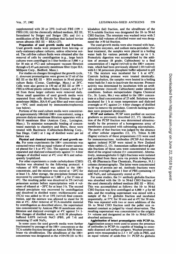
Rabbit antisera (PCSP-As) directed against puri-fied L. donovani PCSP precipitated componentspresent in culture media which had supportedpromastigote growth (Fig. 1). The specificity ofthe reaction was revealed by the following con-trols: normal preimmune rabbit sera did notyield precipitin bands against the used promasti-gote culture media, nor did the PCSP-As showprecipitin lines against unused culture media. Amajor band, the first to appear in gel diffusionsbetween PCSP-As and used culture media, fusedand showed antigenic identity with EF (as pro-vided by G. Slutzky). With additional incuba-tion, other minor precipitin bands became obvi-ous, indicating the presence of several antigens
C....
liI
t A( .
K.
/~a H
i i:,,f
(I
'C->. B*
FIG. 1. Antigenic reactivity of PCSP-As with usedpromastigote culture media and EF. Gel diffusionreactions at (A) 6 h and (B) 27 h between a 3 x globulinfraction of rabbit PCSP-As (As) and: two differentsamples of 10Ox Amicon PM-10-concentrated, used,day-4 RE III medium (wells 1 and 3); EF, 2.5 mg/ml(well 2); unused 100x concentrated RE III mediumcontrol well (4); and preimmune rabbit serum (wells 5.and 6). Several promastigote antigens released in theused culture medium formed precipitin lines withPCSP-As, suggesting their cell surface origin. Theheaviest line, which appeared earliest, is contiguouswith that of EF, indicating the antigenic identitybetween EF and the major antigenic constituent in theused RE III medium. Multiple lines between well 3,but not well 1, indicated the presence of several minorantigens in some samples of used media.
in the used culture media that reacted withPCSP-As.
In this report, antigens in used growth mediathat were detected and specifically precipitatedin gels with PCSP-As were tentatively referredto as shed membrane antigens (SMA). Thisprovisional designation does not suggest that amechanism for their genesis has been estab-lished.IEP analyses of antigens present in used growth
media. Two major antigens, SMA-A (a faster-migrating antigen) and SMA-B (a slower-migrat-
INFECT. IMMUN.
on June 22, 2018 by guesthttp://iai.asm
.org/D
ownloaded from

LEISHMANIA-SHED ANTIGENS 561
\*I
A
_____ B
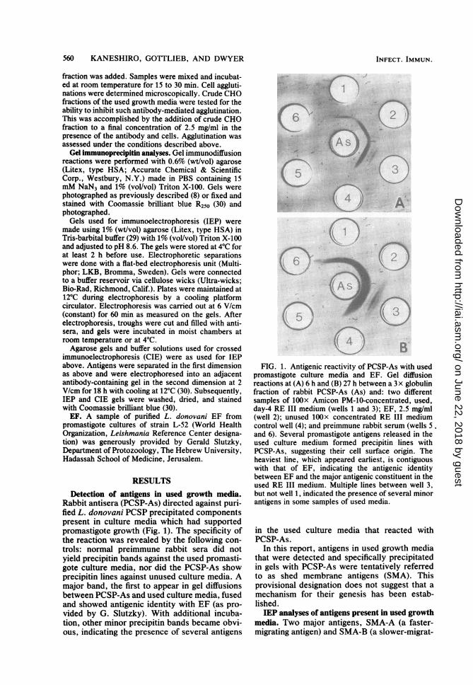
FIG. 2. Resolution of major and minor SMA inAmicon PM-10-concentrated RE III - BSA mediumfrom early-stationary-phase promastigote cultures by(A) CIE and (B) IEP. The anode to the right is the firstdimension in both A and B. The major SMA weredesignated SMA-A and -B. The most cathodic antigen,SMA-C, did not form contiguous precipitin arcs orpeaks with SMA-A or -B. The most anodic migratingantigen, SMA-D, formed a precipitin arc that inter-sected with that of SMA-A. Minor peaks contiguouswith that of the major SMA appeared as shoulders(asterisk in A). Other minor antigens (arrowheads)were often detected when high concentrations of anti-gens were used. Troughs in IEP gels (B) and theantibody gel in CIE analyses (A) contained IgG frac-tions of PCSP-As. The CIE antibody gel (A) contained2.4 mg of PCSP-As IgG per ml.
ing antigen), were distinguished in IEP reactionsagainst PCSP-As in all four types of used culturemedia. Examples of IEP and CIE gel reactionsemploying used RE III - BSA are shown in Fig.2. Examples of gel reactions in which the threeother used media were analyzed are illustratedbelow (see Fig. 7A). The immunoprecipitin linesof SMA-A and -B were contiguous, indicating acommon antigenic site(s). In some analyses,other components that were contiguous withthese major lines were observed in IEP gels.Similarly, in CIE gels these appeared as shoul-ders of the two major SMA peaks (asterisks inFig. 2A and 7B). The spur observed on thecathodal edge of SMA-A and anodal edge ofSMA-B in Fig. 2A suggested that the PCSP-Ascontained some antibodies that recognized anantigenic site different from that common toboth SMA-A and -B.Another antigen, SMA-C, which was also
present in used culture media, gave weakerprecipitin bands in IEP or peaks in CIE gels thandid the major SMA-A or -B (Fig. 2). This indicat-ed that SMA-C was either present in lowerconcentrations than either major SMA in the
used medium, or the antisera had lower concen-trations of antibodies to this antigen. This pre-cipitin band was not always detected in IEP gels.Antigen SMA-C is apparently antigenically dif-ferent from SMA-A and -B, as indicated by thelack of antigenic identity among the respectiveprecipitin arcs.The most anodic antigen detected with PCSP-
As, SMA-D, was only occasionally observed inIEP and CIE gels (Fig. 2B). Its appearancedepended on the lot of culture medium analyzed,as well as the antiserum used. This precipitinband was not contiguous with, but crossed, theprecipitin arc of SMA-A. Other minor antigenswere also observed in some analyses (arrow-heads in Fig. 2).
Cross-reactivity between antigens of used cul-ture media and the surface of promastigotes. Thefollowing studies were carried out to ascertainthe relationships between the major SMA andantigens solubilized from isolated PCSP. Prelim-inary results by gel diffusions (data not shown)demonstrated that used, but not unused, culturemedia and solubiized PCSP antigens gave linesof antigenic identity against PCSP-As. Also,comparison of electrophoretic mobilities in CIEgels of solubilized PCSP and SMA demonstratedthat the major antigens in both of these prepara-tions had similar migration characteristics. Thedistances from the origin that PCSP antigensmigrated corresponded closely to those of SMA-A and -B. These closely corresponding migra-tion distances suggested that the two majorPCSP antigens and the two major SMA havesimilar, although not necessarily identical, prop-erties.To elucidate the relationship between PCSP
antigens and those in used culture media, analy-ses of SMA and solubilized PCSP were conduct-ed with absorbed antisera. In such assays, sam-ples of PCSP-As previously absorbed with the10- to 50-kd CHO fraction, which containedSMA-A as shown by IEP (cf. Fig. 7C below),was used. The absorbed PCSP-As did not formprecipitin bands against the used culture mediain IEP gels corresponding to either SMA-A or-B. Moreover, no precipitin arcs were detectedin IEP reactions between the ¢50 kd CHOfraction of used media and the above absorbedserum (Fig. 3B). A concentrated, unfractionatedsample of used RE III medium analyzed in thesame manner showed neither SMA-A nor -Barcs, but an SMA-C arc was present (Fig. 3C).Thus, SMA-A absorbed antibodies to SMA-Aand -B but not antibodies directed against SMA-C, which further verified that the two majorSMA had common antigenic sites and that SMA-C was antigenically distinct.The IEP and CIE patterns of Triton X-100
extracts of L. donovani PCSP indicated that the
VOL. 37, 1982
on June 22, 2018 by guesthttp://iai.asm
.org/D
ownloaded from

562 KANESHIRO, GOTTLIEB, AND DWYER
antiserum contained precipitating antibodies to avariety of antigenic constituents of the PCSP(Fig. 3D and 4A). Approximately 16 componentswere resolved by CIE. The same Triton extractwas also analyzed by both IEP and CIE, em-ploying the PCSP-As preabsorbed with the 10-to 50-kd CHO fraction of used culture medium.In such gels (Fig. 3E and 4B), the major immu-noprecipitin peaks were absent, further indicat-ing that SMA-A and -B and the major PCSPantigens are antigenically related.To further establish the relationship between
the antigens in PCSP and the used culture medi-um, we performed the following experiments.Azide-free PCSP-As IgG fraction was absorbedwith live, intact promastigotes and tested for theability to precipitate used culture medium anti-gens. A 1-ml amount of the IgG fraction (30 mg
of protein per ml) was absorbed with 3 x 10⁹promastigotes. Samples of this absorbed frac-tion failed to form precipitin arcs correspondingto either SMA-A or -B in IEP gel reactions.These results demonstrated the antigenic cross-reactivity of the two major SMA with externally
/
E C
A
I
I,
L-~
FIG. 3. Absorption of antibodies to SMA-B andseveral cell surface antigens by SMA-A. A sample ofconcentrated day-4 RE III medium (A) analyzed byIEP illustrates the typical pattern showing SMA-A, -B,and -C. In this analysis, unabsorbed PCSP-As wasplaced in the troughs. The globulin fraction of thisantiserum was also absorbed with the 10- to 50-kdCHO fraction of used RE III medium (which containedSMA-A as shown in Fig. 3B, C, and E. The a50-kdCHO fraction of used RE III medium (which containedSMA-B, as shown by other IEP analyses), was ana-lyzed by IEP, using this absorbed serum (B). Theabsence of precipitin arcs indicates antigenic cross-reactivity between SMA-A and -B. A sample of un-fractionated, concentrated day-4 RE III medium wassimilarly analyzed with the absorbed serum (C); thepresence of precipitin arc SMA-C indicates the lack ofantigenic cross-reactivity between SMA-A and -C. ATriton X-100 extract of PCSP was analyzed by IEP,using PCSP-As (D) and the same globulin fraction afterabsorption with the 10- to 50-kd CHO fraction of usedRE III medium (E). Several precipitin arcs present inthe IEP gel shown in (D) were absent in the gel shownin (E), indicating that SMA-A cross-reacted with sev-eral PCSP antigens.
~~~BFIG. 4. Absorption of cell surface antigens with
SMA-A from She used culture medium, as resolved byCIE. Triton X-100 extracts of PCSP were separated inthe first dimension by electrophoresis (anode to theright) and then electrophoresed into PCSP-As-contain-ing gels in the second dimension (anode at the top).The antibody gel shown in Fig. A contained 6% (voVvol) of the 3x globulin fraction of PCSP-As. Thisanalysis resolved about 16 precipitin components. Theantibody gel B contained 4% (vol/vol) of the same 3xPCSP-As globulin fraction as used in gel A, except thatit had been preabsorbed with the 10- to 50-kd CHOfraction (which contained SMA-A, as shown by otherIEP analyses). The two major precipitin peaks, as wellas several other smaller components seen in CIE gelA, were absent in CIE gel B, indicating that SMA-Ashared antigenic cross-reactivity with components inthe PCSP. The absence of the major antigen peaks isobvious in Fig. 4B.
I
INFECT. IMMUN.
on June 22, 2018 by guesthttp://iai.asm
.org/D
ownloaded from

LEISHMANIA-SHED ANTIGENS 563
oriented components in the surface of intactpromastigotes. The results of these absorptionstudies were equivocal with regard to the rela-tionship between the minor used culture mediumcomponents SMA-C and -D and the exposedpromastigote surface antigens.The addition of PCSP-As to a suspension of
live, intact promastigotes rapidly agglutinatedthe cells; aggregates of hundreds of cells formedwithin 15 min. Addition of the crude CHOfraction inhibited the agglutination; only a fewaggregates of three to four promastigotes couldbe detected after 1 h of incubation. Controlswith PBS alone, preimmune sera, or PBS withthe crude CHO fraction from used culture mediadid not cause distinct cellular agglutination. Theglobulin fraction of PCSP-As that was preab-sorbed with the 10- to 50-kd CHO fraction fromused culture media was also tested for its abilityto agglutinate cells. Promastigotes mixed withthis preabsorbed globulin fraction did not imme-diately agglutinate. Aggregates of 6 to 10 cellswere observed after 1 h of incubation, althoughmost cells were free and not part of aggregates.The results strongly suggest that SMA-A and
-B of used culture media are antigenically cross-reactive with those antigens exposed on thesurface of promastigotes. Further, these compo-nents are the major determinants recognized bythe rabbit hyperimmune serum to isolated PCSPand responsible for the observed agglutination.Accumulation of SMA in used growth media. It
seemed possible that the accumulation of SMAin culture media might be due to cell lysis. Toassess this possibility and to examine SMAproduction rates in different culture media, weprepared samples from three different usedgrowth media, i.e., 199 + FBS, RE III, and REIII - BSA. Quantities of SMA present in thesevarious media were estimated by rocket IEP(Fig. 5). Maximum peak areas were assumed tobe proportional to the concentration of SMA-Aor -B or both. Antigens SMA-C and -D could notbe specifically identified in these rocket IEPgels; however, minor components were oftendetected within the rocket peak (arrow in Fig.SB). Hence, the maximum peak areas of rocketsin these analyses do not represent total SMA inculture media. Photocopies of the gels weremade, and individual peaks were excised andweighed. The smallest peak obtained in thesestudies was arbitrarily designated as 1 immuno-precipitin unit (IPU; Fig. 6). Quantitation ofSMA represented by other peak weights wascalculated as direct proportions to that smallestpeak.Growth curves of L. donovani promastigotes
in three different culture media are shown in Fig.6. The doubling time of cells during the log phaseof growth was: 199 + FBS, 14 h; RE III, 19 h;
A
0t12345671!8910 1 2 3 4 5 6 7 8 9B
0 2 3 4 5 6 7 8FIG. 5. Quantitation of SMA in used culture media
via rocket IEP. Examples of rocket IEP analyses forquantitation of SMA accumulated with culture age areshown in (A) (199 + FBS) and (B) (RE III). Thenumbers indicate the culture age in days. In these gels,antigen wells were filled with 20 ,ul of unconcentrated,used media obtained by direct membrane filtration ofcultures. Gels shown in this figure contained 4% (vollvol) of the PCSP-As IgG fraction (anode at the top).When volumes of antigens or antisera concentrationsor both in gels were altered to obtain complete,quantifiable peaks, an antigen sample, previously ana-lyzed on a 4% antibody gel, was employed for propercalibration. Minor precipitin peaks were often detect-ed within the largest peak (arrow in B) and wereprobably due to minor antigens present in used culturemedia, e.g., SMA-C or -D. These analyses showedthat SMA accumulated in media during culture growthand could be quantified and compared with respect toage and other culture conditions.
and RE III - BSA, 17 h. A distinct stationaryphase was observed with organisms grown in199 + FBS. Promastigotes grown in the chemi-cally defined media, RE III and RE III - BSA,reached the same maximal densities as those in199 + FBS, but these cultures rapidly entered adeath phase. Dead cells were first seen at day 9in 199 + FBS cultures and at day 6 in RE III andRE III - BSA cultures.The amount ofSMA produced during promas-
tigote growth in these three different media(estimated by rocket IEP as described above)are also shown in Fig. 6. In 199 + FBS, SMAaccumulation was rapid during log phase, thenprogressed at a constant but lower rate duringstationary phase. In RE III and RE III - BSAmedia, although the death phase was accompa-nied by a rapid decrease in cell numbers (celllysis), SMA levels in the media remained con-
VOL. 37, 1982
on June 22, 2018 by guesthttp://iai.asm
.org/D
ownloaded from

564 KANESHIRO, GOTTLIEB, AND DWYER
108
-S
C 107LU
A(I
106
0w
I-C-)
106,
100
10 C
TIME (days)FIG. 6. Comparison of L. donovani promastigote growth curves and the corresponding accumulation of SMA
in three different culture media. Growth curves of promastigotes in 199 + FBS (0), RE III (A), and RE III -BSA (LI) show that maximal cell densities achieved in the three media were similar. However, cells in 199 + FBSshowed a distinct stationary phase, whereas those grown in chemically defined media (RE III and RE III - BSA)entered a death phase immediately after reaching maximal cell densities. The accumulation of SMA in 199 + FBS(0), RE III (A), and RE III - BSA (A) with age was estimated by rocket IEP analyses. The maximum peak areaof the smallest peak obtained by rocket IEP in these studies was arbitrarily designated as 1 IPU. Peak areasobtained from the rest of the samples of used culture media were calculated as directly proportional to the area ofthe smallest peak. Accumulation of SMA was greatest in the serum-supplemented medium, 199 + FBS, and leastin the chemically defined medium lacking BSA. These quantitations of SMA also show that, although thecultures were in a death phase during which extensive cell lysis occurred, the amount of SMA in the mediumremained constant. This is particularly evident in RE III and RE III - BSA cultures and indicates that significantamounts of SMA were apparently not released from intracellular stores upon cell lysis. The data represent singleexperiments for 199 + FBS and RE III - BSA and the mean of two experiments for RE III.
stant. These results suggest that there were nodetectable soluble stores of intracellular compo-nents antigenically related to SMA that werereleased by the lysis of promastigotes.The rates of SMA production (IPU/day) dur-
ing the log phase of growth were: 199 + FBS,3.46; RE III, 1.21; and RE III - BSA, 0.75.Results of the rocket IEP assays indicated that,on a per cell basis, greater detectable quantitiesof SMA accumulated in the serum-containinggrowth medium. The cumulative results of theseassays suggested that the major SMA werereleased continuously into the culture media byintact, live promastigotes throughout their cellcycle, and not produced as a result of their lysis.
Physicochemical characteristics of SMA. Todetermine the relationship between the two ma-jor antigenically cross-reactive componentsSMA-A and -B, we carried out the followingpreliminary studies. Antigen SMA-A was readi-ly separated from other SMA components by
ultrafiltration through an Amicon XM-50 mem-brane filter (cf. 10- to 50-kd CHO fraction; Fig.7C). SMA-B was retained by this filtration pro-cedure, residual SMA-A in the .50-kd CHOAmicon XM-50 fraction was subsequently re-moved from SMA-B by DEAE-cellulose columnchromatography (manuscript in preparation;e.g., Fig. 7D).
Analysis of L. donovani promastigote EF (15,24-26) indicated that its migration in IEP gelswas similar to that of SMA-A (Fig. 7E). Currentgel diffusion results, as indicated above, demon-strated antigenic identity between this EF andSMA-A. Furthermore, PCSP-As preabsorbedwith the 10- to 50-kd CHO fraction did not formprecipitin lines against EF in gel diffusions (datanot shown).The following observations indicated that
SMA-A and -B, including their antigenic sites,were carbohydrate in nature: (i) immunoprecipi-table activity was retained after heating at 100°C
INFECT. IMMUN.
on June 22, 2018 by guesthttp://iai.asm
.org/D
ownloaded from
LEISHMANIA-SHED ANTIGENS 565
A SMA-SA
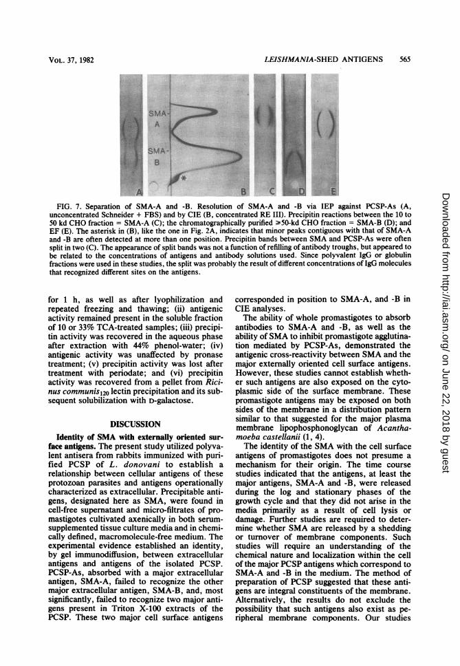
FIG. 7. Separation of SMA-A and -B. Resolution of SMA-A and -B via IEP against PCSP-As (A,unconcentrated Schneider + FBS) and by CIE (B, concentrated RE III). Precipitin reactions between the 10 to50 kd CHO fraction = SMA-A (C); the chromatographically purified >50-kd CHO fraction = SMA-B (D); andEF (E). The asterisk in (B), like the one in Fig. 2A, indicates that minor peaks contiguous with that of SMA-Aand -B are often detected at more than one position. Precipitin bands between SMA and PCSP-As were oftensplit in two (C). The appearance of split bands was not a function of refilling of antibody troughs, but appeared tobe related to the concentrations of antigens and antibody solutions used. Since polyvalent IgG or globulinfractions were used in these studies, the split was probably the result of different concentrations of IgG moleculesthat recognized different sites on the antigens.
for 1 h, as well as after lyophilization andrepeated freezing and thawing; (ii) antigenicactivity remained present in the soluble fractionof 10 or 33% TCA-treated samples; (iii) precipi-tin activity was recovered in the aqueous phaseafter extraction with 44% phenol-water; (iv)antigenic activity was unaffected by pronasetreatment; (v) precipitin activity was lost aftertreatment with periodate; and (vi) precipitinactivity was recovered from a pellet from Rici-nus communis120 lectin precipitation and its sub-sequent solubilization with D-galactose.
DISCUSSIONIdentity of SMA with externally oriented sur-
face antigens. The present study utilized polyva-lent antisera from rabbits immunized with puri-fied PCSP of L. donovani to establish arelationship between cellular antigens of theseprotozoan parasites and antigens operationallycharacterized as extracellular. Precipitable anti-gens, designated here as SMA, were found incell-free supernatant and micro-filtrates of pro-mastigotes cultivated axenically in both serum-supplemented tissue culture media and in chemi-cally defined, macromolecule-free medium. Theexperimental evidence established an identity,by gel immunodiffusion, between extracellularantigens and antigens of the isolated PCSP.PCSP-As, absorbed with a major extracellularantigen, SMA-A, failed to recognize the othermajor extracellular antigen, SMA-B, and, mostsignificantly, failed to recognize two major anti-gens present in Triton X-100 extracts of thePCSP. These two major cell surface. antigens
corresponded in position to SMA-A, and -B inCIE analyses.The ability of whole promastigotes to absorb
antibodies to SMA-A and -B, as well as theability ofSMA to inhibit promastigote agglutina-tion mediated by PCSP-As, demonstrated theantigenic cross-reactivity between SMA and themajor externally oriented cell surface antigens.However, these studies cannot establish wheth-er such antigens are also exposed on the cyto-plasmic side of the surface membrane. Thesepromastigote antigens may be exposed on bothsides of the membrane in a distribution patternsimilar to that suggested for the major plasmamembrane lipophosphonoglycan of Acantha-moeba castellanii (1, 4).The identity of the SMA with the cell surface
antigens of promastigotes does not presume amechanism for their origin. The time coursestudies indicated that the antigens, at least themajor antigens, SMA-A and -B, were releasedduring the log and stationary phases of thegrowth cycle and that they did not arise in themedia primarily as a result of cell lysis ordamage. Further studies are required to deter-mine whether SMA are released by a sheddingor turnover of membrane components. Suchstudies will require an understanding of thechemical nature and localization within the cellof the major PCSP antigens which correspond toSMA-A and -B in the medium. The method ofpreparation of PCSP suggested that these anti-gens are integral constituents of the membrane.Alternatively, the results do not exclude thepossibility that such antigens also exist as pe-ripheral membrane components. Our studies
VOL. 37, 1982
on June 22, 2018 by guesthttp://iai.asm
.org/D
ownloaded from

566 KANESHIRO, GOTTLIEB, AND DWYER
also did not eliminate the possibility that thepresence ofSMA in the media is the result of thesecretion of SMA by intracellular sites.
Relationship between SMA and EF. In contrastto this study, previous investigation of leishma-nial extracellular antigens have employed poly-valent antisera directed against whole promasti-gotes or crude cellular homogenates or both.The precipitating antigens identified by thesereagents have been referred to, by other work-ers, as excretory factor or EF (15, 20, 24-26),antigenically active glycoproteins (7), and exo-metabolites (21, 22). However, to date, thecellular origin and localization of these antigenshave remained undefined. Currently, it is un-clear whether the substances described by thesedifferent investigators are, in fact, identical. Inthis report, we have shown an antigenic identitybetween EF and the major SMA. Furthermore,we observed that EF and SMA-A had similarelectrophoretic characteristics in IEP gels.
Physicochemical characteristics of SMA. Re-sults of various treatments of the extracellularantigens indicated that both SMA-A and -B areCHOs. These results confirmed earlier data forthe CHO nature of EF (15, 24, 27). Previously,two forms of EF were reported, and it wasconcluded that the two forms were the result ofadsorption to serum proteins (27). In that regard,we have demonstrated that the distinction be-tween SMA-A and -B in our studies cannot bethe result of interactions with serum proteins, asidentical results were obtained in the chemicallydefined medium that lacks proteins (RE III -BSA). Similarly, different forms of leishmanialexometabolites have been identified and attrib-uted to aggregation/degradation processes oc-curring during their purification and analyses(22). However, preliminary results of metaboliclabeling studies lead us to conclude that thepresence of SMA-A and -B in our immunochem-ical analyses cannot be explained by aggregationof chemically identical molecules. Samples ofSMA that were metabolically labeled with[14C]inositol or [14C]stearic acid were analyzedby IEP and subsequently subjected to radioau-tography. The results suggested that SMA-B,but not SMA-A, contained inositol and stearicacid (rm.anuscript in preparation). Purification ofthe various SMA components is needed to fur-ther establish the chemical identity of thesemolecules.
Significance to SMA. The large amounts ofSMA released by promastigotes suggest thatthese components may be continuously shed bythe cells during natural infections of vertebrateor insect hosts. In that regard, EF has beenshown to be produced by amastigotes withinmacrophages (24), and in vitro-generated EF hasbeen used to render normally resistant macro-
phages susceptible to leishmanial infection (18).To date, the major extracellular antigens havebeen used to distinguish strains and species ofthese morphologically indistinguishable proto-zoans (20). In this regard, further knowledge ofthe chemical nature of these extracellular andsurface antigens will prove useful to better iden-tify these parasites.
ACKNOWLEDGMENTSWe thank T. Nash and J. Dvorak for useful suggestions and
for making available their research facilities; C. Grady fortechnical assistance; and G. Slutzky for the sample of L.donovani EF.
This work was supported in part by U.S. Public HealthService grant AI-16530 from the National Institute of Healthto M.G.
LITERATURE CITED1. Bailey, C. F., and B. Bowers. 1981. Localization of lipo-
phosphonoglycan in membranes of Acanthamoeba byusing specific antibodies. Mol. Cell. Biol. 1:358-369.
2. Berman, J. D., and D. M. Dwyer. 1981. Expression ofLeishmania antigens on the surface membrane of infectedhuman macrophages in vitro. Clin. Exp. Immunol.44:342-348.
3. Berman, J. D., D. M. Dwyer, and D. J. Wyler. 1979.Multiplication of Leishmania in human macrophages invitro. Infect. Immun. 26:375-379.
4. Bowers, B., and E. D. Korn. 1974. Localization of lipo-phosphonoglycan on both sides ofAcanthamoeba plasmamembrane. J. Cell Biol. 62:533-540.
5. Chang, K. P., and D. M. Dwyer. 1978. Leishmania dono-vani-hamster macrophage interactions in vitro: cell entry,intracellular survival, and multiplication of amastigotes. J.Exp. Med. 147:515-530.
6. Clinton, B. A., N. C. Palczuk, and L. A. Stauber. 1972.Leishmania donovani: partial characterization of someflagellate cytoplasmic immunogens. J. Immunol.108:1570-1577.
7. Decker-Jackson, J. E., and B. M. Honigberg. 1978. Glyco-proteins released by Leishmania donovani: immunologicrelationships with host and bacterial antigens and prelimi-nary biochemical analysis. J. Protozool. 25:514-525.
8. Dwyer, D. M. 1972. Antigenic comparison of Tricho-monas, Histomonas, Dientamoeba and Entamoeba. II.Gel diffusion methods. J. Protozool. 19:326-332.
9. Dwyer, D. M. 1976. Antibody-induced modulation ofLeishmania donovani surface membrane antigens. J. Im-munol. 117:2081-2091.
10. Dwyer, D. M. 1977. Leishmania donovani: surface mem-brane carbohydrates of promastigotes. Exp. Parasitol.41:341-358.
11. Dwyer, D. M. 1979. Membrane interactions betweenLeishmania and host cells, p. 130-134. In D. Schlessinger(ed.), Microbiology-1979. American Society for Micro-biology, Washington, D.C.
12. Dwyer, D. M. 1980. Isolation and partial characterizationof surface membranes from Leishmania donovani pro-mastigotes. J. Protozool. 27:176-182.
13. Dwyer, D. M. 1981. Structural, chemical and antigenicproperties of surface membranes isolated from Leishman-ia donovani, p. 9-28. In G. M. Slutzky (ed.), The bio-chemistry of parasites. Pergamon Press, Oxford.
14. El-On, J., D. J. Bradley, and J. C. Freeman. 1980. Leish-mania donovani action of excreted factor (EF) upon thehydrolytic enzyme activity of macrophages from micewith genetically different resistance to infection. Exp.Parasitol. 49:167-174.
15. El-On, J., L. F. Schnur, and C. L. Greenblatt. 1979.Leishmania donovani: physicochemical, immunological,
INFECT. IMMUN.
on June 22, 2018 by guesthttp://iai.asm
.org/D
ownloaded from
LEISHMANIA-SHED ANTIGENS 567
and biological characterization of excreted factor frompromastigotes. Exp. Parasitol. 47:254-269.
16. Gottlieb, M. 1977. A carbohydrate-containing antigenfrom Trypanosoma cruzi and its detection in the circula-tion of infected mice. J. Immunol. 119:465-470.
17. Gottleb, M., and D. M. Dwyer. 1981. Leishmania dono-vani: surface membrane acid phosphatase activity ofpromastigotes. Exp. Parasitol. 52:117-128.
18. Handman, E., and C. L. Greenblatt. 1977. Promotion ofleishmanial infections in non-permissive host macro-phages by conditioned medium. Z. Parasitendk. 53:143-149.
19. Hendricks, L. D., D. E. Wood, and M. E. Hajduk. 1978.Haemoflagellates: commercially available liquid media forrapid cultivation. Parasitology 76:309-316.
20. Sdhnur, L. F., A. Zuckerman, and C. L. Greenblatt. 1972.Leishmanial serotypes as distinguished by the gel diffu-sion of factors excreted in vitro and in vivo. Isr. J. Med.Sci. 8:932-942.
21. Semprevivo, L. H. 1978. Exometabolites of Leishmaniadonovani prosmatigotes. I. Isolation and initial character-ization. Proc. Soc. Exp. Biol. Med. 159:105-110.
22. Semprevivo, L. H., and B. M. Honigberg. 1980. Exome-tabolites of Leishmania donovani promastigotes. II.Spontaneous changes in exometabolite after isolation. Z.Parasitenkd. 62:201-211.
23. Shaw, J. J., and R. Lainson. 1975. Leishmaniasis inBrazil. X. Some observations on intradermal reactions ofdifferent trypanosomatid antigens of patients suffering
from cutaneous and mucocutaneous leishmaniasis. Trans.R. Soc. Trop. Med. Hyg. 69:323-335.
24. Slutzky, G. M., J. El-On, and C. L. Greenblatt. 1979.Leishmanial excreted factor: protein-bound and freeforms from promastigote cultures of Leishmania tropicaand Leishmania donovani. Infect. Immun. 26:916-924.
25. Slutzky, G. M. and C. L. Greenblatt. 1979. Analyses bySDS-polyacrylamide gel electrophoresis of an immunolog-ically active factor of Leishmania tropica from growthmedia, promastigotes, and infected macrophages. Bio-chem. Med. 21:70-77.
26. Slutzky, G. M., L. F. Schnur, R. L. Jacobson, and C. L.Greenblatt. 1980. Lectin specificities of leishmanial ex-creted factor. Isr. J. Med. Sci. 16:559-560.
27. Steiger, R. F., and C. D. V. Black. 1980. Simplified de-fined media for cultivating Leishmania donovani promas-tigotes. Acta Tropica 37:195-198.
28. Steger, R. F., and E. Steiger. 1977. Cultivation of Leish-mania donovani and Leishmania braziliensis in definedmedia: nutritional requirements. J. Protozool. 24:437-441.
29. Svendsen, P. J., and N. H. Azelsen. 1972. A modifiedantigen-antibody crossed electrophoresis characterizingthe specificity and titre of human precipitins againstCandida albicans. J. Immunol. Methods 1:169-176.
30. Weeke, B. 1973. General remarks on principles, equip-ment, reagents and procedures. A manual of quantitativeimmunoelectrophoresis: methods and applications.Scand. J. Immunol. 2:15-35.
VOL. 37, 1982
on June 22, 2018 by guesthttp://iai.asm
.org/D
ownloaded from