dr. kristina wilson, dvm, dacvr kwilson@uvsonline
TRANSCRIPT


References
Basic ultrasound physics
Overview of equipment and technology
Ultrasound artifacts
Scanning techniques
Terminology

Indications
Advantages and Disadvantages
Systematic approach
Relative organ echogenicity
NORMAL vs. ABNORMAL

Nyland and Mattoon:
Diagnostic Small Animal
Ultrasound, 2nd edition.
Pennick and D’Anjou
Atlas of Small Animal
Ultrasonography

What is ultrasound? Sound waves at higher frequency
than human hearing (>20 kHz) Diagnostic ultrasound uses 2-15 MHz
Frequency inverse related to depth High frequency, low penetration
High frequency, higher attenuated
Absorbed energy is lost as HEAT
Frequency direct related to resolution High frequency, high resolution
axial resolution 7.5 MHz ~ 0.3 mm

TRANSMISSION: sound passes through
ATTENUATION: sound energy lost REFLECTION
Is the basis of u/s image
Acoustic impedance of tissue
Velocity x density
Tissue interfaces
SCATTER Tiny uneven interfaces within
tissue
Creates parenchymal “echotexture”

REFRACTION “BENDING” of sound
beam as passes through tissues of different velocities at curved interface
ABSORPTION Energy lost and
converted to heat
Safety considerations High frequency:
greater absorption: greater heat

Transducer
Wave forms created by
transducer
Vibrations of piezoelectric crystals
when electricity applied or sound
received
Transducer is “emitting” < 1 %,
“listening” >99% of time
Sound Beam
3-D, thin slice
creates artifacts
Focal zone
Narrowest beam, best resolution

Sector Transducers
(real time B-mode)
Electronic
Curvilinear array
Phased array
Linear array
Mechanical
Annular array
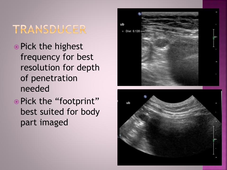
Pick the highest
frequency for best
resolution for depth
of penetration
needed
Pick the “footprint”
best suited for body
part imaged

Scanner Computer- magic happens
Image generated from returning echoes Time to return of echo = depth of pixel (y axis)
Intensity of echoes = brightness and grayscale
Direction of returned echo = location in image (x axis)
Assume returning echoes traveled at 1540 m/s Avg velocity of sound in fluid/soft tissue is 1540 m/s
Velocity actually variable across tissues encountered
Air 331 m/s, fat 1450 m/s, bone 4080 m/s
Velocity depends on density and physical stiffness
Differing velocities cause acoustic impedance
Responsible for creation of some artifacts

Depth Always set to be able to see
the deepest margin of organ being imaged
Focus Set within region of most
interest
Set where measurements are taken
Overall gain Often left alone
May need to change if poor contact (increase) or if abdominal fluid (decrease)
TGC near and far fields
Slides set to (b)right for deeper structures

Helpful
Acoustic
enhancement
Acoustic shadowing
Dirty shadow
Clean shadow
Not helpful
Reverberation
Mirror Image
Side-Lobe
Slice thickness
Edge shadowing
Electrical
interference

Acoustic
enhancement
“through
transmission”
Structure fluid filled
Low attenuation:
increases intensity
of returned echoes
Adjust far field gain

Acoustic Shadowing Clean shadow
Sharp edge, pure black
solid or high reflective structure (bone, foreign body, solid feces, barium or pure gas)
Dirty shadow Mixed echogenicity with
fuzzy edges
inhomogenous structures that contain gas and semisolid material (cloth, soft feces, food in stomach)
Both can “hide” deeper structure

Reverberation
Common artifact
Occurs at highly
reflective interface:
gas, metal
Sound bounces back
and forth between
reflective surfaces
and probe
“Comet tails”

Mirror image
At reflective
interfaces-
especially
diaphragm/ lung
“mismaps” location
based on travel
time
Mistake thoracic
pathology


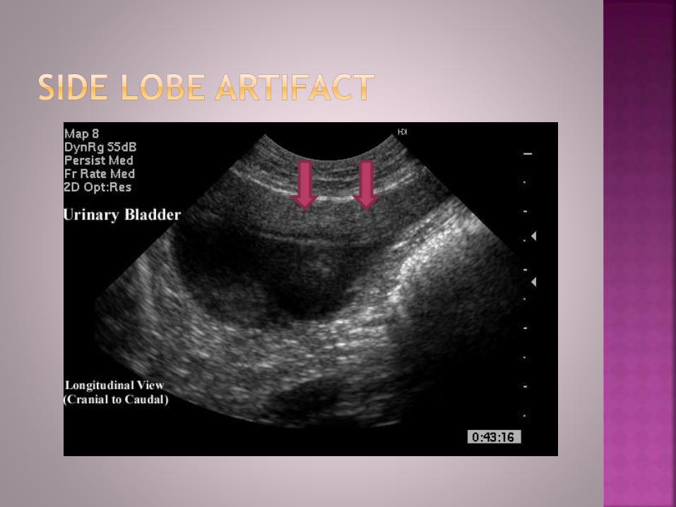
Side lobe artifact
Intense echoes from
lateral lobes are
mismapped as being
within main lobe
Occurs with high
reflective interfaces
lateral to anechoic
object in main beam
Correct by lower
gain, lower frequency,
change orientation or
deeper focus

Slice thickness High reflective
structure within “slice” along with anechoic structure
“pseudo-sludge” in UB/GB Look for “curved”
surface of sludge
Change position of probe, reposition animal

Edge Shadowing At edge of curved
structures
Cystic structures or structures of different acoustic impedance
Refraction- sound redirected and not returned to probe “Loss” of thin wall
structure mimic rupture bladder
Change angle of insonation?

Electrical
interference
Clippers,
radiowaves,
centrifuge,
fluorescent lights,
other equipment


Patient prep Fasting 12 hours
Shaved, clean skin
Gel or alcohol
Patient position Dorsal recumbency
Use troughs
Sedation if needed
Change positions Left lateral: right
liver/ kidney
Standing: bladder, GB

Standard orientation
of images Sagittal/ dorsal plane
view: cranial patient to left of image
Transverse ventral view: right side of patient to left
Right intercostal view: dorsal to left
Left intercostal: ventral to left
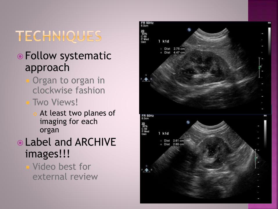
Follow systematic approach Organ to organ in
clockwise fashion
Two Views! At least two planes of
imaging for each organ
Label and ARCHIVE images!!! Video best for
external review

Echogenicity Hypoechoic- darker
Hyperechoic- brighter
Anechoic- no echoes, black
Normoechoic- expected
Isoechoic- equal to
Mixed
Texture Coarse or fine
Patchy or mottled
Nodular
Complex (cavitary)

Echotexture See previous slide
Shape Asymmetric
Irregular
Round, flat, triangular
Margins Irregular vs smooth
Bumpy
Ill-defined
Size Enlarged, small
MEASURE organ!
Location The left kidney is
located more caudal than normal…
In right cranial abdomen, there is…
Function Motility- hyper or hypo
Urine “jets”
hypovascular
Contrast enhancement Not commonly done in
routine studies

Combinations of
sonographic signs
will help prioritize
differential
diagnoses list
ie: enlarged,
hyperechoic liver w/
normal GB in anorexic
jaundiced cat =
lipidosis

Advantages
Non- invasive
Most often does NOT require anesthesia
CAN see inside of organs
CAN see thru abdominal fluid
Disadvantages
Relative costly test
Costly equipment
Highly user dependent
Takes time to perform
CANT see thru air or barium
Is it better than a
CAT scan, doc???
Diagnostic test: know indications
Abnormal organ function/ enzymes
Abdominal fluid or loss of detail on rads
Palpable mass/ mass on rads
Abdominal pain
Vomiting/ diarrhea
Hematuria/ stranguria, Cushings disease, cancer
staging, hypercalcemia, IMHA, VPCs/ arrhythmia,
anal sac tumor, GI foreign body, etc
Guide cystocentesis, aspirate/ biopsy, injections

Systematic approach Same for every scan
Know anatomy!
PRACTICE
Learn NORMALS Variants-age, breed,
sex, fat vs thin
Species differences
Recognize abnormal Changes in sonographic
signs

SiLK Spleen> liver> kidney
cortex
New normals? Cats: renal cortex hyper
to liver
Dogs: renal cortex iso
to liver
Liver always hypo to spleen
Lymph nodes = spleen

Liver
Gallbladder
Stomach
Pancreas- left limb
Spleen
Left kidney
Left adrenal gland
Urinary bladder
Urethra/ prostate
Medial iliac nodes
Intestine
Mesenteric nodes
Right kidney
Right adrenal gland
Right dorsal liver
Porta hepatis
Duodenum/ papilla
Pancreas- right limb

Largest abd organ Lobation: differentiate lobes
with fluid
intercostal views for caudate lobe, deep chest, small liver or porta hepatis
Vessels- PV wall hyper to HV, HA not seen w/o doppler
Size: subjective Left liver to caudal edge of
stomach
Tapered, sharp tips
Echotexture Medium echo- hypo to spleen,
iso to falciform
Coarse, uniform parenchyma

Normal cat Normal dog

Right dorsal
intercostal view
Caudate lobe
Porta hepatis-
CVC, PV, Ao
Hepatic nodes
cvcpv


Enlarged, Hypoechoic
DDX:
Infection (bacterial, viral)
Inflammation (immune
mediated hepatitis, systemic
inflammation)
Amyloidosis
Infiltrative neoplasia
(lymphoma, mast cell)
“reactive” processes (EMH,
congestion, drugs/toxin)

Enlarged, Hyperechoic
DDX CAT
Hepatic lipidosis
Endocrinopathy (diabetes)
Lymphoma, mast cell (rarely)
DDX DOG
Vacuolar hepatopathy-
endocrine or primary
Medication- corticosteroids
Chronic inflammation
w/fibrosis
Copper?
falciform
liver

Enlarged, Nodular
Benign- vacuolar
hepatopathy with
hyperplastic nodules
Neoplasia- lymphoma,
histiocytic sarcoma,
metastatic neoplasia
Fungal disease
Hepatocutaneous
syndrome

Small, irregular, nodular
Cirrhosis w/ nodular
regeneration
Often ascites
Portal hypertension
Normal size, nodular
Benign hyperplasia
Active hepatitis with
nodular regeneration

Small liver, normal architecture
NORMAL variant-dog
Microvascular dysplasia
Atrophy from chronic low-grade disease
Portosystemic shunt
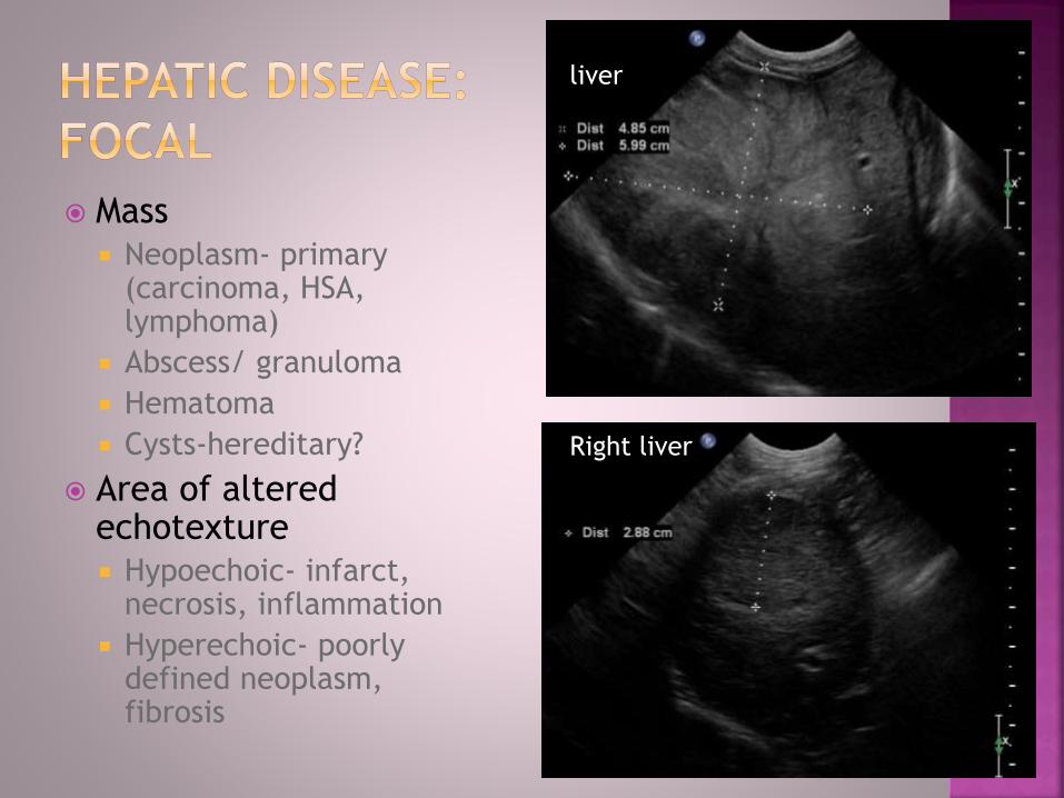
Mass
Neoplasm- primary (carcinoma, HSA, lymphoma)
Abscess/ granuloma
Hematoma
Cysts-hereditary?
Area of altered echotexture
Hypoechoic- infarct, necrosis, inflammation
Hyperechoic- poorly defined neoplasm, fibrosis
liver
Right liver

Thin wall 1-2 mm
Anechoic bile Some sludge normal esp
fasting dogs
Size- subjective Contracts w/ meal
Appears to take up 1/3 to ½ of right liver
Cat 2.5 to 4 cm
Dog 3-6 cm
Shape- tear drop Cystic duct-tapered end

CAT
Bacterial
Immune mediated
Viral- FIP?
DOG
Bacterial
Immune mediated?

Mucocoele
Most often associated with endocrine disease
Hypo-to anechoic, hyper strands/ striations,
ENLARGED,“Stellate”, “kiwi”

Cholesterol/ bile salts
Associated with endocrine disease
Obstructive
- GB enlarged
- stone doesn’t move
Non-obstructive
Gravity dependent
“sand”

Head, body, tail Head: transverse left
intercostal view
Tail movable
Echotexture hyperechoic
Finely granular
Splenic v > a, anechoic
Size: variable Cat <1 cm thick at
hilus
Dog 1-2.5 cm thick

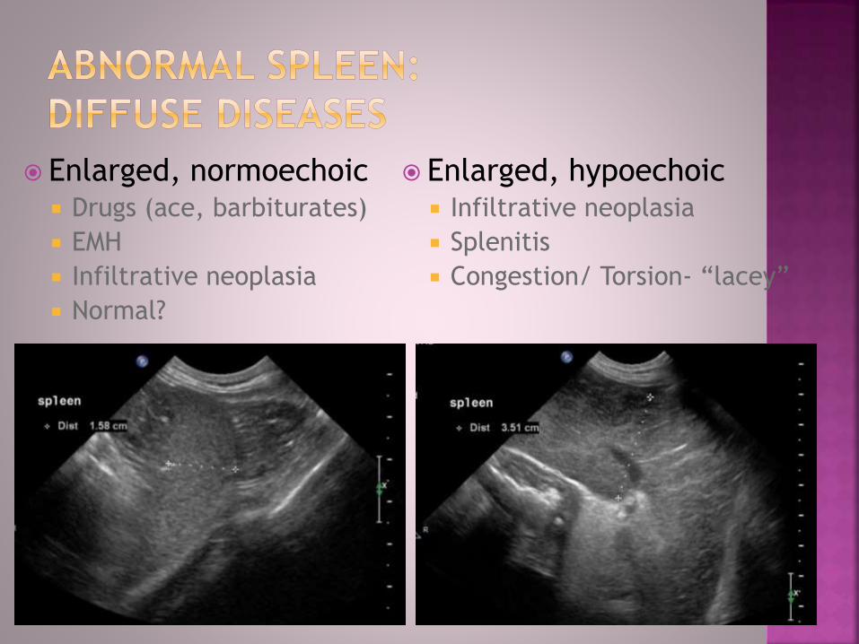
Enlarged, normoechoic Drugs (ace, barbiturates)
EMH
Infiltrative neoplasia
Normal?
Enlarged, hypoechoic Infiltrative neoplasia
Splenitis
Congestion/ Torsion- “lacey”

Enlarged, multi-nodular Neoplasia
Round, hypoechoic nodules- histiocytic, lymphoma
Miliary nodular- lymphoma, mast cell
Abscess/ granulomas
Round, often complex nodules
spleen

Masses Hypoechoic- benign, round cell, HSA
Hyperechoic- benign, round cell, leioSA, myelolipoma
Mixed echoic- old hematoma, HSA round cell, leiomyo
Complex/ cavitary-HSA, hematoma
Area of abnormal echotecture Infarct
Contusion
Necrosis
Neoplasia

Hemangiosarcoma- Single or multiple
ANY APPEARANCE but often complex
free fluid
Metastatic disease
liver

Anatomy: Cortex, medulla,
diverticulae, pyramids, pelvis, sinus
Cortex hyper to Medulla
Sharp definition between C/M
Right kidney intercostal
Size Cats/small dogs 3.5-4.5 cm
50 lb = 5 cm, then 10 lbs per cm up to max about 9 cm
If >10 cm, too big

Right kidney- longitudinal
ventral vs intercostal view

Plane of imaging
parasagittal sagittal
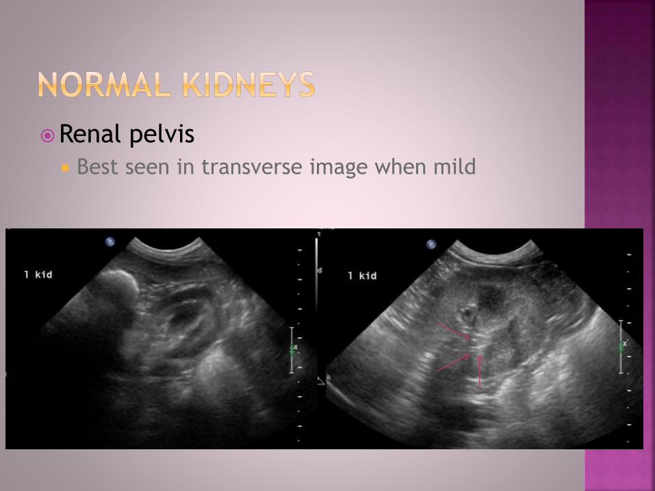
Renal pelvis
Best seen in transverse image when mild

Hyperechoic renal cortices
Overweight males

Enlarged, smooth contour, retained
architecture
Nephritis
Infectious- viral (cat), bacterial
immune mediated and amyloidosis
Toxin
Neoplasia-lymphoma
Portosystemic shunt
Unaltered animal- normal
Compensatory hypertrophy

Enlarged, lumpy, distorted architecture
Neoplasia
Lymphoma
Renal carcinoma
Metastatic- hemangiosarcoma
Abscess/ granulomas
Ascending/ sepsis
Fungal granulomas
‘Acute on chronic’ disease
Renal lymphoma in CRF cat

Small, irregular, distorted architecture
Chronic renal disease
Immune/toxin/unknown
Chronic pyelonephritis
Chronic congenital disease (dysplasia)
Renal cortical infarcts
r kid

Renal cortical infarcts
Hyperechoic striation, triangular wedge or large region
Often causes atrophy and indentation

Pyelectasia
Slight/mild
polyuria of any cause
Early obstruction- blocked cat
Pyelonephritis
Moderate/ severe
Obstruction- ureteral
Pyelonephritis

Pyelectasia continued
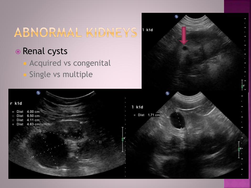
Renal cysts
Acquired vs congenital
Single vs multiple

“Medullary rim” Hyperechoic band at
junction of cortex and medulla Non-specific
Hypercalcemia- mineral deposits in tubules
inflammation- lyme?
l kid

Reduced CM definition
Blurred junction
Cortex/ medulla similar echogenicity
Non-specific

Anatomy Apex- cranioventral
Neck- tapered sphincter
Trigone- caudodorsal
Wall Thickness depends on fullness
Most thick at apex
Mucosa smooth
Ureteral papillae
Location Neck cranial to pubis
Intrapelvic bladder
Anechoic urine Suspended “specks”- fat
droplets, concentrated urine in cats

Ureteral papillae
Cranial border of trigone
Urine “jets”
Common location for stone obstruction

Fat droplets
Stay suspended/ don’t settle out

Calculi
Non-radiopaque stones
“Sand”
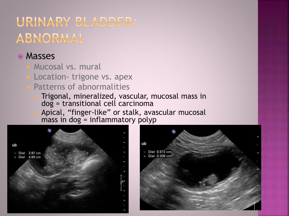
Masses Mucosal vs. mural
Location- trigone vs. apex
Patterns of abnormalities Trigonal, mineralized, vascular, mucosal mass in
dog = transitional cell carcinoma Apical, “finger-like” or stalk, avascular mucosal
mass in dog = inflammatory polyp
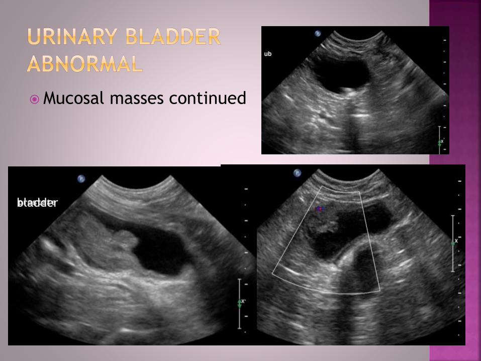
Mucosal masses continued
bladder

Masses continued:
Mural
Hematoma
Soft tissue sarcoma (leiomyoma/ leiomyosarcoma)

Neutered
Small, less than 2
cm width
Hypoechoic, smooth
Intact
Variable size
Bilobed shape transverse
Smooth contour
Hyperechoic, uniform

Anatomy
Best viewed empty
Cardia, fundus, body
and pyloric antrum
Pyloric sphincter
Wall
Layered like intestine
Varies 2-5 mm thick
Rugal folds thicker
Contracts 3-5/ min

Jejunum wall
Cats up to 3.0 mm
Dogs up to 3.5 mm
Five distinct layers-
mucosa thickest
Lumen
peristalsis
Gas/ small amt fluid only
Solid material abnormal
diameter >1.5 cm
abnormal in cats
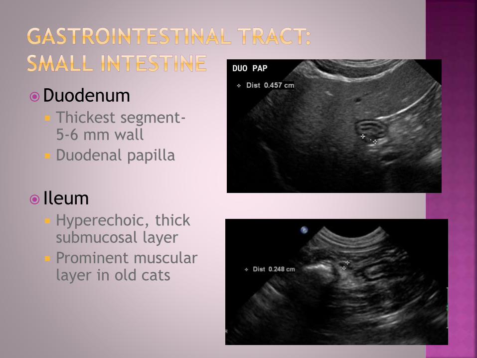
Duodenum Thickest segment-
5-6 mm wall
Duodenal papilla
Ileum Hyperechoic, thick
submucosal layer
Prominent muscular layer in old cats

Dogs:
Peanut, bilobed shape
Cortex and medulla
Size varies 4-7 mm
diameter
Cats
More round shape
Hypoechoic
Size <5 mm diameter

Dogs
Right limb easier
<1.5 cm height
Uniformly hypoechoic
(iso to liver)
Cats
Left limb easier to see
5-7 mm diameter limbs
Old cats- panc duct visible

Mesenteric (jejunal)
Paired along mesenteric
vessels
Dogs <6 mm, Cats < 4 mm
Hyperechoic
Medial iliac
Right/left lateral views
Dogs <7 mm
Hard to see in cats
Hyperechoic
Questions???