© 1998 Wiley-Liss, Inc.
Journal of Clinical Laboratory Analysis 12:179–189 (1998)
Ultrasensitive and Rapid Enzyme Immunoassay(Thin Aqueous Layer Immune Complex Transfer Enzyme
Immunoassay) for Antibody IgG to HIV-1 p17 Antigen Setsuko Ishikawa, 1 Seiichi Hashida, 1 Kazuya Hashinaka, 1 Akio Adachi, 2
Shinichi Oka, 3 and Eiji Ishikawa 1*1Department of Biochemistry, Miyazaki Medical College, Kiyotake, Miyazaki, Japan
2Department of Virology, School of Medicine, University of Tokushima, Kuramoto, Tokushima, Japan3AIDS Clinical Center, International Medical Center of Japan, Toyama, Shinjuku, Tokyo, Japan
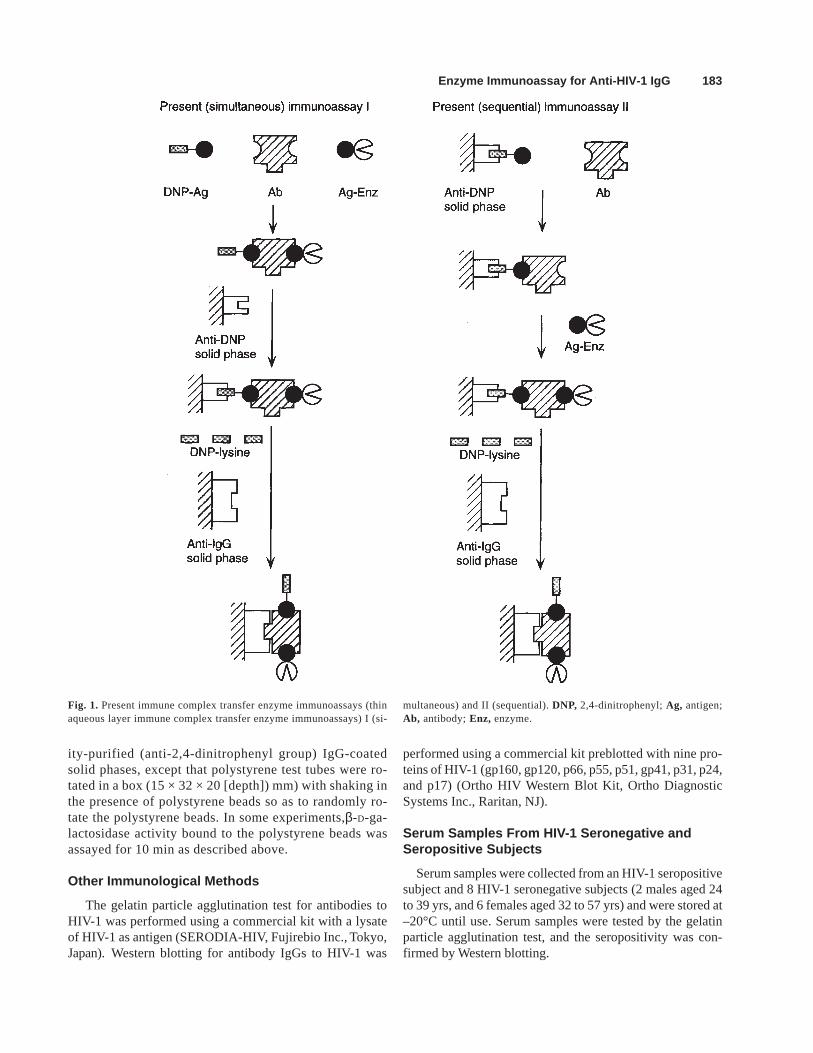
The immune complex transfer enzyme im-munoassay for antibody IgG to HIV-1 p17antigen was performed in two different ways(the present immunoassays I and II) withinshorter periods of time than previously re-ported. In the present (simultaneous) immu-noassay I, antibody IgG to HIV-1 p17 antigenin 10 µL of serum samples was incubatedsimultaneously with 2,4-dinitrophenyl-mal-tose binding protein-recombinant p17(rp17)fusion protein and rp17-β-D-galactosidaseconjugate in a total volume of 22 µL for 10min to form the immune complex compris-ing the three components. The reaction mix-ture was incubated with a polystyrene beadof 6.35 mm in diameter coated with affinity-purified (anti-2,4-dinitrophenyl group) IgG for5 min in a styrol test tube (13.3 × 54 mm and2.1 g) to trap the immune complex. Afterwashing, the polystyrene bead was incu-bated with 30 µL of εN-2,4-dinitrophenyl-L-lysine solution in a polystyrene tube (12 ×75 mm) coated with affinity-purified (antihu-man IgG γ-chain) IgG for 10 min to transferthe immune complex. In the present (se-quential) immunoassay II, a polystyrenebead of 6.35 mm in diameter coatedsuccessively with affinity-purified (anti-2,4-dinitrophenyl group) IgG and 2,4-dinitro-phenyl-maltose binding protein-rp17 fusionprotein was incubated in a styrol test tube(13.3 × 54 mm and 2.1 g) sequentially withantibody IgG to HIV-1 p17 antigen in 10 µLof serum samples in a total volume of 16 µLfor 5 min and subsequently with rp17-β-D-galactosidase conjugate in a volume of 10µL for 5 and 10 min. The immune complexformed on the polystyrene bead was trans-ferred to a polystyrene tube coated with af-finity-purified (antihuman IgG γ-chain) IgG for5 and 10 min in the same way as in thepresent immunoassay I. During the incuba-
tions, the styrol test tubes containing thepolystyrene beads and reaction mixtureswere shaken, and the polystyrene test tubeswere rotated with shaking, so that the poly-styrene beads were rotated randomly, andsmall drops (16 to 30 µL) of the reaction mix-tures evenly contacted all parts of the solidphase surfaces during the incubations,though only small parts of the solid phasesurfaces were contacted at one time. Theintent was to continuously mix thin aqueouslayers of the reaction mixtures covering thesolid phase surfaces with the rest of the re-action mixtures. (Therefore, these immu-noassays were called thin aqueous layerimmunoassays.) β-D-Galactosidase activitybound to the polystyrene tubes was assayedby fluorometry for 30 and 60 min. The presentimmunoassays I and II, in which only 15 to 25min were used for the immunoreactions, wereas sensitive if not more so than the previousimmune complex transfer enzyme immunoas-say requiring 150 min for the immunoreactions.In these earlier immunoreactions, the immunecomplex comprising the three componentsformed by 30 min incubation was trappedonto two polystyrene beads (3.2 mm in di-ameter) coated with affinity-purified (anti-2,4-dinitrophenyl group) IgG for 60 min, and wasthen transferred to two polystyrene beads(3.2 mm in diameter) coated with affinity-pu-rified (antihuman IgG γ-chain) IgG for 60 minin a total volume of 150 µL. Furthermore,the present (sequential) immunoassay II(and probably I) could become approximately10 times more sensitive by assaying boundβ-D-galactosidase activity for a longer periodof time (10 h), since β-D-galactosidase ac-tivity, bound nonspecifically in the presenceof serum samples from HIV-1 seronegativesubjects, was considerably low. J. Clin. Lab.Anal. 12:179–189, 1998. © 1998 Wiley-Liss, Inc.
Key words : antibody; human immunodeficiency virus type 1; p17; enzyme immunoassay
*Correspondence to: Eiji Ishikawa, M.D., Ph.D., Professor of Biochemistry,Department of Biochemistry, Miyazaki Medical College, Kiyotake, Miyazaki889-16, Japan. Received 21 October 1997; Accepted 31 October 1997
180 Ishikawa et al.
INTRODUCTION
Ultrasensitive enzyme immunoassays (immune complextransfer enzyme immunoassays) for antibody IgGs to p17,p24, and reverse transcriptase (RT) of HIV-1 have been de-veloped using recombinant p17, p24, and RT (rp17, rp24, andrRT) as antigens (1-7). Antibody IgGs to HIV-1 were reactedwith 2,4-dinitrophenyl-recombinant protein conjugates andrecombinant protein-β-D-galactosidase conjugates for 3 h, andthe immune complexes formed, comprising the three compo-nents, were trapped onto polystyrene beads coated with af-finity-purified (anti-2,4-dinitrophenyl group) IgG byovernight incubation. After washing, the immune complexeswere eluted from the polystyrene beads with eN-2,4-dinitrophenyl-L-lysine and were transferred to polystyrenebeads coated with affinity-purified (antihuman IgG γ-chain)IgG by 3 h incubation. Bound β-D-galactosidase activity wasassayed by fluorometry for 2.5 h. The volume of serumsamples used was 10 µL. These immunoassays have madethe diagnosis of HIV-1 infection with urine (1-4) and wholesaliva samples (5,6) reliable, and have been shown to be moreuseful as a corroborative test for HIV-1 infection with highersensitivities and specificities than Western blotting (7). No-tably, antibody IgG to p17 of HIV-1 could be detected as earlyas or even earlier than antibodies to HIV-1 by conventionalmethods (7,8). In addition, an ultrasensitive enzyme immu-noassay for p24 antigen of HIV-1 has also been developed byapplying the principle of immune complex transfer (9,10).On the basis of these developments, the window period afterHIV-1 infection, during which diagnosis of the infection isnot possible due to the absence of detectable antibodies toHIV-1, has been shortened by simultaneous detection of bothp24 antigen and antibody IgGs to p17 and RT (8). Recently, ithas been made possible to perform more sensitive immunecomplex transfer enzyme immunoassay for antibody IgG toHIV-1 p17 antigen within shorter periods of time (1 to 2.5 hfor the immunoreactions described above) by incubation withshaking (11).
This report describes the immune complex transfer enzymeimmunoassay for antibody IgG to HIV-1 p17 antigen that canbe performed within much shorter periods of time (15 to 25min) and with a sensitivity as high as that of the previouslyreported ones.
MATERIALS AND METHODS
Buffers
The regularly used buffers were 10 mmol/L sodium phos-phate buffer, pH 7.0, containing 1 mmol/L MgCl2, 0.1%bovine serum albumin (fraction V, Intergen Company, Pur-chase, NY) and 0.1% NaN3 (buffer A) and 10 mmol/L so-dium phosphate buffer, pH 7.0, containing 0.1 mol/L NaCl,1 mmol/L MgCl2, 0.01% bovine serum albumin and 0.1%NaN3 (buffer B).
2,4-Dinitrophenyl- b-D-Galactosidase
1. aN-6-Maleimidohexanoyl-eN-2,4-dinitrophenyl-L-lysine: An aliquot (0.45 ml) of 5 mmol/L eN-2,4-dini-trophenyl-L-lysine-HCl (Tokyo Kasei Kogyo, Tokyo, Japan)in 0.1 mol/L sodium phosphate buffer, pH 7.0, containing 5mmol/L EDTA was incubated with 0.05 ml of 30 mmol/L N-succinimidyl-6-maleimidohexanoate (Dojindo Laboratories,Kumamoto, Japan) in N,N-dimethylformamide at 30°C for30 min.
2. 2,4-Dinitrophenyl-β-D-galactosidase: An aliquot (7.5 µL,22.5 nmol) of the aN-6-maleimidohexanoyl-eN-2,4-dinitrophenyl-L-lysine solution was incubated with β-D-ga-lactosidase (Boehringer Mannheim GmbH, Mannheim,Germany) (2.7 mg, 5 nmol) in 0.3 ml of 0.1 mol/L sodiumphosphate buffer, pH 6.0, containing 5 mmol/L EDTA at 30°Cfor 30 min. After the addition of 0.193 ml of 0.1 mol/L so-dium phosphate buffer, pH 6.0, the reaction mixture was sub-jected to gel filtration using the centrifuged column procedure(12) with a column (1.1 × 5.3 cm) of Sephadex G-50 fine(Pharmacia Biotech AB, Uppsala, Sweden), equilibrated in10 mmol/L sodium phosphate buffer, pH 7.0, containing 0.1mol/L NaCl. The average number of dinitrophenyl groupsintroduced per β-D-galactosidase molecule was 3.9, which wascalculated from the absorbance at 280 nm and 360 nm (13).
Recombinant p17 (rp17) Antigen of HIV-1
DNA fragment for the gene of HIV-1 p17 antigen was pre-pared by PCR using pNL4-3 (14) as a template and was in-serted into pMAL-c2 (New England Biolabs, Beverly, MA)to produce rp17 as a fusion protein with maltose binding pro-tein. The recombinant proviral clone used (pNL4-3) containedDNA from HIV-1 isolates NY5 (GenBank accession numberHIVNL43) and LAV (15), and the sequence for p17 derivedfrom NY5. Primers used for PCR were obtained fromBex Co., Ltd, Tokyo, Japan, and their sequences were5´-CGGGATCCGGTATTGAAGGTCGTATGGGTGCG-AGAGCGTCGGTA and 5´-GGTCTAGATCAGTAAT-TTTGGCTGACCTGGCT for N- and C-termini, respectively,of p17 antigen. The plasmid and the PCR product were di-gested with Bam HI and Xba I (New England Biolabs) be-fore the insertion.
Escherichia coli (competent, DH5α, Life TechnologiesOriental, Inc., Tokyo, Japan) was transformed with the plas-mid containing the p17 DNA fragment. After induction withisopropyl-1-thio-β-D-galactopyranoside the transformed cellswere sonicated in 15 mmol/L sodium phosphate buffer, pH7.5, containing 5 mmol/L EDTA. The sonic supernatant wasapplied to a column of DEAE Sepharose (Pharmacia BiotechAB) equilibrated with the same buffer. The fusion proteinwas eluted from the column with the same buffer containing75 mmol/L NaCl, and, following the instruction of New En-gland Biolabs, was processed successively for affinity chro-matography with amylose resin (New England Biolabs),
Enzyme Immunoassay for Anti-HIV-1 IgG 181
digestion with biotinyl factor Xa (New England Biolabs) andcolumn chromatography with streptavidin gel (New EnglandBiolabs). Subsequently, rp17 was separated from maltosebinding protein by elution from a column of SP SepharoseFast Flow (Pharmacia Biotech AB) with 15 mmol/L sodiumphosphate buffer, pH 6.7, containing 0.5 mol/L NaCl afterwashing with the same buffer containing 0.1 mol/L NaCl.Homogeneity of the purified rp17 was confirmed by electro-phoresis.
2,4-Dinitrophenyl-Maltose Binding Protein-Rp17Fusion Protein
Maltose binding protein-rp17 fusion protein (1.2 mg) in0.4 ml of 0.1 mol/L sodium phosphate buffer, pH 7.0, wasincubated successively at 30°C for 30 min with 13.5 µL of 50mmol/L N-succinimidyl-S-acetylmercaptoacetate in N,N-dimethylformamide, for 5 min with 5 µL of 1 mol/Lglycylglycine, pH 7.0 and 20 µL of 0.1 mol/L EDTA, pH 7.0,and for 10 min with 50 µL of 1 mol/L NH2OH, pH 7.0. Thereaction mixture was incubated for 1 h with 150 µL ofaN-6-maleimidohexanoyl-eN-2,4-d in i t rophenyl-L-lysine solution, which had been prepared by incubating30 µL of 100 mmol/L eN-2,4-dinitrophenyl-L-lysine inN,N-dimethylformamide, 15 µL of 100 mmol/L N-succinimidyl-6-maleimidohexanoate in N,N-dimethyl-formamide and 105 µL of 0.1 mol/L sodium phosphatebuffer, pH 7.0 at 30°C for 1.5 h. The reaction mixture wassubjected to gel filtration with Sephadex G-50 fine(Pharmacia Biotech AB) to separate 2,4-dinitrophenyl-maltose binding protein-rp17 fusion protein from lowmolecular weight-substances. The average number of 2,4-dinitrophenyl groups introduced per fusion protein mol-ecule was 3.9, which was calculated from absorbances at 280nm and 360 nm (13).
Rp17-b-D-Galactosidase Conjugate
Mercaptoacetylated rp17 antigen was conjugated to β-D-galactosidase (EC 3.2.1.23) from E. coli (BoehringerMannheim, GmbH) using 2,4-phenylenedimaleimide (1).
Polystyrene Beads Coated With Affinity-Purified(Anti-2,4-Dinitrophenyl Group) IgG and(Antihuman IgG g-Chain) IgG
Colored polystyrene beads with a diameter of 3.2 mm andwhite polystyrene beads with a diameter of 6.35 mm (ImmunoChemical, Okayama, Japan) were coated with affinity-puri-fied (anti-2,4-dinitrophenyl group) IgG (50 µg/ml) by physi-cal adsorption as described previously(16). White polystyrenebeads with a diameter of 3.2 mm (Immuno Chemical) werecoated with affinity-purified (antihuman IgG γ-chain) IgG (10µg/ml) by physical adsorption as described previously (16).The coated polystyrene beads were stored in buffer A con-taining 0.1 mol/L NaCl at 4°C until use.
Polystyrene Beads Coated With 2,4-DinitrophenylMaltose Binding Protein-Rp17 Fusion Protein
White polystyrene beads with a diameter of 6.35 mm coatedwith affinity-purified (anti-2,4-dinitrophenyl group) IgG asdescribed above were incubated with 15 µL/bead of buffer Acontaining 0.1 mol/L NaCl and various amounts of 2,4-dinitrophenyl-maltose binding protein-rp17 fusion protein instyrol test tubes (13.3 × 54 mm and 2.1 g, one bead/tube) for5 min. The incubation was performed at room temperaturewith shaking as described below. The coated polystyrene beadswere stored in buffer A containing 0.1 mol/L NaCl at 4°Cuntil use.
Polystyrene Tubes Coated With Affinity-Purified(Anti-2,4-Dinitrophenyl Group) IgG and(Antihuman IgG g-Chain) IgG
Polystyrene tubes (12 × 75 mm, Maxisorp, A/S Nunc, DK-4000 Roskilde, Denmark) were coated by physical adsorp-tion with 0.4 ml of 0.1 mol/L sodium phosphate buffer, pH7.5, containing 0.1% NaN3 and 20 µg of affinity-purified (anti-2,4-dinitrophenyl group) IgG or 10 µg of affinity-purified(antihuman IgG γ-chain) IgG (16). The coated polystyrenetubes were stored with 0.5 ml of buffer A containing 0.1 mol/L NaCl at 4°C until use.
Binding of 2,4-Dinitrophenyl- b-D-Galactosidase toAffinity-Purified (Anti-2,4-Dinitrophenyl Group)IgG-Coated Solid Phases
Two different solid phases coated with affinity-purified(anti-2,4-dinitrophenyl group) IgG were incubated with 2,4-dinitrophenyl-β-D-galactosidase in three different ways.
First, a polystyrene bead with a diameter of 6.35 mmcoated with affinity-purified (anti-2,4-dinitrophenyl group)IgG was incubated with 15, 50 and 150 µL of buffer A con-taining 0.1 mol/L NaCl and 7.7 amol/µL of 2,4-dinitro-phenyl-β-D-galactosidase in a styrol test tube (13.3 × 54mm and 2.1 g) at room temperature for 1.25 and 2.5 min.The incubation was performed by shaking the test tube in abox (16 × 16 × 20 [depth] mm), so that the polystyrenebead was rotated randomly and the 2,4-dinitrophenyl-β-D-galactosidase solution contacted evenly all parts of the poly-styrene bead surface during the incubation to continuouslymix thin aqueous layer of the 2,4-dinitrophenyl-β-D-galac-tosidase solution with the rest. The shaking rate was 180per min, and the shaking width was 25 mm. The polysty-rene bead was washed three times with 2 ml of buffer Band was incubated with a mixture of 100 µL of buffer Band 50 µL of 3 × 10⁻⁴ mol/L 4-methylumbelliferyl-β-D-ga-lactoside for 10 min. The enzyme reaction was stopped byaddition of 2.5 ml of 0.1 mol/L glycine-NaOH buffer, pH10.3, and the fluorescence intensity for bound β-D-galac-tosidase activity was measured with a spectrofluoro-
182 Ishikawa et al.
photometer (RF-510, Shimadzu Corporation, Kyoto, Japan)using 360 nm for excitation and 450 nm for emission analy-sis. The fluorescence intensity of 1 × 10⁻⁸ mol/L 4-methyl-umbelliferone was adjusted to 100.
Second, a polystyrene bead with a diameter of 6.35 mmcoated with affinity-purified (anti-2,4-dinitrophenyl group)IgG was incubated with 150 µL of buffer A containing 0.1mol/L NaCl and 0.77 and 7.7 amol/µL of 2,4-dinitrophenyl-β-D-galactosidase in a small glass test tube (10 × 75 mm) atroom temperature for 1.25 and 2.5 min. The small glass testtube was shaken in a box (15 × 15 × 60 [depth] mm). Theshaking rate was 180 per min, and the shaking width was 25mm. The whole surface of the polystyrene bead remained inthe 2,4-dinitrophenyl-β-D-galactosidase solution during theincubation. β-D-galactosidase activity bound to the polysty-rene bead was assayed as described above.
Third, a polystyrene test tube (12 × 75 mm) coated withaffinity-purified (anti-2,4-dinitrophenyl group) IgG was in-cubated with 15, 50, and 150 µL of buffer A containing 0.1mol/L NaCl and 7.7 amol/µL of 2,4-dinitrophenyl-β-D-ga-lactosidase at room temperature for 1.25 and 2.5 min. Thepolystyrene test tube was washed three times with 2 ml ofbuffer B, and β-D-galactosidase activity bound to the poly-styrene test tube was assayed for 5 to 20 min by incuba-tion with a mixture of 50 µL of buffer B and 25 µL of 3 ×10⁻⁴ mol/L 4-methylumbelliferyl-β-D-galactoside. Theenzyme reaction was stopped by addition of 2.5 ml of 0.1mol/L glycine-NaOH buffer, pH 10.3, and the fluorescenceintensity for bound β-D-galactosidase activity was mea-sured as described above. All the incubations were per-formed by rotating the polystyrene test tubes in a box (15× 32 × 20 [depth] mm), so that the 2,4-dinitrophenyl-β-D-galactosidase and 4-methylumbelliferyl-β-D-galactosidesolutions contacted evenly all parts of the polystyrene testtube surface coated with affinity-purified (anti-2,4-dinitrophenyl group) IgG during the incubation. The rotationrate was 40 per min.
Previous Immune Complex Transfer EnzymeImmunoassay Using Polystyrene Beads
The previous immune complex transfer enzyme immu-noassay was performed as described previously (11). Analiquot (10 µL) of serum samples was incubated for 0.5 hwith 140 µL of buffer A containing 0.4 mol/L NaCl, 50µg of inactive β-D-galactosidase (β-D-galactosidase-mutein, Boehringer Mannheim GmbH) and 100 fmol eachof 2,4-dinitrophenyl-maltose binding protein-rp17 fusionprotein and rp17-β-D-galactosidase conjugate and was in-cubated for 1 h with two colored polystyrene beads (3.2mm in diameter) coated with affinity-purified (anti-2,4-dinitrophenyl group) IgG in a test tube. The colored poly-styrene beads were washed three times with 2 ml of bufferB and were incubated for 1 h with two white polystyrene
beads (3.2 mm in diameter) coated with affinity-purified(antihuman IgG γ-chain) IgG in a test tube containing 150µL of buffer A containing 0.1 mol/L NaCl and 1 mmol/LeN-2,4-dinitrophenyl-L-lysine. β-D-galactosidase activitybound to the white polystyrene beads was assayed for upto 10 h as described above.
Present Immune Complex Transfer EnzymeImmunoassays (Thin Aqueous Layer ImmuneComplex Transfer Enzyme Immunoassays) I and II
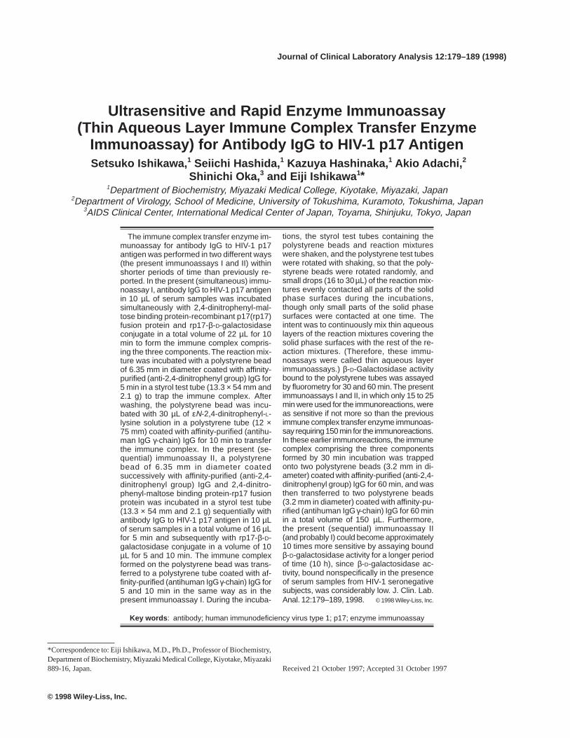
Antibody IgG to HIV-1 p17 antigen was measured in twodifferent ways (Fig. 1).
Present (simultaneous) immunoassay I
An aliquot (10 µL) of serum samples was incubated in astyrol test tube (13.3 × 54 mm and 2.1 g) for up to 15 minwith 12 µL of buffer A containing 0.6 mol/L NaCl, 5 µg ofinactive β-D-galactosidase, 100 fmol of 2,4-dinitrophenyl-maltose binding protein-rp17 fusion protein, and 100 fmolof rp17-β-D-galactosidase conjugate, and subsequently in-cubated for up to 10 min with a polystyrene bead with adiameter of 6.35 mm coated with affinity-purified (anti-2,4-dinitrophenyl group) IgG. The polystyrene bead waswashed three times with 2 ml of buffer B and was incubatedfor up to 20 min with 30 µL of buffer A containing 0.1 mol/LNaCl and 1 mmol/L eN-2,4-dinitrophenyl-L-lysine in a poly-styrene test tube (12 × 75 mm) coated with affinity-purified(antihuman IgG γ-chain) IgG. β-D-galactosidase activitybound to the polystyrene test tube was assayed for 60 min asdescribed above.
Present (sequential) immunoassay II
A polystyrene bead with a diameter of 6.35 mm, whichhad been coated successively with affinity-purified (anti-2,4-dinitrophenyl group) IgG and 2,4-dinitrophenyl-maltose bind-ing protein-rp17 fusion protein, was incubated in a styrol testtube (13.3 × 54 mm and 2.1 g) with a mixture of 10 µL ofserum samples and 6 µL of buffer A containing 0.82 mol/LNaCl and 5 µg of inactive β-D-galactosidase for up to 10min and subsequently with 10 µL of buffer A containing0.4 mol/L NaCl and 200 fmol of rp17- β-D-galactosidaseconjugate for up to 30 min. The polystyrene bead waswashed three times with 2 ml of buffer B and was incu-bated with 30 µL of buffer A containing 0.1 mol/L NaCland 1 mmol/L eN-2,4-dinitrophenyl-L-lysine in a polysty-rene test tube (12 × 75 mm) coated with affinity-purified(antihuman IgG γ-chain) IgG for up to 20 min. β-D-galac-tosidase activity bound to the polystyrene test tube wasassayed for up to 10 h as described above.
All the incubations in the present immunoassays I andII were performed in the same way as described above forbinding of 2,4-dinitrophenyl-β-D-galactosidase to affin-
Enzyme Immunoassay for Anti-HIV-1 IgG 183
ity-purified (anti-2,4-dinitrophenyl group) IgG-coatedsolid phases, except that polystyrene test tubes were ro-tated in a box (15 × 32 × 20 [depth]) mm) with shaking inthe presence of polystyrene beads so as to randomly ro-tate the polystyrene beads. In some experiments,β-D-ga-lactosidase activity bound to the polystyrene beads wasassayed for 10 min as described above.
Other Immunological Methods
The gelatin particle agglutination test for antibodies toHIV-1 was performed using a commercial kit with a lysateof HIV-1 as antigen (SERODIA-HIV, Fujirebio Inc., Tokyo,Japan). Western blotting for antibody IgGs to HIV-1 was
performed using a commercial kit preblotted with nine pro-teins of HIV-1 (gp160, gp120, p66, p55, p51, gp41, p31, p24,and p17) (Ortho HIV Western Blot Kit, Ortho DiagnosticSystems Inc., Raritan, NJ).
Serum Samples From HIV-1 Seronegative andSeropositive Subjects
Serum samples were collected from an HIV-1 seropositivesubject and 8 HIV-1 seronegative subjects (2 males aged 24to 39 yrs, and 6 females aged 32 to 57 yrs) and were stored at–20°C until use. Serum samples were tested by the gelatinparticle agglutination test, and the seropositivity was con-firmed by Western blotting.
Fig. 1. Present immune complex transfer enzyme immunoassays (thinaqueous layer immune complex transfer enzyme immunoassays) I (si-
multaneous) and II (sequential). DNP, 2,4-dinitrophenyl; Ag, antigen;Ab, antibody; Enz, enzyme.
184 Ishikawa et al.
RESULTS
Rapid Binding to (Anti-2,4-Dinitrophenyl Group)IgG-Coated Solid Phases of 2,4-Dinitrophenyl- b-D-Galactosidase in Thin Aqueous Layers Coveringthe Solid Phase Surfaces
The time course of binding to affinity-purified (anti-2,4-dinitrophenyl group) IgG-coated solid phases of 2,4-dinitrophenyl-β-D-galactosidase in thin aqueous layerscovering the solid phase surfaces was examined in three dif-ferent ways.
First, a polystyrene bead of 6.35 mm in diameter coatedwith affinity-purified (anti-2,4-dinitrophenyl group) IgG wasincubated with different volumes (15, 50, and 150 µL) of 2,4-dinitrophenyl-β-D-galactosidase solution (7.7 amol/µL) in astyrol test tube (13.3 × 54 mm and 2.1 g) by shaking in a box(16 × 16 × 20 [depth] mm), so that the polystyrene beadwas rotated randomly. The 2,4-dinitrophenyl-β-D-galac-tosidase solution evenly contacted all parts of the poly-styrene bead surface during the incubation, although onlya small part of the polystyrene bead surface was contactedat any given time. Random rotation served to continuouslymix the thin aqueous layer covering the polystyrene beadsurface with the rest of the 2,4-dinitrophenyl-β-D-galac-tosidase solution (Table 1). The shaking rate was 180 permin, and the shaking width was 25 mm.
Second, a polystyrene bead of 6.35 mm in diameter coatedwith affinity-purified (anti-2,4-dinitrophenyl group) IgG wasincubated with 150 µL of 2,4-dinitrophenyl-β-D-galactosidasesolutions (0.77 and 7.7 amol/µL) in a smaller glass test tube(10 × 75 mm) with shaking (Table 1). In this experiment, thewhole surface of the polystyrene bead always remained inthe 2,4-dinitrophenyl-β-D-galactosidase solution during theincubation, although the incubation was performed with shak-ing as described above.
Third, a polystyrene tube (12 × 75 mm) coated with 0.4 ml
of affinity-purified (anti-2,4-dinitrophenyl group) IgG wasincubated with different volumes (15, 50 and 150 µL) of 2,4-dinitrophenyl-β-D-galactosidase solution (7.7 amol/µL) withrotation, so that the 2,4-dinitrophenyl-β-D-galactosidase so-lution evenly contacted all parts of the coated polystyrenetube surface during the incubation (Table 1). The rotation ratewas 40 per min.
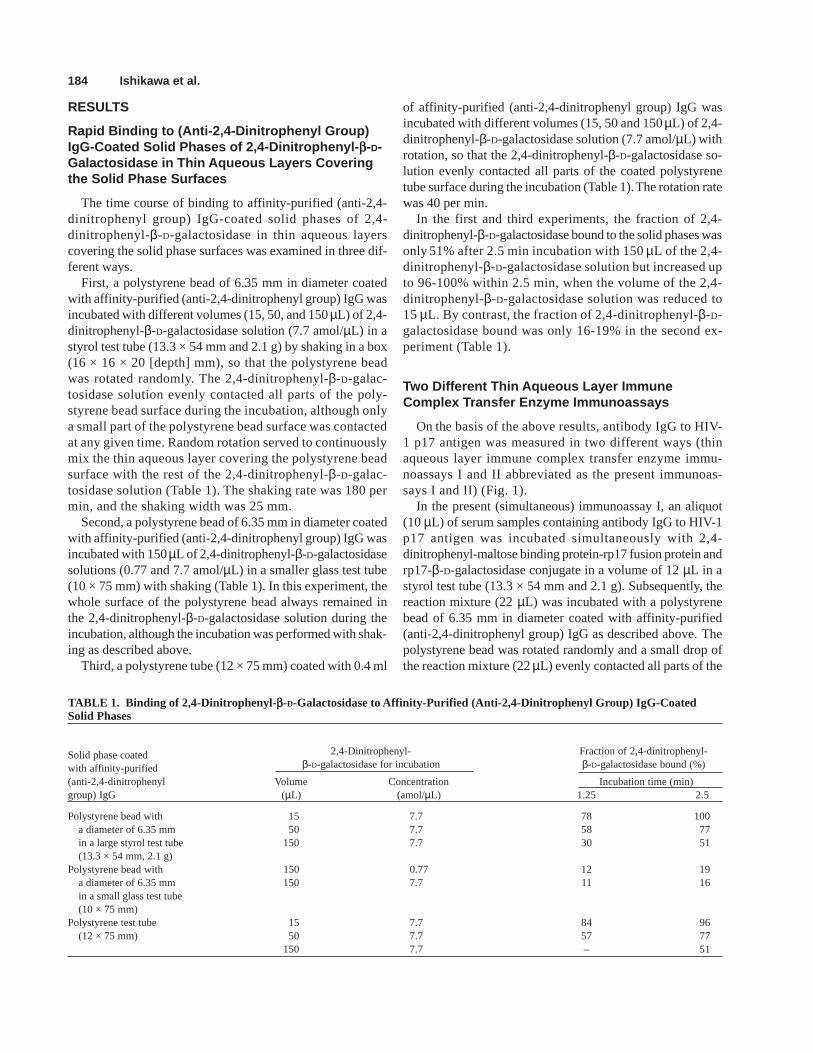
In the first and third experiments, the fraction of 2,4-dinitrophenyl-β-D-galactosidase bound to the solid phases wasonly 51% after 2.5 min incubation with 150 µL of the 2,4-dinitrophenyl-β-D-galactosidase solution but increased upto 96-100% within 2.5 min, when the volume of the 2,4-dinitrophenyl-β-D-galactosidase solution was reduced to15 µL. By contrast, the fraction of 2,4-dinitrophenyl-β-D-galactosidase bound was only 16-19% in the second ex-periment (Table 1).
Two Different Thin Aqueous Layer ImmuneComplex Transfer Enzyme Immunoassays
On the basis of the above results, antibody IgG to HIV-1 p17 antigen was measured in two different ways (thinaqueous layer immune complex transfer enzyme immu-noassays I and II abbreviated as the present immunoas-says I and II) (Fig. 1).
In the present (simultaneous) immunoassay I, an aliquot(10 µL) of serum samples containing antibody IgG to HIV-1p17 antigen was incubated simultaneously with 2,4-dinitrophenyl-maltose binding protein-rp17 fusion protein andrp17-β-D-galactosidase conjugate in a volume of 12 µL in astyrol test tube (13.3 × 54 mm and 2.1 g). Subsequently, thereaction mixture (22 µL) was incubated with a polystyrenebead of 6.35 mm in diameter coated with affinity-purified(anti-2,4-dinitrophenyl group) IgG as described above. Thepolystyrene bead was rotated randomly and a small drop ofthe reaction mixture (22 µL) evenly contacted all parts of the
TABLE 1. Binding of 2,4-Dinitrophenyl- b-D-Galactosidase to Affinity-Purified (Anti-2,4-Dinitrophenyl Group) IgG-CoatedSolid Phases
Solid phase coated 2,4-Dinitrophenyl- Fraction of 2,4-dinitrophenyl-
with affinity-purified β-D-galactosidase for incubation β-D-galactosidase bound (%)
(anti-2,4-dinitrophenyl Volume Concentration Incubation time (min)group) IgG (µL) (amol/µL) 1.25 2.5
Polystyrene bead with 15 7.7 78 100a diameter of 6.35 mm 50 7.7 58 77in a large styrol test tube 150 7.7 30 51(13.3 × 54 mm, 2.1 g)
Polystyrene bead with 150 0.77 12 19a diameter of 6.35 mm 150 7.7 11 16in a small glass test tube(10 × 75 mm)
Polystyrene test tube 15 7.7 84 96(12 × 75 mm) 50 7.7 57 77
150 7.7 – 51
Enzyme Immunoassay for Anti-HIV-1 IgG 185
polystyrene bead surface during the incubation, although itcontacted only a small part of the polystyrene bead surface atany given time. Random rotation served to continuously mixthe thin aqueous layer covering the polystyrene bead surfacewith the rest of the reaction mixture. After washing, the poly-styrene bead and 30 µL of eN-2,4-dinitrophenyl-L-lysine so-lution were added to a polystyrene tube (12 × 75 mm) coatedwith affinity-purified (antihuman IgG γ-chain) IgG, and thepolystyrene tube was shaken with slow rotation in a box (15× 35 × 20 [depth] mm) to elute and transfer the immune com-plex to the polystyrene tube surface.
In the present (sequential) immunoassay II, a polystyrenebead with a diameter of 6.35 mm, which had been coatedsuccessively with affinity-purified (anti-2,4-dinitrophenylgroup) IgG and 2,4-dinitrophenyl-maltose binding protein-rp17 fusion protein, was incubated with a mixture of 6 µL ofa buffer and 10 µL of serum samples containing antibodyIgG to HIV-1 p17 antigen and subsequently with 10 µL of abuffer containing rp17-β-D-galactosidase conjugate in a sty-rol test tube (13.3 × 54 mm and 2.1 g) as described above.The immune complex formed on the polystyrene bead wastransferred to a polystyrene tube coated with affinity-puri-fied (antihuman IgG γ-chain) IgG as in the present immu-noassay I described above.
Time Courses of the Immunoreactions in thePresent (Simultaneous) Immunoassay I
Serum from an HIV-1 seropositive subject was diluted withserum from an HIV-1 seronegative subject, and 10 µL of thediluted serum was incubated with 2,4-dinitrophenyl-maltosebinding protein-rp17 fusion protein and rp17-β-D-galactosi-dase conjugate for up to 15 min to form the immune complexcomprising the three components, and was subsequently in-cubated with a polystyrene bead coated with affinity-purified(anti-2,4-dinitrophenyl group) IgG for 5 min to trap the im-mune complex. β-D-galactosidase activity bound to the poly-styrene bead that was assayed for 10 min reached themaximum within 10 min, indicating that the formation of theimmune complex was completed within 10 min.
The immune complex was formed for 10 min and wastrapped for up to 10 min. Bound β-D-galactosidase activityreached the maximum within 3 min, indicating that trappingthe immune complex was completed with 3 min.
The immune complex was formed for 10 min, trapped for5 min, and was then transferred for up to 20 min. Transfer-ence of the immune complex was almost completed within10 min.
Amount of 2,4-Dinitrophenyl-Maltose BindingProtein-Rp17 Fusion Protein for CoatingPolystyrene Beads
Polystyrene beads of 6.35 mm in diameter were coatedsequentially with affinity-purified (anti-2,4-dinitrophenyl
group) IgG and various amounts of 2,4-dinitrophenyl-mal-tose binding protein-rp17 fusion protein and were reacted withserum from an HIV-1 seropositive subject and rp17-β-D-ga-lactosidase conjugate. β-D-galactosidase activity bound tothe polystyrene beads increased with increasing amountsof 2,4-dinitrophenyl-maltose binding protein-rp17 fusionprotein but was only approximately 20% higher whencoated with 800 fmol than when coated with 400 fmol. Inthe following experiments, therefore, 800 fmol per beadwere used for coating.
Time Courses of the Immunoreactions in thePresent (Sequential) Immunoassay II
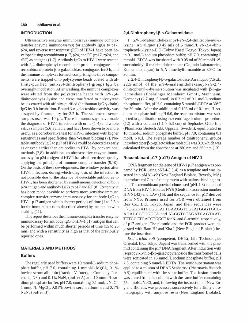
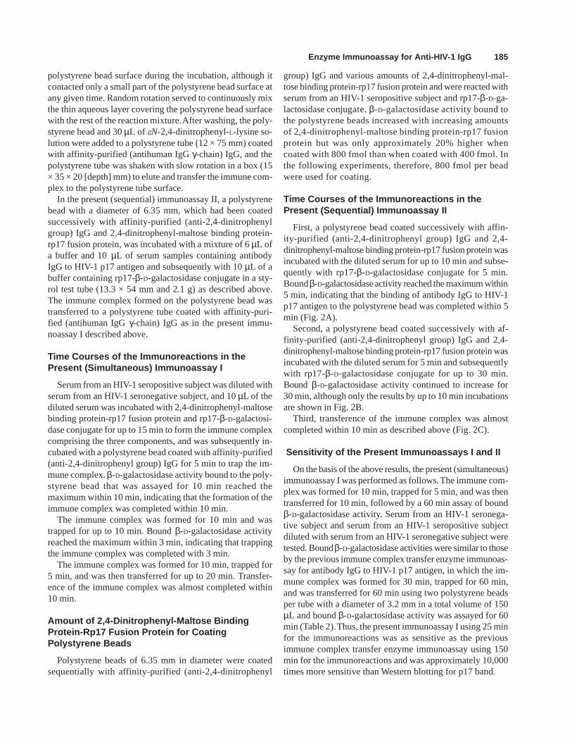
First, a polystyrene bead coated successively with affin-ity-purified (anti-2,4-dinitrophenyl group) IgG and 2,4-dinitrophenyl-maltose binding protein-rp17 fusion protein wasincubated with the diluted serum for up to 10 min and subse-quently with rp17-β-D-galactosidase conjugate for 5 min.Bound β-D-galactosidase activity reached the maximum within5 min, indicating that the binding of antibody IgG to HIV-1p17 antigen to the polystyrene bead was completed within 5min (Fig. 2A).
Second, a polystyrene bead coated successively with af-finity-purified (anti-2,4-dinitrophenyl group) IgG and 2,4-dinitrophenyl-maltose binding protein-rp17 fusion protein wasincubated with the diluted serum for 5 min and subsequentlywith rp17-β-D-galactosidase conjugate for up to 30 min.Bound β-D-galactosidase activity continued to increase for30 min, although only the results by up to 10 min incubationsare shown in Fig. 2B.
Third, transference of the immune complex was almostcompleted within 10 min as described above (Fig. 2C).
Sensitivity of the Present Immunoassays I and II
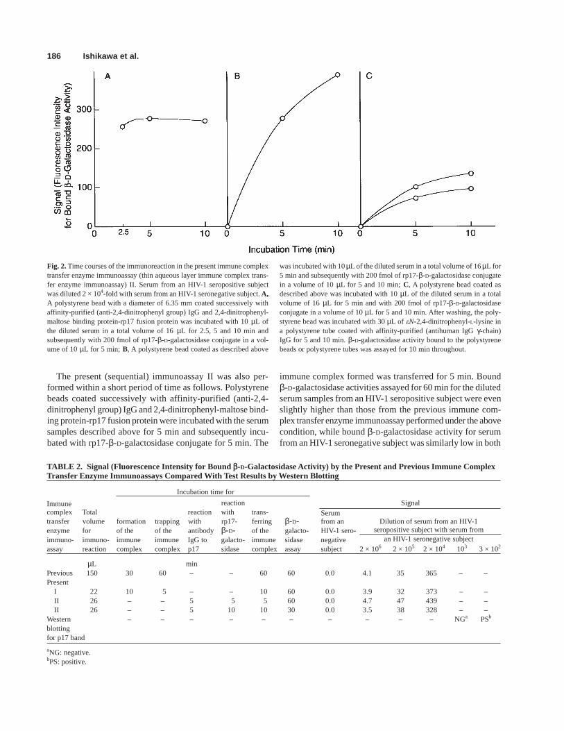
On the basis of the above results, the present (simultaneous)immunoassay I was performed as follows. The immune com-plex was formed for 10 min, trapped for 5 min, and was thentransferred for 10 min, followed by a 60 min assay of boundβ-D-galactosidase activity. Serum from an HIV-1 seronega-tive subject and serum from an HIV-1 seropositive subjectdiluted with serum from an HIV-1 seronegative subject weretested. Bound β-D-galactosidase activities were similar to thoseby the previous immune complex transfer enzyme immunoas-say for antibody IgG to HIV-1 p17 antigen, in which the im-mune complex was formed for 30 min, trapped for 60 min,and was transferred for 60 min using two polystyrene beadsper tube with a diameter of 3.2 mm in a total volume of 150µL and bound β-D-galactosidase activity was assayed for 60min (Table 2). Thus, the present immunoassay I using 25 minfor the immunoreactions was as sensitive as the previousimmune complex transfer enzyme immunoassay using 150min for the immunoreactions and was approximately 10,000times more sensitive than Western blotting for p17 band.
186 Ishikawa et al.
The present (sequential) immunoassay II was also per-formed within a short period of time as follows. Polystyrenebeads coated successively with affinity-purified (anti-2,4-dinitrophenyl group) IgG and 2,4-dinitrophenyl-maltose bind-ing protein-rp17 fusion protein were incubated with the serumsamples described above for 5 min and subsequently incu-bated with rp17-β-D-galactosidase conjugate for 5 min. The
immune complex formed was transferred for 5 min. Boundβ-D-galactosidase activities assayed for 60 min for the dilutedserum samples from an HIV-1 seropositive subject were evenslightly higher than those from the previous immune com-plex transfer enzyme immunoassay performed under the abovecondition, while bound β-D-galactosidase activity for serumfrom an HIV-1 seronegative subject was similarly low in both
Fig. 2. Time courses of the immunoreaction in the present immune complextransfer enzyme immunoassay (thin aqueous layer immune complex trans-fer enzyme immunoassay) II. Serum from an HIV-1 seropositive subjectwas diluted 2 × 10⁴-fold with serum from an HIV-1 seronegative subject. A,A polystyrene bead with a diameter of 6.35 mm coated successively withaffinity-purified (anti-2,4-dinitrophenyl group) IgG and 2,4-dinitrophenyl-maltose binding protein-rp17 fusion protein was incubated with 10 µL ofthe diluted serum in a total volume of 16 µL for 2.5, 5 and 10 min andsubsequently with 200 fmol of rp17-β-D-galactosidase conjugate in a vol-ume of 10 µL for 5 min; B, A polystyrene bead coated as described above
was incubated with 10 µL of the diluted serum in a total volume of 16 µL for5 min and subsequently with 200 fmol of rp17-β-D-galactosidase conjugatein a volume of 10 µL for 5 and 10 min; C, A polystyrene bead coated asdescribed above was incubated with 10 µL of the diluted serum in a totalvolume of 16 µL for 5 min and with 200 fmol of rp17-β-D-galactosidaseconjugate in a volume of 10 µL for 5 and 10 min. After washing, the poly-styrene bead was incubated with 30 µL of eN-2,4-dinitrophenyl-L-lysine ina polystyrene tube coated with affinity-purified (antihuman IgG γ-chain)IgG for 5 and 10 min. β-D-galactosidase activity bound to the polystyrenebeads or polystyrene tubes was assayed for 10 min throughout.
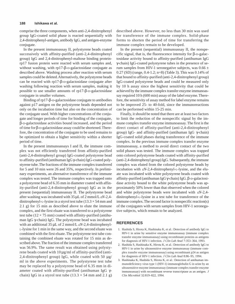
TABLE 2. Signal (Fluorescence Intensity for Bound b-D-Galactosidase Activity) by the Present and Previous Immune ComplexTransfer Enzyme Immunoassays Compared With Test Results by Western Blotting
Incubation time for
Immune reaction Signalcomplex Total reaction with trans- Serumtransfer volume formation trapping with rp17- ferring β-D- from an Dilution of serum from an HIV-1enzyme for of the of the antibodyβ-D- of the galacto- HIV-1 sero- seropositive subject with serum fromimmuno- immuno- immune immune IgG to galacto- immune sidase negative an HIV-1 seronegative subjectassay reaction complex complex p17 sidase complex assay subject 2 × 10⁶ 2 × 10⁵ 2 × 10⁴ 103 3 × 10²
µL minPrevious 150 30 60 – – 60 60 0.0 4.1 35 365 – –Present
I 22 10 5 – – 10 60 0.0 3.9 32 373 – –II 26 – – 5 5 5 60 0.0 4.7 47 439 – –II 26 – – 5 10 10 30 0.0 3.5 38 328 – –
Western – – – – – – – – – – NGa PSb
blottingfor p17 band
aNG: negative.bPS: positive.
Enzyme Immunoassay for Anti-HIV-1 IgG 187
the present and previous immunoassays (Table 2). Thus, thepresent immunoassay II, in which the immunoreactions wereperformed within only 15 min in total, was even slightly moresensitive than the previous one using 150 min for theimmunoreactions described above and was 10,000 times moresensitive than Western blotting for p17 band.
The sensitivity of the present immunoassay II did not sig-nificantly change even when the period of time for assay ofbound β-D-galactosidase activity was reduced to 30 min, pro-vided that the incubation with rp17-β-D-galactosidase conju-gate and transference of the immune complex were performedboth for a longer period of time (10 min) (Table 2). Thus, thepresent immunoassay II, performed within 55 min (25 minfor the immunoreactions and 30 min for β-D-galactosidaseassay), was as sensitive as the previous immune complex trans-fer enzyme immunoassay requiring 3.5 h (2.5 h for theimmunoreactions and 1 h for β-D-galactosidase assay) andwas 10,000 times more sensitive than Western blotting forp17 band.
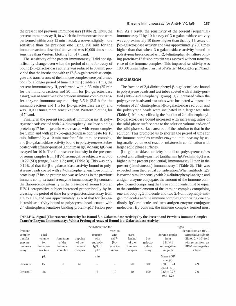
Finally, in the present (sequential) immunoassay II, poly-styrene beads coated with 2,4-dinitrophenyl-maltose bindingprotein-rp17 fusion protein were reacted with serum samplesfor 5 min and with rp17-β-D-galactosidase conjugate for 10min, followed by a 10 min transfer of the immune complex,and β-D-galactosidase activity bound to polystyrene test tubescoated with affinity-purified (antihuman IgG γ-chain) IgG wasassayed for 10 h. The fluorescence intensity in the presenceof serum samples from HIV-1 seronegative subjects was 0.66±0.27 (SD) (range, 0.4 to 1.2 ; n=8) (Table 3). This was only0.14% of that for β-D-galactosidase activity bound to poly-styrene beads coated with 2,4-dinitrophenyl-maltose bindingprotein-rp17 fusion protein and was as low as in the previousimmune complex transfer enzyme immunoassay. By contrast,the fluorescence intensity in the presence of serum from anHIV-1 seropositive subject increased proportionally by in-creasing the period of time for β-D-galactosidase assay from1 h to 10 h, and was approximately 35% of that for β-D-ga-lactosidase activity bound to polystyrene beads coated with2,4-dinitrophenyl-maltose binding protein-rp17 fusion pro-
tein. As a result, the sensitivity of the present (sequential)immunoassay II by 10 h assay of β-D-galactosidase activitywas approximately 10 times higher than that by 1 h assay ofβ-D-galactosidase activity and was approximately 250 timeshigher than that when β-D-galactosidase activity bound topolystyrene beads coated with 2,4-dinitrophenyl-maltose bind-ing protein-rp17 fusion protein was assayed without transfer-ence of the immune complex. This improved sensitivity was100,000 times higher than that of Western blotting for p17 band.
DISCUSSION
The fraction of 2,4-dinitrophenyl-β-D-galactosidase boundto polystyrene beads and test tubes coated with affinity-puri-fied (anti-2,4-dinitrophenyl group) IgG increased when thepolystyrene beads and test tubes were incubated with smallervolumes of 2,4-dinitrophenyl-β-D-galactosidase solution andthe polystyrene beads were incubated in a larger test tube(Table 1). More specifically, the fraction of 2,4-dinitrophenyl-β-D-galactosidase bound increased with increasing ratios ofthe solid phase surface area to the solution volume and/or ofthe solid phase surface area out of the solution to that in thesolution. This prompted us to shorten the period of time forthe immune complex transfer enzyme immunoassay by us-ing smaller volumes of reaction mixtures in combination withlarger solid phase surfaces.
β-D-galactosidase activity bound to polystyrene tubescoated with affinity-purified (antihuman IgG γ-chain) IgG washigher in the present (sequential) immunoassay II than in thepresent (simultaneous) immunoassay I (Table 2). This wasexpected from theoretical consideration. When antibody IgGis reacted simultaneously with 2,4-dinitrophenyl-antigen andantigen-enzyme conjugate, the amount of the immune com-plex formed comprising the three components must be equalto the combined amount of the immune complex comprisingone antibody IgG molecule and two 2,4-dinitrophenyl-anti-gen molecules and the immune complex comprising one an-tibody IgG molecule and two antigen-enzyme conjugatemolecules. By contrast, the immune complex formed must
TABLE 3. Signal (Fluorescence Intensity for Bound b-D-Galactosidase Activity) by the Present and Previous Immune ComplexTransfer Enzyme Immunoassays With a Prolonged Assay of Bound b-D-Galactosidase Activity
Incubation time for SignalImmune reaction Serum from an HIV-1complex Total reaction with trans- Serum samples seropositive subjecttransfer volume formation trapping with rp17- ferring β-D- from diluted 2 × 10⁷-foldenzyme for of the of the antibody β-D- of the galacto- 8 HIV-1 with serum from animmuno- immuno- immune immune IgG to galacto- immune sidase seronegative HIV-1 seronegativeassay reaction complex complex p17 sidase complex assay subjects subject
µL min Mean ± SD(range)
Previous 150 30 60 – – 60 600 0.94 ± 0.24 4.9(0.63–1.3)
Present II 26 – – 5 10 10 600 0.66 ± 0.27 7.9(0.4–1.2)
188 Ishikawa et al.
comprise the three components, when anti-2,4-dinitrophenylgroup IgG-coated solid phase is reacted sequentially with2,4-dinitrophenyl-antigen, antibody IgG, and antigen-enzymeconjugate.
In the present immunoassay II, polystyrene beads coatedsuccessively with affinity-purified (anti-2,4-dinitrophenylgroup) IgG and 2,4-dinitrophenyl-maltose binding protein-rp17 fusion protein were reacted with serum samples and,without washing, with rp17-β-D-galactosidase conjugate asdescribed above. Washing process after reaction with serumsamples could be deleted. Alternatively, the polystyrene beadscan be reacted with rp17-β-D-galactosidase conjugate afterwashing following reaction with serum samples, making itpossible to use smaller amounts of rp17-β-D-galactosidaseconjugate in smaller volumes.
Binding of rp17-β-D-galactosidase conjugate to antibodiesagainst p17 antigen on the polystyrene beads depended notonly on the incubation time but also on the concentration ofthe conjugate used. With higher concentrations of the conju-gate and longer periods of time for binding of the conjugate,β-D-galactosidase activities bound increased, and the periodof time for β-D-galactosidase assay could be shortened. There-fore, the concentration of the conjugate to be used remains tobe optimized to obtain a higher sensitivity within a shorterperiod of time.
In the present immunoassays I and II, the immune com-plex was not efficiently transferred from affinity-purified(anti-2,4-dinitrophenyl group) IgG-coated polystyrene beadto affinity-purified (antihuman IgG γ-chain) IgG-coated poly-styrene tube. The fractions of the immune complex transferredfor 5 and 10 min were 26 and 35%, respectively. In prelimi-nary experiments, an alternative transference of the immunecomplex was tested. The immune complex was trapped ontoa polystyrene bead of 6.35 mm in diameter coated with affin-ity-purified (anti-2,4-dinitrophenyl group) IgG as in thepresent (sequential) immunoassay II. The polystyrene beadafter washing was incubated with 35 µL of 2 mmol/L eN-2,4-dinitrophenyl-L-lysine in a styrol test tube (13.3 × 54 mm and2.1 g) for 15 min as described above to elute the immunecomplex, and the first eluate was transferred to a polystyrenetest tube (12 × 75 mm) coated with affinity-purified (antihu-man IgG γ-chain) IgG. The polystyrene bead was incubatedwith an additional 35 µL of 2 mmol/L eN-2,4-dinitrophenyl-L-lysine for 1 min in the same way, and the second eluate wascombined with the first eluate. The polystyrene test tube con-taining the combined eluate was rotated for 15 min as de-scribed above. The fraction of the immune complex transferredwas 56.9%. The same result was obtained using polysty-rene beads coated with 10 µg/ml of affinity-purified (anti-2,4-dinitrophenyl group) IgG, while coated with 50 µg/ml in the above experiments. The polystyrene test tubemay be replaced by a polystyrene bead of 6.35 mm in di-ameter coated with affinity-purified (antihuman IgG γ-chain) IgG in a styrol test tube (13.3 × 54 mm and 2.1 g)
described above. However, no less than 30 min was usedfor transference of the immune complex. Solid-phaseforms to shorten the period of time for transferring theimmune complex remain to be developed.
In the present (sequential) immunoassay II, the nonspe-cific signal, that is, the fluorescence intensity for β-D-galac-tosidase activity bound to affinity-purified (antihuman IgGγ-chain) IgG-coated polystyrene tubes in the presence of se-rum samples from HIV-1 seronegative subjects, was 0.66 ±0.27 (SD) (range, 0.4-1.2; n=8) (Table 3). This was 0.14% ofthat bound to affinity-purified (anti-2,4-dinitrophenyl group)IgG-coated polystyrene beads and could be measured onlyby 10 h assay since the highest sensitivity that could beachieved by the immune complex transfer enzyme immunoas-say required 10 h (600 min) assay of the label enzyme. There-fore, the sensitivity of assay method for label enzyme remainsto be improved 25- to 40-fold, since the immunoreactionscan be performed within 15-25 min.
Finally, it should be noted that there are at least two factorsto limit the reduction of the nonspecific signal by the im-mune complex transfer enzyme immunoassay. The first is thedirect contact of affinity-purified (anti-2,4-dinitrophenylgroup) IgG- and affinity-purified (antihuman IgG γ-chain)IgG-coated solid phases during transference of the immunecomplex. In the previous immune complex transfer enzymeimmunoassay, a method to avoid direct contact of the twosolid phases was tested. The immune complex was trappedonto colored polystyrene beads coated with affinity-purified(anti-2,4-dinitrophenyl group) IgG. Subsequently, the immunecomplex was eluted from the colored polystyrene beads byincubation with eN-2,4-dinitrophenyl-L-lysine, and the elu-ate was incubated with white polystyrene beads coated withaffinity-purified (antihuman IgG γ-chain) IgG. β-D-galactosi-dase activity bound to the white polystyrene beads was ap-proximately 50% lower than that observed when the coloredand white polystyrene beads were incubated with eN-2,4-dinitrophenyl-L-lysine in a test tube for transference of theimmune complex. The second factor is nonspecific reaction(s)of the conjugates with serum samples from HIV-1 seronega-tive subjects, which remain to be analyzed.
REFERENCES
1. Hashida S, Hirota K, Hashinaka K, et al.: Detection of antibody IgG toHIV-1 in urine by sensitive enzyme immunoassay (immune complextransfer enzyme immunoassay) using recombinant proteins as antigensfor diagnosis of HIV-1 infection. J Clin Lab Anal 7:353–364, 1993.
2. Hashida S, Hashinaka K, Hirota K, et al.: Detection of antibody IgG toHIV-1 in urine by ultrasensitive enzyme immunoassay (immune com-plex transfer enzyme immunoassay) using recombinant p24 as antigenfor diagnosis of HIV-1 infection. J Clin Lab Anal 8:86–95, 1994.
3. Hashinaka K, Hashida S, Hirota K, et al.: Detection of antihuman im-munodeficiency virus type 1 (HIV-1) immunoglobulin G in urine by anultrasensitive enzyme immunoassay (immune complex transfer enzymeimmunoassay) with recombinant reverse transcriptase as an antigen. JClin Microbiol 32:819–822, 1994.
Enzyme Immunoassay for Anti-HIV-1 IgG 189
4. Hashida S, Hashinaka K, Saitoh A, et al.: Diagnosis of HIV-1 infection bydetection of antibody IgG to HIV-1 in urine with ultrasensitive enzymeimmunoassay (immune complex transfer enzyme immunoassay) usingrecombinant proteins as antigens. J Clin Lab Anal 8:237–246, 1994.
5. Ishikawa S, Hashida S, Hashinaka K, et al.: Diagnosis of HIV-1 infec-tion with whole saliva by detection of antibody IgG to HIV-1 withultrasensitive enzyme immunoassay using recombinant reverse tran-scriptase as antigen. J Acquir Immune Defic Syndr Human Retrovirol10:41–47, 1995.
6. Ishikawa S, Hashida S, Hashinaka K, et al.: Whole saliva dried onfilter paper for diagnosis of HIV-1 infection by detection of antibodyIgG to HIV-1 with ultrasensitive enzyme immunoassay using recom-binant reverse transcriptase as antigen. J Clin Lab Anal 10:35–41, 1996.
7. Hashida S, Hashinaka K, Nishikata I, et al.: Immune complex transferenzyme immunoassay that is more sensitive and specific than Westernblotting for detection of antibody immunoglobulin G to human im-munodeficiency virus type 1 in serum with recombinant pol and gagproteins as antigens. Clin Diag Lab Immunol 2:535–541, 1995.
8. Hashida S, Hashinaka K, Nishikata I, et al.: Shortening of the windowperiod in diagnosis of HIV-1 infection by simultaneous detection ofp24 antigen and antibody IgG to p17 and reverse transcriptase in se-rum with ultrasensitive enzyme immunoassay. J Virol Methods 62:43–53, 1996.
9. Hashida S, Hashinaka K, Nishikata I, et al.: Measurement of humanimmunodeficiency virus type 1 p24 in serum by an ultrasensitive en-
zyme immunoassay, the two-site immune complex transfer enzymeimmunoassay. J Clin Microbiol 33:298–303, 1995.
10. Hashida S, Hashinaka K, Nishikata I, et al.: Ultrasensitive and morespecific enzyme immunoassay (immune complex transfer enzyme im-munoassay) for p24 antigen of HIV-1 in serum using affinity-purifiedrabbit anti-p24 Fab´ and monoclonal mouse anti-p24 Fab´. J Clin LabAnal 10:302–307, 1996.
11. Ishikawa S, Hashida S, Hashinaka K, et al.: More sensitive immunecomplex transfer enzyme immunoassay for antibody IgG to p17 ofHIV-1 with shorter incubation time for immunoreactions and largervolumes of serum samples. J Clin Lab Anal 11:244–250, 1997.
12. Penefsky HS: A centrifuged-column procedure for the measurementof ligand binding by beef heart F1. Methods Enzymol LVI:527–530, 1979.
13. Eisen HN, Carsten ME, Belman S: Studies of hypersensitivity to lowmolecular substances. III. The 2,4-dinitrophenyl group as a determi-nant in the precipitin reaction. J Immunol 73:296–308, 1954.
14. Adachi A, Gendelman HE, Koenig S, et al.: Production of acquiredimmunodeficiency syndrome-associated retrovirus in human and non-human cells transfected with an infectious molecular clone. J Virol59:284–291, 1986.
15. Wain-Hobson S, Sonigo P, Danos O, Cole S, Alizon M: Nucleotidesequence of the AIDS virus, LAV. Cell 40:9–17, 1985.
16. Ishikawa E, Kato K: Ultrasensitive enzyme immunoassay. Scand JImmunol 8(Suppl. 7):43–55, 1978.


