
UNIT 2: MECHANISMS OF INHERITANCETopic #1: A History of DNA
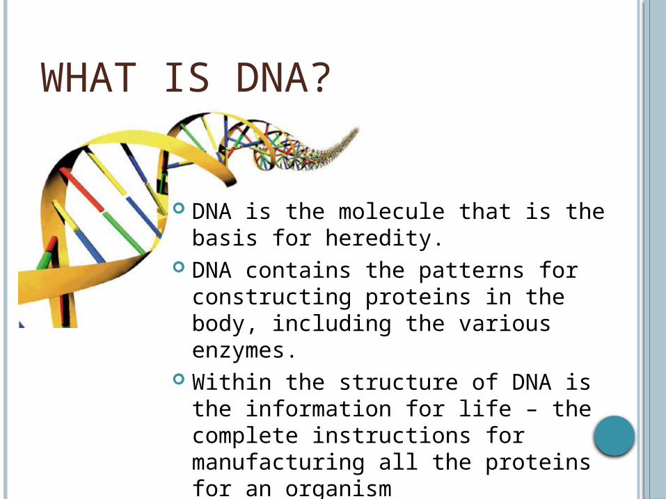
WHAT IS DNA?
DNA is the molecule that is the basis for heredity.
DNA contains the patterns for constructing proteins in the body, including the various enzymes.
Within the structure of DNA is the information for life – the complete instructions for manufacturing all the proteins for an organism

A DNA TIMELINE
Between Mendel (1866) and present time, many scientists have contributed to our knowledge of inheritance.
DNA Interactive Timeline Your task is to create a timeline of events,
from Mendel to present time.

MULTIDISCIPLINARY COLLABORATION CONTRIBUTIONS TO DNA RESEARCH Many individual and group research
projects have added to our knowledge of DNA
Scientists involved with the research come from a variety of backgrounds:Physics – invented the X-ray techniques
used to view DNAChemistry – discovering the ratios of A-T
and G-C as well as theorizing the bonds that hold the molecule together
Biology – research with viruses, bacteria, plants and animals.

THE RACE TO DISCOVER Watson and Crick are credited as the co-
discoverers of DNA – but the race was political.
The X-ray diffraction technique that they used was developed by Maurice Wilkins and Rosalind Franklin to view DNA
Another lead scientist (Linus Pauling) from the U.S.A. was denied a visa to England to study the X-ray photographs because of his political associations.

MECHANISMS OF INHERITANCETopic #2: The Structure of DNA Nucleotides

THE STRUCTURE OF DNA DNA is most often described as a
double helix It resembles a twisted ladder The backbone of the ladder is made of
sugar and phosphate molecules The rungs of the ladder are formed by
two nitrogen bases that are connected by hydrogen bonds

A NUCLEOTIDE DNA is a polymer made of repeating
subunits called nucleotides Nucleotides have 3 parts:
1) A simple sugar (deoxyribose)2) A phosphate group3) A nitrogenous base
DNA stands for deoxyribonucleic acid

DEOXYRIBOSE SUGAR STRUCTURE The simple sugar (Deoxyribose) in DNA gives
it its name (Deoxyribonucleic acid)
O
OH H
CH2

PHOSPHATE GROUP The phosphate group of DNA consists
of one atom of phosphorus surrounded by 4 oxygen atoms
P O
O
O
O

NITROGENOUS BASES A nitrogenous base is a carbon ring
structure that contains one or more atoms of nitrogen
In DNA there are 4 possible nitrogenous bases:1) Adenine (A)2) Guanine (G)3) Cytosine (C)4) Thymine (T)

CHAINS OF NUCLEOTIDES Nucleotides join together to form long
chains. The phosphate group of one nucleotide
bonds to the deoxyribose sugar of an adjacent nucleotide
The nitrogen bases stick out like the teeth of a zipper

COMPLIMENTARY BASE PAIRSIn DNA, adenine (A) always pairs
with thymine (T) and guanine (G) always pairs with cytosine (C).
Therefore, the amount of adenine is always the same as thymine, and the amount of cytosine is always the same as guanine

EXAMPLES:
If 33.4% of the bases in a sample are guanine, what are the percentages of the other bases?

EXAMPLE: Write the complimentary strand of DNA:
CGACTTGCA

ACTIVITY: MAKE A DNA MOLECULE
Directions:1) Colour your nucleotides: Use a
different colour for your deoxyribose sugar, phosphate group, and each type of nucleic acids
2) Cut out the nucleotides3) Pair and tape the complimentary
bases together4) Twist into a helix shape – Voila!
You have DNA

MECHANISMS OF INHERITANCETopic #3: DNA Replication
Objectives: Identify the number of hydrogen bonds between complimentary base pairs Describe the 5 steps of DNA Replication
http://www.pbs.org/wgbh/aso/tryit/dna/shockwave.html

BASE PAIRING As you know, Adenine pairs with Thymine Cytosine pairs with Guanine The complimentary base pairs are are
bound together by hydrogen bonds
The A-T bond has 2 hydrogen bonds The G-C bond has 3 hydrogen bonds

BASE PAIRING

DNA REPLICATION DNA is the only molecule know that is
capable of duplicating itself. This is done through the process of
replication During replication, the weak hydrogen bonds
are broken The two edges of the ladder “unzip”

SEMICONSERVATIVE REPLICATION DNA replication is known as
semiconservative replication because only the parent strands are conserved – the original double-strand is not.
Semiconservative replication produces two “half-old, half-new” strands of DNA

REPLICATION STEP 1: HELICASE Step 1 of replication: DNA Helicase unwinds
the parental DNA strand (it picks a weak point between a A-T where there are only 2 hydrogen bonds

REPLICATION STEP 2: BINDING PROTEINS Step 2 of replication: DNA binding proteins then stabilize the
unzipped strand

REPLICATION STEP 3: LEADING STRAND SYNTHESIS DNA is always synthesized from the 5’ end to
the 3’ end. The strand where the open end is
synthesizing the 5’ end is called the leading strand. It is copied first.

REPLICATION STEP 4: LAGGING STRAND SYNTHESIS On the opposite strand, the DNA is replicated
in chunks called Okazaki Fragments

REPLICATION STEP 5: DNA LIGASE LINKS TOGETHER OKAZAKI FRAGMENTS
Once the base pairs have been replicated, DNA Ligase links together all of the Okazaki fragments to the growing strand.


MECHANISMS OF INHERITANCETopic #4: DNA vs. RNA
Objectives: Describe the differences between DNA & RNA Know when RNA is used Describe 3 types of RNA Identify where RNA is located in the cell

FROM GENE TO PROTEIN The sequence of nucleotides in DNA
contains information for making proteins.
Proteins fold into complex, 3D shapes to become structures and regulators of cell functions.
E.g. Filaments in muscle tissueEnzymes

DNA IS THE BLUEPRINT FOR RNA RNA is made from DNA during the
process of transcription. DNA is “unzipped”, and the parent
strand of DNA is used as a template for making RNA.

THE PURPOSE OF RNA Ribonucleic Acid (RNA) plays an
important role in the synthesis of proteins.
There are a number of different types of RNA:1) rRNA – ribosomal RNA2) tRNA – transfer RNA3) mRNA – messenger RNA

MESSENGER RNA Messenger RNA
(mRNA) brings instructions from DNA in the nucleus of the cell to the cytoplasm.
Ribosomes use this information to assemble amino acids and make proteins
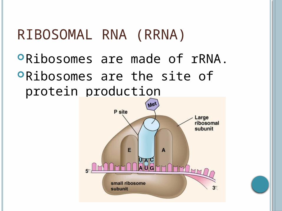
RIBOSOMAL RNA (RRNA)
Ribosomes are made of rRNA. Ribosomes are the site of protein
production
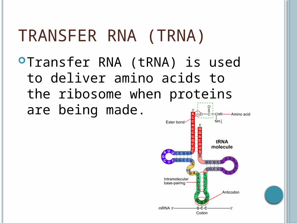
TRANSFER RNA (TRNA)Transfer RNA (tRNA) is used to
deliver amino acids to the ribosome when proteins are being made.

DIFFERENCES BETWEEN DNA & RNA
RNA contains the nitrogenous base uracil instead of thymine
RNA contains ribose instead of deoxyribose
RNA is single strandedRNA carries genetic information
found in DNA from the nucleus to the ribosomes in the cytoplasm

ASSIGNMENT – FOR YOUR NOTES Read Pages 288 – 290 in your textbook. Using the rest of the space on your page,
explain how a car being built on an assembly line is like proteins being synthesized in the cell.
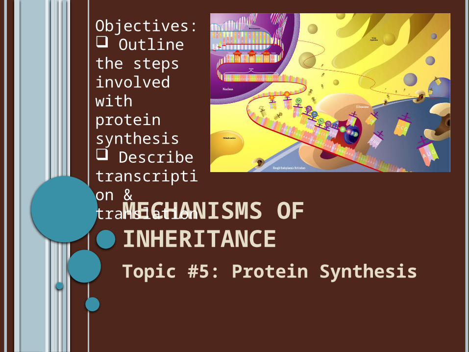
MECHANISMS OF INHERITANCETopic #5: Protein Synthesis
Objectives: Outline the steps involved with protein synthesis Describe transcription & translation

GENES The DNA that makes up
the genome can be subdivided into information bytes called genes.
Each gene encodes a protein that performs a specialized function in the cell. This is the central dogma of molecular biology.
The human genome contains >25000 genes.
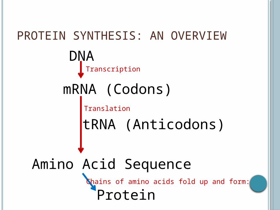
PROTEIN SYNTHESIS: AN OVERVIEW
DNA
mRNA (Codons)
tRNA (Anticodons)
Amino Acid Sequence
Transcription
Translation
ProteinChains of amino acids fold up and form:

TRANSCRIPTION & TRANSLATION In the process of protein synthesis, cells
use a 2-step process to read each gene and produce the string of amino acids that makes up a protein.
The two steps are:1) Transcription2) Translation

TRANSCRIPTION: MAKING A GENETIC MESSENGER
Proteins are produced in the cytoplasm of the cell. However, during protein synthesis, DNA does not leave the nucleus of the cell.
Therefore, a message needs to be sent out of the membrane that “tells” the ribosome directions for producing a protein.
These directions are mRNA

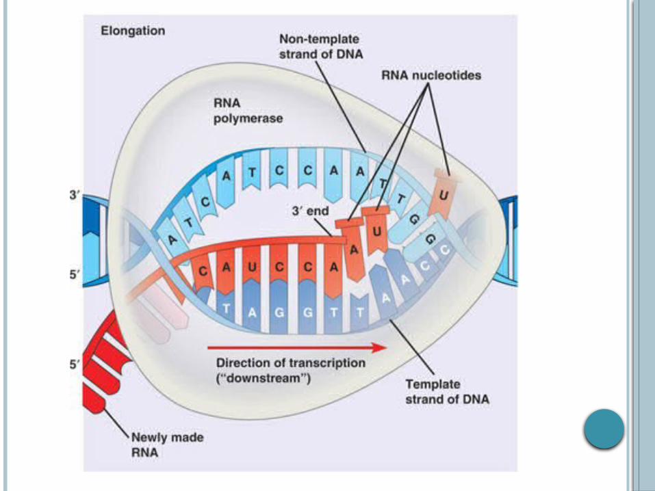
TRANSCRIPTION Transcription is a 3-step process.
1) Enzymes unzip the DNA molecule in the region of the gene that is being transcribed.
2) Free RNA nucleotides form base pairs with their complimentary nucleotides on the DNA strand.
3) The mRNA strand breaks away and the DNA strands rejoin. The mRNA strand then leaves the nucleus and enters the cytoplasm.

TRANSCRIPTION EXAMPLE:
A DNA molecule has the sequence below. What is the corresponding mRNA code?
A T T A C G A T C T G
Remember, instead of thymine, RNA
uses uracil!

TRANSCRIPTION EXAMPLE: A DNA molecule has the sequence below.
What is the corresponding mRNA code?
A T T A C G A T C T GU A A U G C U A G U C

TRANSLATION After mRNA is formed in the nucleus, the
mRNA travels from the nucleus to ribosomes in the cytoplasm.
This is where translation takes place. The mRNA attaches itself to a ribosome and
protein synthesis begins.

CODONS & ANTI-CODONS A group of 3 nitrogenous bases on the mRNA
strand, is called a codon tRNA that are circulating in the cytoplasm of
the cell contain 3 exposed nitrogen bases – the anti-codon – which correspond to the codons on the mRNA

TRNA CARRIES AMINO ACIDS
Each type of tRNA molecule has a specific anti-codon, which relates to a different amino acid that the tRNA carries.
There are 64 possible combinations of nitrogenous bases that the anticodon can have: 4x4x4
These 64 combinations correspond to 21 possible amino acids

DICTIONARY OF MESSENGER RNA CODE WORDS: THESE CORRESPOND TO THE CODONS
STARTING & STOPPING THE AMINO ACID CHAIN
Some of the codes provided by the mRNA are not for amino acids.
These codes, called terminators end protein synthesis (kind of like a period ends a sentence)
Other codes, called initiators are responsible for turning protein synthesis on.
AUG is the codon code to start.

EXAMPLES OF PROTEIN SYNTHESIS
http://learn.genetics.utah.edu/content/begin/dna/transcribe/

MECHANISMS OF INHERITANCETopic #6: Mutations

MUTATION A mutation is any change in DNA sequence. Mutations can be caused by:
1) Errors in replication 2) Errors in transcription 3) Errors in cell division 4) External agents

CAUSES OF MUTATIONS
Mutations in DNA sequences generally occur through one of two processes: 1) DNA damage from environmental agents such
as UV light, nuclear radiation, or certain chemicals 2) Mistakes that occur when a cell copies its DNA
in preparation for cell division

FACTS ABOUT MUTATIONS
Most mutations are minor Many mutations are harmful Some mutations are lethal Very few mutations are helpful.
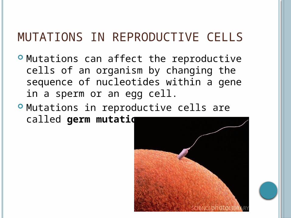
MUTATIONS IN REPRODUCTIVE CELLS
Mutations can affect the reproductive cells of an organism by changing the sequence of nucleotides within a gene in a sperm or an egg cell.
Mutations in reproductive cells are called germ mutations
MUTATIONS IN BODY CELLS
Mutations in body cells are called somatic mutations
If a cell’s DNA is changed, a mutation may impair the function of the cellE.g. Mutation in a stomach cell may cause it to stop producing stomach acid
When the cell divides, the new cells will also have the same mutation.

TYPES OF MUTATIONS 1) Substitutions (Point Mutations) 2) Insertions 3) Deletions 4) Inversion 5) Translocation 6) Nondisjunction

SUBSTITUTIONS/POINT MUTATION A substitution or point mutation is when one
base-pair is replaced by another.
E.g G to C or A to G
GATCGGATTA changes to GATGGGATTA

INSERTIONS
An insertion is when one or more base pairs is added to a sequence
E.g. CGATGG changes toCGAATGG

DELETIONS
A deletion is when one or more base pairs is lost from a sequence.
E.g. CGATGG changes toCATGG

INVERSIONS
An inversion is when a piece of a chromosome breaks off and reattaches itself in reverse order.
E.g. ATGGACGTTAC changes toATGCATTGCAG

TRANSLOCATIONS
A translocation is when a broken piece of a chromosome attaches to a non-homologous chromosome.
E.g. Chromosome #1 Chromosome #2ATGGCATTACG CAGTATGGCAGTCATTACG

NONDISJUNCTIONS
Nondisjunction is when a pair of chromosomes fail to separate during cell division.

POSSIBLE RESULTS FOR MUTATIONS
1) Silent Mutation2) Substitution3) Premature Stop4) Codon Deletion or Insertion5) Frame Shift

SILENT MUTATION
A silent mutation is when a base pair is substituted but the change still codes for the amino acid in the sequence.
Example: TCT and TCC both code for the amino acid Serine

SUBSTITUTION
A substitution is when a base pair is substituted and the new codon codes for a different amino acid.
E.g. TCT codes for Serine and CCT codes for proline
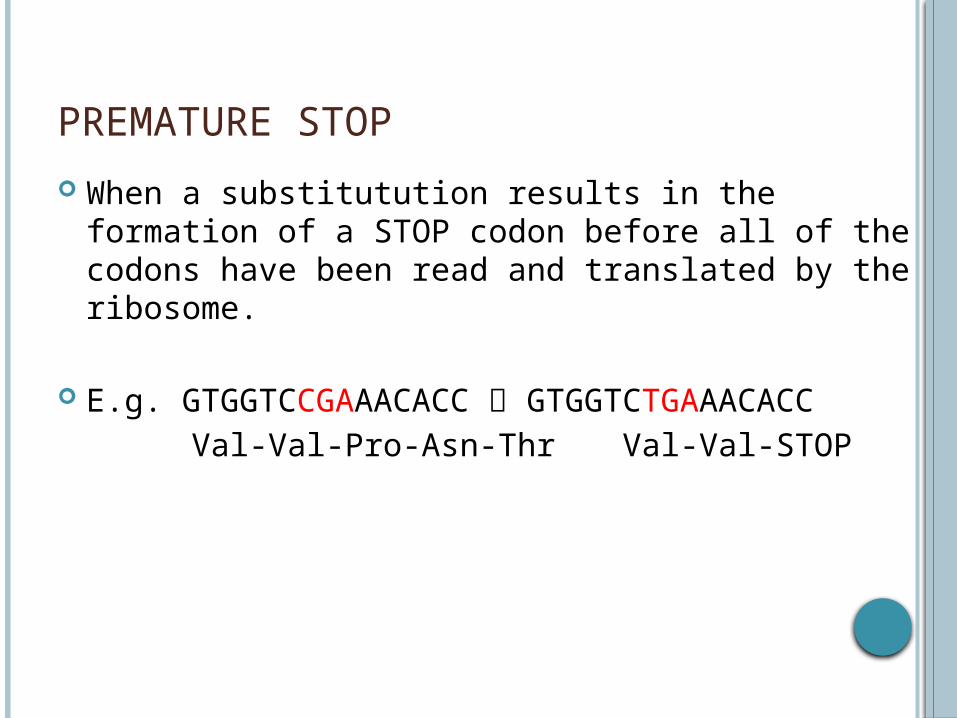
PREMATURE STOP
When a substitutution results in the formation of a STOP codon before all of the codons have been read and translated by the ribosome.
E.g. GTGGTCCGAAACACC GTGGTCTGAAACACC Val-Val-Pro-Asn-Thr Val-Val-STOP

CODON DELETION OR INSERTION
A whole new amino acid is added or one is missing from the mutant protein
Example:GTGGTCCGAAACACC GTGGTCTGCCGAAACACCVal-Val-Pro-Asn-Thr Val-Val-Cys-Pro-Asn-Thr

FRAME SHIFT
When a deletion or insertion results in a different base pair being the beginning of the next codon, changing the whole sequence of amino acids.
E.g. GTGGTCCGAAACACCT GTGGTCGAAACACCT Val-Val-Pro-Asn-Thr Val-Val-Glu-Thr-Pro


