DR.RATHER WASEEM YOUSUF
MVSC VETY CLINICAL MEDICINE
RBC & WBC ABNORMALITIES AND
THEIR INTERPRETATION
HEMATOLOGY
Study of blood and blood forming tissues
Key components of hematologic system are:BloodBlood forming tissues
Bone marrow Spleen Lymph system

WHAT DOES BLOOD DOES Transportation
Oxygen
Nutrients Hormones Waste Products
Regulation Fluid, electrolyte Acid-Base balance
Protection Coagulation Fight Infections
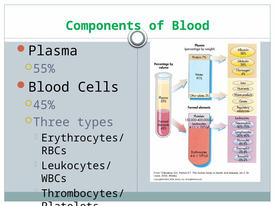
Components of BloodPlasma
55%Blood Cells
45%Three types
Erythrocytes/RBCs
Leukocytes/WBCs
Thrombocytes/Platelets
Erythrocytes/Red Blood Cells
Composed of hemoglobinErythropoiesis
= RBC production Stimulated by hypoxia Controlled by erythropoietin
Hormone synthesized in kidneyHemolysis
= destruction of RBCsReleases bilirubin into blood stream
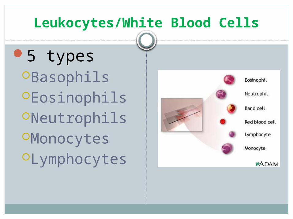
Leukocytes/White Blood Cells
5 typesBasophilsEosinophilsNeutrophilsMonocytesLymphocytes
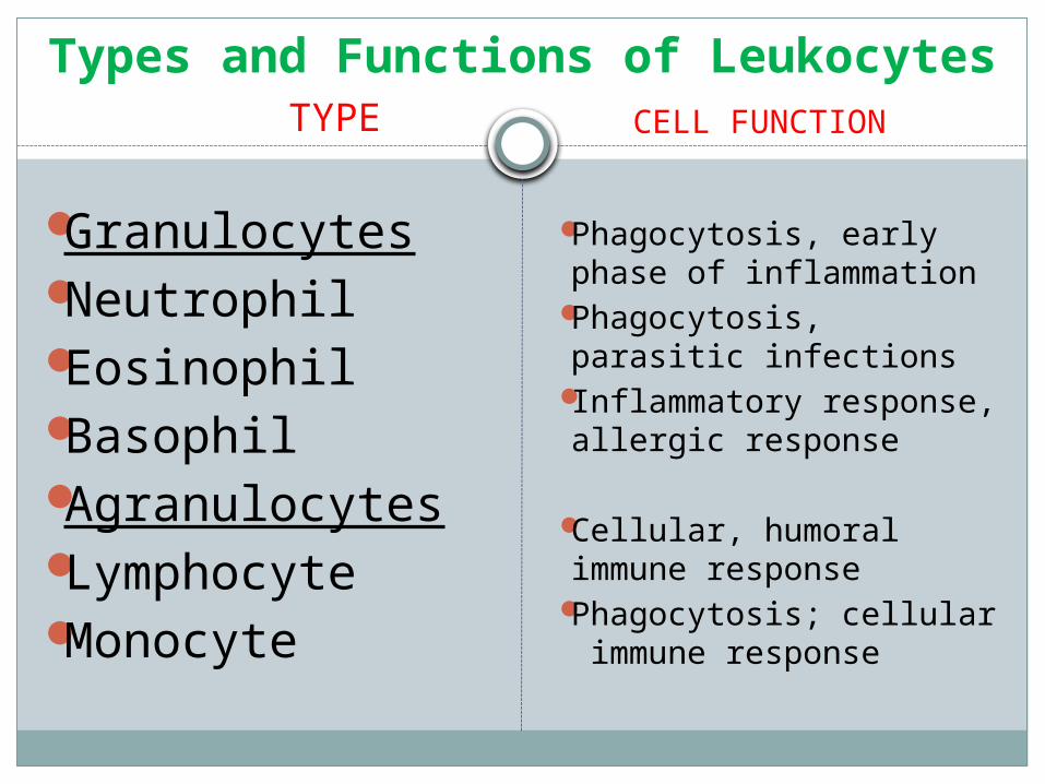
Types and Functions of Leukocytes
GranulocytesNeutrophilEosinophilBasophilAgranulocytesLymphocyteMonocyte
Phagocytosis, early phase of inflammation
Phagocytosis, parasitic infections
Inflammatory response, allergic response
Cellular, humoral immune response
Phagocytosis; cellular immune response
TYPE CELL FUNCTION

Abnormalities of RBCs
Abnormal erythrocyte morphology is found in pathological states that may be :
- abnormalities in size (anisocytosis).
- In shape (poikilocytosis). - In hemoglobin content. - presence of inclusion
bodies
1-Variation in erythrocyte size (anisocytosis)
1-Microcytosis:Morphology: Decrease in the red cell size. Red cells are
smaller than normal.
Found in:- Iron deficiency anemia..- Lead poisoning.- Anemia of chronic disease.
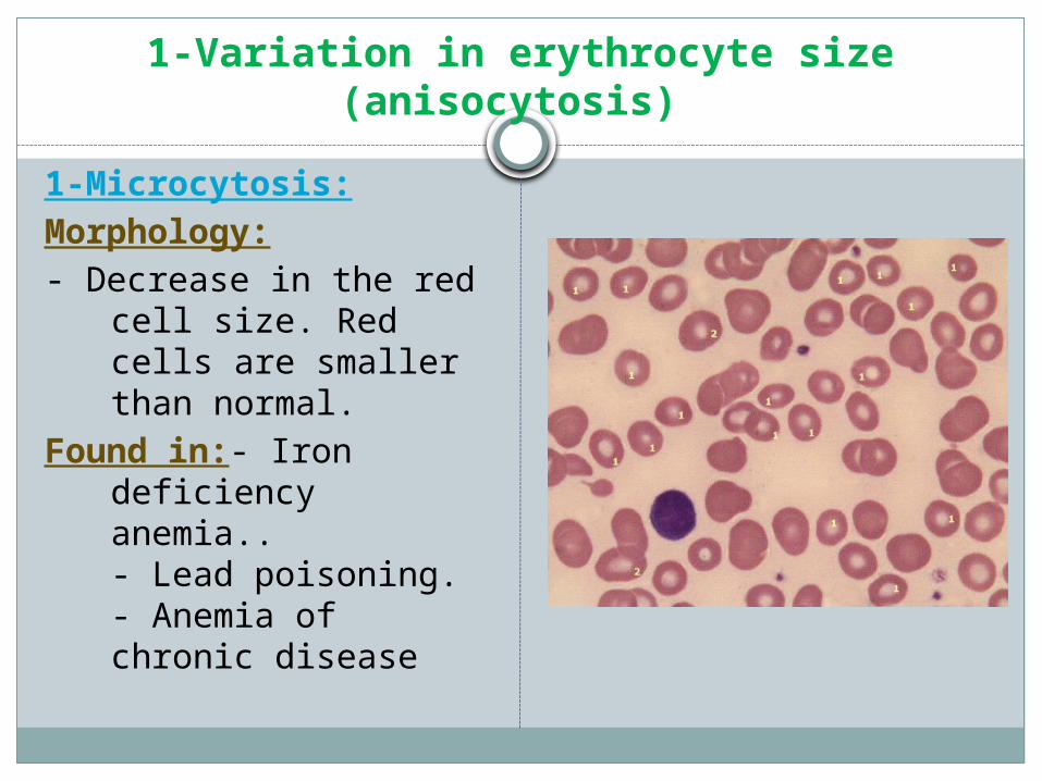
1-Variation in erythrocyte size (anisocytosis)
1-Microcytosis:Morphology:- Decrease in the red
cell size. Red cells are smaller than normal.
Found in:- Iron deficiency anemia..- Lead poisoning.- Anemia of chronic disease
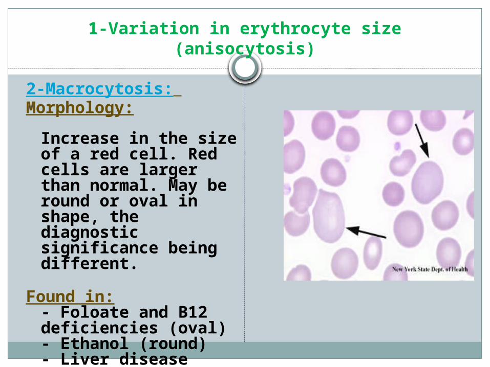
1-Variation in erythrocyte size (anisocytosis)
2-Macrocytosis: Morphology:
Increase in the size of a red cell. Red cells are larger than normal. May be round or oval in shape, the diagnostic significance being different.
Found in:- Foloate and B12 deficiencies (oval)- Ethanol (round)- Liver disease (round)- Reticulocytosis (round)
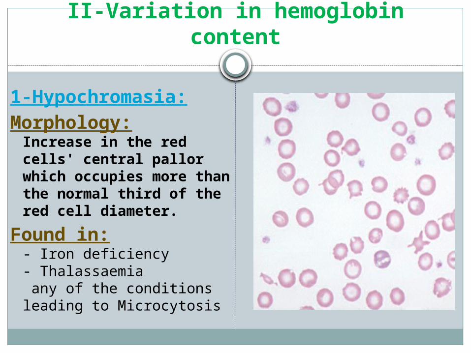
II-Variation in hemoglobin content
1-Hypochromasia:Morphology:
Increase in the red cells' central pallor which occupies more than the normal third of the red cell diameter.
Found in: - Iron deficiency- Thalassaemia any of the conditions leading to Microcytosis
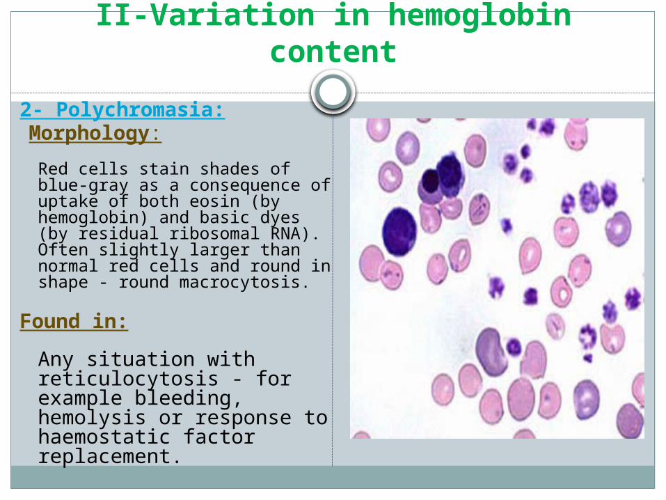
II-Variation in hemoglobin content
2- Polychromasia: Morphology:
Red cells stain shades of blue-gray as a consequence of uptake of both eosin (by hemoglobin) and basic dyes (by residual ribosomal RNA). Often slightly larger than normal red cells and round in shape - round macrocytosis.
Found in:Any situation with reticulocytosis - for example bleeding, hemolysis or response to haemostatic factor replacement.
Anemia Classifications
Microcytic, hypochromic
Microcytic, normochromic
Normocytic, normochromic
Macrocytic, normochromic
NORMOCYTIC, NORMOCHROMIC ANEMIA
MCV normalMCHC normalExamples:
acute blood lossaplastic anemiamost leukemia'sbone marrow infiltration
MICROCYTIC, HYPOCHROMIC ANEMIA
MCV decreased MCHC decreasedExamples:
iron deficiencythalassemialead poisoninganemia of chronic disease
MICROCYTIC, NORMOCHROMIC ANEMIA
MCV decreasedMCHC normalExamples:
mid-stage iron deficiencythalassemia
MACROCYTIC, NORMOCHROMIC ANEMIA
MCV increasedMCHC normalExamples:
folate deficiencyvitamin B12 deficiencypernicious anemia
III- Variation of red cells shape (Poikilocytosis)
RBCs may have different shapes:
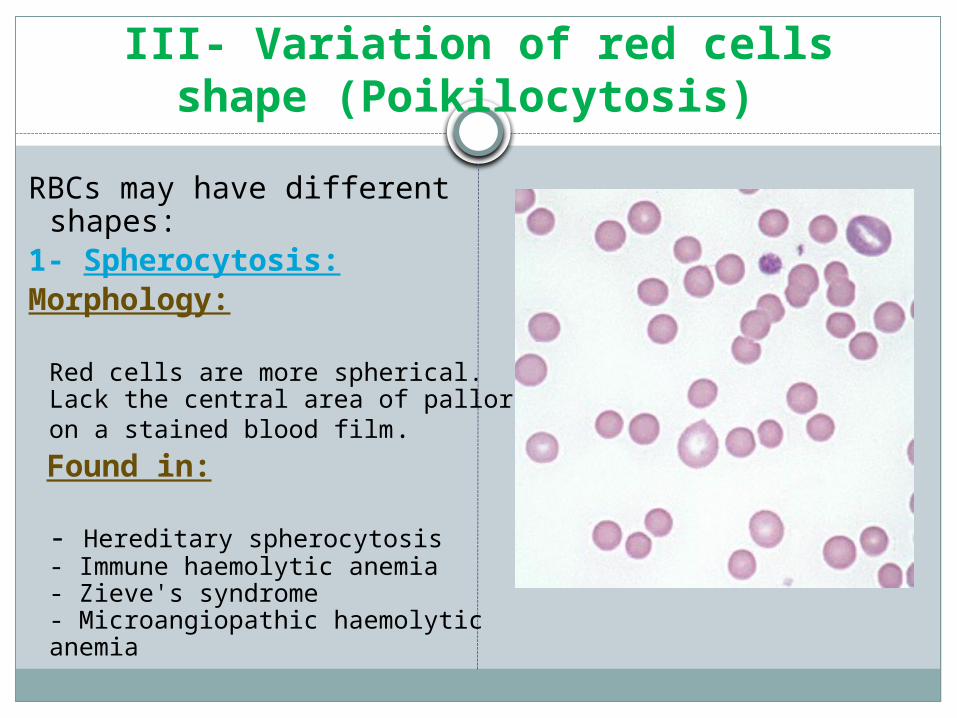
1- Spherocytosis: Morphology:
Red cells are more spherical. Lack the central area of pallor on a stained blood film.
Found in:
- Hereditary spherocytosis- Immune haemolytic anemia- Zieve's syndrome- Microangiopathic haemolytic anemia

III- Variation of red cells shape (Poikilocytosis)
2-Target Cells:Morphology:Red cells have an area of
increased staining which appears in the area of central pallor.
Found in:-Obstructive liver disease
-Severe iron deficiency- Thalassaemia- Haemoglobinopathies - Post splenectomy
III- Variation of red cells shape (Poikilocytosis)
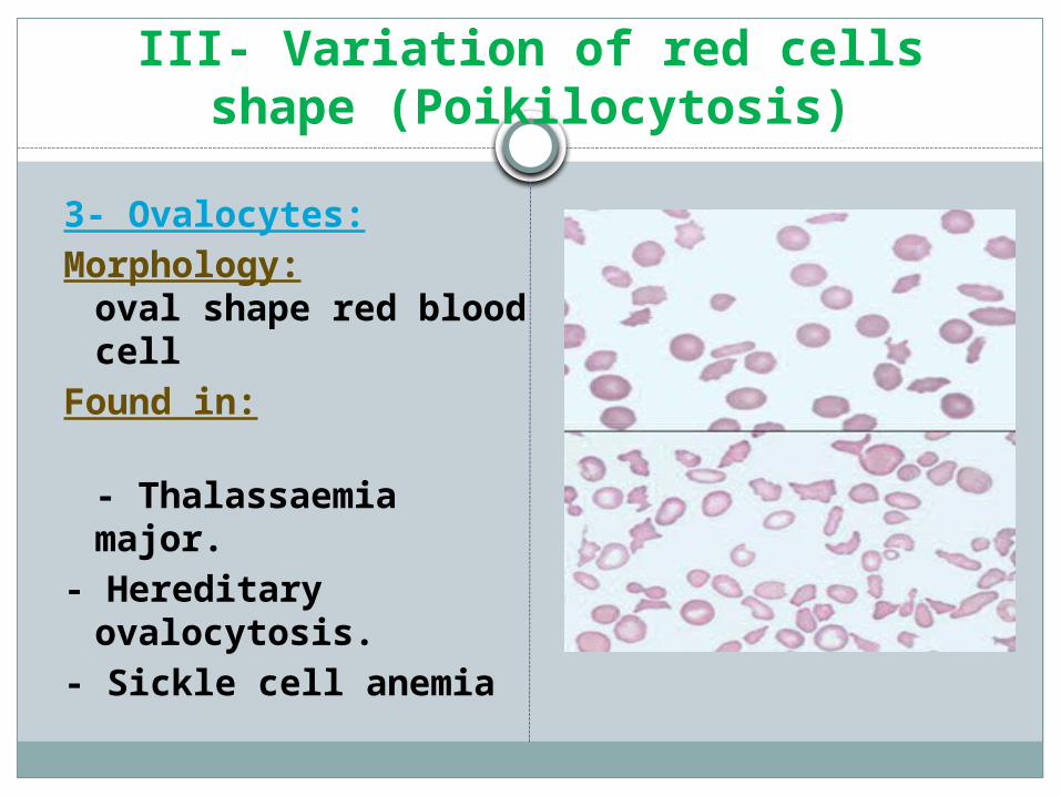
3- Ovalocytes:Morphology:
oval shape red blood cell
Found in:
- Thalassaemia major.- Hereditary ovalocytosis. - Sickle cell anemia
III- Variation of red cells shape (Poikilocytosis)
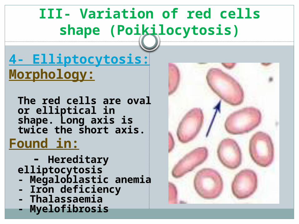
4- Elliptocytosis:Morphology:
The red cells are oval or elliptical in shape. Long axis is twice the short axis.
Found in: - Hereditary elliptocytosis
- Megaloblastic anemia- Iron deficiency - Thalassaemia- Myelofibrosis
III- Variation of red cells shape (Poikilocytosis)
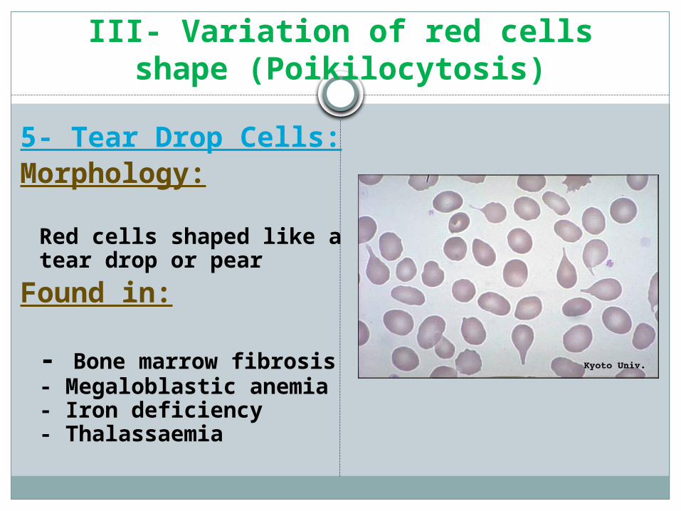
5- Tear Drop Cells:Morphology:
Red cells shaped like a tear drop or pear
Found in:
- Bone marrow fibrosis- Megaloblastic anemia- Iron deficiency- Thalassaemia
III- Variation of red cells shape (Poikilocytosis)
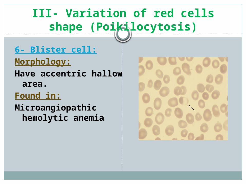
6- Blister cell:Morphology: Have accentric hallow
area.Found in: Microangiopathic
hemolytic anemia
III- Variation of red cells shape (Poikilocytosis)
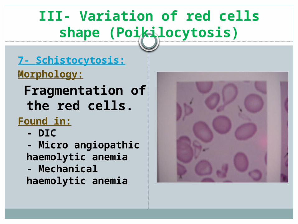
7- Schistocytosis:Morphology: Fragmentation of
the red cells. Found in:
- DIC - Micro angiopathic haemolytic anemia- Mechanical haemolytic anemia
III- Variation of red cells shape (Poikilocytosis)
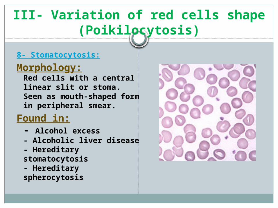
8- Stomatocytosis:Morphology:
Red cells with a central linear slit or stoma. Seen as mouth-shaped form in peripheral smear.
Found in:- Alcohol excess- Alcoholic liver disease- Hereditary stomatocytosis- Hereditary spherocytosis
III- Variation of red cells shape (Poikilocytosis)
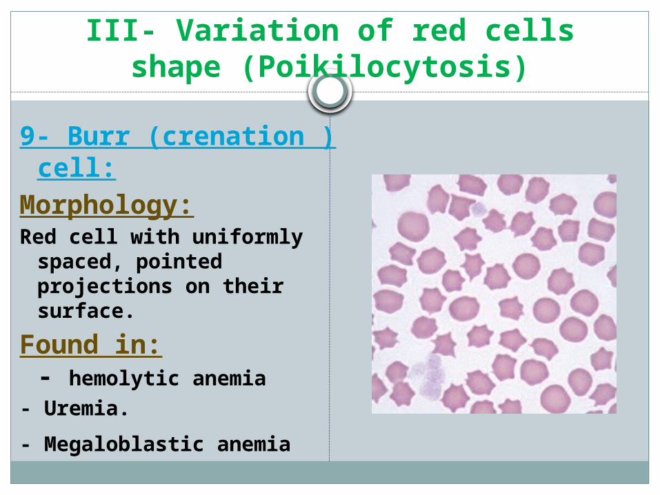
9- Burr (crenation ) cell:
Morphology:Red cell with uniformly
spaced, pointed projections on their surface.
Found in:- hemolytic anemia
- Uremia.- Megaloblastic anemia
III- Variation of red cells shape (Poikilocytosis)
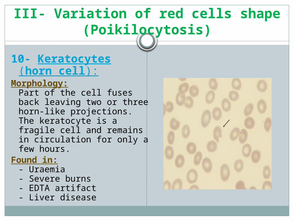
10- Keratocytes (horn cell):
Morphology:Part of the cell fuses back leaving two or three horn-like projections. The keratocyte is a fragile cell and remains in circulation for only a few hours.
Found in:- Uraemia- Severe burns- EDTA artifact- Liver disease
III- Variation of red cells shape (Poikilocytosis)
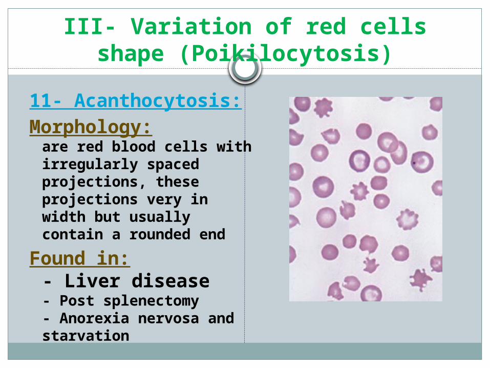
11- Acanthocytosis:Morphology:
are red blood cells with irregularly spaced projections, these projections very in width but usually contain a rounded end
Found in:- Liver disease - Post splenectomy- Anorexia nervosa and starvation
III- Variation of red cells shape (Poikilocytosis)
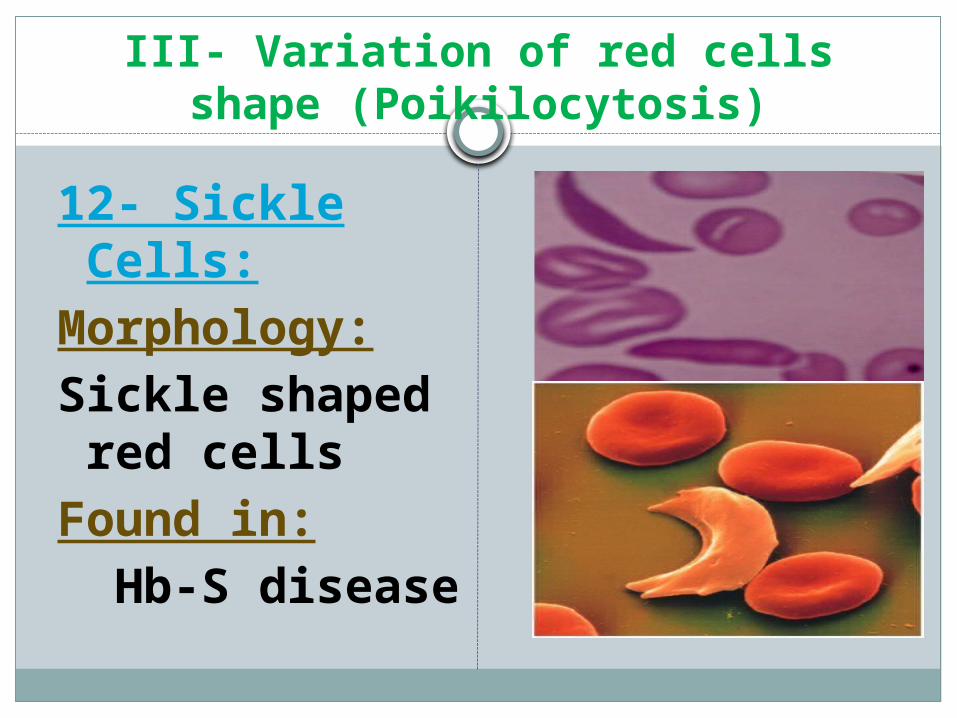
12- Sickle Cells:Morphology: Sickle shaped red
cells Found in: Hb-S disease

III- Variation of red cells shape (Poikilocytosis)
13- Rouleaux Formation:
Morphology:
Stacks of RBC's resembling a stack of coins.
Found in:
-Hyperfibrinogenaemia
-Hyperglobulinaemia
III- Variation of red cells shape (Poikilocytosis)
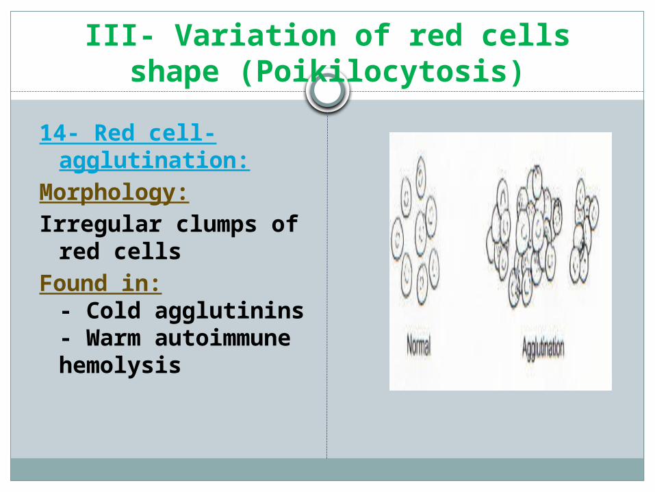
14- Red cell-agglutination:
Morphology: Irregular clumps of red
cellsFound in:
- Cold agglutinins- Warm autoimmune hemolysis
III- Variation of red cells shape (Poikilocytosis)
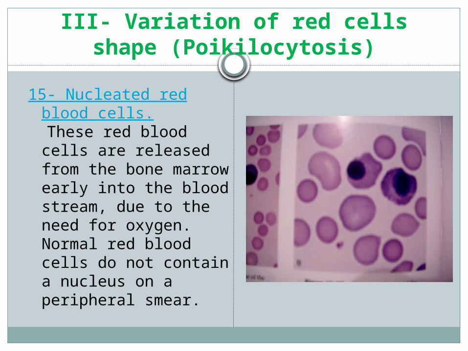
15- Nucleated red blood cells.
These red blood cells are released from the bone marrow early into the blood stream, due to the need for oxygen. Normal red blood cells do not contain a nucleus on a peripheral smear.
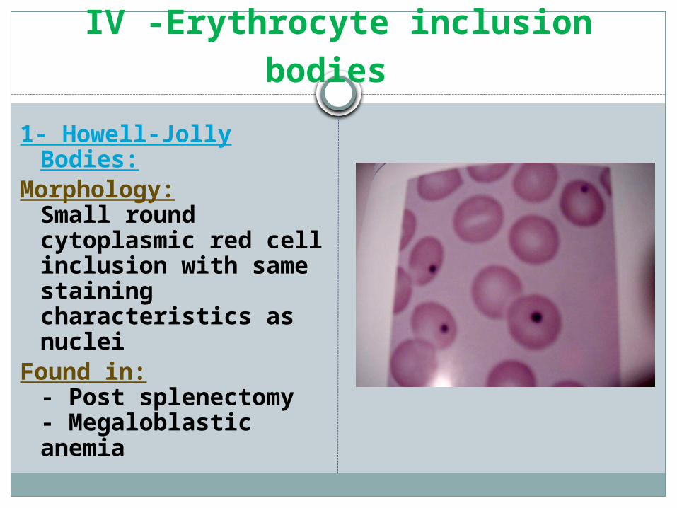
IV -Erythrocyte inclusion bodies 1- Howell-Jolly Bodies:Morphology:
Small round cytoplasmic red cell inclusion with same staining characteristics as nuclei
Found in:- Post splenectomy- Megaloblastic anemia
IV -Erythrocyte inclusion bodies
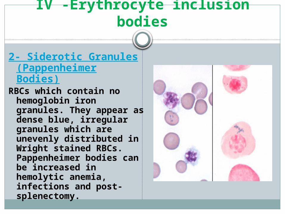
2- Siderotic Granules (Pappenheimer Bodies)
RBCs which contain no hemoglobin iron granules. They appear as dense blue, irregular granules which are unevenly distributed in Wright stained RBCs. Pappenheimer bodies can be increased in hemolytic anemia, infections and post-splenectomy.
IV -Erythrocyte inclusion bodies
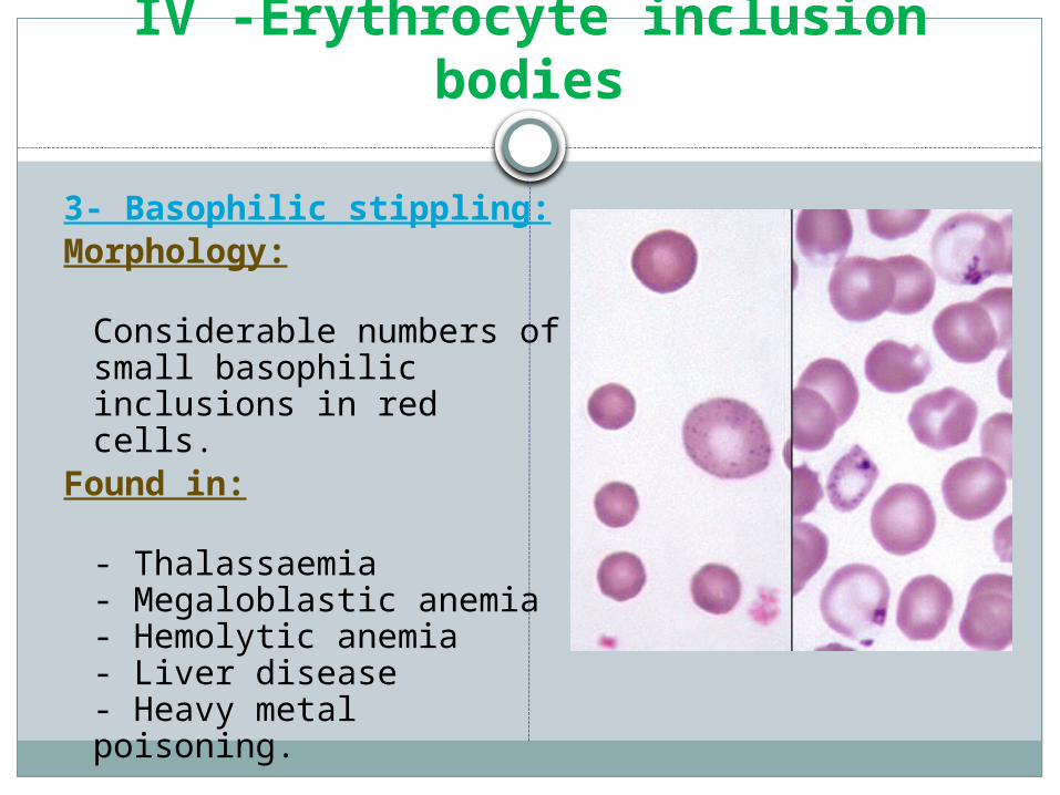
3- Basophilic stippling: Morphology:
Considerable numbers of small basophilic inclusions in red cells.
Found in:
- Thalassaemia- Megaloblastic anemia- Hemolytic anemia - Liver disease- Heavy metal poisoning.
IV -Erythrocyte inclusion bodies
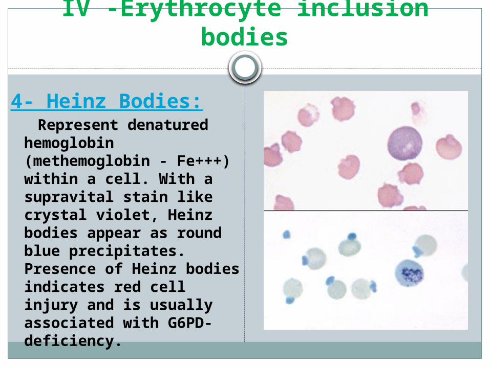
4- Heinz Bodies: Represent denatured
hemoglobin (methemoglobin - Fe+++) within a cell. With a supravital stain like crystal violet, Heinz bodies appear as round blue precipitates. Presence of Heinz bodies indicates red cell injury and is usually associated with G6PD-deficiency.
IV -Erythrocyte inclusion bodies
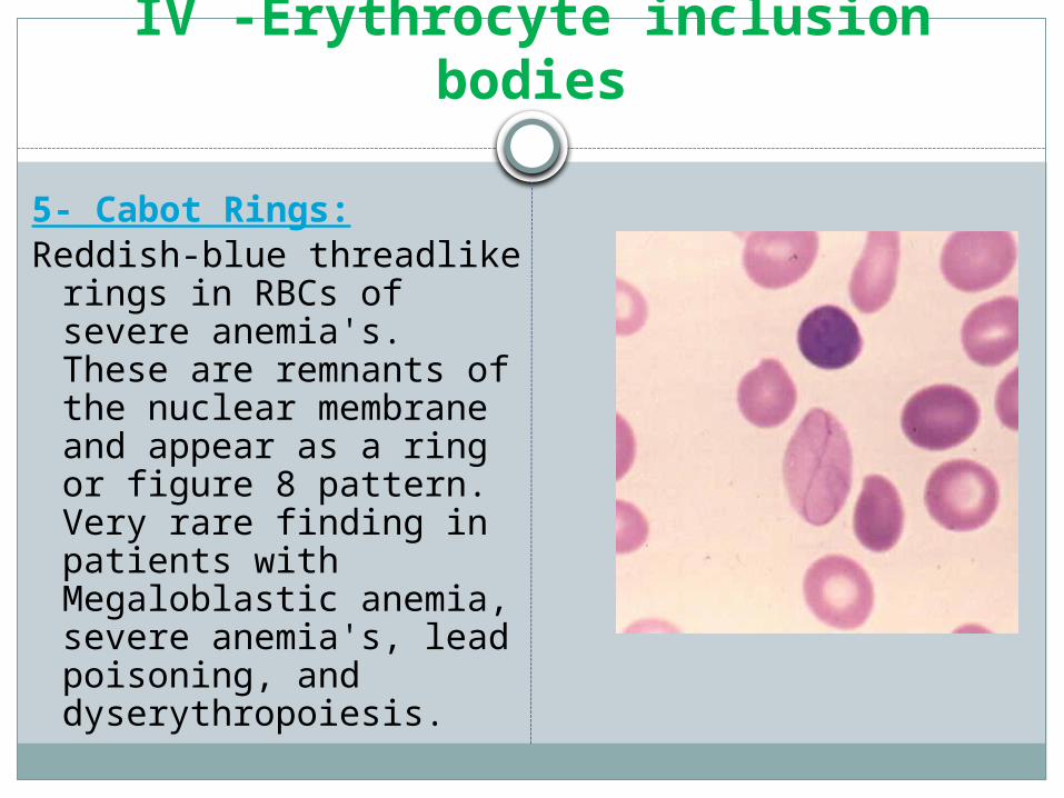
5- Cabot Rings: Reddish-blue threadlike rings
in RBCs of severe anemia's. These are remnants of the nuclear membrane and appear as a ring or figure 8 pattern. Very rare finding in patients with Megaloblastic anemia, severe anemia's, lead poisoning, and dyserythropoiesis.
IV -Erythrocyte inclusion bodies
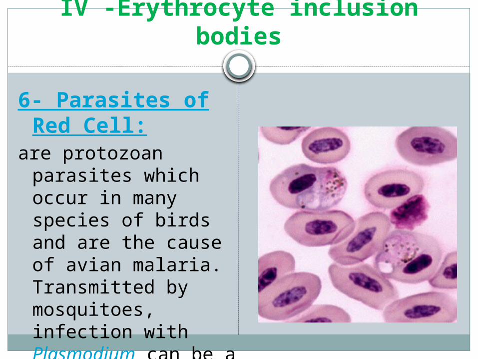
6- Parasites of Red Cell:
are protozoan parasites which occur in many species of birds and are the cause of avian malaria. Transmitted by mosquitoes, infection with Plasmodium can be a cause of hemolytic anemia
Erythrocyte inclusion bodies
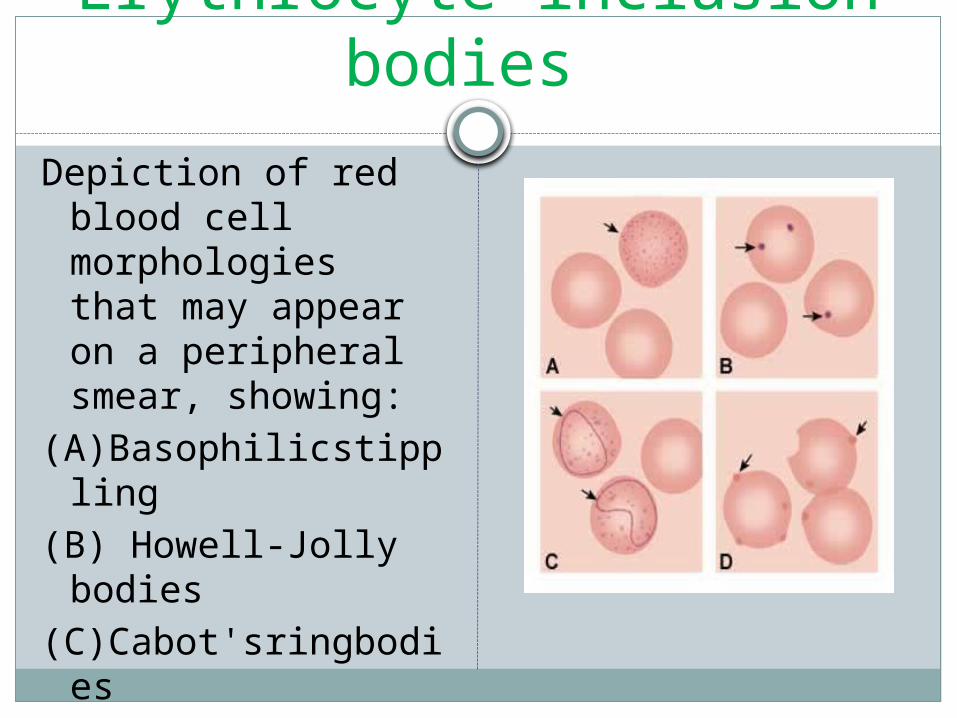
Depiction of red blood cell morphologies that may appear on a peripheral smear, showing:
(A)Basophilicstippling
(B) Howell-Jolly bodies
(C)Cabot'sringbodies(D) Heinz's bodies.

Leukocytes (White Blood Cells)
The leukocytes, or white blood cells, constitute only 1% of the total blood volume.
They originate in the bone marrow and circulate throughout the lymphoid tissues of the body.
There they function in the inflammatory and immune processes.
They include: the granulocytes
Neutrophils – 55-65% Eosinophils – 1-4% Basophils – 0-1%
the lymphocytes – 20-40% the monocytes – 3-8%
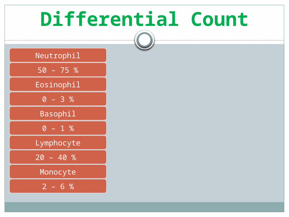
Differential CountNeutrophil
50 – 75 %
Eosinophil
0 – 3 %
Basophil
0 – 1 %
Lymphocyte
20 – 40 %
Monocyte
2 – 6 %
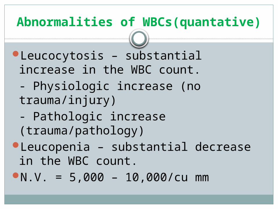
Abnormalities of WBCs(quantative)
Leucocytosis – substantial increase in the WBC count.
- Physiologic increase (no trauma/injury) - Pathologic increase (trauma/pathology)Leucopenia – substantial decrease in
the WBC count.N.V. = 5,000 – 10,000/cu mm
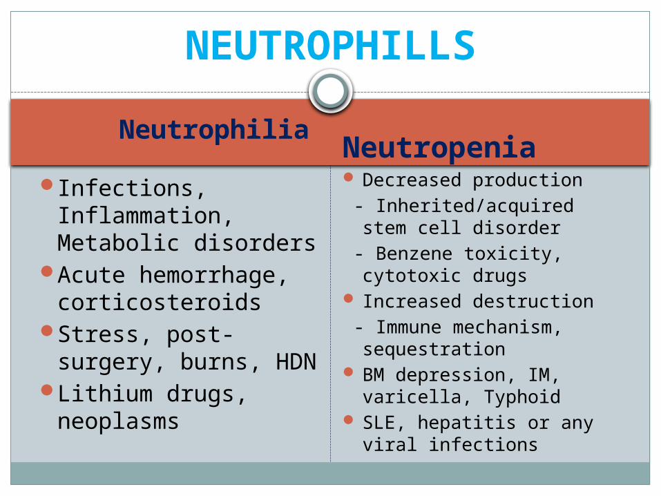
Neutrophilia NeutropeniaInfections,
Inflammation, Metabolic disorders
Acute hemorrhage, corticosteroids
Stress, post-surgery, burns, HDN
Lithium drugs, neoplasms
Decreased production - Inherited/acquired stem
cell disorder - Benzene toxicity, cytotoxic
drugs Increased destruction - Immune mechanism,
sequestration BM depression, IM,
varicella, Typhoid SLE, hepatitis or any viral
infections
NEUTROPHILLS
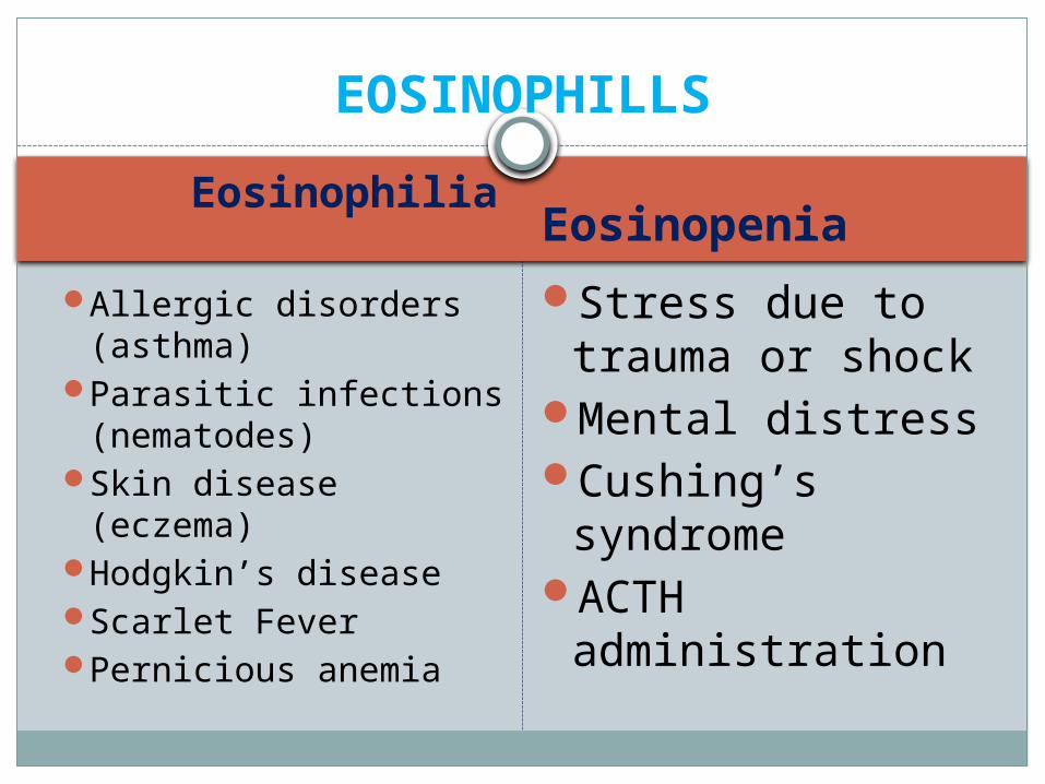
Eosinophilia Eosinopenia
Allergic disorders (asthma)
Parasitic infections (nematodes)
Skin disease (eczema)Hodgkin’s diseaseScarlet FeverPernicious anemia
Stress due to trauma or shock
Mental distressCushing’s
syndromeACTH
administration
EOSINOPHILLS
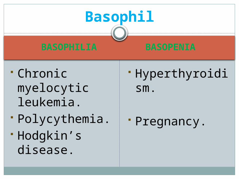
BASOPHILIA BASOPENIA
Chronic myelocytic leukemia.
Polycythemia. Hodgkin’s disease.
Hyperthyroidism.
Pregnancy.
Basophil
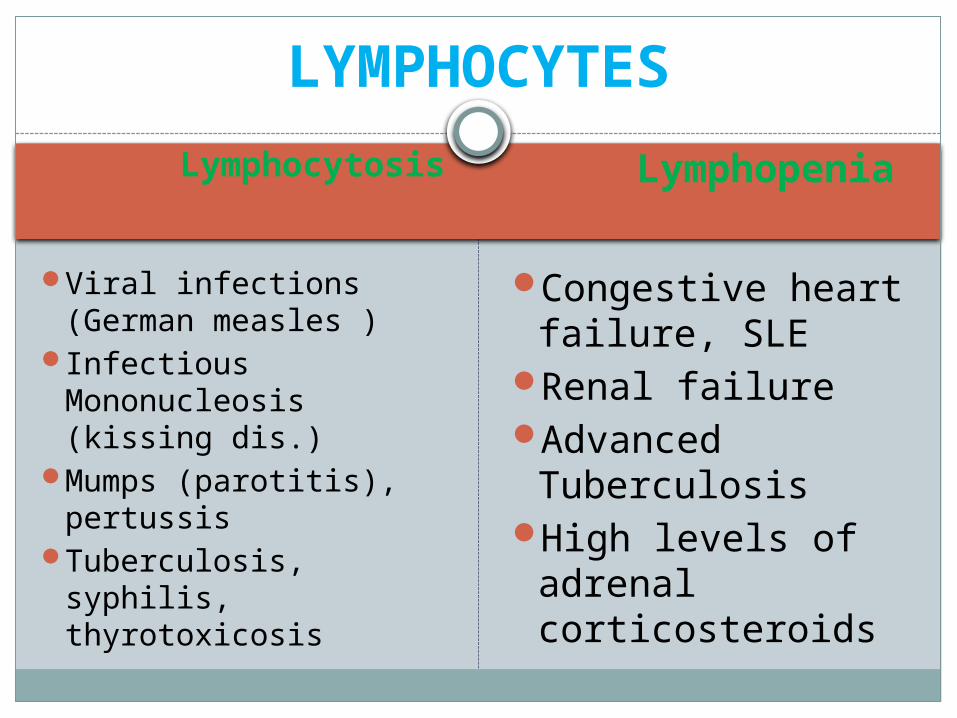
Lymphocytosis Lymphopenia
Viral infections (German measles )
Infectious Mononucleosis (kissing dis.)
Mumps (parotitis), pertussis
Tuberculosis, syphilis, thyrotoxicosis
Congestive heart failure, SLE
Renal failureAdvanced
TuberculosisHigh levels of
adrenal corticosteroids
LYMPHOCYTES
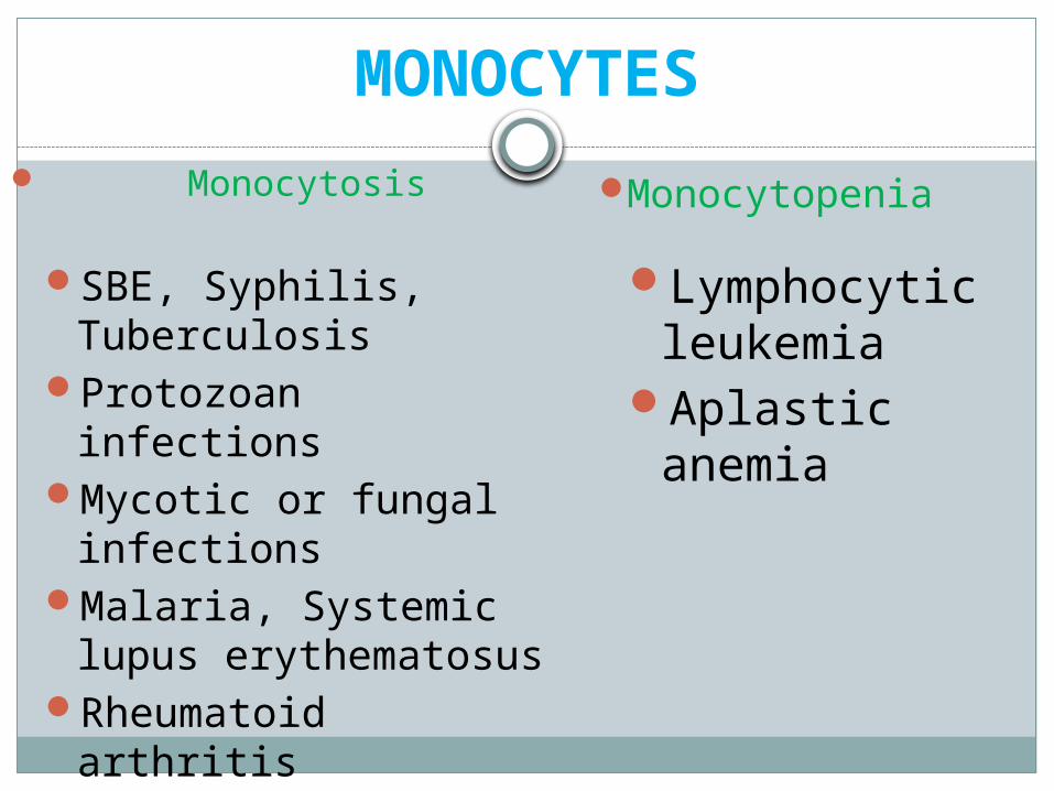
MONOCYTES
SBE, Syphilis, Tuberculosis
Protozoan infectionsMycotic or fungal
infectionsMalaria, Systemic
lupus erythematosusRheumatoid arthritis
Monocytosis Monocytopenia
Lymphocytic leukemia
Aplastic anemia
Abnormalities of WBCs(qualitative)
Morphologic abnormalities involving either the nucleus or cytoplasm
Functional abnormalities
Inherited or Acquired
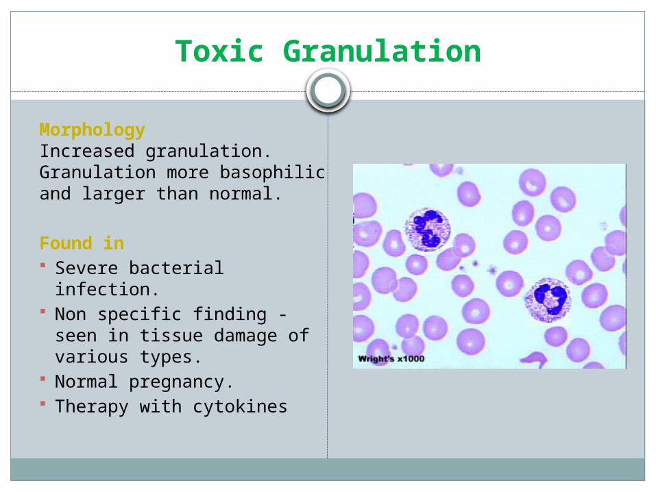
Toxic Granulation
MorphologyIncreased granulation. Granulation more basophilic and larger than normal.
Found in Severe bacterial infection. Non specific finding - seen in
tissue damage of various types. Normal pregnancy. Therapy with cytokines
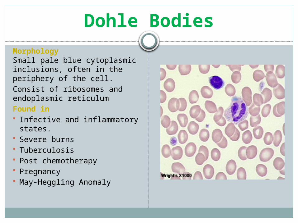
Dohle BodiesMorphologySmall pale blue cytoplasmic inclusions, often in the periphery of the cell.Consist of ribosomes and endoplasmic reticulumFound in Infective and inflammatory states. Severe burns Tuberculosis Post chemotherapy Pregnancy May-Heggling Anomaly
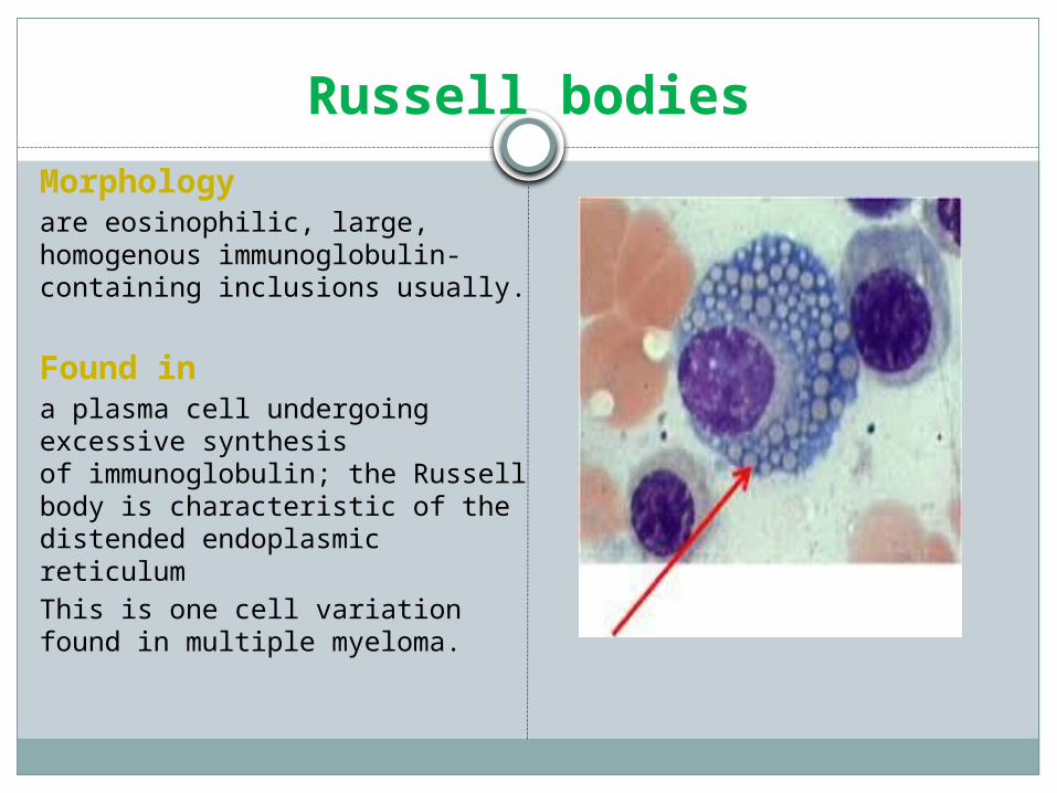
Russell bodiesMorphologyare eosinophilic, large, homogenous immunoglobulin-containing inclusions usually.
Found ina plasma cell undergoing excessive synthesis of immunoglobulin; the Russell body is characteristic of the distended endoplasmic reticulumThis is one cell variation found in multiple myeloma.
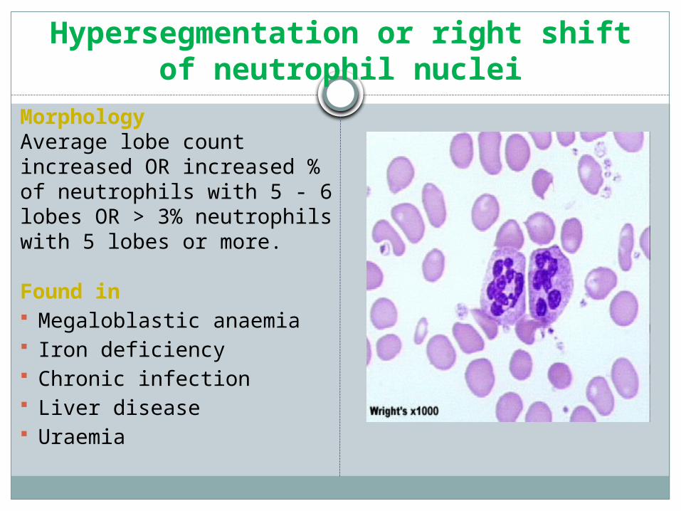
Hypersegmentation or right shift of neutrophil nuclei
MorphologyAverage lobe count increased OR increased % of neutrophils with 5 - 6 lobes OR > 3% neutrophils with 5 lobes or more.
Found in Megaloblastic anaemia Iron deficiency Chronic infection Liver disease Uraemia
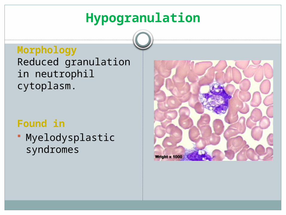
Hypogranulation
MorphologyReduced granulation in neutrophil cytoplasm.
Found in Myelodysplastic syndromes
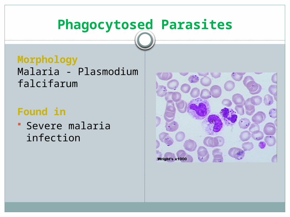
Phagocytosed Parasites
MorphologyMalaria - Plasmodium falcifarum
Found in Severe malaria infection
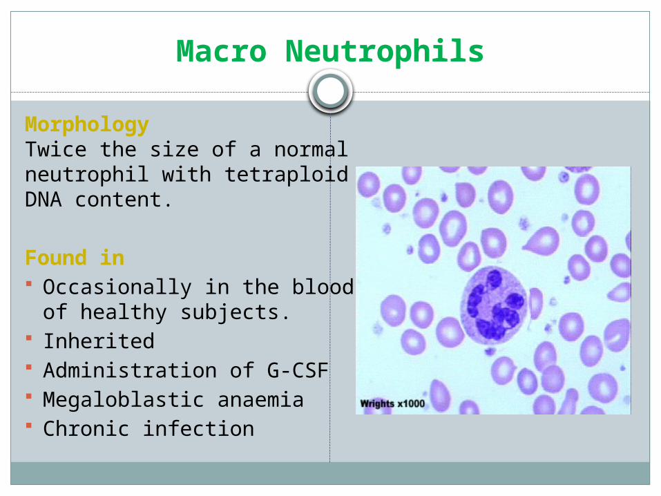
Macro Neutrophils
MorphologyTwice the size of a normal neutrophil with tetraploid DNA content.
Found in Occasionally in the blood of
healthy subjects. Inherited Administration of G-CSF Megaloblastic anaemia Chronic infection
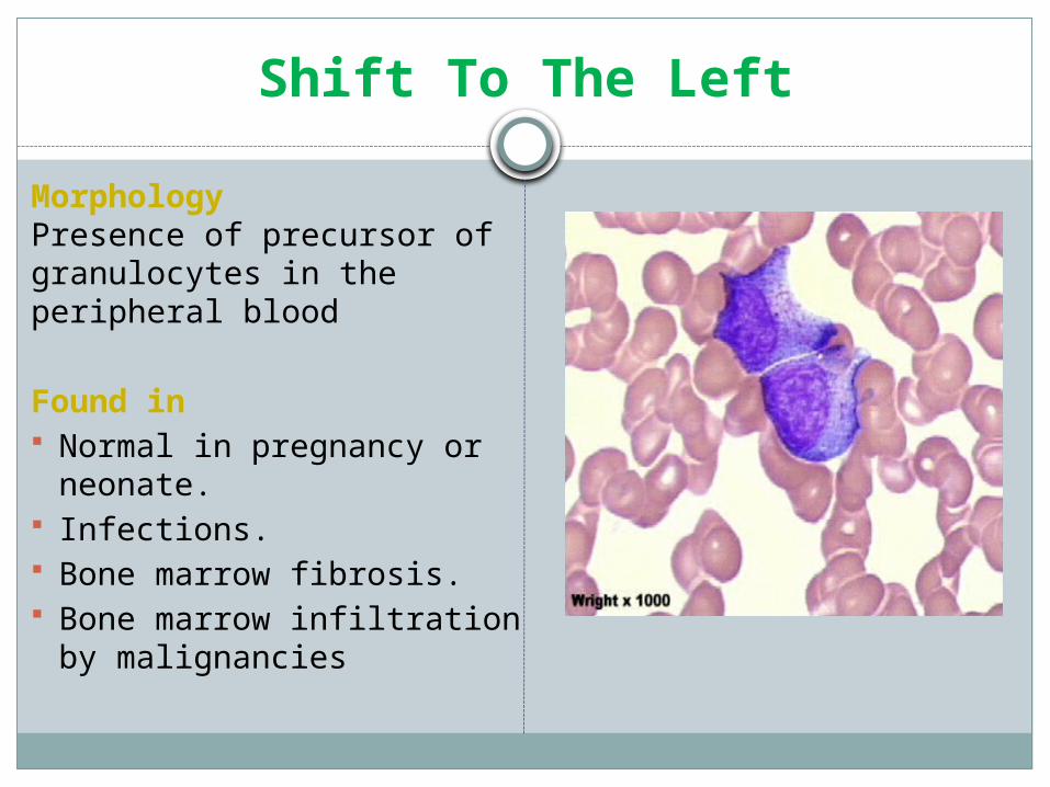
Shift To The Left
MorphologyPresence of precursor of granulocytes in the peripheral blood
Found in Normal in pregnancy or neonate. Infections. Bone marrow fibrosis. Bone marrow infiltration by
malignancies
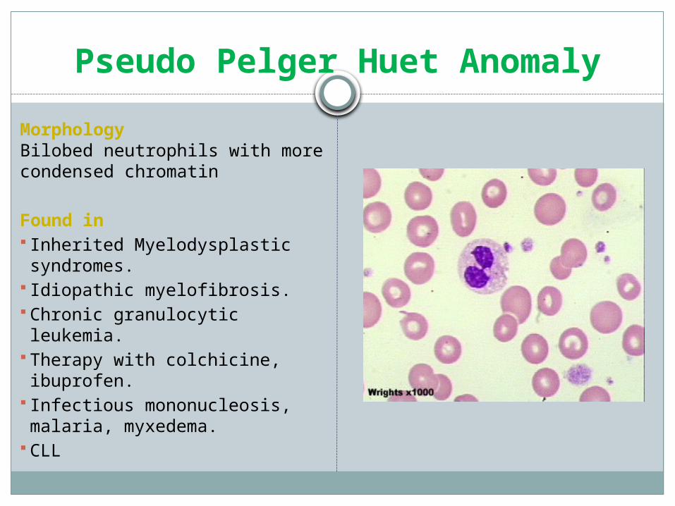
Pseudo Pelger Huet Anomaly
MorphologyBilobed neutrophils with more condensed chromatin
Found in Inherited Myelodysplastic
syndromes. Idiopathic myelofibrosis. Chronic granulocytic leukemia. Therapy with colchicine, ibuprofen. Infectious mononucleosis, malaria,
myxedema. CLL
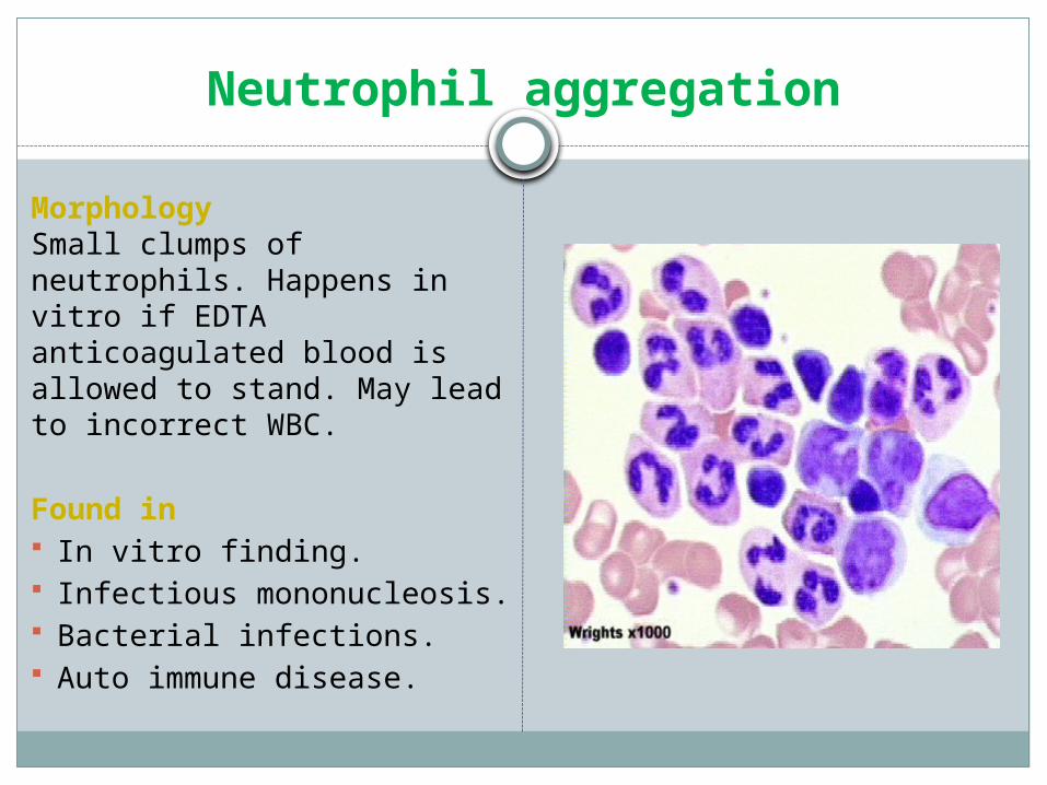
Neutrophil aggregation
MorphologySmall clumps of neutrophils. Happens in vitro if EDTA anticoagulated blood is allowed to stand. May lead to incorrect WBC.
Found in In vitro finding. Infectious mononucleosis. Bacterial infections. Auto immune disease.
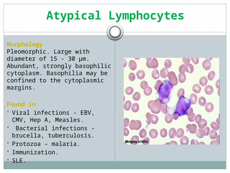
Atypical Lymphocytes
MorphologyPleomorphic. Large with diameter of 15 - 30 µm. Abundant, strongly basophilic cytoplasm. Basophilia may be confined to the cytoplasmic margins.
Found in Viral infections - EBV, CMV, Hep A,
Measles. Bacterial infections - brucella,
tuberculosis. Protozoa – malaria. Immunization. SLE.

Thank You


