chapter 8 - defence against disease1 defence against disease chapter 8 pages 245-284 rbc leukocyte...
TRANSCRIPT

Chapter 8 - Defence Against Disease 1
Defence Against Disease
Chapter 8
Pages 245-284
RBC
Leukocyte (WBC)
Leukocyte (WBC)

2
ImmunityImmunityInfection:Defn: entry of a pathogen into the body of
a organism (host) that might cause disease.
ImmunityDefn: reactions that occur in a person in
response to an infection
The immune system• The immune system is able to
distinguish foreign material from material that is made by the body.
• The immune system has two kinds of responses to the entry of foreign material.
1. Non- Specific Immunity: involves a natural immunity that is non-specific.
2. Specific Immunity (adaptive immunity): the action of specific white blood cells (lymphocytes) to a specific antigen (pathogen or part of) which acts to neutralize the pathogen (also invokes production of memory cells)

Self and Non-self• All cells have marker proteins on their plasma
membrane
• These proteins are the products of the MHC genes. Each person has different MHC genes.
• Therefore marker proteins are specific to each person/organism
• Cells with the body's own marker proteins are accepted as “self”. These proteins are not antigenic to our own immune system.
• Cells with foreign markers are recognised as “non-self”. These marker proteins are antigenic for us.
How Does The Body Know What Cells To Attack
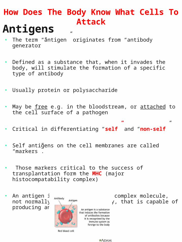
Antigens• The term “antigen” originates from “antibody
generator”
• Defined as a substance that, when it invades the body, will stimulate the formation of a specific type of antibody
• Usually protein or polysaccharide
• May be free e.g. in the bloodstream, or attached to the cell surface of a pathogen
• Critical in differentiating “self” and “non-self”
• Self antigens on the cell membranes are called “markers”.
• Those markers critical to the success of transplantation form the MHC (major histocompatability complex)
• An antigen is typically a large complex molecule, not normally present in the body, that is capable of producing an immune response
How Does The Body Know What Cells To Attack

IMMUNE SYSTEMIMMUNE SYSTEM
Consists of Three Lines of Consists of Three Lines of DefenceDefence
Chapter 8 - Defence Against Disease 5
Pathogen invades Tissue/ Cell
Non Specific DefenceSpecific Defenceacquired
resistanceBarrie
rs
1
Physiological
Mechanisms
ChemicalMechanis
ms
Phagocytes and
NK Cells
Inflammation
BasophilsMast Cells
and platelets
Histamines &
phagocytosis
B Cells
T Cells
Memory Cells
Antibodies
Humoral Immunity
Cell Mediated Immunity
32


Chapter 8 - Defence Against Disease 7
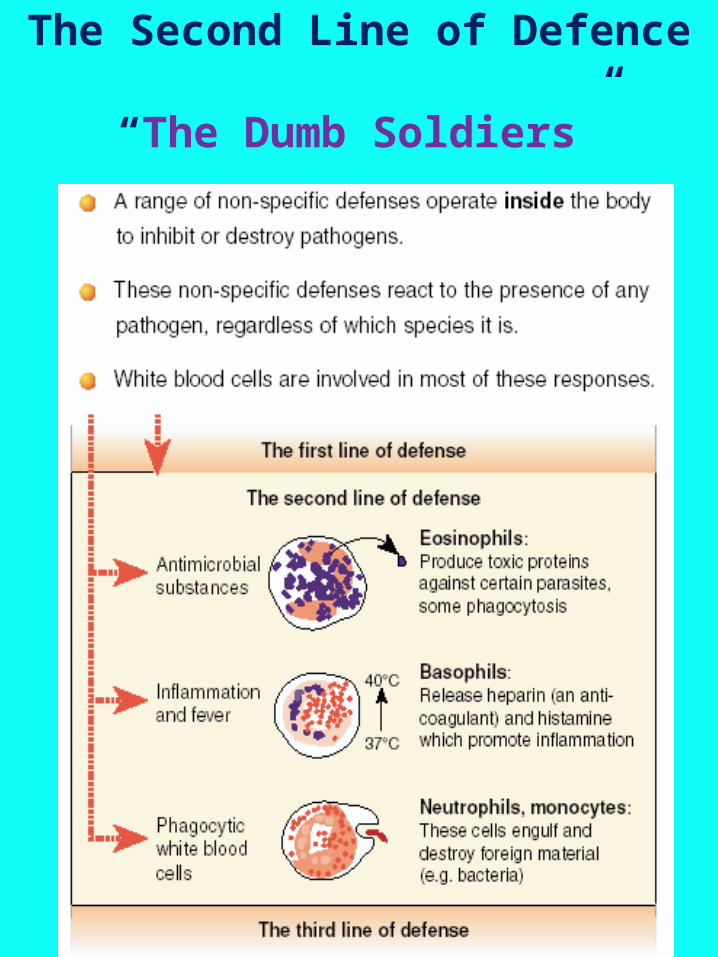
Non-Specific Immunity
reading: 246 – 250 Quick Check:1-7 Biozone: 147-148

Non-specific mechanisms
• Part of body’s natural immunity
• Provide protection
• Are present at birth
• Either prevent entry of pathogens or destroy them
• Limit the onset and development of infection
• Directed against a wide range of pathogens

9
Non-specific mechanisms:
Barriers: 1st line of defence

Chapter 8 - Defence Against Disease 10
1. The First Line of Defence
‘The Wall”• The best action against micro-
organisms is to prevent their entry into the body altogether.
• The first line of defence against infection takes place at the body surfaces.

Skin
• An intact skin acts as a barrier against entry by micro-organisms. A cut or abrasion will allow entry of bacteria or viruses.
– Hardening of outer layers• Provides a physical barrier
– Anti-bacterial and anti-fungal secretions• Produced by sweat glands, sebaceous
(oil) glands, bacterial flora of the skin– Lack of moisture
• Limits growth of microorganisms

12
The First Line of Defence continued….continued….
Mucous Membranes
• secreted by the cells lining your respiratory tract
• traps bacteria which are then swept upwards to the back of the throat by the action of cilia.
• some of the mucus and bacteria is then swallowed, coughed or sneezed out, or blown out through the nose.
• promote growth of natural flora whose secretions limit pathogen growth

Chapter 8 - Defence Against Disease 13
Natural Secretions
• Many secretions of the body contain bactericidal agents. Tears and saliva contain lysozyme, an enzyme that cause bacteria to lyse or burst. Acid in the stomach also kills many bacteria.
Peristalsis Diarrhoea eliminates pathogens by
movement towards the anus for elimination
Vomiting also results in removal of pathogens from body
The First Line of Defence continued….continued….

14
Enzymes– Lysozyme in tears, saliva, sweat, nasal
secretions and tissue fluids breaks up (lyses) the cell wall of certain bacteria
Natural Flora
• Many different bacteria are normally found on the skin, gut and in the vagina. These bacteria are harmless to the body and occur naturally.
• The presence of these bacteria can inhibit the growth of pathogenic bacteria as they compete for nutrients and space.
The First Line of Defence continued….continued….

Gastro-intestinal secretions
– HCl in stomach, alkaline fluids e.g. bile in duodenum• Are of a pH which is outside the
range of tolerance for many microorganisms
Hairs and cilia
– Filter inhaled air– Remove micro-organisms and other
antigenic material (e.g. pollen)
Chapter 8 - Defence Against Disease 15
The First Line of Defence continued….continued….

16
Non-specific mechanisms:
2nd Line of defence
17
The Second Line of Defence
“The Dumb Soldiers”

PHAGOCYTES
Phagocytes (group of cells)
• (particular) white blood cells• formed in the bone marrow• very motile and can move between
cells• engulf and destroy micro-organisms
and foreign materials through phagocytosis
• include the following groups:
1.Neutrophils
2.Monocytes/ Macrophages
3.Eosinophils
Chapter 8 - Defence Against Disease 18
The Second Line of Defence continued …

(a) SEM (4300x) : macrophage pulling rod-shaped E.coli towards it with long cytoplasmic extensions. Several bacteria on the macrophage’s surface are being engulfed
(b) Events of phagocytosis

Monocytes • Largest of the white blood cells• become macrophages when they leave the
bloodstream
Macrophages • gather in various tissues such as the lungs, liver,
kidneys and brain.• are particularly active against micro-organisms that
can live inside the cells of the person they infect.• engulf bacterium
Neutrophils• The most numerous of the phagocytotic cells• Granulated nucleus• Attacks bacteria• Die after engulfing bacterial pathogen• Their dead cells become the bulk of • ‘pus’ at wounds
The Second Line of Defence continued …
Macrophage destroying bacterial cells

Eosinophils • can be phagocytotic.• secrete enzymes to kill parasitic worms
among other pathogins
The Second Line of Defence continued …
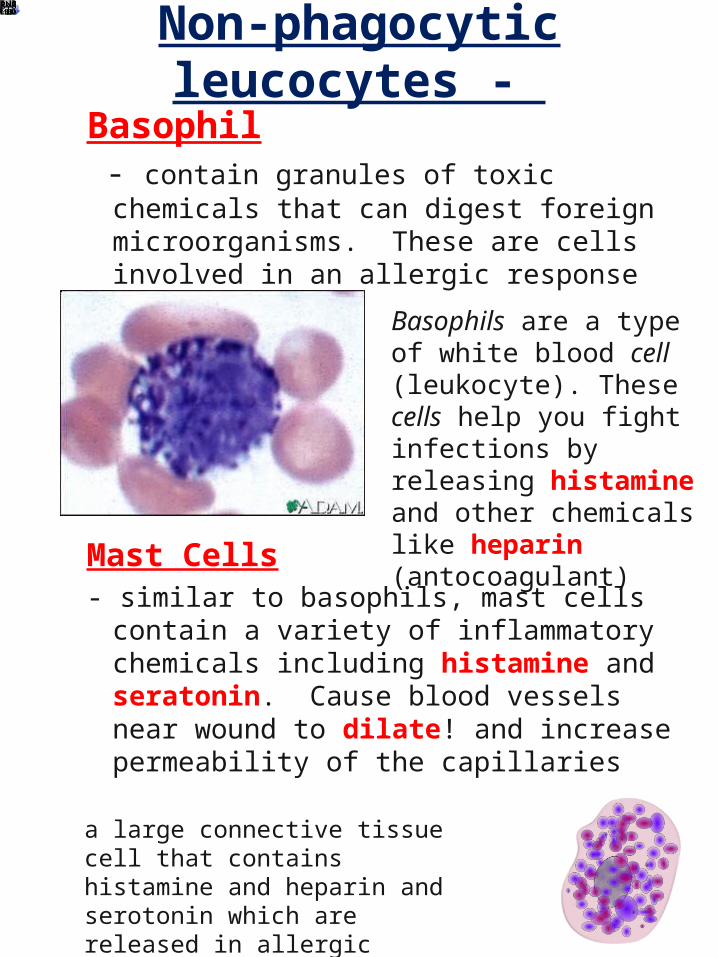
Non-phagocytic leucocytes -
Basophil - contain granules of toxic chemicals
that can digest foreign microorganisms. These are cells involved in an allergic response
Mast Cells- similar to basophils, mast cells contain
a variety of inflammatory chemicals including histamine and seratonin. Cause blood vessels near wound to dilate! and increase permeability of the capillaries
a large connective tissue cell that contains histamine and heparin and serotonin which are released in allergic reactions or in response to injury ...
Basophils are a type of white blood cell (leukocyte). These cells help you fight infections by releasing histamine and other chemicals like heparin (antocoagulant)

LEUKOCYTES
LEUKOCYTES
All produced in the Bone Marrow from Stem Cells
Granular Leukocytes
Have large, lobbed nuclei and distinctive granules in their
cytoplasm
Agranular Leukocytes
Cytoplasm usually lacks granules and the
nucleus is more rounded
Neutrophils
•Most numerous WBC•Main phagocytotic cell•Ingest bacteria and phagocytize dead cells
Eosinophils
•Produce enzymes which detoxify foreign proteins and fight parasatistic infection
Basophils
•Produce and release heparin and histamine in response to injury or infection
Lymphocytes
(T and B)
•Some produce antibodies (B Cells) and others attack invading cells directly (T Cells)
Monocytes/ Macrophag
es
•Largest WBC• monocytes grow into macrophages•Phagocytotic cells that don’t usually die after consuming pathogen
23

Chapter 8 - Defence Against Disease 24
The Second Line of Defence continued …

Chapter 8 - Defence Against Disease 25
Complement Proteins
Phagocytes are able to recognise foreign bodies with the aid of complement proteins.
Complement proteins help phagocytes by:
1. Sticking to invading microorganisms to become more readily identifiable by phagocytes.
2. Some stimulate phagocytes to become more active.
3. Some attract phagocytes to the site of infection.
4. Some complement proteins destroy the membranes of invading micro-organisms.
The Second Line of Defence continued …

Compliment Proteins continued …
Consequences of complement fixation
Membrane attack complex results in lesions/holes in foreign cell. These result in death
Amplifies inflammatory response because fixation causes the release of vasodilators and chemotaxis chemicals
Foreign cell is made sticky and easier to
phagocytise
OPSONIZATION
Complement: at least 20 different types of plasma proteinsUsually in inactive formBind to sugars or protein on foreign cell “complement fixation”

Natural killer cells (NK cells)
• are a type of lymphocyte (like macrophages)
• police the body in blood and lymph
• lyse and kill cancer cells and virus-infected cells
• act against any such target (i.e. non-specific)
• recognise certain sugars on invader’s surface
• are not phagocytic: attack membrane of target cell and cause it, and its nucleus, to disintegrate
The Second Line of Defence continued …

28
Interferons.
• are a group of antiviral chemicals
• are secreted by some cells when they are infected by virus particles.
• act on uninfected cells making them more resistant to the virus.
• interfer with virus replication
• stimulate macrophages to destroy virus infected cells
• are produced very early during viral infection.
• if a person develops a cold or flu, then the interferons have failed.
The Second Line of Defence continued …

Chapter 8 - Defence Against Disease 29
Fever
The Second Line of Defence continued …
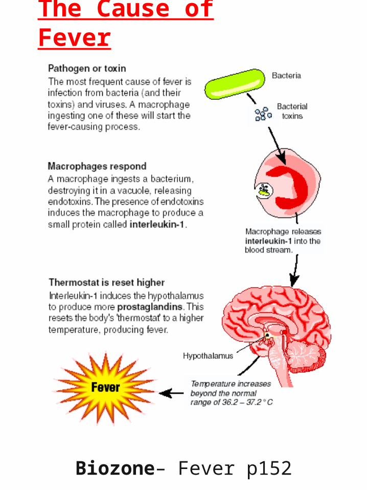
30
The Cause of Fever
Biozone– Fever p152

Inflammatory response
Four signs of Inflammation
1. Redness1. Increased blood flow to area brings
chemical and cellular agents to the site of injury and potential infection
2. Heat 1. Results from increased blood flow
3. Swelling1. Because blood vessels become
more permeable and “leak” more fluid into surrounding tissues
2. This encourages lymphatic return past stationary lymphocytes in lymph nodes
4. Pain• Adaptive because we protect area
and prevent further damage
The Second Line of Defence continued …

Inflammatory response continued …
Prevents spread of damaging agents to nearby tissues
Disposes of cell debris and pathogens
Sets the stage for repair

Chapter 8 - Defence Against Disease 33
Inflammation
The Second Line of Defence continued …

Chapter 8 - Defence Against Disease 34
Inflammation

Chapter 8 - Defence Against Disease 35

Key Elements of the 2nd Line of Defence
FEATURE PRODUCED/
FOUND
FUNCTION KEY FEATURES
Leokocyte
Monocytes
Macrophages
Neutrophils
Basophils
Eosinophils
Nk Cells
Mast Cells
Stem Cells
Interferons
Compliment Proteins
Vasodilation
Inflammation
Fever
Interleukin-1
Prostaglandins
Pyrexia -
Cytokines
Histamines
Seratonin Chapter 8 - Defence Against Disease 36

37
Specific Immunity 3nd Line of defence

Chapter 8 - Defence Against Disease
The Lymphatic System
Function:
• 1. take up excess tissue fluid and return it to the bloodstream
• 2. absorb fats at the intestinal villi and transport to the circulatory system
• 3. defend against disease

Chapter 8 - Defence Against Disease
The Lymphatic System
Components include:– Bone Marrow (stem cells differentiate into
lymphocytes
– Lymphocytes: T and B (plasma & memory) Cells, Macrophages & NK cells
– Lymph vessels
– Lymph organs: Thymus, spleen,
– Lymph nodes: tonsils, adenoids, armpits, groin etc
– Lymph: fluid of the vessels containing cells of the lymph and foreign material (antigens) that have drained into it!

Chapter 8 - Defence Against Disease 40

Chapter 8 - Defence Against Disease 41

Chapter 8 - Defence Against Disease 42
Lymph Nodes
• Biozone– The Lymphatic System p153

43
Specific ImmunityThe Third Line of
Defence
Once a pathogen or other foreign material has entered the body, it is
not only bombarded with your non-specific defences but is subject to attack by cells of
immune system, the T and B Lymphocytes.
This system is slower to take action but is more specific in its attack.
T and B Cells are the bodies special forces

Chapter 8 - Defence Against Disease 44
Specific ImmunityThe Third Line of Defence

Chapter 8 - Defence Against Disease 45
• This line of defence requires a specific response to a particular infection by the immune system and results in adapted or acquired immunity.
• The specific immunity acquired is generally long lasting, often for life.
• This third line of defence involves special white blood cells known as lymphocytes.
• Lymphocytes attack the particular invader, but also remember the attack (memory cells) so that a latter infection may be stopped more rapidly.
The Second Line of Defence continued …

Chapter 8 - Defence Against Disease 46
Two main groups of lymphocytes are involved in specific immunity:
B Cells
T Cells
• B Cells mature in the bone marrow to produce
• T Cells leave the bone marrow and mature in the thymus gland, where they turn into T-lymphocytes or T-cells.
• B and T Cells work together and help each other!!
The Second Line of Defence continued …

Chapter 8 - Defence Against Disease
B Cells & theHumoral (Antibody
Mediated) Immunity
Ballistics

B Cells & theHumoral (Antibody
Mediated) Immunity
• B Cells provide - Humoral (Antibody Mediated) Immunity
• they can produce large quantities of antibodies in response to a foreign antigen
• Must recognise ‘non-self’ antigen by binding to it to its receptor site
• Requires a helper T- Cell to activate the B Cell
• Once activated by a Helper T Cell, it divides madly producing two types of daughter cells including:
• Plasma B cells• Memory B Cells
• These cells are clones of the original activated cell and thuis produce the same antibody. This is known as the Clonal Selection Theory (see page 255) 48

Chapter 8 - Defence Against Disease 49
B-CellsB-Cells

Chapter 8 - Defence Against Disease 50
B-Cells in Action

Chapter 8 - Defence Against Disease 51

Chapter 8 - Defence Against Disease 52
B-memory Cells
• When the plasma cells produce new antibodies and B-cells, some produced differentiate into other cells called B-memory cells.
• B-memory cells have the same antigen-antibody specificity as the original parent B-cell.
• Memory cells can survive for many years or even life.
• If a second infection ever occurs, the B-memory cells react faster and more vigorously than the initial infection.
• Remain in circulation, producing small quantities of antibody

Chapter 8 - Defence Against Disease 53
B-Plasma Cells
• Produce and secrete huge quantities of antibody molecules
• These antibodies bind with antigens forming an antibody-Antigen complex
• Plasma cells are relatively short living & broken down following infection

Chapter 8 - Defence Against Disease 54
Antibodies- Antibodies- “immunoglobulins
” • B-cells have immunoglobulins on their surfaces.
• Immunoglobulins are proteins that identify antigens.
• Immunoglobulins are also called antibodies.
• The immunoglobulins of each B-cell have a specific structure and recognise only one kind of antigen.
• There are millions of antigens that the body must be able to respond. In response to this millions of different B-cells are produced with different immunoglobulins on their surfaces.
• Self – tolerance is the ability of the immune system to recognise and ignore its own tissues early in development
• Auto-immune disorder is the condition occuring when the body attacks its own tissues

Chapter 8 - Defence Against Disease 55
The Structure of The Structure of AntibodiesAntibodies

Chapter 8 - Defence Against Disease 56
Action of Antibodies

Chapter 8 - Defence Against Disease 57
Action of Antibodies

• immunoglobulins – ig for short
• Whenon the surface of a Lymphocyte – they are receptor sites. Off, they are antibodies!
• both t and b cells have immunoglobulins on their surface
• secreted immunoglobulins are called antibodies
• it is the binding of antigen to receptor which triggers the specific immune response
• during maturation, the genes that determine ig structure are continually being rearranged. this leads to new combinations of shape and charge in the antigen binding site
• antibodies can combine with two antigens at once. this can cause clumping, or agglutination
• the antigen – antibdy complex promotes phagocytosis
• activates complement proteins
• neutralizes the binding site of an antigenChapter 8 - Defence
Against Disease 58
“immunoglobulins
”more facts!

Clonal selection Theory
When an antigen enters the body it probably passes many B cells before it meets one
with the immunoglobulan with which it can combine. In effect the antigen ‘selects’ the
B cell that will lead to its death
• Antigen ‘selects’ B Cell and its immunoglobulan
• B cell rapidly reproduces (mitosis) to produce identical daughter cells
• Each of these reproduces rapidly to produce a large clone of cells
• Cell cloned in this way will have exactly the same DNA and antibodies
• Most will differentiate into in plasma B cells, others into memory cells
Chapter 8 - Defence Against Disease 59

Chapter 8 - Defence Against Disease 60
T-CellsT-Cells
• When T-cells mature in the thymus, many different types of T-cells are produced which recognise many different antigens.
Types of T - Cells
Helper T-cells (Th)
• Release chemicals which attract phagocytes
• Stimulate cell division in B-Cells• Produce chemicals that stimulate other
T Cells
Helper T - Cells
Cytotoxic (Killer) T
Cells
Memory T Cells

Chapter 8 - Defence Against Disease 61

Chapter 8 - Defence Against Disease 62
T-CellsT-Cells
Cytotoxic T-Cells (Tc)
• Another type of T-cell, cytotoxic T-cells (Tc), kills body cells that have been infected with a virus.
• Tc cells kill the infected cell by secreting proteins that punch holes in the membrane of the cell and the contents ooze out.
• Tc cells can only kill a virus when it is inside a cell.
• Some Tc cells also destroy cancer cells.
Suppressor T Cells (Tsc)
• regulates immune response by turning it off when the infection passes

Chapter 8 - Defence Against Disease 63
T-CellsT-Cells
Cytotoxic T-Cells (Tc)
• Another type of T-cell, cytotoxic T-cells (Tc), kills body cells that have been infected with a virus.
• Tc cells kill the infected cell by secreting proteins that punch holes in the membrane of the cell and the contents ooze out.
• Tc cells can only kill a virus when it is inside a cell.
• Some Tc cells also destroy cancer cells.
Memory T Cells (Tm)
• Remain in circulation (spleen) for many years after infection

Chapter 8 - Defence Against Disease 64
Allergies and Hypersensitivity

Chapter 8 - Defence Against Disease 65
Allergies and Hypersensitivity

Chapter 8 - Defence Against Disease 66
IgE binds with mast cells which are now sensitised to the allergen- producing histamines – inflammatory response – increase blood volume; permeability of vessels,
Allergies and Hypersensitivity

Chapter 8 - Defence Against Disease 67
Blood Groups – Blood Antigens

Chapter 8 - Defence Against Disease 68

• A red blood cell antigen, the rhesus factor is present on the red blood cells of a majority of people. Such people are rhesus positive (RH+). If the antigen is absent a person is rhesus negative (Rh-). If a person who is Rh- and comes into contact with RH+ blood will respond by producing antibodies against the antigen. This can become critical in pregnancy
Chapter 8 - Defence Against Disease 69
Rhesus Incompatibility

Chapter 8 - Defence Against Disease 70
Rhesus Incompatibility

Chapter 8 - Defence Against Disease 71
Acquiring Specific Immunity
• Acquired Immunity is the term used to describe when antibodies are required to form immunity from a specific antigen.
• Passive Immunity is when antibodies are received by an outside source (vaccination).
• Passive naturally occuring Passive induced
• Active Immunity is when antibodies are produced within a person (B-cells and T-cells).).
• Active naturally occuring Active induced

Chapter 8 - Defence Against Disease 72
Acquiring Specific Acquiring Specific ImmunityImmunity

Chapter 8 - Defence Against Disease 73
Acquiring Specific Immunity

Chapter 8 - Defence Against Disease 74
Acquiring Specific Immunity

Classification of BacteriaGram Stains pages 214-215
• 1984, bacteriologist, Joachim Gram developed the gram stain
• Gram stain distinguishes between two main groups of bacteria
• Important stain to help identify which drugs are useful
• Gram Positive bacteria take up the violet colour of the stain– Gram + have a cell wall layer of teichoic acid– Are particularly susceptible to penicillin and
sulphonamide drugs• Gram Negative bacteria fail to take up the stain and by
default stain pink– Gram – have no teichoic acid in their walls and
smaller amounts of disaccharides and amino acids– Outer layer of lipid compounds enables these
bacteria to resist penicillin and other drugs– Also makes phagocytosis of the bacteria very difficult– Effective drugs include streptomycin, tetracycline
– S. empidermis: gram negative (susceptible to penicillin)
– S. ecoli: gram negative (resistant to penicillin)75